Embed Size (px)
Citation preview

The SearchLight DNA™ report is designed to describe the mutations identified in a pet's tumor. It also describesdiagnostic, prognostic, and treatment-related biomarker associations with those mutations that are based oncuration of more than 274 primary canine publications and inference from human cancer biomarker publicationsand databases. The report is thus built on a foundation of peer-reviewed research to support clinical decision-making. The front pages summarize actionable mutations and their associations. These pages are followed by in-depth biomarker summaries, evidence statements, and links to supporting articles.
Overview of Findings
Evidence Summaries
Clinical Trials
Variants of Uncertain
Significance
Gene ListReferences
Test Description
The Clinician’s Report The Client’s Report
SearchLight DNATM
Orientation
Sample ID: OID12345MCT Page 1 of 19
Therapeutically Actionable Genomic Findings1 Pharmacogenomic BiomarkerTreatment Options Based on Genomic Alterations
2 Actionable Biomarkers3 Matching Drugs
SearchLight DNATM
Clinician Report
Summary of Key Findings
Vidium Animal Health | 445 N. 5th Street Phoenix, AZ 85004
1-833-794-0318 | www.vidiumah.com
Tumor Histology-Related Findings4 Diagnostic Biomarkers3 Prognostic Biomarkers5 Clinical Trials
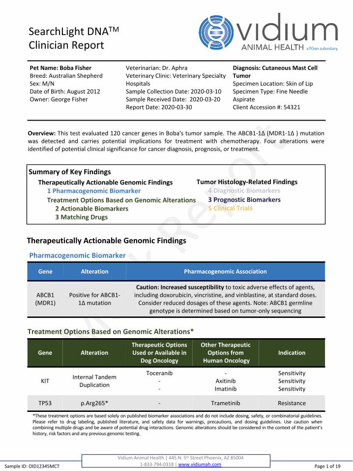
Overview: This test evaluated 120 cancer genes in Boba's tumor sample. The ABCB1-1Δ (MDR1-1Δ ) mutation
was detected and carries potential implications for treatment with chemotherapy. Four alterations were
identified of potential clinical significance for cancer diagnosis, prognosis, or treatment.
Pharmacogenomic Biomarker Gene Alteration Pharmacogenomic Association
ABCB1
(MDR1)
Positive for ABCB1-
1Δ mutation
Caution: Increased susceptibility to toxic adverse effects of agents,
including doxorubicin, vincristine, and vinblastine, at standard doses.
Consider reduced dosages of these agents. Note: ABCB1 germline
genotype is determined based on tumor-only sequencing
Pharmacogenomic Biomarker
Treatment Options Based on Genomic Alterations*
Gene AlterationTherapeutic Options Used or Available in
Dog Oncology
Other Therapeutic Options from
Human OncologyIndication
KITInternal Tandem
Duplication
Toceranib
-
-
-
Axitinib
Imatinib
Sensitivity
Sensitivity
Sensitivity
TP53 p.Arg265* - Trametinib Resistance
Therapeutically Actionable Genomic Findings
Pet Name: Boba FisherBreed: Australian Shepherd
Sex: M/N
Date of Birth: August 2012
Owner: George Fisher
Diagnosis: Cutaneous Mast Cell TumorSpecimen Location: Skin of Lip
Specimen Type: Fine Needle
Aspirate
Client Accession #: 54321
Veterinarian: Dr. Aphra
Veterinary Clinic: Veterinary Specialty
Hospitals
Sample Collection Date: 2020-03-10
Sample Received Date: 2020-03-20
Report Date: 2020-03-30
*These treatment options are based solely on published biomarker associations and do not include dosing, safety, or combinatorial guidelines.
Please refer to drug labeling, published literature, and safety data for warnings, precautions, and dosing guidelines. Use caution when
combining multiple drugs and be aware of potential drug interactions. Genomic alterations should be considered in the context of the patient’s
history, risk factors and any previous genomic testing.

Page 2 of 19
Vidium Animal Health | 445 N. 5th Street Phoenix, AZ 850041-833-794-0318 | www.vidiumah.com
SearchLight DNATM
Clinician Report
Gene Alteration Diagnostic Rolein Dog Cancers
Diagnostic Rolein Human Cancers
CDKN2B Copy Number Loss
GNB1 p.Gly116Phe -
KIT Internal Tandem Duplication -
TP53 p.Arg265* -
Diagnostic Biomarkers
Tumor Histology-Related Findings
Sample ID: OID12345MCT
Diagnosis: Cutaneous Mast Cell TumorSpecimen Location: Skin of LipSpecimen Type: Fine Needle AspirateClient Accession #: 54321
Veterinarian: Dr. AphraVeterinary Clinic: Veterinary Specialty HospitalsSample Collection Date: 2020-03-10Sample Received Date: 2020-03-20Report Date: 2020-03-30
Prognostic Biomarkers
Gene Alteration Prognostic Rolein Dog Cancers
Prognostic Rolein Human Cancers
CDKN2B Copy Number Loss Poor Outcome -
KIT Internal Tandem Duplication Poor Outcome -
TP53 p.Arg265* Poor Outcome Poor Outcome
Pet Name: Boba FisherBreed: Australian ShepherdSex: M/NDate of Birth: August 2012Owner: George Fisher

Page 3 of 19
Vidium Animal Health | 445 N. 5th Street Phoenix, AZ 850041-833-794-0318 | www.vidiumah.com
SearchLight DNATM
Clinician Report
Tumor Histology Clinical Trials
CancerAAHSD000002 - Targeted electrochemotherapy cancer treatment for local cancer control
CancerAAHSD000080 - Evaluating a targeted telomerase vaccine to stimulate anti-tumor immunity and prolong survival times in dogs and cats with various cancers
CancerAAHSD000083 - Endoscopic/laparoscopic treatment of inoperable cancer with a targeted endoscopic electroporation system (EES)
CancerAAHSD004609 - Combination of electrochemotherapy and gene therapy with canine IL-12
CancerAAHSD004967 - Integrating immunotherapy with radiotherapy to improve tumor control for canine cancer patients
Clinical Trials Summary*
*Clinical trials are identified utilizing the American Veterinary Medical Association (AVMA) Animal Health Studies Database (AAHSD) athttps://ebusiness.avma.org/aahsd/study_search.aspx based on tumor type match, then general cancer (solid tumors, hematological malignancies, neoplasia).Vidium Animal Health is not an active participant in the clinical trials shown here and has no ownership stake in any of the treatments under study.
Sample ID: OID12345MCT
Diagnosis: Cutaneous Mast Cell TumorSpecimen Location: Skin of LipSpecimen Type: Fine Needle AspirateClient Accession #: 54321
Veterinarian: Dr. AphraVeterinary Clinic: Veterinary Specialty HospitalsSample Collection Date: 2020-03-10Sample Received Date: 2020-03-20Report Date: 2020-03-30
Pet Name: Boba FisherBreed: Australian ShepherdSex: M/NDate of Birth: August 2012Owner: George Fisher

Gene Alteration Summaries
Pet Name: Boba Fisher
Gene Summary
Page 4 of 19
Vidium Animal Health | 445 N. 5th Street Phoenix, AZ 85004
1-833-794-0318 | www.vidiumah.com
Gene: ABCB1Alteration: Positive for ABCB1-1Δ mutation
The ABCB1 gene encodes the protein ATP-binding cassette sub-family B member 1 and is pronounced "Ay-
Bee-See-Bee-Won". It is also called MDR1 ("Emm-Dee-Arr-Won") or "Multi-Drug Resistance One". The
membrane-associated protein encoded by this gene is a member of the superfamily of ATP-binding cassette
(ABC) transporters. ABC proteins transport various molecules across extra- and intra-cellular membranes.
ABC genes are divided into seven distinct subfamilies (ABC1, MDR/TAP, MRP, ALD, OABP, GCN20, White).
This protein is a member of the MDR/TAP subfamily. Members of the MDR/TAP subfamily are involved in
multidrug resistance. The protein encoded by this gene is an ATP-dependent drug efflux pump for xenobiotic
compounds with broad substrate specificity. It is responsible for decreased drug accumulation in multidrug-
resistant cells and often mediates the development of resistance to anticancer drugs. This protein also
functions as a transporter in the blood-brain barrier. Mutations in this gene are associated with colchicine
resistance and inflammatory bowel disease. Alternative splicing and the use of alternative promoters results
in multiple transcript variants. Germline mutations in this gene are common in many herding dog breeds.
Due to the disruptive effect of these mutations on function of the MDR1 protein, such mutations could lead
to drug accumulation in the central nervous system and other tissues. Subsequent adverse reactions to many
drugs, including many common chemotherapy agents, may occur.
Variant SummaryA polymorphism (referred to as ABCB1-1delta) occurs in a subset of dog breeds, including many herding
breeds. The ABCB1-1delta polymorphism is a 4-base pair deletion that causes a shift in the reading frame
that results in premature truncation of P-glycoprotein and loss of P-glycoprotein function. Dogs that are
homozygous or heterozygous for this polymorphism can experience increased toxicity for chemotherapeutic
agents that are substrates for ABCB1, such as doxorubicin, vincristine, and vinblastine. Dogs without this
polymorphism (non-mutant) show standard susceptibility to chemotherapy-associated adverse effects, and a
dosing adjustment based on ABCB1 status is not needed. (Mealey et al. J Vet Intern Med 2008; Mealey et al.
Vet Clin North Am Small Anim Pract 2013).
Sample ID: OID12345MCT

Gene Alteration Summaries
Gene SummaryCDKN2B stands for "cyclin dependent kinase inhibitor 2B" and is pronounced "See-Dee-Kay-Enn-Too-Bee".This gene lies adjacent to the tumor suppressor gene CDKN2A in a region that is frequently mutated anddeleted in a wide variety of tumors. This gene is itself a tumor suppressor that encodes a cyclin-dependentkinase inhibitor, which forms a complex with CDK4 or CDK6, and prevents the activation of the CDK kinases.Thus the encoded protein functions as a cell growth regulator that controls cell cycle G1 progression. Theexpression of this gene was found to be dramatically induced by TGF beta, which suggested its role in theTGF beta induced growth inhibition. Two alternatively spliced transcript variants of this gene, which encodedistinct proteins, have been reported. Mutations in CDKN2B can disrupt its ability to negatively regulate cellgrowth and thereby lead to excessive cell growth.
Variant SummaryCDKN2A and the adjacent CDKN2B genes (CDKN2A/B) are tumor suppressors commonly deleted in humanglioblastoma (56%), mesothelioma (45%), esophageal cancer (39%), bladder cancer (32%), melanoma (31%),head and neck carcinoma (31%), pancreatic cancer (28%), diffuse large B-cell lymphoma (27%), lungsquamous cell carcinoma (26%), lung adenocarcinoma (17%), cholangiocarcinoma (17%), sarcoma (15%),stomach cancer (11%), low grade glioma (11%), adrenocortical carcinoma (7%), liver cancer (6%), and othercancers. CDKN2A/B is also frequently mutated or deleted in canine cancers including malignant melanoma(~68%), histiocytic sarcoma (~63%), osteosarcoma (~42-70%), T-cell lymphoma (~40%), pulmonaryadenocarcinoma (~40%), urothelial carcinoma (~26%), head and neck squamous cell carcinoma (~25%),hemangiosarcoma (~22-28%), KIT-mutant mast cell tumors (~21%), and glioma (~10%). CDKN2A/B deletionleads to loss of functional protein (the p16 and p14 tumor suppressors) and disruption of the tumorsuppressive effects of these proteins. In human cancers, CDKN2A/B deletion has been associated with poorprognosis in sarcomas. In dogs, CDKN2A/B deletion and/or promoter methylation has also been associatedwith high grade in lymphoma.
Diagnostic Biomarker SummaryArray comparative genomic hybridization analysis of 109 canine mast cell tumors identified loss of thechromosome 11 locus encompassing CDKN2A and CDKN2B to be significantly enriched in KIT-mutant mastcell tumors (21.2%). (Mochizuki H et al. Chromosome Res 2017).
Associated human data. CDKN2B deletions are common in human glioblastoma (55%), mesothelioma (43%),esophageal cancer (35%), bladder cancer (31%), melanoma (29%), head and neck carcinoma (28%),pancreatic cancer (27%), lung squamous cell carcinoma (26%), diffuse large B-cell lymphoma (25%), lungadenocarcinoma (16%), sarcoma (15%), cholangiocarcinoma (14%), stomach cancer (11%), low grade glioma(11%), liver cancer (5%), and other cancers (TCGA Pan-Cancer Atlas accessed via cBioPortal).
Page 5 of 19Vidium Animal Health | 445 N. 5th Street Phoenix, AZ 85004
1-833-794-0318 | www.vidiumah.com
Gene: CDKN2BAlteration: Copy Number Loss
Pet Name: Boba Fisher
Sample ID: OID12345MCT
Gene Alteration Summaries
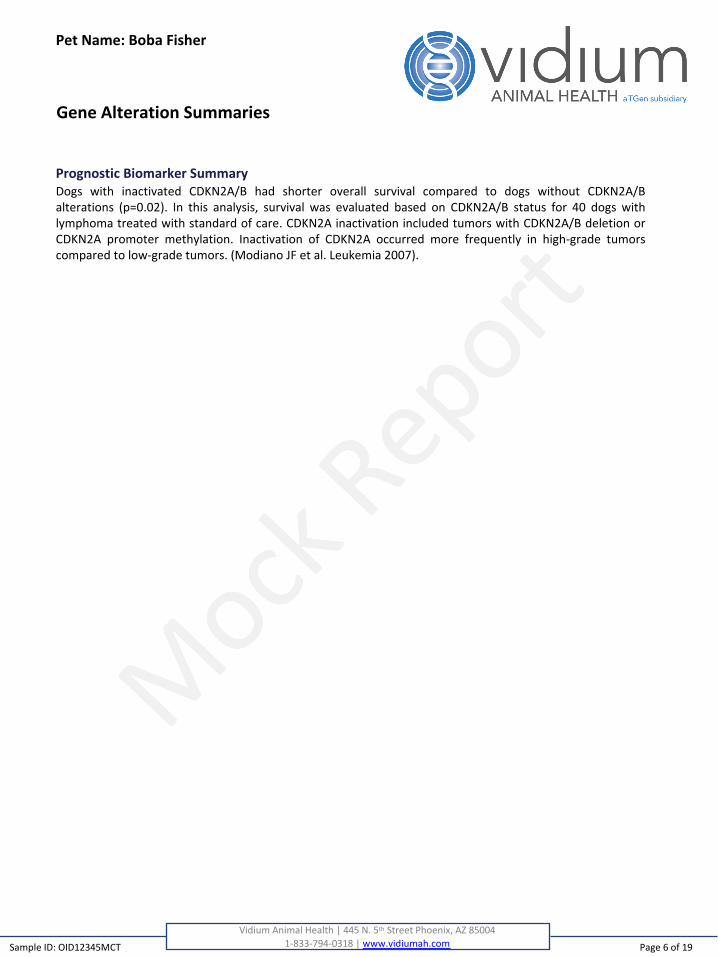
Prognostic Biomarker SummaryDogs with inactivated CDKN2A/B had shorter overall survival compared to dogs without CDKN2A/Balterations (p=0.02). In this analysis, survival was evaluated based on CDKN2A/B status for 40 dogs withlymphoma treated with standard of care. CDKN2A inactivation included tumors with CDKN2A/B deletion orCDKN2A promoter methylation. Inactivation of CDKN2A occurred more frequently in high-grade tumorscompared to low-grade tumors. (Modiano JF et al. Leukemia 2007).
Page 6 of 19
Vidium Animal Health | 445 N. 5th Street Phoenix, AZ 850041-833-794-0318 | www.vidiumah.com
Pet Name: Boba Fisher
Sample ID: OID12345MCT

Gene Alteration Summaries
Gene SummaryThe GNB1 gene encodes the G protein subunit beta 1, and is pronounced "Gee-Enn-Bee-Won".Heterotrimeric guanine nucleotide-binding proteins (G proteins), which integrate signals between receptorsand effector proteins, are composed of an alpha, a beta, and a gamma subunit. These subunits are encodedby families of related genes. This gene encodes a beta subunit. Beta subunits are important regulators ofalpha subunits, as well as of certain signal transduction receptors and effectors. Alternative splicing results inmultiple transcript variants. GNB1 is a candidate oncogene. Mutations in this gene may result in alteredfunction of the protein and therefore hyperactivation of downstream signaling pathways that are oncogenic.
Variant SummaryThis GNB1 missense variant has been detected in one study in 12.3% of 81 mast cell tumors (MCT) including11% (8/72) of cutaneous and 22% (2/9) of subcutaneous MCTs. These GNB1 missense mutations have beenpreviously identified in human acute lymphoblastic B-cell leukemia. The adjacent amino acid is involved in Gprotein subunit binding, but the specific effects of this variant remain unknown.
Diagnostic Biomarker SummaryPotentially activating GNB1 mutations were identified for the first time through multi-platform sequencing ascommonly occurring in canine mast cell tumors (MCTs). GNB1 mutations occurred in 17.3% of 81 MCTs (72cutaneous, 9 subcutaneous; 48 fresh, 33 FFPE) with higher frequency in subcutaneous MCTs (44%) comparedto cutaneous (13.9%). GNB1 G116F mutations were present in 10/81 cases (12.3%). (Vozdova M et al. VetComp Oncol 2020).
Page 7 of 19Vidium Animal Health | 445 N. 5th Street Phoenix, AZ 85004
1-833-794-0318 | www.vidiumah.com
Gene: GNB1Alteration: p.Gly116Phe
Pet Name: Boba Fisher
Sample ID: OID12345MCT

Gene Alteration Summaries
Gene SummaryThe KIT (pronounced "Kit") gene provides instructions for making a member of a protein family called
receptor tyrosine kinases. Receptor tyrosine kinases transmit signals from the cell surface into the cell
through a process called signal transduction. The KIT protein is found in the cell membrane of certain cell
types where a specific protein, called stem cell factor, attaches (binds) to it. This binding turns on (activates)
the KIT protein, which then activates other proteins inside the cell by adding a cluster of oxygen and
phosphorus atoms (a phosphate group) at specific positions. This process, called phosphorylation, leads to
the activation of a series of proteins in multiple signaling pathways. KIT is an oncogene, and mutations in this
gene can cause hyperactivation of the signals that lead to cell division. The common analogy is that
mutations in oncogenes "put the foot on the accelerator of cell growth" and allow cancer to develop. The
signaling pathways stimulated by the KIT protein control many important cellular processes such as cell
growth and division (proliferation), survival, and movement (migration). KIT protein signaling is important for
the development and function of certain cell types, including reproductive cells (germ cells), early blood cells
(hematopoietic stem cells), white blood cells called mast cells, cells in the gastrointestinal tract called
interstitial cells of Cajal (ICCs), and cells called melanocytes. Melanocytes produce the pigment melanin,
which contributes to hair, eye, and skin color.
Variant SummarySomatic activating KIT mutations occur in ~13-50% of canine mast cell tumors (MCTs), ~35-74% of canine
gastrointestinal stromal tumors (GISTs), and ~2-8% of malignant melanomas. In MCT, mutations
predominantly occur as internal tandem duplications (ITD) in KIT exons 8 and 11, leading to constitutive
activation of the KIT receptor tyrosine kinase through modulation of extracellular and juxtamembrane
domain regulatory activity. KIT ITDs in canine MCT have been associated with prognosis and response to
tyrosine kinase inhibitors. KIT exon 11 ITDs are the most common KIT mutations in canine MCTs. This variant
is a KIT exon 11 ITD.
Diagnostic Biomarker SummaryActivating KIT mutations, predominantly internal tandem duplications (ITDs), commonly occur in canine mast
cell tumors (MCTs). Reports of KIT mutation frequencies in MCT vary widely, from ~9% to 50%. Reported
frequencies depend on cohort size and makeup - e.g. they are more frequent in higher grade MCTs.
Reported frequencies also depend on sequencing regions and platforms - e.g. many studies only evaluate
one or two KIT exons and use only targeted sequencing approaches. Estimates of KIT mutation frequency in
the largest cohorts utilizing the most comprehensive sequencing methods report a more narrow range from
17% to 30%. Exon 11 ITDs or deletions are the most common KIT mutations in MCT followed by exon 8 ITDs
or substitutions and rare mutations (ITDs, substitutions, or deletions) in exons 9, 12, and 17. KIT ITDs in MCT
have been shown to drive constitutive KIT activation and downstream pro-growth signaling. (Reviewed with
Consensus Guidelines in Thamm DH et al. Vet Comp Oncology 2019).
Page 8 of 19
Vidium Animal Health | 445 N. 5th Street Phoenix, AZ 85004
1-833-794-0318 | www.vidiumah.com
Gene: KITAlteration: Internal Tandem Duplication
Pet Name: Boba Fisher
Sample ID: OID12345MCT

Gene Alteration Summaries
Prognostic Biomarker SummaryActivating KIT mutations, predominantly internal tandem duplications (ITDs) in exon 11, have shownassociations with canine mast cell tumor (MCT) prognosis (i.e. histologic grade and post-surgical outcomes) inretrospective and prospective studies. Prospective trials have found significant association between exon 8or 11 ITDs and progression-free interval (but not overall outcome) as well as with shorter progression-freesurvival in dogs treated with toceranib. Retrospective studies have found significant association of KITmutation in exons 8, 9, and 11 with high grade (both Kiupel and Patnaik systems) based on univariateanalyses. They have also found significant association of exon 11 or 12 ITDs with recurrence, metastasis,progression-free survival, disease-free interval, survival time, and death, typically based on univariateanalyses. KIT exon 11 mutations have also been associated with increased cell proliferation indices such asAgNOR frequency, Ki67 index, and mitotic counts. Given variability in cohort sizes, clinical annotation, andKIT mutation assessment methods (e.g. a single region versus multiple regions versus all regions of the gene)in these studies, the status of KIT mutations as independent prognostic factors are still in need of definitivevalidation. Per the consensus publication of the joint Veterinary Cancer Society and American College ofVeterinary Pathologists’ Oncology-Pathology Working Group (OPWG), KIT “mutation presence/absence is amore objective measurement than either histologic grade or mitotic index/count, both of which can beassociated with considerable observer bias. Thus, this measure could still provide important objectiveinformation useful in decision-making.” (Reviewed with Consensus Guidelines in Thamm DH et al. Vet CompOncology 2019).
Therapeutic Evidence SummaryAxitinib – SensitivityThis study evaluated a canine MCT-cell line, CMMC1, bearing four KIT mutations including Q255del, K415E,an exon 11 ITD (Y573_N590dup), and F507fs. CMMC1 was shown to be hypersensitive to the tyrosine kinaseinhibitor, axitinib, relative to a KIT wild-type cell line, HRMC, based on multi-point drug-dose-response andcell-counting assays (IC50 of 9 nM for CMMC1 versus > 10 µM for HRMC). CMMC1 also showed constitutivelyhigh levels of KIT phosphorylation and axitinib-dose-dependent reduction of those levels based on Westernblotting whereas HRMC showed constitutively high phospho-KIT levels independently of drug dose.(Takeuchi Y et al. J Vet Pharmacol Ther 2012).
Imatinib – SensitivityThis study evaluated a canine MCT-cell line, CMMC1, bearing four KIT mutations including Q255del, K415E,an exon 11 ITD (Y573_N590dup), and F507fs. CMMC1 was shown to be hypersensitive to the tyrosine kinaseinhibitor, imatinib, relative to a KIT wild-type cell line, HRMC, based on multi-point drug-dose-response andcell-counting assays (IC50 of 42.3 nM for CMMC1 versus > 10 µM for HRMC). CMMC1 also showedconstitutively high levels of KIT phosphorylation and axitinib-dose-dependent reduction of those levels basedon Western blotting whereas HRMC showed constitutively high phospho-KIT levels independently of drugdose. (Takeuchi Y et al. J Vet Pharmacol Ther 2012).
CoMS is a canine mast cell tumor cell line that has been shown to bear the exon 11 juxtamembrane domainKIT mutation. CoMS was shown to be sensitive to imatinib in this study. (Kobayashi M et al. Oncol Rep 2017).
Page 9 of 19Vidium Animal Health | 445 N. 5th Street Phoenix, AZ 85004
1-833-794-0318 | www.vidiumah.com
Pet Name: Boba Fisher
Sample ID: OID12345MCT

Gene Alteration Summaries
Toceranib – SensitivityThis registration study for toceranib - a multi-center, placebo-controlled, double-blind, randomized clinicaltrial in 153 dogs with MCT - additionally evaluated association between KIT genotype by PCR for exon 11 and12 ITDs and clinical outcomes. KIT exon 11 or 12 ITDs were detected in 20% of cases (30/150). In thetoceranib group, exon 11 and 12 KIT-mutant MCTs were more likely to respond than KIT wild-type MCTs(60.0%, 12/20 versus 31.3%, 20/64), although KIT mutations were not associated with time to progression orduration of response. (London CA et al. Clin Cancer Res 2009).
This study, the first Phase I dose-escalation study of the tyrosine kinase inhibitor, toceranib (SU11654)evaluated KIT exon 11 ITD status in 22 dogs with MCT treated with SU11654. KIT exon 11 ITDs were found in50% (11/22) of MCT cases. KIT exon 11 ITD status as well as lymph node involvement were significantlyassociated with initial response to therapy. KIT ITD-positive cases also survived longer (36.9 v 15.4 weeks)and the median time to progression in KIT-mutant versus wild-type MCTs was 21.0 weeks v 3.9 weeks,though these associations were not statistically significant. (London CA et al. Clin Cancer Res 2003).
Page 10 of 19
Vidium Animal Health | 445 N. 5th Street Phoenix, AZ 850041-833-794-0318 | www.vidiumah.com
Pet Name: Boba Fisher
Sample ID: OID12345MCT

Gene Alteration Summaries
Gene SummaryPronounced "Tee-Pee-Fifty-Three", the TP53 gene provides instructions for making a protein called tumorprotein p53 (or p53). This protein acts as a tumor suppressor, which means that it regulates cell division bykeeping cells from growing and dividing (proliferating) too fast or in an uncontrolled way. The p53 protein islocated in the nucleus of cells throughout the body, where it attaches (binds) directly to DNA. When the DNAin a cell becomes damaged by agents such as toxic chemicals, radiation, or ultraviolet (UV) rays from sunlight,this protein plays a critical role in determining whether the DNA will be repaired or the damaged cell will self-destruct (undergo apoptosis). If the DNA can be repaired, p53 activates other genes to fix the damage. If theDNA cannot be repaired, this protein prevents the cell from dividing and signals it to undergo apoptosis. Bystopping cells with mutated or damaged DNA from dividing, p53 helps prevent the development of tumors.Because p53 is essential for regulating DNA repair and cell division, it has been nicknamed the "guardian ofthe genome." Mutations in this gene are some of the most common mutations in cancer. They may impactthe ability of the p53 protein to perform its critical functions in protecting against DNA damage.
Variant SummaryInactivating TP53 mutations (truncating mutations and/or hotspot missense mutations in the DNA-bindingdomain) are the most common mutations in canine and human cancers. In canine cancers, they are mostcommonly observed in osteosarcoma, hemangiosarcoma, and histiocytic sarcoma (>40% of cases) and lesscommonly (<20%) in B-cell lymphoma, pulmonary adenocarcinoma, mast cell tumors, malignant melanoma,and glioma. They are also associated with poor prognosis in many human cancers. In canine cancer cell lines,they have also been shown to correlate with resistance to the MEK inhibitor trametinib.
Diagnostic Biomarker SummaryAssociated human data. Truncating mutations in TP53 occur in multiple tumor types, including breastcancer, colorectal cancer, lung cancer, sarcoma, adrenocortical carcinoma, glioma, Spitzoid tumor, andmultiple other tumor types (COSMIC). TP53 truncating mutations occur in esophageal cancer (34%), headand neck squamous cell carcinoma (32%), lung squamous cell carcinoma (30%), ovarian cancer (27%), uterinecarcinosarcoma (23%), pancreatic cancer (22%), lung adenocarcinoma (20%), stomach cancer (18%),colorectal cancer (17%), bladder cancer (16%), chromophobe renal cell carcinoma (15%), sarcomas (13%),breast cancer (12%), adrenocortical carcinoma (12%), liver cancer (12%), low grade gliomas (11%), uterinecancer (10%), glioblastoma (8%), melanoma (7%), diffuse large B-cell lymphoma (5%), mesothelioma (5%),acute myeloid leukemia (5%), and other cancers. (TCGA Pan-Cancer Atlas accessed via cBioPortal).
Prognostic Biomarker SummaryTP53 mutations were identified in 24/59 (40.7%) of osteosarcoma cases assessed by polymerase chainreaction and single-strand conformational polymorphism analysis. Dogs with osteosarcoma bearing TP53mutations had a significantly shorter median survival time (81 days, 95% confidence interval: 73-89 days)than dogs bearing wild-type TP53 (256 days, 95% confidence interval: 119-392 days, p=.026). (Kirpensteijn Jet al. Vet Surg 2008).
Page 11 of 19
Vidium Animal Health | 445 N. 5th Street Phoenix, AZ 850041-833-794-0318 | www.vidiumah.com
Gene: TP53Alteration: p.Arg265*
Pet Name: Boba Fisher
Sample ID: OID12345MCT
TP53 truncating mutations were significantly associated with the presence of metastases, but not withsurvival time or chemotherapy response in canine histiocytic sarcoma. (Asada H et al. Res Vet Sci 2019).
Associated human data. Deep sequencing of human splenic marginal zone lymphoma found that TP53mutations are an independent marker of reduced overall survival (HR 2.36, p=0.03). (Parry M et al. ClinCancer Res 2015).
In a meta-analysis evaluating the relationship between TP53 mutation and prognosis in humanosteosarcoma, TP53 mutation was found to be associated with reduced 2-year overall survival (RR=1.79).(Chen Z et al. Dis Markers 2016).
In a meta-analysis of patients with EGFR or ALK mutant non-small cell lung cancer treated with targetedtyrosine kinase inhibitors, TP53 mutation was associated with shorter progression free survival and overallsurvival (HR=1.88, HR=1.92). (Qin K et al. BMC Cancer 2020).
In a meta-analysis study including 888 patients with esophageal cancer, patients with TP53 mutant tumorswere found to have reduced overall survival when compared to patients whose tumors did not have TP53mutations (HR 1.48). (Fisher OM et al. Gut 2017).
In a targeted sequencing study of 283 primary pancreatic ductal adenocarcinoma, TP53 mutation wasassociated with reduced overall survival when compared to tumors without TP53 mutations (median OS 37.4vs 65.0 months). (McIntyre CA et al. Cancer 2020).
In a whole exome sequencing study of 87 hepatocellular carcinomas, TP53 mutation was associated with ahigher recurrence rate (89 vs 40 percent, p=0.006), shorter disease-free survival (median DFS 7.9 vs 42.9months), and a trend toward reduced overall survival (median OS 26.0 vs 83.2 months), compared to tumorswithout TP53 mutations. (Cleary SP et al. Hepatology 2013).
In acute lymphoblastic leukemia (ALL), patients with TP53 mutation showed shorter overall survival,compared to patients with TP53 wildtype tumors (median 11.5 months vs not reached). Patients withalterations in both alleles of TP53 (2 hits) showed the shortest overall survival (median OS 2 hits, 7.1 months,single mutation, 63.5 months, deletion alone, not reached, TP53 wt, 75.5 months). (Stengel A et al. Blood2014).
In human acute myeloid leukemia, TP53 mutations are associated with worse prognosis. In a studyevaluating prognostic subgroups of AML with myelodysplasia-related changes, TP53 mutations occurred in22% of cases, were mostly found in the complex karyotype AML subgroup, and predicted poor clinicaloutcome. (Devillier R et al. Oncotarget 2015).
In human chronic lymphocytic leukemia, TP53 mutation is associated with poor prognosis. None of thepatients with TP53-mutant tumors had a complete response to fludarabine-based chemotherapy. Themedian progression-free survival for was 23.3 months for patients with TP53 mutation compared to 62.2months for those without TP53 mutation. Patients with TP53 mutant tumors also showed reduced overallsurvival (29.2 vs 84.6 months). (Zenz T et al. J Clin Oncol 2010).
Gene Alteration Summaries
Page 12 of 19
Vidium Animal Health | 445 N. 5th Street Phoenix, AZ 850041-833-794-0318 | www.vidiumah.com
Pet Name: Boba Fisher
Sample ID: OID12345MCT

Longitudinal analysis of diffuse large B-cell lymphomas found enrichment for TP53 mutations in therelapsed/refractory setting, and association of TP53 mutation with inferior progression-free survival andoverall survival. (Rushton CK et al. Blood Adv 2020).
TP53 mutation was associated with poor outcome in a pan-cancer analysis of over 3000 tumors representingtwelve tumor types (HR 1.19, p=0.049). Tumor types where TP53 showed significant associations with pooroutcome included kidney cancer, head and neck squamous cell carcinoma, and acute myeloid leukemia.(Kandoth C et al. Nature 2013).
Therapeutic Evidence Summary
Trametinib - ResistanceIn a preclinical study, drug sensitivity was evaluated in a panel of genomically characterized canine cancercell lines. Whole exome sequencing was performed for 33 canine cancer cell lines, spanning 10 differenttumor types. TP53 mutations were detected in 36% (12/33) of the cell lines. Seven of the 9 osteosarcoma celllines were trametinib resistant, including the three osteosarcoma cell lines with activating mutations ingenes in the MAPK pathway: BRAF, NRAS, and KRAS. Across the full panel of cell lines, TP53 mutations wereassociated with trametinib resistance. (Das S et al. Mol Cancer Ther 2019).
Gene Alteration Summaries
Page 13 of 19
Vidium Animal Health | 445 N. 5th Street Phoenix, AZ 850041-833-794-0318 | www.vidiumah.com
Pet Name: Boba Fisher
Sample ID: OID12345MCT

Sample ID: OID12345MCT
Clinical Trials
Vidium Animal Health | 445 N. 5th Street Phoenix, AZ 85004
1-833-794-0318 | www.vidiumah.com
Clinical Trial Options Based on Tumor Histology
Tumor Histology Clinical Trials Location
CancerAAHSD000002 - Targeted electrochemotherapy
cancer treatment for local cancer control
Guardian Veterinary
Specialists
Brewster, New York;
New York, New York
Cancer
AAHSD000080 - Evaluating a targeted telomerase
vaccine to stimulate anti-tumor immunity and
prolong survival times in dogs and cats with
various cancers
Guardian Veterinary
Specialists
Brewster, New York;
New York, New York
Cancer
AAHSD000083 - Endoscopic/laparoscopic
treatment of inoperable cancer with a targeted
endoscopic electroporation system (EES)
Guardian Veterinary
Specialists
Brewster, New York;
New York, New York
Cancer
AAHSD004609 - Combination of
electrochemotherapy and gene therapy with
canine IL-12
Veterinary Oncology Services
New York, New York
Cancer
AAHSD004967 - Integrating immunotherapy with
radiotherapy to improve tumor control for canine
cancer patients
Colorado State University
Fort Collins, Colorado
Clinical trials are being conducted for dogs with cancer. Trials in the United States for dogs with mast cell
tumor or for dogs with cancer in general are listed below and are based on investigator self-curated deposition
into the AVMA AAHSD (https://ebusiness.avma.org/aahsd/study_search.aspx). Additional enrollment eligibility
criteria may apply or additional trials may be available.
Pet Name: Boba Fisher
Page 14 of 19

Variants of Uncertain Significance
ATR (Copy Number Loss)ATR is the gene that encodes the ATR serine/threonine kinase, and is pronounced "Ay-Tee-Ahr". Theprotein encoded by this gene is a serine/threonine kinase and DNA damage sensor, activating cell cyclecheckpoint signaling upon DNA stress. The encoded protein can phosphorylate and activate severalproteins involved in the inhibition of DNA replication and mitosis, and can promote DNA repair,recombination, and apoptosis. This protein is also important for fragile site stability and centrosomeduplication. Defects in this gene are a cause of Seckel syndrome 1. Other mutations in this gene may limitthe resulting protein's ability to detect and repair damaged DNA and tumorigenesis may result.
EGFR (p.Ile287Val)The EGFR gene provides instructions for making a receptor protein called the epidermal growth factorreceptor, and is pronounced "Ee-Jee-Eff-Ahr". The receptor for epidermal growth factor spans the cellmembrane so that one end of the protein remains inside the cell and the other end projects from theouter surface of the cell. This positioning allows the receptor to attach (bind) to other proteins, calledligands, outside the cell and to receive signals that help the cell respond to its environment. Ligands andreceptors fit together like keys into locks. Epidermal growth factor receptor binds to at least sevendifferent ligands. The binding of a ligand to epidermal growth factor receptor allows the receptor toattach to another nearby epidermal growth factor receptor protein (dimerize), turning on (activating) thereceptor complex. As a result, signaling pathways within the cell are triggered that promote cell growthand division (proliferation) and cell survival. EGFR is an oncogene. Mutations in the gene that make thisprotein less likely to turn off or more likely to activate can cause proliferation and tumorigenesis.
Page 15 of 19
Vidium Animal Health | 445 N. 5th Street Phoenix, AZ 850041-833-794-0318 | www.vidiumah.com
The following variants were detected in Boba's tumor sample. These variants are considered variants ofuncertain significance, meaning the functional impact of the alteration on gene function is unknown orthe role of the mutation in tumor diagnosis, prognosis, or treatment is unknown. Future research mayreveal a role for these mutations in cancer.
Appendix
Pet Name: Boba Fisher
Sample ID: OID12345MCT

1. Asada H et al. Clinical significance of the two-base insertion mutation in the TP53 gene in caninehistiocytic sarcoma. Res Vet Sci (2019) https://pubmed.ncbi.nlm.nih.gov/30852355
2. Chen Z et al. TP53 Mutations and Survival in Osteosarcoma Patients: A Meta-Analysis of Published Data.Dis Markers (2016) https://pubmed.ncbi.nlm.nih.gov/27239089
3. Cleary SP et al. Identification of driver genes in hepatocellular carcinoma by exome sequencing.Hepatology (2013) https://pubmed.ncbi.nlm.nih.gov/23728943
4. Das S et al. Identifying Candidate Druggable Targets in Canine Cancer Cell Lines Using Whole-ExomeSequencing. Mol Cancer Ther (2019) https://pubmed.ncbi.nlm.nih.gov/31175136
5. Devillier R et al. Role of ASXL1 and TP53 mutations in the molecular classification and prognosis of acutemyeloid leukemias with myelodysplasia-related changes. Oncotarget (2015)https://pubmed.ncbi.nlm.nih.gov/25860933
6. Fisher OM et al. The prognostic value of TP53 mutations in oesophageal adenocarcinoma: a systematicreview and meta-analysis. Gut (2017) https://pubmed.ncbi.nlm.nih.gov/26733670
7. Kandoth C et al. Mutational landscape and significance across 12 major cancer types. Nature (2013)https://pubmed.ncbi.nlm.nih.gov/24132290
8. Kirpensteijn J et al. TP53 gene mutations in canine osteosarcoma. Vet Surg (2008)https://pubmed.ncbi.nlm.nih.gov/18986312
9. Kobayashi M et al. A decrease in ubiquitination and resulting prolonged life-span of KIT underlies the KIToverexpression-mediated imatinib resistance of KIT mutation-driven canine mast cell tumor cells. OncolRep (2017) https://pubmed.ncbi.nlm.nih.gov/28765927
10. London CA et al. Phase I dose-escalating study of SU11654, a small molecule receptor tyrosine kinaseinhibitor, in dogs with spontaneous malignancies. Clin Cancer Res (2003)https://pubmed.ncbi.nlm.nih.gov/12855656
11. London CA et al. Multi-center, placebo-controlled, double-blind, randomized study of oral toceranibphosphate (SU11654), a receptor tyrosine kinase inhibitor, for the treatment of dogs with recurrent(either local or distant) mast cell tumor following surgical excision. Clin Cancer Res (2009)https://pubmed.ncbi.nlm.nih.gov/19470739
12. McIntyre CA et al. Alterations in driver genes are predictive of survival in patients with resectedpancreatic ductal adenocarcinoma. Cancer (2020) https://pubmed.ncbi.nlm.nih.gov/32573775
13. Mochizuki H et al. Genomic profiling of canine mast cell tumors identifies DNA copy number aberrationsassociated with KIT mutations and high histological grade. Chromosome Res (2017)https://pubmed.ncbi.nlm.nih.gov/28058543
14. Modiano JF et al. Predictive value of p16 or Rb inactivation in a model of naturally occurring canine non-Hodgkin's lymphoma. Leukemia (2007). https://pubmed.ncbi.nlm.nih.gov/16990767
15. Parry M et al. Genetics and Prognostication in Splenic Marginal Zone Lymphoma: Revelations from DeepSequencing. Clin Cancer Res (2015) https://pubmed.ncbi.nlm.nih.gov/25779943
16. Qin K et al. Prognostic value of TP53 concurrent mutations for EGFR- TKIs and ALK-TKIs based targetedtherapy in advanced non-small cell lung cancer: a meta-analysis. BMC Cancer (2020)https://pubmed.ncbi.nlm.nih.gov/32299384
17. Rushton CK et al. Genetic and evolutionary patterns of treatment resistance in relapsed B-celllymphoma. Blood Adv (2020) https://pubmed.ncbi.nlm.nih.gov/32589730
18. Stengel A et al. TP53 mutations occur in 15.7% of ALL and are associated with MYC-rearrangement, lowhypodiploidy, and a poor prognosis. Blood (2014) https://pubmed.ncbi.nlm.nih.gov/24829203
19. Takeuchi Y et al. Biological effect of tyrosine kinase inhibitors on three canine mast cell tumor cell lineswith various KIT statuses. J Vet Pharmacol Ther (2012) https://pubmed.ncbi.nlm.nih.gov/21480930
Page 16 of 19
Vidium Animal Health | 445 N. 5th Street Phoenix, AZ 850041-833-794-0318 | www.vidiumah.com
References
Pet Name: Boba Fisher
Sample ID: OID12345MCT

20. Thamm DH et al. Prognostic and predictive significance of KIT protein expression and c-kit genemutation in canine cutaneous mast cell tumours: A consensus of the Oncology-Pathology WorkingGroup. Vet Comp Oncol (2019) https://pubmed.ncbi.nlm.nih.gov/31264352
21. Vozdova M et al. Recurrent gene mutations detected in canine mast cell tumours by next generationsequencing. Vet Comp Oncol (2020) https://pubmed.ncbi.nlm.nih.gov/31999054
22. Zenz T et al. TP53 mutation and survival in chronic lymphocytic leukemia. J Clin Oncol (2010).https://pubmed.ncbi.nlm.nih.gov/20697090
Page 17 of 19Vidium Animal Health | 445 N. 5th Street Phoenix, AZ 85004
1-833-794-0318 | www.vidiumah.com
References
Pet Name: Boba Fisher
Sample ID: OID12345MCT
Alteration frequencies in human cancers are derived from the COSMIC Cancer Gene Census(https://cancer.sanger.ac.uk/census) and the TCGA pan-cancer cohort, as accessed through cBioPortal(https://www.cbioportal.org)Gene summaries are based on gene descriptions provided by the National Library of Medicine andNational Center for Biotechnology Information (https://www.ncbi.nlm.nih.gov/gene)Mealey et al. ABCB1-1Delta polymorphism can predict hematologic toxicity in dogs treated withvincristine. J Vet Intern Med (2008) https://pubmed.ncbi.nlm.nih.govMealey et al. Adverse drug reactions in veterinary patients associated with drug transporters. Vet ClinNorth Am Small Anim Pract (2013) https://pubmed.ncbi.nlm.nih.gov/23890239
Additional Supporting Information1.
2.
3.
4.

Genes Evaluated by SearchLight™ DNA
Page 18 of 19 Vidium Animal Health | 445 N. 5th Street Phoenix, AZ 85004
1-833-794-0318 | www.vidiumah.com
SearchLight DNA™ detects multiple types of gene mutations:• Single nucleotide variants, small nucleotide insertions and deletions (SNVs) occurring in selected
commonly mutated regions in oncogenes (“Selected Exons”) or in any coding region of a tumorsuppressor gene (“All Coding Exons”).• Copy number variants (CNVs) including copy number gains encompassing oncogenes and copy number
losses encompassing tumor suppressor genes.• Internal tandem duplications (ITDs) occurring in oncogenes.• Pharmacogenomic variants in genes that regulate drug processing.
Pet Name: Boba Fisher
Sample ID: OID12345MCT
ABCB1
AKT1 AKT3 ALK APC ARAF
ARID1A
ASXL1
ATM ATR ATRX
BAP1
BRAF
BRCA1
BRCA2
BTK CALR
CBL CCND1
CCND2
CCND3
CCNE1
CDK12
CDK4
SNV Selected Exons ● ● ● ● ● ● ● ● ●SNV All Coding Exons ● ● ● ● ● ● ● ● ●
CNV ● ● ● ● ● ● ● ● ● ● ● ● ● ● ● ● ● ●ITD
Pharmacogenomic ●
CDK6
CDKN
2A
CDKN
2B
CHEK
2CRK
LCSF
3RCTN
NB1
DDR2DNMT3A
EGFR
ERBB2
ERRFI
1ESR
1EZH
2FA
NCAFA
NCCFA
NCGFA
NCLFBX
W7FG
F3FG
FR1
FGFR
2FG
FR3
FLCN
SNV Selected Exons ● ● ● ● ● ● ● ● ● ● ●SNV All Coding Exons ● ● ● ● ● ● ●
CNV ● ● ● ● ● ● ● ● ● ● ● ● ● ● ● ● ●ITD
Pharmacogenomic
FLT3
FOXL2
GNAQGNAS
GNB1H3F3
AHRAS
IDH1IDH2
IKZF1 JAK1 JAK2 KDR
KIT KMT2D
KRAS
MAP2K1
MAP2K2
MAPK1MDM2
MDM4MEN
1MET MLH
1
SNV Selected Exons ● ● ● ● ● ● ● ● ● ● ● ● ● ● ● ● ● ●SNV All Coding Exons ● ●
CNV ● ● ● ● ● ● ● ● ● ● ●ITD ● ●
Pharmacogenomic
MSH2
MSH3
MSH6
MTOR
MYC MYCN MYD88
NF1 NF2 NFE2L2
NOTCH1
NPM1
NRAS
NT5C2
PALB2
PDGFR
A
PIK3CA
PIK3R1
PMS2
POLD1
POLE
PTCH1
PTEN
PTPN11
SNV Selected Exons ● ● ● ● ● ● ● ● ● ● ● ●SNV All Coding Exons ● ● ● ● ● ● ● ● ● ● ●
CNV ● ● ● ● ● ● ● ● ● ● ● ● ● ● ● ● ● ●ITD
Pharmacogenomic
RAC1
RAF1
RB1
REL RET RICTO
RRU
NX1SD
HBSD
HDSET
D2SF3
B1SM
AD4
SMAR
CA4
SMAR
CB1
SMO
STAT3
STK11
TP53
TRAF3
TSC1
TSC2
U2AF1
VEGFA
VHL
SNV Selected Exons ● ● ● ● ● ● ● ● ●SNV All Coding Exons ● ● ● ● ● ● ● ● ● ● ● ● ●
CNV ● ● ● ● ● ● ● ● ● ● ● ● ● ● ● ● ● ●ITD
Pharmacogenomic

Assay Description
Limitations
Page 19 of 19Vidium Animal Health | 445 N. 5th Street Phoenix, AZ 85004
1-833-794-0318 | www.vidiumah.com
SearchLight DNA™ is a Next Generation Sequencing targeted tumor-only assay that provides for the detection of single nucleotidevariants (SNVs), small nucleotide insertions and deletions (indels), copy number variants (CNVs), internal tandem duplications(ITDs), and polymorphisms in tumor tissue. Genomic DNA is extracted from the patient’s tumor samples and the isolated DNA isthen prepared using a custom hybrid capture panel (Agilent). Library preparation includes shearing, purification, adaptor ligationand PCR amplification. Libraries are then clustered on a flow cell and sequenced using the Illumina MiSeq or NextSeq. Sequencedata are analyzed using validated bioinformatics tools (SearchLight DNA™ Pipeline 1.0) and canine polymorphism databases. Thereference genome assembly used for alignment is CanFam 3.1. Each tumor’s candidate cancer-specific mutations are queriedagainst Vidium’s proprietary knowledgebase which contains thousands of canine cancer biomarker associations derived fromprimary peer-reviewed literature to identify potential pharmacogenomic, diagnostic, prognostic, and therapeutic associations.Additionally, this knowledgebase contains human cancer biomarker associations inferred via genomic and proteomic alignmentsand conservation scores from the Clinical Interpretation of Variants in Cancer (CIViC version 05/01/20) and Catalogue of SomaticMutations in Cancer (COSMIC version 91) databases. ABCB1 germline genotype is determined based on tumor-only sequencing.SNVs are reported when present at ≥ 3% allele fraction. Allele fractions are dependent on tumor purity. Tumor purity is not takeninto account when calculating allele fractions. Reported CNVs (gains/losses) are identified based on comparison to a copy numberbaseline generated from normal tissues across major breed clades and tissue types. Reported CNVs may be focal, arm-level, orchromosome-level. ITDs are reported only for KIT and FLT3 in selected exons. Pharmacogenomic polymorphisms are reported onlyfor ABCB1 (also known as MDR1). Indeterminate results may occur due to poor sample quality or sequencing coverage. Meantarget coverage for tumor sample DNA is ≥ 200x (unique reads) and ≥ 89% of target bases bear ≥ 100x coverage.
Samples with a tumor content less than 30% may have reduced sensitivity and lead to false negative results. It is also possible thatthe sample contains a mutation below our established limit of detection or in a genetic region not included in our assay. Alterationspresent in repetitive or high GC content region or non-coding areas may not be detected. Indels larger than 40bp may not bedetected. Copy number signal relative to background noise inherent in DNA from FFPE samples may affect sensitivity of reportingCNV gains/losses. The lack of a variant call does not necessarily indicate the absence of a variant since technical limitations toacquire data in some genetic regions may limit assay detection. ABCB1 germline genotype is inferred from tumor-only sequencingand it remains possible, though unlikely, that either ABCB1 loss of heterozygosity in the tumor or somatic acquisition of an ABCB1mutation could interfere with accurate genotyping.
This test was developed, and performance characteristics determined, by Vidium Animal Health. This test has not been approvedby the U.S. FDA. The FDA has determined that such clearance or approval for veterinary diagnostics is not necessary. This test isused for clinical purposes for veterinary patients. It should also be noted that the data interpretations are based on our currentunderstanding of genes and variants and are current as of the report date. Alterations are listed alphabetically, and not in order ofstrength of evidence or appropriateness for the patient’s disease. When the report does identify variants with therapeuticimplications, this does not promise or guarantee that a particular drug or treatment regimen will be effective or helpful in thetreatment of disease in any patient, and the selection of any drug for patient treatment is done at the discretion of the treatingveterinarian. These treatment options are based solely on published biomarker associations and do not include dosing, safety, orcombinatorial guidelines. Please refer to drug labeling, published literature, and safety data for warnings, precautions, and dosingguidelines. Use caution when combining multiple drugs and be aware of potential drug interactions. Genomic alterations should beconsidered in the context of the patient’s history, risk factors and any previous genomic testing. Variants of Unknown Significance(VUS) may be associated with potential therapies in the future. Vidium does not update reports or send notification regardingreclassification of these alterations. Vidium Animal Health's services, including but not limited to the results contained in thisreport, are governed by Vidium's Terms & Conditions, which are available by email by requesting them at [email protected].
Electronically Signed By
Disclaimers
Confidential and Proprietary Information NoticeThis document is CONFIDENTIAL and contains proprietary information and intellectual property of Vidium Animal Health that shallnot be used, disclosed, or reproduced, in whole or in part, for any purpose, other than in accordance with the Terms andConditions of Service or other written agreement with Vidium Animal Health. Any use, disclosure, or reproduction of anyinformation in this document, other than for the treatment of the specific pet for which the information is intended, is strictlyprohibited unless Vidium Animal Health otherwise agrees in writing. Vidium Animal Health retains title to this document and allproprietary information and intellectual property of Vidium Animal Health contained herein.
Pet Name: Boba Fisher
Sample ID: OID12345MCT