Embed Size (px)
Citation preview

ISSN 1607-8322ISSN (Online) 2220-5799
ANAESTHESIA, PAIN& INTENSIVE CARE
An International Journal of Anaesthesiology, PainManagement, Intensive Care & Resuscitation
Vol. 15, No. 1 June 2011
ANAESTHESIA, PAIN& INTENSIVE CARE
An International Journal of Anesthesiology, Pain Management, Intensive Care & Resuscitation
VOL. 15, NO. 1 June 2011
‘Anaesthesia, Pain & Intensive Care’ is indexed by PakMediNet, Medlip.Index Medicus, Index Copernicus, Embase & EMCare. Indexation by Medline,CINAHL, ExtraMed and others pending.
Listed with: National Library of Medicine CatalogueNLM ID: 101313795 [Serial]
Registered by Pakistan Medical & Dental Council (PMDC).Recognized by Higher Education Commision.Permission granted by District Magistrate Islamabad for publication.
General Information: The journal is published twice a year in the monthsof June and December. Please direct inquiries regarding subscriptions, singlecopies and back issues, changes of addresses, and other correspondence tothe Publications Office. Advertising inquiries should also be sent to the sameaddress. The 'Anaesthesia, Pain & Intensive Care' is published every sixmonths. See us at FACE BOOK. All articles represent the opinions of theauthors and do not reflect official policy of the journal. All rights are reservedto the publisher. No part of the journal may be reproduced or transmitted inany form or by any means, electronic or mechanical, including photocopying,regarding, or via any retrieval system, without written permission from thepublisher.
Subscription Rates: The rates for a one-year subscription of the Journal arePak Rupees 1000 for subscribers in Pakistan, Pak Rupees 2000 for institutionsin Pakistan, Pak Rupees 2000 for subscribers from SAARC countries andPak Rupees 4000 from elsewhere.
Cover Design: First Intubation RobotPhoto credits: Dr. Thomas M. Hemmerling
Mailing Address: Editor ‘APICARE’,60-A, Nazim-ud-Din Road, F-8/4, Islamabad (Pakistan)E-mail: [email protected]
[email protected]: www.apicareonline.com
www.apicare.net.pkPhone: +92-321-5149709
‘Anaesthesia, Pain & Intensive Care’ is the official journal of;
Published by: Dr. Tariq Hayat Khan
Pakistan ResuscitationSociety (PARES)
Pakistan Society ofAnaesthesiologists (PSA)
Society for Treatment & Studyof Pain (STSP
Managing Editor:Tariq Hayat Khan
Editor-in-Chief:Brig. M. Salim, SI(M)
Editors:Tariq Hayat KhanSaid Abuhasna
Assistant Editors:Samina IsmailDr. Pranav Bansal
Statisticians:Nadia NisarIrum Abid
Editorial Advisory Board:PAKISTAN:Khalid BashirIqbal MemonWaqas Ahmed QaziSaeeda HayderGohar AfshanShahab NaqviFazal HameedRana Altaf AhmadSalman WarisFaisal SalimINDIA:S. K. MalhotraPramila BajajUK:Ikhlaq DinUSA:Rashed A. HasanKSA:Ehsan ul HaqueIRAN:Abdul Hameed Ch.

CONTENTSEDITORIAL VIEWS
Low sodium; a high risk in perioperative Zulfiqarr Ahmed 1pediatric patients
Sepsis in my view Said Abuhasna 4
ORIGINAL ARTICLES
A comparison of APACHE II and APACHE IV Tülin Akarsu Ayazoglu 7scoring systems in predicting outcome in patientsadmitted with wtroke to an intensive care unit
A comparative study of supraclavicular versus Safdar Hussain 13infraclavicular approach for central venous Riaz Ahmed Khancatheterization
Oral gabapentin reduces hemodynamic response Tahira Iftikhar, Arshad Taqi 17to direct laryngoscopy and tracheal intubation Asiya Sibtain, Suhail Anjum,
Iftikhar Awan
Comparison of prophylactic ephedrine against prn Abdul Rehman, Harris Baig 21ephedrine during spinal anesthesia for caesarian M. Zameer Rajput, Huma Zebsections
Endotracheal reintubatioin in post-operative Abdul-Zahoor 25cardiac surgical patients Nor Azlina
Influence of working conditioins on job satisfactioin Shidhaye, Divekar 30in Indian anesthesiologists: a cross sectioinal survey Gaurav Goel, Shidhaye Rabul
An audit on ventilator associated pneumonia in the Asoka Gunaratne 38Intensive Care Unit at Teaching Hospital Karapitiya, Dhammika VidanagamaGalle, Sri Lanka
CASE REPORTS
Development of negative pressure pulmonary oedema Muhammad Saqib, Maqsood Ahmad 42secondary to postextubation laryngospasm Raheel Azhar Khan
ANAESTH, PAIN & INTENSIVE CARE 2011;15(1) JUNE 2011

Perioperative anaphylactic shock in patient with Iclal Ozdemir Kol, Cevdet Duger 45unruptured hepatic hydatic cyst: a case report Kenan Kaygusuz, Sinan Gursoy
Cengiz Aydin, Caner Mimaroglu
Removal of a large hydatid cyst in spleen Maqsood Ahmad, Muhammad Saqib 48Mumtaz Ahmad, Muhammad Raees
Dental braces bracing a throat pack to cause difficulty Mansoor Aqil 51in its removal
Anesthetic management of the parturient with combined Tahira Batool, Bushra Babur 54protein C an dS dificiency Shahida Tasneem
Tension pneumothorax caused by ventilating rigit Safdar Hussain, Riaz Ahmed Khan 57bronchoscopy for removal of foreign body Muhammad Iqbal
CASE SERIES
Intenventional pain management techniques can be Ishrat Bano, Waqas Ashraf Chaudhary 60helpful in headache management Muhammad Ashfaq
REVIEW ARTICLES
The causes, prevention and management of post spinal Muhammad Kashif Rafique 65backache: an overview Arshad Taqi
CLINIQUIZ
Radiofrequency Neurotomy Tariq Hayat Khan 70
LETTERS TO EDITOR
Need to close the ‘closed suction in-line catheter’ port! Manpreet Singh, Dheeraj Kapoor 72
TRENDS & TECHNOLOGY 73
ACADEMIC ACTIVITIES 75
CALENDAR 76
CLINIPICS
Intubating robot 77
ANAESTH, PAIN & INTENSIVE CARE 2011;15(1) JUNE 2011

INSTRUCTIONS FOR AUTHORSThe ‘APICARE’ agrees to accept manuscripts prepared in accordancewith the Uniform Requirement for Submission of Manuscripts Submittedto Biomedical Journals published in the British Medical Joumal 1991;302:334N1,
All material submitted for publication should be sent exclusively to the‘APICARE’.
ETHICAL CONSIDERATIONSIf tables, illustrations or photographs, which have been already published,are included, a letter of permission for their re-publication should beobtained from author(s) as well as the editor of the journal where itwas previously printed. Permission to reproduce photographs of patientswhose identity is not disguised should be sent with the manuscriptotherwise the eyes will be blackened out.
MATERIAL FOR PUBLICATION
The material submitted for publication may be in the form of an OriginalResearch, a Review Article, a Case Report, Recent Advances, NewTechniques, Debates, Book Review/CDs on Clinical/Medical Education,or a Letter to the Editor. Original articles should normally report originalresearch of relevance to clinical anaesthesiology, pain management,intensive care or resuscitation, and may appear either as papers oras short communications. The paper should be of about 2000 words,with no more than six tables or illustrations, short communicationsshould be of about 600 words, with one table or illustration and nomore than five references. Clinical case reports and brief or negativeresearch findings may appear in this section. Review article shouldconsist of structured overview of some relatively narrow topic providingbackground, recent development with refrence of original literature.
Letters should normally not exceed 400 words, have no more than 10references, and be signed by al the authors. An article based ondissertation submitted as part of the requirement for a fellowship ofthe postgraduate degree awarding medical institutions can be sent forpublication after the Research and Training Monitoring Cell (RTMC)have approved it. Editorials are written by invitation.
Each manuscript should include a title page (containing e-mail address,fax and phone numbers of the corresponding author), abstract, text,acknowledgments, references, tables, and legends.
Each component should begin on a new page, in the following sequence:title page: abstract and keywords: text; acknowledgments; references;tables (each table, complete with title and footnotes, on a separatepage); and legends for illustrations.
The manuscript should be typed in double spacing on 8 1/2? x 11?(21.5cm x 28.0cm) white bond paper with one-inch (2.5cm) margin onboth sides. It should not exceed 20 pages, excluding tables andreferences. There should be no more than 40 references in an OriginalArticle, and no more than 100 in a Review Article.
TABLES AND ILLUSTRATIONSTables and illustrations should be submitted separately, and legendsto illustrations should be typed on a separate sheet. Each table shouldhave a title and be typed in double space without horizontal and verticallines on an 8 1/2? x 11? (21.5 x28.0 cm) paper.Tables should benumbered consecutively with Roman numerals in the order they arementioned in the text. Page number should be in the upper right corner,if abbreviations are used, they should be explained in footnotes. Whengraphs, scatter grams, or histograms are submitted, the numericaldata on which they are based should be supplied.
S.I. UNITSSystem Intemational (Sl) Unit measurements should be used. All drugsmust be mentioned in generic form. The commercial name may,however, be mentioned with in brackets.
FIGURES AND PHOTOGRAPHSShould be sent only when data cannot be expressed in any other form.These must be unmounted, glossy prints in sharp focus, 5? x 7?, (12.7
x 17.3 cm) in size. These may be in black and white or in colour.Negatives, transparencies, and X-ray films should not be submitted.The number of the figure, the name of the author(s) should be printedon the back of each figure/photograph, and must be cited in the textin consecutive order. Legends must be typed on a separate sheet ofpaper. Legends for photographs should indicate the magnification,internal scale and the method of staining. Photographs in publishedarticles will not be returned.
REFERENCESShould be numbered in the order in which they are cited in the text.At the end of the article, the full list of references should give the namesand initials of all authors (unless there are more than six when onlythe first three should be given followed by et al). The authors’ namesare followed by the title of the article; the title of the journal abbreviatedaccording to the style of the Index Medicus. e.g.: Hall, RR. The headingof tissues by Co, laser. BrJ. Surg: 1971 58:222-225. Reference tobooks should give the names of editors, place of publication, publisher,and year.
PEER REVIEWEvery paper will be reviewed by a member d the editorial reviewcommittee, and one or more external reviewers. Statistical analysiswail be examined by a statistician.
ABSTRACTAbstracts of onginal article should be prepared with a structured format.The elements addressed could include objectives, design, setting,patients or other participants, interventions, and outcome measures,the result, and conclusions. Please label each section clearly with theappropriate subheading. Review article, Case report and other requirea short, unstructured abstract Commentaries do not require abstract.
INTRODUCTIONThis should include the purpose of the article. The rationale for thestudy or observation should be summarised; only strictly pertinentreferences should be cited; the subject should not be extensivelyreviewed Data or conclusions from the work being reported should notbe presented.
METHODSThe selection of the observational or experimental subjects (patientsor experimental animals, including controls) should be described clearly.Tar is methods and the apparatus used should be identified (with themanufacturers name and address in parentheses), and proceduresdescribed in sufficient detail to allow other workers to reproduce theresults References to established methods should be given, includingstatistical methods; references and brief descriptions for methods thathave been published but are not wall known should be provided; newor substantially modified methods should be descriebed, giving reasonsfor using them, and evaluating their limitations. All drugs and chemicalsused should be identified precisely, including generic name(s), dose(s),and route(s) of administration.
RESULTSThese should be presented in a logical sequence in the text, tables,and illustrations. All the data in the tables or illustrations should not berepeated in the text; only important observations should be emphasizedor summarised.
DISCUSSIONThe authors comment on the results, supported with contemporaryreferences, including arguments and analysis of identical work doneby other workers. A summary is notrequired. Brief acknowledgementmay be made at the end.
MODEL SUBMISSION LETTERA model submission letter is available on our website:www.apicareonline.com to be filled in and signed by the correspondingauthor. The letter is a required to the submitted with the manuscripts.

ANAESTH, PAIN & INTENSIVE CARE 2011;15(1) JUNE 2011 1
Perioperative fluid therapy is aimed at providing maintenancefluid requirements, at correcting fluid deficit and at providingthe volume of fluid needed to maintain adequate tissuesperfusion. It gets more important in pediatric populationas the little shift in the small total volume of intracellularand extracellular compartments in these patients is multipliedmany folds in its effects . Perioperative fluid therapy hasbeen suggested to be a medical prescription adapted to thepatient status, the type of operation and the expectedevents in the postoperati ve period of which both thevolume and the composition matter.
The landmark article in which Holliday and Segar1 proposedthe rate and composition of parenteral maintenance fluidsfor hospitalized children has been the mainstay of muchof our practice of fluid administration in the perioperativeperiod even to this day. However, the glucose, electrolyte,and intravascular volume requirements of the pediatricsurgical patient may be quite different than the originalpopulation described, and consequently, use of traditionalhypotonic fluids proposed by Holliday and Segar has beenquestioned, e.g. hyperglycemia and hyponatremia, in thepostoperative surgical patient. T here is significantcontroversy reg arding the c hoice of isotonic v ersushypotonic fluids in the postoperative period2.
Holliday and Seg ar calculated maintenance electrolytesfrom the amount delivered by the same volume of humanmilk. Daily sodium and potassium requirements are 3mmol/kg and 2 mmol/kg respectively in children. Thus,the combination of maintenance fluid requirements andelectrolyte requirements results in a hypotonic electrolytesolution. Since the publication of this paper , the usualintravenous maintenance f luid gi ven to c hildren b ypediatricians for decades has been one fourth-to one half-strength saline and usually 5% dextrose3.
The dextrose is added to prevent assumed hypoglycemiain infants and smaller children. Although, very important
EDITORIAL VIEW
Low sodium; a high risk in perioperative pediatric patientsZulfiqar Ahmed, M.B.,B.S. F.A.A.P.
Staff Anesthesiologist, Children's Hospital of Michigan; Director of Research/Assistant Professor of Anesthesiology,Wayne State University, 3901 Beaubien Detroit, MI 48201-2196 (USA)
in this g roup of patients , the risk of preoperati vehypoglycemia has been demonstrated to be low in normalhealthy infants and c hildren (1-2%), despite prolong edfasting periods4-6 as energy requirements during anesthesiaare close to basal metabolic rate. Although neonates havea higher metabolic rate and an increased risk of perioperativehypoglycemia and lipolysis, but during anesthesia, even inneonates, both oxygen consumption and metabolic rateare decreased, and this may lead to reduced intraoperativeglucose requirements.
Hyperglycemia, on the other hand, can induce osmoticdiuresis and consequently deh ydration and electrolytedisturbances. Several animal studies have also demonstratedthat hyperglycemia will increase the risk of hypoxic-ischemicbrain or spinal cord damag e. Conversely, administeringglucose containing solutions (to prev ent hypoglycemia)has predisposed the pediatric patients to dangerously lowlevels of sodium. T he fact is that dextrose containingsolutions with lo w sodium is still administered as aperioperative fluid of choice in many parts of the world.This practice has already led to many cases of hyponatremiaand brain injury or death7. For practical purposes, in theperi-operative environment, D5 0.45% solution is hypotonic.The sodium in such glucose containing solutions needs tobe low to maintain isotonicity. These solutions becomeeffectively hypotonic once the fluid enters the blood streamand the glucose becomes metabolized. T his may occurwhen these solutions are utilized in the intraoperative orpost-operative time period. Recent studies have focusedattention on the incidence of postoperative hyponatremiaand associated morbidity and mor tality rates, generatingdebate on the advisability of perioperative fluid therapyand calling into question both the effecti veness of thisstrategy and the quantities used8.
Improper fluid therapy has just compounded the problemof hyponatremia, that ma y have other causes as w ell,

Low sodium; a high risk in perioperative pediatric patients
ANAESTH, PAIN & INTENSIVE CARE 2011;15(1) JUNE 20112
including pituitary or adrenal insufficiency, brain injuriesor brain tumors associated with salt losses, and inappropriatesecretion of ADH. Plasma ADH is often increased inpostoperative period as a result of hypovolemia, stress,pain, or traction of dura mater. The combination of ADHsecretion and infusion of hypotonic fluids will producedilutional hyponatremia. Normally, the kidneys are able toexcrete in excess of 20l/d of electrolyte-free water. Inwater intoxication, dilutional and hypotonic hyponatremiaensues from a rapid intake of a large volume of parenteralelectrolyte-free fluid in excess of renal excretion over ashort period of time. As free water is retained, hyponatremiadevelops. The resultant h yponatremia causes osmoticmovement of free w ater across cell membrane fromextracellular to intra-celllular compartment and the brainis the most seriously damaged organ9. Some of the riskfactors are postmenarchal female gender, and prepubescentchildren. In post menarc hal women, estrogen seems toimpair the ability of brain to adapt to h yponatremia.Children are more susceptible to brain edema then adultsbecause of the ratio of brain size and intracranial capacity.By the age of six years, the brain size of a child is the samesize as adult while the skull continues to grow until the age16 to adult size. Hence the capacity of CSF to buffer thebrain expansion is relatively less in children then adults.
In older infants the occurrence of iatrogenic hyponatremiain this way has led to a critical reappraisal of the validityof the Hollida y-Segar method for not only calculatingmaintenance f luid requirements, but also the c hoice ofsolution, in the postoperative period. The emphasis needsto be laid, now, on prevention of hyponatremia, which isthe most common electrolyte disorder in hospitalizedpatients, with an incidence of approximately 1%-4%10-13.In fact, excess total body water in the presence of a smallserum sodium concentration can result in an increase ofextracellular water, cerebral edema, and potential brainherniation. Cerebral edema can manifest as nausea,headache, confusion, letharg y, convulsions, seizures, orcoma. Radiological diagnosis of cerebral edema is difficult,if not impossible. Other signs and symptoms may includehemiparesis, ataxia, nystagmus, tremor, rigidity, aphasia,muscle cramps, and fasciculations12,13. Severe hyponatremiais also associated with cardiopulmonar y dysfunction,including ar rhythmias, h ypotension, h ypoxemia, andpulmonary edema12. In the perioperati ve period, thesesigns may easily be confused with adverse effects of the
anesthetic drugs and agents being used, thus delaying theproper and adequate treatment of the actual cause. Oftenthe respiratory arrest is the first manifestation of suchelectrolyte imbalance because the hyponatremia progressunnoticed till it is too late . T he mor tality rate ofhyponatremia in hospitalized patients is reported to be 7-to 60-fold more frequent compared with normonatremiccontrols14.
Anesthesiologists should maintain an index of suspicionfor hyponatremia from water intoxication in patients withneurologic symptoms during the perioperati ve period.Routine preoperative instructions regarding maximumperioperative water intake and inquiry into any concurrentalternative medical therapies ma y help to a void thispreventable complication. A careful intraoperati vemonitoring and adaptation of the infusion rate as neededis crucial because the glucose and fluid requirements mayvary widely between subjects. Conceptionally, the distinctionbetween maintenance requirements, deficits and ongoingloss is helpful. Although the pathophysiological basis forparenteral fluid therapy was clarified in the first half ofthe 20th century, some aspects still remain controversial.
Dextrose containing solutions are an inappropriate choicefor perioperati ve f luid losses suc h as blood loss andinsensible loss and urine output, and by all means, in infantsand young children, 5% dextrose solutions should beavoided; 1% or 2% dextrose in lactated Ring er may bemore appropriate 15. Only c hildren who are risk forhypoglycemia should receive dextrose containing solution.These children include neonates in the first few da ys oflife, patients on total parenteral solutions , children withlow body w eight (less then 3rd percentile) or bor n todiabetic mothers among others.
It ma y be reasonable to c hoose a solution for f luidreplacement which has a composition comparable to thecomposition of the fluid which must be replaced. In anycase, only isotonic solutions should be used in clinicalsituations which are known to be associated with increasesin antidiuretic hormone (ADH) secretion. In this context,it is important to realize that in contrast to lactated Ringer'ssolution, the use of normal saline can lead to hyperchloremicacidosis in a dose-dependent fashion16.

Editorial View
ANAESTH, PAIN & INTENSIVE CARE 2011;15(1) JUNE 2011 3
In summary, administration of dextrose containing fluidsin pediatric patients in the peri-operati ve environmentshould be strongly discouraged and should be reserved inpatients at real risk of hypoglycemia. If in doubt bloodglucose should be monitored and patient should be followedclosely in the post operative period. The fluid therapy inpediatric patients, especially during the perioperative period,must be tailored to the indi vidual patient and carefulmonitored. Prevention of iatrogenic hyponatremia is aneasy to implement practice with a high dividend. "First ofall, do no harm".
REFERENCES
1. Holliday M, Segar W. The maintenance need for water inparenteral f luid therapy . P ediatrics 1957;19:823-832.
2. Bailey AG, McNaull PP, Jooste E, Tuchman JB. Perioperativecrystalloid and colloid fluid management in children: whereare we and how did we get here? Anesth Analg. 2010 Feb1;110(2):375-90.
3. Murat I, Dubois MC. Perioperative fluid therapy in pediatrics.Pediatric Anesthesia 2008;18(5):363-370.
4. 18. Aun CS, Panesar NS. Paediatric glucose homeostasisduring anaesthesia. Br J Anaesth 1990; 64: 413-418.
5. 19. Dubois M, Gouyet L, Murat I. Lactated Ringer with 1%dextrose: an appropriate solution for peri-operati ve fluidtherapy in c hildren.Paediatr Anaesth 1992; 2: 99-104.
6. 20. Hongnat J , Murat I, Saint-Maurice C . Evaluation ofcurrent paediatric guidelines for f luid therapy using tw odifferent dextrose hydrating solutions. Paediatr Anaesth1991; 1: 95-100.
7. Lonqvist P E. Editorial: Inappropriate perioperative fluidmanagement in children: time for a solution?! P ediatricAnesthesia 2007;17:203-205.
8. Fernández AR, Ariza MA, Casielles JL, Gutiérrez A, de lasMulas M. Postoperative hyponatremia in pediatric patients.Rev Esp Anestesiol R eanim. 2009;56(8):507-10.
9. Arieff A. I., Ayus J. C., Fraser C. L. Hyponatraemia anddeath or permanent brain damage in healthy children. BMJ.1992;304(6836):1218-1222.
10. Fraser CL, Areiff AI. Epidemiology, pathophysiology, andmanagement of hyponatremic encephalopathy. Am J Med1997;102:67-77.
11. Moritz ML, Ayus JC. Disorders of water metabolism inchildren: hyponatremia and h ypernatremia. Pediatr Rev2002;23:371-80.
12. Anderson RJ , Chung HM, Klug e R, Sc hrier R W.Hyponatremia: a prospective analysis of its epidemiologyand the pathogenetic role of vasopressin. Ann Intern Med1985;102:164-8.
13. Rig gs JE. Neurologic manifestations of electrolytedisturbances. Neurol Clin 2002;20:227-39.
14. Bhananker SM, Paek R, Vavilala MS. Water Intoxicationand Symptomatic Hyponatremia After Outpatient Surgery.A & A 2004;98(5):1294-1296
15. Sümpelmann R, Mader T, Dennhardt N, Witt L, Eich C,Osthaus WA. A novel isotonic balanced electrolyte solutionwith 1% glucose for intraoperative fluid therapy in neonates:results of a prospecti ve m ulticentre obser vationalpostauthorisation safety study (PASS). Paediatr Anaesth.2011 May 13. doi: 10.1111/j.1460-9592.2011.03610.x.
16. Steurer MA, Berg er TM. Infusion therapy for neonates ,infants and c hildren. Anaesthesist. 2011;60(1):10-22.
APICARE UPGRADED
We proudly announce that Anaesthesia, Pain & Intensive Care has been upgradedby Higher Education Commission of Pakistan to 'Y' category. It is indeed a greattribute to continuous and dedicated hard work by the members of Editorial Board,our respected reviewers, cont ributors, researchers as well as our sp onsors.Congratulations!

ANAESTH, PAIN & INTENSIVE CARE 2011;15(1) JUNE 20114
Sepsis is a disease process that exists on a spectr um thatincreases in severity from sepsis to severe sepsis to septicshock. The common thread between these elements is adisseminated inf lammatory response to infectioncharacterized by clinical and laborator y findings. Severesepsis is complicated by organ dysfunction. It is the numberone cause of death in the noncoronary intensive care unit.More than 750,000 Americans develop severe sepsis eachyear in the USA, while the w orldwide toll is unkno wn.Cases of severe sepsis are expected to rise in the futurewith the increase in the awareness and sensitivity for thediagnosis, number of immunocompromised patients, useof invasive procedures, number of resistant microorganisms,and the growth of the elderly populations1. Septic shockis sepsis with refractory hypotension. Over the last decadeseveral strategies to manage septic patients have emergedand have been summarized in inter national guidelinessupported by international medical specialty organizations.Despite extensive research indicating the benefits of thesetherapies in the manag ement of se psis, the debate iscontinuing and research is gearing up2.
In the past three decades, enormous investment has beenmade in enhancing critical care resources , yet, mortalityfrom severe sepsis ranges from 28% to 50% or greater. A2001 study reported that the treatment of severe sepsisresulted in an a verage cost of $2200 per case , with anationwide annual total cost of over $16.7 billion. 2,3
Any type of bacteria, and fungi and (rarely) vir uses mayproduce this condition. Toxins released by the bacteria orfungi may cause tissue damage, and may lead to low bloodpressure and poor organ function. Some researchers thinkthat blood clots in small ar teries are responsible for lowblood flow and poor organ function.
Septic shock occurs most often in the v ery old and thevery young. It also occurs in people who have other illnesses;and has a crude mortality rate of 45% and claims the lives
EDITORIAL VIEW
Sepsis in my viewSaid Abuhasna, MD
Chairman, Department of Critical Care Medicine; Associate Professor in Medicine, FMHS - UAEU;Chairman of Ethics and Consultation Service, Tawam Hospital, Al Ain, (United Arab Emirates)E-mail: [email protected]
of 90,000 people eac h year in the USA alone .3 Anepidemiological survey in France of over 100,000 intensivecare unit (ICU) admissions, indicates the incidence of septicshock before or following admission to ICU is rising andnow affects almost 10% of this patient population.4 Giventhe scale and associated costs of this problem,3,5 it is notsurprising that developing solutions has been a focus ofresearchers, clinicians, and the phar maceutical industry.The intensive care specialists took the challenge to overcomethe cur rent situation and to reduce se psis mor talitysignificantly by implementing evidence based clinicalstandards for the diagnosis and treatment of se psisworldwide. New strategies, including tight glycemic control,early hemodynamic g oal-directed therapy, infusion ofactivated protein C, and use of corticosteroids (still fordebate), have shown some promise in prevention and/ortreatment of sepsis and septic shock..
Risk factors for septic shock include; diabetes, diseases ofthe g enitourinary system or intestinal system, AIDS ,indwelling catheters (those that remain in place for extendedperiods, especially intravenous lines and urinary cathetersand plastic and metal stents used for drainage), leukemia,long-term use of antibiotics , recent use of steroidmedications and many more.
Sepsis is defined as the presence of infection in associationwith SIRS. The presence of SIRS is, of course, not limitedto sepsis, but in the presence of infection, an increase inthe number of SIRS criteria obser ved should aler t theclinician to the possibility of endothelial dysfunction,developing organ dysfunction, and the need for aggressivetherapy. Certain biomarkers have been associated with theendothelial dysfunction of sepsis; however, the use ofsepsis-specific biomark ers has not yet translated toestablishing a clinical diagnosis of sepsis in the emergencydepartment (ED). There is a promise of procalcitonin useas a marker in early identification of such septic patients.
Editorial View
ANAESTH, PAIN & INTENSIVE CARE 2011;15(1) JUNE 2011 5
With sepsis, at least one of the following manifestationsof inadequate organ function/perfusion is typically seen:
• Alteration in mental state
• Hypoxemia; PaO2 < 72 mmHg at FiO2 of 0.21; overtpulmonary disease not the direct cause of hypoxemia
• Elevated plasma lactate level
• Oliguria (urine output < 30 ml or 0.5 ml/kg for atleast 1 h)
Severe sepsis is defined as sepsis complicated by end-organdysfunction, as signaled by altered mental status, an episodeof hypotension, elevated creatinine concentration, orevidence of disseminated intravascular coagulopathy (DIC).
Septic shock is defined as a state of acute circulatory failurecharacterized by persistent ar terial hypotension despiteadequate f luid resuscitation or b y tissue hypoperfusion(manifested by a lactate concentration greater than 4 mg/dl)unexplained by other causes. Patients receiving inotropicor vasopressor agents may not be hypotensive by the timethat they manifest hypoperfusion abnormalities or organdysfunction.
We all agree that treatment strategies of sepsis should startin the emergency room and we should start the antibioticswithin the hour after blood work is drawn. The success oftreatment depends upon early detection of high-risk patients,appropriate antimicrobials, source control, hemodynamicoptimization (clarity in f luid therapy and v asopressorselection), and the results of large-scale efforts to implementbundles of care. Recently, the sepsis surviving campaignhas issued the latest recommendations for treatment ofseptic shock, but the debate about the use of steroids isstill going on. In my opinion, it has a definiti ve role andshould be used in refractory hypotension.
In 2001, a landmark paper, "Early goal-directed therapy inthe treatment of severe sepsis and septic shock", alteredthe clinical landscape of sepsis management. Two hundredand sixty-three patients with severe sepsis, defined as twoSIRS criteria, a source of infection, and a serum lactate>4mmol/l, and systolic blood pressure <90 mmHg afteradequate fluid challenge, were randomized to receive eitherstandard therapy or early goal-directed therapy (EGDT).During the first six hours of care, patients in the EGDTarm received statistically significantly more intra venousfluids, inotropes, and blood transfusions. By moving anaggressive, algorithmic resuscitation strategy to the proximal
phase of critical infection and inf lammation, Rivers andcolleagues demonstrated a 16% absolute reduction in in-hospital mor tality. T his reduction in mor tality w asaccompanied by a decreased use of vasopressors andmechanical v entilation o ver the first 72 hours ofhospitalization. These results spurred a renewed interestin improving sepsis management in the ED and led tonumerous implementation studies and quality improvementinitiatives, showing improved in-hospital, 28 day, and up-to-one-year mor tality with implementing EGDT 5.
We recognized more than a decade ago that the widespreadand perhaps indiscriminate use of an extremely expensiveand marginally effective therapy for se ptic shock couldhave serious economic implications for many hospitals .One of these is Drotrecogin Alpha Activated protein C6.
Many times in humans, sepsis is caused by fungi or gram-positive bacteria. Drugs that are effective against endotoxinor gram-negative bacteria may not have the same effect onother pathogens. The report continues: In sepsis there aremultiple clinical, microbiologic, and host derived indicatorsof prognosis that are difficult to control, such as severityof underlying disease , co-morbidities, degree of organdysfunction, and adequacy of antibiotic therapy. Remarkably,Bernard and his colleagues, in a landmark New EnglandJournal of Medicine ar ticle describing the so-calledPROWESS trial, demonstrated that drotrecogin alfa orrecombinant human activated protein C has anti-thrombotic,anti-inflammatory and pro-fibrinolytic properties. Treatmentwith this human activated protein C (marketed by Eli Lillyas Xigris®), significantly reduces mortality in patients withsevere sepsis. The treatment was effective regardless ofage, severity of illness, the number of dysfunctional organsor systems, the site of the infection and the type of infectingorganism.5,6
At the integrated hospital system level, I believe drotrecoginalfa requires widespread coordination of phar macydepartment efforts to appropriately utilize this new entity.Intrasystem coordination is essential in the sharing of dataabout the number of sepsis cases, their clinical characteristics,and outcomes with and without the use of drotrecoginalfa7. Integrated systems should have a systemwide approachto drotrecogin alfa use , emphasizing a judicious andcircumspect prescribing behavior on the part of all clinicians.
A retrospective analysis using electronic database forpatients who received drotrecogin alfa from J une 2008until April 2011 was conducted at our 20-bed intensi vecare unit (ICU) at a governmental hospital in Al Ain, United
Sepsis in my view
ANAESTH, PAIN & INTENSIVE CARE 2011;15(1) JUNE 20116
Arab Emirates. Among the 41 patients who recei veddrotrecogin alfa, the indication w as appropriate for 32(78%). We conclude that strictly following the institutionalprotocols can have a big impact in minimizing wastage bybetter selection of candidates for drotrecogin alfa.
REFERENCES
1. World health day 2011: Challenge to combat AntimicrobialResistance, Surviving Sepsis campaign.
2. Nash DB, Johnson NE, Gottlieb JE, et al. Monoclonalantibodies for septic shock: In or out of the barn door?
3. Bone RC, Balk RA, Cerra FB, et al. Definitions for sepsisand organ failure and guidelines for the use of innovativetherapies in sepsis. The ACCP/SCCM Consensus Conference
Committee. American College of Chest Physicians/ Societyof Critical Care Medicine . Chest 1992;101(6):1644-55.
4. Annane D, Aegerter P, Jans-Guincestre MC, Guidet B .Current e pidemiology of se ptic shock. T he CUB-RéaNetwork. Am J R espir Crit Care Med 2003;168:165-72.
5. Letarte J, Longo CJ, Pelletier J, Nabonne B, Fisher HN.Patient characteristics and costs of severe sepsis and septics h o ck i n Q u e b e c . J C r i t C a r e 2 0 0 2 ; 1 7 :39-49.
6. Bernard GR, Vincent JL, Later re PF, et al. Efficacy andsafety of recombinant human activated protein C for severesepsis. NEJM 2001;344:699-709.
7. Matthay MA. Severe sepsis - A new treatment with bothanticoagulant and anti-inflammatory properties.(ed). NEJM2001;344:759-761.
NOT-TO-BE-USED ABBREVIATIONS
In 2001, The Joint Commission issued a Sentinel Event Alert on t he subject ofmedical abbreviations, and just one year later, its Board of Commissioners approveda National Patient Safety Goal requiring accredited organizations to develop andimplement a list of abbreviations not to use. In 2004, The Joint Commission createdits “do not use” list of abbreviations as part of the requirements for meeting thatgoal.
On the list used by the European Association of Science Editors (www.ease.org.uk),Tom Lang has posted a link t o a list of abbreviations that one organization hasrecommended should not be used, apparently because they have been associatedwith confusion leading to serious adverse events (http://www.jointcommission.org/facts_about_the_off icial_/).
Some of these abbreviations are very common in health research. Experience withmanuscripts from different parts of the world shows that some of them --particularlythe ones that use Greek letters and other symbols not available on the keyboard-- may cause character conversion errors, and t hat these errors are not alwaysdetected at pr oof stage. If do sages of r adiation or dr ugs are involved, t hepotential for accidents may be worth considering.

ANAESTH, PAIN & INTENSIVE CARE 2011;15(1) JUNE 2011 7
INTRODUCTION
Stroke is a major health problem and the second majorcause of death w orldwide. As the population ag es, itssignificance will grow1,2. The Oxford Vacular Study reportedthat the incidence of cerebrovascular events was 1.2-foldhigher than coroner events3. There are about 5.5 milliondeaths yearly and an estimated loss of 49 million disability
ORIGINAL ARTICLE
A comparison of APACHE II and APACHE IV scoringsystems in predicting outcome in patients admitted
with stroke to an intensive care unitTülin Akarsu Ayazoglu*
*Chief Assistant, Department of Anesthesiology and Reanimation, SB High Graduate Education and Training Hospital, Clinic Kartal/Kosuyolu, _stanbul (Türkiye)
Correspondence: Dr.Tülin Akarsu Ayazoglu, Department of Anesthesiology and Reanimation, SB High Graduate Educationand Training Hospital, Clinic Kartal /Kosuyolu, _stanbul (Türkiye); e-mail: [email protected]
ABSTRACTIntroduction: Stroke is the second major cause of death worldwide. APACHE IV is a successful scoring systemassessing severity of illness and prognosis of ICU patients . The objective of this study w as to compareAPACHE IV scoring system for patients admitted with stroke with APACHE II scoring system Methodology:We included all patients with the diagnosis of stroke, who were admitted to intensive care unit of our hospitalfor tracheal intubation and mec hanical ventilation, between 1 J anuary 2008 and 1 F ebruary 2009 fromprospectively collected ICU database. Observed mortality rates were compared with predicted mortality ratesfor both the APACHE IV and APACHE II scoring systems, SMR, sensitivity and specificity were determined.The mortality percentages were predicted using the APACHE IV system and were compared with the observeddata. The statistical analysis was carried out using SPSS for Windows version 15.0. The qualitative variableswere compared to a _2 (chi-squared) test.
Results: Fifty five patients were included in the study, with an average age of 76.5±11.5 years for male patientsand 72±5 years for females. The overall mortality observed was 34.54% in all the patients (19/55 patients).Apache IV predicted mortality rate sensititivity and specificity were 94.7% and 94.4% respectively, SMR of0.95 and diagnostics value was 94.5%. Apache II predicted mortality rate sensitivity and specificity 100% and86.1%, SMR of 0.79 and diagnostics value was 90.9%.
Conclusion: Predicting outcome in stroke patients is difficult due to the variability in etiology, presentationand underlying patho-physiology. We conclude that APACHE IV scoring system is equally better as theAPACHE II system in predicting mor tality rate in ICU stroke patients. APACHE IV (score of >84) givesprobably a more reliable prediction of high risk of death in patients with strok e than APACHE II (score>24).
Key Words: Intensive care unit; mortality prediction; APACHE IV; APACHE II; strokeCitation: Ayazoglu TA. Validation of the APACHE IV scoring system in patients with stroke: A comparison with the APACHE IIsystem. Anaesth Pain & Intensive Care 2011;15(1):7-12.
adjusted life years worldwide4,5. Stroke can occur at anyage, but half of all strokes occur in people aged over 70years. About 80% of all acute strokes are ischaemic, usuallyresulting from thrombotic or embolic occlusion of acerebral artery6.
The survival, recovery and final outcome in stroke patientsdepends on various variables such as neurological damage,
A comparison of APACHE II and APACHE IV scoring systems in predicting outcome....
ANAESTH, PAIN & INTENSIVE CARE 2011;15(1) JUNE 20118
age, hypertension, diabetes, smoking, atrial fibrillation(AF)7,8 and social factors. Intensive care units (ICUs) haveplayed a vital role in the practice of stroke patients. TheICU provides advanced and resource-intensive treatmentfor the sickest hospitalized patients. Critically ill patientsfrequently require mec hanical ventilation, circulator ysupport, and other assist devices; but it is still not clearwhether intensive care treatment does provide any help topatients with stroke, since most of them have a very poorprognosis despite intensive care treatment.9-13
The use of scoring systems to predict risk of mortalityand evaluating outcome in critically ill patients is importantin modern evidence-based medicine. Clinicians can predictthe outcome for patients, who are severely ill and for thosewho have a g ood prognosis. Measuring the sev erity ofdisease and prognosis in patients in the ICU is v eryimportant, because it effects the quality of patient careacross ICUs, but this cannot be done without some objectiveindex of disease severity. Predictive scoring systems canprovide a stable fundamental principle and help clinicaldecision making. The other objective is to identify ICUsrequiring longer or shorter length of stay (LOS). Accurateprediction of LOS of stroke patients in ICUs is critical toICU outcome assessment, its resource manag ement andfloor management.
APACHE (Acute Ph ysiology and Chronic HealthEvaluation) scoring system 11 takes into considerationvarious parameters like physiological variables, vital signs,urine output, neurological score , along with ag e relatedparameters and comorbid conditions, which may have asignificant impact on the outcome of these critically illpatients.
APACHE II has been used worldwide for measuring ICUperformance14,15. The system, outlined by Knaus16 et al.in 1985, has been validated in many clinical trials, and isa commonly used ICU sev erity of illness estimation.APACHE II estimates risk, based on data available withinthe first 24 h of ICU stay.
APACHE III was developed in 199117 and this systemwas designed to predict an individual's risk of dying in ahospital. Disease-specific scoring systems ha ve beendeveloped for several important subgroups treated in theICU. APACHE IV is the new est standardized scoringsystem to assess the severity of illness and prognosis inthe ICU and new v ariables added to APACHE III likemechanical ventilation, thrombolysis, impact of sedationon Glasgow Coma Scale, rescaled Glasgow Coma Scale,
PaO2: F iO2 and disease-specific subg roups.18-20
We compared the performance of the APACHE IV systemwith APACHE II in ICU stroke patients.
METHODOLOGY
This study was carried out at an 11 bedded ICU. Fifty fivepatients, ³65 yrs of age, who had been admitted withstroke into the ICU, were included in the study . Thesepatients were either admitted from emerg ency room ortransfered from another hospital; evaluated clinically andCT scans were performed to confirm the diagnosis. Thenecessity of tracheal intubation and mechanical ventilationwas the leading cause of admission to ICU. We includedall ICU patients with the diagnosis of stroke between 1January 2008 and 1 February 2009 from a prospecti velycollected ICU database.
The patients aged under 65 years old or readmitted duringthe study period and those transferred from other ICUsor with a stay of less than 24 h were excluded.
The day after ICU admission the worst values on APACHEIV and APACHE II variables(worst measurement observedduring 24 h follo wing ICU admission) w ere abstractedfrom clinical and laboratory records and APACHE scoreswere calculated using an online APACHE score calculator.Observed mortality rates were compared with predictedmortality rates for both the scoring systems and standardizedmortality ratios (SMR) and sensitivity, and specificity weredetermined. APACHE -IV predicted ICU-LOS of strokepatients were compared with observed ICU-LOS and dayson mechanical ventilation.
Statistical analysis was carried out using a software package(SPSS for Windows; version 15.0) and p values less than0.05 were considered significant. All data were tested fornormal distribution with the K olmogorov-Smirnov testbefore further statistical analysis. Differences between studygroups were assessed using the Mann Whitney U test. TheWilcoxon signed rank test was used for paired comparisonsof abnormal distribution variables into the groups. Thequalitative variables was compared to a _2 (c hi-squared)test.
Receiver operating c haracteristic (ROC)21 cur ve, is agraphical plot of the sensitivity, or tr ue positive rate vsfalse positive rate (1_specificity or 1-tr ue negative rate),for a binary classifier system as its discrimination thresholdis varied. The ROC can also be represented equivalently

Original Article
ANAESTH, PAIN & INTENSIVE CARE 2011;15(1) JUNE 2011 9
by plotting the fraction of true positives out of the positives(TPR = true positive rate) vs. the fraction of false positivesout of the negatives (FPR = false positive rate). The areaunder the ROC curve was measured to test discrimination.
The SMR with 95% confidence intervals were calculatedand the differences betw een obser ved and predictednumbers of ICUs deaths were analyzed.
RESULTSIn this study the average age of male patients was 76.5±11.5and of female patients w as 72±5 years . There was nodifference between gender (p>0,05); but the age was themost significant factor for stroke associated mortality inboth sexes (p=0,000) (Table 1)
LOS: lenght of stay Vent.D: Ventilated Day
Predicted ICU lenght of stay was significantly short bothin non-survivors and survivors group (p<0.05).
APACHE IV, APS and APACHE II scores were significantlyelevated non-sur vivors g roups (p=0.000)(T able4).
Twenty three patients had hemorrhagic infarction (41.8%)and thirty two had ischemic infarction (58.1%). Twelvepatients out of 23 of the hemorrhagic group (52.17%) andseven out of 32(21.8%) in the ischemic infarction groupdied. The overall mortality observed was 34.54% in all thepatients (19/55 patients) (Table 2).
Table 1: Demographic variables
Non-survivors SurvivorsGender p
N % N %
Female 9 47,4 10 27,8 0,146Male 10 52,6 26 72,2Total 19 100 36 100
Mean±SD Range Mean±SD Range
Age 0,000(years) 77,1±6,5 65-88 69,1±4,3 65-79
**p < 0.01
Mean observed ICU-LOS (19±8 days) for non-survivors,and (16±6) for sur vivors was significantly g reater thanAPACHE -IV predicted ICU-LOS. Length of ventilationperiod was18±8 days in ICU for non-survivors, and 13±7days for survivors (p<0.05)(Table 3).
Table 2. Stroke subtypes
Subtypes N Non-survivors Survivors
Ischemic 32 7/32 (21.8%) 24 /32(78.2%)
Hemorrhagic 23 12/23 (52.17%)** 11/23 (47.83%)
Total 55 19/55 36/55
Table 4. Comparison of non-survivors and survivors scoringsystems
Non-survivors Survivors PN Mean±SD Range N Mean±SD Range
APS score 19 89.6±13.7 74.0-115.0 36 68.1±10.6 45.0-91.0 0.000
AP II score 19 28.9±3.7 25.0-40.0 36 21.4±3.1 14.0-27.0 0.000
AP II Pred
M.Rate 19 0.66±0.10 0.53-0.91 36.00 0.41±0.10 0.19-0.61 0.000
AP IV score 19 105.4±14.9 84.0-139.0 36 79.9±11.6 50.0-103.0 0.000
AP IV Pred
M.Rate 19 0.65±0.11 0.50-0.89 36.00 0.38±0.09 0.17-0.52 0.000
APACHE-IV, APS and APACHE-II scores were significantly elevated in non-survivors groups (p=0,000).APS = Acute Physiology Score *APS is the acute physiology score derived from APACHE IVAP II = Acute Physiology and Chronic Health Evaluation-IIAP IV= Acute Physiology and Chronic Health Evaluation-IVPred. M.Rate = Predicted mortality rate
Table 3. APACHE-IV LOS ICU and ventilation period: Comparison of non-survivors and survivors
Non-survivors SurvivorsP
N Mean±SD Range N Mean±SD Range
LOS ICU day 19 19±8 7-39 36 16±6 9-45 0.037
LOS Vent.D 19 18±8 7-39 36 13±7 6-45 0.012
Predicted ICU LOS 19 5.5±0.8 3.9-7.5 36 6±0.8 4.7-8 0.021
Tables 4, 5 and 6 pro vide patient data in relation toAPACHE IV and II scores, observed deaths and predictedmortality rates.
Table 5: Apache-IV predicted mortality rate * situationcrosstabulation
SituationTotal
Non Survivorssurvivors
deaths 18 2 20 %36.3
discharged 1 34 35
Observed 19 36 55 %34.5
Sensitivity = 18/19 = 94.7% Specificity = 34/36 = 94.4% Diagnostics value (18+34) + 55=94.5%SMR 19/20=0.95
PredictApache IV

A comparison of APACHE II and APACHE IV scoring systems in predicting outcome....
ANAESTH, PAIN & INTENSIVE CARE 2011;15(1) JUNE 201110
The mean APACHE IV score was 88.7 (±17.6), sensitivitywas 94.7%, specifity was 94.4%, diagnostics value was94.5% and was SMR of 0.95. Mean APACHE II score was24 (±4.9) and sensiti vity was 100%, specifity w as 86.1,diagnostics value w as 90.9% and w as SMR of 0.79.
APACHE IV predicted deaths were 36.36% and APACHEII were 43.63%. Obser ved mor tality rate w as 34.54%(Table 5 and 6 ).
The area under ROC curve was 93% for APACHE IV and98% for APACHE II (Fig 2,3), (Table 7). The predictabilityof APACHE II was more sensitive than APACHE IV butAPACHE IV predictions w as more selector and morereliable than APACHE II.
Table 6: APACHE-II predicted mortality rate * SituationCrosstabulation
SituationTotalNon Survivors
survivors
deaths 19 5 24 %43,6
discharged 0 31 31
Observed 19 36 55 %34,5
The sensitivity = 19/19 = 100% Specificity = 31/36 = 86,1% Diagnostics value (19+31)/55= 90,9%SMR=19/24=0.79
PredictApache II
Table 7: Area Under the Curve; Test Result Variable(s)
Area Std. Error(a) AsymptoticSig.(b)
APACHE IVScore .935 .033 .000 .871 .999
APACHE IIScore 0.981 0.014 0.000 0.95 1.00
Asymptotic 95%Confidence Interval
The square under the curve 93% (confidence interval 0,87 - 0,99; p<.001) was found APACHEIV. The distinction of non-survivors situation was 93%. The square under the curve 98%(confidence interval 0,95 - 1,00; p<.001) was found APACHE II. The distinction of non-survivorssituation was 98%
Acute Physiology Score (APS) was derived from APACHEIV. Mean APS score was 75.5 (±15.5). APACHE IV, APSand Apache II scores were significantly different betweeensurvivors and non-survivors groups (p=0.000). All scoreswere significantly higher in non-sur vivors. It w as alsoobserved that the likelihood of mortality increased as thescore increased
DISCUSSION
In the ICU, risk adjustment and mortality prediction hasusually been performed using severity score taxonomiessuch as the APACHE score, the Simplified Acute PhysiologyScore (SAPS) or the Mor tality Prediction Model (MPM)and their updated derivatives22. Apache IV model is themost recent v ersion and it used the same v ariables asAPACHE III21 but new variables added and disease-specificsubgroups.
The results from our study demonstrate that the APACHEIV prognostic scoring system better predicts mortality ratethan APACHE II scoring system.
Stroke severity at onset and patient ag e are the mostimportant factors for predicting prognosis1. Burtin et al.emphasized that age was the most significant independent
Observed Mortality rate Predicted mortality rate
50%
0%
45%
40%
35%
30%
25%
20%
15%
10%
5%
APACHE IV APACHE IIFigure.1: Comparison of observed vs predicted mortality rates
Figure 2: APACHE IV score ROC curve
ROC Curve
Sens
itivi
ty
1.0
0.0
0.2
0.4
0.6
0.8
0.0 0.2 0.4 0.6 0.8 1.0
1 - SpecificityDiagonal segments are produced by ties.

Original Article
ANAESTH, PAIN & INTENSIVE CARE 2011;15(1) JUNE 2011 11
risk factor for stroke-associated mortality in both sexes.11
In this study there w as no difference betw een gender(p>0.05) but the age of non-survivors was seen to be morethan the survivors (p=0.000) (Table 1).
The total mortality observed was 34.54%. The patientswith hemorrhagic infaction group had a higher mortality(52.17% vs 21.8%) than those with isc hemic infarction(Table 2). Per Thorvaldsen et al re ported that the case-fatality rates for stroke at 28 days varied from 15% to 49%among men and from 18% to 57% among w omen18.Bhalla A. et al. re ported an overall mortality, due to allcauses, of 34% in all strok e patients 23. In our studymortality rate is similar to other studies.
The APACHE system is the only v alidated ICU risk-adjustment model that provides performance informationabout two separate outcomes of care, e.g. mortality andICU length-of-stay (LOS).
Prediction of duration of a patient's sta y in the ICU ,however, is difficult and less studied than the predictionof mortality24. Prolonged stay in the ICU not only increasesthe overall costs and consumes more resources, but alsolimits the number of beds available for use.
Kakar et al experienced that the predictive ICU length ofstay and mortality percentage did not correlate in severeacute pancreatitis25. We found that APACHE IV predictedICU lenght of stay was not correlated and significantlyshort for both non-survivors and survivors groups p<0.05(Table 3).
APACHE IV, APS and APACHE II scores were elevated
in non-survivors groups. It was observed that the likelihoodof mortality increased as the score increased (T able 4).
Daley at al point outed that APACHE II has been widelyused for measuring ICU perfor mance but this scoringsystem was not disease spesific26. Bhattacharyya et al foundit to overestimate ICU performance and suggested thatAPACHE IV might be more relev ant to estimate ICUperformance27.
The SMR of 0.95 and predicted mortality rate sensitivitywas 94.7% and the specificity was 94.4% for APACHEIV. SMR of 0.79 and predicted mortality rate sensitivitywas 100% and the specificity was 86.1% for APACHE II.The correctness was 94.5% for APACHE IV and 90.9%for APACHE II .
We found that AP ACHE IV w as more sensiti ve thanAPACHE II in our study (Table 5-7, Figure 2,3)
APACHE IV scoring system better predicts mortality ratethan APACHE II scoring system in our study, which maybe the result of having disease-specific subg roups andincluding a specific reason for ICU admission in its riskprediction. Thus, this may be a better alter native and agood, effective predictor of short term outcome in elderlystroke patients in ICU.
CONCLUSION
Predicting outcome in stroke patients is difficult due tothe variability in etiolog y, presentation and underlyingpatho-physiology. In this study, APACHE IV (score of>84.5) is probably a more reliable prediction of high riskof death in patients with stroke than APACHE II (score>25.5). APACHE IV score is a valid mode of predictingoutcome in stroke patient. Further comprehensive studiesare needed to supplement our finding.
REFERENCES
1. Ingall T. Stroke-incidence, mortality, morbidity and risk. J.Insur. Med 2004;36(2):143-52
2. M Kelly-Hayes. Influence of age and health behaviours onstroke risk:lessons from longitudinal studies. Journal of theAmerican Geriatrics Society 2010;58:325-8.
3. Rothwell PM, Coull AJ, Silver LE, Fairhead JF, Giles MFet al. Population-based study of event-rate, incidence, casefatality, and mor tality for all acute v ascular events in allarterial ter ritories (Oxford V ascular Study). Lancet2005;366:1773-83
Figure 3. APACHE II score ROC curve
0.0 0.2 0.4 0.6 0.8 1.01 - Specificity
Diagonal segments are produced by ties.
ROC Curve
Sens
itivi
ty1.0
0.0
0.2
0.4
0.6
0.8

A comparison of APACHE II and APACHE IV scoring systems in predicting outcome....
ANAESTH, PAIN & INTENSIVE CARE 2011;15(1) JUNE 201112
4. Feigin VL. Stroke epidemiology in the developing world.Lancet 2005; 25;365:2160-61
5. Cox AM, McKevitt C, Rudd AG, Wolfe CD. Socioeconomicstatus and stroke. Lancet Neurol 2006;5:181-8
6. Warburton E, Ala wneh JA, Clatworthy PL, Mor ris RS.S t roke Manag enent . C l in Ev idence(Onl ine ) .2008;16:pii:0201(Abstract).
7. Weimar C, Ziegler A, K önig IR, Diener HC . Predictingfunctional outcome and survival after acute ischemic stroke. J Neurol. 2002;249(7):888-95.
8. König IR, Andreas Ziegler A, Bluhmki E, Hacke W, BathPMW. Predicting long-term outcome after acute ischemicstroke: a simple index w orks in patients from controlledclinical trials. Stroke. 2008;39(6):1821-6.
9. Foerch C,Kessler KR,Steckel DA,Steinmetz H, Sitzer M.Survival and quality of life outcome after mec hanicalventilation in elderly stroke patients. J Neurol NeurosurgPsychiatry 2004;75:988-93
10. R G. Holloway, CG. Benesch, WS. Burgin, JB. Zentner.Prognosis and Decision Making in Sev ere Stroke JAMA2005;294:725-33
11. Burtin P, Bollaer t PE, Feldmann L, Lelarg e P, Bauer P,Larcan A. Prognosis of patients with strok e undergoingmechanical ventilation. Intensive Care Med. 1994;20:32-36.
12. Strand T, Asplund K, Eriksson S, Hägg E, Lithner F, WesterPO. Stroke unit care: who benefits? Comparisons withgeneral medical care in relation to prognostic indicators onadmission. Stroke.1986;17:377-81
13. RS.Howard, DM K ullmann, NP Hirsc h. Admission toneurological intensive care: who, when, and why? J NeurolNeurosurg Psychiatry 2003;74:1112- 9
14. Gupta R, Arora VK. Performance evaluation of APACHEII score for an Indian patient with respirator y problems.Indian J Med Res 2004;119:273-82.
15. Ludwigs U, Csatlos M, Hulting J . Predicting in.hospitalmortality in acute m yocardial infarction: Impact ofthrombolytic therapy on APACHE II performance. ScandCardiovasc J 2000:34;371-6.
16. Knaus WA, Draper EA, Wagner DP, et al. APACHE II: Aseverity of disease classification system. Crit Care Med1985;13:818-29
17. Knaus WA, Wagner DP, Draper EA et al. T he APACHEIII prognostic system. Risk prediction of hospital mortalityfor critically ill hospitalized adults. Chest 1991;100:1619-36.
18. Zimmerman JE, Kramer AA, McNair AA, Douglas S, FernR.Acute Ph ysiology and Chronic Health Ev aluation(APACHE). IV: ICU length of stay benchmarks for today'scritically ill patients . Crit Care Med 2006;34:2517-29
19. Zimmerman JE, Kramer AA, McNair DS, Malila FM. CritCare Med. 2006;34:1297-310
20. Zimmerman JE, Kramer AA, McNair DS , Malila FM,Schaffer VL. Crit Care Med 2006 Oct;34(10):2517-29
21. Hanley JA, McNeil BJ. The meaning and use of the areaunder a recei ver operating c haracteristic (ROC) cur ve.Radiology 1982;143(1):29-36
22. PA Mendez Tellez, D.Todd. Predicting Patient Outcomes,Futility, and Resource Utilization in the Intensive Care Unit:The Role of Severity Scoring Systems and General OutcomePrediction Models.Mayo Clinic Proceedings 2005;80(2):161-323.
23. Bhalla A, Gupta OP, Gupta SB. Predicting mor tality instroke. Neurology India 2002;50(3): 279-81
24. Copeland.Fields L, Griffin T, Jenkins T, Buckley M, WiseL.C. Comparison of outcome prediction made by physicians,by nurses and by using the mortality prediction model. Am.J.Critical Care 2001;10:313-19
25. Kakar P,Govil D, Gupta S, Srinivasan S, Mehta P. Validationof APACHE IV in patients with severe acute pancreatitis.Critical Care 2008;12(Suppl 2):P500
26. Daley J, Jencks S, Draper D. Predicting hospital.associated mortality for medicare patients . A method for patientswith stroke, pneumonia, acute myocardial infarction, andcongestive heart failure. JAMA 1988;260: 3617-24
27. Bhattacharyya M, Todi S. APACHE IV: benchmarking inan Indian ICU . Critical Care 2009;13(Suppl 1):P510

ANAESTH, PAIN & INTENSIVE CARE 2011;15(1) JUNE 2011 13
INTRODUCTION
Central venous catheter (CV C) placement is a routineprocedure in the management of critically ill patients inIntensive Care Units (ICU) and Operating Rooms (OR).Central venous access is indicated when peripheral veins
A comparative study of supraclavicular versus infra-clavicular approach for central venous catheterizationSafdar Hussain (MCPS)*, Riaz Ahmed Khan (MCPS,FCPS)**, Muhammad Iqbal (FCPS)***,
Muhammad Shafiq (FCPS)***
*Registrar, **Associate Professor and HoD, ***Assistant Professor,Department of Anesthesiology and Surgical ICU, Rehman Medical Institute, Hayatabad, Peshawar (Pakistan)
Correspondence: Col (R) Dr. Riaz Ahmed Khan, Department of Anesthesiology and Surgical ICU, Rehman Medical Institute,Phase 5, Hayatabad, Peshawar (Pakistan); Email: [email protected]
ABSTRACT
Objective: Supraclavicular approach to subclavian vein catheterization is still being employed less often thantraditional infraclavicular approach. The purpose of this study was to compare the two techniques regardingnumber of attempts, success rate of catheterization and complications associated with the procedure .
Place of study: Surgical Intensive Care Unit (SICU) of Rehman Medical Institute, Peshawar (Pakistan).
Duration of study: 1st June 2010 to 30th December 2010
Method: We included 144 adult patients of either sex undergoing central venous catheterization for variousindications, selected by nonrandom sampling, in the study. They were divided into the supraclavicular andinfraclavicular groups (72 in each group). Right subclavian vein of the patient was chosen in all patients forcatheterization. Variables for comparison included number of attempts, success or failure of catheterizationand complications associated with the procedure in each group. Statistical analysis was done by applying Chi-square test and Student's Independent Samples T-test.
Results: The overall success rate was 95.83% for right supraclavicular and 87.50% for right infraclavicularapproach (p>0.05). The number of successful attempts for supraclavicular and infraclavicular approacheswere 1.13 ± 0.42 and 1.35 ± 0.69 respectively (P=0.029). The complication rate was higher in the supraclaviculargroup, but the difference was not statistically significant.
Conclusion: The supraclavicular approach to subclavian vein cannulation was found to be a more successfulmethod for adult central venous catheterization with complications comparable to the more commonly usedinfraclavicular approach.
Key W ords: Central v enous catheterization; infracla vicular approac h; supracla vicular approac hCitation: Hussain S, Khan RA, Iqbal M, Shafiq M. A comparative study of supraclavicular versus infraclavicular approach for centralvenous catheterization. Anaesth Pain & Intensive Care 2011;15(1):13-16.
ORIGINAL ARTICLE
are inaccessible, for volume resuscitation, administrationof potent vasoactive drugs, frequent blood sampling, totalparenteral nutritional support, hemodialysis, hemodynamicmonitoring, transvenous cardiac pacing, and administrationof long term chemotherapy.1-3
Oral gabapentin reduces hemodynamic response to direct laryngoscopy and tracheal intubation
ANAESTH, PAIN & INTENSIVE CARE 2011;15(1) JUNE 201114
The subcla vian v ein access has been the standardrecommended approach for central venous catheterizationboth for shor t and long ter m use. The advantages areattributed to its large size, patient comfort and lowest rateof catheter related infections.4,5 It also carries a lower riskof thrombosis when compared to femoral or inter naljugular vein cannulation.6,7
Since the first report of percutaneous catheterization ofthe subclavian vein, the infraclavicular approach has beenwidely used.8,9 Unfortunately this approach is associatedwith a few well known complications like subclavian arterialpuncture, pneumo- and hemothorax, which may be dueto vague anatomical landmarks such as controversial skinentry points and ambiguous targets located far from theinsertion site.10 Sometimes these complications are life-threatening.11-13 Moreover, the approach is influenced bychanges in patient's position and shoulder retraction. 14
As an alternative, the supraclavicular approach for subclavianvein was suggested by Yoffa.15 This route to the subclavianvein has some distinct advantages over the infraclavicularapproach. However, it is less often taught and utilized forreasons that are not clear14. Perhaps most of the practitionershave not been trained and taught this technique. Secondly,there may be a fear of directly entering into the pleuralcavity and damage to vital str uctures, and there may beinitial difficulty in identifying the landmarks, the angle andproper direction of the needle , resulting in failures .
We compared the tw o techniques regarding number ofattempts, success rate of catheterization and complicationsassociated with the procedure.
METHODOLOGY
This prospective, randomized, comparati ve study w asconducted in the SICU of Rehman Medical Institute ,Hayatabad, Peshawar (Pakistan) from 1st June 2010 to 30thDecember 2010. Permission was obtained from hospitalethical committee and infor med consent was obtainedeither from the patient or from next of kins to carry outthe procedure. A total of 72 patients, requiring subclavianvein catheterization for various indications, were includedin each of the two groups by nonrandom selection. Rightsided supraclavicular and infraclavicular approaches wereused in Group A and Group B patients respectively. Bothgroups were studied with respect to number of attempts,success or failure of procedure and any complications
associated with the procedures. Size 16 or 18 G Arrow™(Teleflex International Ireland) central venous catheters(Saldinger technique) were used in the study. Size of thecatheter and single or triple lumen were selected accordingto need of the individual patients. Size 18 (No-33) and size16 (No-39) catheters were used in Group A and size 18(No-42) and size 16 (No-30) catheters were used in GroupB patients. Each skin puncture was defined as an attemptand maximum 3 attempts were allowed in either approachand in case of failure, alternate approach (internal jugular)was used for catheterization. All successful cann ulationswere confir med by post-procedure c hest radiog raphy.
Data were analyzed by SPSS version 15.0 for calculationof descriptive and inferential statistics. The Chi square testwas used for comparing qualitati ve variables, while theStudent's Independent Samples T-test was used to comparemeans. A p ² 0.05 denoted significance .
PROCEDURE
Patients to be catheterized were placed in supine positionwith head turned to the left side. No roll towel was keptbetween interscapular region, nor a head do wn positionwas used in the study, as it was impracticable on ICU beds.Anterior region of neck and upper chest was cleaned withpovidone-iodine solution. All ase ptic precautions wereused by the operator. Procedure site was draped with steriletowels. Lignocaine plain 1% solution (3-4 ml) was injectedto anaesthetize the puncture site and subcutaneous tissue.The claviculosternomastoid angle was identified either byasking the patient to raise his/her head or b y palpation.Correct identification of this angle is critical to the successof supraclavicular approach. The needle with attac hedsyringe was inserted at the claviculosternomastoid angle,bisecting it in a direction, 10 degrees from the sagittal planeand 35 degrees posteriorly from the coronal plane. Needlewas advanced behind the clavicle and directed towards thecontralateral nipple. This approach allows for the shortestdistance to the target vessel (2-3 cm) and for the first ribto act as a ph ysical bar rier to reduce the risk ofpneumothorax. Bevel of the needle was directed medially(9 o'clock position) to facilitate threading of the guide wirein the direction of superior vena cava (Fig. 1). Right sidedapproach was used because of the lower location of pleuraldome, more direct route to superior vena cava, being awayfrom subclavian artery and absence of thoracic duct onthis side.

Original Article
ANAESTH, PAIN & INTENSIVE CARE 2011;15(1) JUNE 2011 15
Standard approach was used for the infraclavicular approachby selecting point of needle entry 1 cm below the clavicleat the junction of middle and medial third of the clavicleand directing the needle towards the suprasternal notch.
RESULTS
There were 54 males and 18 females in Group A, and 47males and 25 females in group B; the differences were notstatistically significant. The mean age of the patients ingroup A w as 38.26±8.72 years and in g roup B it w as40.42±9.52 years (p=N.S.)
Results of the successful attempts and the frequencydistribution of successful catheterizations are gi ven inTable 1.
Figure 1(a&b): Supraclavicular approach
Supraclavicular Infraclavicularn=72 n=72
1 62(86.11) 49(68.05) 111(77.08) 0.042
2 05(6.94) 06(8.33) 11(7.64)
3 02(2.77) 08(11.11) 10(6.94)
Unsuccessful 03(4.16) 09(12.50) 12(8.33)
Table 1: Frequency distribution of No. of attempts
Attempts Approaches n(%) Total n(%)n=144
P value
Overall success rate w as 95.8% (69/72) for rightsupraclavicular approach and 87.5% (63/72) for rightinfraclavicular approach. Catheterization failed in 3 patients
(4.16%) in Group A and in 9 patients (12.50%) in GroupB. Comparison of successful attempts is given in Table 2.
Malpositioning of catheter (threaded in contralateralsubclavian) was noted in 2 patients in Group A and ipsilateralinternal jugular v ein in 1 patient in Group B , whereaspneumothorax and subcla vian ar terial puncture w asencountered in 1 and 3 patients respectively in Group A;only 1 arterial puncture was seen in Group B as shown inTable 3. The complication rate was not significant withinor inbetween the two groups.
Table 3: Comparison of complications in two Groups (n=72 each)
Complication Group A Group B Total P valuen(%) n(%) n(%)
Malposition 2(2.80) 1(1.40) 3(2.08)
Pneumothorax 1(1.40) 0 1(0.07) N.S.
Arterial puncture 3(4.20) 1(1.40) 4(3.47)
Total 6(8.33) 2(2.80) 8(5.55)
Mean±SD 1.13 ± 0.42 1.35 ± 0.69 1.23 ± 0.58 0.029
Table 2: Comparison of successful attempts of CVC (n=132)
Attempts Total(n=132)
P valueSupraclavicular(n=69)
Infraclavicular(n=63)

Oral gabapentin reduces hemodynamic response to direct laryngoscopy and tracheal intubation
ANAESTH, PAIN & INTENSIVE CARE 2011;15(1) JUNE 201116
DISCUSSIONNumerous modifications of Yoffa's original supraclaviculartechnique15 have been sug gested and tested in cada verstudies and prospective case series. Garcia et al evaluated83 attempts at subcla vian vein catheterization using amodified supracla vicular approac h.17 Successfulcatheterization was achieved in 98.6% of the attempts with2 pneumothoraces and 3 subcla vian ar tery punctures.
These findings are in ag reement with our present study,where the right supracla vicular approach (as per Yoffatechnique) showed success in 95.83% of cases, as comparedto a success rate of 87.50% for the right infracla vicularapproach. Moreover, the complication rates of the presentstudy are also similar with 1.4% pneumothorax and 4.2%arterial punctures recorded, and total complications of8.33% compared well to Yoffa's 6.02%.
Identification of landmarks was critical to the success ofsupraclavicular approach. We found that supraclavicularapproach was comparatively easy in thin medium buildpatients but was difficult in obese patients with short necks.Further, difficulty was faced in unconscious patients whocould not lift their head for identification ofclaviculosternomastoid angle. In such cases manual palpationof the angle was used which usually led to success.
The literature demonstrates the effecti veness of thesupraclavicular approach using Yoffa's original techniqueas well as modifications to landmarks, angles and patientposition. No central v enous access is without potentialcomplications and no one tec hnique is ideal for ev erypatient.
Large scale , m ulticentre studies ma y help in bettercomparison between the tw o techniques. A thoroughknowledge of anatom y and familiarity with m ultipleapproaches is the route to successful CVC.
CONCLUSION
We conclude that the supracla vicular approach was themore successful method of central venous catheterizationcompared to the infraclavicular approach.
REFERENCES
1. Oksuz H, Senoglu N, Yildiz H, Demirkiran H. Anatomicalvariations of the clavicle and main vascular structures intwo pediatric patients: subclavicular vein cannulation withsupraclavicular approac h. Inter national J ournal ofAnatomical Variations 2009; 2:51-53.
2. Celinski SA, Seneff MG. Central venous catheterization.Procedures, Techniques and Minimally Invasive Monitoringin Intensive Care Medicine 4th Edition. Edited b y: IrwinRS, Rippe JM, Lisbon A, Heard SO . Lippincott Williamsand Wilkins. Philadelphia. 2007:19-37.
3. Czarnik T, Gawda R, Perkowski T, Weron R. SupraclavicularApproach is an Easy and Safe Method of Subclavian VeinCatheterization Even in Mechanically Ventilated Patients:Analysis of 370 Attempts . Anesthesiolog y 2009;111:2:334-9
4. Paoletti F, Ripani U, Antonelli M, Nicoletta G . CentralVenous Catheters: Obser vations on the ImplantationTechnique and its Complications . Minerva Anesthesiol.2005;71:555-580.
5. Jessen MO. Anatomical Basis of Central Venous CatheterFracture. Clin Anat. 2008;21:108-110.
6. Patrick SP, Tijunelis MA, J ohnson S , Herber t ME.Supraclavicular Subcla vian Vein Catheterization: T heForgotten Central Line . WestJEM. 2009;10:110-114.
7. McGoo DC, Gould MK. Prev enting Complications ofCentral V enous Catheterization. N Engl J Med.2003;348:1123-33.
8. Aubaniac R. Subclavian intravenous injection; advantagesand technic. Presse Med. 1952;60:1456.
9. Jung CW, Seo JH, Lee W, Bahk JH. A novel supraclavicularapproach to the right subcla vian vein based on three-dimensional computed tomog raphy. Anesth Analg .2007;105:200-4.
10. Moosman DA. The anatomy of infraclavicular subclavianvein catheterization and its complications. Surg GynecolObstet. 1973;136:71-4.
11. McGee DC, Gould MK. Prev enting complications ofCentral V enous Catheterization. N Engl J Med.2003;348:1123-33.
12. Schummer W, Schummer C, Rose N, Niesen WD, SakkaSG. Mechanical Complications and Malpositions of CentralVenous Cann ulations b y Experienced Operators . AProspective Study of 1794 Catheterizations in Critically IllPatients. Intensive Care Med. 2007;33:1055-9.
13. Fortune JB, Feustel P. Effect of Patient Position on Sizeand Location of the Subcla vian Vein for P ercutaneousPuncture. Arch Surg. 2003;138:996-1000.
14. Bahk JH, Ryu HG. Position of the Shoulder for SubclavianApproach. Anesthesiology. 2005;103:208-9.
15. Yoffa D. Supracla vicular Subclavian Venepuncture andCatheterization. Lancet. 1965;2:614-17.
16. Singh PK, Ali Z, Rath GP, Prabhakar H. Catheter Malpositionfollowing Supraclavicular Approach for Subclavian VeinCatheterisation. M.E.J . Anesth. 2008;19:1405-10
17. Garcia JM, Mispreta LA, Pinho R V. P ercutaneousSupraclavicular Superior Vena Caval Cannulation. SurgGynecol Obstet. 1972;134:839-41.

ANAESTH, PAIN & INTENSIVE CARE 2011;15(1) JUNE 2011 17
INTRODUCTION
Endotracheal intubation is required for maintenance ofthe airway and protection against aspiration of the gastriccontents1. Direct laryngoscopy and intubation result in anincrease in BP and HR2,3, the so called 'pressor response'.Tachycardia and h ypertension cause an imbalance inmyocardial oxygen demand and supply, predisposing it toischemia, infarction and hear t failure . P atients with
Oral gabapentin reduces hemodynamic response todirect laryngoscopy and tracheal intubation
Tahira Iftikhar*, Arshad Taqi**, Asiya Sibtain***, SuhailAnjum****, IftikharAwan*****
*Registrar, **Consultant Anaesthesiologist, *** AnaesthesiologistHameed Latif Hospital Lahore (Pakistan)
**** Senior Registrar, Department of Anaesthesiology, Lahore General Hospital, Lahore (Pakistan)***** Senior Registrar, Department of Anaesthesiology, Services Hospital, Lahore (Pakistan)
Correspondence: Dr. Tahira Iftikhar; 461-B, Iqbal Park Rifle Range Road, Lahore Cant (Pakistan);Ph: +923334346422; E-mail: [email protected]
ABSTRACT
Background: Laryngoscopy and tracheal intubation increase blood pressure (BP) and hear t rate (HR). Westudied the effect of gabapentin 800 mg given orally one hour before surg ery on hemodynamic responsesto laryngoscopy and tracheal intubation.
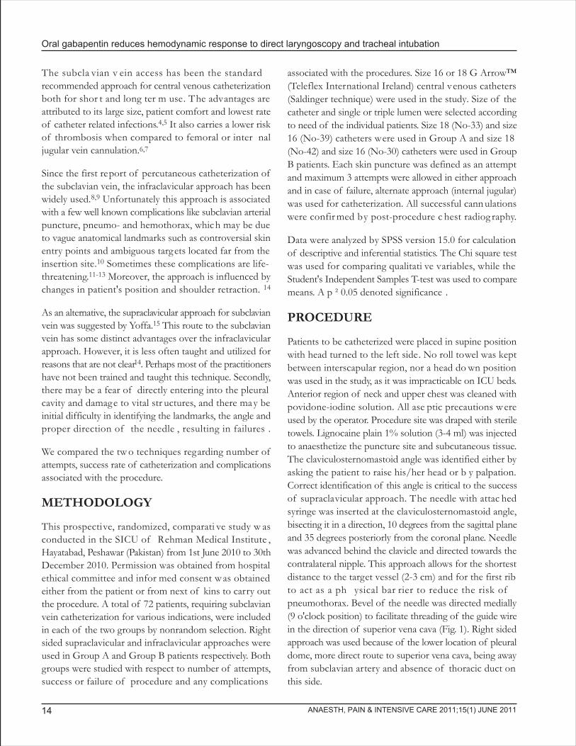
Methods: Sixty patients were randomly allocated to one of the two groups. Group I received 800 mg ofgabapentin and Group II received placebo with sip of water one hour before the induction of anaesthesia.After standard induction technique, study variables, pulse and noninvasive BP (systolic, diastolic and mean)and HR were noted every minute for first five minutes then at 10 and 15 minutes. Relevant demographic dataand study variables were recorded.
Results: Mean systolic BP with Gabapentin w as lower compared to placebo but it w as significant at 1min(136±22vs149±23), 2min (120±21vs136±24), 10min (107±12vs118±16) and 15 min (106±13vs116±13) afterintubation (P<0.05). Mean diastolic BP with gabapentin was significantly lower at 3min (69±15vs74±17) afterintubation with P<0.05. Mean BP with gabapentin was significantly lower at 2min (91±18vs103±18), 10min(79±12vs88±13) and 15 min (79±14vs86±12) after intubation at P<0 .05. Decrease in HR with gabapentinwas significant at 10min (92±15vs101±18) and 15 min (87±14vs99±16) after intubation (p<0.05).
Conclusion: Oral gabapentin decreases the response to laryngoscopy and intubation on systolic BP at 2 minand 15 min; mean ar terial pressure at 2, 10 and 15 min and HR at 10 and 15 min follo wing laryngoscopy.
Key Words: Gabapentin; pressor response; laryngoscopy; tracheal intubation
Citation: Iftikhar T, Taqi A, Sibtain A, Anjum S, Awan I. Oral gabapentin reduces hemodynamic response to direct lar yngoscopyand tracheal intubation. Anaesth Pain & Intensive Care 2011;15(1):17-20.
ORIGINAL ARTICLE
preexisting coronary artery disease and underlying cardiacdysfunction are particularly vulnerable to these changes4.Patients with uncontrolled h ypertension also sho wexaggerated response to lar yngoscopy and intubation.Different pharmacological agents have been used to obtundthis response.
Ultra short acting opioids increase the depth of anaesthesiafor a shor t period5. The beta bloc ker esmolol is used

Oral gabapentin reduces hemodynamic response to direct laryngoscopy and tracheal intubation
ANAESTH, PAIN & INTENSIVE CARE 2011;15(1) JUNE 201118
because of its cardioselective adrenergic receptor blockingproperties and ultra shor t duration of action6,7. Severalstudies have assessed the effecti veness of esmolol inblunting the hemodynamic response to laryngoscopy andtracheal intubation.
Recently, gabapentin has been recommended to decreasethe cardiovascular response to laryngoscopy and intubation8.It was approved in 1994 by FDA for the control of partialseizures with the combination of other antiseizure drugs9.In 2002 it w as shown to be effecti ve for post her peticneuralgia10 and other painful neuropathies 11, and ner verelated pains. Originally developed as an anticonvulsant,it is effective in controlling neuropathic pain, to treat acutepostoperative pain and reduce postoperati ve opioidrequirements in clinical trials . We planned to ev aluategabapentin for attenuation of response in BP and HR ondirect laryngoscopy and tracheal intubation in normotensivepatients undergoing elective surgery.
METHODOLOGY
The study was conducted at Department of Anaesthesia,Hameed Latif Hospital Lahore, from May 2007 to July2008. After obtaining appro val from hospital ethicalcommittee, 60 American Society of Anaesthesiologistsclass I and II. adult patients, planned for elective surgerywere randomly allocated either to g abapentin group orcontrol group. Pregnant patients, known hypertensive andischemic heart disease patients w ere excluded from thestudy. The patients in extremes of age were also excluded.
Patient's demographic data e.g. age, sex, weight, diagnosisand the surgical procedures were noted. Group I patientsreceived 800 mg oral gabapentin, while Group II patientsreceived placebo capsules one hour prior to surgery in thepre operative area. All patients received inj. nalbuphine 0.1mg/kg approximately 5 minutes before intubation. Inductionof general anaesthesia w as done with inj. thiopentonesodium 5 mg/kg and inj. rocuronium 0.6mg/kg. Patientswere ventilated with facemask and bag for 3 minutes andthen intubated after direct lar yngoscopy by a trainedanaesthetist. HR, systolic, diastolic and mean arterial BPswere recorded just before intubation as a baseline and then1, 2, 3, 4, 5, 10 and 15 minutes after intubation. Data wascollected on a specified proforma and analyzed by computerwith software SPSS version 11. Independent variables weregabapentin and placebo while de pendent variables wereHR, systolic, diastolic and mean arterial BP.
Descriptive statistics were calculated. The ratio betweengenders w as described in percentag es while ag e w asdescribed as mean and standard deviation. Mean HR,systolic, diastolic and mean BPs were compared by usingpaired 't' test. P< 0.05 w as considered as significant.
RESULTS
The demographic data was comparable between the groups.There was no statistical difference in gender distributionor mean age between two groups (Table 1&2)
The patients in gabapentin group as compared to placebogroup, showed lower Mean HR but it w as statisticallysignificant only at 10 and 15 min after intubation as p valuewas <0.05 only at these time intervals (Fig 1). Mean systolicBP with gabapentin was lower compared to placebo butit was significant at 1,2,10 and 15 minute after intubation(P<0.05). Mean diastolic BP in g abapentin g roup wassignificantly lower at 3-minute after intubation with P=0.05.Mean arterial BP with gabapentin was significantly lowerat 2, 10 and 15 min ute after intubation (P=0.05)(Fig 2).
DISCUSSION
The results of our study suggest that there was a generalizedtrend to wards less haemodynamic response in thegabapentin group as compared to the placebo group butit gained statistical significance only at some specific points.
Table 2: Comparison of mean age of the subjects (in years)
Study groups N Mean±SD
Gabapentin 30 37±12
Placebo 30 36±14
Statistical Analysis: t =0.246 P = 0.8 (P>0.05)There was no statistically significant difference of meanage between two study groups
Table 1: Gender distribution of the subjects under study [N(%)]
Gender Total
Gabapentin Placebo
Male 17(57.7) 19(63.3) 36(60)
Female 13(43.33) 11(36.67) 24(40)
Total 30(100) 30(100) 60(100)
Study groups
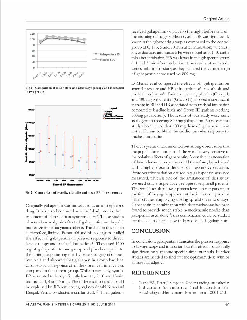
Original Article
ANAESTH, PAIN & INTENSIVE CARE 2011;15(1) JUNE 2011 19
Originally gabapentin was introduced as an anti-epilepticdrug. It has also been used as a useful adjunct in thetreatment of chronic pain syndromes12,13. These studiesobserved an analgesic effect of gabapentin but they didnot realize its hemodynamic effects. The data on this subjectis, therefore, limited. Fassoulaki and his colleagues studiedthe effect of gabapentin on pressor response to directlaryngoscopy and tracheal intubation.14 They used 1600mg of gabapentin to one g roup and placebo capsule tothe other group, starting the day before surgery at 6 hoursintervals and sho wed that g abapentin g roup had lesscardiovascular response at all the obser ved intervals ascompared to the placebo group. While in our study, systolicBP was noted to be significantly low at 1, 2, 10 and 15min,but not at 3, 4 and 5 min. The difference in results couldbe explained by different dosing regimes. Shashi Kiran andDeepak Verma conducted a similar study15. Their patients
Fig 1: Comparison of HRs before and after laryngoscopy and intubationin two groups
Fig 2: Comparison of systolic, diastolic and mean BPs in two groups
received gabapentin or placebo the night before and onthe morning of surgery. Mean systolic BP was significantlylower in the gabapentin group as compared to the controlgroup at 0, 1, 3, 5 and 10 min after intubation; whereas ,lower diastolic and mean BPs were noted at 0, 1, 3, and 5min after intubation. HR was lower in the gabapentin group0, 1 and 3 min after intubation. T he results of our studywere similar to this study, as they had used the same strengthof gabapentin as we used i.e. 800 mg.
D. Memis et al compared the effects of gabapentin onarterial pressure and HR at induction of anaesthesia andtracheal intubation16. Patients receiving placebo (Group I)and 400 mg gabapentin (Group II) showed a significantincrease in BP and HR associated with tracheal intubationcompared to baseline levels and Group III (patients receiving800mg gabapentin). The results of our study were sameas the group receiving 800 mg gabapentin. Moreover thisstudy also showed that 400 mg dose of gabapentin wasnot sufficient to blunt the cardio vascular response totracheal intubation.
There is yet an undocumented but strong observation thatthe population in our part of the world is very sensitive tothe sedative effects of gabapentin. A consistent attenuationof hemodynamic response could therefore , be achievedwith a higher dose at the cost of excessive sedation.Postoperative sedation caused b y g abapentin was notmeasured, which is one of the limitations of this study.We used only a single dose pre-operatively in all patients.This would result in lower plasma levels in our patients atthe time of laryngoscopy and intubation as compared toother studies emplo ying dosing spread o ver two days.Gabapentin in combination with dexamethasone has beenfound to provide much stable hemodynamic profile thangabapentin used alone17; this combination could be studiedfor the sedati ve effects with lo w doses of gabapentin.
CONCLUSION
In conclusion, gabapentin attenuates the presser responseto laryngoscopy and intubation but this effect is statisticallysignificant only at some specific time inter vals. Furtherstudies are needed to find out the optimum dose with orwithout an adjunct.
REFERENCES
1. Carrie ES., Peter J. Simpson. Understanding anaesthesia:Ind ica t ions for endotrac hea l in tubat ion .4 thEd.Michigan.Heinemann Professional; 2001:213.
Oral gabapentin reduces hemodynamic response to direct laryngoscopy and tracheal intubation
ANAESTH, PAIN & INTENSIVE CARE 2011;15(1) JUNE 201120
APICARE ON MEDPEDIA
The journal maintains a group page on Medpedia. This site is intended to connecthealthcare providers as well as general public on t he net. The site of fers richopportunities to indulge in health related discussions and to connect with likeminded people. Please log on to;http://www.medpedia.com/groups/455-APICARE
2. Kayhan Z, Aldemir D , Mutlu H, Ogus E. W hich isresponsible for the haemodynamic response due tolaryngosopy and trac heal intubation? Catec holamines,vasopressin or angiotensin? European jour nal ofAnesthesiology. 2005;22:780-85.
3. Morgan GE, Mikhail MS, Murray MJ. Clinical anesthesiology:Airway management. 4th Ed. New Y ork. McGraw HillCompanies Inc; 2002:110. .
4. Ismail S, Azam SI, Khan FA. Effect of age on haemodynamicresponse to tracheal intubation. A comparison of young,middle-aged and elderly patients. Anaesth Intensive Care2002;30:608-14.
5. Kim JT, Shim JK, Kim SH, Ryu HG, Yoon SZ, Jeon YS etal. R emifentanil vs . lignocaine for atten uating thehaemodynamic response during rapid sequence inductionusing propofol.Clin Dr ug In vestig. 2007;27:269-77.
6. Figueredo E, Garcia EM. Assessment of the efficacy ofesmolol on the haemodynamic c hanges induced b ylaryngoscopy and trac heal intubation: A meta-analysis .ActaAnaesthesiolScand 2001;45:1011-22.
7. Ugur B, Ogurlu M, Gezer E, Nuri O, Gursoy F. Effects ofesmolol, lidocaine and fentanyl on haemodynamic responsesto endotracheal intubation: a comparative study.Clin DrugInvestig. 2007;27:269-77.
8. Kong V, Irwin G. Gabapentin: A multimodal perioperativedrug. British J ournal of Anaesthesia. 2009;9:775-786.
9. Bazil CW. New antie pileptic dr ugs.Neurologist. 2002;8:71-8.
10. Jensen MP, Chiang YK, Wu J. Assessment of pain qualityin a clinical trial of g abapentin extended release forpostherpetic neuralgia. Clin J P ain. 2009;25:286-92.
11. Serpell MG. Gabapentin in neuropathic pain syndromes: Arandomised, double-blind, placebo-controlled trial. P ain.2002;99:557-66
12. Fassoulaki A, Stamatakis E, P etropoulos G, Siafaka I,Hassiakos D, Sarantopoulos C. Gabapentin attenuates latebut not acute pain after abdominal h ysterectomy. Eur JAnaesthesiol 2006;22:136-41.
13. Fassoulaki A, T riga A, Melemeni A, Sarantopoulos C .Multimodal analgesia with gabapentin and local anestheticsprevents acute and c hronic pain after breast surg ery forcancer. AnesthAnalg 2005;101:1427-32.
14. Fassoulaki A, Melemeni A, P araskeva A, Petropoulos G.Gabapentin atten uates the pressor response to directlaryngoscopy and trac heal intubation. Br J Anaesth.2006;96:769-73.
15. Kiran S, Verma D. Evaluation of gabapentin in attenuatingpressor response to direct lar yngoscopy and trac healintubation. SAJAA 2008;14:43-46.
16. Memis D, Turan A, K aramanlioglu B, Seker S. Ture M.Gabapentin reduces cardiovascular responses to laryngoscopyand tracheal intubation. European Journal of Anaesthesiology2006;23:686-690.
17. Koc S, Memis D, Sut N. The preoperative use of gabapentin,dexamethasone and their combination in varicocele surgery:a randomized controlled trial. AnesthAnalg 2007;105(4):1137-42.

ANAESTH, PAIN & INTENSIVE CARE 2011;15(1) JUNE 2011 21
INTRODUCTION
Hypotension in patients who receive subarachnoid block(SAB) is a potentially serious issue, which is known to leadto significant morbidity if not manag ed effectively andurgently. In obstetric applications, profound hypotensioncan potentially lead to serious hypoxia and hypovolemia inthe mother and the fetus. As placental blood flow is directlyproportional to the maternal blood pressure, the hypotensioncan lead to to placental hypoperfusion and fetal asphyxia.1The current incidence of hypotension following SAB isup to 8o% of patients without prophylactic therapy.1 To
Comparison of prophylactic ephedrine vs prnephedrine during spinal anesthesia for
caesarian sectionsAbdul Rehman*, Harris Baig*, M. Zameer Rajput**, Huma Zeb***
*Consultant anesthesiologist, **Professor of anesthesiology, ***Medical Officer AnesthesiaShifa International Hospital Islamabad (Pakistan)
Correspondence: Dr. Abdul Rehman, Consultant Anesthesiologist, Shifa International Hospital, H-8/4, Islamabad (Pakistan).Cell:+923335227329; email:[email protected]
ABSTRACTObjective: The objective of this study was to compare the hemodynamic effects of use of prophylacticintravenous ephedrine with ephedrine use on as needed basis in patients receiving spinal anesthesia for caesareansections.Study design: A double blind, randomized, comparative trialSetting: Department of Anaesthesiology, Critical Care, and Pain Management, Shifa International HospitalIslamabad.Duration: October 2007 to March 2008Methodology: Seventy patients were recruited who were scheduled to receive spinal anaesthesia for C-section.The patients were randomized into two groups (A and B). In patients of Group A (control group) ephedrinewas used to treat h ypotension when indicated, while in Group B (inter vention group), patients receivedprophylactic ephedrine soon after the subarachnoid block. Hemodynamic changes were recorded and the datawas analysed.Results: In Group A, the blood pressure dropped in a higher number of patients [23 (65.7%)], as comparedto Group B [6(17.1%)]. This difference was statistically significant (p<0.001).Conclusion: Prophylactic ephedrine is better than ephedrine prn in prevention of hypotension in patientsreceiving spinal analgesia for C-Section.
Key Words: Subarachnoid block; hypotension; caesarean section; ephedrine.Citation: Rehman A, Baig H, Rajput MZ, Zeb H. Comparison of prophylactic ephedrine against prn ephedrine during spinal anesthesiafor caesarian sections. Anaesth Pain & Intensive Care 2011;15(1):21-24.
ORIGINAL ARTICLE
prevent this problem, various methods have been consideredappropriate. Expansion of intravascular volume can beachieved with preload with crystalloids or colloids. Thoughthis is a common practice for electi ve cases, it does notoffer full protection against hypotension.2 Other optionsinclude, but are not limited to , left uterine displacement(LUD) and occasional use of ionotropic suppor t.
Ephedrine has been the vasopressor of choice to controlspinal hypotension for many years, but the controversiesstill exist about the best regimen of its use; whether to useit in intermittent boluses or in infusion, whether to use itprophylactically or just to use it prn to control hypotension

Comparison of prophylactic ephedrine against prn ephedrine during spinal anesthesia for caesarian sections
ANAESTH, PAIN & INTENSIVE CARE 2011;15(1) JUNE 201122
once it does occur. We conducted this study to comparethe hemodynamic control b y e phedrine when usedprophylactically with its prn use after the occur rence ofhypotension.
METHODOLOGY
A double blind, randomized, comparati ve trial w asconducted at Department of Anesthesiology, Critical Care,and Pain Manag ement, Shifa Inter national Hospital,Islamabad, after g etting approval from Hospital EthicsCommittee and informed consent of the parturients, fromOctober 2005 to March 2006. Seventy obstetric patientsscheduled for elective caesarean section with AmericanSociety of Anesthesiologist's physical status (ASA-PS) Ior II were randomly divided into two groups A and B with35 patients in each group. Patients on antihypertensives,diabetics, and pregnancy induced h ypertensive patientswere excluded. Patients with pre-eclampsia and eclampsiawere also excluded from the study . Patients with fixedcardiac output (mitral stenosis or aor tic stenosis),coagulopathy (platelet count less than 80,000), abr uptioplacentae, placenta previa, severe fetal distress and cordprolapse were also ex cluded from the study as SAB iscontraindicated in these cases.
After appropriate preoperative preparation, patients weretransferred from the ward to the obstetric operating room.Baseline BP and HR were measured. An intravenous linewith 18G IV cannula was established. Lactated Ringer'ssolution 15ml/kg was infused to all patients 30 min utesbefore the SAB as a standard protocol. Patients were thendivided into tw o g roups A and B in random order .
Spinal hypotension was defined as a ³ 30% drop of systolicBP from the baseline reading.
SAB was instituted in left lateral position with hyperbaricbupivacaine bupivacaine 0.75% with dextrose 8.25%(Abocaine Spinal™ -Abbott Laboratories (Pakistan) Ltd®)1.6 ml injected in the subarac hnoid space over 15 sec atL3-4 through 25G pencil point needle (Unises CorporationTokyo-Japan) after infiltrating 1% lignocaine 1 ml locally.Patients in Group B received prophylactic ephedrine 15mgintravenously, simultaneously with the administration ofhyperbaric bupivacaine. Then patients were placed in asupine position with the table in left lateral tilt. Oxyg enwith facemask was initiated at 3 litres/min to all patients.BP and HR were measured every two minutes initially, tilldelivery of the baby and then every five minutes till theend of the operation. Lactated Ringer's solution 5 ml/kg/hr
was infused as a maintenance f luid. Synthetic oxytocin(Syntocinon™) 5 IU was injected IV after delivery of thebaby in all patients. In both groups, hypotension if occurredwas treated by a second dose of ephedrine 10mg IV inorder to maintain the systolic BP within ±10% of thebaseline. The patients were shifted to post anaesthesia careunit (PACU) and vital signs monitored.
The data collected included systolic , diastolic and meanarterial pressures and hear t rates. Statistical analysis wasperformed through SPSS version 12. Descriptive statisticswere presented as tables. Chi-square test was applied tocompare the mean values of systolic blood pressure . Pvalue ² 0.05 w as considered statistically significant.
RESULTS
The demographic data of the patients and the indicationsof the surgery are given in Table 1. No statistical differencewas found between two groups regarding mean age, bodyweight and indications of c-section.
Table 2: Comparison of hemodynamic parameters in twogroups (Mean±SD)
Group A Group BParameter (Control Group) (Intervention Group)
n=35 n=35
Systolic 116±7 118±10
Diastolic 66±13 68±10
Mean 82±12 87±11
Baseline HR 102±12 95±15
Table 1: Comparison of demographic data of mothers in twogroups
Group A Group Bn=35 n=35
Age (yrs) (Mean±SD) 31±4 27±4
Body wt. (Kg) (Mean±SD) 63±4 64±5
Indications for c-section [N(%)]
Breech 13(37.1) 11(31.4)
Feto-pelvic disproportion 12(34.2) 17(48.6)
Previous c-sections 10(28.5) 7(20)
The baseline hemodynamic parameters in two groups werecomparable, with no statistical difference (T able 2).
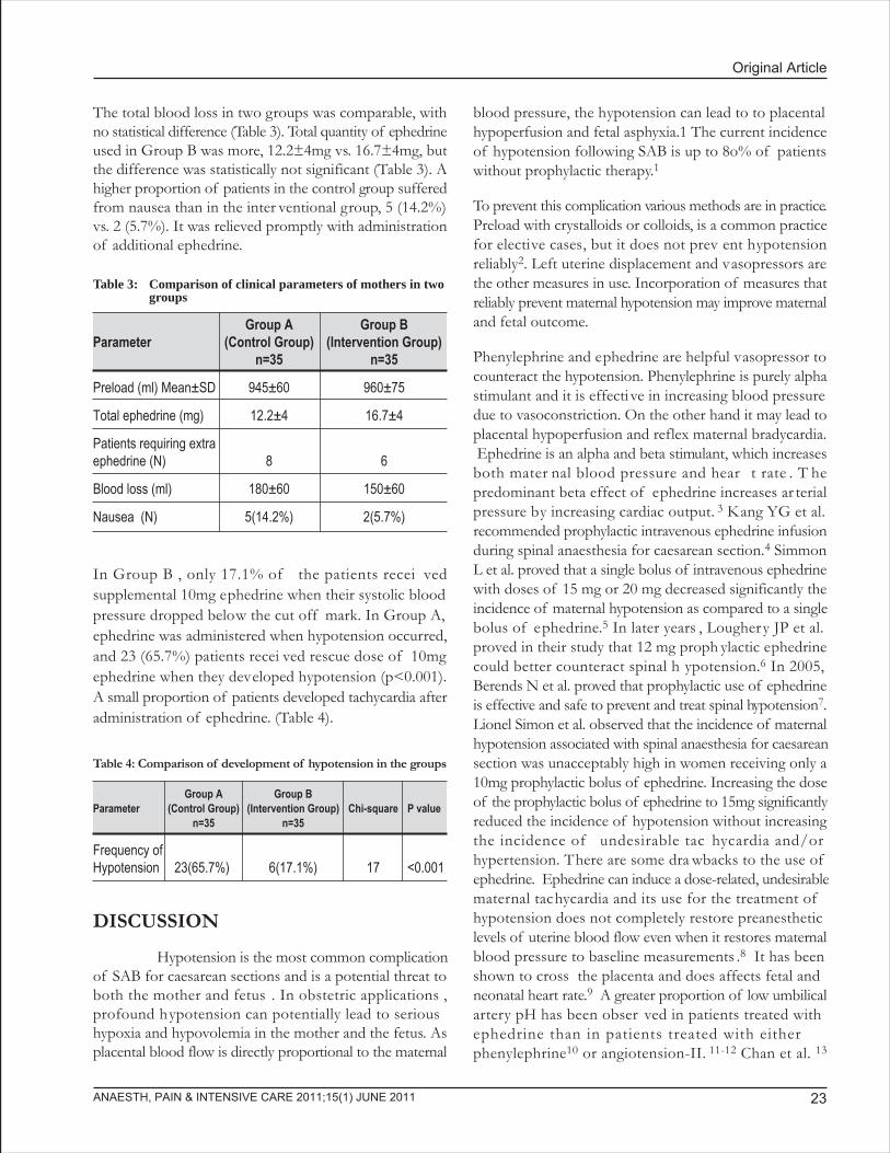
Original Article
ANAESTH, PAIN & INTENSIVE CARE 2011;15(1) JUNE 2011 23
The total blood loss in two groups was comparable, withno statistical difference (Table 3). Total quantity of ephedrineused in Group B was more, 12.2±4mg vs. 16.7±4mg, butthe difference was statistically not significant (Table 3). Ahigher proportion of patients in the control group sufferedfrom nausea than in the inter ventional group, 5 (14.2%)vs. 2 (5.7%). It was relieved promptly with administrationof additional ephedrine.
In Group B , only 17.1% of the patients recei vedsupplemental 10mg ephedrine when their systolic bloodpressure dropped below the cut off mark. In Group A,ephedrine was administered when hypotension occurred,and 23 (65.7%) patients recei ved rescue dose of 10mgephedrine when they developed hypotension (p<0.001).A small proportion of patients developed tachycardia afteradministration of ephedrine. (Table 4).
DISCUSSION
Hypotension is the most common complicationof SAB for caesarean sections and is a potential threat toboth the mother and fetus . In obstetric applications ,profound hypotension can potentially lead to serioushypoxia and hypovolemia in the mother and the fetus. Asplacental blood flow is directly proportional to the maternal
Table 3: Comparison of clinical parameters of mothers in twogroups
Group A Group BParameter (Control Group) (Intervention Group)
n=35 n=35
Preload (ml) Mean±SD 945±60 960±75
Total ephedrine (mg) 12.2±4 16.7±4
Patients requiring extraephedrine (N) 8 6
Blood loss (ml) 180±60 150±60
Nausea (N) 5(14.2%) 2(5.7%)
blood pressure, the hypotension can lead to to placentalhypoperfusion and fetal asphyxia.1 The current incidenceof hypotension following SAB is up to 8o% of patientswithout prophylactic therapy.1
To prevent this complication various methods are in practice.Preload with crystalloids or colloids, is a common practicefor elective cases, but it does not prev ent hypotensionreliably2. Left uterine displacement and vasopressors arethe other measures in use. Incorporation of measures thatreliably prevent maternal hypotension may improve maternaland fetal outcome.
Phenylephrine and ephedrine are helpful vasopressor tocounteract the hypotension. Phenylephrine is purely alphastimulant and it is effective in increasing blood pressuredue to vasoconstriction. On the other hand it may lead toplacental hypoperfusion and reflex maternal bradycardia. Ephedrine is an alpha and beta stimulant, which increasesboth mater nal blood pressure and hear t rate . T hepredominant beta effect of ephedrine increases ar terialpressure by increasing cardiac output. 3 Kang YG et al.recommended prophylactic intravenous ephedrine infusionduring spinal anaesthesia for caesarean section.4 SimmonL et al. proved that a single bolus of intravenous ephedrinewith doses of 15 mg or 20 mg decreased significantly theincidence of maternal hypotension as compared to a singlebolus of ephedrine.5 In later years , Loughery JP et al.proved in their study that 12 mg proph ylactic ephedrinecould better counteract spinal h ypotension.6 In 2005,Berends N et al. proved that prophylactic use of ephedrineis effective and safe to prevent and treat spinal hypotension7.Lionel Simon et al. observed that the incidence of maternalhypotension associated with spinal anaesthesia for caesareansection was unacceptably high in women receiving only a10mg prophylactic bolus of ephedrine. Increasing the doseof the prophylactic bolus of ephedrine to 15mg significantlyreduced the incidence of hypotension without increasingthe incidence of undesirable tac hycardia and/orhypertension. There are some dra wbacks to the use ofephedrine. Ephedrine can induce a dose-related, undesirablematernal tachycardia and its use for the treatment ofhypotension does not completely restore preanestheticlevels of uterine blood flow even when it restores maternalblood pressure to baseline measurements .8 It has beenshown to cross the placenta and does affects fetal andneonatal heart rate.9 A greater proportion of low umbilicalartery pH has been obser ved in patients treated withephedrine than in patients treated with eitherphenylephrine10 or angiotension-II. 11-12 Chan et al. 13
Table 4: Comparison of development of hypotension in the groups
Group A Group BParameter (Control Group) (Intervention Group) Chi-square P value
n=35 n=35
Frequency ofHypotension 23(65.7%) 6(17.1%) 17 <0.001
Comparison of prophylactic ephedrine against prn ephedrine during spinal anesthesia for caesarian sections
ANAESTH, PAIN & INTENSIVE CARE 2011;15(1) JUNE 201124
compared ephedrine infusion and f luid preload for theprevention of spinal hypotension during caesarean section.The best proph ylaxis of maternal hypotension duringcaesarean section is still controversial. McGrathe et al.14
showed that ephedrine was superior to phenylephrine inrestoring uterine blood flow and fetal oxygenation duringritodrine infusion and e pidural anaesthesia inducedhypotension in gravid ewes. Hall et al.15 compared infusionsof ephedrine and phenylephrine during spinal anaesthesia,Some authors proposed using angiotension-II instead ofephedrine to avoid maternal tachycardia and fetal academia,but it is not readily available. Thus, ephedrine remains thevasopressor of choice in obstetrics.
We used prophylactic ephedrine in a dose of 15 mg withintermittent boluses of ephedrine prn, and found that theformer was better in controlling mater nal hypotension(p<0.001).
CONCLUSION
We conclude that prophylactic use of ephedrine is moreefficient for maintenance of blood pressure during spinalanaesthesia for caesarean section as compared to its pr nuse.
REFERENCES
1. Turkoz A, Togal T, Gokdeniz R, I Topraks H, Esroy O.Effectiveness of intravenous ephedrine infusion duringspinal anaesthesia for caesarean section based on maternalhypotension, neonatal acid-base status and lactate lev els.Anaesth Intensive Care 2002;30:316-20.
2. Ueyama H, Yan-ling H, Tanigami H, Mashimo T, YoshivaI. Effects of crystalloid and colloid preload on blood volumein the parturient undergoing spinal anaesthesia for electivecaesarean section. Anesthesiolog y 1999;91:1571-6.
3. Lee A, Warwick D, Knee N, fanzca. A quantitative, systemicreview of randomized controlled trials of ephedrine versusphenylepherine for the management of hypotension duringspinal anaesthesia for caesarean deli very. Anesth Analg2002;94:920-6.
4. Kang YG, Abouleish E, Caritis S. Prophylactic intravenousinfusion during spinal anaesthesia for caesarean section.Anesth Analg 1982;61:839-42.
5. Simlon L, Provenchere S,de Saint Blanquat L, Boula y G,Hamza J. Dose of prophylactic intravenous ephedrine duringspinal anaesthesia for caesarean section. J Clin Anesth2001;13:366-9.
6. Loughery JP, Walsh F, Gardiner J. Prophylactic intravenousbolus e phedrine for caesarean section under spinalanaesthesia. Eur J Anesthesiol 2002;191:63-8.
7. Berends N, Teunkens A, Vandermeersch E, Van de VeldeM. A randomized trial comparing low-dose combined spinal-epidural anaesthesia and conventional epidural anaesthesiafor caesarean in sever preeclampsia.Acta Anaesthesiol Belg2005;56(2):155-62.
8. Vincent RD, Jr, Werhan CF, Norman PF et al. Prophylacticangiotension 11 infusion during spinal anaesthesia for electivecaesarean section. Anesthesiolog y 1988;88:1475-1479.
9. Wright RG, Shnider SM, Levinson G, Rolbin SH, Parer JT.The effect of maternal administration of ephedrine on fetalheart rate and variability. Obstet Gynecol 1981;51:734-738.
10. Thomas DG, Robson SC, Redfern N, Hughes D, Boys RJ.Randomized trial of bolus phenylepherine or ephedrine formaintenance of arterial pressure during spinal anaesthesiafor caesarean section. Br J Anaesth 1996;76:61-65.
11. Ramin SM, Ramin KD, Cox K, Magnees RR, Shearer VE,Gant NF. Comparison of prophylactic angiotension II versusephedrine infusion for prevention of maternal hypotensionduring spinal anaesthesia. Am J Obstet Gynecol1994;171:734-739.
12. Shearer VE, Ramin SM, Wallace DH, Dax JS, Gilstrap LC.Fetal effects of proph ylactic e phedrine and mater nalhypotension during regional anaesthesia for caesarean section.J Matern Fetal Med 1996;5:79-84.
13. Chan WS, Irwin MG, Tong WN, Lam YH. Prevention ofhypotension during spinal anaesthesia for caesarean section:ephedrine infusion v ersus f luid preload. Anaesthesia1997;52:908-913.
14. McGrath JM, Chestnut DH, Vincent RD, Jr, et al. Ephedrineremains the vasopressor of the c hoice for treatment ofhypotension during ritodrine infusion and e piduralanaesthesia. Anesthesiolog y 1994;80:1073-1081.
15. Hall PA, Bennett A, Wilkies MP, Lewise M. Spinal Anesthesiafor caesarean section. Comparison of infusion ofphenylephrine and ephedrine. Br J Anaesth 1994;73:471-474.
ANAESTH, PAIN & INTENSIVE CARE 2011;15(1) JUNE 2011 25
INTRODUCTION
Endotracheal reintubation is not uncommon among criticallyill patients after open heart surgery1. The overall incidencefor reintubation in a general surgical intensive care unit isgenerally considered 4%, but varies dramatically between
Endotracheal reintubation in post-operativecardiac surgical patients
Abdul-Zahoor MD, FCPS*, Nor Azlina MD, MSc**
*Consultant AnesthesiologistKing Khaled Eye Specialist Hospital, P. O. Box 7191, Riyadh 11462 (Saudi Arabia)
**Consultant AnesthesiologistNational Heart Institute, 145, Jalan Tun Razak, 50400, Kuala Lumpur (Malaysia)
Correspondence: Dr. Abdul-Zahoor MD, FCPS, Consultant Anesthesiologist, King Khaled Eye Specialist Hospital,P. O. Box 7191, Riyadh 11462 (Saudi Arabia); Phone: +966 509003709; Fax No: +966 1 4821908;E-mail: [email protected] or [email protected]
ABSTRACTBackground: The reported incidence of reintubation in patients who were weaned from mechanical ventilationafter cardiac surgery is 6.6%4 in a retrospective study, but little work has been done prospectively to find outthe incidence and causes for reintubation in a cardiac surgical ICU. We conducted this study to find outincidence and the causes of endotracheal reintubation in patients who were electively ventilated after openheart surgery and were extubated after fulfilling preset criteria for extubation.
Methodology: A total of 1229 consecutive patients were included in the study. On arrival to ICU after cardiacsurgery, all patients were electively ventilated with standardized ventilatory parameters. Routine monitoringof all patients was done and patients were extubated once they met the criteria for extubation. The patients,who met the reintubation criteria, were reintubated and the reason(s) noted. Once they stabilized and fulfilledthe extubation criteria, they were extubated.
Results: A total of 47(3.82%) patients required reintubation after weaning from the ventilation during thestudy period, and in 5(10.63%) patients out of these, reintubation was needed more than once. We found ahigher incidence of reintubation, 11.84 % and 10.63%, in patients after single and double valve replacementsurgery respectively. The incidence was much lower (2.14%) among coronary artery bypass grafting (CABG)patients.
Conclusion: The patients undergoing valve replacement surgery are more prone to reintubation in postoperativeperiod as compared to CABG patients. Impending respiratory failure, cardiovascular (hemodynamic) instabilityand impaired conscious level are the common indications for reintubation.
Key Words: Extubation failure; coronary artery bypass grafting; reintubation; cardiac surgery intensive careunit; open heart surgery; valve replacement.Citation: Zahoor A, Azlina N. Endotracheal reintubation in post-operative cardiac surgical patients. Anaesth Pain & Intensive Care2011;15(1):25-29.
ORIGINAL ARTICLE
1-13%, depending on the underlying disease process 2.Similarly a 10% incidence has been re ported in 745consecutive admissions in medical ICU patients that weremechanically ventilated for a minimum of 6 hours3. Thereported incidence of reintubation in patients who wereweaned from mechanical ventilation after cardiac surgery

Endotracheal reintubation in post-operative cardiac surgical patients
ANAESTH, PAIN & INTENSIVE CARE 2011;15(1) JUNE 201126
is 6.6%4 in a retrospective study but little work has beendone to find out the incidence and causes for reintubationin a cardiac surgical ICU prospectively.
Reintubation is not only associated with increased durationof mechanical ventilation but also the ICU and hospitallength of stay5. Reintubation is known to be an independentcause, which adds to the mortality; patients who requiredreintubation have poor prognosis with a mor tality rateexceeding 30-40% 6, ir respective of the cause forreintubation. The major causes of reintubation are usuallyrelated to respiratory or cardiovascular system but couldbe multi-factorial6 and may possibly be prevented, to someextent, by improving the care7.
We studied the incidence and the causes of endotrachealreintubation in patients who were electively ventilated afteropen heart surgery and were extubated after fulfilling thepreset criteria for extubation.
METHODOLOGY
After approval by the institutional review board of ourhospital, all adult patients , who underw ent open hear tsurgery and were electively ventilated after the surgery inthe post-cardiac surgery intensive care unit (CICU), wereincluded in the study. It was a descriptive study. The datawas prospectively collected for a period of seven months,from January to July 2008. A total of 1229 patients, admittedduring this period, were enrolled in the study.
Routine monitoring of all patients w as done includingECG, arterial oxygen saturation (SaO2), end tidal carbondioxide (EtCO2), central v enous pressure and in vasivearterial blood pressure . Pulmonar y ar tery pressure ,pulmonary capillary wedge pressure or left atrial pressurewere monitored where indicated (Table 3).
Arterial blood gases were checked hourly for the first fourhours and were subsequently repeated after every changein the ventilatory parameters. Urine output and surgicalbleed through the chest drains was measured on an hourlybasis. Hematocrit was aimed to be kept at 30 % or aboveand appropriate transfusion w as given when indicated.
All patients, on arrival to ICU, were put on standardizedventilatory parameters unless indicated otherwise . Theywere initially put on volume controlled or pressure controlledmode (Table 3). A positive end expiratory pressure (PEEP)of 5 cmH2O or more w as added where indicated. Aninspired oxygen fraction (FiO2) of ³50% and a tidalvolume of 10ml/kg body weight was set to all patients .
The ventilatory parameters were adjusted according to theresults of arterial blood gases (ABG's).
An early extubation was aimed in all patients but no timeschedule was fixed like a fast trac k. All patients w ereextubated without any dela y, when they met the presetcriteria for extubation (Table 1).
Table 2: Pre-set criteria for re-intubation
Respiratory:• Impending respiratory failure:• in PaCO2 ³ 10 mmHg/hr, _in the pH ³ 0.10/hr, PaO2
² 60 mmHg or SaO2 ² 90% on FiO2 ³ 50%• work of breathing• Upper respiratory obstruction• Excessive pulmonary secretions
A consideration w as also gi ven to the frequency ofrequirement and dosag e of inotropic , c hronotropic,vasodilator and/or vasoconstrictor drugs used. Extubationwas delayed if a patient was on more than three drugs atthat same time. Diaphoresis, confusion, stroke, renal failureor compromised renal functions and anxiety w ere alsoconsidered as causes for extubation dela y. Patients werere-intubated when indicated according to preset criteriafor reintubation (Table 2).
Table 1: Pre-set Criteria for extubation
Respiratory:• Minimal respiratory support (1-2 h):
SIMV rate ² 6 breaths/min; Pressure support ²10cmH2O;PEEP ² 5cmH2O; FiO2 ² 50%.
• Arterial blood gas:SaO2 ³ 90% on Fio2 ² 50%, PaO2 ³ 80mmHg on Fio2² 50%, PaCo2 30-45mmHg
• No frequent airway suctioning needed
Cardiovascular:• Mean arterial pressure 70-110mmHg.• Stable (sinus) rhythm or a rhythm other than sinus not
adversely affecting the BP.• Heart rate 60-110/min.
Conscious level:• Mentally alert, Obeying verbal commands, protecting airway,
intact cough and gag reflex.
Vasoactive drugs:Dopamine²10 µg/Kg/min; Dobutamine ² 10 µg/kg/min; Epinephrine² 0.05µg/min; Norepinephrine ² 0.2 µg/kg/min; Glyceral Trinitrate(GTN) ² 5 µg/kg/min; Milrinone ² 0.5 µg/kg/min and Sodiumnitroprusside (Nepride) ² 2 µg/kg/min
Miscellaneous:• Chest drainage ³ 100 ml/hr• Muscle power ³ grade-3
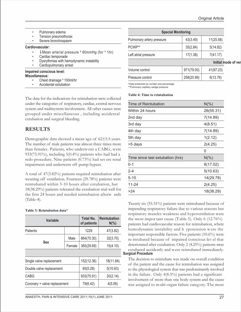
Original Article
ANAESTH, PAIN & INTENSIVE CARE 2011;15(1) JUNE 2011 27
The data for the indications for reintubation were collectedunder the categories of respiratory, cardiac, central nervoussystem and multisystem involvement. All other causes weregrouped under miscellaneous , including accidentalextubation and surgical bleeding.
RESULTS
Demographic data showed a mean age of 62±5.5 years.The number of male patients was almost three times morethan females. Patients, who underw ent a CABG, were933(75.91%), including 5(0.4%) patients who had had aredo procedure. Nine patients (0.73%) had sev ere renalimpairment and underwent off pump bypass.
A total of 47(3.82%) patients required reintubation afterweaning off ventilation. Fourteen (29.78%) patients werereintubated within 5-10 hours after extubation, but18(38.29%) patients tolerated the extubation trial well forthe first 24 hours and needed reintubation afterw ards(Table-4).
Twenty six (55.31%) patients were reintubated because ofimpending respiratory failure due to various reasons butrespiratory muscles weakness and hypoventilation werethe most impor tant cause (T able 5). Only 6 (12.76%)patients had cardiovascular reason for reintubation, wherehemodynamic instability and h ypotension w ere theimportant responsible factors. Five patients (10.6%) werere-intubated because of impaired conscious lev el thatdeteriorated after extubation. Only 2 (4.25%) patients wereextubated accidently and were reintubated immediately.
The decision to reintubate was made on overall conditionof the patient and the cause for reintubation was assignedto the physiological system that was predominantly involvedin the failure . Only 4(8.5%) patients had a significantinvolvement of more than one body system and the causewas assigned to m ulti-organ failure categ ory. The most
• Pulmonary edema• Tension pneumothorax• Severe bronchospasm
Cardiovascular:• Mean arterial pressure ² 60mmHg (for ³ 1hr)• Cardiac temponade• Dysrythmias with hemodynamic instability• Cardiopulmonary arrest
Impaired conscious level:Miscellaneous:
• Chest drainage ³ 100ml/hr• Accidental extubation
Table 3: Reintubation data*
Total No. Reintubationof patients N(%)
Patients 1229 47(3.82)
SexMale 864(70.30) 32(3.70)
Female 365(29.69) 15(4.10)
Surgical Procedure
Single valve replacement 152(12.36) 18(11.84)
Double valve replacement 65(5.28) 5(10.63)
CABG 933(75.91) 20(2.14)
Coronary + valve replacement 79(6.42) 4(5.06)
Variable
Special Monitoring
Pulmonary artery pressure 43(3.49) 11(25.58)
PCWP** 35(2.84) 5(14.82)
Left atrial pressure 17(1.38) 7(41.17)
Initial mode of ventilation
Volume control 971(79.00) 41(87.23)
Pressure control 258(20.99) 6(12.76)
*Data presented as number and percentage**Pulmonary capillary wedge pressure
Table 4: Time to reintubation
Time of Reintubation N(%)
Within 24 hours 26(55.31)
2nd day 7(14.89)
3rd day 4(8.51)
4th day 7(14.89)
5th day 1(2.12)
>5 days 2(4.25)
0
Time since last extubation (hrs) N(%)
0-1 8(17.02)
2-4 5(10.63)
5-10 14(29.78)
11-24 2(4.25)
>24 18(38.29)
Endotracheal reintubation in post-operative cardiac surgical patients
ANAESTH, PAIN & INTENSIVE CARE 2011;15(1) JUNE 201128
common combination of multi-organ involvement wasimpending respirator y failure tog ether with impairedconscious level.
Eighteen patients (38.29 %) that were reintubated once ormore than once ev entually died. None of them had atracheostomy because they all died for v arious reasonswithin two weeks of reintubation. T he cause of deathdetermination was beyond the scope of this study; hence,has been ignored.
Five (10.63%) patients, needed reintubation more thanonce (Table 3). T he incidence of reintubation was notdifferent betw een male and female patients andcorresponded closely to their ratio . We found a higherincidence of reintubation, 18/152 (11.84 %) and 5/65(10.63%), in single and double valve replacement surgeryrespectively. The incidence was much lower [20/933 (2.14%)]among CABG patients.
DISCUSSION
We admitted 1229 consecutive patients after cardiac surgeryin our ICU over a period of 7 months and studied themprospectively. A total of 47(3.82%) patients failed theextubation trial. Patients undergone a single or doublevalve surgery had a higher incidence of reintubation in
Table 5: Causes of reintubation
Causes N(%)
Impending respiratory failure:
Due to Pneumonia / excessive secretions, non cardiacpulmonary edema, lung collapse, aspiration,bronchospasm, respiratory muscle weakness, upperairway obstruction, hypoventilation syndrome andkinked / blocked tube.
Cardiovascular:Severe myocardial ischemia or acute Infarction, severearrhythmia with Hemodynamic instability, severehypotension (low output syndrome), congestive heartfailure and cardiac arrest.
Impaired conscious level 5(10.6)
Accidental extubation 2(4.25)
Surgical bleeding 2(4.25)
Multi organ involvement 4(8.5)
Miscellaneous 2(4.25)
Total 47(100)
26(55.31)
6(12.76)
comparison to CABG surg ery. T he incidence ofreintubation was 4/79(5.06%) in patients who had CABGtogether with a valve replacement.
The higher incidence of reintubation among the v alvereplacement surgery indicates some correlation betweenthe extubation failure and valve surgery. The exact causeis unclear but it is known that pulmonary functions mightdeteriorate in the immediate postoperati ve period andmight take time to return to the preoperative values8. Thepossible mechanism could be the poor compliance of thelungs to accommodate the corrected cardiac output aftervalve surgery. The reported incidence of persistent pleuraleffusion for weeks after valve replacement surgery is 45%9
and that could ha ve been contributed to pulmonar ymalfunction and subsequent need for reintubation in ourvalve replacement patients.
Our overall incidence (3.82%) of reintubation was almosthalf of the reported incidence (6.6%) after cardiac surgery4.The reasons of this difference may be related either to thedifference in the type of surgery or to a difference in thepolicy of fast track protocol at some centers, in an attemptto reduce the ICU stay and cost10.
The most common (55.31%) cause for reintubation in ourstudy was impending respiratory failure that manifestedwith increased work of breathing, accessory muscle use,hypoxia and/or hypercapnea, hypoventilation and respiratoryacidosis, especially for those who were reintubated withinthe first 24 hours. Half of our reintubated patients developedpneumonia. In a case control study , the incidence ofpneumonia was significantly higher (47%vs10%) in patientsneeding reintubation11. Dries and colleagues also foundan increased incidence of nosocomial pneumonia in patientswho failed extubation trial12, which confirms our findings.
Pulmonary edema and upper airw ay obstr uction wereamong the important respiratory causes for reintubation,especially for those who were reintubated after the secondday of extubation. Interestingly, causes related to airwaypatency and secretions manifested only after extubation.
The incidence of asymptomatic myocardial ischemia hasbeen reported to be 52% after CABG13. In our study 6/47(12.76%) of the patients had a cardiac reason forreintubation, where myocardial ischemia or acute infarctionwere the common reasons, that led to low output syndromeand heart failure among CABG patients, while pulmonaryhypertension w as the impor tant reason in the v alve

Original Article
ANAESTH, PAIN & INTENSIVE CARE 2011;15(1) JUNE 2011 29
replacement surgical patients.
It is well known that patients, who undergo myocardialrevascularization procedures, are par ticularly prone tostroke, encephalopathy and other neurologic dysfunction,because they are relati vely old and ha ve atheroscleroticdisease. They are also subject to cerebral embolization andcerebral h yperthermia after the discontin uation ofcardiopulmonary bypass14,15. We found impaired consciouslevel (10.6%) to be the third impor tant reason forreintubation in our study. Two of those patients ultimatelydeveloped stoke and died later on.
CONCLUSION
Endotracheal reintubation is not uncommon among criticallyill patients after open hear t surgeries. The incidence washigher in patients undergone valve replacement surgery incomparison to CABG surg ery. Impending respirator yfailure, cardiovascular (hemodynamic) instability andimpaired conscious level were the most important indicationsfor reintubation.
REFERENCES
1. Kurt M, Boek en U, Litmathe J , F eindt P, Gams E.Oxygenation failure after cardiac surgery: Early reintubationversus treatment by nasal continuous positive airway pressure(NCPAP) or non-in vasive positive pressure v entilation(NPPV). Monaldi Arch chest Dis 2008;70:71-5.
2. Demling, RH, Read T, Lind LJ, Flanagan HL. Incidenceand morbidity of extubation failure in surgical intensi vecare patients. Crit. Care Med;16:573-7.
3. Epstein K, Ciubotaru RL. Independent effects of etiologyof failure and time to reintubation on outcome for patientsfailing extubation Am J Respir. Crit Care Med1998;158(2):489-93.
4. Rady, Mohamed Y , R yan, T homas MB. P erioperativepredictors of extubation failure and the effect on clinicaloutcome after cardiac surg ery. Crit Care Med1999;27(2):340-47.
5. Gowardman JR, Huntington D, Whiting J. The effect ofextubation failure on outcome in a multidisciplinary Australianintensive care unit. Crit Care R esus 2006;8:328-33.
6. Jordi R, Emili D, Marta R, Jordi V. Risk factors for developingpneumonia within 48 Hours of Intubation. Am. J. Resir.Crit. Care Med 1999;159(6):1742-46.
7. Sibu P, Saha, Norma L, Cindy B, Victor A, Ferraris. AdvancedCare for Patients after Coronary artery bypass graft. InterJ Ang 2005;14:141-43.
8. Mustafa KY, Nour MM, Shuhaiber H, Y ousof AM.Pulmonary function before and sequentially after v alvereplacement surg ery with cor relation to preoperati vehemodynamic data. Am Rev Respir Dis1984;130(3):400-6.
9. Moujahed L, Richard B, Brigitte D, François M, Louis PB.Pleural Effusions Following Cardiac Surgery. Prevalence,Risk Factors, and Clinical Features. CHEST 2009;136(6):1604-11.
10. Reyes A, Vega G, Blancas R, Morato B, Moreno JL, TorrecillaC, Cereijo E. Early vs conventional extubation after cardiacsurgery with cardiopulmonary bypass. Chest 1997;112(1):193-201.
11. Torres A, Gatell JM, Aznar E, el Ebiar y M, Puig DL,Gonzalez J. Reintubation increases the risk of nosocomialpneumonia in patients needing mechanical ventilation. AmJ Respir Crit Care Med 1995;152:137-41.
12. Dries DJ, McGonigal MD, Malian MS, Bor BJ, Sullivan C.Protocol-driven ventilator weaning reduces use of mechanicalventilation, rate of early reintubation and ventilator-associatedpneumonia. J Trauma 2004;56:943-51.
13. Smith, Randall C, Leung, Jacqueline M, Mangano, DennisT. Postoperative myocardial ischemia in patients undergoingcoronary ar tery b ypass g raft surg ery. Anesthesiolog y1991;74:464-73.
14. Mora CT, Murkin JM. The central nervous system responsesto cardiopulmonar y b ypass. In: Mora CT , ed.Cardiopulmonary bypass: principles and tec hniques ofextracorporeal circulation. New York: Springer-Verlag. 1995:pp114-46.
15. Herskowitz A, Mangano DT. The inflammatory cascade: afinal common pathw ay for perioperati ve injur y?Anesthesiology 1996;85:454-57.

ANAESTH, PAIN & INTENSIVE CARE 2011;15(1) JUNE 201130
Influence of working conditions on job satisfaction inIndian anesthesiologists: a cross sectional survey
Shidhaye R.V., MD, DA,*, Divekar D.S., MD, DA*, Gaurav Goel, MD*,Shidhaye Rahul, MD, MHS***
*Professor, ** ResidentDepartment of Anesthesiology and Critical CarePravara Institute of Medical Sciences, Loni 413736 (India)
***Lecturer:Indian Institute of Public Health, Hyderabad (India)
Correspondence: Dr.R.V.Shidhaye, Shraddha Clinic, Babhaleshwar Tal. Rahata, Dist. Ahmednagar 413737 (India); Phone:02422 253459; Cell: +91 9822034601; E mail: [email protected]
ABSTRACTBackground: Studies related to job satisfaction in Indian anesthesiologists are very limited which promptedus to design this study to quantify the level of job satisfaction among Indian anesthesiologists and to identifythe factors responsible for satisfaction/dissatisfaction.
Study Type and Design: Cross-sectional study based upon a confidential survey.
Location: Pravara Institute of Medical Sciences, Loni (India).
Duration: One year.
Methods: A set of questions was handed over personally to the anesthesiologists at National and state levelanesthesiology conferences and CMEs, and filled proformas were collected. Confidentiality and anonymityof the participants was maintained. Main outcome measures were demographics, anesthesia practice, overalljob satisfaction, anaesthetic assistance , surg eons' perceived attitude , attitude to wards other colleagueanesthesiologists, and patients' perceived attitude towards them.
Results: Response rate was 96%. Seventy eighty percent respondents re ported full satisfaction. F emaleanesthesiologists and male anesthesiologists w orking in teac hing hospitals w ere more satisfied.(P < 0.01). Forty nine percent respondents were satisfied with the assistance in operating rooms; 51% feltthey were duly respected by the surgeons; and 50% expressed satisfaction with recognition of their servicesby patients. Two main factors for the dissatisfaction were lack of resources/equipment and low recognitionof anesthesia services by the patients.
Conclusions: Although job satisfaction level in Indian anesthesiologists is quite high, still there is a need toset the standards related to number of working hours, number of night call duties per week, enforcing properassistance, raising the profile of anesthesiologists among general public, improving funding and resources forOT, which would help reduce occupational stress and further improve efficiency and job satisfaction amonganesthesiologists.
Keywords: Indian anesthesiologists; job satisfaction; working conditions.Citation: Shidhaye RV, Divekar DS, Goel G, Shidhaye R. Influence of working conditions on job satisfaction in Indian anesthesiologists:a cross sectional survey. Anaesth pain & Intensive Care 2011;15(1):30-37.
ORIGINAL ARTICLE

Original Article
ANAESTH, PAIN & INTENSIVE CARE 2011;15(1) JUNE 2011 31
INTRODUCTIONThe scope of work of anesthesiologists in hospital practicehas expanded in the past few decades . Anesthesiologistswork as specialists in emerg ency care, in intensive caremedicine and in the manag ement of acute and c hronicpain. Some anesthesiologists ha ve taken up researc h,teaching or administrative responsibilities in addition. Yetthese accomplishments have not necessarily resulted in animproved recognition of their important role in healthcaresystem1-3. Low recognition is perhaps not only limited tothe general public and the media, but surgical and nursingcolleagues have also been involved. Previous studies haveshown that recognition of the anesthesiologist as a medicaldoctor by the patient varies from 65 to 82% 1-3 but veryfew kno w of their precise role in the hospital.Anesthesiologists are overworked due to a huge gap betweendemand and supply4. Conflicting demand is regarded as arisk factor for o verwork5. Government of India hasindicated a shortage of nearly 6000 anesthesiologists andhas re ported that only 30% of the population hassatisfactory access to proper anesthesia services of which80% are urban beneficiaries4. It is essential to know whetherthose who are already practicing the specialty are fullysatisfied with the job and if not, then wh y so? J obsatisfaction is one of the central v ariables in work andorganizational psychology and is seen as an impor tantindicator of working life quality 6,7. Satisfied employeestend to be more producti ve and creati ve. Studies ha veshown a direct correlation between physician satisfactionand patient satisfaction.8
Several studies ha ve been conducted to find out jobsatisfaction and quantify effects of stressors amonganesthesiologists from different regions of differentcountries9-13. Studies related to job satisfaction in Indiananesthesiologists are very limited, which prompted us todesign this study to quantify the level of job satisfactionand to identify the factors responsible for satisfaction/dissatisfaction.
METHODOLOGYApproval from the institutional ethics committee w asobtained and a questionnaire w as distributed to theanesthesiologists at national and state level anesthesiologyconferences and CMEs to be filled in. The confidentialityand anonymity was maintained. Questionnaire used b yJenkins K et al 11 for a survey of professional satisfactionamong Canadian anesthesiologists w as used with fewmodifications. National conferences are attended b yresidents and senior anesthesiologists from teaching as wellas community hospitals from all o ver India. State lev el
conference and CMEs were also selected because manyanesthesiologists from private sector, nonteaching hospitalsattend it. Thus, we assume that our sample is representativeof all groups of anesthesiologists from India.
We distributed 200 proformas based on previous similarstudies12,13.
Main Outcome Measures: Demographics, type of andstanding in anesthesia practice , overall job satisfaction,nature of anesthesia assistance available, surgeons' attitude,attitude of colleague anesthesiologists , and patients'perceived attitude.
Anesthesiologists having more than 8 years of practicewere grouped as seniors and those having less than 8 yearsof practice as juniors. Professors and associate professorswere included in the senior group where as residents, andlecturers were included in the junior group.
Overall job satisfaction and satisfaction with available ORassistance was recorded on a fi ve-point Lik ert scale .Anesthesiologists were asked to indicate the factors whichcontributed to job satisfaction and dissatisfaction from thelist handed o ver to them. T he breadth of clinicalresponsibilities w as examined looking at ser vicecommitments in OR, in intensive care unit (ICU), in acuteand chronic pain management, in consultation clinic andin offsite work like in private clinics, in radiolog y or inother areas . In volvement in researc h, teac hing andadministration was also noted. Assistance available in theOR w as look ed at in areas , e .g. transfer of patients ,application of monitors, insertion of venous and arterialcatheters, induction and emergence, and obtaining drugsand equipment. Response was sought regarding perceptionof the surgeons' attitudes towards anesthesiologists, andthe public's attitude toward anesthesia, as perceived by theanesthesiologist. Anesthesiologists w ere asked if theyexplained their intraoperati ve role to the patientspreoperatively, whether patients knew that they were medicaldoctors and whether they talked to the lay public aboutthe role of anesthesia during an operati ve procedure. Afive-point scale was used for questions of satisfaction,dissatisfaction and perceived attitudes. All five-point scaleswere also recategorized into binary variables where 1, 2,3 represented one group and 4, 5 the other.
The statistical analysis was performed by Stata 10 software. Comparison of categoric variables among and betweengroups and subgroups were performed using Chi squaredanalyses. A p-value of <0.05 was considered statisticallysignificant.
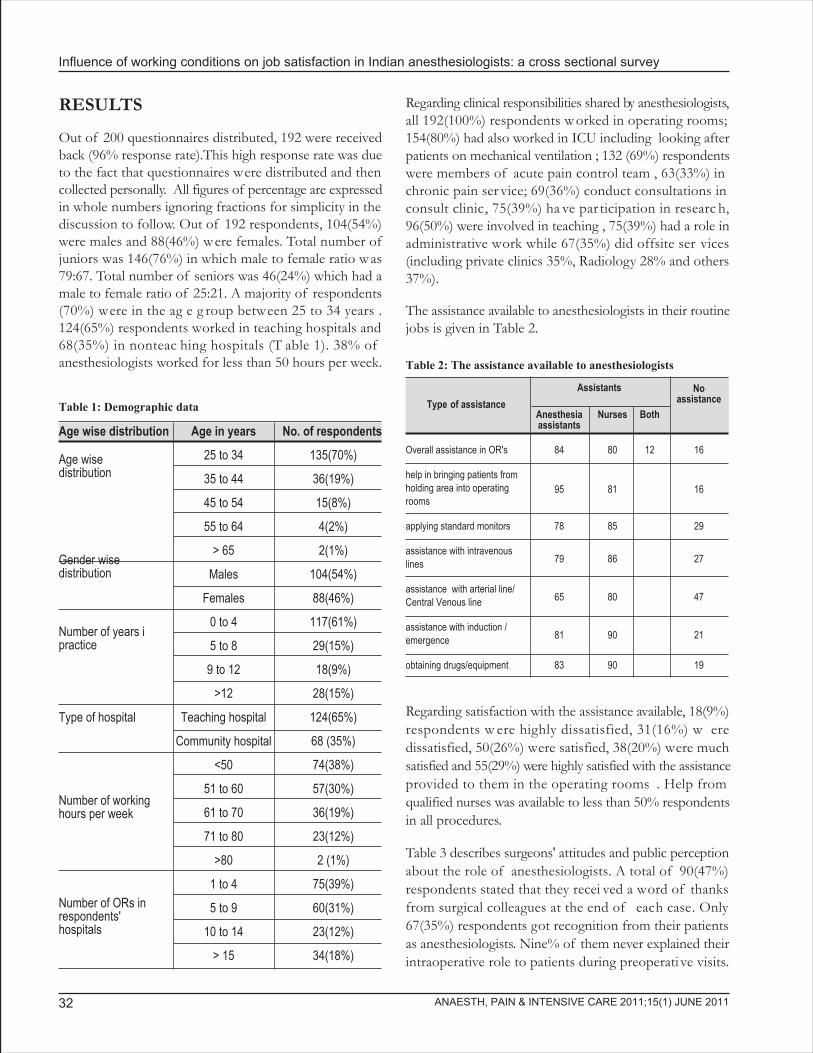
Influence of working conditions on job satisfaction in Indian anesthesiologists: a cross sectional survey
ANAESTH, PAIN & INTENSIVE CARE 2011;15(1) JUNE 201132
RESULTS
Out of 200 questionnaires distributed, 192 were receivedback (96% response rate).This high response rate was dueto the fact that questionnaires were distributed and thencollected personally. All figures of percentage are expressedin whole numbers ignoring fractions for simplicity in thediscussion to follow. Out of 192 respondents, 104(54%)were males and 88(46%) were females. Total number ofjuniors was 146(76%) in which male to female ratio was79:67. Total number of seniors was 46(24%) which had amale to female ratio of 25:21. A majority of respondents(70%) were in the ag e g roup between 25 to 34 years .124(65%) respondents worked in teaching hospitals and68(35%) in nonteac hing hospitals (T able 1). 38% ofanesthesiologists worked for less than 50 hours per week.
Regarding clinical responsibilities shared by anesthesiologists,all 192(100%) respondents worked in operating rooms;154(80%) had also worked in ICU including looking afterpatients on mechanical ventilation ; 132 (69%) respondentswere members of acute pain control team , 63(33%) inchronic pain ser vice; 69(36%) conduct consultations inconsult clinic, 75(39%) ha ve par ticipation in researc h,96(50%) were involved in teaching , 75(39%) had a role inadministrative work while 67(35%) did offsite ser vices(including private clinics 35%, Radiology 28% and others37%).
The assistance available to anesthesiologists in their routinejobs is given in Table 2.
Table 1: Demographic data
Age wise distribution Age in years No. of respondents
25 to 34 135(70%)
35 to 44 36(19%)
45 to 54 15(8%)
55 to 64 4(2%)
> 65 2(1%)
Males 104(54%)
Females 88(46%)
0 to 4 117(61%)
5 to 8 29(15%)
9 to 12 18(9%)
>12 28(15%)
Type of hospital Teaching hospital 124(65%)
Community hospital 68 (35%)
<50 74(38%)
51 to 60 57(30%)
61 to 70 36(19%)
71 to 80 23(12%)
>80 2 (1%)
1 to 4 75(39%)
5 to 9 60(31%)
10 to 14 23(12%)
> 15 34(18%)
Number of workinghours per week
Number of ORs inrespondents'hospitals
Number of years ipractice
Gender wisedistribution
Age wisedistribution
Regarding satisfaction with the assistance available, 18(9%)respondents w ere highly dissatisfied, 31(16%) w eredissatisfied, 50(26%) were satisfied, 38(20%) were muchsatisfied and 55(29%) were highly satisfied with the assistanceprovided to them in the operating rooms . Help fromqualified nurses was available to less than 50% respondentsin all procedures.
Table 3 describes surgeons' attitudes and public perceptionabout the role of anesthesiologists. A total of 90(47%)respondents stated that they recei ved a word of thanksfrom surgical colleagues at the end of each case. Only67(35%) respondents got recognition from their patientsas anesthesiologists. Nine% of them never explained theirintraoperative role to patients during preoperati ve visits.
Table 2: The assistance available to anesthesiologists
Type of assistance
Noassistance
Anesthesia Nurses Bothassistants
Overall assistance in OR's 84 80 12 16
help in bringing patients fromholding area into operatingrooms
95 81 16
applying standard monitors 78 85 29
assistance with intravenouslines 79 86 27
assistance with arterial line/Central Venous line 65 80 47
assistance with induction /emergence 81 90 21
obtaining drugs/equipment 83 90 19
Assistants

Original Article
ANAESTH, PAIN & INTENSIVE CARE 2011;15(1) JUNE 2011 33
Table 3: Surgeons' attitudes and public perception
Questions
Response and number of respondentsN(%)
1 2 3 4 5
How would you rate the surgeons' attitude towards anesthesiologists? #. 4 (2) 15(8) 75(39) 48(25) 50(26)
Do surgical colleagues consult you for medical problems* 6(3) 15(8) 77(40) 54(28) 40(21)
Do surgical colleagues readily accept your decision in cancellation of cases* 6(3) 33(17) 42(22) 52(27) 59(31)
Do surgical colleagues readily accept your choice of anaesthetic technique* 4(2) 2(1) 15(8) 75(39) 96(50)
Do surgical colleagues pressurize you for time taken for assessment/ induction* 54(28) 57(30) 44(23) 10(5) 27(14)
Do surgical colleagues ask if they may start the case* 13(7) 17(9) 8(4) 33(17) 121(63)
Do surgical colleagues thank you at the end of the case* 6(3) 4(2) 42(22) 50(26) 90(47)
Do you explain your intraoperative role to the patients during preoperative visits* 6(3) 12(6) 48(25) 36(19) 90(47)
Do your patients know you are an anaesthesiologist* 19(10) 29(15) 48(25) 29(15) 67(35)
# Grading: 1-'No respect at all'; 2-Some respect; 3- Indifferent; 4-Moderate degree of respect ; 5-'Consider equal in status'* Grading: 1-Never; 2-Rarely; 3-Sometimes; 4-Frequently; 5-Always
Figure I: Aspects of practice bringing the most dissatisfaction.
Role of an anesthesiologist in the healthcare system w asdescribed as a 'P erioperative physician' by 105 (55%)respondents, as 'part of a multidisciplinary surgical team'by 77(40%) and as 'providing a service to the surgeon' by10(5%). No respondent chose the option of 'mainly as atechnician' or 'just a job'.
Overall job satisfaction was rated as 1 (totally dissatisfied)by 4(2%) respondents , 2 by 2(1%), 3 b y 37(19%), 4 b y111(58%) and 5 (totally satisfied) by 38(20%) respondents.
The commonest reasons given for job satisfaction were;
1. Good quality of patient care; 44(23%)2. Intellectual stimulation; 25(13%)
3. Interaction with anesthesia colleagues; 25(13%)4. The magic about anesthesia; 25(13%)
Figure I depicts the aspects of practice bringing the mostdissatisfaction. Lack of recognition by patients (49%) andLack of resources/equipment (46%) are the tw o mostcommon reasons gi ven b y the respondents fordissatisfaction.
Comparison of job satisfaction on the basis of gender,seniority and place of w ork is sho wn in T able 4
Females were more fully satisfied than males (79:70) (p <0.01).
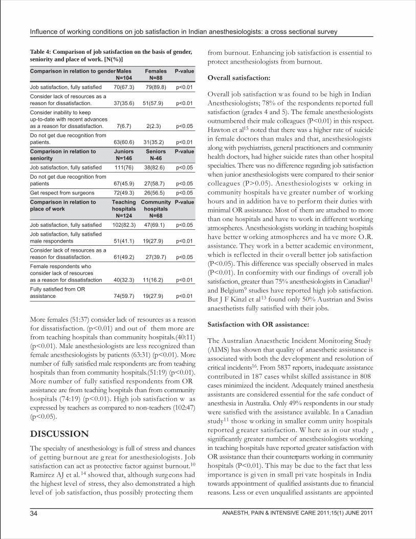
Influence of working conditions on job satisfaction in Indian anesthesiologists: a cross sectional survey
ANAESTH, PAIN & INTENSIVE CARE 2011;15(1) JUNE 201134
More females (51:37) consider lack of resources as a reasonfor dissatisfaction. (p<0.01) and out of them more arefrom teaching hospitals than community hospitals.(40:11)(p<0.01). Male anesthesiologists are less recognized thanfemale anesthesiologists by patients (63:31) (p<0.01). Morenumber of fully satisfied male respondents are from teachinghospitals than from community hospitals.(51:19) (p<0.01).More number of fully satisfied respondents from ORassistance are from teaching hospitals than from communityhospitals (74:19) (p<0.01). High job satisfaction w asexpressed by teachers as compared to non-teachers (102:47)(p<0.05).
DISCUSSIONThe specialty of anesthesiology is full of stress and chancesof getting burnout are g reat for anesthesiologists . Jobsatisfaction can act as protective factor against burnout.10
Ramirez AJ et al. 14 showed that, although surgeons hadthe highest level of stress, they also demonstrated a highlevel of job satisfaction, thus possibly protecting them
from burnout. Enhancing job satisfaction is essential toprotect anesthesiologists from burnout.
Overall satisfaction:
Overall job satisfaction w as found to be high in IndianAnesthesiologists; 78% of the respondents reported fullsatisfaction (grades 4 and 5). The female anesthesiologistsoutnumbered their male colleagues (P<0.01) in this respect.Hawton et al15 noted that there was a higher rate of suicidein female doctors than males and that, anesthesiologistsalong with psychiatrists, general practitioners and communityhealth doctors, had higher suicide rates than other hospitalspecialties. There was no difference regarding job satisfactionwhen junior anesthesiologists were compared to their seniorcolleagues (P>0.05). Anesthesiologists w orking incommunity hospitals ha ve greater number of workinghours and in addition ha ve to perform their duties withminimal OR assistance. Most of them are attached to morethan one hospitals and have to work in different workingatmospheres. Anesthesiologists working in teaching hospitalshave better w orking atmospheres and ha ve more O.R.assistance. They work in a better academic environment,which is ref lected in their overall better job satisfaction(P<0.05). This difference was specially observed in males(P<0.01). In conformity with our findings of overall jobsatisfaction, greater than 75% anesthesiologists in Canadian11
and Belgium9 studies have reported high job satisfaction.But J F Kinzl et al13 found only 50% Austrian and Swissanaesthetists fully satisfied with their jobs.
Satisfaction with OR assistance:
The Australian Anaesthetic Incident Monitoring Study(AIMS) has shown that quality of anaesthetic assistance isassociated with both the development and resolution ofcritical incidents16. From 5837 reports, inadequate assistancecontributed in 187 cases whilst skilled assistance in 808cases minimized the incident. Adequately trained anesthesiaassistants are considered essential for the safe conduct ofanesthesia in Australia. Only 49% respondents in our studywere satisfied with the assistance available. In a Canadianstudy11 those w orking in smaller comm unity hospitalsreported g reater satisfaction. W here as in our study ,significantly greater number of anesthesiologists workingin teaching hospitals have reported greater satisfaction withOR assistance than their counterparts working in communityhospitals (P<0.01). This may be due to the fact that lessimportance is gi ven in small pri vate hospitals in Indiatowards appointment of qualified assistants due to financialreasons. Less or even unqualified assistants are appointed
Table 4: Comparison of job satisfaction on the basis of gender,seniority and place of work. [N(%)]
Comparison in relation to genderMales Females P-valueN=104 N=88
Job satisfaction, fully satisfied 70(67.3) 79(89.8) p<0.01
Consider lack of resources as areason for dissatisfaction. 37(35.6) 51(57.9) p<0.01
Consider inability to keepup-to-date with recent advancesas a reason for dissatisfaction. 7(6.7) 2(2.3) p<0.05
Do not get due recognition frompatients. 63(60.6) 31(35.2) p<0.01
Comparison in relation to Juniors Seniors P-valueseniority N=146 N-46Job satisfaction, fully satisfied 111(76) 38(82.6) p<0.05
Do not get due recognition frompatients 67(45.9) 27(58.7) p<0.05
Get respect from surgeons 72(49.3) 26(56.5) p<0.05
Comparison in relation to Teaching Community P-valueplace of work hospitals hospitals
N=124 N=68Job satisfaction, fully satisfied 102(82.3) 47(69.1) p<0.05
Job satisfaction, fully satisfiedmale respondents 51(41.1) 19(27.9) p<0.01
Consider lack of resources as areason for dissatisfaction. 61(49.2) 27(39.7) p<0.05
Female respondents whoconsider lack of resourcesas a reason for dissatisfaction 40(32.3) 11(16.2) p<0.01
Fully satisfied from ORassistance 74(59.7) 19(27.9) p<0.01
Original Article
ANAESTH, PAIN & INTENSIVE CARE 2011;15(1) JUNE 2011 35
on lower pay scales. Most of the teaching hospitals runnursing schools, which conduct nursing courses as well asparamedical certificate courses like OR assistants course;thus nursing students are available to work in ORs. Morenumber of fully satisfied anesthesiologists w orking inteaching hospitals than their counterparts in communityhospitals where OR assistance is inadequate , points outthat there is a close association between quality and typeof OR assistance a vailable and better job satisfaction.
Perceived public attitude:
One of the aspects of practice bringing the mostdissatisfaction is lac k of recognition b y patients. It isexpected that recognition of an individual doctor increaseswith the seniority in practice , which is more or less tr uewith doctors from other clinical specialties; but in case ofanesthesiologists, seniority makes minimal difference asthey always play their role behind the curtain. Results fromour study are comparable with other studies in this aspect.No significant difference was found between senior andjunior anesthesiologists when compared, in respect ofrecognition from patients (P>0.05). Male anesthesiologistsare less recognized b y their patients than femaleanesthesiologists (P<0.01). T o a cer tain extentanesthesiologists are themselv es responsible for poorrecognition by patients as they maintain a low profile. Only28% anesthesiologists give talks to the la y public aboutanesthesia; and 9% of them do not ev en explain theirintraoperative role to patients on the preoperati ve visits.It is possible to raise awareness among patients by layingmore emphasis on pre-anesthetic meetings and also b yproviding an information sheet preoperatively to outpatients.This may help in improving patients understanding of therole of the anesthesiologists. This was also ref lected inPresidential address delivered at ISACON-2006, Mysore,(Karnataka) on 27 December 2006 5 in whic h it w asmentioned that--- “public awareness of anesthesia servicesand anesthesiologists is far from satisfactor y even now.This is basically due to our fault in mark eting ourselves.We have to constantly try to make the public aware of theimportance of our work by establishing pre anesthesiaclinics, patient counseling, pain clinics, trauma and criticalcare areas as well as promote paramedical courses to trainanesthesia technicians or assistants. These steps will enhanceour public imag e and create fa vorable public opinion.Moreover, we have to utilize the services of 'informationand technology science' in publishing about anesthesia,anesthesia related problems and in gi ving instruction inthe management of disasters.”
Perceived surgeons' attitudes:
A good relationship with the surgeon is of fundamentalimportance in anesthesiologist's practice 17. P oorinterpersonal relationships may lead to considerable stress.Anesthesiologists may feel unable to c hange or controlsituations in an en vironment, where the surg eon iscommonly perceived to be in charge.11 Younger physicianstend to find interpersonal relationships more stressful thantheir older colleagues do. Jenkins K et al11 found that thesenior Canadian respondents got higher regard from thesurgeons in comparison to the y ounger respondents.However, we did not observe such difference (P>0.05); inour study, 51% respondents felt highly reg arded by thesurgeons (graded 4 or 5 on the Likert scale). In a study ofCalifornian anesthesiologists18, 96% indicated they oftenor always had a good working relationship with the surgeonsbut slightly o ver half did not believ e that surg eonsunderstood the risks of anesthesia. Surgeons pressurizeanesthesiologists to proceed with cases instead of postponethe cases in spite of high risks and to hasten anaestheticprocedures18. This conf lict of interests is a source ofdisharmony in OR. Forty-nine percent of our respondentssaid that they were usually consulted for difficult medicalproblems. T hough most surg eons acce pted theanesthesiologists' choice of anesthetic technique (89%)only 58% respected their decision to postpone cases .Nineteen percent of the anesthesiologists felt frequentlypressurized for time in assessing patients and inducinganesthesia. All these factors definitely affect job satisfaction.
Factors affecting job satisfaction/dissatisfaction:
The en vironment in whic h indi viduals w ork has atremendous effect on the level of 'pride' in themselves andthe work they do. A job that is interesting and that permitsone to contribute one's skills and ideas is very importantin respect of anesthesiologists. As long as sufficient resourcesare at their disposal, anesthesiologists are able to managetheir high demanding tasks and task-related stressors veryefficiently13. An interesting job as the sole factor w ouldnot suffice for adequate job satisfaction. Improving workingconditions and pro viding adequate resources w oulddefinitely help in enhancing job satisfaction13. Two mainfactors caus ing d issa t i s fac t ion are l ac k ofresources/equipment and lack of recognition by patients.It is felt that hospital administrators are reluctant insanctioning and releasing funds for purchasing equipmentsand drugs related to anesthesiology. Equipment necessaryfor safe monitoring and improving quality of anesthesia

Influence of working conditions on job satisfaction in Indian anesthesiologists: a cross sectional survey
ANAESTH, PAIN & INTENSIVE CARE 2011;15(1) JUNE 201136
service will not be purchased unless statutory bodies laydown minimum standards of monitoring care. Budgetaryconstraints are faced by anesthesiologists in teaching aswell as in comm unity hospitals , which is ref lected inresources being equally limited to both of them (P>0.05).More female anesthesiologists, especially those working inteaching hospitals, considered lack of resources as a reasonfor dissatisfaction (P<0.05). Other factors indicated forbringing dissatisfaction were long/unpredictable workinghours, being unduly blamed for complications , lack ofrecognition by surgeons, inadequate financial compensation,unrealistic expectations of the clientele, hospital politics,and not being able to remain abreast with the latestknowledge/ technical applications. At present it is theresponsibility of each individual anesthesiologist to keephim/her informed of newer developments, but unlessthere is statutator y compulsion e .g. compulsor y CMEactivity, this fact will not be view ed seriously .
Overall, 78% anesthesiologists w ere satisfied b y theirprofessional work. Yet, it is felt that this number may beincreased by improving on factors identified in the presentstudy. Increasing intellectual stimulation, allowing betterquality of care, improving interaction with patients andproviding adequate OR assistance should be seriouslyconsidered. Departmental funding should be increased soas to meet the requirements of newer drugs, monitors, andequipment to perform newer techniques. Statutory bodieslike University Grants Commission, Medical Council ofIndia, Indian Society of Anesthesiologists etc. must laydown regulations requiring local administrations andauthorities to follow minimum monitoring standards andprovide anesthesia equipments like modern anesthesia workstations to improve standard of care as well improve jobsatisfaction. Better communication and team work by theentire surgical team would enhance professional satisfactionof anesthesiologists. Raising the profile of anesthesiologist,both in the eyes of the public and fellow health professionalsshould be taken on a priority basis. Patient education is animportant method to raise the anesthesiologists' imag eamongst the public.
LIMITATIONS
Our sample size is relati vely small and ma y not berepresentative of all anesthesiologists from India, as it doesnot include a large number of practicing anesthesiologists,who never attend any conference or CME or workshop;it is not yet mandator y in India to ear n CME points forrenewal of registration and license to practice .
CONCLUSION
Although job satisfaction level in Indian anesthesiologistsis quite high, still there is a need to set the , which wouldhelp reduce occupational stress and fur ther improveefficiency and job satisfaction among anesthesiologists .Authorities, e.g. Indian Society of Anesthesiologists mayurge large scale multicentre studies to lay down standardsrelated to number of working hours per day and per week,number of night call duties per w eek, making properassistance mandatory, preparing standard protocols andguidelines for anaesthetic management of different clinicalcases, providing medicolegal protection etc..
REFERENCES
1. Le May S, Dupuis G, Harel F, Taillefer MC, Dubé S, HardyJF. Clinimetric scale to measure surgeons' satisfaction withanesthesia ser vices. Can J Anaesth.2000;47(5):398-405.
2. García-Sánchez MJ, Prieto-Cuéllar M, Galdo-Abadín JR,Palacio-Rodríguez MA. [Can we change the patient's imageof the anaesthesiologist?] R ev Esp Anestesiol R eanim.1996;43(6):204-7.
3. Hennessy N, Harrison DA, Aitkenhead AR.The effect ofthe anaesthetist's attire on patient attitudes. The influenceof dress on patient perception of the anaesthetist's prestige.Anesthesia. 1993;48(3):219-22.
4. PRESIDENTIAL ADDRESS - 2006. Indian J . Anaesth.2007;51(1):10-12.
5. Seeley HF.The practice of anesthesia--a stressor for themiddle-aged? Anesthesia. 1996;51(6):571-4.
6. Dormann C, Zapf D. Job satisfaction: a meta-analysis ofstabilities. J Organ Behav 2001;22:483-504.
7. Warner TH. The effects of job satisfaction and organizationalcommitment on intent to leave among nurse anaesthetists:a comparative study. Diss Abstr Int Sect A: Humanit SocSci 2001;61:49-66.
8. Neuwirth ZE. An essential understanding of physician-patient communication. Part II. . J Med Pract Manag e1999;15:68-72.
9. Dercq JP, Smets D, Somer A, Desantoine D. A survey ofBelgian anesthesiologists. Acta Anaesth Belg 1998;49:193-204.

Original Article
ANAESTH, PAIN & INTENSIVE CARE 2011;15(1) JUNE 2011 37
APICARE ON FACEBOOKThe readers can participate in all act ivities and know all that is going on related to APICAREby logging in to Facebook. Our address is; http://www.facebook.com/apicare
Apicare Journal GroupThe journal maintains a group for all facebook enthusiasts to participate in group discussions,upload photos or documents etc. related to anesthesiology. The calendar of events notif iesin advance about all important conferences, symposia or workshops in the region as well as theworld. The readers are urged to upload any event which is planned to be held in the comingmonths. Please log on to;http://www.facebook.com/group.php?gid=57260551059
Pain FellowsThe journal maintains a group for all facebook enthusiasts to participate in group discussions,upload photos or documents etc. related to pain and pain management. The calendar of eventsnotif ies in advance about all important conferences, symposia or workshops about pain andpain management in the region as well as the world. The readers are urged to upload any eventwhich is planned to be held in the coming months. Please log on to;http://www.facebook.com/group.php?gid=178359883480
Pakistan Resuscitation SocietyThe journal maintains a page on facebook for resuscitation enthusiasts to participate in groupdiscussions, upload photos or documents etc. related to resuscitation. It is also off icial pageof Pakistan Resuscitation Society (PARES). Please log on to;http://www.facebook .com/pages/Pak ist an-Resuscit at ion-Societ y/313782574532
10. Kluger MT, Townend K, Laidlaw T. Job satisfaction, stressand burnout in Australian specialist anaesthetists. . Anesthesia.2003;58(4):339-45.
11. Jenkins K, Wong D.A survey of professional satisfactionamong Canadian anesthesiologists . . Can J Anaesth.2001;48(7):637-45.
12. Kluger MT, Bryant J. Job satisfaction, stress and burnoutin anaesthetic technicians in New Zealand. Anaesth IntensiveCare. 2008;36(2):214-21.
13. J F Kinzl, H Knotzer, C Traweger, W Lederer, T Heidegger,A Benzer. Influence of working conditions on job satisfactionin anaesthetists Br J Anesthesia 2005:94:211-215.
14. Ramirez AJ, Graham J, Richards MA, Cull A, Gregory WM.Mental health of hospital consultants: the effect of stressand satisfaction at w ork. Lancet 1996; 347: 724-8.
15. Hawton K, Clements A, Sakarovitch C, Simkin S, Deeks JJ.Suiside in doctors: a study of risk according to g ender,seniority and specialty in medical practitioners in Englandand Wales. Journal of Epidemiology and Community Health2001; 5:296-300.
16. Kluger MT, Bukofzer M, Bullock M., Anaesthetic assistants:their role in the development and resolution of anaestheticincidents. Anaesth Intensi ve Care 1999; 27:269-74.
17. Irita K, Takahashi S. [Lack of communication betweenanesthesiologists and surgeons: comparison of questionnairesurvey among anesthesiologists with that among surgeonsconcerning pre-anesthetic evaluation of surgical patients].Masui. 2000;49(1):7-17.
18. Gaba DM, Howard SK, Jump B. Production pressure inthe work environment. California anesthesiologists' attitudesand experiences . Anesthesiolog y 1994;81(2):488-500.

ANAESTH, PAIN & INTENSIVE CARE 2011;15(1) JUNE 201138
INTRODUCTION
Ventilator associated pneumonia (V AP) is a for m ofnosocomial infection that occurs in patients recei vingmechanical ventilation for longer than 48hrs1. The incidenceof VAP is around 22.8%2, and patients receiving ventilatorysupport account for 86% of the cases of nosocomialpneumonia3. The mortality rate attributable to V AP isaround 27%4, and the costs of treatment of these infections
An audit on ventilator associated pneumonia in theIntensive Care Unit at Teaching Hospital Karapitiya,
Galle, Sri LankaAsoka Gunaratne*, Dhammika Vidanagama**,
*Consultant Anesthesiologist, **Consultant MicrobiologistTeaching Hospital Karapitiya, Galle (Sri Lanka)
Correspondence: Dr. Asoka Gunaratne, Consultant Anesthesiologist, Teaching Hospital Karapitiya, Galle (Sri Lanka);e-mail: [email protected]
ABSTRACTBackground: Critical care is one of the most expensive services provided by a hospital. The aim of this auditwas to ascer tain the incidence of v entilator associated pneumonia in the intensi ve care unit.
Type of study: A descriptive study
Place of study: Teaching Hospital Karapitiya, Galle (Sri Lanka)
Duration of study: 1st June 2010 to 30th August 2010
Methodology: All patients, who were admitted to ICU and who stayed there for more than 48hrs during aperiod of three months were studied. Infections were identified on clinical parameters such as fever and onlaboratory investigations such as full count, CRP and cultures.
Results: Out of 82 patients, 48(58.5%) were subsequently discharged to the ward and 30(36.6%) succumbedto their illness. 68(82.9%) were ventilated and 26 of them had an underlying pathology related to an infection.A total of 20(29.4%) patients of this ventilated group subsequently developed a lower respiratory tract infection.The main nosocomial infection was ventilator associated pneumonia and had an incidence of 21.9%. Themost prevalent organisms were mixed gram negative bacilli and Acinetobacter spp.
Conclusion: Nosocomial infections are a cause of increased mortality and morbidity in the intensive careunit. Awareness of the risk factors tog ether with simple preventive measures and sur veillance will help toreduce its occurrence.
Key words: Nosocomial infection; ventilator associated pneumoniaCitation: Gunaratne A, Vidanagama D. An audit on ventilator associated pneumonia in the Intensive Care Unit at Teaching HospitalKarapitiya, Galle, Sri Lanka. Anaesth Pain & Intensive Care 2011;15(1):38-41.
ORIGINAL ARTICLE
contribute to a significant por tion of the expenditureincurred in maintaining an intensi ve care unit. TeachingHospital Karapitiya, Galle, Sri Lanka is one of the maintertiary care centers in the country. All the general medicaland surgical patients requiring intensive care are admittedto the main intensi ve care unit where the audit w asconducted. The main objective of this study was to ascertainthe incidence of ventilator associated pneumonia in theintensive care unit.

Original Article
ANAESTH, PAIN & INTENSIVE CARE 2011;15(1) JUNE 2011 39
METHODOLOGY
Permission to conduct this prospective audit was obtainedfrom the director Teaching Hospital Karapitiya. The auditwas conducted for three months from 1st J une 2010 to30th August 2010. We studied all the patients admitted tothe main ICU and who stayed there for more than 48hrs,regardless of whether they were ventilated or not duringthis period. Reports of cultures from tracheal secretionswere obtained from the de partment of microbiolog y.However, we not able to perfor m bronchoscopicallycollected samples for quantitative cultures due to lack offacilities.
RESULTS
Total number of patients admitted to main ICU for morethan 48hrs w as 82; out of whic h 48(58.5%) w eresubsequently disc harged to the w ard and 30(36.6%)succumbed to their illness . Of the 82 patients studied68(82.9%) were ventilated and only 26 of them had anunderlying pathology related to an infection (Table 2). Atotal of 20(29.4%) patients of this v entilated g roupsubsequently developed a lower respiratory tract infection(Table 2). The majority of them [14(70%)] were in categoriesthat had no infection in any par t of the body at the timeof admission to the ICU or at the time of commencingventilation (Table 2). We used the follo wing criteria todiagnose lo wer respirator y tract infection (LR TI).
1. New shadow developing in the chest X- ray2. Temperature > 99º F3. Course crepitations on chest auscultation4. White cell count > 11,000
We used the follo wing criteria to diagnose V AP.
1. New shadow developing in the chest X-ray2. Temperature < 96.8 or > 990 F3. Ventilated for more than 48 hrs4. White cell count > 11,000 or < 4,0005. Cultures positi ve from endotrac heal secretions
All 20 patients, who developed an LRTI, had their trachealsecretions cultured and 2 of them failed to g row anyorganisms in the endotracheal secretions. However, if wehad the facilities to perform bronchoscopic sampling forquantitative cultures, all of them may have been positivefor bacterial invasion. The organisms cultured are shownin Table1.
VAP was diagnosed in 18 (21.9%) patients on these criteria.
Table 1: The organisms cultured
Organism Frequency %
Acinetobacter spp 5 25
Pseudomonas spp 3 66.6
Coliform spp 2 10
Mixed gram negative bacilli 6 33.3
Staphylococcus aureus 2 10
No growth 2 10
Total 20 100
DISCUSSIONThe patients needing intensive care usually have low hostdefense immunity. Immunosupression primarily due to therelease of interleukins and other anti inflammatory agentscreates a state sometimes ter med as immunoparalysis5.Endotracheal intubation compromises local defensemechanisms such as coughing, sneezing and mucocilliaryclearance. Upper airway and oral secretions can pool abovethe cuff of an endotracheal tube and line the tube, forminga biofilm, starting as early as 12 hrs after intubation. Thebiofilm contains larg e amounts of bacteria that can bedisseminated into the lungs b y the v entilator inducedinspiration6-8. The presence of the endotracheal tube alsoprovides a direct route for colonized bacteria to enter thelower respiratory tract. Exogenous colonization arises fromcross transmission from the hands of health care workersor visitors9.
Table 2: Patient categories developing LRTI followingventilation
Patient category No. Ventilated LRTI
Head Injury 14 14 4
Trauma other than head injury 6 6 1
Poisoning 5 5 3
Post-op elective Ventilation 9 9 4
Medial reasons without infection 11 8 2
Infections from Medical wards 7 4 0
Infections from other wards 30 22 6
Total 82 68 20

An audit on ventilator associated pneumonia in the Intensive Care Unit....
ANAESTH, PAIN & INTENSIVE CARE 2011;15(1) JUNE 201140
Cook and colleagues noted that the administration ofparalytic agents was an independent predictor of nosocomialpneumonia in their study of 1,014 mechanically ventilatedpatients10. Sedative drugs and stress ulcer prophylaxis haveall been implicated as risk factors.
In a re port from the National Nosocomial InfectionSurveillance (NNIS) system involving data from 498,998patients, 83% of episodes of nosocomial pneumonia wasassociated with mec hanical ventilation11. Lang er andcolleagues showed an increased incidence from 5% inpatients who received 1 day of mechanical ventilation to69% in those who received 30 days of venilation12. Oneof the largest databases related to nosocomial infection inintensive care is the EPIC study 13. In this 1-da y pointprevalence study, information was obtained on all patientswho occupied a bed in an intensive care unit over 24hrs.10,038 patients were recruited from 1,407 western Europeanintensive care units. 2,064 of them (21%) had an intensivecare unit acquired infection. The incidence of nosocomialpneumonia in the EPIC study w as 47% and 31% in theNNIS study.
For many years VAP has been diagnosed b y the clinicalcriteria published by Johanson et el in 1972, which includethe appearance of a new pulmonar y infiltrate , fever,leukocytosis and purulent tracheobroncheal secretions14.However these criteria are non specific . VAP is mostaccurately diagnosed b y quantitati ve cultures andmicroscopic examination of lo wer respirator y tractsecretions which are best obtained by bronchoscopicallydirected techniques, such as the protected specimen brushand bronc hoalveolar la vage. T hese tec hniques ha veacceptable repeatability and inter pretation of results isunaffected by antibiotics administered concur rently forinfections in extra pulmonar y sites as long as theantimicrobial therapy has not been changed for <72 hrsbefore bronchoscopy15,16. The accuracy of quantitativecultures and microscopic examination of lower respiratorytract secretions for the diagnosis of VAP was validated byChastre et al17,18. However, other published studies haveconcluded that bronchoscopically directed techniques werenot more accurate for diagnosis of VAP than clinical X-ray criteria combined with cultures of tracheal aspirates19-
22.
Prevention has a key part to play in limiting VAP. Severalstudies have reported high rates of contamination withpotentially pathogenic organisms by the hands of healthcareworkers23,24. Hand disinfection with the use of alcohol
based antiseptic hand rub solutions have been shown tobe effective in reducing hand contamination25. Chlorhexidineoral rinse twice daily has been used b y some workers toreduce oral colonization26. However, no evidence basedprotocols for oral care ha ve been tested to decrease theincidence of VAP in patients recei ving mec hanicalventilation. According to the studies done so far , stressulcer prophylaxis does not pla y a significant role in thedevelopment of VAP. Saline lavage of endotracheal tubesbefore suctioning can dislodg e bacteria from theendotracheal tubes in to the lower airways increasing therisk of VAP27. Positioning the patient in a semi recumbentposition prevents reflux and aspiration. Maintaining anadequate cuff pressure decreases the likelihood of secretionsleaking around the cuff . Use of tubes with por ts forcontinuous subglottic suctioning can decrease the incidenceof VAP by 50%28,29 and therefore the oropharynx shouldbe thoroughly suctioned. Daily interruption of continuoussedation and paralysis can shor ten the duration ofmechanical ventilation. Infection surveillance can reducenosocomial infection rates when incorporated with infectionprevention prgrammes30.
CONCLUSION
VAP, although often preventable, is a cause of increasedmortality and morbidity; it is also responsible for excessiveresource expenditure in the intensive care unit. Awarenessof the risk factors and attention to simple prev entivemeasures such as hand hygiene can reduce the incidenceof these infections . Sur veillance tog ether with amultidisciplinary approach of prevention is the otherimportant aspect of its limitation.
REFERENCES
1. Kollef MH. T he prev ention of v entilator associatedpneumonia. N Engl J Med. 1999;340(8):627-34.
2. Safdar N, Dezfulian C, Collard HR, Saint S . Clinical andeconomic consequences of ventilator associated pneumonia:a systemic review. Crit Care Med. 2005;33(10):2184-93.
3. Richards MJ , Edw ards JR, Culv er DH, Ga ynes RP.Nosocomial infections in medical intensive care units in theUnited States . Crit Care Med. 1999;27:887-92.
4. Craven DE. Epidemiolog y of v entilator associatedpneumonia. Chest. 2000;117(4):1865-75.
5. Docke WD, Randow F, Syrbe HP et al. Monocyte deactivationin septic patients:restoration by IFN treatment. Nat Med.1997;3:678-81.

Original Article
ANAESTH, PAIN & INTENSIVE CARE 2011;15(1) JUNE 2011 41
6. Kunis KA, Puntillo KA. Ventilator associated pneumoniain the ICU:its pathophysiology, risk factors and prevention.Am J Nurs. 2003;133(8):64A-64G.
7. Olson ME, Har mon BG , K ollef MH. Silv er coatedendotracheal tubes associated with reduced bacterial burdenin the lungs of mec hanically v entilated dogs . Chest.2002;121:863-70.
8. Morehead RS, Pinto SJ. Ventilator associated pneumonia.Arch Intern Med. 2002;160(13):1926-30.
9. Wang JT, Chang SC , Ko WJ, et al. A hospital acquiredoutbreak of methicillin resistant Staph ylococcus aureusinfection initiated b y a surg eon car rier. J Hosp Infect.2001;47:104- 09.
10. Cook DJ, Walter SD, Cook RJ, et al. Incidence of and riskfactors for ventilator associated pneumonia in critically illpat ients. Ann Inter n Med. 1998;129:433-40.
11. Richards MJ , Edw ards JR, Culv er DH, Ga ynes RP.Nosocomial infection in combined medical surgical intensivecare units in the United States . Infect Control HospEpidermiol 2000;21:510-15.
12. Langer M, Mosconi P, Cigada M, Mandelli M. Long ter mrespiratory support and risk of pneumonia in critically illpatients. Am R ev R espir Dis 1989;140:302-05.
13. Vincent JL, Bihari D, Suter PM, et al. T he prevalence ofnosocomial infection in intensive care units in Europe: theresults of the EPIC study . J AMA 1995;274:639-44.
14. Johanson W G, Pierce AK, Sanford JP , T homas GD.Nosocomial respiratory infections with gram negative bacilli.The significance of colonization of the respiratory tract.Ann Intern Med 1972;77:701-6.
15. Timsit JF, Misset B, Renaud B, Goldstein FW, Carlet J. Effectof previous antimicrobial therapy on the accuracy of themain procedures used to diagnose nosocomial pneumoniain patients who are using ventilation. Chest 1995:108:1036-40.
16. Souweine B, Verber B, Bedos JP, Gachot B, et al. Diagnosticaccuracy of protected specimen brush and bronchoalveolarlavage in nosocomial pneumonia: impact of previousantimicrobial treatment. Crit Care Med 1998;26:236-44.
17. Chastre J, Fagon JY, Bor net-Lecso M, Calv at S, et al.Evaluation of bronchoscopic techniques for the diagnosisof nosocomial pneumonia. Am J R esp Crit Care Med1995;152:231-40.
18. Chastre J, Viau F, Brun P, Pierre J, et al. Prospective evaluationof the protected specimen br ush for the diagnosis ofpulmonary infections in ventilated patients. Am Rev RespiDis 1984;130:924-9.
19. Torres A, El-Ebiary M, Padro L, Gonzalez J, et al. Validationof different tec hniques for the diagnosis of ventilatorassociated pneumonia. Comparison with immediatepostmortem pulmonary biopsy. Am J Respi Crit Care Med1994;149:324-31.
20. Marquette CH, Copin MC , Wallet F, Neviere R, et al.Diagnostic tests for pneumonia in v entilated patients:Prospective evaluation of diagnostic accuracy using histologyas a diagnostic g old standard. Am J Respi Crit Care Med1995;151:1878-88.
21. Kirtland SH, Corely DE, Winterbauer RH, SpringmeyerSC, et al. The diagnosis of ventilator associated pneumonia.A comparison of histological, microbiological and clinicalcriteria. Chest 1997;112:445-7.
22. Fabregas N, Ewig S, Torres A, El-Ebiary M. et al. Clinicaldiagnosis of ventilator associated pneumonia revisited:comparative validation using immediate post mortem lungbiopsies. Thorax 1999;54:867-73.
23. Rangel-Frausto MS, Wiblin T, Blumberg HM, et al. Nationalepidemiology of mycoses survey (NEMIS): variation inrates of blood stream infection due to Candida species inseven surgical intensive care units and six neonatal intensivecare units. Clin Infect Dis 1999;29:253-58.
24. Pittet D, Dharan S, Touveneau S, Sauvan V, Perneger TV.Bacterial contamination of the hands of hospital staff duringroutine patient care . Arch Intern Med 1999;159:821-26.
25. Pittet D, Hugonnet S, Harbarth S, et al. Effectiveness of ahospital wide programme to improve compliance with handhygiene. Lancet 2000;356:1307-12.
26. DeRiso AJ, Ladowski JS, Dillon TA, Justice JW, PeersonAC. Chlorhexidine gluconate 0.12% oral rinse reduces theincidence of total nosocomial respirator y infection andsystemic antibiotic use in patients undergoing heart surgery.Chest 1996;109(6):1556-61.
27. Moore T. Suctioning tec hniques for the remo val ofrespiratory secretions . Nurs Stand 2003;18(9):47-55.
28. Craven DE. Preventing ventilator associated pneumoniain adults: sowing seeds of change. Chest 2006;130(1):251-60.
29. Shorr AF, O'Malley PG. Continuous subglottic suctioningfor the prev ention of ventilator associated pneumonia:potential economic implications. Chest 2001;119(1):228-35.
30. Haley RW, Culver DH, W hite JW, et al. T he efficacy ofinfection surveillance and control programmes in preventingnosocomial infections in US hospitals . Am J Epider miol1985;121:182-205.

ANAESTH, PAIN & INTENSIVE CARE 2011;15(1) JUNE 201142
INTRODUCTION
NPPE, also called Post-obstruction Pulmonary Edema(POPE), is a complication that can develop in the immediatepostoperative period. The first case reports of NPPE werepublished by Oswalt et al. in 19771. We encountered threecases of NPPE over a period of eighteen years, (incidence0.06% of all general anesthetics) which is almost similarto other workers2,3. It is a rare condition, but it may resultin a fatal outcome if diagnosis and treatment are delayed.
CASE 1:A 22 years old, young soldier, ASA-I, underwent plannedhaemorrhoidectomy under thiopentone - O 2/N2O -isoflurane - relaxant anaesthsia. The patient was intubatedand inj. pethidine 50mg was given IV. At the end of theprocedure, the effects of relaxant w ere reversed andextubation was done with the patient fully recovered fromanaesthesia. A few seconds later the patient had a briefspell of laryngospasm after which his SpO2 started falling.
Development of negative pressure pulmonary oedemasecondary to postextubation laryngospasm
Muhammad Saqib, FCPS*, Maqsood Ahmad, FCPS**, Raheel Azhar Khan, FCPS***
*Consultant Anaesthesiologist, PAF Hospital Rafiqui, Shorkot Cantt** Consultant Anaesthesiologist, Combined Military Hospital Gujranwala***Consultant Anaesthesiologist, Combined Military Hospital Multan
Correspondence: Muhammad Saqib, Consultant Anaesthesiologist, PAF Hospital Rafiqui, Shorkot Cantt (Pakistan);Cell: 03124792234; e-mail: [email protected]
ABSTRACTNegative Pressure Pulmonary Oedema (NPPE) immediately after general anaesthesia is a rare but life threateningcomplication, caused by an increased fluid in the interstitial spaces and alveoli due to forced inspiratory effortsagainst tightly closed glottis. Once developed, it impairs gas exchange and causes hypoxemia and if not treatedpromptly may lead to respirator y failure. Management involves maintaining airway, diuretics and positi vepressure ventilation. Affected cases recover completely with appropriate treatment but death ma y occur iftreatment is delayed. We present here three cases that developed post-extubation NPPE after short spells oflaryngospasm. All of the three cases recovered completely after management with diuretics and venitlatorysupport with added PEEP.
Key Words: Negative pressure pulmonary oedema; extubation; postoperative complicationCitation: Saqib M, Ahmad M, Khan RA. Development of post extubation negative pressure pulmonary oedema in three cases. Anaesthpain & Intensive Care 2011;15(1):42-44.
CASE REPORT
Positive pressure was applied with mask and 100% O2 butSpO2 kept on falling. Chest auscultation showed bilateralfine crepts, so it was decided to re-intubate the patient. Onlaryngoscopy frank blood tinged froth was noticed comingout of the glottis . A diagnosis of NPPE due to post-extubation laryngospasm was made, the patient was givenrelaxants, IPPV continued and shifted to ICU for ventilatorysupport. Portable X-ray chest showed bilateral opacities ofacute pulmonary oedema. ECG trace and cardiac enzymeswere ordered and found to be within normal limits. Afterabout 4 hours of ventilatory support chest auscultationconfirmed his complete recovery from pulmonary oedemaso the relaxant w as reversed, he w as weaned off theventilator and extubated. He recovered completely withoutany sequelae.
CASE 2:A 4 years old female c hild, ASA-1, weighing 15 kg, wasscheduled for repair of a tongue laceration under generalanaesthesia in emergency. Child had been NPO for 6 hours

Original Article
ANAESTH, PAIN & INTENSIVE CARE 2011;15(1) JUNE 2011 43
before the procedure. She was induced with thiopentone60mg and intubated orally after relaxation with inj. Succinylcholine 20mg. A throat pac k was placed in phar ynx.Anaesthesia was maintained with O2/N2O, isoflurane andinj. Tracrium™. At the end of procedure, the throat packwas removed and extubation w as done after thoroughsuction. The patient went into severe laryngospasm afterextubation. Airway was maintained and 100% O2 was givenwith face mask but her SpO 2 dropped rapidly and shedeveloped bradycardia with a hear rate of 50/min. Classicsigns of NPPE were present e.g. bilateral fine crepts andbilateral diffuse opacities on chest X-ray. Inj. atropine 0.2mg IV was repeated twice and the patient was re-intubated.Laryngoscopy revealed frank blood tinged froth from thelarynx. IPPV with100% O2 improved the SpO2. She wassedated and relaxed and later on shifted to pediatric ICUfor ventilatory support with a PEEP of 4 cm of H2O.After 4 hours of ventilatory support lungs became dry andshe was weaned off and extubated after reversal of therelaxants.
CASE 3:A 25 years old male , ASA-1, a moderate smoker for thelast eight years , was planned for a testicular biopsy forinfertility, under GA. T he course of anaesthesia withintubation and IPPV remained uneventful. At the end ofthe procedure, the relaxant was reversed and extubationwas done. The patient developed severe laryngospasmsoon after extubation and his SpO 2 star ted falling .Ventoline™ nebulisation was done. Laryngospasm did notrespond to any medication or maneuver. Administrationof 100% O2 by mask failed to raise SpO2 to normal levels,so he w as re-intubated to enable us positi ve pressureventilation with PEEP. During intubation typical pink frothwas noticed to fill the mouth ca vity. Auscultation of thechest revealed bilateral rales. IPPV was continued and hewas shifted to ICU for ventilatory support. He was giveninj. Dexamethasone 4mg IV, inj. Furusamide 40mg IV andventilatory support with 8cmH2O of PEEP. Eight hoursof ventilation dried his lungs, so he was weaned off theventilator. His postoperative investigations revealed nocardiac, pulmonary or neurological deficit.
DISCUSSION
NPPE, also called POPE, results from transudation offluid, first from pulmonary capillaries into interstitial spacesand then into the alveoli. It occurs after complete obstructionof the airway usually due to a brief but severe laryngospasm.Young healthy muscular patients, undergoing head and
neck surgeries and/or painful procedures are more proneto this condition. NPPE is a potentially dangerous conditionwith a multifactorial pathogenesis. The central mechanismis a large inspiratory force generated against an obstructedor closed upper airway. The resultant decreased intrathoracicpressure leads to increased venous return to the right sideof the heart and increased hydrostatic pulmonary capillarypressure. Elevation of pulmonary capillary pressure resultsin decreased left ventricular compliance that may resultfrom the right ventricular distention and shift of the cardiacseptum to the left. The negative intrathoracic pressure alsoresults in an increased afterload imposed on the left ventricle,causing a further decrease in left ventricular stroke volume.4,5
The autoPEEP during obstruction does not allow fluid totransudate, but as soon as airway opens, this autoPEEP islost and under the effects of increased interstitial pressure,fluid pours first into interstitium and then into the alveoli.Fortunately, because of the lung's unique ultra str uctureand its capacity to increase lymph f low, the pulmonar yinterstitium usually accommodates large increases in capillarytransudation before interstitial par tial pressure becomespositive. When this reserve capacity is exceeded, pulmonaryedema develops4,5.
Pulmonary edema is often divided into four stages:
Stage I: Only interstitial pulmonary edema is present.
Stage II: Fluid fills the interstitium and begins to fill thealveoli.
Stage III: Alveolar f looding occurs; many alv eoli arecompletely flooded with no air.
Stage IV: Marked alveolar f looding spills over into theairways as froth.
All of our cases of NPPE in volved post-extubationlaryngospasm. Other mechanical causes of upper airwayobstruction resulting in NPPE reported in literature includehanging, laryngeal growths, strangulation, sleep apnea, andbiting down on or kinking of the endotracheal tube whileintubated.6-8 Croup and epiglottitis are common causes ofupper airway obstruction leading to NPPE in c hildren.9Warner et al 10 re ported NPPE dev eloping afteradministration of muscle relaxants at the beginning of aninhalation induction of anesthesia in healthy infants. Othercauses of airway obstruction in unconscious patient maybe tongue fall against the posterior pharynx, glottic edema,secretions, vomitus or blood in the airw ay, foreign body

Development of negative pressure pulmonary oedema secondary....
ANAESTH, PAIN & INTENSIVE CARE 2011;15(1) JUNE 201144
like forgotten throat pac k or exter nal pressure on thetrachea most commonly from a neck hematoma.
Signs and symptoms of NPPE include tachypnea, shortnessof breath, pulmonar y rales, frothy sputum production,decreased oxygen saturation, and evidence of upper airwayobstruction. Chest radiog raphs ma y sho w signs ofpulmonary edema.
None of the patients on whom we have reported had anyhistory of cardiac disease and eac h had a neg ativepreoperative ph ysical exam 6. After establishing anoncardiogenic etiolog y with relati ve cer tainty, thedifferential diagnosis should include an aspiration of gastriccontents, adult respirator y distress syndrome (ARDS),volume overload, anaphylaxis, and airw ay obstruction.Perhaps the hardest to differentiate from the diagnosis ofpost obstructive pulmonary edema is that of aspiration ofgastrointestinal contents. This is due to the fact that theonset of both processes may closely resemble each other(struggling patient, difficult intubation) and the clinicalpresentation (wheezing, dyspnea, hypoxia) is very oftenidentical, but this condition is difficult to treat and itsmortality is very high9,10.
Airway obstr uction should be manag ed b y gi vingsupplemental oxygen and opening the airw ay. If thesemaneuvers fail then refractor y laryngospasm should betreated aggressively with a small dose of succinyl choline(10-20 mg) and temporary positive pressure ventilation with100% oxygen. Some anaesthesiologists routinely use IVlignocaine 10-20 mg at the time of extubation to preventsevere coughing and lar yngospasm11. Endotrac healintubation may occasionally be necessar y to reestablishventilation. If pulmonary edema develops it is similar tocardiogenic pulmonary edema and is managed in a similarway but reintubation and positi ve pressure v entilatorysupport with PEEP are extremely useful and full recoveryfrom pulmonar y edema occurs without any residualpulmonary or cardiac damag e in most of the cases .
CONCLUSION
Postextubation laryngospasm in the immediate postoperativeperiod is an impor tant complication and it ma y lead tosignificant morbidity or mortality like pulmonary edema,cardiac arrest, brain damage or death. Prompt establishmentof airway, use of short acting muscle relaxant to relieve
severe laryngospasm and IPPV with PEEP ma y be lifesaving in these situations.
REFERENCES
1. Oswalt CE, Gates GA, Holmstrom FMG: Pulmonary edemaas a complication of acute airway obstruction. JAMA 1977;38:1833-1835.
2. Deepika K, K enaan CA, Bar rocas AM, et al: Neg ativepressure pulmonar y edema after acute upper airw ayobstruction. J Clin Anesth 9:403-408, 1997
3. W. Christopher Patton, Champ L. Baker, Jr., J South OrthopAssoc. 2000;9(4)
4. Peters J, Kindred MK, Robotham JL: Transient analysis ofcardiopulmonary interactions: I. Diastolic ev ents. J ApplPhysiol 64:1506-1517, 1988
5. Peters J, Kindred MK, Robotham JL: Transient analysis ofcardiopulmonary interactions: II. Systolic ev ents. J ApplPhysiol 64:1518-1526, 1988
6. Wei l MH: Pulmonar y edema: cardiog enic vsnoncardiogenic.Emerg Med 1991;87:65-68.
7. Willms D, Shure D: Pulmonary edema due to upper airwayobstruction in adults . Chest 94:1090-1092, 1988
8. Liu EJ, Yih PS: Negative pressure pulmonary oedema causedby biting and endotrac heal tube occlusion: a case fororopharyngeal airways. Singapore Med J 40:174-175, 1999
9. Bonadio WA, Losek JD: T he characteristics of childrenwith epiglottitis who develop the complications of pulmonaryedema. Arch Otolaryngol Head Neck Surg 117:205-207,1991
10. Warner LO, Martino JD, Davidson PJ, et al: Negative pressurepulmonary oedema: a potential hazard of muscle relaxantsin a wake infants . Can J Anaesth 37:580-583, 1990
11. E. Kesimci, B. Aslan, T. Gümüs & O. Kanbak : ManagementOf Unpredıcted Postoperatıve Negatıve Pressure PulmonaryEdema: A Report Of Two Cases . The Internet Journal ofAnesthesiology. 2007 Volume 12 Number 1

ANAESTH, PAIN & INTENSIVE CARE 2011;15(1) JUNE 2011 45
INTRODUCTION
Hydatid cyst disease is an infection most frequently causedby the lar val form of a parasite named Ec hinococcusgranulosus1,2. Hydatid cysts of the liver have been treatedsurgically for many years b y several surgical techniquesincluding marsupialization, evacuation, and filling the cystwith saline after evacuation of the endocyst2-4. Surgery forhydatid cysts of the liver has been associated with spillageof the antigenic hydatid fluid into the peritoneal cavity ordirect contact with the bloodstream resulting in seriousIgE-mediated anaphylactic reaction3-7. Most anaphylacticreactions encountered in open surgery for hepatic hydatidcysts occur when the cysts are deeply seated into the liver,and thus require a hepatotomy4,6. We present a case reportof such a reaction during an open surgery of hydatid cystof the liver.
CASE REPORT
Perioperative anaphylactic shock in a patient withunruptured hepatic hydatid cyst: a case report
Iclal Ozdemir Kol, MD*, Cevdet Duger, MD**, Kenan Kaygusuz, MD*, Sinan Gursoy, MD*,Cengiz Aydin, MD***, Caner Mimaroglu, MD****
*Associate Professor, **Assistant Professor, ****ProfessorDepartments of Anesthesiology and Reanimation
***ProfessorDepartment of General SurgeryCumhuriyet University, Faculty of Medicine, 58140 Sivas, (Turkey).
Correspondence: Dr. Cevdet Duger, Department of Anesthesiology and Reanimation, Cumhuriyet University, School ofMedicine, 58140 Sivas, (Turkey); Tel: 0090346 2580125; Fax: 0090346 2581305; e-mail: [email protected]
ABSTRACTHydatid cyst disease is an infection most frequently caused by the larval form of a parasite named Echinococcusgranulosus. Spillage of hydatid fluid during open surgery has been shown to result in serious anaph ylacticreaction. We report a case of 46 years old male with hydatid cyst of liver, who had a sudden onset of intra-operative hypotension, tachycardia, flushing, edema and bronchospasm. He was managed with adrenaline,antihistaminics, steroids, supplementary fluids and vasopressors, and after successful resuscitation, was shiftedto ICU for further management. Four days later, he was weaned off from vasopressors and ventilatory supportand shifted to the surgical ward. Early diagnosis and intervention are crucial for successful management ofthe anaphylactic reactions.
Key words: Hydatid cyst; anaphylactic shock; liverCitation: Kol IO, Duger C, Kaygusuz K, Gursoy S, Aydin C, Mimaroglu C. Perioperative anaphylactic shock in a patient with unrupturedhepatic hydatid cyst: a case report. Anaesth Pain & Intensive Care 2011;15(1):45-47.
CASE REPORT
A 46-year-old male complained of abdominal and f lankpain and fever for many months . Ultrasonography andcomputed tomography of the abdomen showed a cyst ofthe liver. Preoperative examination of the cardiovascularand respiratory systems was normal and he had no historyof allergy. The patient was premedicated with 0.07 mg/kgmidazolam IM. T he anesthesia w as induced with 0.05µg/kg fentanyl, 0.1 mg/kg v ecuronium and 7 mg/kgthiopentone sodium IV. Endotracheal intubation was doneusing a 8.5mm endotrac heal tube . T he patient w ascontinuously monitored by electrocardiography (ECG),noninvasive blood pressure monitor, pulse oximetry andend-tidal capnography. Anaesthesia was maintained with50% O 2:50% N 2O and isof lurane. T he patient w ashemodynamically stable at the onset of the operation with

Perioperative anaphylactic shock in a patient with unruptured hepatic hydatid cyst: a case report
ANAESTH, PAIN & INTENSIVE CARE 2011;15(1) JUNE 201146
blood pressure (BP) at 120/80 mmHg and heart rate (HR)90/min. Until approaching the cyst, hemodynamic signscontinued to remain stable. Just before touching the cyst20 mg diphenhydramine and 4 mg dexamethasone sodiumwere injected IV. After the starting of cyst excision procedurethere was a sudden increase of HR to 140 bpm and adecrease of BP to 80/40 mmHg . 10 mg e phedrine wasadministered and infusion rate of fluids was increased.Instantly, a radial arterial catheter was passed to measureblood pressure invasively. Suddenly, the patient desaturatedfrom 99% to 85% and his BP dropped to 60/30 mmHg.He was with 100% O 2 and isof lurane was switched off.Prednisolone 2 mg/kg, ephedrine 20 mg, adrenaline 0.1mg and ranitidine 40 mg w ere administered IV. Due topersistent hypotension, infusion of dopamine at a rate of20 µg/kg/h and an infusion of adrenaline at a rate of 0.2mg/h were star ted. Ar terial blood g as (ABG) analysisshowed hypoxemia (PaO2: 52 mmHg, PaCO2: 60 mmHg,pH: 7.1, serum bicarbonate: 22 mmol/l and SpO 2: 88%on FiO2 of 1). His BP was 30/10 mmHg now. The patienthad diffuse erythema of whole of the body, edema of headand nec k and bronc hospasm. A central v enouscatheterization was administered and central venous pressure(CVP) was measured to be 6 mmHg. After the BP rose to70/40 mmHg, the operation was allowed to proceed. Thecyst was excised in toto. After a few moments BP increasedto 90/50 mmHg, and remained stable till the end of theoperation at 90/60 mmHg. HR was then 130 bpm andABG's now showed improvement (PaO2: 69 mmHg,PaCO2: 47 mmHg, pH: 7.2, serum bicarbonate: 22 mmol/land SpO2: 90%). The patient was admitted to ICU andelectively ventilated for 21 hours. He was extubated afterfull normalisation of ABG's and hemodynamic status atthe 21st hour and discharged from ICU to surgical wardat the 4th day.
DISCUSSIONAnaphylaxis is a severe, life-threatening, hypersensitivityreaction7. In recent years anaph ylactic reactions duringanesthesia are becoming a common problem8. However,the incidence of intraoperative anaphylaxis due to hydatidcyst has been reported to be low at 0.2-3.3% and is usuallyassociated with spillage of its highly antig enic contentsinto the body ca vities or systemic circulation 9. It ma ypresent with signs lik e flushing, edema, bronchospasm,hypotension and tac hycardia10-12. T he present re portdescribes a typical anaphylactic reaction as manifested bysevere hypotension, tachycardia, f lushing, and edema.
The allergic reaction can be in a rang e from a mildhypersensitivity reaction to a fatal anaphylactic shock. In
this case, the cystic walls were intact, and we believe thathigh intracystic pressure coupled with blunt dissectionmust have been the cause of leakage of cystic fluid intothe bloodstream. In our case, there was an anaphylacticreaction secondary to the diffusion of the highly antigenichydatid fluid directly into the bloodstream, as there w asno macroscopic rupture of the cyst. In the literature fewanaphylactic reactions ha ve been re ported withoutmacroscopic r uptures of the h ydatid cysts 13,14.
In an operation for a hydatid cyst removal, the possibilityof an anaphylactic or anaphylactoid reaction should alwaysbe considered when there is unexpected, sudden, sev erehypotension and tac hycardia. Bronchospasm, skin andmucosal erythema may be late or obscured signs . Earlyaggressive therapy with intravenous adrenaline is crucialand the drug of choice in the management of such cases15.In addition, inhalation anesthetics should be stopped justafter the diagnosis of anaph ylaxis, 100% o xygenadministered and the intravascular volume replaced withcolloid or crystalloid fluids. Histamine receptor antagonistsmay be administered. The use of vasopressors should beconsidered.
CONCLUSION
In conclusion, in hydatid cyst surgery of the liver, anesthetistsmust remain vigilant for an anaph ylactic reaction. Anintubated patient with anaphylactic reaction provides easycontrol of airway and early diagnosis and intervention arecrucial for a successful outcome in a patient withanaphylactic shock.
REFERENCES1. Malamou-Mitsi VM, Papa L, Vougiouklakis T, Peschos D,
Kazakos N, Grekas G , et al. Sudden death due to anunrecognized cardiac h ydatid cyst. J F orensic Sci2002;47:1062-1064.
2. Cotran RS, Kumar V, Robbins SI. Infectious diseases: fungal,protozoal and helminthic diseases and sarcoidosis . In:Lichenberg F, editor. Robbins pathologic basis of disease.Philapdelphia P A: WB Saunders; 1989:385-433.
3. Gonzalez EM, Selas PR, Martiner B, Garcia G, Carazo FP,Pascual MH. R esults of surgical treatment of he patichydatidosis: current therapeutic modifications. World J Surg1991;15:254-260
4. Magistrelli P, Masetti R, Coppola R, Messia A, Nuzzo G ,Picciocchi A. Surgical treatment of hydatid disease of theliver. Arch Surg 1991;126:518-522

Case Report
ANAESTH, PAIN & INTENSIVE CARE 2011;15(1) JUNE 2011 47
1ST INTERNATIONAL CONFERENCE ON INTERVENTIONAL PAINMEDICINE & NEUROMODULATION
The 1st International Conference on Interventional Pain Medicine & Neuromodulation is takingplace in t he his toric cit y of W roclaw (P oland) on S aturday, 3r d S eptember 2011.
The conference programme includes sessions on interventions for chronic pain and cancer painas well as neuromodulation treatment modalities.
There are also a limit ed number of places for the hands-on session on ult rasound guided-interventional procedures.
Interventional Section of the Polish Pain SocietyNeuromodulation Society of PolandFoundation for the Promotion of Pain MedicinePain Management & Neuromodulation CentreGuy's & St Thomas' Hospital, London, UK
For further information please log on to;www.painandneuromodulationwroclaw.blogspot.com
5. Ta_pınar V, Erdem D , Erk G , Ba ydar M, Gö_ü_ N .Intraoperative anaphylaxis caused by a hydatid cyst. Casereport. Turkiye Klinikleri J Anest R eanim 2004;2:36-40.
6. Khanna P, Garg R, P awar D. Intraoperative anaphylaxiscaused by a hepatic hydatid cyst. Singapore Med J 2011;52:18-19.
7. Khoury G, Jabbour-Khoury S, Soueidi A, Nabbout G ,Baraka A. Anaphylactic shock complicating laparoscopictreatment of h ydatid cysts of the li ver. Surg Endosc1998;12:452-454.
8. Harper NJ, Dixon T, Dugué P, Edgar DM, Fay A, Gooi HCve ark. Suspected anaph ylactic reactions associated withanaesthesia. Anaesthesia 2009;64:199-211.
9. Chhabra A, Arora MK, Gupta A, Dehran M, Ag arwala S.Intraoperative anaphylaxis with a complicated pulmonaryhydatid cyst. P aediatr Anaesth 2007;17:195-197.
10. Inal MT, Memi_ D, Akın F, Sezer A. Allergic R eactionDuring Hydatid Cyst Operation: Case Report. Trakya UnivTip Fak Derg 2008;25:238-240.
11. Kervancıo_lu R, Ba yram M, Elbeyli L. CT findings inpulmonary hydatid disease. Acta Radiol 1999;40:510-514.
12. Pedrosa I, Saiz A, Arrazola J, Ferreiros J, Pedrosa CS. Hydatiddisease: radiologic and pathologic features and complications.Radiographics 2000;20:795-817.
13. Sanei B, Hashemi SM, Mahmoudieh M. Anaphylactic ShockCaused by Nonr uptured Hydatid Cyst of the Li ver. JGastrointest Surg 2008;12:2243-2245.
14. Koppen S, Wejda B, Dormann A, Seeko H, HuchzermeverH, Junghanss T. Anaphylactic shock caused by rupture ofan echinococcal cyst in a 25-year-old asylum seeker fromGeorgia. Can J Surg 1996;39:293-296.
15. Sheffer AL. Anaphylaxis : clinical aspects. Allergy AsthmaProc 2004;25:31-32.

ANAESTH, PAIN & INTENSIVE CARE 2011;15(1) JUNE 201148
INTRODUCTIONHydatidosis is endemic in many Mediterranean countries,the Middle East, South America, Australia, New Zealand,Africa and other par ts of the world. It is caused b y aninfection from echinococcus granulosus larvae which canlead to the dev elopment of cysts. The most frequentlyaffected organs are the li ver and lungs. Splenic hydatidcysts alone are very rare. Humans are incidental host whocontract the disease by ingesting highly infective eggs ofadult echinococcus, harbouring in the small intestine ofthe definitive hosts like dogs and other canine animals .Splenic hydatid cysts, being a rare entity, can occur primarilyor in association with hepatic, pulmonary or multi organhydatidosis1. Open splenectomy is the standard procedurefor benign disorders and laparoscopic hydatid cystectomyhas also been re ported. We report a case of incidentalfinding of this condition in an old hypertensive lady, whopresented with unusual complaints.
CASE REPORTA 79 years old female , weighing 85 kg, presented withcomplaints of malaise and generalized aches and pains inmultiple joints. She was given symptomatic treatment fortwo weeks but this did not impro ve her symptoms. She
CASE REPORT
Removal of a large hydatid cyst in spleenMaqsood Ahmad, FCPS*, Muhammad Saqib, FCPS**, Mumtaz Ahmad, FCPS***,
Muhammad Raees, FCPS*****Consultant anesthesiologist, CMH Gujranwala.**Consultant anesthesiologist, PAF Hospital Rafiqui Shorkot Cantt.*** Consultant surgeon, Nescom Hospital Islamabad.****Consultant surgeon, CMH Gujranawala.
Correspondence: Dr Maqsood Ahmad, FCPS Classified Anaesthetist Combined Military Hospital Gujranwala (Pakistan)
ABSTRACTHydatid disease is caused by the infestation of larvae of Taenia Echinococcus (TE). Humans are infectedthrough faeco-oral route by the ingestion of food and milk, contaminated by dog faeces containing the ovaof parasite or direct contact with dogs. After coming out of eggs in the gut, larvae get into the portal circulationand pass through the liver which acts as the first filter. Most of the larvae settle in the liver and lungs, rarelypassing to other organ like brain, spleen and mesenter y. We describe an incidental finding of huge hydatidcyst spleen in an old lady who presented with long standing g eneralized vague complaints. The completeremoval of cyst was performed with partial splenectomy.Key words: Hydatid cyst; splenectomy; laparotomy; old ageCitation: Ahmad M, Saqib M, Ahmad M, Raees M. Removal of a large hydatid cyst in spleen. Anaesth Pain & Intensive Care2011;15(1):48-50.
had these complaints for the last 15 years. She was on tabatenolol 50 mg daily for her hypertension for the last 15years. She reported with these complaints off and on todifferent outdoors and w as always given symptomatictreatment, whic h contin ued for 2-3 years with noimprovement. Interestingly , she had no abdominalcomplaints. Her investigations, e.g. blood complete picture,urine RE, blood sugar, blood urea and electrolytes and RAfactor were within normal limits.
Figure 1: CT scan abdomen showing hydatid cyst in thespleen
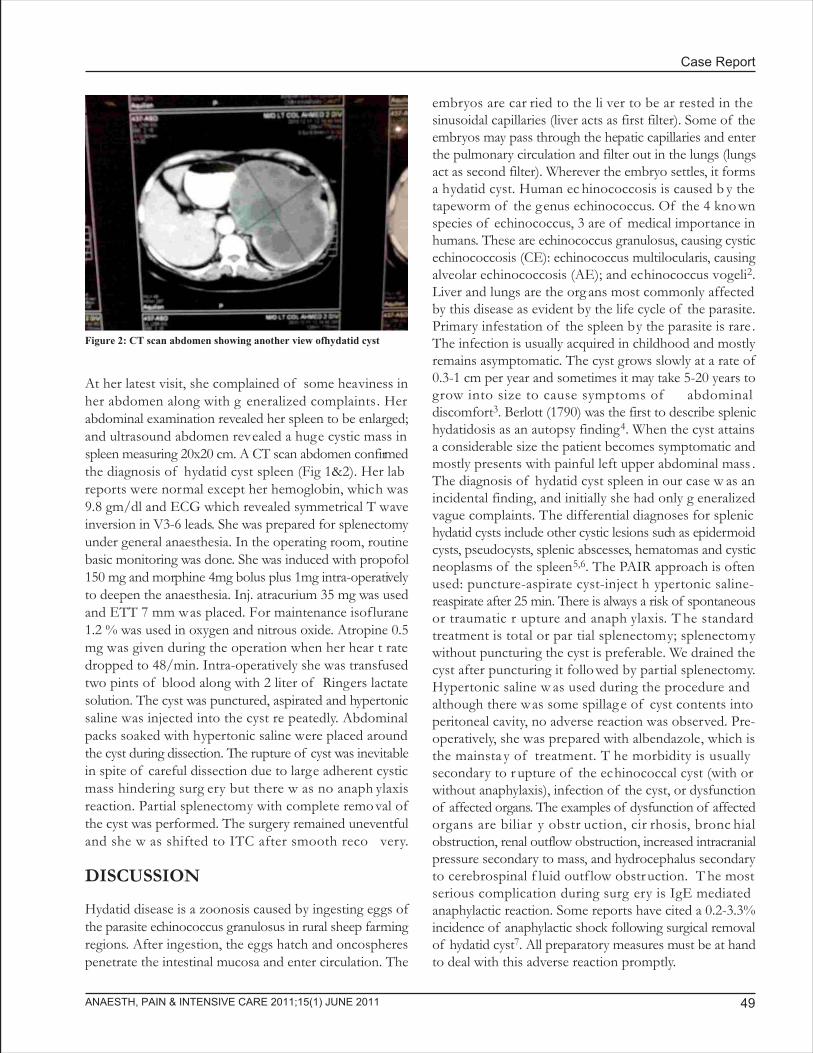
Case Report
ANAESTH, PAIN & INTENSIVE CARE 2011;15(1) JUNE 2011 49
Figure 2: CT scan abdomen showing another view ofhydatid cyst
At her latest visit, she complained of some heaviness inher abdomen along with g eneralized complaints. Herabdominal examination revealed her spleen to be enlarged;and ultrasound abdomen revealed a huge cystic mass inspleen measuring 20x20 cm. A CT scan abdomen confirmedthe diagnosis of hydatid cyst spleen (Fig 1&2). Her labreports were normal except her hemoglobin, which was9.8 gm/dl and ECG which revealed symmetrical T waveinversion in V3-6 leads. She was prepared for splenectomyunder general anaesthesia. In the operating room, routinebasic monitoring was done. She was induced with propofol150 mg and morphine 4mg bolus plus 1mg intra-operativelyto deepen the anaesthesia. Inj. atracurium 35 mg was usedand ETT 7 mm was placed. For maintenance isoflurane1.2 % was used in oxygen and nitrous oxide. Atropine 0.5mg was given during the operation when her hear t ratedropped to 48/min. Intra-operatively she was transfusedtwo pints of blood along with 2 liter of Ringers lactatesolution. The cyst was punctured, aspirated and hypertonicsaline was injected into the cyst re peatedly. Abdominalpacks soaked with hypertonic saline were placed aroundthe cyst during dissection. The rupture of cyst was inevitablein spite of careful dissection due to large adherent cysticmass hindering surg ery but there w as no anaph ylaxisreaction. Partial splenectomy with complete removal ofthe cyst was performed. The surgery remained uneventfuland she w as shifted to ITC after smooth reco very.
DISCUSSION
Hydatid disease is a zoonosis caused by ingesting eggs ofthe parasite echinococcus granulosus in rural sheep farmingregions. After ingestion, the eggs hatch and oncospherespenetrate the intestinal mucosa and enter circulation. The
embryos are car ried to the li ver to be ar rested in thesinusoidal capillaries (liver acts as first filter). Some of theembryos may pass through the hepatic capillaries and enterthe pulmonary circulation and filter out in the lungs (lungsact as second filter). Wherever the embryo settles, it formsa hydatid cyst. Human ec hinococcosis is caused b y thetapeworm of the genus echinococcus. Of the 4 knownspecies of echinococcus, 3 are of medical importance inhumans. These are echinococcus granulosus, causing cysticechinococcosis (CE): echinococcus multilocularis, causingalveolar echinococcosis (AE); and echinococcus vogeli2.Liver and lungs are the org ans most commonly affectedby this disease as evident by the life cycle of the parasite.Primary infestation of the spleen by the parasite is rare .The infection is usually acquired in childhood and mostlyremains asymptomatic. The cyst grows slowly at a rate of0.3-1 cm per year and sometimes it may take 5-20 years togrow into size to cause symptoms of abdominaldiscomfort3. Berlott (1790) was the first to describe splenichydatidosis as an autopsy finding4. When the cyst attainsa considerable size the patient becomes symptomatic andmostly presents with painful left upper abdominal mass .The diagnosis of hydatid cyst spleen in our case w as anincidental finding, and initially she had only g eneralizedvague complaints. The differential diagnoses for splenichydatid cysts include other cystic lesions such as epidermoidcysts, pseudocysts, splenic abscesses, hematomas and cysticneoplasms of the spleen5,6. The PAIR approach is oftenused: puncture-aspirate cyst-inject h ypertonic saline-reaspirate after 25 min. There is always a risk of spontaneousor traumatic r upture and anaph ylaxis. T he standardtreatment is total or par tial splenectomy; splenectomywithout puncturing the cyst is preferable. We drained thecyst after puncturing it followed by partial splenectomy.Hypertonic saline w as used during the procedure andalthough there was some spillag e of cyst contents intoperitoneal cavity, no adverse reaction was observed. Pre-operatively, she was prepared with albendazole, which isthe mainsta y of treatment. T he morbidity is usuallysecondary to r upture of the echinococcal cyst (with orwithout anaphylaxis), infection of the cyst, or dysfunctionof affected organs. The examples of dysfunction of affectedorgans are biliar y obstr uction, cir rhosis, bronc hialobstruction, renal outflow obstruction, increased intracranialpressure secondary to mass, and hydrocephalus secondaryto cerebrospinal f luid outf low obstruction. T he mostserious complication during surg ery is IgE mediatedanaphylactic reaction. Some reports have cited a 0.2-3.3%incidence of anaphylactic shock following surgical removalof hydatid cyst7. All preparatory measures must be at handto deal with this adverse reaction promptly.

Removal of a large hydatid cyst in spleen
ANAESTH, PAIN & INTENSIVE CARE 2011;15(1) JUNE 201150
11TH APICARE CONFERENCE-PSA RAWALPINDI-ISLAMABAD
14-16 October, 2011, PC Hotel Bhurban (Murree Hills)
Three day conference will b e held b y PSA Rawalpindi-Islamabad chapter atpicturesque place of Bhurban (Murree Hills) near Islamabad-t he capital cit y ofPakistan. It will comprise of plenary sessions and discussion penals. The scientif icprogramme will be announced shortly.
Theme of the Conference: "Quality Care - at Minimal Cost"
We look forward to see you in Bhurban.
For further details:Dr Shahab NaqviConvener, APICON 11HoD Anaesthesia and Intensive CareAFIC/NIHD, RawalpindiTel: +9251 927 2928 (O); +9251 927 1034 (R); Fax: +9251 927 1015-16Cell: +92345 543 3525; www.pacta.pk/conf.html
CONCLUSION
Hydatid cyst spleen is a rare but important diagnosis. Anearly diagnosis and treatment had an almost complete curefrom the disease. Surgical excision is the mainstay of thetreatment, but all preparatory measures must be undertakento treat possible anaphylactic reaction.
REFERENCES
1. Murtaza B, Gondal ZI, Mehmood A, ShahSS, Abbasi MH,Tmimy MS, et al. Massie splenic h ydatid cyst. JCPSP2005;15(9):568-70.
2. Kir. A, Baran. E, Simultaneous operation for hydatid cystof right lung and liver. Thorac cardiovasc Surgeon.1995;43:62-4.
3. Wani NA, Tak S, Shah ND, Bashir A, Arif SM Bullet injurycausing r upture of spleen with h ydrated cyst. JK Prac1998;5:55-56.
4. Muro J et al. demonstration angiog raphica del quisitehidatidico de bazo . R ev Clin Esp 1969;115:433-438.
5. Pedrosa I, Saiz A, Arrazola j, et al. Hydatid disease: radiologicand pathologic features and complications. Radiolgraphics.2000;20:795-817.
6. Durgun V, K apan S, K apan M, et al. Primar y splenichydatidosis. Dig Surg. 2003;20:38-41.
7. Jakubowski MS, Barnard DE, Anaphylactic shock duringoperation for h ydatid disease. Anesthesiolog y 1971;34:197-9.

ANAESTH, PAIN & INTENSIVE CARE 2011;15(1) JUNE 2011 51
INTRODUCTION
Throat packs are commonly used in patients undergoingoral, nasal and dental procedures. We encountered difficultyin its removal at the time of extubation of trachea in apatient who was having dental braces fixed to lingual surfaceof her teeth. We found our throat pack was entangled ina hook of the braces that led to difficulty in its retriev al.The cause of this difficulty in removal of the throat packis unusual and to our knowledge no such event has beenreported in the literature. This case report emphasizes theneed of extra care while removing the throat pack to avoiddamage to these costly braces that ma y result inembarrassment to the anaesthetist and/or lead to acompensation claim.
CASE REPORT
A 23 years old female patient presented to our hospitalwith nasal deformity secondary to road side accident. Shewas scheduled for elective open septorhinoplasty undergeneral anaesthesia. Her w eight was 74Kg and height157cm. Her airway examination and neck movements werenormal. She was wearing lingual dental braces as par t ofher treatment for dental deformity. All baseline investigationse.g. haemoglobin, haematocrit, platelet count, urea,creatinine, prothrombin and APTT w ere within normallimits. She was graded as ASA 1 physical status. She was
CASE REPORT
Dental braces bracing a throat pack to causedifficulty in its removal
Mansoor Aqil, MBBS, FCPS
Associate Professor Anesthesiology, College of Medicine, King Saud University/Consultant anesthesiologist,King Abdul Aziz University Hospital, Riyadh 11411, (Saudi Arabia)
Correspondence: Dr. Mansoor Aqil, P.O box 245, Riyadh 11411, (Saudi Arabia); Phone: 0096614775703 (office); 00966507221058(mobile); Fax: 0096614786100 Ext. 7525; Email: [email protected]
ABSTRACTWe present an unusual case of dental braces entangled into a throat pack, thus making it impossible to removeit from the mouth cavity of a patient. The patient, who was emerging from anesthesia, had to be reanesthetised,to enable removal of the pack.
Key words: Throat pack; anesthesia complication; dental bracesCitation: Aqil M. Dental braces bracing a throat pack to cause difficulty in its removal. Anaesth Pain & Intensive Care 2011;15(1):51-53.
advised to be fasting from midnight and was premedicatedwith 2mg tab. lorazepam two hours before surgery with asip of water.
On arrival in the holding area of the operating room, anIV line was established with a 20G cannula on the dorsumof the hand. In the operating room, ECG electrodes, pulseoximeter probe and non invasive blood pressure cuff wereapplied. She was pre-oxygenated with oxygen 6 l/min forabout 3 minutes. Anesthesia was induced with inj. propofolin the dose 2 mg/kg and fentanyl 2 µg/kg. For endotrachealintubation, neurom uscular bloc k w as attained withcisatracurium in the dose of 10 mg. She was intubatedwithout any difficulty with 7.5 mm internal diameter RAE(Portex®) cuffed endotrac heal tube (ETT). Afterconfirmation of proper position of the ETT, it was fixedat 21 cm in the midline and intermittent positive pressureventilation of the lungs was started with anesthesia machine(Datex Ohmeda AS 5™). To avoid soiling of the airway,the throat of the patient was packed around the ETT witha cotton ribbon gauze pack by one of our trainee doctorand the tail of the pack was left outside the mouth andtied to the ETT. To remind the presence of pack in thethroat, a sticker was applied to the ETT connector quoting“Caution! throat pack inside”. The placement of the throatpack was notified in the anesthesia record sheet and alsoendorsed to the circulating nurse. Anesthesia was maintainedwith sevoflurane in oxygen-air mixture and inter mittent
Dental braces bracing a throat pack to cause difficulty in its removal
ANAESTH, PAIN & INTENSIVE CARE 2011;15(1) JUNE 201152
doses of fentanyl. The intraoperative course was uneventfuland duration of surgery was 110 minutes.
At the end of the surgery, residual neuromuscular blockwas reversed. On establishment of adequate breathing weplanned to extubate the trachea. The oral cavity was clearedof any blood clot by using a flexible suction catheter size14F and removal of throat pack was attempted by applyinga gentle traction on the tail of the throat pack. We noticedunusual resistance in retrieving the throat pack. We plannedto retrieve it under direct vision with the help of a McIntosh laryngoscope and Magill's forceps. To get a deeperplane of anaesthesia, inspiratory sevoflurane concentrationwas increased to get its end tidal concentration 3-4%. Onattaining adequate depth of anaesthesia, laryngoscope wasplaced in the oral cavity and found that the threads of thethroat pack were entangled in a hook of the lingual dentalbraces near the second molar tooth. Throat pack was gentlyfreed from the hook and was removed after some difficulty.Residual blood clots w ere remo ved and trac hea w asextubated. The patient was sent to post anesthesia careunit where she stayed for about 40 minutes and was shiftedto the ward with full recovery.
DISCUSSION
Lingual braces are used by orthodontists to straighten thetooth position. T hey consist of wire brac kets that areattached to the lingual side of the teeth. They are usuallyinvisible from outside and very costly. This is an unusualand unexpected cause of difficulty in removal of throatpack and should be considered in the patients with lingualdental braces, undergoing surgery under general anesthesiarequiring endotrac heal intubation and throat pac k.
Throat packs are commonly used under general anesthesiain the surgery in the oral or nasal ca vity and procedureson nasolacrimal duct. The common purpose of their useis to absorb blood or debris, body secretions or externalfluids and prevent the seepage into respirator y tract.1-3
Occasionally, they are used to provide seal around the ETTor to stabilise the ETT or supraglottic devices.4-6 We triedto search the literature to find out the reported complicationsof throat pack but did not find any report of difficulty inits retrieval due to entanglement in a hook of a dentalbrace. The reported complications are pain in the throat1,7-10, injury to lingual and h ypoglossal nerve11, unilaterallaryngeal and hypoglossal paralysis12, soft palate paresis13,forgotten throat pack leading to airway obstruction14-24
and unilateral pharyngeal plexus injury.25
We recommend that during pre-anesthesia assessment, wemust enquire about the presence of dental braces, especiallythe lingual braces, and have more thorough inspection tofind out any uncovered hook. Throat packs, if used, shouldbe gently removed under direct vision, as an undue tractionon it out may result in damage to the costly braces. Suchincident may be an embarrassment to the anaesthetist andis likely to result in a compensation claim.
REFERENCES
1. Jaiswal V, Bedford GC. Review of the use of throat packsin nasal surg ery. J Lar yngol Otol. 2009;123(7): 701-4.
2. Al-Mazrou KA, Abdullah KM, ElGammal MS, Ansari RA,Turkistani A, Abdelmeguid ME. Laryngeal mask airway vs.uncuffed endotracheal tube for nasal and paranasal sin ussurgery: paediatric airway protection. Eur J Anaesthesiol.2010;27(1):16-9.
3. Hill EE, Rubel B. A practical review of prevention andmanagement of ingested/aspirated dental items. Gen Dent.2008;56(7):691-4.
4. Kim CS, Lee BH, Shim JY, Chung WH. Clinical Study ofPharyngeal Throat Pack - Cases of V-pack Application tothe Endotrac heal T ubes. K orean J Anesthesiol.1980;13(3):38-43.
5. Gordon R. H, and Renae T. Tracheal Injury: A Cause forUnexplained Endotracheal Cuff leak During MediastinalDissection. Anesth Analg 1997;84:684-5.
6. Weber T, Salvi N, Orliaguet G, Wolf A. Cuffed vs non-cuffed endotracheal tubes for pediatric anesthesia. PaediatrAnaesth. 2009;19 Suppl 1:46-54.
7. Erkalp K, Korkut YA, Meric A, K ahya V, Gedikli O, SuOK, Saitoglu L. Pharyngeal packing is a predisposing factorfor postoperative aphthous stomatitis in nasal surg ery.Otolaryngol Head Nec k Surg . 2010;142(5):672-6.
8. Griffiths DPG, Lindop MJ, Samuels SI, Roberts GDD.Pharyngeal packs and the incidence of post-operative sorethroat. Anaesthesia 1973;28:320-30
9. Fine J, Kaltman S, Bianco M. (1988) Prev ention of sorethroat after nasotracheal intubation. Journal of Oral andMaxillofacial Surgery 46:946-7.
10. Basha S, McCoy E, Ullah R. T he efficacy of pharyngealpacking during routine nasal surg ery - a prospecti verandomised controlled study. Anaesthesia 2006;61: 1161-5.
11. Evers KA, Eindhoven GB, Wierda JM. T ransient nervedamage follo wing intubation for trans-sphenoidalhypophysectomy. Can J Anaesth. 1999;46(12):1143-5
Case Report
ANAESTH, PAIN & INTENSIVE CARE 2011;15(1) JUNE 2011 53
12. Tesei F, Poveda LM, Strali W, Tosi L, Magnani G, FarnetiG. Unilateral laryngeal and hypoglossal paralysis (Tapia'ssyndrome) following rhinoplasty in general anaesthesia: casereport and review of the literature. Acta OtorhinolaryngolItal. 2006;26(4):219-21.
13. Berendes J. Rhinolalia aperta after the use of a postnasalpack as a medico-leg al problem. HNO . 1979;27(11):377-9.
14. NHS Evidence - National Library of Guidelines formerlya Specialist Librar y of the National Librar y for Health.http://www.library.nhs.uk/GuidelinesFinder/ViewResource.aspx?resID=314420&tabID=288. Accessed on10.06.2010.
15. Ozer C, Ozer F, Sener M, Yavuz H. A forgotten gauze packin the nasophar ynx: an unfor tunate complication ofadenotonsillectomy. Am J Otolar yngol. 2007 Ma y-Jun;28(3):191-3.
16. Scheck P. A pharyngeal pack fixed on to the tracheal tube.Anaesthesia 1981;(36):892-5.
17. Najjar MF, Kimpson J. A method for preventing throat packretention. Anesthesia and Analg esia 1995;80:204-12.
18. To EWH, Tsang WM, Yiu F, Chan M. (2001) A missingthroat pack. Anaesthesia 56:383-4.
19. Walton SL. Post-extubation foreign body aspiration: a casereport. American Association of Nurse Anaesthetists Journal1997;65:147-9
20. Burden RJ, Bliss A. (1997) Residual throat pack - a furthermethod of prevention. Anaesthesia 52:811.
21. Stone, JP, Collyer J. Aide-memoir to pharyngeal pack removal.Anesthesia and Analgesia 2003;96:301-8.
22. Preeti K. Manghnani PK, Shinde V . S, Chaudhari L. S .Critical incidents during anaesthesia; An A udit. Indian J.Anaesth 2004;48:287-294.
23. Sixth Report of the Victorian Consultati ve Council onAnaesthetic Mortality and Morbidity pag e 4/7. Availablef r o m U R L h t t p : / / w w w . h e a l t h . v i c . g o v. a u /vccamm/major/6th/5.htm. Accessed on 10.06.2010
24. Knepil GJ, Blackburn CW. Retained throat packs: results ofa national survey and the application of an organisationalaccident model. Br J Oral Maxillofac Surg 2008;46(6):473-6.
25. Robert W. M, David Z, Andrew M, Charles B. B. Unilateralpharyngeal plexus injury following use of an oropharyngealpack during third-molar surg ery. Journal of oral andmaxillofacial surgery 1990;48(10):1102-1104.
DR PRANAV BANSAL JOINS APICAREDr Pranav Bansal has recently joined APICARE as assistant editor. He is currently serving asAssociate Professor at Dept . of A naesthesiology, Teerthanker Mahaveer Medical College,Moradabad, U.P. (India). He is a member of editorial board of the 'Internet Journal of MedicalEducation' and 'Internet Journal of Anesthesiology'. He is also a peer reviewer for 'Journal ofLocal and R egional A nesthesia', 'Jour nal of P ain R esearch', Do vepress, Ne wZealand
As well as 'Kathmandu University Medical Journal' of Nepal. He will be responsible for our permanentchapter, 'Cliniquiz'. This page is based upon a clinical case scenario, a technological developmentor newer trends in the practice of the relevant f ields, followed by MCQ's of one best optiontype.
We welcome Dr Pranav Bansal to the world of APICARE.
ANAESTH, PAIN & INTENSIVE CARE 2011;15(1) JUNE 201154
INTRODUCTION
Protein C is a 62-KD, Vitamin K dependent glycoproteinsynthesized in the liver, and circulates in the blood as aninactive zymogen at a concentration of 4µg/ml. Its activationinto activated protein C (aPC), is catalyzed b y thrombinwhen it is bound to the endothelial proteoglycanthrombomodulin.This activated form exerts its anticoagulantactivity primarily through inactivation of factors Va andVIIIa1,which are required for factor X acti vation andthrombin generation. The catalytic activity of activatedprotein C is enhanced by protein S. The results of deficiencyof these can result in dee p vein thrombosis, pulmonaryembolism, thrombophlebitis, neonatal purpura fulminans,liver cir rhosis and w arfarin-induced skin necrosis .2
Protein S is also a vitamin K-de pendent plasma proteindiscovered in 1977. Patients, deficient in either protein Sor C, or who express a dysfunctional protein S , are at arisk for repetitive thrombosis.1 Under normal circumstances,pregnancy is associated with a h ypercoagulable state.2Venous thromboembolism is among the leading causes ofmaternal death in developed countries.3-5 We describe theuse of neuraxial anesthesia for cesarean section in aparturient who presented with both protein C and Sdeficiency.
CASE REPORT
Anesthetic management of the parturient withcombined protein C and S deficiency
Tahira Batool*, Bushra Babur**, Shahida Tasneem***.
*Professor of Gynae & Obstetrics, **Associate specialist in Gynae & Obstetrics, ***Head Department of AnesthesiologyKRL General Hospital, Islamabad (Pakistan).
Correspondence: Dr Tahira Batool, Professor of Obstetrics and Gynecology, KRL General Hospital Islamabad (Pakistan);Phone +92519106270.
ABSTRACTProtein S is a vitamin K de pendent co-factor of protein C . Deficiency of Protein C and S results in ahypercoagulable state, which is treated with anticoagulation. This is the first report of a patient with combinedprotein C and S deficiency , who underwent a Cesarean section under spinal anesthesia in KRL HospitalIslamabad (Pakistan).
Key words: Protein C and S deficiency; spinal anesthesia; caesarean sectionCitation: Batool T, Babur B, Tasneem T. Anesthetic management of the parturient with combined protein C and S deficiency. AnaesthPain & Intensive Care 2011;15(1):54-56.
CASE REPORT
A 34 years old primigravida, with a past medical history ofepilepsy controlled on two antiepileptic medications, haddeveloped deep venous thrombosis of left leg at 30 weeksof gestation and w as on anticoagulant therapy i.e . lowmolecular weight heparin according to her w eight. Herwork up for hereditary thrombophilia was done and shewas found to have deficiency of both protein C and S. Shehad developed bursitis of left knee joint and mumps at 36weeks of gestation. Her elective C- section was decided at37 weeks of gestation. A coagulation profile obtained priorto surgery showed her PT 14, APTT 40, INR 1.0 andplatelets 205000.
Anticoagulants were stopped 24 hours prior to her delivery.Spinal anesthesia was chosen for the surgery. The patientwas preloaded with inj. Ring er's lactate solution in theoperating room and placed in the sitting position. A spinalanesthesia was given at the L3-L4 level using a 25 gaugepencil point needle. The patient received 2.0 cc of 0.5%(hyperbaric) bupivacaine to obtain a T4 level of anesthesia.The patient was placed in the left uterine displacementposition and had an uneventful Cesarean section. She wasmonitored in the post anesthesia care unit for two hours.She had by this time recovered motor and sensory functions
Case Report
ANAESTH, PAIN & INTENSIVE CARE 2011;15(1) JUNE 2011 55
bilaterally and was discharged from the recovery room andsent to the high dependency unit. In the immediate postop period she was given two fresh frozen plasmas and oneunit of blood. Her repeat coagulation profile was carriedout. The results were in normal range. She was restartedon aspirin and heparin on 2nd post operative day and wasdischarged on day 7 after shifting to oral w arfarin as ananticoagulant for further management.
DISCUSSION
Protein S is a vitamin K de pendent co-factor of proteinC. Protein C acts by neutralizing activated factors V andVIII and by an inhibitory action on plasminogen activator.6Deficiency of both protein C and S results in increasedincidence of venous thrombosis. Deficiency of these maybe hereditary or acquired. Hereditary disease is an autosomaldominant disorder, with homozygotes generally dying ininfancy due to massive thrombosis. Heterozygotes generallyhave their first thrombotic event by their mid-twenties7.Acquired disease is usually due to he patic disease .7
The frequency of venous thromboembolism from proteinC and S deficiency ranges from 7 to 17%.6 There is alsoa risk of spontaneous abor tion. Pregnant patients withprotein C and S deficiency can be manag ed with either acombination of aspirin and subcutaneous heparin or lowmolecular weight heparins (LMWHs).8 LMWHs do notcross the placenta8-9 and thus have a fetal safety profileequivalent to that of unfractionated heparin. Patients shouldnot be offered neuraxial anesthesia unless the PTT is withinthe normal range if they are manag ed with aspirin andheparin.10 The effect of low molecular weight heparincannot be determined by a lab test, so the patient must beoff it for at least 24 hours prior to neuraxial anesthesia.Tachycardia, hypotension, and hypothermia increase thelikelihood of thrombosis and should also be a voided10.The risk of D VT among health y pregnant w omenundergoing elective cesarean section is lo w and generalmedical thromboprophylaxis is probably not justified. 11
To the best of our knowledge, this is the first report of apatient with combined protein C and S deficiency , whounderwent a Cesarean section under spinal anesthesia. Oursearch revealed three previous case reports of patients withonly protein S deficiency, who presented in labor.12,13 outof these, two patients delivered by caesarean section doneunder spinal anesthesia without complication. T he third
patient had been on heparin, also. She had had a combinedspinal/epidural anesthetic vaginal delivery.
Spinal anesthesia has an added benefit of being associatedwith a lo wer incidence of DVT. It is also prefer red aspregnant patients are at increased risk for aspiration priorto intubation for general anesthesia and there is an increasedrisk of difficult intubation. A regional anesthetic techniqueis thus preferred whenever possible, and general anesthesiashould be avoided.
CONCLUSION
In summary, neuraxial techniques can be used safely inpatients with protein C and S deficiency as long as anappropriate laboratory workup is done and the patient hasbeen off anticoagulants for an adequate period of time.
REFERENCES
1. Comp PC, Nixon RR, Cooper MR, and Esmon CT. FamilialProtein S Deficiency is Associated with R ecurrentThrombosis. Journal of Clinical Investigation 1984; 74:2082-2088. (s)
2. William's Obstetrics. F Gary Cunningham et al. 21st Edition.P824.The McGraw Hill Company Inc., New York, NY, 2001.(s)
3. Chang J, Elam-Evans LD, Berg CJ, et al. Pregnancy-relatedmortality surveillance-United States, 1991-1999. MMWRSurveillSumm 2003; 52:1-8.Medline
4. Lewis G, Drife JO, Clutton-Brock T, et al. Why MothersDie, 2000-2002. T he Sixth R eport of the ConfidentialEnquiries into Maternal Deaths in the United Kingdom.London: RCOG Press, 2004.
5. Health Canada. Special Report on Maternal Mortality andSevere Morbidity in Canada-Enhanced Sur veillance: ThePath to Prevention. Ottawa: Minister of Public Works andGovernment Ser vices Canada, 2004. www .phac-aspc.gc.ca/rhs-ssg/srmm-rsmm/page1-eng.php. Accessed11/26/2008.
6. Faught W, Garner P, Jones G, and Ivey B. Changes in proteinC and protein S lev els in nor mal pregnancy. AmericanJournal of Obstetrics and Gynecology 1995;172:147-150.(s)
7. Thrombophillic disorders and fetal loss: a meta-analysis .Rey E, Kahn SR, David M, Shrier I. Lancet 2003;361:901-908(s)
8. Melissari E, Parker CJ, Wilson NV, et al. Use of low molecular

Anesthetic management of the parturient with combined protein C and S deficiency
ANAESTH, PAIN & INTENSIVE CARE 2011;15(1) JUNE 201156
Gratitude to ReviewersAPICARE acknowledges with sincere gratitude the continued commitment and dedication towards progress of the journalby sparing time and putting in enormous effort to review the received articles for publication. We are particularlyindebted to the following peer reviewers for their help;
Professor Mehdi MumtazEx-Professor of Anesthesia & Critical CareConsultant in critical care / Anesthesiology,Christie Hospital, Manchester (UK).drmhmumtaz@yahoo. co.uk
Talal Khan, MDAssistant Professor of Anesthesiologyand Pain MedicineMarc Asher Comprehensive Spine CenterUniversity Kansas Medical Center (USA)[email protected]
Brig Asif GulConsultant in critical care / Anesthesiology,AFIC [email protected]@gmail.com
Zulfiqar Ahmed, M.B.,B.S. F.A.A.P.Assistant Professor of AnesthesiologyDirector of ResearchChildren's Hospital of MichiganWayne State University3901 BeaubienDetroit, MI 48201-2196 (USA)[email protected]
Shamsuddin AkhtarAssociate Professor,Director of Medical StudiesYale University School of MedicineNew haven, CT 06520 (USA)[email protected]
Jack Vander BeekConsutant4609 Toutle Ct SEOlympia, WA 98501 (USA)[email protected]
Richard B. Schwartz, MD, FACEPChair, Department of Emergency MedicineMedical College of Georgia1120 15th Street, AF2037Augusta, GA [email protected]
Jawad Hasnain, MD, MBA, [email protected]
Mamoona Shaikh [email protected]
Shireen Ahmad, M.D.Associate Professor,Associate Chair for Faculty DevelopmentDepartment of AnesthesiologyNorthwestern Univ. Feinberg School of [email protected]
Asim Mahmood, M.D.Chief of pediatric Anesthesia division,Department of AnesthesiologyMt. Sinai Medical Center & Sinai childrenhospital1500 S. California Ave.Chicago, IL 60608 (USA)[email protected]
weight heparin in pregnancy. ThrombHaemost 1992;68:652-656.
9. Forestier F, Daffos F, Capella-Pavlovsky M. Low molecularweight heparin (PK 10169) does not cross the placentaduring the second trimester of pregnancy study by directfetal blood sampling under ultrasound. T hromb R es1984;34:557-560.
10. Miller RD. Anesthesia, 5th edition, V olume 1, Churchill
Livingstone Publishing, New York p.1150.
11. Jacobsen AF, Drolsum A, Klow NE, Dahl GF, Qvigstad E,Sandset PM. Deep Vein Thrombosis after elective cesareansection. Thrombosis Research 2005;115:461-467.
12. Abramovitz SE, Beilin Y. Anesthetic Management of theParturient with Protein S Deficiency and Isc hemic HeartDisease. Anesthesia and Analgesia 1999;89:709.
13. Fan SZ, Yeh M, Tsay W. Cesarean section in a patient withprotein S deficiency. Anesthesia 1995;50:251-253.

Editorial View
ANAESTH, PAIN & INTENSIVE CARE 2011;15(1) JUNE 2011 57
INTRODUCTION
Ventilating rigid bronc hoscopy is commonly used forremoving foreign bodies from the tracheo-bronchial tree.Foreign body impaction in the airway is a common causeof death in c hildren1. Haemor rhage, post-procedurelaryngeal oedema and tracheo-bronchial lacerations canoccur during v entilating bronc hoscopy2. T ensionpneumothorax is the most serious consequence of rigidbronchoscopy3,4. T his occurs when a one w ay v alvemechanism dev elops because of trac heo-bronchialinstrumentation leading to a rent allowing the air to enterthe pleural cavity during positive pressure ventilation butnot allowing it to leave during expiratory phase. As the airbuilds up in the pleural space, ipsilateral lung is compressedfollowed b y mediastinal shift and compression ofcontralateral lung and intrathoracic vasculature. This leadsto severe hypoxemia and cardio vascular compromise.Tension pneumothorax should be suspected in mechanicallyventilated patients who suddenly decompensate 5. Earlydiagnosis and prompt manag ement are essential for anoptimum outcome. Therefore, this complication must bekept in mind and should be ruled out whenever ventilation
CASE REPORT
Tension pneumothorax caused by ventilating rigidbronchoscopy for removal of foreign body
Safdar Hussain, MCPS*, Riaz Ahmed Khan, MCPS, FCPS**,Muhammad Iqbal, FCPS***
*Consultant, **Associate Professor and Head, ***Assistant ProfessorDepartment of Anaesthesia, Pain and Intensive Care, Rehman Medical Institute, Hayatabad, Peshawar (Pakistan).
Correspondence: Col (R) Riaz Ahmed Khan, Head, Department of Anaesthesia, Pain and Intensive Care, Rehman MedicalInstitute, Hayatabad, Peshawar (Pakistan); e-mail: [email protected]
ABSTRACTTension pneumothorax during ventilating bronchoscopy for foreign body removal is a rare but life threateningcomplication, which if not promptly diagnosed and treated can pro ve fatal. We present a case of tensionpneumothorax, due to a laceration in the right main bronchus caused by bronchoscope, in a one year old child,who underwent bronchoscopy for removal of foreign body (bead). T he child was successfully treated andmanaged by immediate insertion of 14 gauge IV cannula in the pleural cavity followed by chest tube insertion.The laceration was subsequently repaired and foreign body removed by thoracotomy.
Key Words: Ventilating bronchoscopy; Tension pneumothoraxCitation: A. Hussain S, Khan RA, Iqbal M. Tension pneumothorax caused by ventilating rigid bronchoscopy for removal of foreignbody. Anaesth Pain & Intensive Care 2011;15(1):57-59.
is impaired and there is sudden cardiovascular compromiseduring anaesthetic manag ement for v entilatingbronchoscopy6.
CASE REPORT
A one year old female child, weighing 11 kg, was admittedthrough ER department with a one day history of foreignbody inhalation followed by severe bouts of cough anddyspnea. Pre-operative examination sho wed a health yfemale child with symmetrical chest expansion, reducedair entry on the right side and SpO2 of 100%. She was notdyspneic and was normothermic at the time of inspection,without any other medical problem. Pre-op c hest x-rayshowed a foreign body in the right main bronchus (Fig 1).
Without any premedication, anaesthesia w as inducedintravenously with inj. propofol 2mg/kg IV andsupplemented by sevoflurane 3-4 MAC with 100% oxygen.Inj. atropine 0.2mg was given IV and muscle relaxationwas obtained with a small 5-10mg dose of suxamethonium.Rigid bronchoscope was inserted by otolaryngologist and

Tension pneumothorax caused by ventilating rigid bronchoscopy for removal of foreign body
ANAESTH, PAIN & INTENSIVE CARE 2011;15(1) JUNE 201158
Fig 2: Chest x-ray showing a massive right sided pneumothorax withdepression of right hemidiaphragm and shifting of trachea andmediastinum to the left with compression of the left lung
Fig 1: Pre-operative chest x-ray showing a foreign body in the right mainbronchus (arrows)
attempts were made to get hold the foreign body with theforceps. 30 minutes after starting the procedure patient'sSpO2 started falling down to 40%, with cyanosis . Heartrate went upto 200 beats/minute. Child's abdomen wasdistended and on auscultation no breath sounds could beheard over the right hemithorax. The surgeon was askedto stop the procedure and an uncuffed endotracheal tube(size 4.0 ID Endosoft) was put in the trachea under directlaryngoscopy and the patient was manually ventilated with100% oxygen. High airway resistance was encounteredwith only a slight improvement in SpO2, upto 80%. Stillno breath sounds w ere audible on the right side onauscultation. Immediate portable chest x-ray was requested,which showed a massive right sided pneumothorax withdepression of right hemidiaphragm and shifting of tracheaand mediastinum to the left with compression of the leftlung (Fig 2).
A 14 gauge IV cannula was immediately inser ted in the2nd intercostal space in the midclavicular line. A gush ofair leaked out through the cannula and ventilation becameeasier. Within a few min utes SpO 2 rose to 100%.Subsequently, another size-14 cannula was passed in themidaxillary line in the 5th intercostal space and connectedby an IV infusion set to a temporary under-water seal afterremoving the 1st cann ula. Repeat chest x-ray showedmarked improvement with re-expansion of the right lungand mediastinal shift back to its position. (Fig 3)
A chest tube was placed in the right pleural cavity by thethoracic surgeon followed after a day by right thoracotomywhich showed a small 0.7mm rent in the right main bronchusthrough which foreign body was removed and the rent wassutured. Child sho wed unev entful reco very and w asdischarged after a few days.
DISCUSSION
Ventilating rigid bronc hoscopy has inherent risks andcomplications such as bleeding, laryngeal trauma, laryngealoedema, lar yngospasm, bronc hospasm, infection,hypoxemia, tracheobronchial lacerations and tears leadingto pneumothorax 2, pneumomediastinum and surgicalemphysema of the chest wall and neck.
Tension pneumothorax is a rare but life threateningcomplication3,4. Rothmana and Boeckman7 reported thatthe probability of developing pneumothorax during rigidbronchoscopy for the remo val of foreign body isapproximately 1 in 100 cases. It usually results from directtrauma to tracheobroncaheal tree. Therefore, the possibilityof pneumothorax should alw ays be kept in mind when
Fig 3: Post-op chest x-ray showing re-expansion of the right lung

Case Report
ANAESTH, PAIN & INTENSIVE CARE 2011;15(1) JUNE 2011 59
ventilation worsens during ventilating bronchoscopy8. Anearly diagnosis of pneumothorax during anaesthesia,however, is not so easy as the symptoms may be maskedby anaesthesia6. Full monitoring is essential to aler t theanesthesiologist of a w orsening situation. T ensionpneumothorax during surgery may manifest itself by a fallin oxygen saturation, a rapid increase in airway pressure,hypotension and tac hycardia9. In our case , bronchiallaceration caused by rigid bronchoscope was responsiblefor tension pneumothorax. Immediate inter vention torelease the built up pressure in the pleural ca vity can beachieved by a large bore IV cannula and is life saving, tobe followed by chest tube insertion. Vigilance is the priceof safety.
REFERENCES1. Steen KH, Zimmermann T. Tracheobronchial aspiration of
foreign bodies in children: a study of 94 cases. Laryngoscope.1990;100:525-530.
2. Hasdiraz L, Oguzkaya F, Bilgin M, Bicer C. Complicationsof bronchoscopy for foreign body removal: experience in1,035 cases. Ann Saudi Med. 2006;26:283-287.
3. Harar RP, Pratap R, Chadha N, Tolley N. Bilateral tensionpneumothorax following rigid bronchoscopy: a report ofan epignathus in a newborn delivered by the EXIT procedurewith a fatal outcome. J Laryngol Otol. 2005;119:400-402.
4. Gallagher MJ, Muller BJ. Tension pneumothorax duringpediatric bronchoscopy. Anesthesiology. 1981;55:685-686.
5. Brian H, Martin LM. “Pleural disorders in the intensive careunits.” The Washington Manual of Critical Care. 3rd Ed.New Delhi: Williams & Wilkins, 2009
6. Baumann MH, Sahn SA. Tension pneumothorax: diagnosticand therapeutic pitfalls. Crit care Med. 1993;21:177-179.
7. Rothmann BF, Boeckman CR. Foreign bodies in the larynxand tracheobronchial tree in children: a review of 225 cases.Ann Otol Rhino Lar yngol . 1980;89:434-436.
8. Ibrahim AE, Stanwood PL, Freund PR. Pneumothorax andsystemic air embolism during positive-pressure ventilation.Anesthesiology. 1990;90:1479-1481.
9. Hunter AR. Problems of anaesthesia in ar tificialpneumothorax. Proc R Soc Med. 1955;48:765-768.
PAKISTANI ANESTHESIOLOGISTS DIRECTORYAPICARE int ends t o publish a dir ectory cont aining basic inf ormation about all P akistanianesthesiologists serving in or out of the country in a book shape. It will serve as a referenceand have multiple uses, besides enhancing intercommunication between us. It will als o beuploaded on t he net . Please make sure t hat t he directory is as complet e as p ossible byapproaching all the anesthesiologists you know at your end.
Please attach a good quality photograph preferably in JPG format. Print photo with your nameon the back in ink is also accepatable.
Address: Editor 'Apicare', 60-A, Nazim-ud-Din Road, F-8/4, Islamabad.E-mail: [email protected]. [email protected]
City: Name: Date of birth:
Qualif ications Date/College of graduation:
Designation (if any): Institution:
Work address: Off ice phone:
Residential address: Residential phone:
Cell: e-mail:
ANAESTH, PAIN & INTENSIVE CARE 2011;15(1) JUNE 201160
INTRODUCTION
The lifetime prevalence of headache is over 90%. T hereported prevalence of migraine headaches is 18.2% amongfemales and 6.5% among males. Headache is a commoncomplaint for which patients seek relief in the emergencydepartment (ED). The management of headache in UKEDs includes an armamentarium of medications deliveredby the oral, subcutaneous, intramuscular, or intravenousroutes. It has been characterized as a “broad pharmacopeiaof medications” with opioids commonly administered,especially meperidine. While the most effective treatmentfor primary headaches is intravenous prochlorperazine(typically administered with diphenhydramine to counteractthe common side effect of akathisia)1-4, there are othertherapeutic techniques that involve intramuscular injectionsof local anesthetics. This technically simple procedure israpidly accomplished, and results are gratifying in that therelief occurs in fi ve to 10 min utes. T he technique isbecoming increasingly accepted as a therapeutic modalityfor treating headache. In this case series , we used threedifferent procedures to treat headache including occipitalnerve blocks, cervical injections and trigger point injections.
CASE SERIES
Interventional pain management techniques can behelpful in headache management
Ishrat Bano, PhD*, Waqas Ashraf Chaudhary, MD**,Mohammed Ashfaq, MD, PGAc, LicWHO**
* University of Cambridge, Cambridge (UK),**Pain Management Centre, Omer Medical Complex, Raiwind City (Pakistan).
Correspondence: Ishrat Bano , University of Cambridg e, CB2 3QZ, Cambridg e (UK); E-mail: [email protected] .uk,[email protected]
ABSTRACTIn this case series we present three different interventional procedures used to treat headache. The procedures,e.g. occipital ner ve block, cervical injection and trig ger point injection, are described and the suppor tingliterature is reviewed.
Key Words: Emergency department (ED); migraine; cervical injections; trigeminovascular system; cephalgia;occipital nerve block; trigger point injection.Citation: Bano I, Chaudhary WA, Ashfaq M. Interventional pain management techniques can be helpful in headache management.Anaesth Pain & Intensive Care 2011;15(1):60-64.
Patients' length of stay in the ED appears to be shortened.Based on the rapid resolution of headache and othertrigeminovascular system-related signs and symptomsfollowing these injections, connections to the trig eminalsystem appear to be involved. Three patients illustrative ofthe therapeutic response to the lo wer cervical injectionwith inj. bupivacaine are presented.
CASE ONE:(Migraine without Aura: Cervical Injection)
A 31 year old female presented to the ED for pain relieffrom migraine headache, that started three days earlier. Shecomplained of light sensitivity, nausea and vomiting. Routineabortive medications were attempted but without relief .The headache began on the left hemicranium, but becamebilateral and was 10/10 in severity on VAS. The patientdenied having any other medical problem. She was treatedwith 1.5ml of inj. bupivacaine 0.5% at either side of thespinous process of the C-7 v ertebra. In less than fi veminutes she experienced relief of her headache to 1/10.She also described complete relief of her photophobiaand nausea. Subsequently, the patient reported that she did
Case Report
ANAESTH, PAIN & INTENSIVE CARE 2011;15(1) JUNE 2011 61
not have her usual postdromal headac he that typicallyaffected her during the following 24 hours. Nine days latershe returned to the ED with a migraine headache initiatedby the 'smell of a strong cologne'. Again, photophobia andnausea accompanied the headac he. T he patient ag ainreceived bilateral intramuscular injections of 1.5ml of 0.5%bupivacaine at the level of C-7 spinous process. The timefrom the local anesthetic injections to complete headacherelief was exactly sev en minutes. The patient had tw osubsequent visits to the ED for similar migraine headachesover several months and responded consistently to thecervical injection therapy.
CASE TWO:(Episodic Tension Headache: Cervical Injection)
A 47-year-old female complained of a constant frontal,unilateral headache for three days prior to ar rival in theED. Similar headaches would occur 'every now and then'.The patient described the headache as a tight band abouther head. She denied an aura, n umbness, phonophobia,photophobia, nausea, or vomiting. Even though she reportedfeeling 'congested', there was no clinical evidence of sinusitis.The headache was 6/10 in severity. Bilateral lower cervicalinjection with 1.5ml of 0.5% bupi vacaine resulted incomplete resolution (0/10) of the headache in approximatelysix minutes. During follow-up, the patient confirmed thatshe remained headache-free after leaving the ED, and thatshe was able to return immediately to her daily activities.
CASE THREE:(Acute Post-traumatic Headac he: Cer vical Injection)
A 25 year old male sustained a head injur y and orofacialtrauma after collision with a car while driving a motorbike.A brief loss of consciousness occurred. Additionally, therewas avulsion and subsequent reimplantation of the rightmaxillary central incisor as well as extrusive subluxation ofthe mandibular central incisors . Since the accident thepatient had experienced continuous, severe right maxillarydental pain with hypersensitivity to cold water and lighttouch. He also experienced significant dental and gingivalpain in the subluxated but stabilized teeth. His pain w asunresponsive to h ydrocodone with acetaminophen oroxycodone with acetaminophen. In addition, the patientreported a constant, throbbing, posterior headache ratedat 7-10/10 in severity. A C-7 paraspinous intram uscularbupivacaine injection was performed bilaterally. Not onlywas the patient's headac he relieved, his dental pain w asreduced to 1/10 in severity. After the injection, the patient
was able to bite do wn, drink water, and r un cold waterover his previously painful teeth. During the follo w-up,the patient reported that his dental pain remained diminishedand that the headache did not return.
CASE FOUR:(Occipital Neuralgia: Occipital Nerve Blocks)
A 41 year old male presented to the ED for evaluation andmanagement of his headache of recent onset. The patientdescribed inter mittent shooting pains that seemed tooriginate from the right occipital area. T he clinicalpresentation was consistent with the diagnosis of occipitalneuralgia. The headache had been present for over a weekand had not responded to over-the-counter medications.Palpation over the occipital area easily reproduced the pain.He denied any other associated symptoms and denied anyother relevant past medical, social or surgical histor y. Inthe ED the injection of bupi vacaine 0.5% andmethylprednisolone sodium succinate 20 mg with a 25-Gneedle (1.5 cm) into the m uscles of the occipital regionalong the nuchal line, thus blocking superficial occipitalnerves, brought immediate relief to the headac he.
CASE FIVE:(Trigger Point Injections)
A 23 year old female presented for evaluation of headachethat had been present for three da ys. Muscle tendernesswas detected b y palpation bilaterally o ver the anteriortemporal area and several ml of inj. bupivacaine 0.5% wereused to inject these areas. The patient had rapid relief ofher headache.
DISCUSSION
Occipital Nerve Blocks
With this procedure the greater and lesser occipital nervesare anesthetized. These nerves are commonly involved incervicogenic headaches5 and occipital neuralgia6. However,evidence exists to support the use of occipital nerve blocksfor a much larger spectrum of primary headaches7. Thegreater occipital nerve arises from the C-2 nerve root andafter traveling deep in the paraspinal musculature becomessuperficial just below the superior nuchal line and lateralto the occipital protuberance. The nerve travels just medialto the occipital artery at these locations. The lesser occipitalnerve is the ter minal branch of the superficial cer vicalplexus and becomes superficial o ver the inferior nuchal

Interventional pain management techniques can be helpful in headache management
ANAESTH, PAIN & INTENSIVE CARE 2011;15(1) JUNE 201162
Figure1: The injection site for greater occipital nerve block Figure2: The injection for greater occipital nerve block
line. The g reater occipital ner ve block technique, firstinvolves identifying the nerve at its point of entry to thescalp along the superior nuchal line midway between themastoid process and occipital protuberance. (Fig. 1) Thepatient will report pain as the nerve is palpated along thisdistribution. The point of maximal tenderness should beidentified and used as the injection site 8,9. The ner vetypically exits approximately 3 cm below and 1.5 cm lateralto the inion or bony prominence of the occipital skull.(Fig. 2) T he scalp should be cleansed with alcohol oranother appropriate antise ptic. Local anesthetic ag entscommonly used include 2% lignocaine or 0.5% bupivacaine.A corticosteroid is often added to the anesthetic beinginjected. To reduce patient discomfort, the superficial skincan be anesthetized by creating a small wheal using a 27-gauge needle with 1% or 2% lignocaine . Alternatively, avapocoolant can also be used to reduce discomfort10,11. A25-G needle is directed to wards the occiput until bonyresistance is felt. A small amount of anesthetic is injectedat that location. The needle is then pulled back until it isjust under the skin then redirected laterally and mediallyas the anesthetic is injected. As par t of the process theparaspinal muscles near the suboccipital region are infiltratedwith the anesthetic. Since the smaller third occipital nerveexits medial to and in close proximity to the greater occipitalnerve, it too is anesthetized (Fig. 2). Once the syringe iscompletely withdrawn, the injected area should be massagedand compressed to allo w for better distribution of theanesthetic. An alternative technique consists of anesthetizingthe nerve at a more distal site over the occipital ridge. Theoccipital artery is palpated one-third of the way from theinion to the mastoid process and the injections are madejust medial to the occipital ar tery and then additionalinjections are made medial and lateral to this point9.Response rates have been re ported to be about 85%9.
Hypesthesia occurs within 1 to 2 minutes, extending upwardson the scalp to the interaural line. Overall, occipital nerveinjections are safe but some adv erse side effects such asdizziness, lightheadedness or local tender ness at the siteof injection may occur. The use of local steroids has beenassociated with alopecia and h ypopigmentation of thesurrounding skin.
Cervical Injections
The bilateral lower cervical injections with bupivacaine isa recently re ported technique for managing headac hepain12,13. Additionally, this procedure also appears to providesome relief to patients with orofacial pain14. The mechanismis currently unknown, but based on the therapeutic responsethe authors suggest that the sensitized trigeminocervicalcomplex is someho w calmed 15-18. Previous w ork hasestablished that cervical and trigeminal afferents as well asother structures with profound antinocice ptive effectsconverge on the brainstem and are known to synapse withthe trigeminocervical complex19,20. Cervical injection isperformed at the lower cervical or upper thoracic dorsalspine. The field is cleaned with antiseptic solution such astriclosan 0.25% (Chlorosept), betadine or alcohol swabs.Using a 25-G 1.5-inch needle, 1.5 ml of 0.5% bupivacaine,1 or 2% lignocaine are the anesthetic options. The needleis inserted 1 to 1.5 inches into the paraspinal muscles, 2-3 cm bilaterally, at C6 or C7 cer vical vertebrae12,21. Theentire amount of anesthetic solution selected (1.5 ml) isdeposited in each injection site. Always withdraw the plungerbefore injecting to ensure that needle is not in a bloodvessel. The injected area can be massag ed afterwards tofacilitate anesthetic absorption and a band-aid can be placedover the injection sites. While the therapeutic response istypically rapid, it can tak e up to 20 min utes before the
Case Report
ANAESTH, PAIN & INTENSIVE CARE 2011;15(1) JUNE 2011 63
medication effect is noted. If patients do not report anypain relief, other therapeutic techniques should be tried12,21.Patients should be warned about injection site sorenesswhich can last anywhere from 24 to 48 hours . Otherpotential minor complications of this procedure include,pain and irritation at the site of injection, vasovagal syncope,and hematoma formation. Patients should be informed ofthese potential risks before the procedure is perfor med.
Trigger Point Injections
Myofascial trigger points have been postulated as an etiologyfor headaches23-27 and trigger point injections have beendescribed as successful in the manag ement of theseheadaches25-27. Brofeldt and Panacek described the reliefof headaches following the injection of anesthetic in thesuboccipital and anterior temporal areas in their 1998article27. T heir tec hnique is described as a tw o-stepprocedure that involves identifying the proper injectionsite and then administrating the injection. To identify theinjection site, various sites on the patients head and neckare palpated with the tips of the index and middle fingersusing firm circular pressure while paying close attentionto the suboccipital and anterior temporal areas. Anteriorpressure is applied to the g eneral area where the g reateroccipital nerve penetrates the semispinalis capitis musclelocated approximately two finger breadths inferior to thesuperior nuchal line and one to two finger breadths lateralto the occipital protuberance. For the anterior temporalarea, pressure is applied to the slight depression just posteriorto the lateral orbital rim and superior to the zyg omaticarch. According to the authors , the appropriate site forinjection is identified when focal pressure re produces orincreases the patient's headache symptoms. The next stepis the intramuscular injection for which a 50/50 mixtureof 2% lignocaine and 0.25% bupivacaine buffered with a1/10 volume of 8% sodium bicarbonate is used. A 27-Gneedle on a 5 ml syring e is inserted through the area ofmaximal tenderness until the needle makes contact withthe cranium. To make contact with the inferior portion ofthe occiput bone, the needle is guided in a 45-60 deg reeangle superiorly. Once the needle is at the periosteum,continuous pressure is applied on the syring e and eachfocally tender area is 'fanned' with 1-5 ml of anestheticsolution by moving the needle in m ultiple directions, inand out of the tender area. Aspiration to avoid injectioninto a vessel is generally not recommended if the solutionis injected simultaneously during fanning of the needle.Once the syring e is withdrawn, the area of injection ismassaged for at least 30 seconds. The injection is considered
successful when focal palpation no longer reproduces theheadache symptoms27. The authors re ported that two-thirds of their patients had a therapeutic response. In thereport by Young et al. the combination of 8 ml of 0.05%bupivacaine mixed with 2% lignocaine is preferred for theinjections9. Their technique involves identifying the injectionsites by palpating for areas of tenderness in the paraspinal,suboccipital and trapezius muscles. A total of 0.5-1 ml areinjected per site , with the dose di vided between threetriangularly oriented sites reached without removing theneedle from under the skin9. When the trapezius muscleis injected near the apex of the lung, the authors pinch themuscle to isolate the muscle and decrease the chance of apneumothorax. Steroids are often added to the anestheticwhen trigger point injections are perfor med9. While theoccipital injection location described by these authors isvery likely an occipital ner ve block and the describedparaspinous injections may be similar to the lower cervicalinjections, the injection of the described trig ger points,including temporal, and trapezius muscles, would appearto have some benefit.
CONCLUSION
In this article we described three different intramuscularanesthetic injections that have reported therapeutic benefitfor managing headac he pain. W hile there are m ultipletherapeutic modalities a vai;able to reliev e headac he,intramuscular anesthetic injections other than g reateroccipital nerve blocks are cur rently not widely utilized,recognized or researched. The clinical importance of cervicalinjections and trigger point injections remains to be furtherclarified. However, this new injection technique appearsto be an effective therapeutic option for the entire spectrumof Inter national Headac he Society (IHS) classifiedheadaches, whether it be migraine, tension headache, clusterheadache or other trig eminal autonomic ce phalalgias.
REFERENCES
1. Drotts DL, Vinson DR. Prochlorperazine induces akathesiain emergency patients. Ann Emerg Med 1999;34:469-75.
2. Coppola M, Y ealy DM, Leibold RA. Randomized,placebocontrolled evaluation of prochlorperazine versusmetoclopramide for emergency department treatment ofmigraine headac he. Ann Emerg Med 1995;26:541-6.
3. Jones J, Sklar D, Dougherty J, White W. Randomized double-blind trial of intravenous prochlorperazine for the treatmentof acute headache. JAMA 1989;261:1174-6.
Interventional pain management techniques can be helpful in headache management
ANAESTH, PAIN & INTENSIVE CARE 2011;15(1) JUNE 201164
4. Ginder S, Oatman B, Pollack M. A prospective study of i.v.magnesium and i.v. prochlorperazine in the treatment ofheadaches. J Emerg Med 2000;18:311-5.
5. The International Classification of Headache Disorders.Second edition. Cephalalgia 2004; Vol 24.suppl 1.
6. Lake AE, Saper JR. Chronic headac he: New advances intreatment strategies . Neurolog y 2002;59:S 8-13.
7. Tobin J, Flitman S. Occipital nerve blocks: when and whatto inject? Headache 2009;49(10):1521-33.
8. Caputi CA, Firetto V . Therapeutic blockade of g reateroccipital and supraorbital ner ves in mig raine patients.Headache 1997;37(3):174-9.
9. Young WB, Marmura M, Ashkenazi A, Evans RW. Greateroccipital nerve and other anesthetic injections for primaryheadache disorders . Headac he 2008;48:1122-5.
10. Reis EC, Holubkov R. Vapocoolant spray is equally effectiveas EMLA cream in reducing immunization pain in school-aged children. Pediatrics 1997;100(6):e5.
11. Hijazi R, Taylor D, Richardson J. Effect of topical alkanevapocoolant spray on pain with intravenous cannulation inpatients in emergency departments: Randomised doubleblind placebo controlled trial. BMJ 2009;338:b215.
12. Mellick LB, McIlrath ST, Mellick GA. Treatment of headachesin the ED with lo wer cervical intramuscular bupivacaineinjections: a1-year retrospecti ve review of 417 patients .Headache 2006; 46(9): 1441-9.
13. Mellick GA, Mellic k LB. Lower cer vical injections forheadache relief . Lett Headac he 2001;41:992-4.
14. Mellick GA, Mellick LB. Regional head and face pain relieffollowing lower cervical intramuscular anesthetic injection.Headache 2003;43:1111-3.
15. Abrahams VC, Anstee G, Richmond FJ, Rose PK. Neckmuscle and trigeminal input to the upper cervical cord andlower medulla of the cat. Can J Ph ysiol Phar macol1979;57:642-51.
16. Piovesan EJ, Kowacs PA, Tatsui CE, Lange MC, Ribas LC,Werneck LC. Referred pain after painful stimulation of thegreater occipital nerve in humans: evidence of convergence
of cer vical afferents on trig eminal nuclei. Ce phalalgia2001;21:107-9.
17. Bartsch T, Goadsby PJ. Stimulation of the greater occipitalnerveinduces increased central excitability of dural afferentinput. Brain 2002;125:1496-509.
18. Bartsch T , Goadsb y PJ . Increased responses intrigeminocervical nociceptive neurons to cervical input afterstimulation of the dura mater . Brain 2003;126:1801-13.
19. Bartsch T, Goadsby PJ. The trigeminocervical complex andmigraine: Cur rent conce pts and synthesis . Cur r P ainHeadache Rep 2003;7:371-6.
20. Mason P. Deconstructing endogenous pain modulations. JNeurophysiol2005;94:1659-63.
21. Mellick LB, Mellick GA. Treatment of acute orofacial painwith lower cervical intramuscular bupivacaine injections: a1-year retrospective review of 114 patients. J Orofac Pain2008;22(1):57-64.
22. Silberstein SD. Practice parameter: Evidence-based guidelinesfor migraine headache (an evidence-based review): Reportof the Quality Standards subcommittee of the AmericanAcademy of Neurology. Neurology 2000;55(6):754-762.
23. Davidoff RA. Trigger points and myofascial pain: towardunderstanding how they affect headac hes. Ce phalalgia1998;18(7):43648.
24. Fernández-de-Las-Peñas C, Alonso-Blanco C, CuadradoML, Gerwin RD, Pareja JA. Myofascial trigger points andtheir relationship to headache clinical parameters in chronictension-type headac he. Headac he 2006;46(8):1264-72.
25. Venancio Rde A, Alencar FG Jr, Zamperini C. Botulinumtoxin, lidocaine, and dry-needling injections in patients withmyofascial pain and headaches. Cranio 2009;27(1):46-53.
26. Harden RN, Cottrill J, Gagnon CM, et al. Botulinum toxina in the treatment of chronic tension-type headache withcervical myofascial trigger points: a randomized, double-blind, placebo-controlled pilot study . Headac he2009;49(5):732-43.
27. Brofeldt BT, Panacek EA. Pericranial injection of localanesthetics for the manag ement of resistant headaches.Acad Emerg Med 1998;5(12):1224-9.

ANAESTH, PAIN & INTENSIVE CARE 2011;15(1) JUNE 2011 65
INTRODUCTION
Backache is a common public health problem and a majorpsychological, physical and economical burden for theindividual and the society,1-2 . Back pain is one of humanity'smost frequent complaints and a common reason forphysician visits. It is estimated that nine out of ten adultsexperience backache at least once in their lifetime, and fiveout of ten working adults have back pain every year. Nocomprehensive data exist for its prevalence in our populationbut it is almost the same as in the w estern population ifnot more.
Back pain after surg ery may result from a m ultitude ofcauses that include posture during surgery, aggravation ofan existing medical condition or needle trauma duringcentral neuraxial blocks,3-4. In rare cases this may by themanifestation of a sinister condition like epidural abscessor haematoma follo wing a central neuraxial bloc k.5
REVIEW ARTICLE
The causes, prevention and management ofpost spinal backache: an overview
Mohammad Kashif Rafique, FCPS*, Arshad Taqi, FCPS*
*Consultant Anesthesiologist, Hameed Latif Hospital, Lahore (Pakistan).
Correspondence: Dr. Arshad T aqi, Consultant Anesthesiologist, Hameed Latif Hospital, Lahore (P akistan); e-mail:[email protected]
SUMMARYBack pain is one of humanity's most frequent complaints, a common reason for physician visits and a majorpsychological, physical and economical burden. Although the frequency of backache is as high as 46% evenafter general anaesthesia, it was the major cause for 13.4% patients refusing spinal anaesthesia. Multiple factorsare involved in the pathogenesis of postoperative back pain and include type and duration of surgery, durationof immobilization, and the position of the patient during spinal puncture . Diagnosis of back pain is notsimple; contributing factors may include needle trauma, surgical positioning, and injection of saline or localanaesthetic into the interspinous ligaments, development of a supraspinous hematoma, excessive stretchingof ligaments after relaxation of paraspinal muscles and localized trauma to the intervertebral disc. Its relationshipwith various types and sizes of spinal needle is yet to be confir med. Some preventive aspects have beendiscussed. Acute post spinal backache usually resolves within 7 days without any treatment but the possibilityof epidural abscess or epidural hematoma must be ruled out. Counselling, hot and cold massage, mild analgesicslike paracetamol or topical NSAIDs ointments may be prescribed.
Key words: Backache; Postoperative back pain; Spinal needle; Transient neurologic symptomsCitation: Rafique MK, Taqi A. The causes, prevention and management of post spinal backache: an overview (Review Article).Anaesth Pain & Intensive Care 2011;15(1):65-69.
INCIDENCE
A significant number of patients complain of backachefollowing anaesthesia and surgery. Although the frequencyof backache is as high as 46% following general anaesthesia6,the patients relate this to their anaesthesia if they haveundergone a central neuraxial block; the myth of invariableinjury to the bac k associated with needles 7. Backachefollowing previous spinal anaesthetic was the major causefor 13.4% patients refusing spinal anaesthesia in a seriesof more than 1000 patients8.
Symptoms varying from “pricking sensation” at the siteof needle inser tion, upper or lo wer back pain or painradiating to the buttocks and legs are all sometimes reportedas backache. 26.6% of more than 100 patients studied byChan complained of injection site tenderness lasting lessthan a week, which should be differentiated from classical“backache” that none of them complained9.
The causes, prevention and management of post spinal backache: an overview
ANAESTH, PAIN & INTENSIVE CARE 2011;15(1) JUNE 201166
Confounding variables like pre-existing backache; durationof surg ery and the patient's posture during surg erycompound the issue. The pain could be of a short duration,lasting from 72 hours to a week or persistent, lasting beyond3 months.
431 out of 918 pregnant patients surveyed by Shaheen andcolleagues had at least one e pisode of backache duringtheir pregnancy; 96 out of these had experienced backachebefore they became pregnant. T his indicates that abouthalf of these patients would have a preexisting backacheif they presented for spinal anaesthesia for Caesareandelivery10.
Controversy exists over the relationship between anaesthetictechnique and the tr ue incidence of postoperative backpain. Regardless of anaesthetic technique, back pain wasseen in almost 25% of the patients who underwent surgicaloperations under general or spinal anaesthesia,11-12. Randeland colleagues at the Uni versity of Michigan comparedthe recovery characteristics of three anaesthetic techniquesfor outpatient orthopaedic surgery. One of the parametersthey measured was post operative back pain and they foundthat epidural followed by spinal and then general anaesthesiahad highest incidence of back pain on first post operativeday but by the third post operative day the difference ofback pain in these three tec hniques was not statisticallysignificant. No patient in this study required any specifictreatment for backache13.
PATHOPHYSIOLOGY
An overview of the anatomical str uctures involved mayhelp understand the nature of post spinal anaesthesia backpain. The back is a complex str ucture with an intricatenetwork of bones, joints, muscles, ligaments, with multilevel crossovers in nerve supply as well as muscular andligamentous attachments. The multiple subdivisions ofmuscle mass, numerous connective tissue planes , andmultiple attachments of tendons o ver small areas ofvertebral periosteum help to explain the prev alence ofneck and bac k pain while sim ultaneously explain thedifficulty in precisely localizing the source of that pain.Branches of the posterior ramus provide sensory fibres tofascia, ligaments, periosteum, and facet joints.
Source of traumatic low back pain may be the vertebralcolumn itself, surrounding muscle, tendons, ligaments, andfasciae, or a combination thereof. Taking into account thisdifficulty in identifying muscle and tendon injur y as the
source of pain and the fact that there are other generatorsof low back pain besides muscles (e.g., fasciae, ligaments,facet joint, intervertebral disc), it becomes clear that diagnosisof back pain is not simple . Deyo and colleagues ha vepointed out that source of acute low back pain cannot beidentified in 85% of patients14 (Table 1).
Multiple factors are in volved in the pathog enesis ofpostoperative back pain and include type and duration ofsurgery, duration of immobilization, and the position ofthe patient during spinal puncture 15. Other contributingfactors include needle trauma, surgical positioning, injectionof saline or local anaesthetic into the interspinous ligamentsand development of a supraspinous hematoma, 16-17.Excessive stretching of lig aments after relaxation ofparaspinal muscles and localized trauma to the intervertebraldisc has also been implicated in causing bac k pain 18.
Persistent Postoperative Back Pain: Schwabe and Hopfstudied persistent back pain after spinal anaesthesia in thenon-obstetric setting using questionnaires at 3 months andthen after 1 year of spinal anaesthesia in 245 patients .Percentage of patients complaining of backache in theirstudy was comparable with the a verage from 11 studiesthey referred to (15.4% vs 18%). Pre-existing bac k painwas the only variable associated with persistent back painafter 3 months of spinal anaesthesia. Most of these patientsdid not link their post-operative complaints of low backpain to the spinal anaesthetic19.
Table 1: Pain sensitive tissues in the spine
Pain sensitive tissues in the spine
• Skin, subcutaneous tissue, and adipose tissue• Capsules of facet and sacroiliac joints• Ligaments: longitudinal spinal, interspinous (mainly posterior),
and sacroiliac• Periosteum• Dura mater and epidural fibroadipose tissue• Vasculature; both arterial and venous• Paravertebral muscles
Backache and transient neurological symptoms
Postoperative back pain is sometimes confused with transientneurological symptoms. Lignocaine has been implicatedas a possible cause of temporary and permanent neurologiccomplications after spinal anaesthesia in many case reports.Follow up of patients who received uncomplicated spinalanaesthesia revealed that some of them developed pain inthe lower extremities after an initial full reco very. This

Review Article
ANAESTH, PAIN & INTENSIVE CARE 2011;15(1) JUNE 2011 67
painful condition that occurs in the immediate postoperativeperiod was named 'transient neurologic symptoms' (TNS).
Frequency of TNS and neurologic complications afterspinal anaesthesia with lignocaine compared to other localanaesthetics was studied in a Cochrane review that lookedat sixteen trials reporting on 1467 patients, 125 of whomdeveloped TNS. The use of lignocaine for spinal anaesthesiaincreased the risk of developing TNS. T here was noevidence that this painful condition w as associated withany neurologic patholog y; the symptoms disappearedspontaneously by the fifth postoperative day.
In another study, the relative risk (RR) for developing TNSafter spinal anaesthesia with lignocaine as compared toother local anaesthetics (bupivacaine, prilocaine, procaine,levobupivacaine, ropivacaine, and 2-chloroprocaine) was7.31 (95% confidence inter val (CI) 4.16 to 12.86). T heauthors concluded that the risk of developing TNS afterspinal anaesthesia was significantly higher with lidocaineas compared to bupivacaine, prilocaine, or procaine20. Riskof TNS with lignocaine does not change when concentrationof lignocaine is reduced from 5% to 2%21.
Anaesthetic factors influencing postoperative backache
The data on post spinal analgesia consists of observationalstudies looking at the effect of variables like needle size,design and technique on the outcome , which is larg elysuccess rate and postdural puncture headache. Postduralpuncture backache (PDPB) is largely included as anothervariable that is not studied closely; a large array of complaintsranging from pain at the site of injection to classicalbackache or pain radiating to the lower limbs are lumpedtogether as backache. Complete neurological evaluation todetermine the cause is larg ely not documented in thesestudies22.
Needle Type and Size: Type and size of spinal needle usedfor subarachnoid block has been studied extensi vely. Asurvey conducted on 274 patients underg oing spinalanaesthesia using 23 or 25 gauge spinal needles found nodifference in the incidence of postoperative backachebetween the groups23. Kandg and colleagues compared 26and 27 g auge needles for spinal anaesthesia in a larg epopulation of 730 ambulatory surgery patients. They noted18-20% incidence of postoperative back pain in the twogroups which was not statistically significant 24. Tarkkilaand colleagues compared Sprotte needle with Quinc keneedle for frequency of postoperati ve headache andbackache in 300 ASA ph ysical status 1 and 2 patients
undergoing minor orthopedic or urologic procedures intheir randomized, prospective trial. Backache was the mostcommon complication, occurring in 18% patients with nodifference between the two groups studied. Sprotte needledid not demonstrate any advantage in reducing the incidenceof post dural puncture headache or backache25. Atraucanneedle was compared with Sprotte and Quincke needlesin a study that failed to demonstrate superiority of any onetype of spinal needle in reducing the incidence ofpostoperative back pain26.
Lowery and Oli ver studied the incidence of postduralpuncture headac he and bac kache fo l lo wingdiagnostic/therapeutic lumbar puncture using a 22G cuttingspinal needle, and after introduction of a 25G pencil pointspinal needle in 99 pediatric patients. They reported postprocedure back pain in 11% of patients in the 22G Quinckeneedle group while none in the 25G pencil point needlegroup. These finding, although o verwhelming, are notsupported by data from adult literature27.
Rebekah and colleagues compared the back pain and patientsatisfaction scores after the administration of a spinalanaesthetic with or without the use of an 18 g augeintroducer needle in 84 patients. They failed to demonstratea difference in back pain or patient satisfaction scores ondischarge from post-anaesthesia care unit or 24, 48 and72, hours postoperatively. Significant increase in the numberof redirections between groups was observed in the non-introducer g roup, which did not affect the results 28.
Technique: Wilder-Smith prospectively followed 697 patientsoperated under spinal anaesthesia to determine the incidenceand contributing factors predisposing to post-spinalanaesthesia backache. Backache was reported by one outof every seven patients (13.1%), which is comparable tofrequency of post-spinal headache. They determined thatthis often neglected additional cause of post-spinal morbiditycan be reduced by the use of atraumatic techniques andwith small-gauge spinal needles for perfor ming lumbarpuncture29.
Shutt and colleagues compared 22G and 25G W hitacareneedles with 26G Quinc ke needles. It was a controlledstudy of 150 women undergoing elective Caesarean deliveryunder spinal anaesthesia in whic h effect of number ofneedle insertions on the postoperative complication ratewas assessed. The significant difference between groups(P < .001) w as attributable entirely to the n umber ofpatients reporting backache after more than two attempted

The causes, prevention and management of post spinal backache: an overview
ANAESTH, PAIN & INTENSIVE CARE 2011;15(1) JUNE 201168
needle insertions. The increased incidence of backachefollowing repeated spinal needle insertion was presumedto be due to soft tissue or periosteal trauma. No backachewas sufficiently severe to be follo wed beyond 72 hoursafter the operation30.
PREVENTION
Needle size and design do not influence the likelihood ofa patient developing postoperative backache. Number ofattempts made before a successful block increase the riskof trauma and lik elihood of postoperative backache.Avoiding neuraxial bloc ks while a patient is recei vingantiplatelet increases the risk of epidural haematoma withresulting acute back pain and neurological injury. There islittle evidence to suggest an association between persistentbackache and spinal anaesthesia; almost all of these patientshave a history of at least one episode. This history shouldbe sought during preanaesthetic interview and the patientsreassured about this lack of association before administeringthem spinal anaesthesia.
MANAGEMENT
Acute post spinal backache is a self limiting condition thatresolves within 7 da ys without any treatment in mostpatients but the symptoms overlap with those of seriousneurological complications like epidural abscess or epiduralhematoma. Conservative management may be institutedafter serious causes of back pain have been r uled out.Patient should be counselled about the reversibility of thecondition. Hot and cold massag e mild analg esics likeparacetamol or topically NSAIDs ointments ma y beprescribed. A follow up would be advisable to r ule outpersistent backache that requires more extensive workupand management.
CONCLUSION
Incidence of PDPB is almost the same as postdural punctureheadache (PDPH). In contrast with PDPH, whic h is adirect consequence of the technique, there is little data toattribute PDPB to dural puncture; exception being seriousconditions like epidural abscess, haematoms and meningitis.PDPB can result from a multitude of causes that includepatient's positioning during surgery, length of surgery andpre-existing backache. It is a self-limiting condition thatresponds to conservative management. There is, however,an established association between intrathecal lidocaineand TNS. We recommend seeking a thorough history for
pre-existing backache from all patients recei ving spinalanaesthesia; complaints of new onset backache after spinalanaesthesia should be investigated for serious causes likeepidural haematoma or abscess before the patients arereassured and symptomatic management ensued. Back painpersisting for more than one week should be referred forfurther investigations.
REFERENCES
1. Manning DC, Rowlingson JC. Back pain and the role ofneural blockade. In: Cousins MJ, Bridenbaugh PO (eds).Neural Blockade in Clinical Anaesthesia and Managementof Pain. Philadelphia, PA: Lippincott-Raven, 1998;879-914.
2. Haddox LD, Bonica JJ. Evolution of the speciality of painmedicine and the multidisciplinary approach to pain. In:Cousins MJ, Bridenbaugh PO (eds). Neural Bloc kade inClinical Anaesthesia and Management of Pain. Philadelphia,PA: Lippincott-Raven, 1998;1113
3. O'donovan N, Healey TE, F aragher EB, Wilkins R G,Hamilton AA. P ostoperative backache: the use of aninflatable w edge. Br J Anaesth 1986;58(3):280-283.
4. Sardin B, Boncoeur MP , Despor t JC , Abder razak M,Guillaume A. Delayed low back pain after spinal anaesthesia.(Abstract . Ar ticle in F rench). Ann F r Anesth R eanim1995;14(2):230-2.
5. Cook T. M., Counsell D ., Wildsmith3 J . A. W. Majorcomplications of central neuraxial bloc k: report on theThird National A udit Project of the R oyal Colleg e ofAnaesthetists. British Journal of Anaesthesia 2009;102(2):179-90.
6. Hickmott KC, Healy TEJ, Roberts SP, Faraghart. Back painfollowing general anaesthesia and surg ery: Evaluation ofrisk factors and the effect of an inflatable lumbar support.Br Journ Surgery 1990:77;571-575
7. Butler R, Fuller J. Back pain following epidural anaesthesiain labour. Can J Anaesthesia 1998;45:724-8.
8. Rhee WJ, Chung CJ, Lim YH, Lee KH, Lee SC. Factors inpatient dissatisfaction and refusal regarding spinal anaesthesia.Korean J Anaesthesiol 2010:59;260-64
9. Chan S T. Incidence of back pain after lumbar e piduralanaesthesia for non-obstetric surgery--a preliminary report.Med J Malaysia 1995;50(3):241-5.
10. Shaheen K, Arif T. Shahida S: Bac kache in Pregnancy.Biomedica 2006:22;(1):12-5.
Review Article
ANAESTH, PAIN & INTENSIVE CARE 2011;15(1) JUNE 2011 69
11. David L, Brown. Spinal, Epidural and Caudal Anaesthesia.In: Miller. R.D., Anaesthesia, Churchill Livingstone, 6th ed,2005;1668-1670.)
12. Tay HB, Low TC, Loke YH: Morbidity from subarachnoidspinal anaesthesia-a prospective study on the post-operativemorbidity from subarachnoid spinal anaesthesia. SingaporeMed J 1989;30(4):350-5.
13. G.I. Randel S.P, Pandit S.K, Brousseau M, Levy L: Recoverycharacteristics of three anaesthetic techniques for outpatientorthopedic surgery. 1993;25-30.
14. Deyo RA, Cherkin D, Conrad D, et al. Cost contro versycrises: low back pain and the health of the public. AnnuRev Public Health 1992;12:141-55.
15. Brattebo G, Wisborg T, Rodt SA, et al. Intrathecal anaesthesiain patients under 45 years: Incidence of postdural puncturesymptoms after spinal anaesthesia with 27-G needles. ActaAnaesthesiol Scand 1993;37:545-548.
16. Covet S, Robin GC, Davidson JT. Back pain after epiduralanalgesia. Anesth Analg 1967;46:259-63.
17. Crawford JS. Lumbar e pidural block in labor : A clinicalanalysis. Brit ish J Anaesthesia 1972;44:66-74.
18. Anil Ag arwal1, K amal Kishore; complications andcontroversies of regional anaesthesia: a review. Indian Journalof Anaesthesia 2009;53:543-553
19. Schwabe K, Hopf HB: persistent bac k pain after spinalanaesthesia in the non-obstetric setting: incidence andpredisposing factors . Br J Anaesth 2001;86(4):535-9
20. Zaric D, Pace NL. Transient neurologic symptoms (TNS)following spinal anaesthesia with lidocaine versus other localanaesthet ics. Anesth Analg 2005;100:1811-16
21. Hampl KF, Schneider MC, Pargger H, Gut J , Drewe J,Drasner K. A similar incidence of transient neurologicsymptoms after spinal anesthesia with 2% and 5% lidocaine.Anesth Analg 1996;83(5):1051-4
22. Pan PH, F ragneto R, Moore C , Ross V. Incidence ofPostdural Puncture Headache and Backache, and SuccessRate of Dural Puncture: Comparison of Two Spinal NeedleDesigns. South Med J ourn. 2004:97(4);359-63
23. Tay HB, Low TC, Loke YH: Morbidity from subarachnoidspinal anaesthesia-a prospective study on the post-operativemorbidity from subarachnoid spinal anaesthesia. SingaporeMed J 1989;30(4):350-5
24. Kang SB, Goodnough DE, Lee YK, Olson RA, BorshofJA, Furiano MM et al. Comparison of 26- and 27-G needlesfor spinal anaesthesia for ambulator y surgery patients.Anesthesiology 1992;76(5):734-8
25. Tarkkila PJ. Heine H. Tervo RR: Comparison of Sprotteand Quincke Needles with Respect to Post Dural PunctureHeadache and Bac kache. Reg Anesth. 1992;17(5):283-7
26. Peter H P, Regina F, Charles M, Vernon R: Incidence ofPostdural Puncture Headache and Backache, and SuccessRate of Dural Puncture: Comparison of Two Spinal NeedleDesigns. Southern Medical J ournal 2004;97(4):359-363
27. Lowery S, Oliver A: Incidence of postdural punctureheadache and backache following diagnostic/therapeuticlumbar puncture using a 22G cutting spinal needle , andafter introduction of a 25G pencil point spinal needle .Paediatr Anaesth. 2008;18(3):230-4
28. Rebekah RB,Chris O,Lee O, Carol D, Charles V, John M:The effect of spinal introducer needle on post operati veback pain. AANA jour nal 2002;70(6):449-452
29. Wilder-Smith OH, Gürtner T: Backache following spinalanaesthesia--a neglected problem?. R eg Anaesth1987;10(1):24-6
30. Shutt LE, Valentine SJ, Wee MYK, Page RG, Prosser A,Thomas T A, Spinal anaesthesia for cesarean section:Comparison of 22-guage and 25-guage Whitacare needleswith 26-guage Quinke needles. Br J Anaesth 1992:69:589-594

ANAESTH, PAIN & INTENSIVE CARE 2011;15(1) JUNE 201170
QUESTIONS
Note: Please choose one best answer.
Q1. A radiofrequency neurotomy is a type of injectionprocedure in which a heat lesion is cr eated oncertain nerves thus interrupting the pain pathways.
a. The heat is produced by resistance in the tip ofthe acti ve electrode to the f lowing cur rent
b. The resistance in the sur rounding tissues isresponsible for heat generation, so that the tip ofthe electrode absorbs heat from the surroundingtissues
c. The electrode tip maintains its temperatureregardless of the temperature of the surroundingtissues
d. If the sur rounding tissues ha ve a highconductance, the temperature rise will betremendous
e. The larger the tip of the electrode, the higher willbe the temperature rise
2. If effective, the neurotomy should provide painrelief for a variable period of time that may be;
a. Nine to fourteen monthsb. Ten to twelve monthsc. Six to eight monthsd. Longer than fourteen monthse. Less than six months
3. The neurotomy procedure requires the following:
a. An IV line, sedatives, an x-ray table, local anestheticsolution, RF cann ulas, RF g enerator.
b. An IV line , sedatives, an x-ra y table , g eneralanesthesia, RF cann ulas, RF g enerator.
c. An IV line, sedatives, an x-ray table, local anestheticsolution, RF cann ulas, RF g enerator, imag eintensifier.
d. An IV line, sedatives, an x-ray table, local anestheticsolution, long spinal needles , RF g enerator.
CLINIQUIZ
Radiofrequency NeurotomyTariq Hayat Khan
e. An IV line, sedatives, an x-ray table, local anestheticsolution, long insulated spinal needles , RFgenerator.
4. The duration of the procedure is highly variableand depends upon many factors.
a. The entire procedure usually tak es between 30and 60 minutes in expert hands
b. The time taken is shor ter if the height of thepatient is less than the a verage height for ag e
c. The duration of the procedure is usually shorterin female patients
d. It is associated with expertise of the operator andthe assistants, the number of nerves to be blockedand the cooperation of the patient
e. It ma y be perfor med quic kly under g eneralanesthesia
5. Regarding RF, it must not be tried if;
a. The pain relief can be achieved with continualuse of NSAIDS
b. Infiltration of local analgesic agents gives adequatepain relief
c. A patient is expected to survive less than one yeard. The pain perception is associated with minimal
numbness of a foote. The patient is a known diabetic
6. Regarding complications of RF it can be saidthat;
a. There is no evidence of any serious complicationsexcept failure to have pain relief
b. There is always full relief of pain even if associatedwith some vascular or neural injury
c. It is a selecti ve procedure with only cutting ofpain pathways without producing cutaneousnumbness or dysesthesia
d. Local infection is common and is a result of tissuedamage by burning

Cliniquiz
ANAESTH, PAIN & INTENSIVE CARE 2011;15(1) JUNE 2011 71
e. Although transient neuritis and dysesthesia havebeen reported, enhancement of pain occurs onlyin 5% of the patients
7. The RF is generall y a safe pr ocedure, but onemust be careful to identify the dev elopment ofsome common side-effects;
a. Hypertension and hyperglycemia
b. Temporary weakness and/or temporary numbness
c. Nausea with or without vomiting
d. Hyperglycemia and nausea
e. Bowel and bladder incontinence
8. Temperature produced depends upon;
a. Current, duration of exposure, tissue resistanceand the skin colour
b. Duration of exposure, proximity of the electrodetip to the ner ves, tissue resistance and the skincolour
c. Current, duration of exposure, tissue resistanceand the diameter of the nerves
d. Impedance, current, duration of exposure andtissue resistance
e. Voltage, duration of exposure, selected frequencyand the type of nerves to be blocked
ANSWERS
1. (b) is the generally accepted correct answer.
2. (a): Nine to four teen months is the g enerally agreedduration of the pain relief with RF, although some authorshave stated a much shorter duration.
3. (c): An IV line, sedatives, an x-ray table, local anestheticsolution, RF cannulas, RF generator and image intensifierare the main requirements of the procedure . A closeattention to the availability of the equipment and requireddrugs will remove many of the later hassles.
4.(d): The time taken to perform RF is associated withexpertise of the operator and the assistants , the numberof nerves to be blocked and the cooperation of the patient.The height and gender of the patient have no effect on
the procedure. It is usually performed by numbing the skinby local anesthetics; and diagnostic blocks and localizationof the ner ves may become difficult or ev en impossibleunder general anesthesia.
5. (a): RF can be used in cancer patients likely to survivemore than three months. RF is only used after successfuldiagnostic block with local anesthetics has been established.These blocks also offer relief from pain caused b y RFitself. If patient is benefitted with continual use of NSAID'sor other pain medications, he should not be subjected toRF; however, theside effects of these dr ugs must becontrolled by appropriate measures.
6. (e): Although transient neuritis, failure to produce painrelief, and dysesthesia have been reported, enhancementof pain occurs only in 5% of the patients. Some evidencedoes exist of occasional serious complications e.g. paralysis,dizziness and bowel and bladder incontinence. Infectioncan be a result of poor adherence to the principles ofasepsis.
7. (b): Common side effects include: temporary weakness,temporary numbness, pain at the procedure site , rarely,more-serious side effects occur , including: long-ter mnumbness, paralysis, dizziness and bo wel and bladderincontinence.
8. (d): Temperature produced depends upon impedance,current, duration of exposure and tissue resistance .Proximity of large blood vessels may lower the temperature.Color of skin or the type of nerves blocked have no effects.
BIBLIOGRAPHY
1. van Bo xem K, et al. Radiofrequency and pulsedradiofrequency treatment of chronic pain syndromes: Theavai lable evidence . P ain Pract ice . 2008;8:385.
2. Barbara W oodward Lips P atient Education Center .Radiofrequency lesioning . R ochester, Minn.: Ma yoFoundation for Medical Education and R esearch; 2002.
3. Bogduk N, et al. A narrative review of lumbar medial branchneurotomy for the treatment of back pain. Pain Medicine.2009;10:1035.
4. Tekin I. A comparison of con ventional and pulsedradiofrequency denervation in the treatment of chronicfacet joint pain. Clinical J ournal of Pain. 2007;23:524.
5. Nath S, et al. Percutaneous lumbar zygapophysial (facet)joint neurotom y using radiofrequency cur rent, in themanagement of chronic low back pain. Spine. 2008;33:1291.

ANAESTH, PAIN & INTENSIVE CARE 2011;15(1) JUNE 201172
Infection sources in ICU require utmost vigilance and care.We hereby describe another probable source of infectionin patients of ICU where closed suction systems (CSS) areapplied for uninterrupted ventilation even during suctioning.Growth of microorganisms increases, whenever theseorganisms get optimum environment for their multiplication.Closed suction inline catheter systems (CISC) are mostoften used in patients who are critical and require ventilationfor prolonged period. Numerous CSS are mark eted bymanufacturers with one advantage or other. Kollef et alsuggested that there is colonization of microorganisms inlower respiratory tract when such systems are placed over24 hours1. Recently, numerous studies suggested that theuse of CISC for more than 24 hours did not increase therisk of ventilator -associated pneumonia and it might besafe for patients 2. Some man ufacturers claim that thisdevice can safely be attac hed for 72 hours contin uouslywith low infection risk.
We routinely use these suction systems in patients on highPEEP v entilation. T he man ufacturer3 claims someadvantages in one CSS with many attachments. This duallumen CISC (Portex ®Suction ProTM 72) contains variousports .These ports are:
Port 1: Attached to endotrac heal tube connecterPort 2: Suctioning port that opens man ually (Figure 1)Port 3: For connection in-line with ventilator inspiratory
circuitPort 4: For attac hment of supplied connectors (for
nebulisation etc.)
This novel device produces g reat f low, high v alue andaccording to manufacturer it can be used for 72 hours. Itprovides an unobstructed evacuation pathway that helpsmake it easier to remo ve secretions, without the risk ofcross contamination and the loc kable end cap prev entsinadvertent suctioning3.
It aids in disconnection of the catheter from the patient'sendotracheal or tracheostomy tubes and a patient labelwith day-of-the-week stickers are pro vided to monitorlength of use. It is also a time saving solution for respiratorytherapists, nurses and medical facilities.
After having used it in various patients in our ICU, it wasobserved that the suction port provided for attachment of
Need to close the 'closed suction in-line catheter' port!Manpreet Singh, MD, FCCP, FIMSA, Dheeraj Kapoor, MD, FCCP
Department of Anesthesiology and Intensive Care, Government Medical College and Hospital, Sector 32, Chandigarh, India-160030
Correspondence: Dr Manpreet Singh, 1223 A, GMCH Campus, Sec 32 B, Chandigarh, (India);Ph:+919646121503; E-mail:[email protected]
LETTERS TO EDITOR
the suction catheter, must have some capping or must havebeen manufactured in a way so that it does not act as asource of infection in critically ill and immunocompromisedpatients. It remains uncapped for 72 hours , and it ma ycome into contact with some unsterile things. This mightbecome a continuous source of infection and a place formicroorganisms growth. The literature is abundant withreports of infections with respiratory tract secretions orcatheter tips as a source, but not from these unusual sites.Further studies are required to samples these sites of CSSdevices; the manufacturers may be suggested to take careof these sites either by capping or some coverings that canbe removed while suctioning and reapplied afterw ards.
REFERENCES
1. Koller MH, Prentice D, Shapiro SD, Fraser VJ,Silver P,Trovilllion E, Wellitz P, von Harz B, St John R: Mechanicalventilation with or without daily changes of in-line suctioncatheters. Am R esp Crit Care med 1997; 156:466-472.
2. Inglis TJ, Lim TM,Ng ML,Tang EK,Hui KP. Structuralfeatures of tracheal tube biofilm formed during prolongedmechanical v entilation .Chest 1995;108:1049-1052.
3. Manufacturer's manual- Smiths Medical Inter nationalLimited.,UK. www.smiths-medical.com.
Figure 1: The site (white arrow) where capping is required

ANAESTH, PAIN & INTENSIVE CARE 2011;15(1) JUNE 2011 73
FDA Recalls Implantable Infusion Pumps andRefill Kits
FDA issued a class I recall of Medtronic's SynchroMed II andSynchroMed EL implantable infusion pumps and refill kits, usedfor the long-term infusion of pain, cancer treatment and anti-spasm medications. Class I recalls are the most serious type ofrecall and in volve situations in whic h there is a reasonableprobability that use of the recalled product will cause seriousadverse health consequences or death. These pumps and refillkits have been recalled because poc ket fills - the unintendedinjection of drugs or fluids into the tissue under the skin at thepump pocket site - have occurred and may result in patient harm,serious injury, and/or death due to drug overdose or underdose.The recalled products were manufactured from June 1998 toJanuary 2003 and distributed from April 1999 to January 2011.
www.fda.gov/medwatch/report.htm
FDA Recalls Central Venous Catheter Trays
Because of leaks in the plunger luer detected during a routinesyringe leak test, v arious types of Cook, Inc , central venouscatheter trays are subject to a class I recall, FD A announced."The potential exists for leakag e and possible loss of sterility.This may lead to serious adverse health consequences and/ordeath," according to an alert from MedWatch, the FDA's safetyinformation and adv erse ev ent re porting prog ram. .
http://www.medscape.com/viewarticle/738433?src=mp&spon=46
Latest Johnson & Johnson Recall Involves Sterility-RiskSutures
Johnson & Johnson, which has been plagued by repeated recallsof its consumer medicines and medical devices o ver the pastyear, has recalled 107 batches of surgical sutures in Decemberdue to potential sterility problems. The recall came to light onWednesday after the United Kingdom Medicines and Healthcareproducts Regulatory Agency (MHRA) described the action onthe agency's website. The action stemmed from potentially faultypackaging seals on the individually wrapped sutures that raiseda contamination risk, J&J said. The potential problem was causedby modifications of manufacturing equipment and has beencorrected, said Barbara Montresor , a spokeswoman for J&J'sEthicon surgical products division.
http://www.medscape.com/viewarticle/738340?src=mp&spon=46
Trends & TechnologyMorphine/Naltrexone Combo Temporarily Withdrawn
King Pharmaceuticals Inc, a wholly owned subsidiary of Pfizer,has voluntarily recalled all dosages of a combination morphinesulfate and naltrexone hydrochloride (Embeda Extended ReleaseCapsules CII) from wholesalers and retailers in the United Statesbecause "a prespecified stability requirement was not met duringroutine testing," a messag e on the product's W ebsite notes.Available data suggest that the issue is unlikely to pose a safetyrisk to patients using the product as prescribed, the company'smessage to patients and prescribers notes.
http://www.medscape.com/viewarticle/739008?src=mp&spon=46
Ventilators for Mass Casualty Scenarios
St. Louis-based Allied Healthcare has released a line of ventilatorsdesigned for mass casualty situations. During natural or man-made disasters, hospitals ma y be o verwhelmed by the sheernumber of patients needing life support, and clinical staff typicallynot qualified to provide life support may be required to assist.Additionally, electric power may become unavailable. The AlliedMass Casualty Ventilators feature simple operation, long batterylife, and are able to operate without exter nal gas connections.
MEDEGRIP
MedeGrip, a simple foam device for w orking with small orbreakable things in the clinic or on the f loor, has received theEuropean CE Mark of approval. It was less than a month agot h a t M e d e G r i p w a ssuccessfully registered withthe FDA as a Class 1 deviceand Access Scientif icincluded it as par t of thePOWERWAND MaximumBarrier Kit. Developed bya PICC line nurse, the devicehelps take away the pain ofhandling things like Luer Loks and pin knots, and makes it a bitsafer to work with glass ampules.
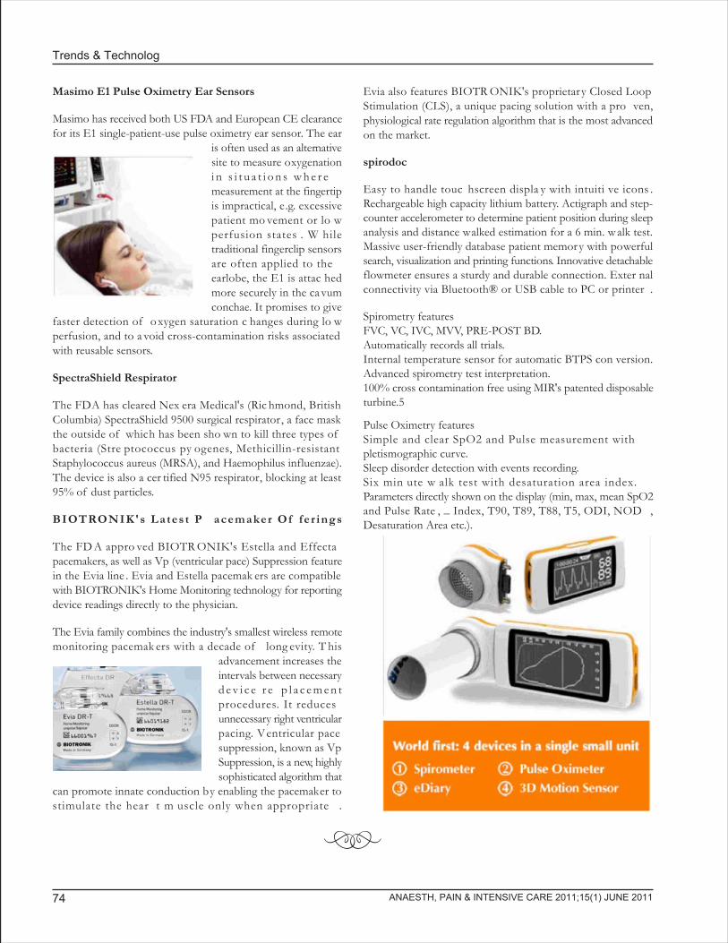
Trends & Technolog
ANAESTH, PAIN & INTENSIVE CARE 2011;15(1) JUNE 201174
Masimo E1 Pulse Oximetry Ear Sensors
Masimo has received both US FDA and European CE clearancefor its E1 single-patient-use pulse oximetry ear sensor. The ear
is often used as an alternativesite to measure oxygenationi n s i t u a t i o n s w h e r emeasurement at the fingertipis impractical, e.g. excessivepatient mo vement or lo wperfusion states . W hiletraditional fingerclip sensorsare often applied to theearlobe, the E1 is attac hedmore securely in the ca vumconchae. It promises to give
faster detection of oxygen saturation c hanges during lo wperfusion, and to a void cross-contamination risks associatedwith reusable sensors.
SpectraShield Respirator
The FDA has cleared Nex era Medical's (Ric hmond, BritishColumbia) SpectraShield 9500 surgical respirator, a face maskthe outside of which has been sho wn to kill three types ofbacteria (Stre ptococcus py ogenes, Methicillin-resistantStaphylococcus aureus (MRSA), and Haemophilus influenzae).The device is also a cer tified N95 respirator, blocking at least95% of dust particles.
BIOTRONIK's Lates t P acemaker Of fe r ings
The FD A appro ved BIOTR ONIK's Estella and Effectapacemakers, as well as Vp (ventricular pace) Suppression featurein the Evia line . Evia and Estella pacemak ers are compatiblewith BIOTRONIK's Home Monitoring technology for reportingdevice readings directly to the physician.
The Evia family combines the industry's smallest wireless remotemonitoring pacemak ers with a decade of long evity. T his
advancement increases theintervals between necessaryd e v i c e r e p l a c e m e n tprocedures. It reducesunnecessary right ventricularpacing. Ventricular pacesuppression, known as VpSuppression, is a new, highlysophisticated algorithm that
can promote innate conduction by enabling the pacemaker tostimulate the hear t m uscle only when appropriate .
Evia also features BIOTR ONIK's proprietary Closed LoopStimulation (CLS), a unique pacing solution with a pro ven,physiological rate regulation algorithm that is the most advancedon the market.
spirodoc
Easy to handle touc hscreen displa y with intuiti ve icons .Rechargeable high capacity lithium battery. Actigraph and step-counter accelerometer to determine patient position during sleepanalysis and distance walked estimation for a 6 min. walk test.Massive user-friendly database patient memory with powerfulsearch, visualization and printing functions. Innovative detachableflowmeter ensures a sturdy and durable connection. Exter nalconnectivity via Bluetooth® or USB cable to PC or printer .
Spirometry featuresFVC, VC, IVC, MVV, PRE-POST BD.Automatically records all trials.Internal temperature sensor for automatic BTPS con version.Advanced spirometry test interpretation.100% cross contamination free using MIR's patented disposableturbine.5
Pulse Oximetry featuresSimple and clear SpO2 and Pulse measurement withpletismographic curve.Sleep disorder detection with events recording.Six min ute w alk test with desaturation area index.Parameters directly shown on the display (min, max, mean SpO2and Pulse Rate , _ Index, T90, T89, T88, T5, ODI, NOD ,Desaturation Area etc.).

ANAESTH, PAIN & INTENSIVE CARE 2011;15(1) JUNE 2011 75
Symposium on Cardiothoracic Anesthesia: ASymposium was organized on 23rd March 2011 by PakistanAssociation of Cardiothoracic Anesthesia (P ACTA) atTABBA Heart Institute Karachi.
The Inaugural session started after recitation of holy Quranfollowed by welcome address by chair organizing committeeDr Hamid Aqil Naqvi consultant TABBA Heart Institute.The oath taking ceremony for new office bearer w asconducted by patron PSA Prof. S Tipu Sultan.
The 2 years activities of PACTA was highlighted by outgoingpresident Prof. Fazal Hameed, who also emphasized tofocus on education and teaching in cardiac anesthesia asa second fellowship has already been started by CPSP. Itwas followed by speeches by incoming president Dr ShahabNaqvi, AFIC, and Prof. S Tipu sultan. They emphasizedthat establishment of PACTA was a dream which is nowa reality and hoped that PACTA would help in promotionof knowledge and improvement in clinical skill and practicein cardiothoracic anesthesia. In the end chief guest CEOTABBA heart institute delivered his speech by appreciatingformation of PACTA and extended his offer to cooperatein future also.
A shield was presented for life time achievement in cardiacanesthesia to Prof. Rehana Kamal from AKU. As she wasout of country Dr Hamid Aqil Naqvi received the shieldon her behalf. Another shield was presented to Prof. YounusKhatri for his contribution in cardiac anesthesia.
The scientific program comprised of two sessions. Theprogram started by a presentation by Dr Zameer Rajput,Shifa Islamabad, who highlighted the c hallenges ofproviding safe anesthesia in lung cancer surgery, followedby Dr Khalid Rasheed a Cardiac surg eon from TABBAHeart Institute , who presented his approac h andmanagement strategies for dealing with urgent & emergentCABG; Prof Najma Amjad, NICVD, spoke on myocardialprotection during off pump surgery; Dr Rana Altaf CPEICMultan express his views on prevention of organ failurefollowing high risk cardiac surg ery. Dr Shahid Sami a
Academic Activitiescardiac surgeon from South City Hospital Karachi discussedsurgical techniques and other relevant issues that affect thedecision-making process In dealing with Functional MR;Dr Safdar g ave his opinion on quality assurance incardiothoracic anesthesia.
In post lunch session Arrhythmia During Period of CABG,Prevention vs. Treatment was discussed by Prof Sadqafrom DUHS, Dr M Hamid described challenges in Pakistanfor open heart pediatric surgery and presented statistics ofpediatric surgeries performed all over Pakistan. For thefirst time a perfusionist Ejaz Haider from AKU and a staffnurse Shenaz Nadeem from T ABBA Hear t Instituteparticipated in suc h for um and presented m yocardialprotection during CPB surgery and fast track cardiac surgery& nursing perspective respectively.
Post-lunch Scientific session included a free paper sessionin which six papers were presented by anesthetists involvedin cardiac anesthesia from all over Pakistan, this generateda lively and exciting discussion for sharing m ulticentreexperiences which was much appreciated by both seniorsand juniors. The whole event was a great success and endedwith hope of promoting a high standard of scientificpresentations in future.
Dr Sadqa AftabProf Anesthesia CHK, DMC, DUHS
Workshop on 'Radiofrequency Neurotomy': A one-day workshop was held at KRL Hospital Islamabad, underthe joint ar rangements of STSP and De pt ofAnesthesiology, Pain Management & Intensive Care of thehospital. Dr. Jamil Sabit was the chief facilitator. Dr. ShahidaTasneem highlighted the importance of pain managementand Dr. Tariq Hayat Khan presented an overview of theRF. Dr. Jamil threw light on various aspects of RF use inpain. A hands-on session was the main event, which wasmuch appreciated by the par ticipants from all o ver thecountry. The workshop was successful in inoculating aninterest in this new pain relief modality in the participants.Certificates were awarded to all of them at the end.

ANAESTH, PAIN & INTENSIVE CARE 2011;15(1) JUNE 201176
Intensive course of regional anesthesia in Dubai UAERashid hospital T rauma centre - DUB AI - U AE (10/07/2011 -14/07/2011)
Peripheral nerve blocks and catheters under ultrasound combined toNS guidance. 5 consecutive days of training with lectures, hands onphantom, models, live case in the Ors, clinical cases, catheters managementin the wards. Requirements to set up a unit of regional anesthesia andanalgesia. Educational material Book of 100 pages, forms and policiesin soft copies. 8 participants/session
2011 Ultrasound Guided - R egional Anesthesia And V ascularAccess WorkshopAugust 17, 2011 -Hershey, Pennsylvania, United States
Fellowship in regional anesthesia and analgesia in DUBAIRashid hospital T rauma Centre - DUB AI - U AE (01/09/2011 -01/09/2012)
Education and research program in regional anaesthesia and analgesiain a trauma centre
30th Annual ESRA Congress 2011Dresden, Germany (07/09/2011 - 10/09/2011)
Building Kno wledge and Science in R egional AnaesthesiaCome learn and network with over 1,500 anaesthesiologists, physiciansand scientists who specialize in regional anaesthesia for surgery, obstetrics,paediatrics and pain control in the lo vely, historic town of Dresden.www.kenes.com/esra2011
10th International Conference on Complexity in Acute Illness(ICCAI)Bonn, Germany (08/09/2011 - 11/09/2011)
10th International Conference on Complexity in Acute Illness (ICCAI)is a highly interdisciplinary, international meeting that focuses on bringingtogether critical care ph ysicians, anesthesiologists, mathematicians,bioengineers, physicists, and computer scientists interested in improvingacute care outcomes through the application of advanced quantitativemethodology to clinical and experimental data to discuss recent advancesin this field.F or more infor mation, please contact Dr . med. Sv [email protected]
3rd International Conference For Pain TreatmentPrizren, Republic of Kosova (09/09/2011 - 11/09/2011)
Professional Health Association (PHA) in cooperation with: Ministryof Health, Republic of Kosova ; Faculty of Medicine, UCCK, Clinicof Anesthesiolog y - Pristina; R egional Hospital “ Prim. Dr . DautMustafa” Prizren ; Albanian P ain Association (APA) and EuropeanFederation of IASP Chapters - EFIC Org anize:3rd Inter nationalconference for Pain Treatment and 2nd Conference of Pain Managementfor Nurses. All health professionals are invited to attend this conference:anesthesiologists, neurologists , surg eons, g ynecologists, dentists ,neurosurgeons, physiotherapists, pharmacists, nurses and other healthworkers who have special interest for the diagnoses and treatment ofpain.
Intensive course of regional anesthesia in Dubai UAERashid hospital T rauma centre - DUB AI - U AE (25/09/2011 -29/09/2011)
CalendarPeripheral nerve blocks and catheters under ultrasound combined toNS guidance. 5 consecutive days of training with lectures, hands onphantom, models, live case in the Ors, clinical cases, catheters managementin the wards. Requirements to set up a unit of regional anesthesia andanalgesia. Educational material: Book of 100 pages, forms and policiesin soft copies. 8 participants/session.
Anesthesia Simulator WorkshopSeptember 22, 2011 -San Francisco, California, United States
Hands-On Difficult Airway Management WorkshopSeptember 22, 2011 -San Francisco, California, United States
Introduction to Transesophageal Echocardiography (iTEE)WorkshopSaturday, September 10, 2011 to Sunday, September 11, 2011
At Intercontinental Buckhead Hotel, Atlanta, Georgia, United Statesof America
The Third International Congress of Obstetric Anaesthesia andPerinatal Medicine with R ecent Advances in Anaesthesiology,Perioperative Medicine and Pain TherapyPoznan, Poland (06/10/2011 - 08/10/2011)
The main goal of the Cong ress is development and delivery of thestate of art updates and refresher courses in the fields of obstetricanaesthesia, perinatal medicine and beyond.
ASA 2011 Annual MeetingOctober 15-19, 2011 -Chicago, Illinois, United States
11th APICARE Conference-PSA Rawalpindi-Islamabad14-16 October, 2011, PC Hotel Bhurban (Murree Hills)
Three day conference will be held by PSA Rawalpindi-Islamabad chapterat picturesque place of Bhurban (Mur ree Hills) near Islamabad-thecapital city of Pakistan. It will comprise of plenary sessions and discussionpenals. T he scientific prog ramme will be announced shor tly.
XVIIth International Congress of Anaesthesiology andIntensive CareNovotel Plovdiv, Bulgaria (27/10/2011 - 30/10/2011)
The Congress programme includes topics on anaesthesia and painmanagement, multidisciplinary treatment of trauma, se psis, clinicalnutrition. The event is organized with the support of Military MedicalAcademy and Bulgarian Society of Parenteral and Enteral Nutrition.For more information, please visit website www.anesthesiology.bg ore-mail [email protected]
European Society of Intensi ve Car e Medicine 201101 Oct 2011 to 05 Oct 2011Location: Berlin, GermanyWebsite: http://www.esicm.org
The Middle East Anaesthesia, P ain Management and CriticalCare Conference;23 - 25 October, 2011Abu Dhabi National Exhibition Centre

ANAESTH, PAIN & INTENSIVE CARE 2011;15(1) JUNE 2011 77
First there was McSleepyTM. Now it's time tointroduce the first intubation robot operatedby remote control. This robotic system namedThe K epler IntubationSystem (KIS), anddeveloped by Dr. Thomas M. Hemmerling,McGill University Health Centre (MUHC)specialist and McGill University Professor ofAnesthesia and his team, ma y facilitate theintubation procedure and reduce somecomplications associated with airw aymanagement. T he w orld's first roboticintubation in a patient w as performed at theMontreal General Hospital by Dr. Hemmerling.The KIS allo ws us to operate a roboticallymounted video-laryngoscope using a joystick
CLINIPICSPh
oto
Cre
dits
: D
r. H
emm
erlin
g's R
esea
rch
Labo
rato
ry.
from a remote w orkstation. After successfullyperforming extensive tests in the airways of medicalsimulation mannequins , whic h closely resembleintubation conditions in humans , clinical testing inpatients has now begun. One day, it might actually bethe standard practice of airw ay manag ement,"concludes Dr . Hemmerling, whose laborator ydeveloped the world's first anesthesia robot, nicknamedMcSleepyTM, in 2008, whic h provides automatedanesthesia delivery.
Source: http://muhc.ca/newsroom/news/introducing-world%E2%80%99s-first-intubation-robot
Intubating robot
Phot
o C
redi
ts :
Dr.
Hem
mer
ling's
Res
earc
h La
bora
tory
.

ANAESTH, PAIN & INTENSIVE CARE 2011;15(1) JUNE 201178
Major General (R) Muhammad Nasim Riaz
Month of June, this year, brought us the sad news of the demise of one of the senior anaesthesiologistsof Pakistan, Major General (R) Muhammad Nasim Riaz who embarked on the journey to his heavenlyabode, on 29 June 2011.
Gen Nasim Riaz was a colleague, a friend, an elder brother, a teacher and mentor to many of us fromthe anaesthesia fraternity of Pakistan Armed Forces. He was born in Attock City on 1st January 1944.He did his g raduation from King Edward Medical College in 1969 and joined the Ar med Forcesduring the 1971 war. He joined the specialty of anaesthesia in 1973 and, after ser ving at differentmilitary hospitals, joined the Ar med Forces Institute of Cardiology & National Institute of HeartDiseases (AFIC-NIHD) in 1981.
He was one of the pioneers in cardiac anaesthesia in Pakistan and worked as cardiac anaesthesiologistin AFIC-NIHD for 20 years, at the end of which he was promoted to the rank of Major General andserved as Advisor in Anaesthesia at Combined Military Hospital, Rawalpindi from 2001 to 2004. Heworked hard for the training and impro vement of anaesthesia care in the Ar med Forces and evenafter his retirement from Army, he played an important role in establishing the cardiac center at thePakistan Institute of Medical Sciences (PIMS), Islamabad.
He was an honest and upright man, a disciplined soldier , a dedicated teacher and a very competentprofessional. The vacuum created by his death is hard to fill. May Allah bless his soul in eternal peace.
Obituary
By: Brigadier Shahab Naqvi