Embed Size (px)
Citation preview
Protein misfolding is the molecular mechanismunderlying MCADD identified in newborn screening
Esther M. Maier1,{, Søren W. Gersting1,{, Kristina F. Kemter1, Johanna M. Jank1, Maria Reindl1,
Dunja D. Messing1, Marietta S. Truger1, Christian P. Sommerhoff2 and Ania C. Muntau1,�
1Department of Molecular Pediatrics, Children’s Research Center, Dr. von Hauner Children’s Hospital, Ludwig-
Maximilians-University, Lindwurmstr. 4, Munich 80337, Germany and 2Department of Clinical Chemistry and Clinical
Biochemistry, Ludwig-Maximilians-University, Munich 80336, Germany
Received January 13, 2009; Revised and Accepted February 16, 2009
Newborn screening (NBS) for medium-chain acyl-CoA dehydrogenase deficiency (MCADD) revealed a higherbirth prevalence and genotypic variability than previously estimated, including numerous novel missensemutations in the ACADM gene. On average, these mutations are associated with milder biochemical pheno-types raising the question about their pathogenic relevance. In this study, we analyzed the impact of 10ACADM mutations identified in NBS (A27V, Y42H, Y133H, R181C, R223G, D241G, K304E, R309K, I331T andR388S) on conformation, stability and enzyme kinetics of the corresponding proteins. Partial to total rescueof aggregation by co-overexpression of GroESL indicated protein misfolding. This was confirmed by acceler-ated thermal unfolding in all variants, as well as decreased proteolytic stability and accelerated thermal inac-tivation in most variants. Catalytic function varied from high residual activity to markedly decreased activityor substrate affinity. Mutations mapping to the b-domain of the protein predisposed to severe destabilization.In silico structural analyses of the affected amino acid residues revealed involvement in functionally relevantnetworks. Taken together, our results substantiate the hypothesis of protein misfolding with loss-of-functionbeing the common molecular basis in MCADD. Moreover, considerable structural alterations in all analyzed var-iants do not support the view that novel mutations found in NBS bear a lower risk of metabolic decompensationthan that associated with mutations detected in clinically ascertained patients. Finally, the detailed insight intohow ACADM missense mutations induce loss of MCAD function may provide guidance for risk assessment andcounseling of patients, and in future may assist delineation of novel pharmacological strategies.
INTRODUCTION
Newborn screening (NBS) for medium-chain acyl-CoA dehy-drogenase deficiency (MCADD; MIM #201450) by tandem-mass spectrometry has successfully been implemented inmany countries worldwide (1). MCADD revealed a notablyhigher birth prevalence (�1:15000) than previously estimated(1–3) and nowadays is the disorder most frequently diagnosedin NBS, together with phenylketonuria (4). The disease leadsto a defect in mitochondrial b-oxidation of fatty acids. Patientsshow a decreased ability to withstand catabolic stress and riskcoma and death due to hypoketotic hypoglycemia duringprolonged fasting or intercurrent illness. In undiagnosed
patients, the disorder shows a significant morbidity and mor-tality. Approximately 20% of patients die during their firstmetabolic crisis and �40% of the survivors show sustainedneurological impairment (5–7). Once diagnosed, however,adverse effects can be prevented by avoidance of fastingduring episodes of catabolic stress. Thus, MCADD is the dis-order thought to most justify early detection by NBS (4).
In patients diagnosed after metabolic decompensation, themutation K304E (c.985A.G) (ACADM gene; OMIM#607008) has been shown to account for 90% of defectivealleles (8) and therefore is considered to be a severe mutationassociated with a high risk of clinical manifestation (9).
†The authors wish it to be known that, in their opinion, the first two authors should be regarded as joint First Authors.
�To whom correspondence should be addressed. Tel: þ49 89 5160 2746; Fax: þ49 89 5160 7792; Email: [email protected]
# 2009. The Author(s)This is an Open Access article distributed under the terms of the Creative Commons Attribution Non-Commercial License (http://creativecommons.org/licenses/by-nc/2.0/uk/) which permits unrestricted non-commercial use, distribution, and reproduction in any medium, provided the original work isproperly cited.
Human Molecular Genetics, 2009, Vol. 18, No. 9 1612–1623doi:10.1093/hmg/ddp079Advance Access published on February 18, 2009
Downloaded from https://academic.oup.com/hmg/article-abstract/18/9/1612/2527170by gueston 01 February 2018

K304E and few other missense mutations identified in symp-tomatic patients were previously shown to induce protein mis-folding and aggregation (10–17).
MCADD patients identified by NBS show a considerablywider genotypic heterogeneity with the mutation K304E beingless prevalent. Numerous novel mutations were unraveledincluding a second prevalent mutation, Y42H (c.199T.C)(3,18–20). After diagnosis, patients carrying these allelic var-iants follow disease management plans to avoid metaboliccrises. Thus, the natural course of MCADD associated withthis new group of mutations is unlikely to be witnessed. Onaverage, these patients express lower disease markers (octanoyl-carnitine and related acylcarnitine ratios in blood) than observedin homo- or heterozygosity for K304E (3,18,20). However, thedifferent genotypes show a high variability of values, and it isgenerally accepted that the biochemical phenotype does notallow for a reliable assessment of the risk associated with thesingle mutations. Insights into the molecular effects of themutations on the corresponding protein would be helpful to esti-mate their pathogenic relevance. Yet, experimental data on themolecular consequences of the various novel missense mutationsidentified in NBS is scarce and mainly refers to the mildtemperature-sensitive Y42H mutation (18,21,22). The aim ofthis study, therefore, was to elucidate the impact of mutationsfound in presymptomatic newborns on conformation, stabilityand catalytic function of the variant MCAD proteins.
MCAD is a member of the acyl-CoA dehydrogenase(ACAD) family of flavoproteins, which catalyzes the first stepof the mitochondrial b-oxidation of medium-chain fatty acids.The ACAD family comprises nine known members, five ofwhich are involved in fatty acid oxidation, four in amino acidoxidation (23). Each subunit of the homotetrameric MCADenzyme is composed of three structural domains of approxi-mately equal size, namely the N-terminal a-domain (residues1–129), the b-domain (residues 130–239) and the C-terminala-domain (residues 240–396). The N- and C-terminaldomains predominantly consist of densely packed a-helices,which shape the core of the tetramer. The middle b-domainsare exposed at the surface of the molecule and comprise twoorthogonal b-sheets. The catalytic centers consisting of thebinding sites for the substrate and the natural cofactor flavinadenine dinucleotide (FAD) are mainly formed by the interfacebetween the b-domain and the C-terminal a-domain. The three-dimensional (3D) structure is a tetrahedral arrangement ofa dimer of dimers with an overall diameter of �90A. Theinteractions between the two monomers forming a dimer areextensive and involve the FAD binding site, whereas betweenthe two dimers predominantly helix–helix interactions arefound, similar to a four-helix bundle structure (23).
In this study, we characterized ten MCAD variants withsingle amino acid substitutions spread over the proteinderived from mutations previously reported from the NBS inBavaria, Germany, including the two prevalent mutationsY42H and K304E (3). We established a high-yield purificationprotocol for recombinant prokaryotic expression of wild-typeand variant MCAD proteins using a maltose bindingprotein (MBP)-tag. The purified proteins were subsequentlycharacterized with respect to oligomerization, enzyme kin-etics, proteolytic stability, thermal inactivation and thermalunfolding. Impressively, all variants showed alterations of
protein function, yet in heterogeneous ways. Catalytic functionvaried from high residual activity to markedly decreasedactivity and severe alteration of substrate affinity. Weobserved aggregation with partial rescue by co-overexpressionof chaperonins, decreased proteolytic stability and acceleratedthermal inactivation in most variants. Thermal unfolding wasfacilitated in all variants. Data on proteolytic and thermal stab-ility revealed that conformational changes and destabilizationare most pronounced in mutations mapping to the b-domain ofthe protein. These results substantiate the hypothesis of proteinmisfolding with loss-of-function being the common molecularbasis in MCADD. The hypothesis holds true not only for thecommon variant K304E, but also for mutations derived frompresymptomatic patients.
RESULTS
Disturbed oligomerization is partially rescuedby co-overexpression of GroESL
Wild-type and ten variant forms of MCAD (Fig. 1 and Table 1)were purified by affinity chromatography and subsequentsize-exclusion chromatography (SEC) to analyze the oligo-meric states of the expressed fusion proteins (Fig. 2). Wild-typeMCAD was eluted in the tetrameric form with an almost negli-gible amount of aggregates (,1%), whereas MCAD variantsshowed a markedly decreased expression of soluble proteinconsistent with proneness to aggregation and degradation ofmisfolded protein. Only one variant (R388S) showed anelution profile identical to wild-type. The remaining variantsrevealed severely disturbed oligomerization consisting of (i)small amounts of tetramers (Y42H, D241G, R309K) with orwithout high molecular weight aggregates, (ii) exclusivelyhigh molecular weight aggregates (A27V, K304E, I331T) or(iii) small amounts of protein fragments of various molecularweights (Y133H, R181C) (Fig. 2A). No monomers or dimerswere observed with any of the variants. R223G was expressed
Figure 1. Structural localization of MCAD missense mutations analyzed. TheMCAD monomer, shown as a ribbon representation, is composed of threestructural domains: the N-terminal a-helix domain (residues 1–129, red),the b-sheet domain (residues 130–239, blue) and the C-terminal a-helixdomain (residues 240–396, green). Amino acid residues affected by mutationsare shown as stick models with carbon atoms in white, oxygen atoms in redand nitrogen atoms in blue.
Human Molecular Genetics, 2009, Vol. 18, No. 9 1613
Downloaded from https://academic.oup.com/hmg/article-abstract/18/9/1612/2527170by gueston 01 February 2018

as truncated, instable protein with no detectable activity (datanot shown) and could not be subjected to SEC.
Tetramer formation was partially rescued by co-overexpres-sion of the chaperonins GroES and GroEL. Co-overexpressingGroESL, seven variants (A27V, Y42H, Y133H, R181C,D241G, K304E, R309K) showed oligomerization profilessimilar to that of wild-type with only small amounts of aggre-gates (5–7%) (Fig. 2B). Co-overexpression of chaperonins didnot enhance protein folding for the two variants I331T andR223G.
In summary, we observed disturbed oligomerization withaggregation and/or degradation in nine of 10 MCAD variantsanalyzed. Our results confirm that ACADM mutations can com-promise protein folding, but misfolding can be mitigated tovarious extent by increasing the amount of available chaperonins.
Variant MCAD proteins show different patternsof enzyme kinetic parameters
Kinetic analyses using the ferricenium ion as an electronacceptor were performed for wild-type and purified MCADvariants (Table 2). All variants displayed Michaelis–Mentenbehavior. Enzyme activity with respect to Vmax was compar-able to wild-type in variants Y42H, R181C and R309K.Reduced maximal activities were found in variants A27V(38% of wild-type), D241G (48%) and K304E (46%).Variant Y133H showed the most pronounced reduction inactivity (3.5%), but still followed Michaelis–Menten kinetics.Vmax of wild-type, Y42H, and K304E are in line with the pre-viously reported data (14,21,24).
For most of the variants, the apparent affinity to the sub-strate octanoyl-CoA was not or only slightly reduced. OnlyR388S showed an excessively decreased substrate affinitywith a 100-fold increase in Km (51.9 mM). This variantshowed a 2.5-fold increase in Vmax. However, the apparentlyhigh activity is of no physiological relevance, since it wouldrequire supraphysiological concentrations of substrate toachieve it.
Km-values displayed in this study are 10-fold lowercompared with previously reported data (14,21,24). This
Figure 2. Disturbed oligomerization of variant MCAD proteins is partiallyrescued by co-overexpression of GroESL. Oligomerization profiles of wild-type (WT) and variant MCAD proteins were determined by size-exclusionchromatography. Soluble high molecular weight aggregates (A) eluted atvolumes of 45–47 ml, tetramers (T) at 58–62 ml. No dimers or monomerswere observed with any of the variants. Elution volumes of .85 ml containedfragments (F) of degraded MCAD or MBP. (A) Profiles withoutco-overexpression of GroESL. Wild-type (WT) and R388S almost exclusivelyeluted as tetramers. All other variants eluted as high molecular weight aggre-gates or low molecular weight fragments with only three variants (Y42H,D241G and R309K) showing small peaks of tetrameric MCAD. Aggregatesand degradation products of Y133H and R181C comprised various molecularweights spread all over the chromatogram. (B) Profiles with co-overexpressionof GroESL. Variants A27V, Y42H, Y133H, R181C, D241G, K304E andR309K showed a rescue of tetramer formation with only small amounts ofaggregates. Only for variant I331T, tetramer formation could not be restored.Note: for WT, the chromatogram without co-overexpression of GroESL isdepicted.
Table 1. cDNA and protein location of MCAD missense mutations analyzed
cDNAa Mature protein
c.155C.T A27Vc.199T.C Y42Hc.472T.C Y133Hc.616C.T R181Cc.742A.G R223Gc.797A.G D241Gc.985A.G K304Ec.1001G.A R309Kc.1067T.C I331Tc.1237C.A R388S
aReference sequence: GenBank accession no. M16827.1. Nucleotidenumbering starts with A of the ATG initiation codon as þ1.
1614 Human Molecular Genetics, 2009, Vol. 18, No. 9
Downloaded from https://academic.oup.com/hmg/article-abstract/18/9/1612/2527170by gueston 01 February 2018
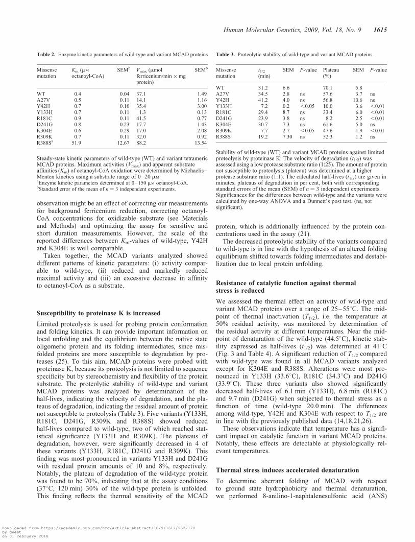
observation might be an effect of correcting our measurementsfor background ferricenium reduction, correcting octanoyl-CoA concentrations for oxidizable substrate (see Materialsand Methods) and optimizing the assay for sensitive andshort duration measurements. However, the scale of thereported differences between Km-values of wild-type, Y42Hand K304E is well comparable.
Taken together, the MCAD variants analyzed showeddifferent patterns of kinetic parameters: (i) activity compar-able to wild-type, (ii) reduced and markedly reducedmaximal activity and (iii) an excessive decrease in affinityto octanoyl-CoA as a substrate.
Susceptibility to proteinase K is increased
Limited proteolysis is used for probing protein conformationand folding kinetics. It can provide important information onlocal unfolding and the equilibrium between the native stateoligomeric protein and its folding intermediates, since mis-folded proteins are more susceptible to degradation by pro-teases (25). To this aim, MCAD proteins were probed withproteinase K, because its proteolysis is not limited to sequencespecificity but by stereochemistry and flexibility of the proteinsubstrate. The proteolytic stability of wild-type and variantMCAD proteins was analyzed by determination of thehalf-lives, indicating the velocity of degradation, and the pla-teaus of degradation, indicating the residual amount of proteinnot susceptible to proteolysis (Table 3). Five variants (Y133H,R181C, D241G, R309K and R388S) showed reducedhalf-lives compared to wild-type, two of which reached stat-istical significance (Y133H and R309K). The plateaus ofdegradation, however, were significantly decreased in 4 ofthese variants (Y133H, R181C, D241G and R309K). Thisfinding was most pronounced in variants Y133H and D241Gwith residual protein amounts of 10 and 8%, respectively.Notably, the plateau of degradation of the wild-type proteinwas found to be 70%, indicating that at the assay conditions(378C, 120 min) 30% of the wild-type protein is unfolded.This finding reflects the thermal sensitivity of the MCAD
protein, which is additionally influenced by the protein con-centrations used in the assay (21).
The decreased proteolytic stability of the variants comparedto wild-type is in line with the hypothesis of an altered foldingequilibrium shifted towards folding intermediates and destabi-lization due to local protein unfolding.
Resistance of catalytic function against thermalstress is reduced
We assessed the thermal effect on activity of wild-type andvariant MCAD proteins over a range of 25–558C. The mid-point of thermal inactivation (T1/2), i.e. the temperature at50% residual activity, was monitored by determination ofthe residual activity at different temperatures. Near the mid-point of denaturation of the wild-type (44.58C), kinetic stab-ility expressed as half-lives (t1/2) was determined at 418C(Fig. 3 and Table 4). A significant reduction of T1/2 comparedwith wild-type was found in all MCAD variants analyzedexcept for K304E and R388S. Alterations were most pro-nounced in Y133H (33.68C), R181C (34.38C) and D241G(33.98C). These three variants also showed significantlydecreased half-lives of 6.1 min (Y133H), 6.8 min (R181C)and 9.7 min (D241G) when subjected to thermal stress as afunction of time (wild-type 20.0 min). The differencesamong wild-type, Y42H and K304E with respect to T1/2 arein line with the previously published data (14,18,21,26).
These observations indicate that temperature has a signifi-cant impact on catalytic function in variant MCAD proteins.Notably, these effects are detectable at physiologically rel-evant temperatures.
Thermal stress induces accelerated denaturation
To determine aberrant folding of MCAD with respectto ground state hydrophobicity and thermal denaturation,we performed 8-anilino-1-naphtalenesulfonic acid (ANS)
Table 3. Proteolytic stability of wild-type and variant MCAD proteins
Missensemutation
t1/2
(min)SEM P-value Plateau
(%)SEM P-value
WT 31.2 6.6 70.1 5.8A27V 34.5 2.8 ns 57.6 3.7 nsY42H 41.2 4.0 ns 56.8 10.6 nsY133H 7.2 0.2 ,0.05 10.0 3.6 ,0.01R181C 29.4 8.7 ns 33.4 6.0 ,0.01D241G 23.9 3.8 ns 8.2 2.5 ,0.01K304E 30.7 7.3 ns 61.6 5.0 nsR309K 7.7 2.7 ,0.05 47.6 1.9 ,0.01R388S 19.2 7.30 ns 52.3 1.2 ns
Stability of wild-type (WT) and variant MCAD proteins against limitedproteolysis by proteinase K. The velocity of degradation (t1/2) wasassessed using a low protease:substrate ratio (1:25). The amount of proteinnot susceptible to proteolysis (plateau) was determined at a higherprotease:substrate ratio (1:1). The calculated half-lives (t1/2) are given inminutes, plateaus of degradation in per cent, both with correspondingstandard errors of the mean (SEM) of n ¼ 3 independent experiments.Significances for the differences between wild-type and the variants werecalculated by one-way ANOVA and a Dunnett’s post test. (ns, notsignificant).
Table 2. Enzyme kinetic parameters of wild-type and variant MCAD proteins
Missensemutation
Km (mM
octanoyl-CoA)SEMb Vmax (mmol
ferricenium/min � mgprotein)
SEMb
WT 0.4 0.04 37.1 1.49A27V 0.5 0.11 14.1 1.16Y42H 0.7 0.10 35.4 3.00Y133H 0.7 0.11 1.3 0.13R181C 0.9 0.11 41.5 0.77D241G 0.8 0.23 17.7 1.43K304E 0.6 0.29 17.0 2.08R309K 0.7 0.11 32.0 0.92R388Sa 51.9 12.67 88.2 13.54
Steady-state kinetic parameters of wild-type (WT) and variant tetramericMCAD proteins. Maximum activities (Vmax) and apparent substrateaffinities (Km) of octanoyl-CoA oxidation were determined by Michaelis–Menten kinetics using a substrate range of 0–20 mM.aEnzyme kinetic parameters determined at 0–150 mM octanoyl-CoA.bStandard error of the mean of n ¼ 3 independent experiments.
Human Molecular Genetics, 2009, Vol. 18, No. 9 1615
Downloaded from https://academic.oup.com/hmg/article-abstract/18/9/1612/2527170by gueston 01 February 2018

fluorescence experiments. The use of the hydrophobic fluoro-phore ANS allowed monitoring of overall unfolding events,since it binds to hydrophobic groups of the denaturingprotein showing a high quantum yield in its bound state, butnot when solved in aqueous buffers (27). ANS fluorescenceanalysis of tetrameric wild-type MCAD revealed a transitionmidpoint (Tm1/2) between folded and unfolded state of52.68C. This is in agreement with previous results obtainedby circular dichroism (16).
Figure 4A shows ANS fluorescence profiles upon thermaldenaturation of wild-type, K304E and two severely distortedvariants. In variant Y133H, a considerably (20-fold) elevatedground state fluorescence signal at 258C was detected in com-parison with wild-type. This finding indicates an increasedhydrophobicity due to partial protein unfolding even withoutthe application of thermal stress. The same was found inR181C and K304E, but to a much lesser extent. Uponthermal denaturation, for all MCAD variants the transitionfrom the native state to the unfolded state occurred at signifi-
cantly lower temperatures than in the wild-type (Fig. 4B andTable 5). The variants Y133H and R181C showed the mostpronounced alterations with Tm1/2 of 42.2 and 40.08C, respect-ively. In addition to the marked left-shift of the curve, Y133Hrevealed a steeper slope of the curve indicating an acceleratedprogress of unfolding with complete denaturation at 468C.
In summary, we observed facilitated unfolding uponthermal stress of various degrees in all variants, but alsopartial unfolding/misfolding in the ground state for some var-iants. The variant Y133H disclosed signs of severe impairmentof structural integrity consistent with the findings from theother stability experiments.
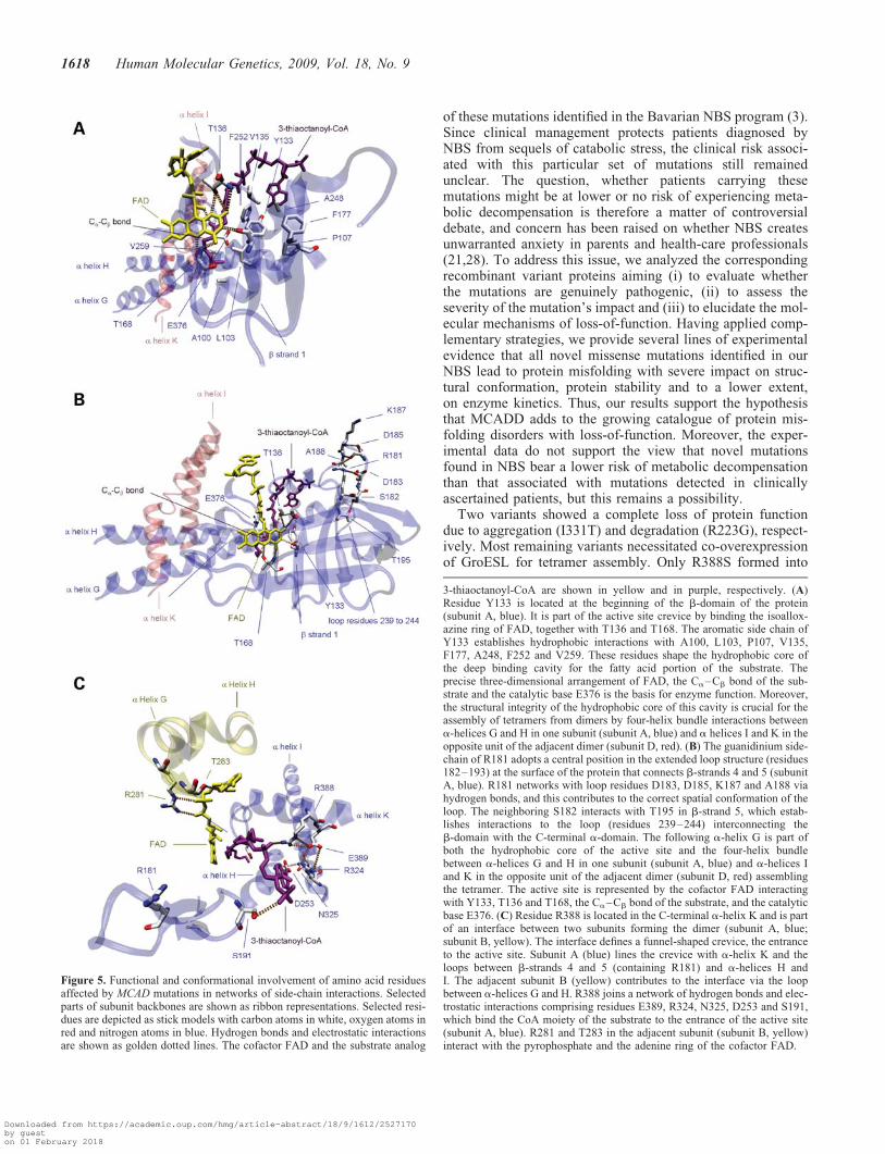
Functional and conformational impact of amino acidreplacements on 3D networks of side-chain interactions
Our experiments revealed severe functional and structuralimpairment, in particular, for the variants Y133H, R181Cand R388S. To gain further insights into how these mutationsexert their deleterious effects on the protein, we used a 3Dstructural model of the porcine MCAD tetramer to investigate,whether the amino acid residues affected are involved in net-works of side-chain interactions with functional and confor-mational impact.
Residue Y133 maps to the b-sheet domain (residues 130–239) and is an essential part of the active site (Fig. 5A). Itdirectly interacts with the cofactor FAD via hydrogen bondformation. Its aromatic side-chain points towards the hydro-phobic core of the deep binding cavity for the fatty acidportion of the substrate establishing hydrophobic interactionswith the residues L103, V135, F177, A248 and F252 which
Figure 3. Variant MCAD proteins show early thermal inactivation andreduced kinetic stability. The effect of thermal stress on enzyme activity ofwild-type (WT) and variant MCAD was analyzed. (A) Thermal inactivationprofiles. Proteins were incubated at increasing temperatures (25–558C) andthe residual enzyme activities were determined. All variants showed a left-shift of the curves in comparison to WT indicating an inactivation of theenzyme at lower temperatures (Table 4, left panel). (B) Kinetic stability at418C. Proteins were incubated at 418C and the residual enzyme activity wasdetermined at incremental time points. For all variants, steeper slopes of thecurves were observed indicating a reduction of half-lives compared withWT (Table 4, right panel). Data points of residual activities in both datasetswere normalized to the initial enzyme activity and subjected to non-linearregression analysis. Error bars represent the mean+SEM of n ¼ 3 indepen-dent experiments.
Table 4. Thermal inactivation and kinetic stability of wild-type and variantMCAD proteins
Missensemutation
Thermal inactivation Kinetic stability at 418CT1/2
(8C)SEM P-value t1/2 (min) SEM P-value
WT 44.5 0.45 20.0 2.36A27V 39.8 0.88 ,0.05 11.8 2.28 nsY42H 39.8 0.52 ,0.05 12.7 1.35 nsY133H 33.6 1.01 ,0.01 6.1 1.14 ,0.01R181C 34.3 1.28 ,0.01 6.8 0.88 ,0.01D241G 33.9 1.08 ,0.01 9.7 0.91 ,0.05K304E 40.6 0.43 ns 12.8 2.53 nsR309K 38.8 1.80 ,0.01 12.9 1.57 nsR388S 42.0 0.37 ns 15.7 0.59 ns
Thermal inactivation and kinetic stability of wild-type (WT) MCAD werecompared with variant MCAD proteins. For thermal inactivation, residualactivities at different temperatures (25–558C) were subjected tonon-linear regression analysis and the midpoints of thermal inactivation(T1/2) were calculated. T1/2-values represent the temperature at 50%residual activity and are given in degree Celsius as means with thecorresponding standard errors of the mean (SEM) of n ¼ 3 independentexperiments. For kinetic stability, the inactivation at 418C was determinedas a function of time. The residual activities were subjected to non-linearregression analysis and the half-lives (t1/2) were calculated. t1/2-valuesrepresent the time point at 50% residual activity and are given in minutesas means with the corresponding standard errors of the mean (SEM) of atleast n ¼ 3 independent experiments. Significances for the differencesbetween wild-type and the variants were calculated by one-way ANOVAand a Dunnett’s post test. (ns, not significant).
1616 Human Molecular Genetics, 2009, Vol. 18, No. 9
Downloaded from https://academic.oup.com/hmg/article-abstract/18/9/1612/2527170by gueston 01 February 2018

line the cavity. To poise the Ca–Cb bond of the fatty acid fordehydrogenation, it is sandwiched between the re-face of theisoalloxazine moiety of FAD and the catalytic base E376. Areplacement of the large, hydrophobic tyrosine by thesmaller, positively charged histidine is supposed to distortthe hydrophobic packing of the binding cavity and by this tolead to a conformational rearrangement of the active sitepocket. As a result, the correct 3D arrangement of FAD, theCa–Cb bond of the substrate and the catalytic base E376will be impaired leading to the marked disturbance inenzyme function. Moreover, A248 and F252 are locatedwithin the a-helix G. Interactions between a-helices G andH in one subunit of the dimer and a-helices I and K in theopposite unit of the adjacent dimer are known to promoteMCAD tetramer assembly from dimers. Hence, conformation-al alteration of the substrate binding cavity with impairment of
the helix–helix interactions due to the Y133H substitutionmight explain the observed distortion of the oligomeric state.
R181 is located at the beginning of an extended loop structure(residues 181–193) at the surface of the protein that connectsb-sheets 4 and 5 (Fig. 5B). The loop contributes to the creviceshaping the entrance to the active site and, due to its strong inter-actions with the beginning of the C-terminal a-domain, plays apivotal role for MCAD tetramerization. R181 networks withD183, D185, K187 and A188 to poise the loop in its proper struc-tural conformation. It is well conceivable that the severe struc-tural distortion of the protein with reduced stability andimpaired tetramer assembly shown for R181C is due to a disrup-tion of this network caused by the replacement of arginine by asmall, polar, uncharged amino acid.
R388 maps to the end of the C-terminal a-domain (residues240–396) and is part of an interface of two subunits thatdefine a funnel-shaped crevice, the entrance to the active sitementioned above (Fig. 5C). It is shaped by a-helix K andthe loops among b-sheets 4 and 5, a-helices H and I anda-helices G and H of the adjacent subunit. R388 forms ahydrogen bond to the AMP portion of the CoA moiety andis part of a side-chain network comprising both hydrogenbonds and electrostatic interactions with the neighboring resi-dues E389 and R324 in the loop between a-helices H andI. A substitution of the large, basic, positively charged arginineby serine might disrupt this network by introducing a gap inthe 3D structure of the crevice. This conformational rearrange-ment at the entrance to the active site will hinder the accessi-bility of the substrate octanoyl-CoA and the interaction withthe AMP portion of the CoA moiety. This is in line with ourobservation that R388S displays a significant decrease in sub-strate affinity for octanoyl-CoA.
DISCUSSION
NBS for MCADD has revealed an increasing number of mis-sense mutations that have never been identified in clinicallydiagnosed patients and we have previously described eight
Figure 4. Variant MCAD proteins show accelerated thermal denaturation andpartial protein unfolding in the ground state for some variants. Thermalunfolding of wild-type (WT) and variants monitored by ANS fluorescence.(A) ANS fluorescence profiles of wild-type, Y133H, R181S and K304E. Inten-sities of the fluorescent dye ANS, which binds to hydrophobic groups of theprotein presented upon unfolding, are plotted as a function of increasing temp-eratures. Ground-state fluorescence was markedly increased for Y133H and, toa lesser extent, for R181C and K304E indicating an increased hydrophobicitydue to partial unfolding of these variants already in the native state. (B)Thermal denaturation of all variants analyzed. Fractions of unfolded proteinare plotted as a function of increasing temperatures and the transition mid-points represent the temperature at half denaturation (fraction unfolded 0.5).All variants showed a marked to moderate left-shift of the curves implyingan increased propensity to unfold upon thermal stress (Table 5). In addition,Y133H showed accelerated unfolding as indicated by the steeper slope ofthe curve and complete denaturation at 468C.
Table 5. Transition midpoints of thermal denaturation of wild-type and variantMCAD proteins
Missense mutation Tm1/2 (8C) SEM P-value
WT 52.6 0.40A27V 48.8 0.69 ,0.01Y42H 50.1 0.19 ,0.01Y133H 42.2 0.11 ,0.01R181C 40.0 0.16 ,0.01D241G 46.4 0.65 ,0.01K304E 48.2 0.16 ,0.01R309K 50.0 0.14 ,0.01R388S 47.0 0.85 ,0.01
Transition midpoints of thermal denaturation obtained by ANSfluorescence of wild-type (WT) MCAD were compared with variantproteins. Three sets of independent experiments were performed. Thetransition midpoints (Tm1/2) were calculated by non-linear regressionanalysis and are given in degree Celsius as means with their correspondingstandard errors of the mean (SEM). Significances for the differencesbetween wild-type and variants were calculated using one-way ANOVAfollowed by a Dunnett’s post test.
Human Molecular Genetics, 2009, Vol. 18, No. 9 1617
Downloaded from https://academic.oup.com/hmg/article-abstract/18/9/1612/2527170by gueston 01 February 2018
of these mutations identified in the Bavarian NBS program (3).Since clinical management protects patients diagnosed byNBS from sequels of catabolic stress, the clinical risk associ-ated with this particular set of mutations still remainedunclear. The question, whether patients carrying thesemutations might be at lower or no risk of experiencing meta-bolic decompensation is therefore a matter of controversialdebate, and concern has been raised on whether NBS createsunwarranted anxiety in parents and health-care professionals(21,28). To address this issue, we analyzed the correspondingrecombinant variant proteins aiming (i) to evaluate whetherthe mutations are genuinely pathogenic, (ii) to assess theseverity of the mutation’s impact and (iii) to elucidate the mol-ecular mechanisms of loss-of-function. Having applied comp-lementary strategies, we provide several lines of experimentalevidence that all novel missense mutations identified in ourNBS lead to protein misfolding with severe impact on struc-tural conformation, protein stability and to a lower extent,on enzyme kinetics. Thus, our results support the hypothesisthat MCADD adds to the growing catalogue of protein mis-folding disorders with loss-of-function. Moreover, the exper-imental data do not support the view that novel mutationsfound in NBS bear a lower risk of metabolic decompensationthan that associated with mutations detected in clinicallyascertained patients, but this remains a possibility.
Two variants showed a complete loss of protein functiondue to aggregation (I331T) and degradation (R223G), respect-ively. Most remaining variants necessitated co-overexpressionof GroESL for tetramer assembly. Only R388S formed into
Figure 5. Functional and conformational involvement of amino acid residuesaffected by MCAD mutations in networks of side-chain interactions. Selectedparts of subunit backbones are shown as ribbon representations. Selected resi-dues are depicted as stick models with carbon atoms in white, oxygen atoms inred and nitrogen atoms in blue. Hydrogen bonds and electrostatic interactionsare shown as golden dotted lines. The cofactor FAD and the substrate analog
3-thiaoctanoyl-CoA are shown in yellow and in purple, respectively. (A)Residue Y133 is located at the beginning of the b-domain of the protein(subunit A, blue). It is part of the active site crevice by binding the isoallox-azine ring of FAD, together with T136 and T168. The aromatic side chain ofY133 establishes hydrophobic interactions with A100, L103, P107, V135,F177, A248, F252 and V259. These residues shape the hydrophobic core ofthe deep binding cavity for the fatty acid portion of the substrate. Theprecise three-dimensional arrangement of FAD, the Ca–Cb bond of the sub-strate and the catalytic base E376 is the basis for enzyme function. Moreover,the structural integrity of the hydrophobic core of this cavity is crucial for theassembly of tetramers from dimers by four-helix bundle interactions betweena-helices G and H in one subunit (subunit A, blue) and a helices I and K in theopposite unit of the adjacent dimer (subunit D, red). (B) The guanidinium side-chain of R181 adopts a central position in the extended loop structure (residues182–193) at the surface of the protein that connects b-strands 4 and 5 (subunitA, blue). R181 networks with loop residues D183, D185, K187 and A188 viahydrogen bonds, and this contributes to the correct spatial conformation of theloop. The neighboring S182 interacts with T195 in b-strand 5, which estab-lishes interactions to the loop (residues 239–244) interconnecting theb-domain with the C-terminal a-domain. The following a-helix G is part ofboth the hydrophobic core of the active site and the four-helix bundlebetween a-helices G and H in one subunit (subunit A, blue) and a-helices Iand K in the opposite unit of the adjacent dimer (subunit D, red) assemblingthe tetramer. The active site is represented by the cofactor FAD interactingwith Y133, T136 and T168, the Ca–Cb bond of the substrate, and the catalyticbase E376. (C) Residue R388 is located in the C-terminal a-helix K and is partof an interface between two subunits forming the dimer (subunit A, blue;subunit B, yellow). The interface defines a funnel-shaped crevice, the entranceto the active site. Subunit A (blue) lines the crevice with a-helix K and theloops between b-strands 4 and 5 (containing R181) and a-helices H andI. The adjacent subunit B (yellow) contributes to the interface via the loopbetween a-helices G and H. R388 joins a network of hydrogen bonds and elec-trostatic interactions comprising residues E389, R324, N325, D253 and S191,which bind the CoA moiety of the substrate to the entrance of the active site(subunit A, blue). R281 and T283 in the adjacent subunit (subunit B, yellow)interact with the pyrophosphate and the adenine ring of the cofactor FAD.
1618 Human Molecular Genetics, 2009, Vol. 18, No. 9
Downloaded from https://academic.oup.com/hmg/article-abstract/18/9/1612/2527170by gueston 01 February 2018

apparently normal tetramers without co-overexpression ofchaperonins. The findings of increased propensity of variantMCAD proteins to form aggregates in Escherichia coli andpartial rescue by increasing the amount of available chapero-nins was regarded as a first evidence for misfolding. Althoughco-overexpression of GroESL can assist to overcome proteinmisfolding in terms of aggregation and by this promote theformation of correctly assembled oligomers (29), it does notcorrect for the mutation induced structural defects of thevariant protein. These defects were then analyzed in detail inthis study.
All purified tetrameric MCAD variants revealed alterationsof structural conformation, yet of various degrees. Interest-ingly, all variants showed thermal instability. Thermal unfold-ing was accelerated with a shift of the transition midpoint(wild-type, 52.68C) by .48C in most variants and down to408C in variant R181C. Thermal inactivation experimentsrevealed a 50% loss-of-function at temperatures ,408C (wild-type 44.58C) in all variants except for K304E and R388S. Thisobservation is of particular interest, since in a considerableshare of patients with MCADD metabolic decompensationoccurs during intercurrent illness with fever. These findingsare in line with the previous data on thermal stability ofother variant MCAD proteins (14–16,21,22). Moreover, weobserved that thermal instability was most pronounced in var-iants mapping to the b-sheet domain of the protein (Y133H,R181C) or to the loop interconnecting this domain and the C-terminal a-helix domain (D241G).
Additional evidence for protein instability arose from ourdata on limited proteolysis. Most variants showed an increasedsusceptibility against proteolytic attack indicating disturbedfolding kinetics (i.e. shift of the folding equilibrium towardsthe unfolded state) and local unfolding. Mild alterationswere observed in variants derived from mutations located inthe tightly packed C-terminal a-domain. Again, the variantsmapping to the b-sheet domain and its subsequent loop dis-closed the most pronounced alterations consistent withsevere conformational changes.
In two variants located in the b-sheet domain (Y133H andR181C), severe alterations of protein conformation wereevident even without thermal or proteolytic stress. Elevatedground state ANS fluorescence revealed an increase in hydro-phobicity and thus a shift to the non-native state of these pro-teins. In Y133H, the induced conformational changes led to asevere destabilization and almost complete loss-of-functionwith a residual catalytic activity of 3.5%. The affectedamino acid residue Y133 is part of the active site in tworespects. Together with T136 and T168, it anchors theco-factor FAD to the protein via hydrogen bonds. In addition,the aromatic side chain of Y133 contributes to the hydro-phobic core lining the binding cavity for the fatty acid. Pre-vious studies characterizing the molecular phenotype ofT168A (c.577A.G), a mutation that has been identified in aclinically diagnosed patient, are in line with our observationsrevealing misfolding, markedly decreased thermal stabilityand catalytic activity (15,16,18). In contrast, R181C showeda residual catalytic activity comparable to wild-type despiteits severe conformational alterations. Our data are consistentwith the notion that the major molecular basis for the patho-genesis of this variant in vivo may be protein instability
under physiological stress pointing to a high risk of metabolicdecompensation during times of illness, especially associatedwith fever. As previously reported, a patient homozygous forthis mutation showed remarkably mild biochemical alterationsin NBS (3), although a mutation affecting the same amino acidresidue (R181L; c.617G.T) was identified in two sympto-matic patients (30). Our experimental data emphasize thatmild biochemical alterations in NBS do not allow implyinga low risk for decompensation.
Mutations located in the N-terminal a-domain showed mod-erate effects on tetramer assembly and thermal stabilitytogether with decreased (A27V) or normal catalytic activity(Y42H). Consistent with our findings, mutations located inthis region have previously been described as mild folding var-iants with normal activity (M124I; c.447G.A), mediumrange activity (R28C; c.157C.T) or moderate thermalinstability (Y42H) (10,21). However, a-helices A, C and Dof this domain form the recognition site of the electron transferflavoprotein, which is the natural electron acceptor of theMCAD protein and responsible for the electron transfer tothe respiratory chain (31). Whether structural changes in thisdomain result in an alteration of electron transfer ratesremains to be elucidated.
In contrast, mutations located in the C-terminal a-domain(K304E, R309K, I331T) often affect helix–helix interactionscrucial for tetramer assembly leading to aggregation(10,12,13,22,23). However, when aggregation was overcomeby co-overexpression of chaperonins, these variants showedconsiderable residual activities. Interestingly, R388S, alsolocated in the C-terminal a-domain, was the only variantthat did not require co-overexpression of GroESL for tetramerformation and its proteolytic and thermal stabilities were onlymoderately affected. In this variant, misfolding resulted in amarked decrease of substrate affinity. Similar effects on Km
have been shown for artificial variants of the homologous argi-nine residue R387 of the ACAD isovaleryl-CoA dehydrogen-ase (32) and have been predicted from crystal structureanalysis for the ACAD glutaryl-CoA dehydrogenase (33).Our data substantiate the essential role of this highly con-served arginine residue for binding of the substrate in ACADs.
The observations obtained from analyzing variants harbor-ing amino acid substitutions in different regions of theprotein show that mutations in the b-sheet domain and theadjacent loop (interconnecting the b-domain and theC-terminal a-domain) are particularly instable and prone tosevere conformational distortion. This is in line with the pre-vious work describing severely impaired biogenesis and stab-ility for two other variants located in the b-domain, T168Aand G170R (c.583G.A) (10,15). Misfolding observed inthese variants has been attributed to the loss of FAD anchor-ing, which has been reported to be crucial for folding and oli-gomerization (34). However, the compiled data including ourvariants spread over the b-domain indicate that not onlyreduced FAD binding, but also the structural and functionalcharacteristics of the b-domain itself account for the proteindestabilization observed.
In conclusion, all mutations analyzed in this study showedsignificant alterations of the molecular phenotype rangingfrom mild to severe. Six of the mutations (Y133H, R181C,R223G, D241G, I331T and R388S) identified in presympto-
Human Molecular Genetics, 2009, Vol. 18, No. 9 1619
Downloaded from https://academic.oup.com/hmg/article-abstract/18/9/1612/2527170by gueston 01 February 2018

matic newborns revealed even more pronounced perturbancesthan the variant K304E, which is generally accepted to beassociated with a severe molecular and clinical phenotype. Ithas to be noted, however, that patients homozygous forK304E may experience a healthy life (2) and our findings ofa severe molecular phenotype do not unequivocally prove asevere clinical phenotype. Standard diagnostic proceduressuch as the determination of blood acylcarnitine concen-trations do not allow for a meaningful prediction of the clinicalcourse. Hence, compiled knowledge gained by documentationof the clinical and biochemical course of specific genotypes incombination with detailed molecular characterization of therespective variants at the protein level will improve geno-type–phenotype correlation. This may assist counseling andrisk assessment of MCADD patients identified in NBS pro-grams. In this context, it has also to be taken into accountthat most patients revealing mutations other than the predomi-nant K304E are compound heterozygous. Moreover, gene–gene interactions and gene–environment interactions such ascellular and environmental conditions or stressors may alsomodify the natural course of the disease (35).
A molecular scenario similar to that now identified forMCADD was recently described for the phenylalaninehydroxylase protein, the enzyme deficient in phenylketonuria(36). In this disease, experimental evidence is now availablethat the well proven efficacy of tetrahydrobiopterin (37,38)is based on a pharmacological chaperone effect. For MCAD,we and others showed that increasing the amount of availablemolecular chaperones (GroESL) prevents MCAD aggregationand leads to higher amounts of functional protein. The persist-ing loss-of-function is then predominantly promoted byinstability against thermal or proteolytic stress. Early degra-dation could thus be overcome using pharmacological chaper-ones, conceivably in combination with proteostasis regulatorsas recently shown for lysosomal storage diseases (39).
In summary, the detailed insight into how ACADM missensemutations induce conformational alteration and protein desta-bilization resulting in loss of enzyme function presented heremay in future provide the basis to delineate novel pharmaco-logical strategies for the potentially fatal MCADD disease.
MATERIALS AND METHODS
Subjects and mutations
In the previous study, we described the spectrum of sequencevariations occurring in newborns with MCADD detected byNBS (3). We identified eight novel missense mutationswithin the ACADM gene that have not yet been characterizedin vitro: A27V (c.155C.T), Y133H (c.472T.C), R181C(c.616C.T), R223G (c.742A.G), D241G (c.797A.G),R309K (c.1001G.A), I331T (c.1067T.C) and R388S(c.1237C.A) (Table 1). Two mutations (R181C andR223G) were found in a homozygous state. The two individ-uals homozygous for R223G displayed markedly elevated bio-chemical markers, whereas the individual homozygous forR181C showed only mild alterations. The remaining mutationswere found in a compound heterozygous state with the twomost prevalent mutations Y42H (c.199T.C) and K304E(c.985A.G). Mutations and biochemical phenotypes of the
patients are summarized in Supplementary Material,Table S1. The structural localization of mutations character-ized in this study was mapped to the 3D model of theporcine MCAD monomer (Fig. 1).
Plasmid construction and site-directed mutagenesis
The cDNA of human ACADM gene (ACADM-encodingpKK223 plasmid obtained as a generous gift from JerryVockley, Pittsburgh, USA) was cloned into the pMAL-c2Xexpression vector (New England Biolabs) encoding a N-terminal MBP-tag and a factor Xa cleavage site. ACADMmutations were introduced using the PCR-based QuikChangeSite-Directed Mutagenesis Kit (Stratagene). After mutagen-esis, all expression vectors were verified by DNA sequencing.
Expression and purification
The expression plasmids were used to transform E.coli strainBL21-CodonPlus (Stratagene). Bacteria were grown in 2 l ofdYT medium (16 g/l tryptone, 10 g/l yeast extract and 5 g/lNaCl) containing 100 mg/ml Ampicillin to OD600 0.6–0.8 at378C. Overexpression of wild-type and variant MBP–MCAD fusion proteins was induced with 0.01 mM
isopropylthio-b-D-galactoside and performed at a reducedgrowth temperature of 288C for 20 h. Bacteria were harvestedby centrifugation and lysed by sonication. Protein purificationwas performed using AKTApurifier and AKTAprime systems(GE Healthcare) at 48C. The crude extract/protein sample wasloaded on a MBPTrap affinity chromatography column (GEHealthcare) equilibrated with column buffer (20 mM Tris–HCl pH 7.4, 200 mM NaCl, 1 mM EDTA, 1 mM DTT) andeluted with the same buffer containing 10 mM maltose afterwashing out unbound bacterial protein. The eluted proteinfractions were pooled and subjected to SEC using a HiLoad16/60 Superdex 200 column (GE Healthcare) equilibratedwith 20 mM HEPES pH 7.0 containing 200 mM NaCl. Thefractions containing tetrameric fusion proteins were pooledand incubated for 12 h at 48C with factor Xa (Novagen) tocleave the MBP-tag of the fusion proteins at a protease tofusion protein ratio (U:mg) of 1:20. Final purification of thecleaved tetrameric MCAD protein was obtained by SEC ona HiLoad 16/60 Superdex 200 column (GE Healthcare) equi-librated with 20 mM HEPES pH 7.0 containing 200 mM
NaCl and reached a purity of .98% SDS/PAGE analysis.Protein concentrations were determined spectrophotometri-
cally using the absorption coefficients at A272 and A448 (14), orthe fluorescent dye binding Quant-iT assay (Invitrogen). Usingthis approach, up to 14 mg of cleaved MCAD tetramericprotein of high purity (.97%) were obtained from 2 lculture. A summary of the purification protocol is given inSupplementary Material, Table S2.
Co-overexpression of chaperonins GroESL
The respective pMAL-c2X MCAD expression plasmids wereco-transformed with pGroESL encoding the proteins GroESand GroEL (40) in E.coli strain DH5a (Invitrogen). Growthof bacteria, expression and purification of proteins wereperformed as described above except for the addition of two
1620 Human Molecular Genetics, 2009, Vol. 18, No. 9
Downloaded from https://academic.oup.com/hmg/article-abstract/18/9/1612/2527170by gueston 01 February 2018
antibiotics, ampicillin (100 mg/ml) and chloramphenicol(50 mg/ml), to the broth.
Analysis of oligomerization
MCAD oligomerization was analyzed by SEC of the MBP–MCAD fusion proteins prior to cleavage with factor Xa. TheMBP-tag increases solubility and yield during expression,but does not significantly affect the oligomeric state of theexpressed proteins [own and previous observation (41)]. Thesize-exclusion column was calibrated using low molecularweight and high molecular weight gel filtration calibrationkits (GE Healthcare). Blue dextran was used to determinethe void volume (V0, 46.0 ml).
Analyses of enzyme kinetic parameters
MCAD activity was determined using the ferricenium-basedspectrophotometric assay described previously (42). Measure-ments were performed in 100 mM HEPES, pH 7.6, containing0.1 mM EDTA at a temperature of 258C with increasing con-centrations of octanoyl-CoA (Sigma Aldrich, Larodan Chemi-cals) ranging from 0 to 20 mM (150 mM for the Km variant). Inorder to correct for varying qualities of octanoyl-CoA withrespect to the degree of purity, amount of crystal water andcationic ligands provided by the manufacturers, concentrationsof oxidizable substrate were determined by complete turn-overusing an excess of enzyme and electron acceptor. All measure-ments were corrected for background reduction of ferricenium.
The assays were performed in at least three independentexperiments. The kinetic parameters Km and Vmax were aver-aged after non-linear regression analysis using the Michae-lis–Menten algorithm implemented in pro Fit (QuantumSoft).
Limited proteolysis by proteinase K
Five micrograms of the purified tetrameric MCAD proteinswere incubated with proteinase K (Sigma) using two differentprotease to substrate ratios, 1:1 and 1:25 by weight. The exper-iments were performed at 378C in 20 mM HEPES buffer pH7.0 containing 200 mM NaCl. Proteolysis was terminated attime points 2, 4, 6, 8, 10, 20, 30, 60 and 120 min by theaddition of the inhibitor phenylmethylsulphonyl fluoride at afinal concentration of 0.4 mM. The protein samples were sub-jected to SDS–PAGE under reducing conditions using 4–12%gradient polyacrylamide gels (Invitrogen). Proteolysis wasmonitored by Coomassie staining and subsequent densitome-try analysis. Densitometry data were quantified by AIDA-software (Raytest), normalized to the intact MCAD proteinprior to proteolysis and analyzed by non-linear regression.The experiments for each protease to substrate ratio were per-formed in triplicates. Significances for the differences betweenwild-type and variant proteins concerning plateau of proteol-ysis (protease to substrate ratio 1:1) and half-lives (proteaseto substrate ratio 1:25) were calculated using one-wayANOVA followed by a Dunnett’s post test. Using the substrateratio of 1:25 resulted in a slower decline of protein and, thus,was more suitable to determine half-lives.
Thermal inactivation experiments
To determine the effect of various temperatures on MCADactivity, aliquots of protein (5 mg/ml) were incubated in100 mM HEPES, pH 7.6 containing 0.1 mM EDTA for15 min at 11 different temperatures ranging from 25 to558C, and then chilled on ice. Enzyme activity was sub-sequently measured at an octanoyl-CoA concentration of10 mM as described above. Residual activities were normalizedto initial enzyme activity at 258C.
For the assessment of thermal effects over time, one aliquotof protein (5 mg/ml) was incubated at 418C with enzymeactivities being determined at nine incremental time points(5–120 min). Residual activities were normalized to enzymeactivity prior to incubation.
Data points were subjected to non-linear regressionanalysis and midpoints of thermal inactivation (T1/2) andhalf-lives (t1/2) were calculated; T1/2 indicates the temperatureand t1/2 marks the time at 50% residual activity, respectively.
Measurements were performed in three independent exper-iments. Significances for the differences in T1/2 and t1/2
between wild-type and variant proteins were calculatedusing one-way ANOVA followed by a Dunnett’s post test.
Thermal denaturation monitored by ANS fluorescence
Fluorescence measurements were performed on a Cary Eclipsefluorescence spectrophotometer equipped with a temperature-controlled Peltier multicell holder (Varian). Samples con-tained MCAD proteins (11.6 mM MCAD subunit) in 20 mM
HEPES buffer at pH 7.0 containing 200 mM NaCl. The dena-turation was monitored following the changes in ANS (pur-chased from Sigma) fluorescence emission: excitation at395, emission at 450 nm (5.0/10.0 nm slit widths). The dena-turation was performed at a rate of 1.28C/min from 20 to358C and from 70 to 858C and at a rate of 0.38C/min from35 to 708C in three independent experiments. The transitionsmidpoints (Tm1/2), indicating the temperature at 50% denatura-tion, were calculated by the differentiation of the increasingpart of the curves. Significances for the differences betweenwild-type and variants were calculated by one-way ANOVAand a Dunnett’s post test.
Statistical analyses
Non-linear regression analyses and statistical tests were per-formed using GraphPad Prism 4.0c (GraphPad Software).
Structural analyses and figure preparation
The 3D structure of tetrameric pig MCAD complexed with thesubstrate analog 3-thiaoctanoyl-CoA (PDB code 1UDY) wasanalyzed using the DeepView-Swiss-PdbViewer (43). Theporcine MCAD protein shows a .90% homology withhuman MCAD, and only homologous amino acid residueshave been analyzed. In the presence of hydrogen atoms,H-bonds were computed with the following constraints: 1.2–2.76A distance, 1208 angles. When hydrogen atoms wereabsent, interactions were computed with the following
Human Molecular Genetics, 2009, Vol. 18, No. 9 1621
Downloaded from https://academic.oup.com/hmg/article-abstract/18/9/1612/2527170by gueston 01 February 2018

constraints: 2.35–3.2A distance, 908 angles. Figures were pre-pared using VMD (44).
SUPPLEMENTARY MATERIAL
Supplementary Material is available at HMG online.
ACKNOWLEDGEMENTS
We are indebted to our patients and to their families; to HeikePreisler, Sylvia Taube and Sabine Streicher for excellent tech-nical assistance; to Uta Nennstiel-Ratzel and Ralph Fingerhutfor providing data on the biochemical phenotypes; to MathiasWoidy for helpful statistical discussions; to Jerry Vockley forproviding the ACADM-encoding plasmid; to Dietrich Rein-hardt for continuous support. This article is part of a thesisby J.M.J. and M.R. to fulfill the requirements for a medicaldegree at the Ludwig-Maximilians-University of Munich.This work was supported by the Bavarian Genome ResearchNetwork (BayGene); and the SHS International Gesellschaftfur klinische Ernahrung mbH.
Conflict of Interest statement. None declared.
FUNDING
Funding to pay the Open Access charge was provided byBavarian Genome Research Network.
REFERENCES
1. Rhead, W.J. (2006) Newborn screening for medium-chain acyl-CoAdehydrogenase deficiency: a global perspective. J. Inherit. Metab. Dis.,29, 370–377.
2. Grosse, S.D., Khoury, M.J., Greene, C.L., Crider, K.S. and Pollitt, R.J.(2006) The epidemiology of medium chain acyl-CoA dehydrogenasedeficiency: an update. Genet. Med., 8, 205–212.
3. Maier, E.M., Liebl, B., Roschinger, W., Nennstiel-Ratzel, U., Fingerhut,R., Olgemoller, B., Busch, U., Krone, N., von Kries, R. and Roscher, A.A.(2005) Population spectrum of ACADM genotypes correlated tobiochemical phenotypes in newborn screening for medium-chainacyl-CoA dehydrogenase deficiency. Hum. Mutat., 25, 443–452.
4. Wilcken, B., Haas, M., Joy, P., Wiley, V., Chaplin, M., Black, C.,Fletcher, J., McGill, J. and Boneh, A. (2007) Outcome of neonatalscreening for medium-chain acyl-CoA dehydrogenase deficiency inAustralia: a cohort study. Lancet, 369, 37–42.
5. Derks, T.G., Reijngoud, D.J., Waterham, H.R., Gerver, W.J., van denBerg, M.P., Sauer, P.J. and Smit, G.P. (2006) The natural history ofmedium-chain acyl CoA dehydrogenase deficiency in the Netherlands:clinical presentation and outcome. J. Pediatr., 148, 665–670.
6. Iafolla, A.K., Thompson, R.J. Jr and Roe, C.R. (1994) Medium-chainacyl-coenzyme A dehydrogenase deficiency: clinical course in 120affected children. J. Pediatr., 124, 409–415.
7. Wilcken, B., Hammond, J. and Silink, M. (1994) Morbidity and mortalityin medium chain acyl coenzyme A dehydrogenase deficiency. Arch. Dis.Child., 70, 410–412.
8. Yokota, I., Indo, Y., Coates, P.M. and Tanaka, K. (1990) Molecular basisof medium chain acyl-coenzyme A dehydrogenase deficiency. An A to Gtransition at position 985 that causes a lysine-304 to glutamate substitutionin the mature protein is the single prevalent mutation. J. Clin. Invest., 86,1000–1003.
9. Gregersen, N., Andresen, B.S. and Bross, P. (2000) Prevalent mutations infatty acid oxidation disorders: diagnostic considerations. Eur. J. Pediatr.,159 (Suppl. 3), S213–S218.
10. Andresen, B.S., Bross, P., Udvari, S., Kirk, J., Gray, G., Kmoch, S.,Chamoles, N., Knudsen, I., Winter, V., Wilcken, B. et al. (1997) Themolecular basis of medium-chain acyl-CoA dehydrogenase (MCAD)deficiency in compound heterozygous patients: is there correlationbetween genotype and phenotype? Hum. Mol. Genet., 6, 695–707.
11. Bross, P., Andresen, B.S., Winter, V., Krautle, F., Jensen, T.G., Nandy,A., Kolvraa, S., Ghisla, S., Bolund, L. and Gregersen, N. (1993)Co-overexpression of bacterial GroESL chaperonins partly overcomesnon-productive folding and tetramer assembly of E.coli-expressed humanmedium-chain acyl-CoA dehydrogenase (MCAD) carrying the prevalentdisease-causing K304E mutation. Biochim. Biophys. Acta, 1182, 264–274.
12. Bross, P., Jespersen, C., Jensen, T.G., Andresen, B.S., Kristensen, M.J.,Winter, V., Nandy, A., Krautle, F., Ghisla, S., Bolundi, L. et al. (1995)Effects of two mutations detected in medium chain acyl-CoAdehydrogenase (MCAD)-deficient patients on folding, oligomer assembly,and stability of MCAD enzyme. J. Biol. Chem., 270, 10284–10290.
13. Jensen, T.G., Bross, P., Andresen, B.S., Lund, T.B., Kristensen, T.J.,Jensen, U.B., Winther, V., Kolvraa, S., Gregersen, N. and Bolund, L.(1995) Comparison between medium-chain acyl-CoA dehydrogenasemutant proteins overexpressed in bacterial and mammalian cells. Hum.
Mutat., 6, 226–231.14. Kieweg, V., Krautle, F.G., Nandy, A., Engst, S., Vock, P., Abdel-Ghany,
A.G., Bross, P., Gregersen, N., Rasched, I., Strauss, A. et al. (1997)Biochemical characterization of purified, human recombinantLys304– .Glu medium-chain acyl-CoA dehydrogenase containing thecommon disease-causing mutation and comparison with the normalenzyme. Eur. J. Biochem., 246, 548–556.
15. Kuchler, B., Abdel-Ghany, A.G., Bross, P., Nandy, A., Rasched, I. andGhisla, S. (1999) Biochemical characterization of a variant humanmedium-chain acyl-CoA dehydrogenase with a disease-associatedmutation localized in the active site. Biochem. J., 337, 225–230.
16. Nasser, I., Mohsen, A.W., Jelesarov, I., Vockley, J., Macheroux, P. andGhisla, S. (2004) Thermal unfolding of medium-chain acyl-CoAdehydrogenase and iso(3)valeryl-CoA dehydrogenase: study of the effectof genetic defects on enzyme stability. Biochim. Biophys. Acta, 1690,22–32.
17. Yokota, I., Saijo, T., Vockley, J. and Tanaka, K. (1992) Impaired tetramerassembly of variant medium-chain acyl-coenzyme A dehydrogenase witha glutamate or aspartate substitution for lysine 304 causing instability ofthe protein. J. Biol. Chem., 267, 26004–26010.
18. Andresen, B.S., Dobrowolski, S.F., O’Reilly, L., Muenzer, J.,McCandless, S.E., Frazier, D.M., Udvari, S., Bross, P., Knudsen, I.,Banas, R. et al. (2001) Medium-chain acyl-CoA dehydrogenase (MCAD)mutations identified by MS/MS-based prospective screening of newbornsdiffer from those observed in patients with clinical symptoms:identification and characterization of a new, prevalent mutation thatresults in mild MCAD deficiency. Am. J. Hum. Genet., 68, 1408–1418.
19. Nichols, M.J., Saavedra-Matiz, C.A., Pass, K.A. and Caggana, M. (2008)Novel mutations causing medium chain acyl-CoA dehydrogenasedeficiency: under-representation of the common c.985 A.G mutation inthe New York state population. Am. J. Med. Genet. A, 146A, 610–619.
20. Waddell, L., Wiley, V., Carpenter, K., Bennetts, B., Angel, L., Andresen,B.S. and Wilcken, B. (2006) Medium-chain acyl-CoA dehydrogenasedeficiency: genotype-biochemical phenotype correlations. Mol. Genet.
Metab., 87, 32–39.21. O’Reilly, L., Bross, P., Corydon, T.J., Olpin, S.E., Hansen, J., Kenney,
J.M., McCandless, S.E., Frazier, D.M., Winter, V., Gregersen, N. et al.(2004) The Y42H mutation in medium-chain acyl-CoA dehydrogenase,which is prevalent in babies identified by MS/MS-based newbornscreening, is temperature sensitive. Eur. J. Biochem., 271, 4053–4063.
22. O’Reilly, L.P., Andresen, B.S. and Engel, P.C. (2005) Two novel variantsof human medium chain acyl-CoA dehydrogenase (MCAD). K364R, afolding mutation, and R256T, a catalytic-site mutation resulting in awell-folded but totally inactive protein. FEBS J., 272, 4549–4557.
23. Kim, J.J. and Miura, R. (2004) Acyl-CoA dehydrogenases and acyl-CoAoxidases. Structural basis for mechanistic similarities and differences.Eur. J. Biochem., 271, 483–493.
24. Nandy, A., Kieweg, V., Krautle, F.G., Vock, P., Kuchler, B., Bross, P.,Kim, J.J., Rasched, I. and Ghisla, S. (1996) Medium-long-chain chimerichuman Acyl-CoA dehydrogenase: medium-chain enzyme with the activecenter base arrangement of long-chain Acyl-CoA dehydrogenase.Biochemistry, 35, 12402–12411.
1622 Human Molecular Genetics, 2009, Vol. 18, No. 9
Downloaded from https://academic.oup.com/hmg/article-abstract/18/9/1612/2527170by gueston 01 February 2018

25. Fontana, A., de Laureto, P.P., Spolaore, B., Frare, E., Picotti, P. andZambonin, M. (2004) Probing protein structure by limited proteolysis.Acta Biochim. Pol., 51, 299–321.
26. Andresen, B.S., Bross, P., Jensen, T.G., Knudsen, I., Winter, V., Kolvraa,S., Bolund, L. and Gregersen, N. (1995) Molecular diagnosis andcharacterization of medium-chain acyl-CoA dehydrogenase deficiency.Scand. J. Clin. Lab. Invest. Suppl., 220, 9–25.
27. Royer, C.A. (1995) Fluorescence spectroscopy. Methods Mol. Biol., 40,65–89.
28. Khoury, M.J., McCabe, L.L. and McCabe, E.R. (2003) Populationscreening in the age of genomic medicine. N. Engl. J. Med., 348, 50–58.
29. Gregersen, N., Bross, P., Andresen, B.S., Pedersen, C.B., Corydon, T.J.and Bolund, L. (2001) The role of chaperone-assisted folding and qualitycontrol in inborn errors of metabolism: protein folding disorders.J. Inherit. Metab. Dis., 24, 189–212.
30. Yang, B.Z., Ding, J.H., Zhou, C., Dimachkie, M.M., Sweetman, L.,Dasouki, M.J., Wilkinson, J. and Roe, C.R. (2000) Identification of anovel mutation in patients with medium-chain acyl-CoA dehydrogenasedeficiency. Mol. Genet. Metab., 69, 259–262.
31. Toogood, H.S., Leys, D. and Scrutton, N.S. (2007) Dynamics drivingfunction: new insights from electron transferring flavoproteins and partnercomplexes. FEBS J., 274, 5481–5504.
32. Volchenboum, S.L., Mohsen, A.W., Kim, J.J. and Vockley, J. (2001)Arginine 387 of human isovaleryl-CoA dehydrogenase plays a crucial rolein substrate/product binding. Mol. Genet. Metab., 74, 226–237.
33. Fu, Z., Wang, M., Paschke, R., Rao, K.S., Frerman, F.E. and Kim, J.J.(2004) Crystal structures of human glutaryl-CoA dehydrogenase with andwithout an alternate substrate: structural bases of dehydrogenation anddecarboxylation reactions. Biochemistry, 43, 9674–9684.
34. Saijo, T. and Tanaka, K. (1995) Isoalloxazine ring of FAD is required forthe formation of the core in the Hsp60-assisted folding of medium chainacyl-CoA dehydrogenase subunit into the assembly competentconformation in mitochondria. J. Biol. Chem., 270, 1899–1907.
35. Gregersen, N., Andresen, B.S., Pedersen, C.B., Olsen, R.K., Corydon, T.J.and Bross, P. (2008) Mitochondrial fatty acid oxidation defects–remaining challenges. J. Inherit. Metab. Dis., 31, 643–657.
36. Gersting, S.W., Kemter, K.F., Staudigl, M., Messing, D.D., Danecka,M.K., Lagler, F.B., Sommerhoff, C.P., Roscher, A.A. and Muntau, A.C.(2008) Loss of function in phenylketonuria is caused by impairedmolecular motions and conformational instability. Am. J. Hum. Genet., 83,5–17.
37. Levy, H.L., Milanowski, A., Chakrapani, A., Cleary, M., Lee, P., Trefz,F.K., Whitley, C.B., Feillet, F., Feigenbaum, A.S., Bebchuk, J.D. et al.(2007) Efficacy of sapropterin dihydrochloride (tetrahydrobiopterin,6R-BH4) for reduction of phenylalanine concentration in patients withphenylketonuria: a phase III randomised placebo-controlled study. Lancet,370, 504–510.
38. Muntau, A.C., Roschinger, W., Habich, M., Demmelmair, H., Hoffmann,B., Sommerhoff, C.P. and Roscher, A.A. (2002) Tetrahydrobiopterin as analternative treatment for mild phenylketonuria. N. Engl. J. Med., 347,2122–2132.
39. Mu, T.W., Ong, D.S., Wang, Y.J., Balch, W.E., Yates, J.R. III, Segatori,L. and Kelly, J.W. (2008) Chemical and biological approaches synergizeto ameliorate protein-folding diseases. Cell, 134, 769–781.
40. Goloubinoff, P., Gatenby, A.A. and Lorimer, G.H. (1989) GroEheat-shock proteins promote assembly of foreign prokaryotic ribulosebisphosphate carboxylase oligomers in Escherichia coli. Nature, 337,44–47.
41. Kapust, R.B. and Waugh, D.S. (1999) Escherichia coli maltose-bindingprotein is uncommonly effective at promoting the solubility ofpolypeptides to which it is fused. Protein Sci., 8, 1668–1674.
42. Lehman, T.C., Hale, D.E., Bhala, A. and Thorpe, C. (1990) Anacyl-coenzyme A dehydrogenase assay utilizing the ferricenium ion. Anal.Biochem., 186, 280–284.
43. Satoh, A., Nakajima, Y., Miyahara, I., Hirotsu, K., Tanaka, T., Nishina,Y., Shiga, K., Tamaoki, H., Setoyama, C. and Miura, R. (2003) Structureof the transition state analog of medium-chain acyl-CoA dehydrogenase.Crystallographic and molecular orbital studies on the charge-transfercomplex of medium-chain acyl-CoA dehydrogenase with3-thiaoctanoyl-CoA. J. Biochem., 134, 297–304.
44. Humphrey, W., Dalke, A. and Schulten, K. (1996) VMD: visual moleculardynamics. J. Mol. Graph., 14 (33–38), 27–38.
Human Molecular Genetics, 2009, Vol. 18, No. 9 1623
Downloaded from https://academic.oup.com/hmg/article-abstract/18/9/1612/2527170by gueston 01 February 2018