Embed Size (px)
Citation preview

Association between Upper Dental Arch Dimensions and Facial Type in Adult
with Class I Normal Occlusion
A (Computerized Study)
Ass. Prof. Neam F.Agha
B.D.S., M.Sc.O.
and
Jassim Ali Jassim Al-E'nizy

INTRODUCTION
Harmonious facial esthetics and optimal functional occlusion have long been
recognized as the two most important goals of orthodontic treatment. (Bishara 2001,
Gallão et al 2013)
The standard or orthognathic face, exhibits a harmonious relationship
between the following parts (Graber and Vanarsdall, 2000):
The facial structure and the cranium.
The mandible and the maxilla.
The maxilla and the maxillary dentition.
The mandible and the mandibular dentition.
The maxillary and mandibular dentition.
The soft tissue profile and the underlying hard tissue structure.
Classification of the Facial Types
1. Frontal View: Facial Index was introduced by The Kollmann for
anthropology in (1892), it relates facial width to facial height, it is calculated by
the formula: "facial height ×100/ zygomatic width" Accordingly, they can be
divided into:
1. Mesoprosopic faces have the ratio between bizygomatic width to the facial
height of 0.88 (the most average ratio).
2. Leptoprosopic faces have a smaller ratio (Long faces).
3. Euryprosopic faces have a bigger ratio (Short faces).

Euryprosopic Mesoprosopic Leptoprosopic
Figure (1) Frontal View of Facial Types
2. Lateral View: In 1957 Bimeler introduced a lateral "suborbital facial index
"that relates suborbital facial height to facial depth .
Ruel et al (1977) described the face into three basic facial patterns:
1. Mesofacial: which is the most average facial pattern.
2. Brachyfacial: which is a horizontal growth facial pattern.
3. Dolicofacial: which is a vertical growth pattern.
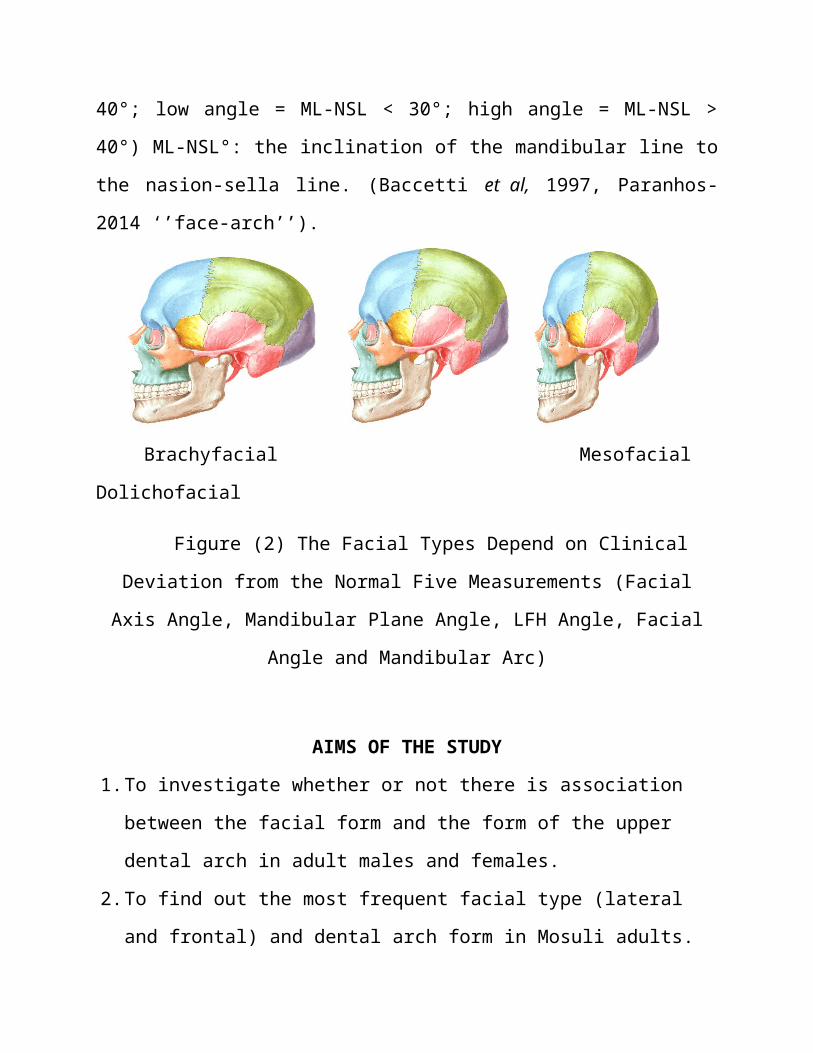
Bimler (1985) used palatal plane (PP): mandibular plane (MP) angle as a
key measurement to describe differences in facial types. He defined (up to 15°
( PP: MP)) angle as a Euryprosopic facial types, (15° to 30°) as mesoprosopic, and
more than (30°) a leptoprosopic facial types. Paranhos-2014’’fac-arc’’
Others use skeletal vertical relationships, on the basis of the ML-NSL values
(Normal = 30° =< ML-NSL =< 40°; low angle = ML-NSL < 30°; high angle =

ML-NSL > 40°) ML-NSL°: the inclination of the mandibular line to the nasion-
sella line. (Baccetti et al, 1997, Paranhos-2014 ‘’face-arch’’).
Brachyfacial Mesofacial Dolichofacial
Figure (2) The Facial Types Depend on Clinical Deviation from the Normal
Five Measurements (Facial Axis Angle, Mandibular Plane Angle, LFH Angle,
Facial Angle and Mandibular Arc)
AIMS OF THE STUDY
1. To investigate whether or not there is association between the facial form and
the form of the upper dental arch in adult males and females.
2. To find out the most frequent facial type (lateral and frontal) and dental arch
form in Mosuli adults.
3. To obtain data in three dimensions (posterior-anterior and lateral) of
craniofacial skeleton of Iraqi adults in Mosul city with normal class I
occlusion, and to define the possible sex differences in craniofacial skeleton in
the three dimensions.
Materials and Methods
The Sample:
The sample of this study involved students from Mosul University
selected randomly from the following colleges (College of Dentistry,
College of Law, College of Education, College of Agriculture and
College of Arts). 448 clinically examined adult subjects, 100 were
selected, and those who fit the criteria of clinical sample selection. Then
cephalometric radiographs and impression were taken for them and only
95 (58 female 61%, 37 male 39%) were selected. The age of the sample
ranged between (18 – 25) years old, They were normal healthy
individuals of Mosul origin.
Criteria for Sample Specification:
1. Full complement of permanent dentition (excluding the third molars).
2. Bilateral Class I molar and canine occlusion
3. There are no:
i. History of previous orthodontic treatment.
ii. Clinical detectable massive interproximal or occlusal caries.
iii. Heavy dental restorations.
iv. Fractured or crowned teeth or fixed prosthodontic therapy.
v. Supernumerary teeth.
vi. History of bad oral habits.
vii. Facial disharmony (deformation).
Materials and Supplies:
A- Diagnostic Instruments : Dental mirrors, Kidney dishes , Cotton &
Disinfectant solution
B-Impression and Cast Materials and Instruments : Wide bladed plaster
spatula ,Rubber bowel ,Upper and lower perforated plastic orthodontic
trays ,Irreversible hydrocolloid impression material & Dental stone.
C-Digitizing Equipments : Sharp pen, Metal ruler, Lab Top (hp) (Pentium IV),
Compact disc ,Computer scanner (hp),.Software Planmeca dimaxis program .
Methods:
The History and Clinical Examination:
The selected students were asked to tell information concerning their names,
ages, history and then subjected to a thorough clinical examination to reassure the
fulfillment of the required sample specifications
Construction of the Study Models:
Individual impression of the both dental arches was taken while the student was seated
on a dental chair after instructing him / her about the procedure in order to cooperate
with the researcher.
The Radiographic Technique:
Under standardized condition, two digital Cephalometrics were taken for
each selected subject, one for lateral view and the other for frontal view. The
subject was set in a standing position with his head fixed by two ear rods laterally
and a locking nasal positioner was then secured against the bridge of the patient's
nose to eliminate the possibility of rotation around ear rods in the sagittal plane and
for future reference in subsequent exposures. Also it acts as a ruler caliber for the
measurement to avoid magnification in the image, so the Frankfort horizontal

plane is kept parallel to the floor. The subject was in centric occlusion during
exposure.
The Landmarks:
1-Incisal Point: The midway point between the incisal edges of the two central
incisors .
2-Canine Point: The cusp tip of the right and left permanent canines .
3- First Molars Point: The mesiobuccal cusp tip of the right and left
permanent first molars .
4- Second Molars Point: The distobuccal cusp tip of the right and left second
permanent molars .
Dental Arch Dimensions
Linear distances were measured on the copy of the study models for the
maxillary dental arches to determine the dental arch width and length.
The linear dimensions are: (Figure 3)
A. Dental Arch Width
The breadth of dental arch is determined by measuring distance between the
corresponding contralateral teeth Daskalogiannakis (2000)that includes:
1-Intercanine Distance (ICD): The linear distance between the cusp tip of the
right and left permanent canines ( Warren and Bishara, 2001; Murad, 2008).

2-Inter First Molar Distance (IMD): The linear distance between the
mesiobuccal cusp tip of the right and left permanent first molars (Salem, 2003).
3-Inter Second Molar Distance (I2MD); the linear distance between the
distobuccal cusp tip of the right and left permanent second molars (Al-Shalabi,
2002).
B. Dental Arch Length:
1-Anterior Arch Length (Canine Vertical Distance) (C.V.D):
The vertical distance from the incisal point perpendicular to the intercanine
distance at the cusp tip (Salem, 2003).
2-Posterior Arch Length (Molar Vertical Distance) (M.V.D):
The vertical distance from the incisal point perpendicular to the intermolar
distance at the mesiobuccal cusp tip of permanent first molars (Ramdan, 2000;
Salem, 2003).
3-Total Arch Length (TAL):
The vertical distance from the incisal point to the line joining the distobuccal
cusp tips of the second permanent molars (Al-Shalabi, 2002).

Dental Arch Form: The size and shape of the arches have considerable
implications in orthodontic diagnosis and treatment planning, affecting the space
available, dental esthetics, and stability of the dentition (Uysala et al, 2005).
Computer Analysis of the Study Models
The models were directly placed on the glass window of the flat bed computer scanner with a metal ruler (Haralabakis et al , 2006). Distortion caused by the scanning procedure was corrected by the use of metal ruler that was scanned
Total arch length
Molar – vertical distance
Anterior arch length
Inter – canine distance
Inter – first molar distance
Inter – second molar distanceFigure (3) Dental Arch Measurements
with each dental cast then corrected automatically by the software Dimaxis program (Mutinelli et al, 2004).
Figure(4) Scanning of the
Dental Cast
Lateral Cephalometric Landmarks:
The following landmarks were used in this study
1.Point N (Nasion): The most anterior point of nasofrontal suture in the midsagittal
plane.
2.Point Or (Orbitale): The lowest point in the inferior margin of the orbit.
3.Point Po (Anatomical Porion): The highest point on the bony external acoustic
meatus.
4.Point ANS (Anterior Nasal Spine): The anterior tip of the sharp bony process of
the maxilla at the lower margin of the anterior nasal opening .
5.Point Ba (Basion): The lowest point on the anterior rim of the foramen magnum
in the mid-sagittal plane.

6.Point Pog (Pogonion): The most anterior point of the bony chin in the median
plane.
7.Point Pt (Pterigoid): The anatomical point representing the foramen rotundum
located at the junction of foramen rotendum with the upper region of the
pterygomaxillary fissure.
8.Point Dc (Condyle): The point in the center of the condylar neck where the
Basion-Nasion plane croses it.
9.Point Pm (Protuberance Menti): The point at the anterior border of the
symphesis between point B and Pogonion where the curvature changes from
concave to convex..
10.Point Me (Menton): The lowest point in the symphyseal shadow of the
mandible.
11. Point Go (Gonion): the most posterior and inferior point at the angle of the
mandible, where the bisector of the angle between tangents to the posterior and
inferior borders of the mandible meets the mandibular outline.
12. Point Gn (Gnathion): The most anterior and inferior point of the bony chin.
13. Point Xi (at the Center of the Ramus): the location of this point is keyed
geometrically to the Frankfort horizontal and pterygoid vertical planes.
Point Xi: This point located at the geographic center of the ramus and it could be
determined by defining four major bony landmarks on the external border of the
mandible and four lines figure(5) ,those are
1. R1-Mandible: the deepest point on the curve of the anterior border of the ramus,
one-half the distance between the inferior and superior curves.
2. R2-Mandible: a point located on the posterior border of the ramus of the
mandible opposing to R1.
3. R3-Mandible: a point located at the center and most inferior aspect of the
sigmoid notch of the ramus of the mandible.

4. R4-Mandible: a point on the lower border of the mandible directly inferior to the
center of the sigmoid notch of the ramus, opposite to R3.
5. A Line Tangent to the Anterior Border of the Ramus at R1 perpendicular to
Frankfort plane and parallel to the pterygoid vertical plane.
6. A Line Tangent to the Posterior Border of the Ramus at R2 perpendicular to
Frankfort plane and parallel to the pterygoid vertical plane.
7. A Line Tangent to the Superior Aspect of the Mandible at point R3 parallel to
Frankfort plane and perpendicular to the pterygoid vertical plane.
8. A Line Was Done at the Lower Aspect of the Mandible parallel to the
Frankfort plane and perpendicular to the pterygoid vertical plane at point R4. The
intersections of these lines would produce a rectangle representing the entire ramus,
from each two contralateral angles a plane can be drawn, the intersection between
these two planes can represent the Xi point (RMO, 2000;Al-Tamimy, 2006)
Figure (5) Diagrammatic Detection of Xi Point in the Mandible (RMO, 2000)
Frontal Cephalometric Landmarks:

The following landmarks were used in this study
1. Point Zy (Zygomatic Arch): At the most lateral border of the center of the
root of the zygomatic arch (right and left).
2. Point Nasion: The most anterior point of nasofrontal suture in the midsagittal
plane.
3. Point Menton: The most caudal point in the outline of the symphesis, it is
regarded as the lowest point of the mandible.
Angular Measurements:
For describing the face Ricketts used five angles. These five angles are:
1. The facial axis angle (Ba-N-Pt-Gn lines) which is formed between the facial
axis or the central axis of the face and the Basion-Nasion plane .This angle gives
information about the chin.
2. The facial angle (FH-N-Pog) which is formed between facial plane (N-Pog)
and (FH) plane. This provides some indication of mandibular prognathism.
3. The lower facial height angle (ANS-Xi-Xi-pm) which is formed by
intersection of a line from anterior nasal spine to the center of the ramus (the Xi
point) and the corpus axis (from the center of the ramus to the supra
pogonion(pm)). It gives an indication of skeletal open bite.
4. The mandibular plane angle (FH-MP) which is formed between frankfort
horizontal and mandibular planes. It was used as an indicator of skeletal
morphology of the mandible.
5. The mandibular bend angle (DcXi-XiPm): measures the angulation of the
condylar process to the body of the mandible.
Frontal Linear Measurements:

Linear measurements (2 Skeletal) were recorded:
1. Zy-Zy: This represents the facial width or interzygomatic distance, measured
from left Zy to right Zy.
Total Anterior Facial Hight (Nasion –Menton); this represents the total anterior facial height. It was measured as the direct distance from nasion to menton.
Dental Arch Forms:
For determination of the dental arch form, the method used by standardization, six
dental cast’s measurements were divided into three sagittal measurements, and
three transverse measurements were utilized to calculate three independent ratios,
which are:
Anterior arch length (canine vertical distance) / inter-canine distance.
Molar vertical distance / inter-first molar distance.
Total arch length / inter-second molar distance.
For each ratio, the average was calculated for the maxillary dental arches for
each sex, then standardization was done for each of three ratios for each subject by
the excel program. Then the mean of these standardized numbers was calculated
for each subject which gave the base for classification as fellows:
Form 1, (Narrow) the three sagittal /transverse ratios are positive (greater than the
mean).the mean of standardized number >+1.
Form 2, (Mid) none of the ratios significantly deviated from the average. the mean
of standardized number between (+1 and -1).
Form 3, (Wide) the three sagittal /transverse ratios are negative (lesser than the
mean). The mean of standardized number <-1.
Lateral Facial Type
In this study the mean of standardization of each five values of the person
was calculated for each gender (by standardization compare the values with the
mean according to the standard deviation) The standardized numbers of the facial
axis angle, the facial angle, the mandibular bend angle multiplied by (-1).So that the
dolichfacial pattern would have a mean of standardized number more than (+1), the
brachyfacial had a mean of standardized number less than (-1) and the mesofacial
had a mean of standardized number between (-1 and +1).
Frontal Facial Type: Determined facial types by calculating the ratio
between interzygomatic distance and total anterior facial height then the face type
for each subject is classified as follows;
Euryprosopic
Facial Index=N-Me (height)/ IZD (Bizygomatic breadth) <average
Mesoprosopic
Facial Index= N-Me (height)/ IZD (Bizygomatic breadth) with in average
Leptoprosopic
Facial Index= N-Me (height)/ IZD (Bizygomatic breadth) > average.
By standardization the facial index the person that has standardized number
more than (+1) would be Leptoprosopic facial type, the person that has standardized
number less than (-1)would be Euryprosopic facial type and the person that has
standardized number between (+1 and -1) would be Mesoprosopic facial type
(Cakirer et al, 2001).
Statistical Analysis:
The data recorded in this research were subjected to computerized statistical
analysis using both Excel-2007, and SPSS version 15 programs
The statistical analysis included
1.Descriptive Statistics (Mean, standard deviation, minimum, maximum) for all
facial measurements angles, liner and ratios taken from the (lateral and

frontal)digital cephalogram, and also for the maxillary dental arch widths, lengths
and ratios for the sample.
t-test” was applied to test the significant differences between the mean of all
dimensions of upper dental arches &facial measurements in Male and Female.
2- Standardized number of the five angles (from lateral cephalometric)and ratio
of the total anterior facial height/interzygomatic distance (from frontal
cephalometric).
3- Standardized number of three ratios(from study models).
Anterior arch length/inter-canine distance.
Molar-vertical distance/inter-first molar distance.
Total arch length/inter-second molar distance.
RESULTS
Dental Arches:
Description and Comparison between Males and Females:
All the variables of the maxillary dental arch widths and, T-test was applied
to see if there were any differences between males and females. It revealed that the
males had larger arch width with a highly significant difference at P < 0.01 in all
width dimensions in maxillary arches as illustrated in Table (1) and Figure (6).
for maxillary dental arch lengths. The statistical analysis showed that there
were high significant differences between males and females in maxillary vertical
distance, all of the vertical measurements showed that males had larger arch length
than females with a high significant difference at P < 0.01 Table (2) and Figure (7).

Table (1) Descriptive Statistics for Maxillary Arch Width with Comparisons between the Genders.
Variable sex Mean ± SD Min Max t-test
Inter Canine
M 36.19 1.91 32.58 41.18
5.450**F 34.11 1.73 30.9 38.52
T 34.92 2.06 30.90 41.18
Inter 1st Molar
M 55.96 2.83 49.56 61.7
5.658**F 52.93 2.31 47.62 59.44
T 54.11 2.92 47.62 61.7
Inter 2nd
Molar D
M 60.07 2.74 53.17 64.78
6.293**F 56.58 2.54 52.34 63.29
T 57.94 3.12 52.34 64.78
NS. : Non Significant at P > 0.05. ** : Significant at P < 0.01. All dimensions in mm
Inter Canine Inter 1st Molar Inter 2nd Molar0
10
20
30
40
50
60
70MaleFemale
Figure (6) Mean of Maxillary Dental Arch Width for both Genders.
Max
illar
y de
ntal
arc
h w
idth
(m
m)

Table (2) Descriptive Statistics for Maxillary Arch Length with Comparison between the Genders.
Variable Sex Mean ± S.D Min. Max. t-value
Anterior Arch Length
M
F
T
8.81
8.31
8.50
0.90
0.83
0.89
6.67
6.56
6.56
10.68
10.35
10.68
2.790**
Molar Vertical D
M
F
T
30.09
28.51
29.13
1.56
1.70
1.81
26.57
24.34
24.34
33.85
33.61
33.85
4.560**
Total Arch Length
M
F
T
44.86
42.37
43.34
1.98
1.83
2.24
40.71
37.53
37.53
48.75
47.31
48.75
6.267**
NS : Non Significant at P > 0.05. ** : Significant at P < 0.01. All dimensions in mm
0
510
1520
2530
3540
45
Anterior archlength
Molar verticaldimension
Total arch length
MaleFemale
Figure (7) Mean of Maxillary Dental Arch Length for both Genders.

Arch Form:
Table (3) and Figure (8) showed the average of the three ratios used in the
determination of the dental arch forms (Anterior arch length/inter-canine distance;
molar-vertical distance/inter-first molar distance, total arch length/inter-second
molar distance) for the maxillary dental arches. there was no significant difference
between males and females in the three ratios.
Table (4) and Figure (9) showed the distribution of the three forms of
maxillary dental arch, It could be noticed that the most prevalent arch form among
the sample was the Mid form (54,05%, 55,17% and 54.73%) followed by the
Narrow form (24,32%, 24,13% and 24.21%), while Wide forms being (21,62%,
20,68% and 21.05%) for males, females and the total sample respectively .
Table (3) Description for the Three Ratios for (Males and Females) with Comparisons between the Genders.
Sex
RatioSex Mean Sd. Min Max T-Value
Canine Vertical / Inter Canine
MF
0.2440.244
0.02690.0236
0.1930.180
0.3130.292 0.023NS
Molar Vertical / Inter 1st Molar
MF
0.54300.5340
0.03030.0266
0.4910.485
0.6220.624 0.342NS
Total Arch Length / Inter 2nd Molar
MF
0.7470.749
0.02940.0278
0.6690.629
0.8450.827 0.162NS
NS: Non Significant at P > 0.05.

Canine vertical/Inter Canine
Molar vertical /Inter 1st Molar
Total arch length/In-ter 2nd Molar
00.10.20.30.40.50.60.70.8
MaleFemale
Figure (8) The Three Ratios for (Males and Females) with Comparisons between the Genders.
Table (4) Distribution of the Three Maxillary Arch Forms for (males,females and the Total) (Frequencies and Percentages).
Males Females Total
Arch Type No. % No. % No. %
Narrow 9 24.32 14 24.13 23 24.21Mid 20 54.05 32 55.17 52 54.73Wide 8 21.62 12 20.68 20 21.05Total 37 100 58 100 95 100

Figure (9) Percentages of Three Maxillary Arch Forms for(Males, Females and the Total).
Lateral Facial Measurements1. Descriptive Statistics:
The description & Comparison of facial measurement & the distribution of
the five angles according to gender There was no significant difference in angular
measurements between males and females by using t-test as showed in Table (5).
Lateral Facial Forms:
The distribution of the lateral facial types among males, females and the
total sample showed in Table (6). This table illustrated that the most frequent
lateral facial type was the Mesofacial type (59,45%, 58,62 % and 58.94%)
followed by Dolichofacial (21,62%, 20,68% and 21.05%) and then the
Brachyfacial type(18,91%, 20,68% and 20%) in males, females and the total
sample respectively.

Table (5) Descriptive Statistics for the Five Angles for (Males, Females and the Total) with Comparison between the Genders.
Variable Sex Mean S.D Min MaxT-
Value
Facial Angle MFT
90.0589.5289.73
2.492.992.80
858383
959696
0.90 NS
Facial Axis Angle MFT
88.5688.4388.48
2.633.072.89
838080
949595
0.21 NS
Mandibular Plane Angle
MFT
22.7624.0223.53
3.864.374.20
151515
323535
1.43 NS
Lower Facial Height Angle
MFT
43.6443.2243.38
3.583.763.68
353635
545555
0.53 NS
Mandibular Bend Angle
MFT
33.6232.4332.89
4.484.154.29
232423
414242
1.31 NS
Table (6) Distribution of the Lateral Facial Types for (Males, Females and the Total) (Frequencies and Percentages).
Males Females Total
Facial Type No. % No. % No. %
Dolichofacial 8 21.62 12 20.68 20 21.05
Mesofacial 22 59.45 34 58.62 56 58.94
Brachyfacial 7 18.91 12 20.68 19 20
Total 37 100 58 100 95 100
Comparison between Males and Females according to lateral facial type .
The difference in angular measurements between males and females in
Dolichofacial , Mesofacial & Brachyfacial by using t-test showed in Table (7). It
was found that there was a significant difference between males and females for
Mandibular plane angle for Dolichofacial & Mesofacial type and a non significant
difference for other angular measurements at P < 0.05.
Association between the lateral Facial Type and Dental arch Forms:
The association between the type of face and the dental arch form in (males,
females and the total) showed in Table (8), expressed as frequencies and
percentages. It was found that the Mid maxillary arch form was associated with
Mesofacial (63.63%, 64.70% and 64.28%).Also we can see that there was an
association between Narrow arch and Dolichofacial type (62.5% , 58.33% and
60%), and there was an association between Wide arch and Brachyfacial type
(57.14%, 58.33% and 57.89%) for males, females and the total sample
respectively.

Dolichofacial Mesofacial Brachyfacial
Variable Sex Mean S.D T-Value Mean S.D T-Value Mean S.D T-Value
Facial AngleM 87.19 1.89
1.110 NS90.02 1.45
1.485 NS93.43 1.27 0.63
NSF 86.18 1.99 89.32 1.89 93.9 1.66
Facial Axis Angle
M 85.78 1.671.297 NS
88.52 1.860.019 NS
91.86 1.77 0.59
NSF 84.50 2.38 88.51 1.71 92.45 2.19
Mandibular Plane Angle
M 27.63 2.662.076 *
22.05 2.762.263 *
19.43 2.81 0.61
NSF 30.27 2.79 23.62 2.47 18.6 2.67
Lower Facial Height Angle
M 48.19 3.440.372 NS
43.05 1.961.010 NS
40.28 2.75 0.23
NSF 48.73 2.88 42.46 2.25 39.95 3.00
Mandibular Bend Angle
M 28.11 2.471.132 NS
33.72 2.881.989 NS
39.57 1.61 1.05
NSF 26.82 2.44 32.43 2.10 38.6 2.01
Table (7) Descriptive Statistics for the Five Angles According to Lateral Facial Type with Comparison between Genders.

Chapter Four Results 99
Table (8) Association of the Three Maxillary Arch Forms According to Lateral Facial Type for (Males ,Females and the Total) (Frequencies and Percentages).
Males Females Total
dolichofacial mesofacial brachyfacial dolichofacial mesofacial brachyfacial dolichofacial mesofacial brachyfacial
Arch Type
No. % No. % No. % No. % No. % No. % No. % No. % No. %
Narrow 5 62.5 4 18.18 0 0 7 58.33 7 20.58 0 0 12 60 11 19.64 0 0
Mid 3 37.5 14 63.63 3 42.86 5 41.67 22 64.70 5 41.67 8 40 36 64.28 8 42.10
Wide 0 0 4 18.18 4 57.14 0 0 5 14.70 7 58.33 0 0 9 16.07 11 57.89
Total 8 100 22 100 7 100 12 100 34 100 12 100 20 100 56 100 19 100

Frontal Facial Measurements1. Description & Comparison between Males and Females in Liner
Measurements
The descriptive statistics of facial measurement of the total anterior facial
height, inter zygomatic distance and the ratio of total anterior facial height and
inter zygomatic distance with The comparison between males, females from frontal
digital cephalometric as showed in Table (9) .It was found that males had higher
values than females with a significant difference at P < 0.01 for all dimensions, but
the ratio was nearly equal and the difference was not significant at P < 0.05.
Table (9) Descriptive Statistics for Liner Measurement and (TAFH/IZD) Ratio for(Males, Females and the Total) with Comparison between Genders.
Males FemalesT-Value
Total
Variable Mean S.D Mean S.D Mean S.D
Total Anterior
Facial Height127.89 7.61 120.32 5.11 5.679 ** 123.28 7.24
Inter Zygomatic
Distance144.42 6.94 135.32 5.27 6.958 ** 138.76 7.32
Ratio
TAFH/IZD0.887 0.05 0.890 0.05 0.181 NS 0.889 0.05
** : Significant at P < 0.01.
2. Frontal Facial Forms
The distribution of the frontal facial types among the males, females and the
total sample showed in Table (10). This table illustrated that the most frequent
facial type was the Mesoprosopic (54.05 %, 60.34% and 57.89%) type followed by

Leptoprosopic (24.32%, 18.96% and 21.05%) and Euryprosopic (21.62%, 20.68%
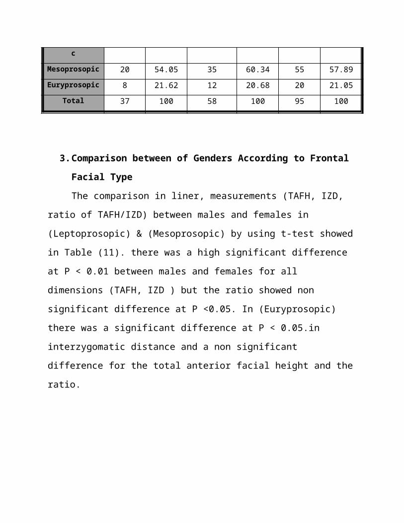
and 21.05%) for males, females and the total sample respectively.
Table (10) Distribution of the Frontal Facial Types for (Males, Females and the Total) (Frequencies and Percentages).
Males Females Total
Facial Type No. % No. % No. %
Leptoprosopic 9 24.32 11 18.96 20 21.05
Mesoprosopic 20 54.05 35 60.34 55 57.89
Euryprosopic 8 21.62 12 20.68 20 21.05
Total 37 100 58 100 95 100
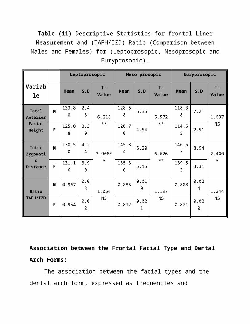
3. Comparison between of Genders According to Frontal Facial Type
The comparison in liner, measurements (TAFH, IZD, ratio of TAFH/IZD)
between males and females in (Leptoprosopic) & (Mesoprosopic) by using t-test
showed in Table (11). there was a high significant difference at P < 0.01 between
males and females for all dimensions (TAFH, IZD ) but the ratio showed non
significant difference at P <0.05. In (Euryprosopic) there was a significant
difference at P < 0.05.in interzygomatic distance and a non significant difference
for the total anterior facial height and the ratio.

Table (11) Descriptive Statistics for frontal Liner Measurement and (TAFH/IZD) Ratio (Comparison between Males and Females) for (Leptoprosopic,
Mesoprosopic and Euryprosopic).
Leptoprosopic Meso prosopic Euryprosopic
Variable Mean S.D T-Value Mean S.D T-Value Mean S.D T-Value
Total Anterior
Facial Height
M 133.88 2.48
6.218 **
128.68 6.35
5.572 **
118.38 7.211.637 NSF 125.08 3.39 120.70 4.54 114.55 2.51
Inter Zygomatic Distance
M 138.50 4.243.908**
145.34 6.206.626 **
146.57 8.942.400 *
F 131.16 3.90 135.36 5.15 139.53 3.31
Ratio TAFH/IZD
M 0.967 0.031.054 NS
0.885 0.0191.197 NS
0.808 0.0241.244 NSF 0.954 0.02 0.892 0.021 0.821 0.020
Association between the Frontal Facial Type and Dental Arch Forms:
The association between the facial types and the dental arch form, expressed
as frequencies and percentages for all the sample showed in Table (12) . It was
found that the Mid maxillary arch form was associated with Mesoprosopic (65%,
62.85% and 63.63) for males, females and the total sample respectively and those
males had higher records than females.
We can see that there was an association between Narrow, maxillary arch
forms and Leptoprosopic (55.55%, 54.54% and 55%) for males, females and the
total sample respectively and that males had higher records than females.
Also we can see that there was an association between Wide maxillary arch
forms with Euryprosopic (62.5%, 58.33% and 60%) for males, females and the
total sample respectively and that males had higher records than females.

6- Association between Lateral and Frontal Facial Types:
The association between the lateral facial types and frontal facial Type
expressed as frequencies and percentages showed in Table (13) . It was found that
the Mesoprosopic facial type associated with Mesofacial (63.63%, 76.47% and
71.42%) for males, females and the total sample respectively and those females
had higher records than males.
We can see that there was an association between Leptoprosopic and
Dolichofacial (62.5%, 66.66% and 65%) for males, females and the total sample
respectively and that females had higher records than males.
We can see that there was an association between Euryprosopic face type
and Brachyfacial (57.14%, 58.33% and 57.89%) for males, females and the total
sample respectively and that females had higher records than males

Males Females Total
Leptoprosopic Mesoprosopic Euryprosopic Leptoprosopic Mesoprosopic Euryprosopic Leptoprosopic Mesoprosopic Euryprosopic
Arch Type No. % No. % No. % No. % No. % No. % No. % No. %
No. %
Narrow 5 55.55 4 20 0 0 6 54.54 8 22.85 0 0 11 55 12 21.81 0 0
Mid 4 44.44 13 65 3 37.5 5 45.45 22 62.85 5 41.66 9 45 35 63.63 8 40
Wide 0 0 3 15 5 62.5 0 0 5 14.28 7 58.33 0 0 8 14.54 12 60
Total 9 100 20 100 8 100 11 100 35 100 12 100 20 100 55 100 20 100
Table(12)Association of three arch forms according to frontal facial types

Table (13) Association of the Frontal with Lateral Facial Types
Males Females Total
Dolichofacial Mesofacial Brachyfacial Dolichofacial Mesofacial Brachyfacial Dolichofacial Mesofacial Brachyfacial
Facial Type No. % No. % No. % No. % No. % No. % No. % No. % No. %
Leptoprosopic 5 62.5 4 18.18 0 0 8 66.66 3 8.82 0 0 13 65 7 12.5 0 0
Mesoprosopic 3 37.5 14 63.63 3 42.85 4 33.33 26 76.47 5 41.66 7 35 40 71.42 8 42.10
Euryprosopic 0 0 4 18.18 4 57.14 0 0 5 14.70 7 58.33 0 0 9 16.07 11 57.89
Total 8 100 22 100 7 100 12 100 34 100 12 100 20 100 56 100 19 100

Three Dimensions Facial FormsThe distribution of the three dimension facial types showed in Table (14) .
This table illustrated that the most frequent facial type was the Three dimension-
average type (56.75%, 55.17% and 55.78%) followed by the Three-dimension long
(24.32%, 22.41% and 23.15%), then Three-dimension short type (18.91%, 22.41%
and 21.05%), for males, females and the total respectively.
Association of Maxillary Arch Forms to Three-Dimension Facial Types
The association between the facial types and the dental arch form expressed
as frequencies and percentages showed in Table (15) .It was found that there was a
highly association between Narrow, maxillary arch forms and Three-dimension
long (77.77%, 84.61% and 81.81%) females had higher records than males. Also
we can see that there was a highly association between Mid maxillary arch form
with Three-dimension average (80.95%, 87.5% and 84.90%) females had higher
records than males. Also we can see that there was a highly association between
Wide maxillary arch forms with the Three-dimension short face type (85.71%,
84.61% and 85%) but here males had higher records than females.

Table (14) Distribution of the Three Dimensions Facial Type
Males Females Total
Facial Type No. % No. % No. %
3D-long 9 24.32 13 22.41 22 23.15
3D-average 21 56.75 32 55.17 53 55.78
3D-short 7 18.91 13 22.41 20 21.05
Total 37 100 58 100 95 100


Males Females Total
3D-long 3D-average 3D-short 3D-long 3D-average 3D-short 3D-long 3D-average 3D-short
Arch Type
No % No % No % No % No % No % No % No % No %
Narrow 7 77.77 2 9.52 0 0 11 84.61 3 9.37 0 0 18 81.81 5 9.43 0 0
Mid 2 22.22 17 80.95 1 14.28 2 15.38 28 87.5 2 15.38 4 18.18 45 84.90 3 15
Wide 0 0 2 9.52 6 85.71 0 0 1 3.12 11 84.61 0 0 3 5.66 17 85
Total 9 100 21 100 7 100 13 100 32 100 13 100 22 100 53 100 20 100
Table (15) Association of the Three Maxillary Arch Forms According to Facial Type Three-Dimension

DISCUSSION
Dental arch dimension
For inter canine distance, the current study for males and females were more
than that of Henrikson (2001) and Al-Zubair (2003), but near that of Salem (2003)
and Al-Hadithy (2005). the mean values of Inter 1st & 2nd molar distance for this
study for males were more than that of Henrikson (2001), Salem (2003) and Al-
Zubair (2003) but near to that of Al-Hadithy (2005) and Haralabakis (2006), while
for females sample this study was larger than that of Henrikson (2001) and Salem
(2003), but near to that of Al-Zubair (2003), Al-Hadithy (2005) and Haralabakis
(2006) , while for the total sample, this study was larger than that of Henrikson
(2001) and Salem (2003) and Kim and Gianelly (2003) but near that of Al-Zubair
(2003), Al-Hadithy (2005) and Haralabakis (2006). The difference might be due to
racial factor or the difference in the analyzing technique. the mean values of total
arch length for this study for males were more than that of Al-Zubair (2003) and
Al-Hadithy (2005), but nearly equal to that of Salem (2003),and less than that of
Haralabakis (2006), but for females and the total sample, the mean value was
nearly equal to that of Salem (2003), Al-Zubair (2003) and Al-Hadithy (2005),and
less than that of Haralabakis (2006) .
We can notice that the most frequent maxillary dental arch form in both
genders was the Mid type. This findings agreed with Borgan (2001) who found
that the most frequent Jordanian maxillary arch form was the Mid form and Salem
(2003) who found that the most frequent Palestinian maxillary arch form was the
Mid form, also Haralabakis (2006) found that the most frequent maxillary arch
form was the Mid form.

In general, it is obvious that the mean values of all measurements taken for
the dental arch length were slightly differed from that of other studies, these slight
differences might be due to the sample selection and ethnic variation.
Facial Types and Dental Arch Forms
persons with Dolichofacial face showed a larger value of Mandibular plane
angle, Lower facial height angle and low value of Facial angle, Facial axis angle
and Mandibular bend angle than other types of face.
Persons with Brachyfacial face showed a low value of Mandibular plane
angle, Lower facial height angle and a large value of Facial angle, Facial axis angle
and Mandibular bend angle than other types of face.
Persons with mesofacial face show a value of five angles in between
the Dolichofacial and the Brachyfacial.
It was found that the mid maxillary arch form was associated with
mesofacial type (63.63 % for males, 64.70% for females, 64.28% for the total).
Also we can see that there was an association between Narrow arch and
Dolichofacial type (62.5% for males, 58.33% for females, 60% for the total). Also
there was an association between Wide arch and Brachyfacial type (57.14% for
males, 58.33% for females, 57.89% for the total).
This means that the person with Dolichofacial face type, mostly has a
Narrow dental arch , the person with Mesofacial face type, mostly has a Mid dental
arch, and the person with Brachyfacial face type, mostly has a Wide dental
arch .This is one of the characters of each facial type that’s supported by (Enlow,
1982; R.M.O, 2000; Al-Taee & Al-Joubori 2014).
there was an association between Narrow, maxillary arch forms and
Leptoprosopic . Also there was an association between Wide maxillary arch forms
and Euryprosopic face type.an association between the face types and the dental

arch form in males and females expressed as frequencies and percentages. It was
found that the Mid maxillary arch form was associated with Mesoprosopic. This
agreed with Salem (2003).
There was also an association between Narrow maxillary arch forms and
Leptoprosopic, and between Wide maxillary arch forms with Euryprosopic face
type.
Our finding were in agreement with Graber (1988) which stated that there
was a sort of correlation between facial morphology and dental arch form. So
Leptoprosopic individuals had Narrow dental arches, while Euryprosopic
individuals had broad, round dental arches; whereas Mesoprosopic individuals
fitted somewhere in between the two types.
Ramadan (2000) stated that the association was not unexpected; Al Shalabi
(2002) agrees on that there is no clear relationship between facial forms and arch
forms.
It was noticed that there was an association between Leptoprosopic and
Dolichofacial, and between Euryprosopic face type and Brachyfacial.
Anterior facial height affects both lateral and frontal facial types so most
but not all of Dolichofacial type had Leptoprosopic this is because the lateral
facial type is affected also by facial depth, but frontal facial type is affected by
facial width instead of facial depth. This was the same for Mesofacial and
Brachyfacial types. So that not every long face of lateral type had the same
characteristics of frontal type, this was the same for short and averaged face.
The lateral facial type represent the relationship between the facial height
and facial depth, the frontal facial type represents the relationship between the
facial height and facial width . As the face is three dimensional object, so we need
three dimensional relationship that represents the relationship between the facial
height, facial depth and facial width.
As the facial bone grows in three dimensions, the face should be studied in
its three dimensions as the transverse dimensions of the face affects all the
determinations of the dentofacial proportion as well as balance and harmony this
was in accordance to (Hatcher and Aboudara, 2004).
In this study the distributions of facial type (lateral, frontal and three
dimension) were nearly the same.
It was noticed that the (Mesofacial, Mesoprosopic, Three-dimension
average)had the most frequent facial type (more than halve 54-60 % ) and then
followed by (Dolichofacial, Leptoprosopic, Three-dimension long) and
(Brachyfacial, Euryprosopic, Three-dimension short) both types had about less
than halve of the sample, each one had about (18-24%) of the sample.
It is found that there was a highly association between Narrow maxillary
arch forms and Three-dimension long ones.(77.77%, 84.61%, 81.81%) Also there
was a highly association between Mid maxillary arch form with a Three-dimension
average (80.95, 87.5%, 84.90%) and between Wide maxillary arch forms with
Three-dimension short face type (85.71%, 84.61%, 85%) for males, females and
the total sample respectively.
Conclusion
In general for this study there was an increase in the association between a
Three-dimension facial type and the upper dental arch form than that of (lateral and
frontal), so that the characteristic features of facial type appeared more clear in the
three dimensions than that of the two dimensions.
References
Al-Shalabi F.S.(2002): The Relationship between Skeletal Facial Form and
Lower Dentalarch Form in an Iraqi Sample Aged 20-25 Years with Class I
Normal Occlusion. "A Computerized Analysis". Master thesis, Baghdad
University.
Al-Tamimy E (2006) The reliability of Ricketts analysis using cephalometric
tracing of Iraqi sample aged 8-10 years. Al-Mustanseria DJ. , Vol. 3(2), 88-96.
Al-Hadithy S.F.(2005): Dental arch dimensions and forms in Sulaimania
Kurdish population sample aged 16-24 years with class I normal occlusion.
Master thesis, Baghdad University.
Al-Zubair N.(2002): Maxillary and mandibular dental arch dimensions and
forms in a sample of Yemeni population aged (18-26) years with class I normal
occlusion. Master thesis, Baghdad University.
Al-Taee H & Al-Joubori S (2014) Dental arches dimensions, forms and its
association to facial types in a sample of Iraqi adults with skeletal and dental
class II-division 1 and class III malocclusion. J Bagh Coll Dentistry; 26(2): 160-
166.
Baccetti T, Antonini A, Franchi l, Tonti M and Tollaro I (1997) Glenoid Fossa
Position in Different Facial Types: a Cephalometric Study British Journal of
Orthodontics.,Vol. 24:55–59
Bishara S.E.(2001): Text book of orthodontics. W.B Saunders Company.
Section I, pp.45-48.
Borgan B.E. (2001): Dental arch dimensions analysis among Jordanian school
children. Master Thesis. Cairo University- Egypt.

Cakirer.B, Hans M.G, Graham G, Aylor J, Tishler P.V And Redline S.(2001):
The Relationship Between Craniofacial Morphology and Obstructive Sleep
Apnea in Whites and in African-Americans. journal of respiratory and critical
care medicine. Vol 163:947-950.
Daskalogiannakis J. (2000): Glossary of orthodontic terms. Quintessence
publishing Co Inc.
Enlow D.H. (1982): Handbook of Facial Growth. 2nd ed. Philadelphia, PA:
WB Saunders.
Gallão S, Faltin K, Santos L, Ary D, Martins L (2013) Facial type measurements influence on transverse dimensions of normal occlusion arches. J Health Sci Inst.;31(3):20-3.
Graber T.M. (1988): orthodontics, principles and practice. W.B. Saunders
company pp:209,210.
Graber T.M. and Vanarsdall R.L. (2000): Orthodontics current principles and
techniques. Third ed Mosby Inc pp:385.
Haralabakis n.b., sifakakis L., papagrigorakis M., papadakis G. (2006): The
correlation of sexual dimorphism in tooth size and arch form. World .j.orthod.
7 (3):254-262.
Hatcher D.C. and Aboudara C.L. (2004): Diagnosis goes digital.
Am.J.Orthod.Dentofac.Orthop. 125: 512 – 515.
Henrikson J., Persson M. and Thilander B. (2001): Long-term stability of dental
arch form in normal occlusion from 13 to 31 years of age. The Euro. J. Orthod.
23 (1): 51-61.
Kim E. and Gianelly A.A.(2003): Extraction vs Nonextraction: Arch Widths and
Smile Esthetics. Angle. Orthod. 73:354–358.
Murad S.M.(2008): Accuracy of measurements made on digital and study models
(A comparative study). Master thesis, Baghdad University..

Mutinelli S., Cozzani M., Manfredi M. and Siciliani G.(2004): Dental arch
analysis system. Progress In Orthodontics. 5(2): 200-2011.
Panhos L, Zaroni M, Carlil J et al (2014) Association between the facial type and morphology of upper central incisor in normal occlusion subjects. Journal of contmperary dental practice, 15(1), 29-33.
Paranhos L, Ramos M, Benedicto E et al (2014) Is there any association between facial type and mandibular dental arch form in subjects with normal occlusion? Acta Scientiarum. Health Sciences Maringá, vol. 36(1), p. 129-134.
R. M. O. (2000): Diagnostic services.
Ramadan O.Z. (2000): Relation between photographic facial Measurements and
lower dental arch measurement in adult Jordanian males with class I normal
occlusion. Master Thesis, Mousl University.
Salem N.M.(2003): Facial and arch form and dimensions in a sample of 16-21
years old Palestinians class I occlusion. Master Thesis. Baghdad University-
Iraq.
Uysala T., Memilib B., Usumezc S. and Sarid Z.(2005): Dental and Alveolar
Arch Widths in Normal Occlusion, Class II division 1 and Class II division 2.
Angle. Orthod. 75:941–947.
Warren J.J. and Bishara S.E. (2001): Comparison of dental arch measurement in
the primary dentition between contemporary and historic samples.
Am.J.Orthod.Dentofac.Orthop. 119:211-215.