Embed Size (px)
Citation preview
BACTERIOLOGICAL AND PHYSICO-CHEMICAL QUALITY OF WATER FROM
VARIOUS SOURCES IN SAMBURU DISTRICT AND EFFICACY OF SELECTED
PLANT PRODUCTS IN WATER PURIFICATION
By
CHELUGET KIPKEMBOI B.Ed (Science) Hons, KU
REG. NO: I56/25II/04
A thesis submitted in partial fulfilment of the requirements for the award of the degree of
Master of Science (Microbiology) in the School of Pure and Applied Sciences
Kenyatta University
JULY 2011

ii
DECLARATION
I Cheluget Kipkemboi, declare that this thesis is my original work and has not been presented for
the award of a degree in any other University or for any other award.
Cheluget Kipkemboi
Signature…………………… Date……………………….
We confirm that the work reported in this thesis was carried out by the candidate under our
supervision
Prof. Kiplagat Kotut,
Department of Plant and Microbial Sciences,
Kenyatta University,
Signature.............................. Date……………………….
Prof. Nkanata M. Gitonga,
Department of Plant and Microbial Sciences,
Kenyatta University,
Signature…………………….. Date……………………….
Dr. Alexander Njue,
Department of Plant and Microbial Sciences,
Kenyatta University,
Signature…………………… Date……………………..

iii
DEDICATION
To my entire family and mostly my dear wife Doreen, and children; Tracy, Sharon, Emmanuel,
Ian and Christian, who despite my long stay away from home during the course of this work,
stood by me, persevered and provided me with much inspiration and energy to go on.
iv
ACKNOWLEDGEMENTS
I would like to express my most sincere appreciation to my supervisors, Prof. Kiplagat Kotut,
Prof. Nkanata Mburugu Gitonga and Dr. Alexander Njue, without whose patience and critical
guidance this work would not have been realized.
My sincere thanks are due to both teaching and non teaching staff of Kenyatta University for
their support and advice. Of particular note are Mr Steve Mwangi and Mr Kamau Kiunyu of the
Department of Plant and Microbial Sciences for their technical support in the field during
sampling and analysis of physico-chemical properties of water.
I am also indebted to Earthwatch institute for supporting my fieldwork through the Communities,
Water and Wildlife Research project and Wamba Mission Hospital for allowing me to use their
facilities. Special thanks also go to all my family members and friends for their support and
encouragement through the study period.
Most of all, I am grateful to the almighty God for the strength and good health that he gave me
throughout the study. Lastly I would like to acknowledge the immense support and
encouragement, which I received from my loving wife Doreen; in more ways than I can mention,
she bore the brunt of this grueling undertaking.
v
TABLE OF CONTENTS
DECLARATION ............................................................................................................................ ii
DEDICATION ............................................................................................................................... iii
ACKNOWLEDGEMENTS ........................................................................................................... iv
TABLE OF CONTENTS ................................................................................................................ v
LIST OF TABLES ......................................................................................................................... ix
LIST OF FIGURES ........................................................................................................................ x
ABBREVIATIONS AND ACRONYMS ...................................................................................... xi
ABSTRACT .................................................................................................................................. xii
CHAPTER ONE: INTRODUCTION .......................................................................................... 1
1.1 Background to the study ............................................................................................ 1
1.2 Problem statement and justification ........................................................................... 2
1.3 Research questions ..................................................................................................... 3
1.4 Hypotheses ................................................................................................................ 4
1.5 Objectives .................................................................................................................. 4
1.5.1 General objective ...................................................................................................... 4
1.5.2 Specific objectives .................................................................................................... 4
1.6 Significance of the study ........................................................................................... 5
CHAPTER TWO: LITERATURE REVIEW ............................................................................. 6
2.1 Bacterial water quality ............................................................................................... 6
2.2 Water quality changes ................................................................................................ 6
2.3 Water quality challenges ............................................................................................ 7
2.4 Biological indicators of water quality ........................................................................ 8
2.4.1 Heterotrophic plate counts ........................................................................................ 8
2.4.2 Thermotolerant coliforms ......................................................................................... 9
2.4.3 Faecal Streptococci and Enterococci ...................................................................... 11
2.4.4 Pathogenic bacteria ................................................................................................. 11
2.4.5 Bacterial water quality risk assessment .................................................................. 13
2.4.6 Physico-chemical indicators of water quality ......................................................... 14
2.4.7 Water pollution by organic matter .......................................................................... 16
2.5 Common water treatment methods ......................................................................... 17
2.5.1 Solar disinfection .................................................................................................... 17
2.5.2 Chemical water treatment methods ......................................................................... 17
2.6 Microbial reduction by coagulation-flocculation .................................................... 20
vi
2.7 Bacterial antibiotic resistance ................................................................................. 20
CHAPTER THREE: MATERIALS AND METHODS ............................................................ 21
3.1 Study area ................................................................................................................ 21
3.2 Water sampling ........................................................................................................ 23
3.3 Field measurements ................................................................................................. 23
3.3.1 pH (pH units) .......................................................................................................... 23
3.3.2 Temperature (°C) .................................................................................................... 24
3.3.3 Electrical conductivity (µS cm-1
) ............................................................................ 24
3.3.4 Dissolved oxygen (DO µg L-1
) ............................................................................... 24
3.4 Laboratory physico-chemical analyses .................................................................... 24
3.4.1 Total alkalinity (TA mg CaCO3 L-1
) ....................................................................... 24
3.4.2 Phosphorus (mg L-1
)................................................................................................ 25
3.4.3 Turbidity (NTU) ...................................................................................................... 25
3.4.4 Most probable number (MPN) of total and faecal coliforms per 100 ml ............... 25
3.4.4.1 Presumptive test ...................................................................................................... 25
3.4.4.2 Confirmatory test .................................................................................................... 27
3.4.4.3 Completed test......................................................................................................... 28
3.4.5 Faecal coliforms (thermotolerant Escherichia coli) ................................................ 28
3.4.5.1 Confirmatory test .................................................................................................... 28
3.4.5.2 Completed test......................................................................................................... 29
3.4.6 Heterotrophic plate counts (CFU mL-1
) .................................................................. 29
3.4.7 Isolation of Salmonella and Shigella spp (presence/absence) ................................ 29
3.4.8 Isolation of Vibrio cholera (presence/absence) ...................................................... 30
3.5 Identification of bacteria .......................................................................................... 30
3.5.1 Gram staining .......................................................................................................... 30
3.5.2 Oxidase test ............................................................................................................. 30
3.5.3 Motility test ............................................................................................................. 30
3.5.4 Indole test ................................................................................................................ 31
3.5.5 Methyl red test ........................................................................................................ 31
3.5.6 Voges-proskauer test ............................................................................................... 31
3.5.7 Citrate test ............................................................................................................... 32
3.6 Bacterial risk level determination ............................................................................ 32
3.7 Preparation of plant material ................................................................................... 33
3.8 Water treatment ....................................................................................................... 33
3.9 Antibacterial activity ............................................................................................... 34
vii
3.9.1 Preparation of extracts ............................................................................................ 34
3.9.2 Sources of test bacteria ........................................................................................... 35
3.9.3 Antibacterial sensitivity testing ............................................................................... 35
3.9.4 Data analysis ............................................................................................................ 36
CHAPTER FOUR: RESULTS .................................................................................................. 37
4.1 Introduction ............................................................................................................. 37
4.2 Physico-chemical properties of water ..................................................................... 37
4.2.1 Temperature ............................................................................................................ 37
4.2.2 pH ............................................................................................................................ 37
4.2.3 Electrical conductivity (µS cm-1
) ............................................................................ 37
4.2.4 Dissolved oxygen (DO mg L-1
) ............................................................................... 40
4.2.5 Salinity .................................................................................................................... 40
4.2.6 Turbidity (NTU) ...................................................................................................... 40
4.2.7 Total alkalinity (TA, mg L-1
CaCO3) ...................................................................... 40
4.2.8 Total phosphorus (TP, µg L-1
) and orthophosphate phosphorus (PO4-P, µg L-1
) ... 41
4.3 Bacterial properties of water sources ....................................................................... 41
4.3.1 Heterotrophic plate counts (HPC, CFU mL-1
) ........................................................ 41
4.3.2 Total coliforms MPN/100 mL................................................................................. 42
4.3.3 Faecal coliforms ...................................................................................................... 43
4.3.4 Occurrence of Vibrio cholerae, Salmonella and Shigella species .......................... 43
4.4 Risk levels ............................................................................................................... 44
4.5 Water purification experiments ............................................................................... 46
4.5.1 Change in heterotrophic bacterial counts ................................................................ 46
4.5.2 Change in water turbidity ........................................................................................ 48
4.5.3 Changes in water pH ............................................................................................... 50
4.6 Bacterial inhibition effects of plant extracts ........................................................... 51
5.1 Physico-chemical quality of water .......................................................................... 52
5.2 Conclusions ............................................................................................................. 62
5.3 Recommendations for further research ................................................................... 63
REFERENCES.............................................................................................................................. 65
APPENDICES .............................................................................................................................. 77
Appendix 1 Location and identity of specific water bodies sampled in Wamba Division,
Samburu District. .................................................................................................... 77
Appendix 2 Selenite F broth used to enrich Salmonella and Shigella spp bacteria. On the
left is sterile broth while on the right is a 24 hour positive enriched bacterial
culture. .................................................................................................................... 78

viii
Appendix 3 Peptone water used to enrich Vibrio cholerae spp bacteria. On the left is
sterile Peptone water while on the right is a 24 hour enriched bacterial culture. ... 79
Appendix 4 Measurement of water turbidity using a colorimeter during treatment. .......... 80
Appendix 5 Laboratory measurement of water pH using a portable pH meter during
treatment. ................................................................................................................ 81
Appendix 6 Assortment of some bacteriological media used to culture bacteria. .............. 82
ix
LIST OF TABLES
Table 1 Bacterial risk level classification based the number (MPN/ 100 mL) of faecal
coliforms present ............................................................................................................ 32
Table 2 Showing inhibition zone diameter interpretive standards………………………… .... ..36
Table 3 Physico-chemical properties of water from the various types of sources in Wamba
Division over the period 26/2/06 to 11/3/07 ................................................................... 39
Table 4 Heterotrophic plate counts (HPC) in water from various sources in Samburu .............. 42
Table 5 Total coliforms counts (MPN/100 mL) in water from various sources in Samburu ...... 42
Table 6 Faecal coliforms counts (MPN/ 100 mL) in water from various sources in Samburu ... 43
Table 7 Frequency of occurrence (%) of Salmonella and Shigella spp in water from different
sources. ........................................................................................................................... 44
Table 8 Bacterial risk level classification for specific water sources .......................................... 44
Table 9 Levels of risk in water from various sources .................................................................. 45
Table 10 Mean percentage reduction in HPC of water samples from various sources after
treatment with plant extracts, alum and sodium hypochlorite ........................................ 47
Table 11 Percentage reduction in water turbidity after treatment of water samples with
extracts from the three test species, alum and sodium hypochlorite .............................. 49
Table 12 Changes in water sample pH obtained following treatment of water samples with the
three plant species investigated as well as alum and sodium hypochlorite over 24
hour period of treatment. ................................................................................................ 50
Table 13Mean zones of inhibition (mm) of selected test bacterial strains by treatment with
plant extracts, alum, sodium hypochlorite and Streptomycin…………………… .…51
x
LIST OF FIGURES
Figure 1 The study area showing the sampling sites………………………………………...22
Figure 2 Multiple tube fermentation technique showing positive presumptive test for faecal
and total coliforms in a 24 hour lactose broth culture……………………………...26
Figure 3 Multiple Tube Fermentation technique showing positive confirmatory test for faecal
and total coliforms in a 24 hour brilliant green bile lactose broth
culture………………………………………………………………………………27
Figure 4 Setup showing incubation of water samples during treatment. Different samples
show different turbidity levels……………………………………………………...34
Figure 5 Percentage changes in water pH, turbidity and HPC of water samples treated with
extracts of the three plant species………………………………..…….…………...48
xi
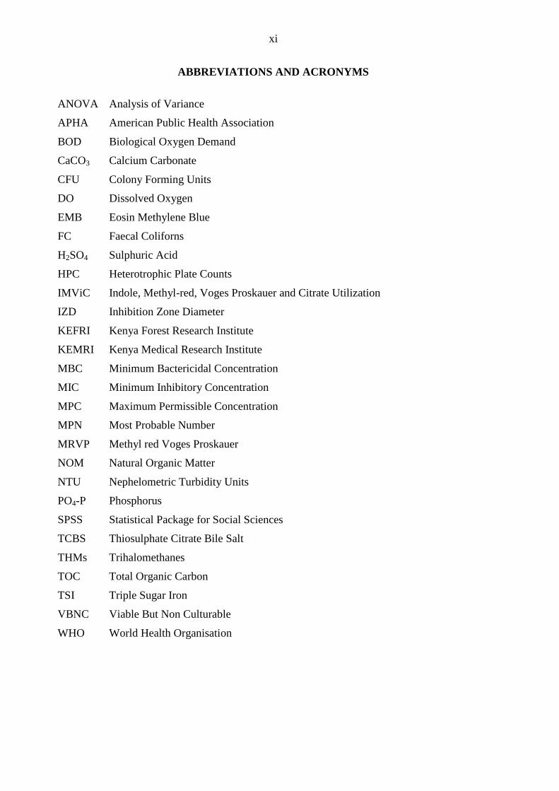
ABBREVIATIONS AND ACRONYMS
ANOVA Analysis of Variance
APHA American Public Health Association
BOD Biological Oxygen Demand
CaCO3 Calcium Carbonate
CFU Colony Forming Units
DO Dissolved Oxygen
EMB Eosin Methylene Blue
FC Faecal Coliforns
H2SO4 Sulphuric Acid
HPC Heterotrophic Plate Counts
IMViC Indole, Methyl-red, Voges Proskauer and Citrate Utilization
IZD Inhibition Zone Diameter
KEFRI Kenya Forest Research Institute
KEMRI Kenya Medical Research Institute
MBC Minimum Bactericidal Concentration
MIC Minimum Inhibitory Concentration
MPC Maximum Permissible Concentration
MPN Most Probable Number
MRVP Methyl red Voges Proskauer
NOM Natural Organic Matter
NTU Nephelometric Turbidity Units
PO4-P Phosphorus
SPSS Statistical Package for Social Sciences
TCBS Thiosulphate Citrate Bile Salt
THMs Trihalomethanes
TOC Total Organic Carbon
TSI Triple Sugar Iron
VBNC Viable But Non Culturable
WHO World Health Organisation
xii
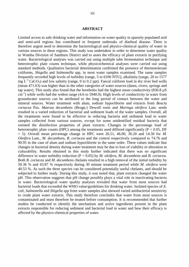
ABSTRACT
Limited access to safe drinking water and information on water quality in sparsely populated arid
and semi-arid regions has contributed to frequent outbreaks of diarheal disease. There is
therefore urgent need to determine the bacteriological and physico-chemical quality of water in
various sources in these regions. This study was undertaken in order to determine water quality
in Wamba Division of Samburu District and to asses the efficacy of plant extracts in purifying
water. Bacteriological analyses was carried out using multiple tube fermentation technique and
heterotrophic plate counts technique, while physicochemical analyses were carried out using
standard methods. Qualitative bacterial determination confirmed the presence of thermotolerant
coliforms, Shigella and Salmonella spp. in most water samples examined. The same samples
frequently recorded high levels of turbidity (range, 5 to 6100 NTU), alkalinity (range, 20 to 1577
mg L-1
CaCO3) and low salinity (range, 0 to 0.2 ppt). Faecal coliform load in dry river bed wells
(mean 471.63) was higher than in the other categories of water sources (dams, rivers, springs and
tap water). This study also found that the boreholes had the highest mean conductivity (830.8 µS
cm-1
) while wells had the widest range (4.6 to 5940.0). High levels of conductivity in water from
groundwater sources can be attributed to the long period of contact between the water and
mineral sources. Water treatment with alum, sodium hypochlorite and extracts from Boscia
coriacea Pax. Maerua decumbens (Brogn.) Dewolf roots and Moringa oleifera Lam. seeds
resulted in a varied reduction of bacterial and sediment loads of the water samples. Overall, all
the treatments were found to be effective in reducing bacteria and sediment load in water
samples collected from various sources, except for some unidentified residual bacteria that
resisted the disinfection properties of plant extracts. Changes in the percentage load of
heterotrophic plate counts (HPC) among the treatments used differed significantly (P < 0.05, DF
= 5). Overall mean percentage change in HPC were 26.51, 46.00, 30.20 and 14.50 for M.
Oleifera Lam., M. decumbens, B. coriacea and the control respectively compared to 74.76 and
90.95 in the case of alum and sodium hypochlorite in the same order. These values indicate that
changes in bacterial density during water treatment may be due to loss of viability or alteration in
culturability. Results obtained in this study further indicated that there was no significant
difference in water turbidity reduction (P < 0.05) by M. oleifera, M. decumbens and B. coriacea.
Both B. coriacea and M. decumbens chelants resulted in a high removal of the initial turbidity by
50.36 % and 43.87 % respectively during 30 minute treatment period while M. oleifera were
40.53 %. As such the three species can be considered potentially useful chelants, and should be
subjected to further study. During this study, it was noted that, plant extracts changed the water
pH. This observation suggests that pH change possibly plays a vital role in inactivating bacteria
in water. Bacteriological water quality analyses revealed that water from most sources had
bacterial loads that exceeded the WHO value/guidelines for drinking water. Isolated species of E.
coli, Salmonella and Shigella spp from water samples also showed varied antibacterial sensitivity
to crude plant water extracts. This study therefore concludes that water from most sources is
contaminated and must therefore be treated before consumption. It is recommended that further
studies be conducted to identify the mechanism and active ingredients present in the plant
extracts responsible for reducing sediment and bacterial load in water and how their efficacy is
affected by the physico-chemical properties of water.
1
CHAPTER ONE: INTRODUCTION
1.1 Background to the study
The Millennium Development Goals include halving the proportion of people without access
to safe drinking water by 2015. The assessment of the health risk from naturally occurring
microbes in drinking water continues to be of a high interest to microbiologists, public health
practitioners and water supply regulators (Richards et al., 1992; Hunter 1993). Although a
number of studies have investigated water supply and quality in sub-Saharan Africa (Shier et
al., 1966), very limited information is available from the more sparsely populated arid and
semi-arid regions.
Samburu district is semi arid with annual rainfall of between 250 - 500 mm. As such, water is
scarce and is the most critical resource in the region. The area supports pastoral communities
that coexist with wildlife. Hence humans, wildlife and livestock compete for available water
resource in ephemeral laggas, natural ponds, man made dams and the only permanent water
source, the Ewaso Nyiro river. Competition intensity varies with season. During the dry
season, the available surface water sources considerably reduce. This coupled with increased
intensity of use by humans, livestock and wildlife leads to a deterioration of water quality.
Contamination results when animal wastes are released into the water.
Livestock movement and migratory wildlife in search of water can contaminate water by
carrying pathogens from one source to another. High water evaporation rates increase the
concentration of dissolved ions in the water, which favour high microbial growth. Ground
water is invariably cleaner than surface water sources in rural areas. However the latter may
need treatment to reduce the load of suspended solids and to kill microorganisms.
2
Removal of suspended solids presents the greatest treatment challenge, and there is a need to
develop and choose technologies that will be sustainable in the medium to long term. In
general, complex solutions should be avoided. Most drinking waters, even with residual
disinfectants, have a natural bacterial population. This is because they use naturally available
carbon and nitrogen sources in water to multiply (Manaia et al., 1990; Reasoner, 1990).
Addition of phosphates to water bodies influences growth of bacteria even at concentrations
of less than 20 µg L-1
(Sathasivan et al., 1997; Lehtola et al., 2002). Animal and human
wastes are major sources of nitrogen, phosphates and pathogenic bacteria contamination of
aquatic environments. These nutrients can promote growth of pathogenic bacteria implicated
in causing disease in humans, wildlife and domestic animals. The type of treatment
technology or combination of technologies to be used depends on the quality of water to be
treated. Hence, there is need for monitoring the quality of water bodies and to explore the
efficacy of various treatment options such as coagulants from plant extracts to reduce
bacterial load and dissolved solutes.
1.2 Problem statement and justification
Information on the bacteriological quality of water from various sources used for domestic
purposes and for livestock watering in Samburu is limited. Livestock faecal wastes may
contain pathogenic microorganisms such as Salmonella and Escherichia coli. When livestock
drink water contaminated with enteric bacteria, they may be exposed to potential pathogens
like Salmonella, which cause salmonellosis in cattle and enteric fever (Typhoid) in humans.
Such waterborne diseases have been shown to be capable of infecting large numbers of
animals over a short time.
The water treatment potential of natural coagulants to purify water in Samburu district has not
been explored. Use of chemical treatments like chlorine as a disinfectant of polluted drinking
3
water is only practiced to a small extent in Samburu. Apart from the high cost of commercial
disinfectants, their use has been reported to lead to the formation of trihalomethane products
(Milot et al., 2000), which are potentially carcinogenic (King et al., 1996). There is, therefore,
a need to develop water purification methods that are cost effective, locally available and
environmentaly friendly. Determination of the physico-chemical properties of water such as
turbidity, pH, salinity, temperature, dissolved oxygen, nitrogen and phosphorus will provide
useful information on overall water quality and its potential to support bacterial growth
(APHA, 2005).
This study describes the microbial quality of water from various sources commonly used by
humans, livestock and wildlife in Wamba Division, Samburu District. The study also
examines the relations between the physico-chemical properties and the microbial properties
of water sources, and assesses the efficacy of plant extracts in purifying water to acceptable
levels for human consumption.
1.3 Research questions
i. What is the level of bacterial contamination of water from common sources in Wamba
Division of Samburu District?
ii. How does the bacterial load and type compare with the type of water source?
iii. How does the bacterial load and type compare with the physico-chemical properties of
the water body?
iv. How effective are the roots extracts of Boscia coriacea Pax., Maerua decumbens
(Brogn.) Dewolf and of seeds of Moringa oleifera Lam. in reducing the bacterial and
sediment load in water?
4
1.4 Hypotheses
i. There is no significant bacterial contamination of water from common water sources
in Wamba Division of Samburu District.
ii. There is no relationship between bacterial load, type and water source.
iii. There is no relationship between bacterial load, type and the physico-chemical
properties of the water.
iv. The roots extracts of Boscia coriacea Pax., Maerua decumbens (Brogn.) Dewolf and
seeds of Moringa oleifera Lam. are not effective in reducing the bacterial and
sediment load in water.
1.5 Objectives
1.5.1 General objective
To determine water quality and to and to assess the efficacy of plant extracts in purifying
water in Wamba Division, Samburu District.
1.5.2 Specific objectives
i. To determine the level of bacterial contamination of water from common
water sources in Wamba Division, Samburu District.
ii. To compare bacterial load, type with type of water source.
iii. To compare bacterial load, type and physico-chemical properties of water.
iv. To determine the efficacy of roots extracts of Boscia coriacea Pax., Maerua
decumbens (Brogn.) Dewolf and seeds of Moringa oleifera Lam. in reducing
the bacterial and sediment load in water.
5
1.6 Significance of the study
This study evaluated the level of health risk that the residents of Wamba division are exposed
to through direct use of water from the various water sources. The documented information
will be important in the formulation of guidelines on water resource use in the division.
Information on the bacterial load in water from different sources will be used by local public
health officers to determine the sources of contamination and to educate the local community
on how to protect the water sources from contamination. Information on the efficacy of the
plant extracts in water purification validates their use as an alternative to chemical treatment,
or forms the basis of their use combination with other water treatment methods to achieve safe
drinking water. This will in the long run provide a cost effective and user-friendly option to be
adopted for domestic household purification of water.
6
CHAPTER TWO: LITERATURE REVIEW
2.1 Bacterial water quality
Dirty and polluted water can contain many harmful organisms including pathogenic bacteria,
which cause diseases like cholera, bacillary dysentery, typhoid, and diarrhea. Disinfection of
water aims to kill these pathogens without leaving any harmful chemical substances in the
water. Coliform bacteria, thermotolerant (faecal) coliforms and Escherichia coli have for
almost a century been used as indicators of the bacterial safety of drinking water (Leclerc et
al., 2001). Water quality guidelines state that drinking water must not contain waterborne
pathogens. More specifically, Escherichia coli or thermotolerant coliforms should not be
present in any 100 ml sample of drinking water (WHO 2004). The guidelines further state that
should this value be exceeded, immediate investigative action must be taken, including
repeated testing, thorough inspection of the water source, and the general hygiene of the water
distribution system.
Unlike other indicators, such as Escherichia coli or total coliforms, low concentrations of
heterotrophic plate count (HPC) bacteria will still be present after treatment of drinking water.
In general, water purification can achieve heterotrophic bacteria concentrations of 10 colony-
forming units (CFU) per millilitre or less in finished water (Fox and Reasoner, 1999).
2.2 Water quality changes
Natural waters are subject to important changes in their microbial quality. These changes have
direct impacts on the decisions made by water authorities striving to maintain safe conditions
in catchments or distribution systems. Correct decision making by water authorities relies
heavily on having access to rapid and accurate bacteriological data (Daniel et al., 2003). This
can be obtained by using HPC, which is a suitable tool for monitoring changes in bacterial
water quality over time for a particular catchment or distribution system (Daniel et al., 2003).
7
Changes in the microbial quality of water may arise from agricultural use, discharges of
sewage, wastewater resulting from human activity, and storm or surface water runoff.
Previous studies have suggested that sewage effluents contain a wide variety of pathogenic
microorganisms whose density and variety are related to the size of the human population, the
seasonal incidence of the illness, and dissemination of pathogens within the community
(Pipes, 1982). Discharge of domestic sewage into water bodies also depletes dissolved oxygen
leading to low dissolved oxygen concentrations and high numbers of enteric bacteria. An
improvement of water quality is associated with an increase in the concentration of dissolved
oxygen and a decrease in the load of faecal coliforms.
Changes in water conductivity results from changes in the mineral composition of water,
which may be caused by seasonal variations in the chemical composition of the various
sources of water. It may also indicate sewage, industrial or agricultural pollution or intrusion
of saline waters. Determination of various water quality properties on a regular basis may,
therefore reveal a need to adjust water treatment according to changes in raw water quality.
2.3 Water quality challenges
Livestock practices that can impact on water quality include both intensive and non-intensive
operations. Intensive agricultural livestock operations (waste management and disposal) have
been identified as point sources of pollution to streams. In water scarce areas, livestock and
wildlife density tends to be high in water catchment areas and near water sources due to the
presence of pasture and water. These animals generate large quantities of wastes. Water
quality changes associated with livestock production include changes in nutrients loads,
(nitrogen and phosphorus), microorganisms (e.g. bacteria, faecal coliforms, Cryptosporidium,
Giardia) and organic material such as livestock wastes. Localized concentration of animal
waste is considered a point source of pollution for surface or ground water. Lack of manure

8
management can adversely affect the water quality of receiving streams, its aquatic life, and
reuse of the water downstream for agricultural, recreational and drinking water purposes.
High animal densities within the catchment area of a water source may lead to the loss of
protective cover of grasses, herbs, and shrubs due to trampling, grazing and browsing action.
This exposes the soil to agents of erosion. Increased erosion results in a loss of organic matter,
fine soil particles, nutrients, and microbes in the soil (Harper and Marble 1988; Schimel et al.,
1985; Belnap, 1995). They may be transported by surface runoff to eventually contaminate
drinking water.
2.4 Biological indicators of water quality
The presence of faecal coliforms (over 99 % of which are Escherichia coli) in a water body is
an indication of possible human/animal waste contamination and the possible presence of
pathogenic bacteria. The detection of Escherichia coli provides definite evidence of faecal
contamination. However, in practice, the detection of thermotolerant (faecal) coliform
bacteria is an acceptable alternative. According to World Health Organisation (WHO, 1997,
2004) standards, faecal coliforms should be absent (0 colony forming units per 100 ml water)
in portable water while total coliforms should be less than 10 colony forming units in any 100
ml water sample. The measurement of faecal coliforms can give an indication of the likely
chlorine demand and also indicates where more intensive treatment is needed.
2.4.1 Heterotrophic plate counts
Heterotrophs are those microorganisms that use organic compounds for most or all of their
carbon requirements (Singleton and Sainsbury, 2001). Most bacteria, including those
associated with drinking water systems, are heterotrophs. A common and universal water
testing method for the general bacteriological quality of water is the heterotrophic plate count
(HPC). This technique assesses the number of bacteria in water that are able to replicate and
9
form visible colonies on a solid nutrient medium under specified test conditions (Australian
Drinking Water Guidelines, 1996). Although not a direct method for the detection of
pathogenic organisms, HPC can be used as a tool to assess the overall quality of water. In
particular, HPC can be used to asses the effectiveness of water treatment processes and to
detect bacterial regrowth within a distribution system (Geldreich, 1996).
The HPC technique has some inherent limitations. One important limitation is that the media
used do not allow for the detection of all bacteria of interest, such as the chemolithotrophic
ammonia-oxidizing bacteria that often colonize chloraminated distribution systems (Cunliffe,
1991). Furthermore, the incubation conditions of HPC, leaves slower growing heterotrophic
organisms undetected (Australian Standard, 1995). Additionally, some bacteria, when
exposed to environmental stresses, are capable of maintaining metabolic activity whilst
developing recalcitrance to culture, commonly referred to as viable but nonculturable (VBNC)
bacteria (McDougald et al., 1998). Chlorine disinfection used in the water treatment process
may also cause sublethal injury of some bacteria (McFeters et al., 1986; Du Preez et al.,
1995), thereby rendering them nonculturable by routine HPC. This has been shown for both
coliform and enteropathogenic bacteria in drinking water (McFeters et al., 1986; du Preez et
al., 1995). The standard HPC procedure may also dilute vital signalling molecules which may
be required for bacterial growth (Kaprelyants and Kell, 1996). Finally, HPC culture may be
impeded by substrate-accelerated bacterial cell death, promoted by the presence of certain
substrates in the HPC medium that were limiting in the environment when bacterial starvation
was initiated (Barer and Harwood, 1999).
2.4.2 Thermotolerant coliforms
Thermotolerant (faecal) coliforms are a subset of total coliforms that possess a more direct
and closer relationship with homeothermic faecal pollution (Geldreich, 1967). These bacteria
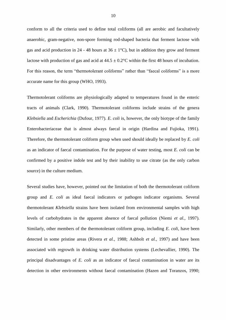
10
conform to all the criteria used to define total coliforms (all are aerobic and facultatively
anaerobic, gram-negative, non-spore forming rod-shaped bacteria that ferment lactose with
gas and acid production in 24 - 48 hours at 36 ± 1°C), but in addition they grow and ferment
lactose with production of gas and acid at 44.5 ± 0.2°C within the first 48 hours of incubation.
For this reason, the term “thermotolerant coliforms” rather than “faecal coliforms” is a more
accurate name for this group (WHO, 1993).
Thermotolerant coliforms are physiologically adapted to temperatures found in the enteric
tracts of animals (Clark, 1990). Thermotolerant coliforms include strains of the genera
Klebsiella and Escherichia (Dufour, 1977). E. coli is, however, the only biotype of the family
Enterobacteriaceae that is almost always faecal in origin (Hardina and Fujioka, 1991).
Therefore, the thermotolerant coliform group when used should ideally be replaced by E. coli
as an indicator of faecal contamination. For the purpose of water testing, most E. coli can be
confirmed by a positive indole test and by their inability to use citrate (as the only carbon
source) in the culture medium.
Several studies have, however, pointed out the limitation of both the thermotolerant coliform
group and E. coli as ideal faecal indicators or pathogen indicator organisms. Several
thermotolerant Klebsiella strains have been isolated from environmental samples with high
levels of carbohydrates in the apparent absence of faecal pollution (Niemi et al., 1997).
Similarly, other members of the thermotolerant coliform group, including E. coli, have been
detected in some pristine areas (Rivera et al., 1988; Ashbolt et al., 1997) and have been
associated with regrowth in drinking water distribution systems (Lechevallier, 1990). The
principal disadvantages of E. coli as an indicator of faecal contamination in water are its
detection in other environments without faecal contamination (Hazen and Toranzos, 1990;
11
Hardina and Fujioka, 1991) and its low survival capability in aquatic environments when
compared with faecal pathogens (Borrego et al., 1983; Cornax et al., 1990).
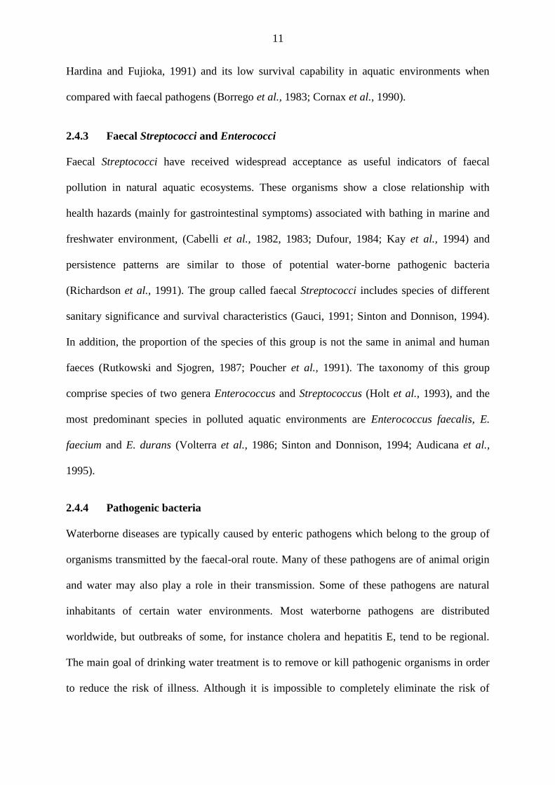
2.4.3 Faecal Streptococci and Enterococci
Faecal Streptococci have received widespread acceptance as useful indicators of faecal
pollution in natural aquatic ecosystems. These organisms show a close relationship with
health hazards (mainly for gastrointestinal symptoms) associated with bathing in marine and
freshwater environment, (Cabelli et al., 1982, 1983; Dufour, 1984; Kay et al., 1994) and
persistence patterns are similar to those of potential water-borne pathogenic bacteria
(Richardson et al., 1991). The group called faecal Streptococci includes species of different
sanitary significance and survival characteristics (Gauci, 1991; Sinton and Donnison, 1994).
In addition, the proportion of the species of this group is not the same in animal and human
faeces (Rutkowski and Sjogren, 1987; Poucher et al., 1991). The taxonomy of this group
comprise species of two genera Enterococcus and Streptococcus (Holt et al., 1993), and the
most predominant species in polluted aquatic environments are Enterococcus faecalis, E.
faecium and E. durans (Volterra et al., 1986; Sinton and Donnison, 1994; Audicana et al.,
1995).
2.4.4 Pathogenic bacteria
Waterborne diseases are typically caused by enteric pathogens which belong to the group of
organisms transmitted by the faecal-oral route. Many of these pathogens are of animal origin
and water may also play a role in their transmission. Some of these pathogens are natural
inhabitants of certain water environments. Most waterborne pathogens are distributed
worldwide, but outbreaks of some, for instance cholera and hepatitis E, tend to be regional.
The main goal of drinking water treatment is to remove or kill pathogenic organisms in order
to reduce the risk of illness. Although it is impossible to completely eliminate the risk of
12
waterborne disease, adopting multi-barrier, source to tap approach to safe drinking water will
reduce the numbers of microorganisms in drinking water. This approach includes protection
of the water source (where possible), the use of appropriate and effective treatment methods,
well-maintained distribution systems, and routine verification of drinking water safety. All
drinking water supplies should be disinfected, unless specifically exempted by the responsible
authority. In addition, all surface and groundwater sources under the direct influence of
surface run-off water should be filtered before treatment.
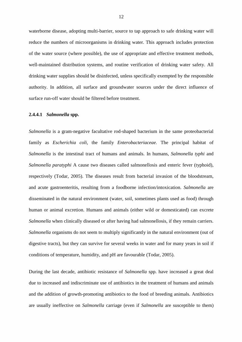
2.4.4.1 Salmonella spp.
Salmonella is a gram-negative facultative rod-shaped bacterium in the same proteobacterial
family as Escherichia coli, the family Enterobacteriaceae. The principal habitat of
Salmonella is the intestinal tract of humans and animals. In humans, Salmonella typhi and
Salmonella paratyphi A cause two diseases called salmonellosis and enteric fever (typhoid),
respectively (Todar, 2005). The diseases result from bacterial invasion of the bloodstream,
and acute gastroenteritis, resulting from a foodborne infection/intoxication. Salmonella are
disseminated in the natural environment (water, soil, sometimes plants used as food) through
human or animal excretion. Humans and animals (either wild or domesticated) can excrete
Salmonella when clinically diseased or after having had salmonellosis, if they remain carriers.
Salmonella organisms do not seem to multiply significantly in the natural environment (out of
digestive tracts), but they can survive for several weeks in water and for many years in soil if
conditions of temperature, humidity, and pH are favourable (Todar, 2005).
During the last decade, antibiotic resistance of Salmonella spp. have increased a great deal
due to increased and indiscriminate use of antibiotics in the treatment of humans and animals
and the addition of growth-promoting antibiotics to the food of breeding animals. Antibiotics
are usually ineffective on Salmonella carriage (even if Salmonella are susceptible to them)
13
because the site of carriage may not allow penetration by the antibiotic. Resistance to
ampicillin, streptomycin, kanamycin, tetracycline, and sulfonamides is commonly observed.
Colistin resistance has not yet been observed. Recently Salmonella typhi strains resistant to
chloramphenicol (the antibiotic most commonly used against typhoid) strains have been
isolated in India, Thailand, and Vietnam.
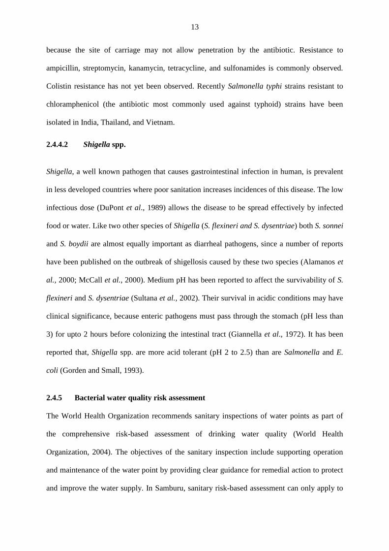
2.4.4.2 Shigella spp.
Shigella, a well known pathogen that causes gastrointestinal infection in human, is prevalent
in less developed countries where poor sanitation increases incidences of this disease. The low
infectious dose (DuPont et al., 1989) allows the disease to be spread effectively by infected
food or water. Like two other species of Shigella (S. flexineri and S. dysentriae) both S. sonnei
and S. boydii are almost equally important as diarrheal pathogens, since a number of reports
have been published on the outbreak of shigellosis caused by these two species (Alamanos et
al., 2000; McCall et al., 2000). Medium pH has been reported to affect the survivability of S.
flexineri and S. dysentriae (Sultana et al., 2002). Their survival in acidic conditions may have
clinical significance, because enteric pathogens must pass through the stomach (pH less than
3) for upto 2 hours before colonizing the intestinal tract (Giannella et al., 1972). It has been
reported that, Shigella spp. are more acid tolerant (pH 2 to 2.5) than are Salmonella and E.
coli (Gorden and Small, 1993).
2.4.5 Bacterial water quality risk assessment
The World Health Organization recommends sanitary inspections of water points as part of
the comprehensive risk-based assessment of drinking water quality (World Health
Organization, 2004). The objectives of the sanitary inspection include supporting operation
and maintenance of the water point by providing clear guidance for remedial action to protect
and improve the water supply. In Samburu, sanitary risk-based assessment can only apply to
14
37 % of the water sources (mainly springs and boreholes) since 63 % (majority) are temporary
sources. According to Lloyd and Bartram (1991) sanitary inspection is not designed to replace
microbiological water quality testing, but rather is a complementary assessment designed to
identify risks to water quality. Owing to the nomadic lifestyle of the Samburu population, and
the increasing competition for water, a risk assesment approach which aims at determining the
most suitable water source for use and enhancing immediate water treatment before use will
be of more immediate health significance than visual sanitary risk inspection.
Waterborne pathogens have been recognized as a significant risk to public health for more
than a century. The risk is based on microbiological standards of drinking water (zero faecal
coliforms and pathogens). Quantitative risk assesment has demonstrated that the risks from
pathogens are greater than the risks from disinfection by products (Craun et al., 2001). The
greater risks of waterborne bacterial disease are known to be associated with contaminated
drinking water. The pathogens that are most widely recognized to cause dangerous
waterborne diseases by the public are Salmonella typhi, Vibrio cholerae, Shigella and
pathogenic E. coli
2.4.6 Physico-chemical indicators of water quality
2.4.6.1 Hydrogen ion potential - pH
The pH of water is a measure of whether it is acidic or alkaline. However, its significance
depends on the buffering capacity of the water. pH affects the acceptability of water for
various uses and influences the solubility of metal ions in water.
2.4.6.2 Turbidity - NTU (Nephelometric Turbidity Units)
Turbidity is a measure of the reduction in the transparency of a water body as a result of light
scattering by suspended particulate matter (Ziegler, 2002). It is a measure of the relative
15
clarity of water (Sadar, 1996). Levels of turbidity in raw water can range from less than 1.0
NTU to more than 1000.0 NTU. Turbidity is not an absolute value, but a relative value
representing a qualitative measurement that can yield different readings based on the method
used (Ankcorn, 2003). Turbidity can also be used to estimate loads for contaminants typically
bound to sediment particles, such as nutrients and bacteria (Ankcorn, 2003). Turbidity
readings may vary between water sources due to water colour, suspended particle size and
particle composition (Packman et al., 1999). Organic particles in water have been shown to
absorb light, and therefore provide different turbidity values compared to water carrying
primarily mineral soils (Lewis, 1996).
The microbiological quality of drinking water can be significantly affected by turbidity. This
is because microbial growth in water is most extensive on the surfaces of particles and inside
loose flocs, which may occur naturally or be formed during treatment. Microbial growth
occurs because nutrients adsorb to surfaces, allowing bacteria to grow more efficiently than
when in free suspension (Brock, 1966 and Stotzky, 1966). A study by Reilly and Kippin
(1983) suggested that turbidity of around 1.0 NTU has no impact on coliforms or HPC.
According to WHO, the indicator value only applies at the treatment works since turbidity
above 1.0 NTU can compromise disinfection by increasing chlorine demand, and hence the
cost of water treatment. However, it is important to optimise the removal of turbidity during
water treatment in order to remove all micro-organisms.
2.4.6.3 Total organic carbon (TOC)
TOC is a non specific measure of the dissolved organic matter present in water. High
concentration can lead to the formation of high levels of chlorination by-products such as
Trihalomethanes (THMs). An abnormal increase in TOC can be an indicator of treatment
failure in the system and it is associated with an increase in assimilable organic carbon
16
(biologically available carbon). This may give rise to an increase in bacterial growth and
formation of biofilms in water distribution systems.
2.4.7 Water pollution by organic matter
Input of organic matter is a normal feature of aquatic systems. The total load usually depends
on the catchment area of the water body and the density of humans and wildlife in areas
where organic wastes are discharged into water bodies through point and non point sources.
Organic wastes discharge into waterbodies increases water turbidity, organic compounds,
nutrients, and the bacterial load in the waterbody (Bitton and Gerba, 1984). Organic wastes
contain a high concentration of both phosphorus and nitrogen. A single cow can produce as
much phosphorus (about 18 kg per year) as 212 ha of forest or more than 57 ha of cropland. It
can also excrete as much nitrogen (58 kg per year) as 68 ha of forest or more than 6 ha of
arable land (Brian, 1980).
Phosphorus enters water bodies either as inorganic phosphate ions or organic polymers and
biodegradable organic compounds in living organisms and detritus. Presence of phosphorus in
a water body promotes bacterial growth (Geildreich, 1996; LeChevallier, 1990; LeChevallier
et al., 1991). A number of studies carried out through batch experiments using drinking water
have shown that the addition of phosphates may lead to increased bacterial culturability
(Miettinen et al., 1997; Sathasivan and Ohgaki, 1999) and sometimes to bacterial growth
(Sathasivan et al., 1997; Lehtola et al., 2002). However, in drinking water in which carbon is
the limiting nutrient for bacterial growth, the addition of phosphates does not induce bacterial
growth (Lyons et al., 1995; Cohen et al., 1999; Appenzeller et al., 2001; Batté et al., 2003).
Treatment of water contaminated by organic matter has always been one of the major public
health concerns. The primary inorganic compound of concern to public health in groundwater
contaminated with livestock wastewater is nitrate-nitrogen, which may cause
17
methemoglobinemia (also called blue-baby syndrome) in infants (Canter, 1997). In most
cases, the recommended treatment method requires the removal of a large part of the organic
load. This is often accompanied by the elimination of nitrogen and phosphorus (Rotereau,
1969; Moletta and Torrijos, 1999; Hamdani, 2002).
2.5 Common water treatment methods
2.5.1 Solar disinfection
Solar disinfection involves storing contaminated drinking water in a transparent container that
is placed in direct sunlight for periods of up to eight hours before consumption (Conroy et al.,
1996; Sommer et al., 1997). This technique is highly effective against a broad range of
pathogens (Conroy et al., 2001; Kehoe et al., 2004; Smith et al., 2000; Wegelin et al., 1994).
Coliforms in water and sewage have been completely inactivated by exposure to sunlight for
about one hour in the presence of methylene blue or rose Bengal (Archer and Juken, 1977).
This method is cheap and can easily be applied by rural communities to disinfect water but
cannot be used to sediment dissolved particulate matter in water.
2.5.2 Chemical water treatment methods
2.5.2.1 Alum coagulation
Use of alum has been traditionally practiced for centuries in many parts of the world (Jahn
and Dirar, 1979; Gupta and Chaudhuri, 1992). In one study, potash alum was evaluated for
household water treatment in a suburban community in Myanmar by adding it to water in
traditional storage vessels (160 L capacity) at 500 mg L-1
. Faecal coliform contamination was
reduced by 90 - 98 % and consumer acceptance of the treated water was high (Oo et al.,
1993). However, because the coagulation-flocculation treatment with alum, iron and other
coagulants requires knowledge and skills to optimize treatment conditions, this type of
treatment is less likely to be performed reliably at point of use for water treatment, especially
18
by semi literate rural communities. Similarly, the relatively high costs of alum and ferric salts,
makes this treatment option not affordable. Some studies have reported that aluminium, a
major component of alum, may induce Alzheimer’s disease (Martyn et al., 1989). Alum also
reacts with the natural alkalinity of water leading to a reduction of pH and low efficiency in
coagulation in cold water (Haarhoff and Cleasby, 1988). This limits the acceptance and use of
alum.
2.5.2.2 Chlorine treatment
Among the drinking water disinfectants, free chlorine is the most widely used and the most
affordable. It is also highly effective against nearly all waterborne pathogens, with notable
exceptions being Cryptosporidium parvum cysts and Mycobacteria species (Sobsey, 1989). At
doses of a few mg L-1
and contact times of about 30 minutes, free chlorine generally
inactivates more than 99.99 % of enteric bacteria and viruses. Despite its disinfection
property, it does not sediment dissolved solids in water hence must be combined with another
sedimentation method.
In water with turbidities ranging from 3.8 to 84.0 NTU, coliforms were detected after the
water was treated with chlorine (free chlorine residuals between 0.1 and 0.5 mg L-1
) after a
minimum contact time of 30 minutes (Sanderson and Kelly, 1964). While studying the
efficiency of chlorination in killing coliforms in unfiltered surface water supplies,
LeChevallier et al., (1981) observed a negative correlation between the effectiveness of
chlorination and turbidity. Chlorine (as hypochlorous acid) also reacts readily with organic
matter containing unsaturated bonds, phenolic groups and nitrogen groups, giving rise to taste
and odour producing compounds (Sawyer and McCarty, 1967) and trihalomethanes (Rook,
1977). Chlorine is also expensive and requires skills to apply, hence not feasible for use by
rural households.
19
2.5.3 Treatment with plant extracts
The mucilage extracted from the seeds of Tamarindus indica Linn. has been used as a
flocculant for removal of sulphate and phosphate ions in aqueous medium. A mucilage dose
of 50 mg L-1
has been reported to remove 74 % and 76 % of sulphates and phosphates
respectively after 30 minutes (Anuradha and Malvika, 2005). Seeds of Moringa species have
been reported to be an effective, simple and low-cost flocculant for turbid surface water that
can be used for household water treatment (Jahn and Dirar, 1979; Jahn, 1981; Olsen, 1987).
Moringa oleifera Lam. seed is a non toxic (Grabow et al., 1985) and natural organic
flocculant.
The treatment potential of other vegetable materials have also been investigated. These
include extracts of Okra and Strychnos potatorum Linn. seeds (Al Samawi and Shokralla,
1996) and Tamarind (Bhole, 1995). The effectiveness of Strychnos potatorum Linn. to
flocculate or precipitate microbes and turbidity in water has also been investigated (Tripathi et
al., 1976). Microbial reductions of about 50 % and 95 % have been reported for plate count
bacteria and turbidity respectively. Past studies have documented an 80 to 99 % turbidity
removal by Moringa oleifera Lam. as the primary coagulant both for raw waters and
synthetics turbid waters (distilled water with turbidity added as kaolin) (Muyibi and Okufu,
1995; Ndbigengesere et al., 1995; Muyibi and Evison, 1996). However, according to Muyibi
and Okufu (1995), Moringa oleifera Lam. might not be an efficient coagulant for water with
low turbidity. Although the water treatment potential of Moringa oleifera Lam. has been
extensively investigated and the plant recommended as a coagulant (Muyibi and Okufu, 1995;
Muyibi and Evison, 1995, 1996; Ndabigengesere and Narasiah 1996, Ndabigengesere et al.,
1995) it is important to establish its effectiveness in waters from different sources and with
different physico-chemical properties.
20
2.6 Microbial reduction by coagulation-flocculation
Optimum coagulation to achieve maximum reduction of turbidity and microbes requires
careful control of coagulant dose, pH, consideration of the quality of the water being treated
as well as appropriate mixing conditions for optimum flocculation. Lack of attention to these
details can result in poor coagulation-flocculation and inefficient removal of particles and
microbes. Under optimum conditions, coagulation-flocculation and sedimentation with alum
and iron can achieve microbial reductions of between 90 to 99 % for all classes of waterborne
pathogens (Payment and Armon, 1989). Greater microbial reductions (more than 99.99 %)
can be achieved with lime coagulation-flocculation at high pH levels (pH more than11).
2.7 Bacterial antibiotic resistance
Antibiotic resistant bacteria have been isolated from a variety of sources such as domestic
sewage, hospitals drinking water, rivers and lakes (Magee and Quinn, 1991; Ogan and Nwiika
1993; Boon and Catanach, 1999). These strains include bacteria that are pathogenic to humans
and many of these show multiple resistance (MAR) (Lancini et al., 1995). Increasing
resistance to commonly used antimicrobial agents in Shigella and Salmonella species has
become a major public health concern worldwide (Mandell et al., 2000). These antibiotic
resistant bacteria are significant environmental contaminants (Huber, 1971) and calls have
been made for antibiotic resistance to be considered when establishing bacteriological water
quality criteria (Bell et al., 1983; El-Zanfaly, 1991).
21
CHAPTER THREE: MATERIALS AND METHODS
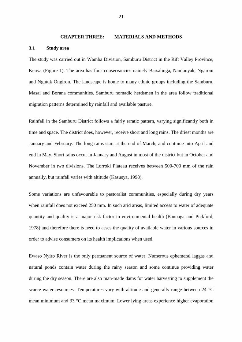
3.1 Study area
The study was carried out in Wamba Division, Samburu District in the Rift Valley Province,
Kenya (Figure 1). The area has four conservancies namely Barsalinga, Namunyak, Ngaroni
and Ngutuk Ongiron. The landscape is home to many ethnic groups including the Samburu,
Masai and Borana communities. Samburu nomadic herdsmen in the area follow traditional
migration patterns determined by rainfall and available pasture.
Rainfall in the Samburu District follows a fairly erratic pattern, varying significantly both in
time and space. The district does, however, receive short and long rains. The driest months are
January and February. The long rains start at the end of March, and continue into April and
end in May. Short rains occur in January and August in most of the district but in October and
November in two divisions. The Lorroki Plateau receives between 500-700 mm of the rain
annually, but rainfall varies with altitude (Kasusya, 1998).
Some variations are unfavourable to pastoralist communities, especially during dry years
when rainfall does not exceed 250 mm. In such arid areas, limited access to water of adequate
quantity and quality is a major risk factor in environmental health (Bannaga and Pickford,
1978) and therefore there is need to asses the quality of available water in various sources in
order to advise consumers on its health implications when used.
Ewaso Nyiro River is the only permanent source of water. Numerous ephemeral laggas and
natural ponds contain water during the rainy season and some continue providing water
during the dry season. There are also man-made dams for water harvesting to supplement the
scarce water resources. Temperatures vary with altitude and generally range between 24 °C
mean minimum and 33 °C mean maximum. Lower lying areas experience higher evaporation
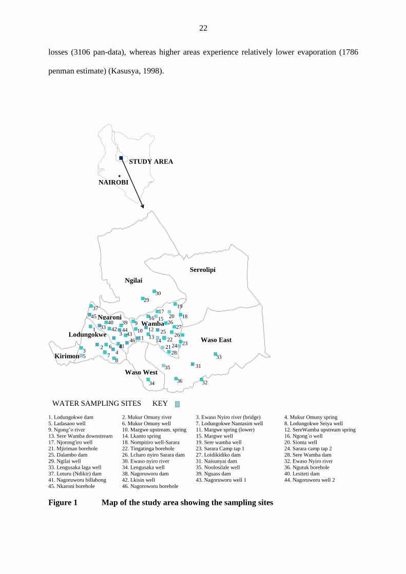
22
losses (3106 pan-data), whereas higher areas experience relatively lower evaporation (1786
penman estimate) (Kasusya, 1998).
1. Lodungokwe dam 2. Mukur Omuny river 3. Ewaso Nyiro river (bridge) 4. Mukur Omuny spring
5. Ladasaoo well 6. Mukur Omuny well 7. Lodungokwe Nantasim well 8. Lodungokwe Seiya well 9. Ngong’o river 10. Margwe upstream. spring 11. Margwe spring (lower) 12. SereWamba upstream spring
13. Sere Wamba downstream 14. Lkanto spring 15. Margwe well 16. Ngong`o well
17. Njorong'iro well 18. Nompitiro well-Sarara 19. Sere wamba well 20. Sionta well 21. Mjiriman borehole 22. Tingatinga borehole 23. Sarara Camp tap 1 24. Sarara camp tap 2
25. Dalambo dam 26. Lcharo nyiro Sarara dam 27. Loidikidiko dam 28. Sere Wamba dam
29. Ngilai well 30. Ewaso nyiro river 31. Naisunyai dam 32. Ewaso Nyiro river 33. Lengusaka laga well 34. Lengusaka well 35. Noolosilale well 36. Ngutuk borehole
37. Loturu (Ndikir) dam 38. Nagoruworu dam 39. Nguass dam 40. Lesiteti dam
41. Nagoruworu billabong 42. Lkisin well 43. Nagoruworu well 1 44. Nagoruworu well 2 45. Nkaroni borehole 46. Nagoroworu borehole
Figure 1 Map of the study area showing the sampling sites
NAIROBI
STUDY AREA
Wamba Ngaroni
Ngilai
Sereolipi
Waso East
Waso West
Lodungokwe
Kirimon
20
3 5
2 4
6
7
1
8
9 16
17 18
19
15
27
21
10 12
11 13 14 22
23 24
28
26 25
26
29
30
32
31
33
34
35
36
37
44
40 39
42
41
43
45
33
46
3
8
WATER SAMPLING SITES KEY
23
3.2 Water sampling
During this study, water from forty six sources in Wamba Division, Samburu District was
sampled and analyzed (Appendix 1). Water from all these sources was water used for drinking
and other household purposes. Some of these sources also served as watering points for both
livestock and wildlife.
Preliminary results of water quality sampled through purposive and random sampling were
used to choose the sampling sites with high microbial loads and diverse pathogenic bacteria.
The water sampling, preservation and tests were performed according to standard methods
(APHA, 2005). Water samples for microbial and chemical analyses were collected from each
source mostly in the morning hours (between 9.00 am to 12 noon) in sterile water sampling
bottles. Where possible, water samples for microbiological analyses were drawn directly from
the water body using sterile 125 ml plastic bottle. When this was not possible, the samples
were drawn using a sterile scooper. At each site, two sterile 125 ml plastic bottles fitted with
screw caps were used for water collection. Water samples for microbial and physico-chemical
analyses were transported to the laboratory in an iced cool box. Microbial analyses were
carried out at Wamba Mission Hospital while physico-chemical analyses were carried out at
Earthwatch camp laboratory.
3.3 Field measurements
3.3.1 pH (pH units)
Water pH was determined using a portable WTW Multiline P4 meter (Weilheim, Germany),
which uses a probe fitted with automatic temperature compensation to 25 °C. This meter
measures hydrogen ion concentration by direct potentiometry. pH readings were taken to the
nearest one decimal place. Where possible, the probe was lowered directly into water and the
meter readings allowed to stabilize for about three minutes before the pH value was taken.
24
3.3.2 Temperature (°C)
Temperature was taken in the field using the dissolved oxygen probe (CellOx325) of a
portable WTW Multiline P4 meter (Weilheim, Germany). The dissolved Oxygen probe has an
in-built temperature sensor, which gives water temperature readings in degrees celcius to one
decimal point. Where possible, the probe was lowered directly into water and the meter
readings allowed to stabilize for about three minutes before the temperature value was taken.
3.3.3 Electrical conductivity (µS cm-1
)
Electrical conductivity was measured in the field using a portable universal multiline P4
WTW (Wilheim Germany) meter. The multiline meter uses a Tetra Con 325 electrical
conductivity probe to measure conductivity. Where possible, the probe was lowered directly
into the water and the meter readings allowed to stabilize for about three minutes before the
electrical conductivity value was taken.
3.3.4 Dissolved oxygen (DO µg L-1
)
Dissolved oxygen was determined in the field using the dissolved oxygen probe (Ox325) of
the universal multiline P4 WTW (Wilheim Germany) meter. Where possible, the probe was
lowered directly into water and the meter readings allowed to stabilize for about three minutes
before the dissolved oxygen value was taken (APHA, 2005). When this was not possible,
samples were carefully collected with a water scooper and readings taken immediately.
3.4 Laboratory physico-chemical analyses
3.4.1 Total alkalinity (TA mg CaCO3 L-1
)
Total alkalinity was determined by the titration of 100 ml water samples with 0.02N standard
HCl using mixed methyl red bromocresol green indicator to determine titration end point.
Sample total alkalinity was computed using the procedure outlined in APHA (2005).
25
3.4.2 Phosphorus (mg L-1
)
Orthophosphate phosphorus was determined by ascorbic acid reduction procedure (APHA,
2005). Water samples were first filtered with pre-washed glass fiber filters (GF/C). To
determine total phosphorus, all forms of phosphorus in water samples were first oxidized to
orthophosphate (PO4 – P). This was achieved by autoclaving a 25 ml water sample at 140 °C
for 40 minutes in the presence of 0.2 g potassium persulfate oxidizing agent. A reagent blank
and standards in a suitable range were prepared from a standard phosphate solution (APHA,
2005). Colour intensity was measured using a digital grading spectrophotometer (Nanocolor
300 D) at a wavelength of 690 nm and the phosphates concentrations determined based on the
standards curve of known phosphate phosphorus concentrations.
3.4.3 Turbidity (NTU)
Turbidity of the water samples were determined by using a colorimeter (Smart - 26617). The
water sample was gently swirled and 10 ml was drawn out using a clean sterile syringe and
transferred into a suitable curvette and readings taken immediately. The turbidity of the
sample was measured against a distilled water blank. Initial turbidity was determined before
treatment of the water sample while change in turbidity was determined after 30 minutes and
24 hours of treatment in the laboratory using a colorimeter.
3.4.4 Most probable number (MPN) of total and faecal coliforms per 100 ml
Analysis of water for the presence of total coliforms was carried out using the multiple tube
fermentation technique (APHA, 2005) which involves three steps: the presumptive, confirmed
and the completed tests.
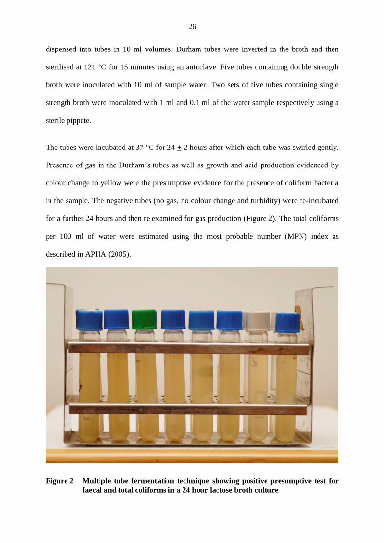
3.4.4.1 Presumptive test
The Presumptive test was carried out to determine total and faecal coliforms present in the
water samples (Figure 2). Double and single strength lactose broth was prepared and
26
dispensed into tubes in 10 ml volumes. Durham tubes were inverted in the broth and then
sterilised at 121 °C for 15 minutes using an autoclave. Five tubes containing double strength
broth were inoculated with 10 ml of sample water. Two sets of five tubes containing single
strength broth were inoculated with 1 ml and 0.1 ml of the water sample respectively using a
sterile pippete.
The tubes were incubated at 37 °C for 24 + 2 hours after which each tube was swirled gently.
Presence of gas in the Durham’s tubes as well as growth and acid production evidenced by
colour change to yellow were the presumptive evidence for the presence of coliform bacteria
in the sample. The negative tubes (no gas, no colour change and turbidity) were re-incubated
for a further 24 hours and then re examined for gas production (Figure 2). The total coliforms
per 100 ml of water were estimated using the most probable number (MPN) index as
described in APHA (2005).
Figure 2 Multiple tube fermentation technique showing positive presumptive test for
faecal and total coliforms in a 24 hour lactose broth culture
27
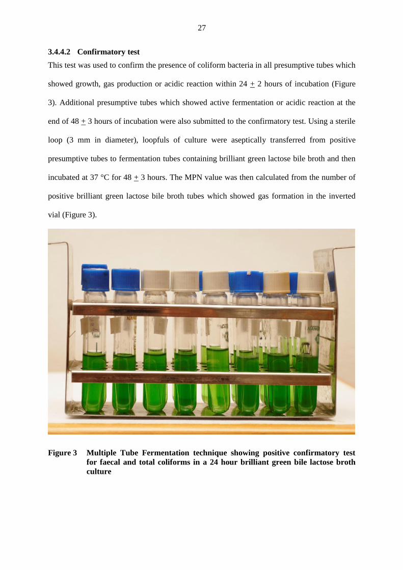
3.4.4.2 Confirmatory test
This test was used to confirm the presence of coliform bacteria in all presumptive tubes which
showed growth, gas production or acidic reaction within 24 + 2 hours of incubation (Figure
3). Additional presumptive tubes which showed active fermentation or acidic reaction at the
end of 48 + 3 hours of incubation were also submitted to the confirmatory test. Using a sterile
loop (3 mm in diameter), loopfuls of culture were aseptically transferred from positive
presumptive tubes to fermentation tubes containing brilliant green lactose bile broth and then
incubated at 37 °C for 48 + 3 hours. The MPN value was then calculated from the number of
positive brilliant green lactose bile broth tubes which showed gas formation in the inverted
vial (Figure 3).
Figure 3 Multiple Tube Fermentation technique showing positive confirmatory test
for faecal and total coliforms in a 24 hour brilliant green bile lactose broth
culture
28
3.4.4.3 Completed test
This test was carried out by submitting 10 % of positive confirmed tubes of brilliant green
lactose bile broth into Eosin Methylene Blue agar (EMB). Using a sterile 3 mm diameter loop,
a culture from each tube of brilliant green lactose bile broth showing gas was streaked on
plates containing Eosin Methylene Blue agar and incubated at 37 °C for 24 + 2 hours. Typical
lactose fermenting colonies were then isolated and transferred to a single strength lauryl
tryptose broth fermentation tubes (with inverted fermentation vials) and incubated at 37 °C for
24 + 2 hours. Presence of turbidity in lauryl tryptose broth and gas in Durham tube within 24
+ 2 hours indicates positive completed test for the presence of total coliforms.
3.4.5 Faecal coliforms (thermotolerant Escherichia coli)
The test for faecal coliforms was conducted simultaneously with the test for total coliforms at
the presumptive stage. Three sets of five tubes containing lauryl tryptose broth were each
inoculated with10 ml, 1 ml and 0.1 ml portions of water samples respectively and incubated at
37 °C for 48 hours. Presence of turbidity in lauryl tryptose broth and gas in Durham tube
within 24 + 2 hours indicates positive completed test for the presence of faecal coliforms.
3.4.5.1 Confirmatory test
This test was carried out by transferring a loopful culture from positive presumptive tubes of
the total MPN test to EC media and incubated at 44.5 °C for 18 to 24 hours (APHA, 2005).
Gas production in fermentation tube within 24 hours confirmed the presence of faecal
coliform bacteria. The MPN value was then calculated from the EC media based on the
number of positive tubes which showed gas formation in the inverted vial.
29
3.4.5.2 Completed test
This test was carried out by carefully streaking Eosin Methylene Blue (EMB) agar plates from
each of the tubes of EC medium showing gas. The plates were then incubated at 37 °C for 18
to 24 hours. Dark blue colonies with green metallic sheen on the EMB agar indicates the
possible presence of Escherichia coli. Colonies with metallic sheen appearance were then
subjected to Indole, Methyl Red, Voges - Proskauer and Citrate utilization tests, (“IMViC”
tests) to confirm Escherichia coli identity.
3.4.6 Heterotrophic plate counts (CFU mL-1
)
Serial dilutions of water sample of 10-3
and 10-4
were prepared by adding 1 ml water sample to
9 ml distilled sterile water. Using a sterile pipette, an amount of 0.5 ml of serially diluted
water sample was inoculated aseptically on nutrient agar contained in presterilized plastic
petri dishes and spread with a bend rod. The plates were incubated at 35 °C for 48 hours after
which all bacterial colonies were counted in all dilutions (APHA, 2005). The dilution factor
was used to calculate the number of total heterotrophic plate counts in the original water
sample.
3.4.7 Isolation of Salmonella and Shigella spp (presence/absence)
One ml water sample was enriched with 10 ml sterile selenite F broth and incubated at 35 °C
for 24 hours. The culture was then carefully streaked on Salmonella - Shigella agar and then
incubated at 35 °C for 24 hours. Typical Salmonella colonies are colourless with a black
center while those of Shigella are colourless, shinny with no black center. Presence of
Salmonella and Shigella spp were confirmed and differentiated using Motility and IMViC
test.
30
3.4.8 Isolation of Vibrio cholera (presence/absence)
An amount of 1 ml of water sample was enriched with 10 ml peptone water and incubated for
6 - 8 hours at 35 °C. The culture was then streaked on to thiosulphate citrate bile salts (TCBS)
agar plates, and then incubated at 37 °C for 24 + 2 hours. Typical Vibrio cholerae colonies are
2 - 3 mm in diameter, yellow in colour, and oxidase positive (Cheesbrough, 1985).
3.5 Identification of bacteria
3.5.1 Gram staining
A pure colony of bacteria was removed using a sterile wire loop and smeared on a clean slide
then heat fixed. The smear was then flooded with gram crystal violet primary stain to stain for
1 minute, after which it was washed off with cold water. The slide was then flooded with
grams iodine mordant and left to seat for 1 minute, and then washed off with safranin
counterstain solution. More counterstain solution was added to the slide to stain for 50
seconds after which it was washed off with cold water and the slide blot dried. Using a light
microscope, the slide was mounted and observed under oil immersion (APHA, 2005).
3.5.2 Oxidase test
A drop of overnight pure bacterial peptone culture was put on the surface of oxidase discs. An
immediate change of the disk from white to purple colour indicated a positive result for Vibrio
cholera (APHA, 2005).
3.5.3 Motility test
SIM medium (CM435) agar was used to detect the presence or absence of motility in bacteria.
This was done by preparing the SIM agar deep tubes and inoculating each tube with
appropriate bacteria using aseptic technique by inserting a straight wire to about one third of
the depth of the medium. The cultures were then incubated for 24 hours at 35 °C after which it
31
was examined for motility and hydrogen sulphide production. Salmonella spp are motile and
produce hydrogen sulphide while Shigella spp are non motile (APHA, 2005).
3.5.4 Indole test
Indole, a nitrogenous compound and a degradation product of amino acid tryptophan of
various bacteria, was detected using Kovac’s method. This was done by preparing an
overnight peptone bacterial culture and adding 0.2 ml of Kovac’s reagent to cover it. The
mixture was then shaken and the reagent allowed to stand for 10 minutes. The appearance of
dark red colour at the top indicated a positive result for presence of E. coli and Vibrio while
no change from the original colour indicated negative result for Salmonella (APHA, 2005).
3.5.5 Methyl red test
A 10 ml tube of Methyl red-Voges-proskauer (MRVP) medium (CM43) was prepared and
aseptically inoculated with the bacterial culture and incubated for 48 hours at 37 °C. A test for
acid production was done by adding 5 drops of 0.4 % w/v methyl red to the test culture and
the colour on the surface was observed immediately. A distinct red colour is a positive test for
E. coli, while yellow is negative.
3.5.6 Voges-proskauer test
A tube of MRVP broth was prepared and aseptically inoculated with 6 hour old bacterial
culture and incubated for 3 days at 35 °C in a water bath. To test for acetyl methyl carbinol, 2
drops of a 0.3 % w/v solution of creatine and 5 ml of 40 % KOH solution was added and then
gently shaken for 30 seconds. The appearance of pink to red colour was a positive result for
the presence of acetyl methyl carbinol, while no colour change (negative) indicate a positive
result for E. coli and Shigella spp (APHA, 2005).
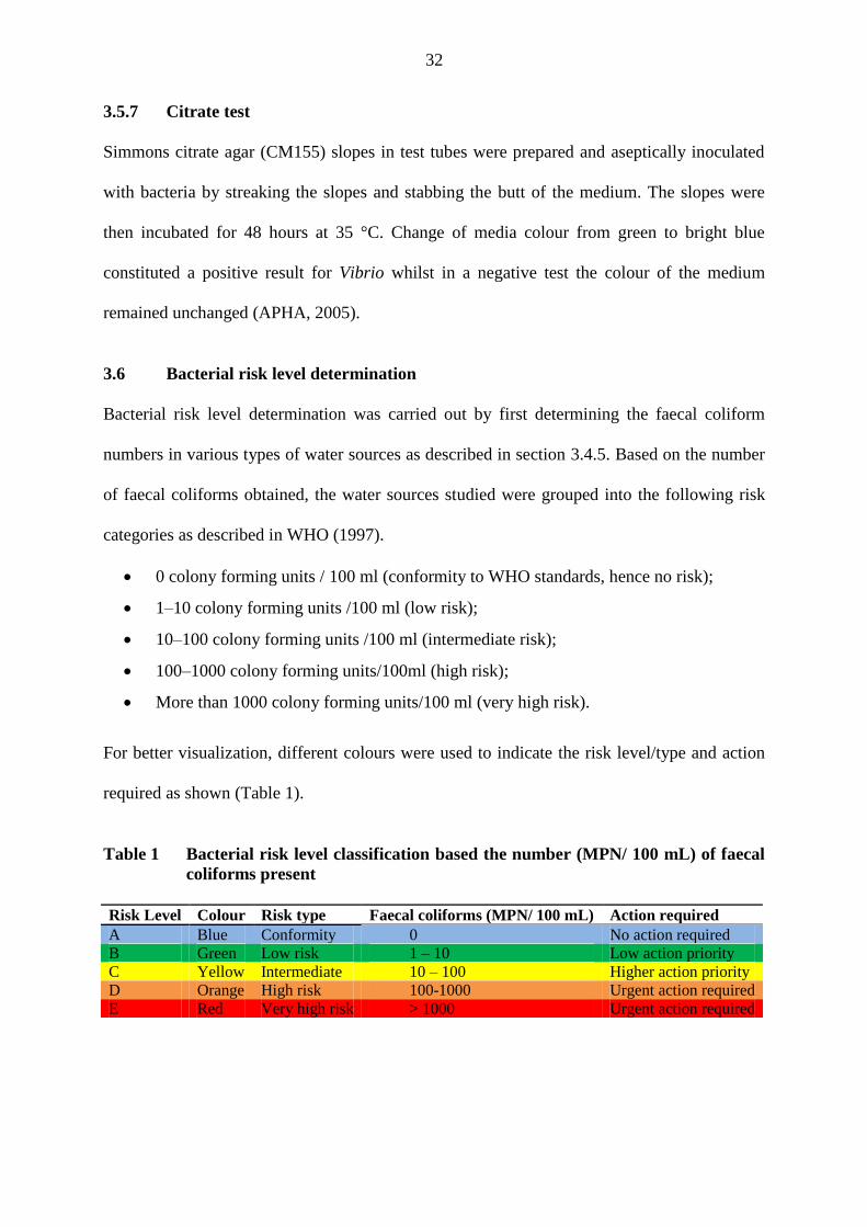
32
3.5.7 Citrate test
Simmons citrate agar (CM155) slopes in test tubes were prepared and aseptically inoculated
with bacteria by streaking the slopes and stabbing the butt of the medium. The slopes were
then incubated for 48 hours at 35 °C. Change of media colour from green to bright blue
constituted a positive result for Vibrio whilst in a negative test the colour of the medium
remained unchanged (APHA, 2005).
3.6 Bacterial risk level determination
Bacterial risk level determination was carried out by first determining the faecal coliform
numbers in various types of water sources as described in section 3.4.5. Based on the number
of faecal coliforms obtained, the water sources studied were grouped into the following risk
categories as described in WHO (1997).
0 colony forming units / 100 ml (conformity to WHO standards, hence no risk);
1–10 colony forming units /100 ml (low risk);
10–100 colony forming units /100 ml (intermediate risk);
100–1000 colony forming units/100ml (high risk);
More than 1000 colony forming units/100 ml (very high risk).
For better visualization, different colours were used to indicate the risk level/type and action
required as shown (Table 1).
Table 1 Bacterial risk level classification based the number (MPN/ 100 mL) of faecal
coliforms present
Risk Level Colour Risk type Faecal coliforms (MPN/ 100 mL) Action required
A Blue Conformity 0 No action required
B Green Low risk 1 – 10 Low action priority
C Yellow Intermediate 10 – 100 Higher action priority
D Orange High risk 100-1000 Urgent action required
E Red Very high risk > 1000 Urgent action required
33
3.7 Preparation of plant material
Roots of Boscia coriacea Pax., and Maerua decumbens (Brogn.) Dewolf were collected from
Wamba, Samburu District with the help of traditional healers. These two plants, indigenous
and common in Samburu are used to treat water and infections of microbial origin. Seeds of
Moringa oleifera Lam. were obtained from KEFRI. All the plant materials collected in the
field were identified and processed in the Department of Plant and Microbial Sciences,
Kenyatta University.
The roots of Boscia coriacea Pax. and Maerua decumbens (Brogn.) Dewolf were washed then
chopped into small pieces and dried in the shade. Dried seeds of Moringa oleifera Lam. were
shelled to remove the seed coat and to obtain the kernels. The root choppings and seed kernels
were each milled using a sterile mill (Thomas–Wiley, Model 4) and stored in a sterile airtight
container at a temperature of 3 °C in a refrigerator until time for use. From each plant, an
amount of 2 g of the ground plant materials was weighed and packed in filter sachets in
readiness for use.
3.8 Water treatment
The study involved 15 different surface water samples with different physico-chemical and
bacteriological qualities. Treatment tests were carried out at laboratory temperatures (24 + 2
°C) inside a laminar flow (Figure 4). The effectiveness of the treatment was assessed
analytically by determining changes in bacterial heterotrophic plate counts (HPC) and
turbidity over a 24 hour period. Heterotrophic plate counts were determined using the spread
plate method while turbidity was determined with the aid of a colorimeter.
An amount of 200 ml of turbid water samples with predetermined bacterial density and
turbidity was dispensed into six 250 ml sterile glass bottles. Using sterile forceps, a sachet of
each plant material was immersed in different water samples. Additionally, 2 grams of alum
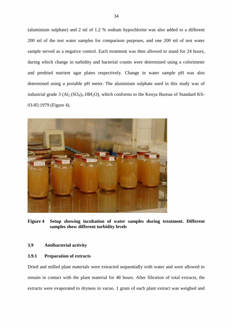
34
(aluminium sulphate) and 2 ml of 1.2 % sodium hypochlorite was also added to a different
200 ml of the test water samples for comparison purposes, and one 200 ml of test water
sample served as a negative control. Each treatment was then allowed to stand for 24 hours,
during which change in turbidity and bacterial counts were determined using a colorimeter
and predried nutrient agar plates respectively. Change in water sample pH was also
determined using a portable pH meter. The aluminium sulphate used in this study was of
industrial grade 3 (Al2 (SO4)3.18H2O), which conforms to the Kenya Bureau of Standard KS-
03-85:1979 (Figure 4).
Figure 4 Setup showing incubation of water samples during treatment. Different
samples show different turbidity levels
3.9 Antibacterial activity
3.9.1 Preparation of extracts
Dried and milled plant materials were extracted sequentially with water and were allowed to
remain in contact with the plant material for 48 hours. After filtration of total extracts, the
extracts were evaporated to dryness in vacuo. 1 gram of each plant extract was weighed and

35
dissolved in 1ml of sterile distilled water. Sterile disks of 6 mm diameter were soaked in the
extract water mixture for one hour. Disks were also soaked in water containing 1 g of alum
and 1ml of 1.2 % sodium hypochlorite.
3.9.2 Sources of test bacteria
Environmental isolates from most polluted water sources in Wamba were used. The isolates
used were E. coli, Salmonella spp, and Shigella spp. as identified based on the IMViC
reactions, cultural characteristics and gram stain reactions. The reference strains used for the
screening were Escherichia coli (ATCC 25922) and Staphylococcus aureus (ATCC 25923).
These bacteria were obtained from stock cultures from Department of Public Health and
Pharmacology, Faculty of Veterinary Medicine, University of Nairobi.
3.9.3 Antibacterial sensitivity testing
Three to five identical colonies from each agar plate were transferred with a sterile wire loop
into a tube containing 5 ml of nutrient broth. The turbidity of each bacterial suspension was
adjusted to give a turbidity value similar to that of a 0.5 McFarland standard, resulting in a
suspension containing approximately 1 to 2 x 10⁸ CFU/ml. Mueller-Hinton agar plates were
inoculated by streaking the swab over the entire sterile agar surface. After allowing the
inoculum to dry at room temperature, 6 mm diameter disks each with fixed concentration of
the extract was tested for antibacterial activity by introducing duplicate disks per plate.
Streptomycin (10 μg ml-1
) was used as a positive control.
The plates were allowed to stand at room temperature for 1 hour and then incubated at 37 °C
for 18 hours. Subsequently, the plates were examined for bacterial growth inhibition and the
inhibition zone diameter (IZD) measured to the nearest millimetre using a transparent ruler. A
standard table of antibiotic susceptibilities (Performance Standards for Antimicrobial
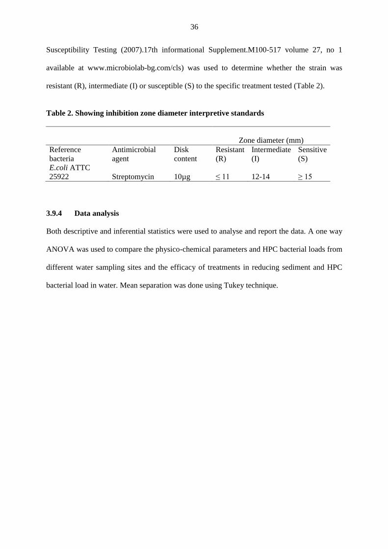
36
Susceptibility Testing (2007).17th informational Supplement.M100-517 volume 27, no 1
available at www.microbiolab-bg.com/cls) was used to determine whether the strain was
resistant (R), intermediate (I) or susceptible (S) to the specific treatment tested (Table 2).
Table 2. Showing inhibition zone diameter interpretive standards
Zone diameter (mm)
Reference
bacteria
Antimicrobial
agent
Disk
content
Resistant
(R)
Intermediate
(I)
Sensitive
(S)
E.coli ATTC
25922 Streptomycin 10µg ≤ 11 12-14 ≥ 15
3.9.4 Data analysis
Both descriptive and inferential statistics were used to analyse and report the data. A one way
ANOVA was used to compare the physico-chemical parameters and HPC bacterial loads from
different water sampling sites and the efficacy of treatments in reducing sediment and HPC
bacterial load in water. Mean separation was done using Tukey technique.
37
CHAPTER FOUR: RESULTS
4.1 Introduction
Water quality determination involved the measurements of among others pH, conductivity,
dissolved oxygen, temperature, alkalinity, salinity, turbidity, nutrient levels and the load of
various waterborne bacteria. These properties are fundamental for evaluating water quality.
4.2 Physico-chemical properties of water
4.2.1 Temperature
Water temperatures varied widely among the study sites with a range of 18.2 to 35.6 °C. The
water sources investigated recorded the following temperature (°C) ranges: springs, 18.2
to29.4; wells, 18.2 to 32.3; dams, 20.8 to 35.6; rivers, 23.2 to 30.8; boreholes, 25.5 to 29.8;
tap, 29 to 31.2 (Table 3). The lowest mean temperature (22.8 °C) was recorded in spring
water while highest mean temperature (30.5 °C) was recorded in tap water (Table 3). Using
the one way ANOVA test, it was established that there was a significant difference in water
temperature of the different sources (P =0.05, DF = 5).
4.2.2 pH
The median pH of water at the various sources ranged from 6.1 to 7.9. The lowest (5.68) and
the highest (10.46) pH values were measured in a shallow well and a dam respectively. Dams
recorded the highest median value of 7.9 with the springs and tap water having a median pH
value of 7.1 and 6.1 respectively (Table 3).
4.2.3 Electrical conductivity (µS cm-1
)
Electrical conductivity (EC) of water at various sources ranged from 3.0 (tap) to 5940 (well)
µS cm-1
. The lowest mean EC (11.17 µS cm-1
) and the highest (830.83 µS cm-1
) were
measured in tap and borehole sources respectively. Using a one way ANOVA test, it was

38
noted that there was no significant difference in mean EC of water from the different sources
(P =0.05, DF= 5) (Table 3).
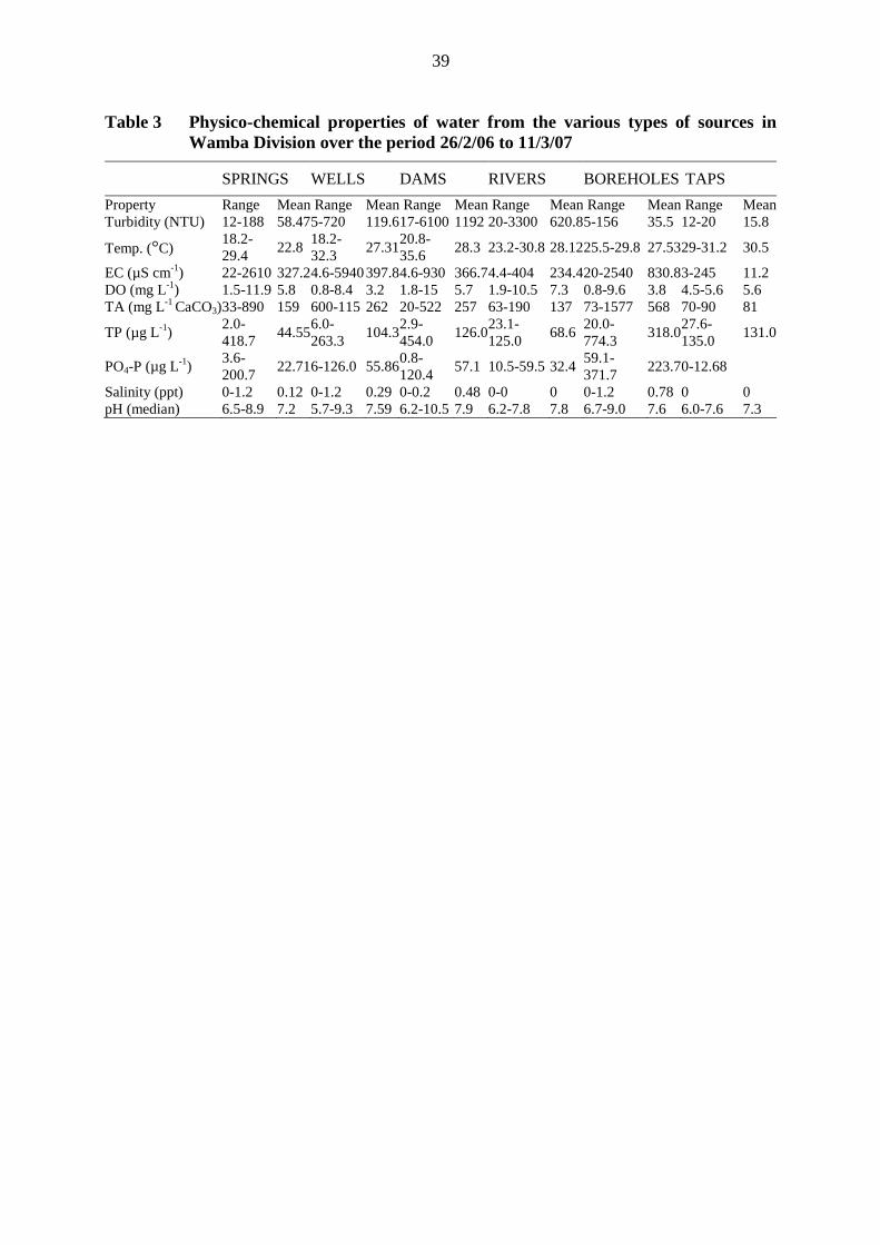
39
Table 3 Physico-chemical properties of water from the various types of sources in
Wamba Division over the period 26/2/06 to 11/3/07
SPRINGS WELLS DAMS RIVERS BOREHOLES TAPS
Property Range Mean Range Mean Range Mean Range Mean Range Mean Range Mean
Turbidity (NTU) 12-188 58.47 5-720 119.6 17-6100 1192 20-3300 620.8 5-156 35.5 12-20 15.8
Temp. (°C) 18.2-
29.4 22.8
18.2-
32.3 27.31
20.8-
35.6 28.3 23.2-30.8 28.12 25.5-29.8 27.53 29-31.2 30.5
EC (µS cm-1
) 22-2610 327.2 4.6-5940 397.8 4.6-930 366.7 4.4-404 234.4 20-2540 830.8 3-245 11.2
DO (mg L-1
) 1.5-11.9 5.8 0.8-8.4 3.2 1.8-15 5.7 1.9-10.5 7.3 0.8-9.6 3.8 4.5-5.6 5.6
TA (mg L-1
CaCO3) 33-890 159 600-115 262 20-522 257 63-190 137 73-1577 568 70-90 81
TP (µg L-1
) 2.0-
418.7 44.55
6.0-
263.3 104.3
2.9-
454.0 126.0
23.1-
125.0 68.6
20.0-
774.3 318.0
27.6-
135.0 131.0
PO4-P (µg L-1
) 3.6-
200.7 22.71 6-126.0 55.86
0.8-
120.4 57.1 10.5-59.5 32.4
59.1-
371.7 223.7 0-12.68
Salinity (ppt) 0-1.2 0.12 0-1.2 0.29 0-0.2 0.48 0-0 0 0-1.2 0.78 0 0
pH (median) 6.5-8.9 7.2 5.7-9.3 7.59 6.2-10.5 7.9 6.2-7.8 7.8 6.7-9.0 7.6 6.0-7.6 7.3
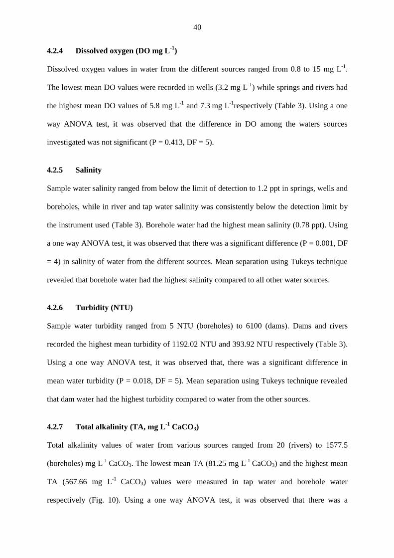
40
4.2.4 Dissolved oxygen (DO mg L-1
)
Dissolved oxygen values in water from the different sources ranged from 0.8 to 15 mg L-1
.
The lowest mean DO values were recorded in wells (3.2 mg L-1
) while springs and rivers had
the highest mean DO values of 5.8 mg L-1
and 7.3 mg L
-1respectively (Table 3). Using a one
way ANOVA test, it was observed that the difference in DO among the waters sources
investigated was not significant (P = 0.413, DF = 5).
4.2.5 Salinity
Sample water salinity ranged from below the limit of detection to 1.2 ppt in springs, wells and
boreholes, while in river and tap water salinity was consistently below the detection limit by
the instrument used (Table 3). Borehole water had the highest mean salinity (0.78 ppt). Using
a one way ANOVA test, it was observed that there was a significant difference (P = 0.001, DF
= 4) in salinity of water from the different sources. Mean separation using Tukeys technique
revealed that borehole water had the highest salinity compared to all other water sources.
4.2.6 Turbidity (NTU)
Sample water turbidity ranged from 5 NTU (boreholes) to 6100 (dams). Dams and rivers
recorded the highest mean turbidity of 1192.02 NTU and 393.92 NTU respectively (Table 3).
Using a one way ANOVA test, it was observed that, there was a significant difference in
mean water turbidity (P = 0.018, DF = 5). Mean separation using Tukeys technique revealed
that dam water had the highest turbidity compared to water from the other sources.
4.2.7 Total alkalinity (TA, mg L-1
CaCO3)
Total alkalinity values of water from various sources ranged from 20 (rivers) to 1577.5
(boreholes) mg L-1
CaCO3. The lowest mean TA (81.25 mg L-1
CaCO3) and the highest mean
TA (567.66 mg L-1
CaCO3) values were measured in tap water and borehole water
respectively (Fig. 10). Using a one way ANOVA test, it was observed that there was a
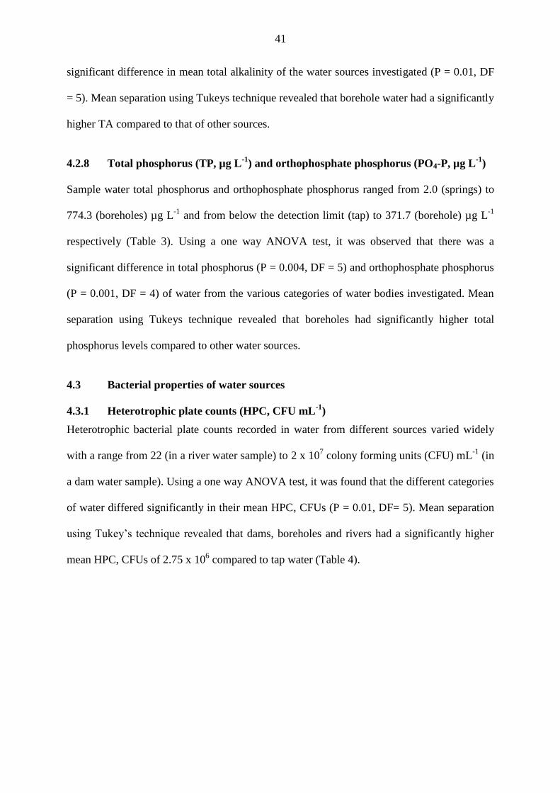
41
significant difference in mean total alkalinity of the water sources investigated (P = 0.01, DF
= 5). Mean separation using Tukeys technique revealed that borehole water had a significantly
higher TA compared to that of other sources.
4.2.8 Total phosphorus (TP, µg L-1
) and orthophosphate phosphorus (PO4-P, µg L-1
)
Sample water total phosphorus and orthophosphate phosphorus ranged from 2.0 (springs) to
774.3 (boreholes) µg L-1
and from below the detection limit (tap) to 371.7 (borehole) µg L-1
respectively (Table 3). Using a one way ANOVA test, it was observed that there was a
significant difference in total phosphorus (P = 0.004, DF = 5) and orthophosphate phosphorus
(P = 0.001, DF = 4) of water from the various categories of water bodies investigated. Mean
separation using Tukeys technique revealed that boreholes had significantly higher total
phosphorus levels compared to other water sources.
4.3 Bacterial properties of water sources
4.3.1 Heterotrophic plate counts (HPC, CFU mL-1
)
Heterotrophic bacterial plate counts recorded in water from different sources varied widely
with a range from 22 (in a river water sample) to 2 x 10⁷ colony forming units (CFU) mL
-1 (in
a dam water sample). Using a one way ANOVA test, it was found that the different categories
of water differed significantly in their mean HPC, CFUs (P = 0.01, DF= 5). Mean separation
using Tukey’s technique revealed that dams, boreholes and rivers had a significantly higher
mean HPC, CFUs of 2.75 x 10⁶ compared to tap water (Table 4).
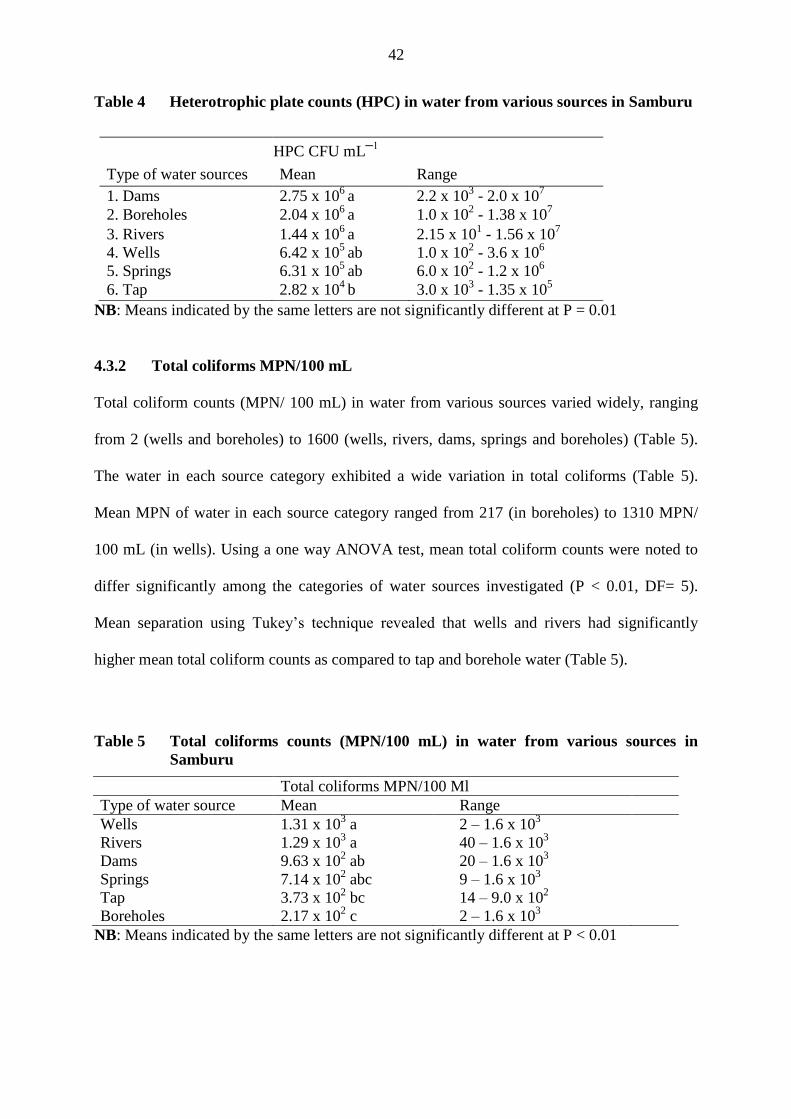
42
Table 4 Heterotrophic plate counts (HPC) in water from various sources in Samburu
HPC CFU mL─1
Type of water sources Mean Range
1. Dams 2.75 x 10⁶
a 2.2 x 10³ - 2.0 x 10
7
2. Boreholes 2.04 x 10⁶
a 1.0 x 10² - 1.38 x 10
7
3. Rivers 1.44 x 10⁶
a 2.15 x 101 - 1.56 x 10
7
4. Wells 6.42 x 10⁵
ab 1.0 x 10² - 3.6 x 10
6
5. Springs 6.31 x 10⁵
ab 6.0 x 10² - 1.2 x 10
6
6. Tap 2.82 x 10⁴
b 3.0 x 10³ - 1.35 x 10
5
NB: Means indicated by the same letters are not significantly different at P = 0.01
4.3.2 Total coliforms MPN/100 mL
Total coliform counts (MPN/ 100 mL) in water from various sources varied widely, ranging
from 2 (wells and boreholes) to 1600 (wells, rivers, dams, springs and boreholes) (Table 5).
The water in each source category exhibited a wide variation in total coliforms (Table 5).
Mean MPN of water in each source category ranged from 217 (in boreholes) to 1310 MPN/
100 mL (in wells). Using a one way ANOVA test, mean total coliform counts were noted to
differ significantly among the categories of water sources investigated (P < 0.01, DF= 5).
Mean separation using Tukey’s technique revealed that wells and rivers had significantly
higher mean total coliform counts as compared to tap and borehole water (Table 5).
Table 5 Total coliforms counts (MPN/100 mL) in water from various sources in
Samburu
Total coliforms MPN/100 Ml
Type of water source Mean Range
Wells 1.31 x 10³ a 2 – 1.6 x 10
3
Rivers 1.29 x 10³ a 40 – 1.6 x 10
3
Dams 9.63 x 10² ab 20 – 1.6 x 10
3
Springs 7.14 x 10² abc 9 – 1.6 x 10
3
Tap 3.73 x 10² bc 14 – 9.0 x 10
2
Boreholes 2.17 x 10² c 2 – 1.6 x 10
3
NB: Means indicated by the same letters are not significantly different at P < 0.01
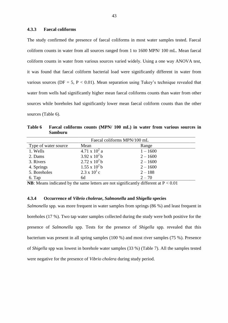
43
4.3.3 Faecal coliforms
The study confirmed the presence of faecal coliforms in most water samples tested. Faecal
coliform counts in water from all sources ranged from 1 to 1600 MPN/ 100 mL. Mean faecal
coliform counts in water from various sources varied widely. Using a one way ANOVA test,
it was found that faecal coliform bacterial load were significantly different in water from
various sources (DF = 5, P < 0.01). Mean separation using Tukey’s technique revealed that
water from wells had significantly higher mean faecal coliforms counts than water from other
sources while boreholes had significantly lower mean faecal coliform counts than the other
sources (Table 6).
Table 6 Faecal coliforms counts (MPN/ 100 mL) in water from various sources in
Samburu
Faecal coliforms MPN/100 mL
Type of water source Mean Range
1. Wells 4.71 x 10² a 1 – 1600
2. Dams 3.92 x 10²
b 2 – 1600
3. Rivers 2.72 x 10²
b 2 – 1600
4. Springs 1.55 x 10²
b 2 – 1600
5. Boreholes 2.3 x 101
c 2 – 188
6. Tap 6d 2 – 70
NB: Means indicated by the same letters are not significantly different at P < 0.01
4.3.4 Occurrence of Vibrio cholerae, Salmonella and Shigella species
Salmonella spp. was more frequent in water samples from springs (86 %) and least frequent in
boreholes (17 %). Two tap water samples collected during the study were both positive for the
presence of Salmonella spp. Tests for the presence of Shigella spp. revealed that this
bacterium was present in all spring samples (100 %) and most river samples (75 %). Presence
of Shigella spp was lowest in borehole water samples (33 %) (Table 7). All the samples tested
were negative for the presence of Vibrio cholera during study period.
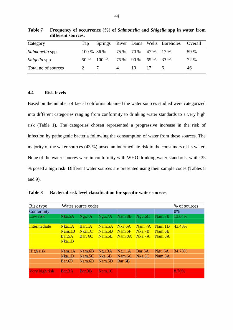
44
Table 7 Frequency of occurrence (%) of Salmonella and Shigella spp in water from
different sources.
Category Tap Springs River Dams Wells Boreholes Overall
Salmonella spp. 100 % 86 % 75 % 70 % 47 % 17 % 59 %
Shigella spp. 50 % 100 % 75 % 90 % 65 % 33 % 72 %
Total no of sources 2 7 4 10 17 6 46
4.4 Risk levels
Based on the number of faecal coliforms obtained the water sources studied were categorized
into different categories ranging from conformity to drinking water standards to a very high
risk (Table 1). The categories chosen represented a progressive increase in the risk of
infection by pathogenic bacteria following the consumption of water from these sources. The
majority of the water sources (43 %) posed an intermediate risk to the consumers of its water.
None of the water sources were in conformity with WHO drinking water standards, while 35
% posed a high risk. Different water sources are presented using their sample codes (Tables 8
and 9).
Table 8 Bacterial risk level classification for specific water sources
Risk type Water source codes % of sources Conformity 0%
Low risk Nka.5A Ngi.7A Ngu.7A Nam.8B Ngu.6C Nam.7B 13.04%
Intermediate Nka.1A Bar.1A Nam.5A Nka.6A Nam.7A Nam.1D 43.48%
Nam.1B Nka.1C Nam.5B Nam.6F Nka.7B Nam.6E
Bar.5A Bar. 6C Nam.5E Nam.8A Nka.7A Nam.3A
Nka.1B
High risk Nam.1A Nam.6B Ngu.3A Ngu.1A Bar.6A Ngu.6A 34.78%
Nka.1D Nam.5C Nka.6B Nam.6C Nka.6C Nam.6A
Bar.6D Nam.6D Nam.5D Bar.6B
Very high risk Bar.3A Bar.3B Nam.1C 8.70%
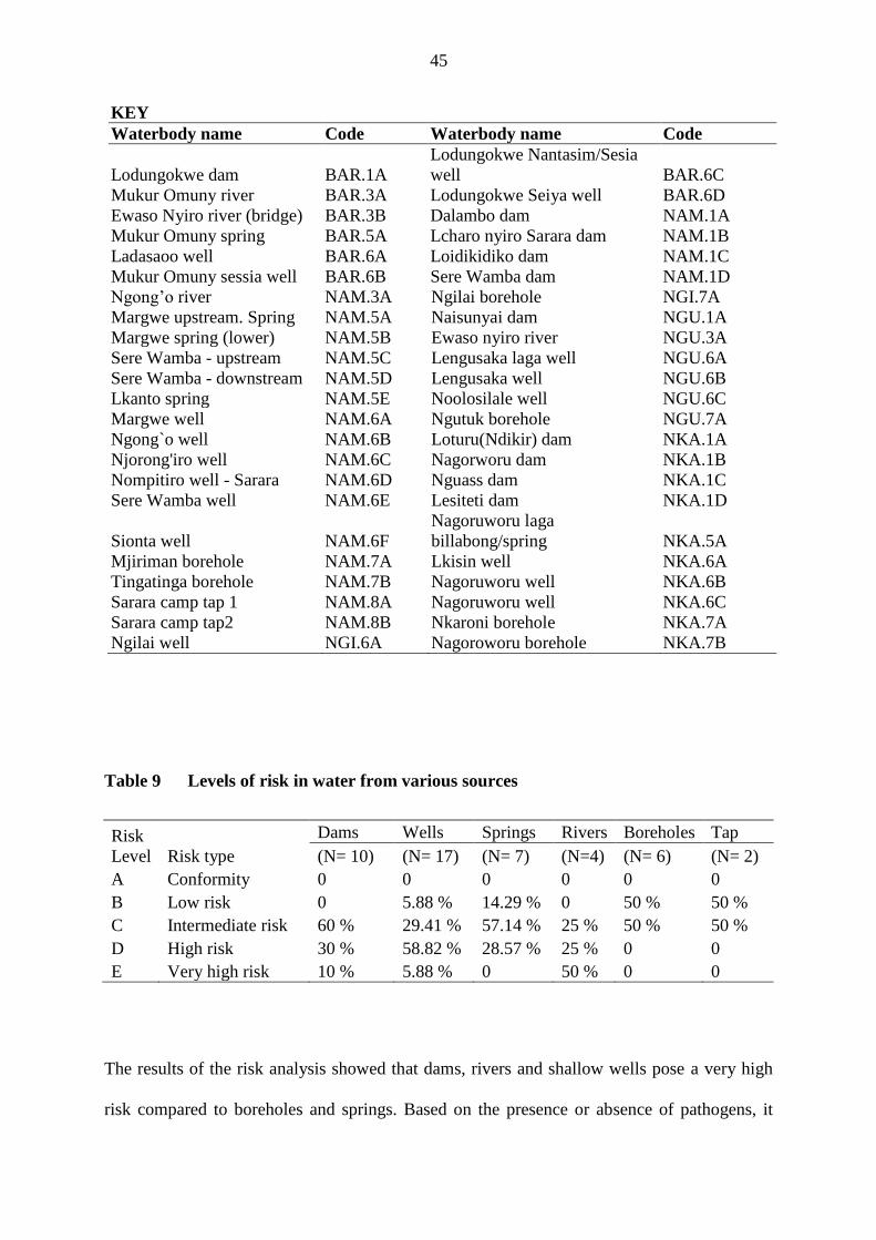
45
KEY
Waterbody name Code Waterbody name Code
Lodungokwe dam BAR.1A
Lodungokwe Nantasim/Sesia
well BAR.6C
Mukur Omuny river BAR.3A Lodungokwe Seiya well BAR.6D
Ewaso Nyiro river (bridge) BAR.3B Dalambo dam NAM.1A
Mukur Omuny spring BAR.5A Lcharo nyiro Sarara dam NAM.1B
Ladasaoo well BAR.6A Loidikidiko dam NAM.1C
Mukur Omuny sessia well BAR.6B Sere Wamba dam NAM.1D
Ngong’o river NAM.3A Ngilai borehole NGI.7A
Margwe upstream. Spring NAM.5A Naisunyai dam NGU.1A
Margwe spring (lower) NAM.5B Ewaso nyiro river NGU.3A
Sere Wamba - upstream NAM.5C Lengusaka laga well NGU.6A
Sere Wamba - downstream NAM.5D Lengusaka well NGU.6B
Lkanto spring NAM.5E Noolosilale well NGU.6C
Margwe well NAM.6A Ngutuk borehole NGU.7A
Ngong`o well NAM.6B Loturu(Ndikir) dam NKA.1A
Njorong'iro well NAM.6C Nagorworu dam NKA.1B
Nompitiro well - Sarara NAM.6D Nguass dam NKA.1C
Sere Wamba well NAM.6E Lesiteti dam NKA.1D
Sionta well NAM.6F
Nagoruworu laga
billabong/spring NKA.5A
Mjiriman borehole NAM.7A Lkisin well NKA.6A
Tingatinga borehole NAM.7B Nagoruworu well NKA.6B
Sarara camp tap 1 NAM.8A Nagoruworu well NKA.6C
Sarara camp tap2 NAM.8B Nkaroni borehole NKA.7A
Ngilai well NGI.6A Nagoroworu borehole NKA.7B
Table 9 Levels of risk in water from various sources
Risk
Level Risk type
Dams Wells Springs Rivers Boreholes Tap
(N= 10) (N= 17) (N= 7) (N=4) (N= 6) (N= 2)
A Conformity 0 0 0 0 0 0
B Low risk 0 5.88 % 14.29 % 0 50 % 50 %
C Intermediate risk 60 % 29.41 % 57.14 % 25 % 50 % 50 %
D High risk 30 % 58.82 % 28.57 % 25 % 0 0
E Very high risk 10 % 5.88 % 0 50 % 0 0
The results of the risk analysis showed that dams, rivers and shallow wells pose a very high
risk compared to boreholes and springs. Based on the presence or absence of pathogens, it
46
was observed that, 23.91 % of the water sources contained no pathogens, while 21.74 % and
54.35 % contained 1or 2 types of pathogens respectively, that is Salmonella or Shigella spp.
None of the water samples analyzed contained Vibrio cholera.
4.5 Water purification experiments
4.5.1 Change in heterotrophic bacterial counts
The efficacy of Boscia coriacea Pax., Maerua decumbens (Brogn.) Dewolf roots and Moringa
oleifera Lam. seeds extracts in reducing heterotrophic bacterial load in water were determined
by considering the percentage change in HPC after treatment of water with the extracts at
various time intervals. Overall mean percentage reduction in HPC in extracts of M. oleifera
Lam., M. decumbens (Brogn.) Dewolf, and B. coriacea Pax. were 26.51, 46.00, and 30.20
respectively compared to percentage reduction of 14.5, 74.8 and 91 percent for the control,
alum and sodium hypochlorite (Table 10). A one way ANOVA test revealed a significant
difference in HPC reduction among treatments (DF = 5, P < 0.05). Mean separation using
Tukey’s test revealed that HPC reduction by sodium hypochlorite and alum was significantly
higher than that of the other treatments. Although the HPC reduction by plant extracts was not
significantly different, only M. decumbens (Brogn.) Dewolf posted a HPC reduction that was
significantly different from that of the control (Table 10).
Change in HPC after 30 minutes.
The percentage change in number of HPC obtained after 30 min of water treatment varied
between 35.88 % - 83.22 %. A one way ANOVA test revealed a significant difference in
percentage reduction of HPC among treatments (DF = 5, P = 0.05). Mean separation using
Tukey’s test revealed that HPC reduction by sodium hypochlorite, alum, M. decumbens
(Brogn.) Dewolf and B. coriacea Pax. were significantly higher than that of the control (Table
10). Among the plant extracts, HPC reduction by M. decumbens (Brogn.) Dewolf was
significantly higher than that of M. oleifera Lam. (Table 10).
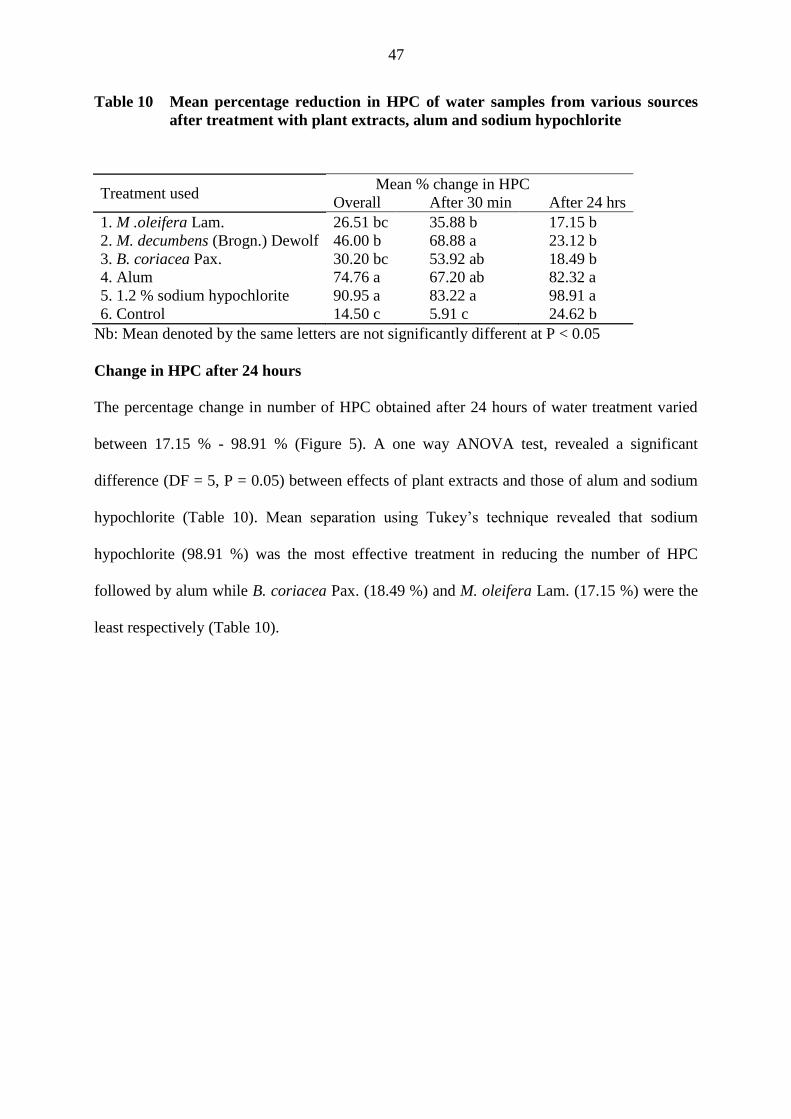
47
Table 10 Mean percentage reduction in HPC of water samples from various sources
after treatment with plant extracts, alum and sodium hypochlorite
Treatment used Mean % change in HPC
Overall After 30 min After 24 hrs
1. M .oleifera Lam. 26.51 bc 35.88 b 17.15 b
2. M. decumbens (Brogn.) Dewolf 46.00 b 68.88 a 23.12 b
3. B. coriacea Pax. 30.20 bc 53.92 ab 18.49 b
4. Alum 74.76 a 67.20 ab 82.32 a
5. 1.2 % sodium hypochlorite 90.95 a 83.22 a 98.91 a
6. Control 14.50 c 5.91 c 24.62 b
Nb: Mean denoted by the same letters are not significantly different at P < 0.05
Change in HPC after 24 hours
The percentage change in number of HPC obtained after 24 hours of water treatment varied
between 17.15 % - 98.91 % (Figure 5). A one way ANOVA test, revealed a significant
difference (DF = 5, P = 0.05) between effects of plant extracts and those of alum and sodium
hypochlorite (Table 10). Mean separation using Tukey’s technique revealed that sodium
hypochlorite (98.91 %) was the most effective treatment in reducing the number of HPC
followed by alum while B. coriacea Pax. (18.49 %) and M. oleifera Lam. (17.15 %) were the
least respectively (Table 10).
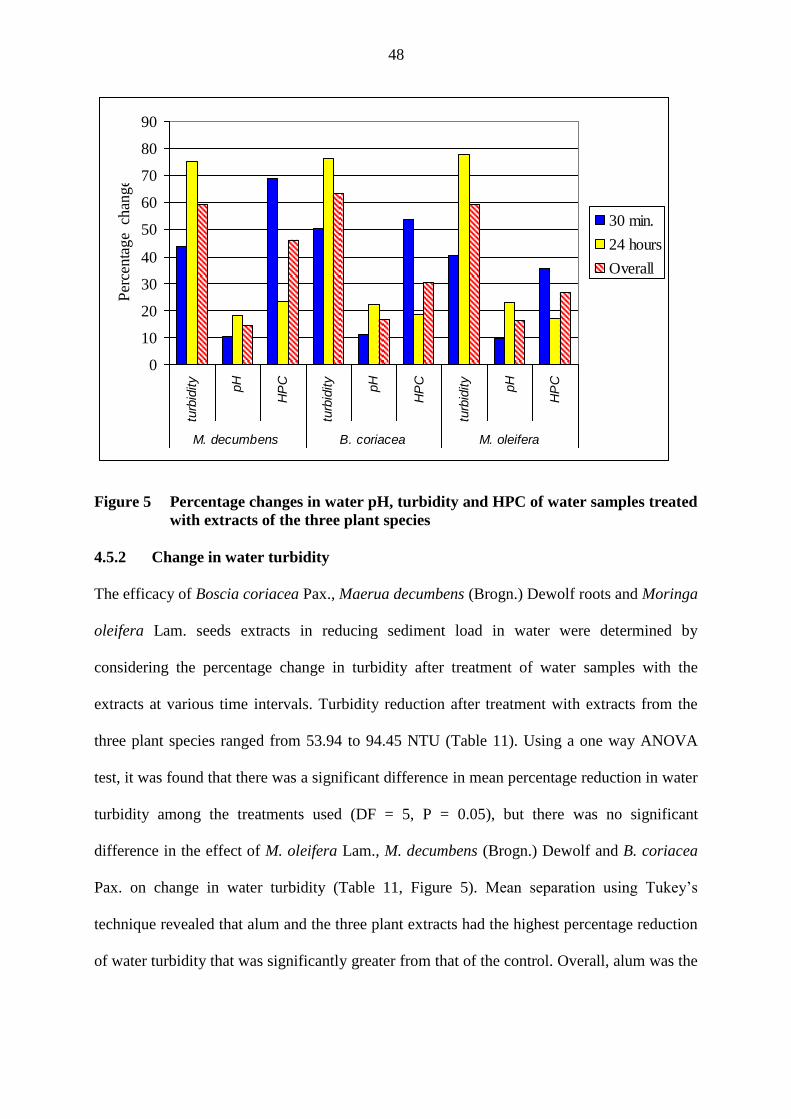
48
0
10
20
30
40
50
60
70
80
90
turb
idity
pH
HP
C
turb
idity
pH
HP
C
turb
idity
pH
HP
C
M. decumbens B. coriacea M. oleifera
Per
centa
ge
chan
ge
30 min.
24 hours
Overall
Figure 5 Percentage changes in water pH, turbidity and HPC of water samples treated
with extracts of the three plant species
4.5.2 Change in water turbidity
The efficacy of Boscia coriacea Pax., Maerua decumbens (Brogn.) Dewolf roots and Moringa
oleifera Lam. seeds extracts in reducing sediment load in water were determined by
considering the percentage change in turbidity after treatment of water samples with the
extracts at various time intervals. Turbidity reduction after treatment with extracts from the
three plant species ranged from 53.94 to 94.45 NTU (Table 11). Using a one way ANOVA
test, it was found that there was a significant difference in mean percentage reduction in water
turbidity among the treatments used (DF = 5, P = 0.05), but there was no significant
difference in the effect of M. oleifera Lam., M. decumbens (Brogn.) Dewolf and B. coriacea
Pax. on change in water turbidity (Table 11, Figure 5). Mean separation using Tukey’s
technique revealed that alum and the three plant extracts had the highest percentage reduction
of water turbidity that was significantly greater from that of the control. Overall, alum was the
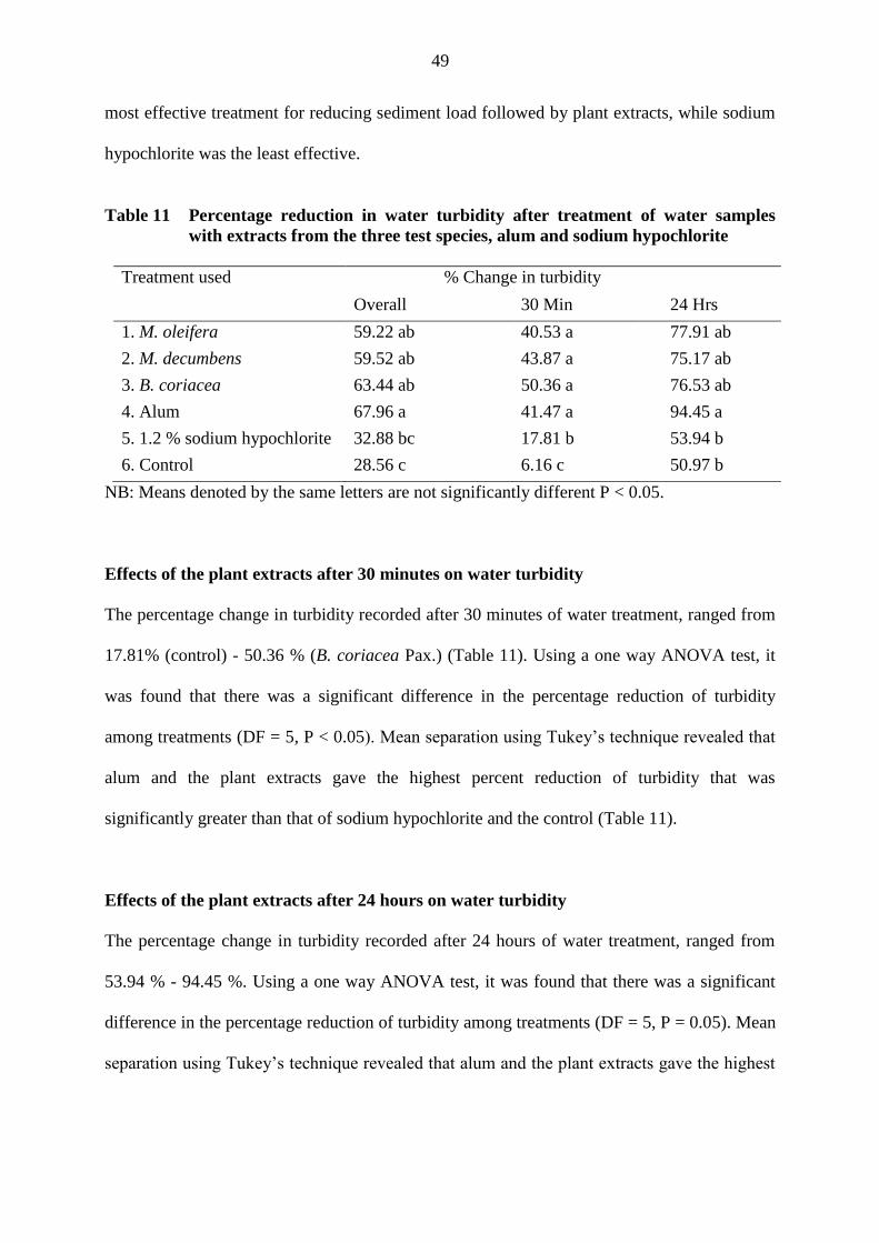
49
most effective treatment for reducing sediment load followed by plant extracts, while sodium
hypochlorite was the least effective.
Table 11 Percentage reduction in water turbidity after treatment of water samples
with extracts from the three test species, alum and sodium hypochlorite
Treatment used % Change in turbidity
Overall 30 Min 24 Hrs
1. M. oleifera 59.22 ab 40.53 a 77.91 ab
2. M. decumbens 59.52 ab 43.87 a 75.17 ab
3. B. coriacea 63.44 ab 50.36 a 76.53 ab
4. Alum 67.96 a 41.47 a 94.45 a
5. 1.2 % sodium hypochlorite 32.88 bc 17.81 b 53.94 b
6. Control 28.56 c 6.16 c 50.97 b
NB: Means denoted by the same letters are not significantly different P < 0.05.
Effects of the plant extracts after 30 minutes on water turbidity
The percentage change in turbidity recorded after 30 minutes of water treatment, ranged from
17.81% (control) - 50.36 % (B. coriacea Pax.) (Table 11). Using a one way ANOVA test, it
was found that there was a significant difference in the percentage reduction of turbidity
among treatments (DF = 5, P < 0.05). Mean separation using Tukey’s technique revealed that
alum and the plant extracts gave the highest percent reduction of turbidity that was
significantly greater than that of sodium hypochlorite and the control (Table 11).
Effects of the plant extracts after 24 hours on water turbidity
The percentage change in turbidity recorded after 24 hours of water treatment, ranged from
53.94 % - 94.45 %. Using a one way ANOVA test, it was found that there was a significant
difference in the percentage reduction of turbidity among treatments (DF = 5, P = 0.05). Mean
separation using Tukey’s technique revealed that alum and the plant extracts gave the highest
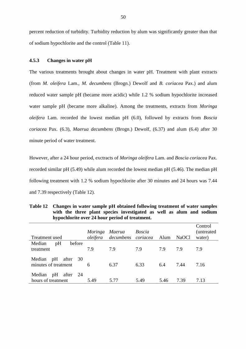
50
percent reduction of turbidity. Turbidity reduction by alum was significantly greater than that
of sodium hypochlorite and the control (Table 11).
4.5.3 Changes in water pH
The various treatments brought about changes in water pH. Treatment with plant extracts
(from M. oleifera Lam., M. decumbens (Brogn.) Dewolf and B. coriacea Pax.) and alum
reduced water sample pH (became more acidic) while 1.2 % sodium hypochlorite increased
water sample pH (became more alkaline). Among the treatments, extracts from Moringa
oleifera Lam. recorded the lowest median pH (6.0), followed by extracts from Boscia
coriacea Pax. (6.3), Maerua decumbens (Brogn.) Dewolf, (6.37) and alum (6.4) after 30
minute period of water treatment.
However, after a 24 hour period, exctracts of Moringa oleifera Lam. and Boscia coriacea Pax.
recorded similar pH (5.49) while alum recorded the lowest median pH (5.46). The median pH
following treatment with 1.2 % sodium hypochlorite after 30 minutes and 24 hours was 7.44
and 7.39 respectively (Table 12).
Table 12 Changes in water sample pH obtained following treatment of water samples
with the three plant species investigated as well as alum and sodium
hypochlorite over 24 hour period of treatment.
Treatment used
Moringa
oleifera
Maerua
decumbens
Boscia
coriacea Alum NaOCl
Control
(untreated
water)
Median pH before
treatment 7.9 7.9 7.9 7.9 7.9 7.9
Median pH after 30
minutes of treatment 6 6.37 6.33 6.4 7.44 7.16
Median pH after 24
hours of treatment 5.49 5.77 5.49 5.46 7.39 7.13
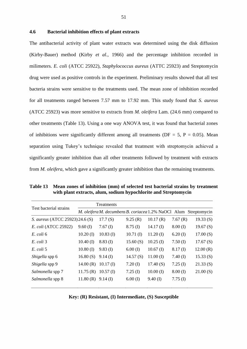
51
4.6 Bacterial inhibition effects of plant extracts
The antibacterial activity of plant water extracts was determined using the disk diffusion
(Kirby-Bauer) method (Kirby et al., 1966) and the percentage inhibition recorded in
milimeters. E. coli (ATCC 25922), Staphylococcus aureus (ATTC 25923) and Streptomycin
drug were used as positive controls in the experiment. Preliminary results showed that all test
bacteria strains were sensitive to the treatments used. The mean zone of inhibition recorded
for all treatments ranged between 7.57 mm to 17.92 mm. This study found that S. aureus
(ATCC 25923) was more sensitive to extracts from M. oleifera Lam. (24.6 mm) compared to
other treatments (Table 13). Using a one way ANOVA test, it was found that bacterial zones
of inhibitions were significantly different among all treatments (DF = 5, P = 0.05). Mean
separation using Tukey’s technique revealed that treatment with streptomycin achieved a
significantly greater inhibition than all other treatments followed by treatment with extracts
from M. oleifera, which gave a significantly greater inhibition than the remaining treatments.
Table 13 Mean zones of inhibition (mm) of selected test bacterial strains by treatment
with plant extracts, alum, sodium hypochlorite and Streptomycin
Test bacterial strains Treatments
M. oleifera M. decumbens B. coriacea 1.2% NaOCl Alum Streptomycin
S. aureus (ATCC 25923) 24.6 (S) 17.7 (S) 9.25 (R) 10.17 (R) 7.67 (R) 19.33 (S)
E. coli (ATCC 25922) 9.60 (I) 7.67 (I) 8.75 (I) 14.17 (I) 8.00 (I) 19.67 (S)
E. coli 6 10.20 (I) 10.83 (I) 10.71 (I) 11.20 (I) 6.20 (I) 17.00 (S)
E. coli 3 10.40 (I) 8.83 (I) 15.60 (S) 10.25 (I) 7.50 (I) 17.67 (S)
E. coli 5 10.80 (I) 9.83 (I) 6.00 (I) 10.67 (I) 8.17 (I) 12.00 (R)
Shigella spp 6 16.80 (S) 9.14 (I) 14.57 (S) 11.00 (I) 7.40 (I) 15.33 (S)
Shigella spp 9 14.00 (R) 10.17 (I) 7.20 (I) 17.40 (S) 7.25 (I) 21.33 (S)
Salmonella spp 7 11.75 (R) 10.57 (I) 7.25 (I) 10.00 (I) 8.00 (I) 21.00 (S)
Salmonella spp 8 11.80 (R) 9.14 (I) 6.00 (I) 9.40 (I) 7.75 (I)
Key: (R) Resistant, (I) Intermediate, (S) Susceptible
52
CHAPTER FIVE: DISCUSSION, CONCLUSIONS AND RECOMMENDATIONS
5.1 Physico-chemical quality of water
Among the physico-chemical properties of water from various sources investigated, pH,
dissolved oxygen, total alkalinity and conductivity values met the requirements for drinking
water as per the guidelines of the World Health Organization (WHO, 2004). However,
turbidity in most dams, rivers, and some wells exceeded the maximum permissible
concentration (MPC) according to WHO (2004) on most occasions.
In general, mean water temperatures in all water sources ranged from 22.8 to 30.5 °C. Water
temperature variation at the various water sources can be attributed to differences in weather
conditions at the time of sampling, the amount of water present in the source and presence of
vegetation shielding water source from direct insolation. During this study it was observed
that compared to other types of water sources, most springs were mainly located in high
altitude areas where they are on most occasions sheltered by trees. Such an environment is
likely to experience cool air temperature which influences water temperature. The dams,
rivers and the shallow wells however were located in lowlands exposed to direct insolation.
Of all the water sources, tap water recorded the highest mean (30.5 °C). This can be attributed
to the fact that the metallic pipes carrying the water at some points in bare rocky areas, were
exposed to direct sunlight. This enabled the water to absorb the heat conducted by the metallic
pipes.
Mean turbidity was highest in dams (1192 NTU) and lowest in boreholes (35.5 NTU). High
turbidity in dams may be due to deposition of soil and organic matter by surface runoff water
coupled with wind erosion during wet and dry seasons respectively. However, animals too
may excrete substantial amount of faecal matter into water directly especially when watering
in dams. Hence non point pollutants mainly affect surface waters, which are exposed
53
compared to water in boreholes, which are protected and therefore usually have a
comparatively lower turbidity (35.5 NTU). According to WHO (2004) it is recommended that
water with a turbidity level of more than 5 NTU, should undergo some form of treatment to
remove turbidity before the water can effectively be disinfected with chlorine. This therefore
means that most water sources in Samburu require some initial treatment to remove sediments
if chlorine disinfection is to be employed.
The electrical conductivity is acknowledged as a quick and reliable measure to indicate the
general chemical quality of water (Hem, 1982; Auer, 1997). The conductivity values obtained
in this study therefore indicate that some water sources have a high concentration of dissolved
solids (minerals). High conductivity values recorded in boreholes can be attributed to the
longer period of contact between the water and the parent rock material whose dissolution
releases mineral ions into the water. The dissolution and infiltration of faecal matter into
sandy aquifer and lateral movement of groundwater along the sandy river bed account for the
high levels of contamination in wells. However the relatively high conductivity and salinity
levels of water observed at some study sites are not likely to affect water consumption by
cattle and sheep and wildlife and their health. Previous studies (Weeth and Haverland, 1961;
Wilson, 1966; Potter and McIntosh, 1974) have demonstrated that conductivity above 15,150
µS cm-1
, lead to reduced water intake and growth rate of cattle while salinities above 26,620
µS cm-1
cause health disorders and subsequent death.
The low values of dissolved oxygen recorded in springs and boreholes are likely to be due to
the fact that they were sheltered from direct wind action by vegetation and concrete cover
respectively. A high mineral content in borehole water (evidenced by the higher salinity)
could have further reduced the solubility of oxygen in water.
54
The median pH recorded in different water sources, were within the WHO recomended range
for drinking water. Although water sources that recorded a pH of around 9 and above are
likely to have reduced levels of faecal bacteria, such high pH values are above the
recommended range for drinking water. Acidic conditions are known to be favourable for the
survival of E. coli both in fresh and saline waters (Rozen and Belkin, 2001).
Mean total alkalinity was highest in boreholes (757.2) and lowest in rivers (136.8) and tap
water (131). The high level of alkalinity and electrical conductivity in boreholes suggests a
possible dominance of bicarbonate, and carbonate ions in water from these sources. Such
highly mineralized, alkaline hard water have an objectionable "soda" taste and hence not very
suitable for drinking. Additionaly, use of mineralized hard water to wash clothes increases the
cost as it does not easily lather with soap. It also causes excessive drying of the skin (remove
natural skin oils) when used for bathing.
Boreholes had the most saline water (mean 0.78 ppt) while rivers had the least (less than 0.1
ppt). These results suggest the existence of a higher load of dissolved salts in deeper water
sources (boreholes) as compared to other shallow ones. The salinity difference in between
surface and bottom water can be attributed to variation in the chemistry of rocks and soils at
different depths, which in turn influences the concentration of introduced cations and anions
such as Na+, K
+, Mg
2+, Fe
2+, NO3
-, CO3
2-, SO4
2-, Cl
- etc. (Fasunwon et al., 2010).
During this study it was noted that most phosphorus entering water sources originates from
non-point sources. Possible non point sources of phosphorus include the natural
decomposition of rocks and minerals, storm water runoff, erosion and sedimentation, and
direct input by animals/wildlife. The presence of higher loads of phosphorus and low levels of
dissolved oxygen in boreholes compared to wells confirms the generalization that phosphorus
is often scarce in the well oxygenated waters (Ricklefs and Schluter 1993). This is due to the
55
fact that as aerobic bacteria decompose organic wastes, they consume oxygen, and in return
release more phosphorus into the water.
Total coliform in all types of water sources exceeded WHO (2004) maximum permissible
load (0/100 mL) for drinking water. Although total coliform organisms may not always be
directly related to the presence of faecal contamination or pathogens in the drinking water,
this study found that all water samples contained both total coliform and faecal coliform.
Hence the total coliform test would still be useful in monitoring the microbial quality of the
raw and treated piped water supplies in the area. During the study it was noted that total and
faecal coliforms counts in wells differed significantly from those in springs with the wells
recording higher total and faecal coliforms counts compared with the springs. These findings
are in agreement with the previous observations (Feachem 1980; Lindskog and Lindskog
1988; Sandiford et al., 1989; Tensay, 1991; Utkilen and Sutton, 1989; White et al., 1972;
Wright, 1985), which suggested that a protected hand-dug well is usually one of the least
contaminated with only spring water being usually cleaner. The higher total and faecal
coliforms counts recorded in wells in Samburu can be attributed to the contamination caused
by lateral movement of water along the sandy laggas where the majority of the wells are
closely situated. Although some shallow wells were well protected from direct access by
animals, the protection appears not to have been effective in reducing water contamination.
This is perhaps due to the fact that most of the animals move along the laggas in search of
water, salt licks and pasture. In the process, they deposit a lot of organic wastes on the lagga
floor. When it rains, the seasonal floods wash off bacteria and organic water into the wells
hence contaminating them.
A higher faecal coliform load in wells (mean 471.63) compared to that in other types of water
sources suggests a recent contamination of the ground water with bacteria of faecal origin.
56
Wells in the study area are mostly shallow and located in dry river beds. Since most river beds
are made of loose sand, they are easily filled up with sand. Although some community
members enclose their wells to protect them from direct fecal contamination by livestock, the
high population of livestock and wildlife that visit the river beds at different times exposes the
wells to some contamination. Hence each morning, these wells have to be cleaned before
drawing drinking or livestock water. These activities contaminate the groundwater sources
and are likely to contribute to high levels of faecal coliforms. Although it has been reported
that indicator and pathogenic bacteria are efficiently retained in soils and are detected at only
low levels in groundwater under field conditions (Liu, 1982; Alhajjar et al., 1988), other
studies have found that heavy rainfall promotes the movement of bacteria and other inorganic
contaminants through soil (Zyman and Sorber, 1988; Nikolaidis et al., 1998). It is therefore
clear that should the groundwater be qualified as drinking water, it must be fully treated and
the wells must be protected from pollutants accordingly.
The risk analysis results of bacteriological water quality shows that consumption of untreated
water from various sources poses a risk to the users (Table 8). Presence of thermotolerant E.
coli in all water sources indicates possible presence of gastro-intestinal pathogens in water.
This is likely since 21.74 % and 54.35 % of water sources contained 1or 2 types of pathogens
respectively that is Salmonella spp or Shigella spp. These pathogens can infect both human
beings and animals. Transmission of these pathogens occurs through faecal contamination of
water in heavily used and unprotected water sources. Hence the consumption of water from
dams, rivers and shallow wells pose very high risk compared to boreholes and springs (Table
7 and 8).
Water treatment using extracts from natural and renewable vegetation has been widely
practiced and appears to be an effective and accepted physico-chemical treatment for
57
household water in some parts of the world (Jahn, 1988). Water treatment results obtained in
this study revealed the capacity of extracts of Boscia coriacea Pax., Maerua decumbens
(Brogn.) Dewolf roots and Moringa oleifera Lam. seeds to reduce heterotrophic bacterial load
in water to some degree. Changes in bacterial density recorded during water treatment may
have been due to loss of viability or alteration in culturability, persistence or aftergrowth of
bacteria. Under treatment conditions it is probable that bacteria may experience metabolic
stress, and as such bacterial cells may enter into a vulnerable but non culturable state
(VBNC).
The ability of pathogenic micro-organisms to exist in VBNC state is well known (Islam et al.,
1993). Rollins and Colwell (1986) have reported that the non culturable cells may remain
viable for a prolonged period of time. Since non culturable cells may still remain
metabolically active and if pathogenic, might maintain their infectiveness (Oliver, 1993), it is
important to determine the viable state of non culturable cells. Rollins and Colwell (1986)
have also reported that non-culturable cells may remain viable for a prolonged period of time.
Some investigators have claimed that non culturable bacteria of selected species can be
resuscitated to the culturable state (Roszak et al., 1984). It is therefore important to use a
highly selective and sensitive method to detect VBNC bacteria prior and after treatment of
water using plant extracts. Such a method should be considered for use during bacteriological
water quality testing.
The results of this study demonstrate that the plant extracts have some water soluble
compounds which have disinfection properties, which lead to the reduction of HPC, especially
within a 30 minute period. However, a decrease in the percentage reduction of bacterial
density after a 24 hour period of water treatment suggests that this disinfection property is lost
after sometime. The results also indicate that the treatments differ in their stability and ability
58
to persist in water to maintain a disinfectant residual. These results are in agreement with
previous work (Eilert et al., 1981; Madsen et al., 1987), which showed that Moringa oleifera
Lam. seed extracts flocculate bacteria and possess antimicrobial activity. According to
Gassenschmidt et al., (1991), the agents responsible of the coagulation and flocculation in
Moringa oleifera Lam. are water soluble proteins.
Among the plant extracts investigated, the greatest antimicrobial activity was due to Moringa
oleifera Lam. (inhibition zone 8 to 13 mm). This suggests that the plant has some metabolic
toxins or broad-spectrum antibiotic compounds. It is known that Moringa oleifera Lam. is
rich in derivatives of benzyl isothiocyanates, a class of compounds with remarkable
antimicrobial activity. It is apparent that Boscia coriacea Pax., and Maerua decumbens
(Brogn.) Dewolf also have some active compounds. However, the nature of the active
compounds present in the two plants remains unknown. Among the test bacteria used
Staphylococcus aureus showed the highest sensitivity to all plant extracts compared to E. coli,
Salmonella and Shigella species. This observation is in agreement with the findings of earlier
studies on medicinal plants (McCutcheon et al., 1992) that showed that, medicinal plants were
less active against gram-negative bacteria than to gram-positive bacteria. Failure of the plant
extracts to completely eliminate the bacteria present suggests that some bacteria present in the
water being treated may have spontaneously developed resistance to antimicrobial activity of
plant extracts or alternatively that the bacteria may have escaped the bacteriocidic effect of the
plant extracts, for instance, after degradation of the active ingredients. It is also possible that
the concentration of active ingredients in the extracts were too low to fully inhibit bacterial
growth. The results of the antimicrobial properties of the plant extracts investigated and
changes over time suggests a need to determine the minimum bactericidal concentration
(MIC) and the optimum time of water storage during treatment to avoid degradation of the
microbial quality of water and biofilm accumulation due to bacterial regrowth.
59
Comparison of efficacy of plant extracts in reducing sediment and bacterial load in water
demonstrates that the three species are almost similar in their performance and that the
potential of Boscia coriacea Pax. and Maerua decumbens (Brogn.) Dewolf extracts needs to
be further evaluated. Although alum, sodium hypochlorite and the three plant extracts
significantly reduced the bacterial load of sample water within a 24 hour period, all cases had
a residual bacterial population that remained in the treated water. The findings of this study
are in close agreement with other studies that reported resistance of bacteria to chlorine
(LeChevallier et al., 1988; Mathieu et al., 1992; Camper et al., 1997). These results suggest
that there were variations in the kinetics of inactivation by the disinfectants, depending on the
bacterial populations involved.
The interpretation of the results of heterotrophic bacteria counts obtained after water treatment
may not be straightforward. This is because indicator bacteria are discrete in water, generally
have a non-random distribution (Lightfoot et al., 1994) and are more likely to be found in
clumps following treatment, rather than being uniformly spread out in the water (Gale et al.,
1997). During this study, the spread plate method was used to culture the heterotrophic
bacteria present in treated water. It is therefore possible that the bacterial cells from the
clumps were dispersed leading to increased false colony forming units counts being obtained.
These observations indicate that other cultural methods of determining residual bacterial
colonies in treated water should be explored. Nevertheless, the results clearly demonstrates
that no single water treatment method is highly effective in reducing water turbidity and
bacterial loads to levels recomended by WHO for drinking water. The varied effectiveness of
plant extracts in purifying water from the various sources may be due to variation in the
physico-chemical properties of water treated. There is, therefore, a need to establish the effect
of these physico-chemical properties of water on efficacy of plant extracts in order to optimize
their use. These findings also indicate the need to determine changes in each type of bacterial
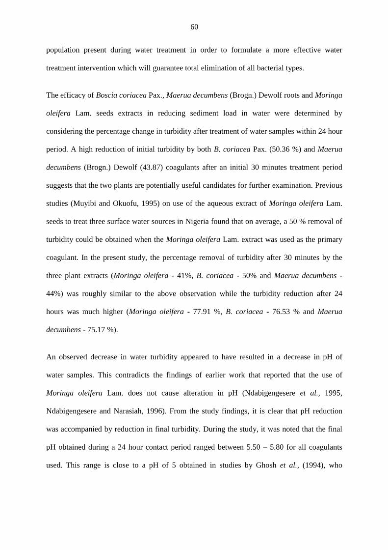
60
population present during water treatment in order to formulate a more effective water
treatment intervention which will guarantee total elimination of all bacterial types.
The efficacy of Boscia coriacea Pax., Maerua decumbens (Brogn.) Dewolf roots and Moringa
oleifera Lam. seeds extracts in reducing sediment load in water were determined by
considering the percentage change in turbidity after treatment of water samples within 24 hour
period. A high reduction of initial turbidity by both B. coriacea Pax. (50.36 %) and Maerua
decumbens (Brogn.) Dewolf (43.87) coagulants after an initial 30 minutes treatment period
suggests that the two plants are potentially useful candidates for further examination. Previous
studies (Muyibi and Okuofu, 1995) on use of the aqueous extract of Moringa oleifera Lam.
seeds to treat three surface water sources in Nigeria found that on average, a 50 % removal of
turbidity could be obtained when the Moringa oleifera Lam. extract was used as the primary
coagulant. In the present study, the percentage removal of turbidity after 30 minutes by the
three plant extracts (Moringa oleifera - 41%, B. coriacea - 50% and Maerua decumbens -
44%) was roughly similar to the above observation while the turbidity reduction after 24
hours was much higher (Moringa oleifera - 77.91 %, B. coriacea - 76.53 % and Maerua
decumbens - 75.17 %).
An observed decrease in water turbidity appeared to have resulted in a decrease in pH of
water samples. This contradicts the findings of earlier work that reported that the use of
Moringa oleifera Lam. does not cause alteration in pH (Ndabigengesere et al., 1995,
Ndabigengesere and Narasiah, 1996). From the study findings, it is clear that pH reduction
was accompanied by reduction in final turbidity. During the study, it was noted that the final
pH obtained during a 24 hour contact period ranged between 5.50 – 5.80 for all coagulants
used. This range is close to a pH of 5 obtained in studies by Ghosh et al., (1994), who
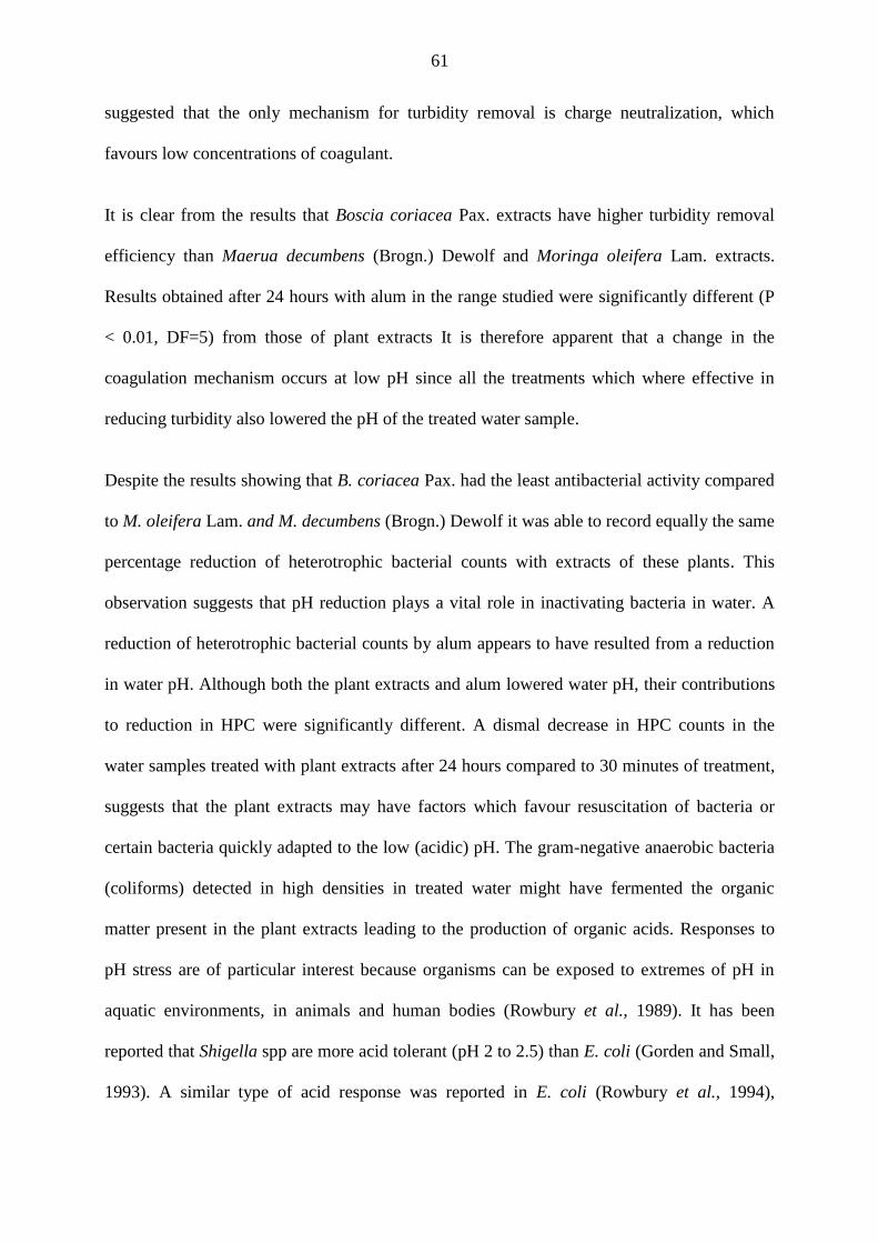
61
suggested that the only mechanism for turbidity removal is charge neutralization, which
favours low concentrations of coagulant.
It is clear from the results that Boscia coriacea Pax. extracts have higher turbidity removal
efficiency than Maerua decumbens (Brogn.) Dewolf and Moringa oleifera Lam. extracts.
Results obtained after 24 hours with alum in the range studied were significantly different (P
< 0.01, DF=5) from those of plant extracts It is therefore apparent that a change in the
coagulation mechanism occurs at low pH since all the treatments which where effective in
reducing turbidity also lowered the pH of the treated water sample.
Despite the results showing that B. coriacea Pax. had the least antibacterial activity compared
to M. oleifera Lam. and M. decumbens (Brogn.) Dewolf it was able to record equally the same
percentage reduction of heterotrophic bacterial counts with extracts of these plants. This
observation suggests that pH reduction plays a vital role in inactivating bacteria in water. A
reduction of heterotrophic bacterial counts by alum appears to have resulted from a reduction
in water pH. Although both the plant extracts and alum lowered water pH, their contributions
to reduction in HPC were significantly different. A dismal decrease in HPC counts in the
water samples treated with plant extracts after 24 hours compared to 30 minutes of treatment,
suggests that the plant extracts may have factors which favour resuscitation of bacteria or
certain bacteria quickly adapted to the low (acidic) pH. The gram-negative anaerobic bacteria
(coliforms) detected in high densities in treated water might have fermented the organic
matter present in the plant extracts leading to the production of organic acids. Responses to
pH stress are of particular interest because organisms can be exposed to extremes of pH in
aquatic environments, in animals and human bodies (Rowbury et al., 1989). It has been
reported that Shigella spp are more acid tolerant (pH 2 to 2.5) than E. coli (Gorden and Small,
1993). A similar type of acid response was reported in E. coli (Rowbury et al., 1994),
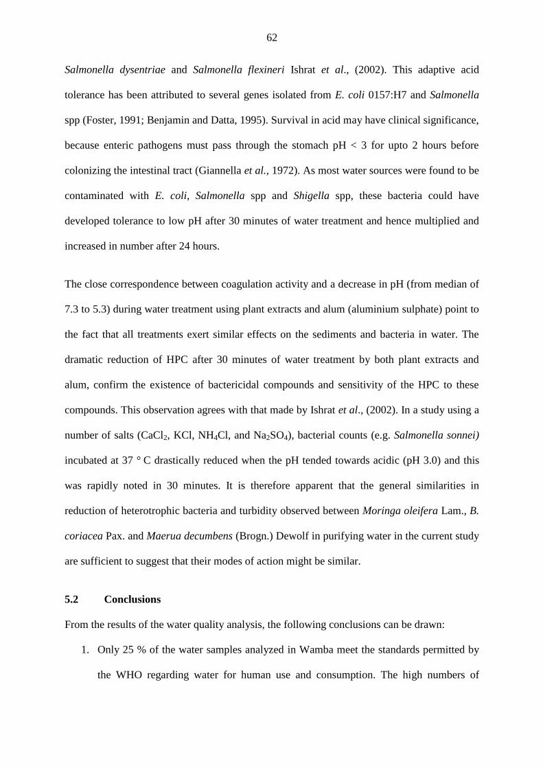
62
Salmonella dysentriae and Salmonella flexineri Ishrat et al., (2002). This adaptive acid
tolerance has been attributed to several genes isolated from E. coli 0157:H7 and Salmonella
spp (Foster, 1991; Benjamin and Datta, 1995). Survival in acid may have clinical significance,
because enteric pathogens must pass through the stomach pH < 3 for upto 2 hours before
colonizing the intestinal tract (Giannella et al., 1972). As most water sources were found to be
contaminated with E. coli, Salmonella spp and Shigella spp, these bacteria could have
developed tolerance to low pH after 30 minutes of water treatment and hence multiplied and
increased in number after 24 hours.
The close correspondence between coagulation activity and a decrease in pH (from median of
7.3 to 5.3) during water treatment using plant extracts and alum (aluminium sulphate) point to
the fact that all treatments exert similar effects on the sediments and bacteria in water. The
dramatic reduction of HPC after 30 minutes of water treatment by both plant extracts and
alum, confirm the existence of bactericidal compounds and sensitivity of the HPC to these
compounds. This observation agrees with that made by Ishrat et al., (2002). In a study using a
number of salts (CaCl2, KCl, NH4Cl, and Na2SO4), bacterial counts (e.g. Salmonella sonnei)
incubated at 37 ° C drastically reduced when the pH tended towards acidic (pH 3.0) and this
was rapidly noted in 30 minutes. It is therefore apparent that the general similarities in
reduction of heterotrophic bacteria and turbidity observed between Moringa oleifera Lam., B.
coriacea Pax. and Maerua decumbens (Brogn.) Dewolf in purifying water in the current study
are sufficient to suggest that their modes of action might be similar.
5.2 Conclusions
From the results of the water quality analysis, the following conclusions can be drawn:
1. Only 25 % of the water samples analyzed in Wamba meet the standards permitted by
the WHO regarding water for human use and consumption. The high numbers of
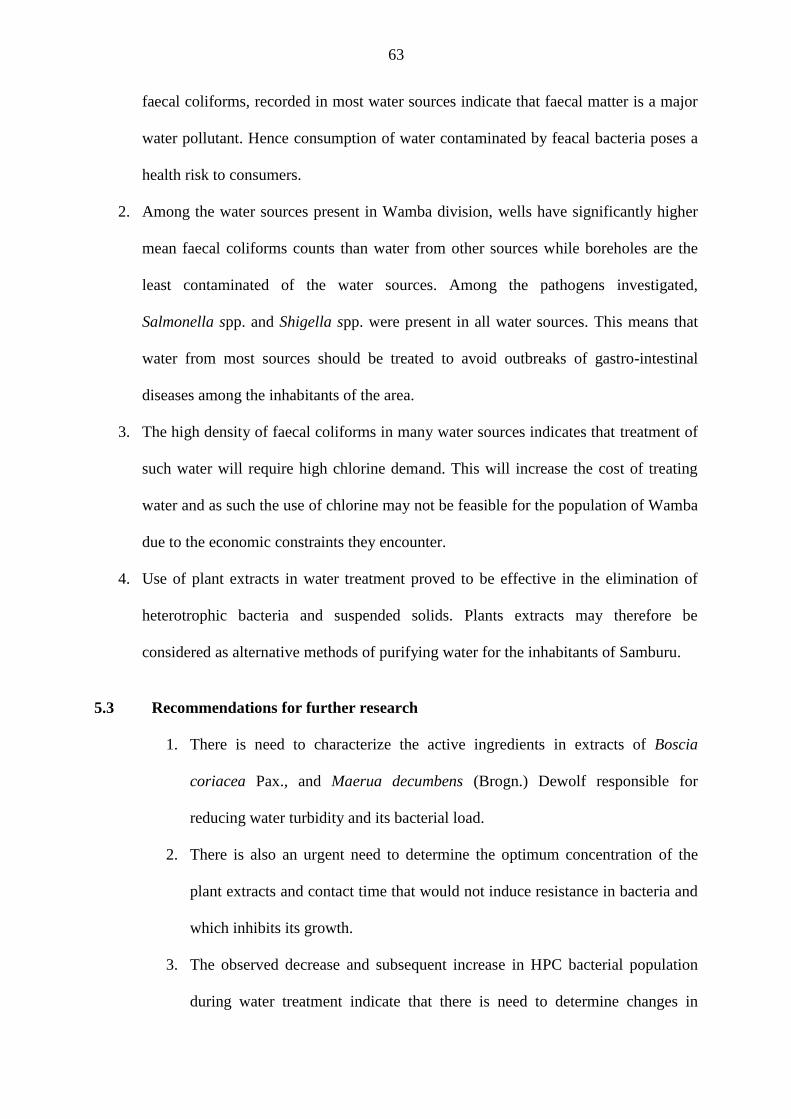
63
faecal coliforms, recorded in most water sources indicate that faecal matter is a major
water pollutant. Hence consumption of water contaminated by feacal bacteria poses a
health risk to consumers.
2. Among the water sources present in Wamba division, wells have significantly higher
mean faecal coliforms counts than water from other sources while boreholes are the
least contaminated of the water sources. Among the pathogens investigated,
Salmonella spp. and Shigella spp. were present in all water sources. This means that
water from most sources should be treated to avoid outbreaks of gastro-intestinal
diseases among the inhabitants of the area.
3. The high density of faecal coliforms in many water sources indicates that treatment of
such water will require high chlorine demand. This will increase the cost of treating
water and as such the use of chlorine may not be feasible for the population of Wamba
due to the economic constraints they encounter.
4. Use of plant extracts in water treatment proved to be effective in the elimination of
heterotrophic bacteria and suspended solids. Plants extracts may therefore be
considered as alternative methods of purifying water for the inhabitants of Samburu.
5.3 Recommendations for further research
1. There is need to characterize the active ingredients in extracts of Boscia
coriacea Pax., and Maerua decumbens (Brogn.) Dewolf responsible for
reducing water turbidity and its bacterial load.
2. There is also an urgent need to determine the optimum concentration of the
plant extracts and contact time that would not induce resistance in bacteria and
which inhibits its growth.
3. The observed decrease and subsequent increase in HPC bacterial population
during water treatment indicate that there is need to determine changes in
64
specific type of bacterial population during water treatment and to understand
the reasons for the observed changes in bacterial populations as a result of
treatment.
4. The sedimentation and antibacterial effects of various combinations of extracts
of Boscia coriacea Pax., Maerua decumbens (Brogn.) Dewolf and Moringa
oleifera Lam., should also be investigated in order to evaluate whether they
have a synergistic effect that would guarantee total elimination of bacteria.
5. Further research on the effects of physico-chemical properties of water on the
purification efficacy of the plant extracts should be carried out.
6. Before adoption of plants extract for water treatment, further research is
necessary to define, optimize and standardize conditions for the use of these
extracts in the treatment for household water or determine its acceptability,
sustainability, costs and effectiveness in reducing waterborne infectious
diseases.
65
REFERENCES
Al Samawi, A.A. and Shokralla E.M. (1996). Investigation into an indigenous natural
coagulant. Journal of Environmental Science Health, 31:1881-97.
Alamanos, Y., Maipa, Levidiotou, S., and Gessouli, E. (2000). A community waterborne
outbreak of gastro-enteritis attributed to Shigella sonnei. Epidemiological infections,
125:499-503.
Alhajjar, B.J., Stramer, S.L., Cliver, D.O. and Harkin, J.M. (1988). Transport modeling
of biological tracers from septic systems. Water Research, 22:907-915.
Ankcorn, P.D. ( 2003). Clarifying Turbidity – The Potential and Limitations of Turbidity as
a Surrogate for Water-Quality Monitoring, Proceedings of the 2003 Georgia Water
Resources Conference, Athens, GA, 23-24 April 2003.
Anuradha Mishra and Malvika Bajpai, (2005). Removal of sulphate and phosphate from
aqueous solutions using a food grade polysaccharide as flocculant. Journal of
colloid and polymer science, 284(4):443-448.
APHA, (2005). Standard Methods for the Examination of Water and Wastewater, 18th ed.
American Public Health Association American Water Works Association and Water
Pollution Control Federation, Washington, DC.
Appenzeller, B.M.R, Batté, M., Mathieu, L., Block, J.C., Lahoussine, V., Cavard, J. and
Gatel, D. (2001). Effect of adding phosphate to drinking water on bacterial growth
in slightly and highly corroded pipes. Water Research, 35(4):248-253.
Archer, A.J., and Jurken, B.J. (1977).Destruction of coliforms in water and sewage water
by dye-sensitized photo-oxidation. Journal of Applied and Environmental
Microbiology, 33:1019-1022.
Ashbolt, N.J., Dorsch, M.R., Cox, P.T. and Banens, B. (1997). Blooming E. coli, what do
they mean? In: D. Kay and C. Fricker [Eds] Coliforms and E. coli, Problem or
Solution? The Royal Society of Chemistry, Cambridge, pp 78-85.
Audicana, A., Perales, I. and Borrego, J.J. (1995). Modification of kanamycin-esculin-
azide agar to improve selectivity in the enumeration of fecal Streptococci from
water samples. Applied and Environmental Microbiology, 61:4178-4183.
Auer, C.K. (1997). Chemical quality of water at waterholes in the Etosha National Park.
Madoqua 20(1):121-128.
Australian Drinking Water Guidelines, (1996). Available at, http://www.nhmrc.gov.au/-
files_nhmrc/file/publications/synopses/eh19.pdf.
Australian Standard, (1995). Water Microbiology. Heterotrophic Colony Count Methods –
Pour Plate Method Using Plate Count Agar. Strathfield, NSW: Standards Australia
Int., AS/NZS 4276.3.1.
Bannaga, S.E.I and Pickford, J. (1978). Water-health relationships in Sudan. Effluent and
Water Treatment journal, 18:559-569.
Barer, M.R. and Harwood, C.R. (1999). Bacterial viability and culturability. Advanced
Microbial Physiology, 41:93-137.
66
Batté, M., Koudjonou, B., Laurent, P., Mathieu, L., Coallier, J., and Prévost, M. (2003). Biofilm responses to ageing and to a high phosphate load in a bench-scale drinking
water system. Water Research, 37:1351-1361.
Bell, J.B., Elliot, G.E. and Smith, D.W. (1983). Influence of sewage treatment and
urbanization on selection of multiple resistance in fecal coliform populations.
Applied and Environmental Microbiology, 46:227-232.
Belnap, J. and Harper, K.T. (1995). The influence of cryptobiotic soil crusts on elemental
content of tissue of two desert seed plants. Arid Soil Research and Rehabilitation,
9:107-115.
Benjamin, M.M and Datta, A.R. (1995). Acid tolerance of Enterohemorrhagic Escherichia
coli. Applied Environmental Microbiology, 61:1669-1672.
Bhole, A.G. (1995). Relative evaluation of a few natural coagulants. Journal of Water Supply
Research Technology, 44:284-90.
Bitton, G. and Gerba, C.P. (1984). Microbial Pollutants: Their Survival and Transport
Pattern to Groundwater. In Groundwater Pollution Microbiology. Bitton, G and
Gerba, C.P. (eds.). John Wiley and Sons, Toronto, Ontario.pp.65-88.
Boon, P.I. and Cattanach, M. (1999). Antibiotic resistance of native and faecal bacteria
isolated from rivers, reservoirs and sewage treatment facilities in Victoria, South-
Eastern Australia. Letters in Applied Microbiology, 28:164-168.
Borrego, J.J., Arrabal, F., Vicente, A. de, Gomez, L.F. and Romero, P. (1983). Study of
microbial inactivation in the marine environment. Journal Water Pollution Control
Federation, 55:297–302.
Brian, M. (1980). Ecology of fresh waters. Blackwell, oxford p. 332.
Brock, T.D. (1966). Principles of Microbial Ecology. Prentice-Hall, Englewood Cliffs, NJ.
Cabelli, V.J., Dufour, A.P., McCabe, L.J. and Levin, M.A. (1982). Swimming-associated
gastroenteritis and water quality. American Journal of Epidemiology, 115:606-616.
Cabelli, V.J., Dufour, A.P., McCabe, L.J. and Levin, M.A. (1983). A marine recreational
water quality criterion consistent with indicator concepts and risk analysis. Journal
Water Pollution Control Federation, 55:1306-1324.
Camper, A.K., Goodrum, L., and Jones, W. (1997). Comparison of chlorine and
monochloramine for the control of biofilm in model distribution system biofilms.
Proceedings of the 1997 Water Quality Technology Conference, Denver, Col. 9–12
November 1997. American Water Works Association, Denver, Col.
Canter, L.W. (1997). Nitrates in groundwater. Lewis Publishers, Boca Chang, S.L.
Cheesbrough, M. (1985). Medical Laboratory Manual for Tropical Countries Cambridge
University Press, United Kingdom.
Clark, J.A. (1990). The presence-absence test for monitoring water quality. In: McFeters, G.
A. (ed) Drinking Water Microbiology. Springer Verlag, Berlin, p. 399-411.
67
Cohen, Y.K., Abernathy, C.G. and Camper, A.K. (1999). Polyphosphates–the solution to
distribution system low residuals, biofilm and pipe corrosion. Proceedings of the
1999 Water Quality Technology Conference, Denver, 31 October – 3 November
1999. American Water Works Association, Denver, Columbia.
Conroy, R.M., Elmore-Meegan, M., Joyce, T., McGuigan, K.G. and Barnes, J. (1996). "Solar disinfection of drinking water and diarrhoea in Maasai children: A controlled
field trial." Lancet (North American Edition), 348(9043):1695-1697.
Conroy, R.M., Meegan, M.E., Joyce, T., McGuigan, K. and Barnes, J. (2001). Solar
disinfection of drinking water protects against cholera in children under 6 years of
age. ArchDis Child, 85:293-295.
Cornax, R., Moriñigo, M. A., Romero, P. and Borrego, J.J. (1990). Survival of pathogenic
microorganisms in seawater. Current Microbiology, 20:293-298.
Craun, G.F., Hauchman, F.S. and Robinson, D.E. (2001). Microbial Pathogens and
Disinfection By-products in Drinking Water. Health Effects and Management of
Risks. ILSI Press, Washington DC.
Cunliffe, D.A. (1991). Bacterial nitrification in chloraminated water supplies. Applied
Environmental Microbiology, 57:3399-3402.
Daniel Hoefel, Warwick L. Grooby, Paul T. Monis, Stuart Andrews, and Christopher P.
Saint, (2003). Enumeration of water-borne bacteria using viability assays and flow
cytometry: a comparison to culture-based techniques. Journal of Microbiological
Methods, 55:585-597.
Du Preez, M., Kfir, R. and Coubrough, P. (1995). Investigation of injury of coliforms after
chlorination. Water Science Technology, 31:115-118.
Dufour, A.P. (1977). Escherichia coli the fecal coliform. In: A.W. Hoadley and B.J. Dutka
[Eds] Bacterial Indicators/Health Hazards Associated with Water. American Society
for Testing and Materials, Philadelphia. pp 48-58.
Dufour, A.P. (1984). Health effects criteria for fresh recreational waters. EPA-600/1-84-004,
US Environmental Protection Agency, Washington, D.C.
DuPont, H.L., Levine, M.M., Hornick, R.B. and Formal, S.B. (1989). Inoculum size in
shigellosis and implications for expected mode of transmission. Journal of
infectious diseases, 159:1126-1128.
Eilert, U., Wolters, B. and Nahrstedt, A. (1981). The antibiotic principle of seeds of
Moringa oleifera and Moringa stenopetala. Planta Medica, 42:55–61.
El-Zanfaly, H.T. (1991).The need for new microbiological water quality criteria.
Environmental protection engineering, 17:41-52.
Fasunwon, O.O., Ayeni, A.O. and Lawal, A.O. (2010). A Comparative Study of Borehole
Water Quality from Sedimentary Terrain and Basement Complex in South-Western,
Nigeria. Research Journal of Environmental Sciences, 4(3):327-335.
Feachem, R. (1980). Bacterial standards for drinking water quality in developing countries.
Lancet, 2:255 - 256.

68
Foster, J.W. and Hall, H.K. (1991). Inducible pH homeostasis and the acid tolerance
response of Salmonella typhimurium. Journal of Bacteriology, 173:5129-5135.
Fox, K.R. and Reasoner, D.J. (1999). Water quality in source water, treatment, and
distribution systems. In: AWWA manual of water supply practices. AWWA M48.
Waterborne pathogens. American Water Works Association, Denver, CO.
Gale, P., van Dijk, P.A.H. and Stanfield, G. (1997). Drinking-water treatment increases
micro-organism clustering: the implications for microbiological risk assessment.
Journal of Water Supply Research and Technology–Aqua, 46(3):117–126.
Gassenschmidt, U., Jany, K.D. and Tauscher, B. (1991). Chemical Properties of Flocculant
- Active Proteins from Moringa oleifera lam, Biol. Chem. Hopper-Seyler, 372:659.
Gauci, V. (1991). Enumeration of Faecal streptococci in seawater. In: Development and
Testing of Sampling and Analytical Techniques for Monitoring of Marine Pollutants
(Activity A): Final Reports on Selected Microbiological Projects, MAP Technical
Reports Series No. 54, United Nations Environment Programme, Athens, 47-59.
Geildreich, E.E. (1996). Microbial Quality of Water Supply in Distribution Systems. CRC
press, Boca Raton. p. 504.
Geldrich, E.E., Best, L.C., Kenner, B.A. and Van Donsel, D.J. (1967). The Bacteriological
aspects of storm water pollution. U. S. Department of Health Education and
Welfare, Washington, D.C.
Ghosh, M., Amirtharajah, A. and Adin, A. (1994). Particle destabilization for tertiary
treatment of municipal wastewater by filtration. Water Science Technology, 30(9):
209–218.
Giannella, R.A., Broitman, S.A. and Zamckeck, N. (1972). Gastric barrier to ingested
microorganisms in man: studies in vivo and invitro. Gut, 13:251–256.
Gorden, J. and Small, P.L.C. (1993). Acid resistance in enteric bacteria. Infect. Immun., 61:
364–367.
Grabow, W.O.K., Slabert, J.L., Morgan, W.S.G. and Jahn, S.A.A. (1985). Toxicity and
mutagenicity evaluation of water coagulated with Moringa oleifera seed
preparations using fish, protozoan, bacterial, enzyme and Ames Salmonella assays.
Water SA, 11:9–14.
Gupta, A. and Chaudhuri, M. (1992). "Domestic water purification for developing
countries." Aqua (Oxford), 45(5):290-298.
Haarhoff, J., Cleasby, J.L., (1988). Comparing aluminum and iron coagulants for in line
filtration of cold waters. Journal of American Water Works Association, 80:168–
175.
Hamdani, A. (2002). Caractérisation et essais de traitement des effluents d’une industrie
laitière: aspects microbiologiques et physico-chimiques. Thèse de doctorat. Faculté
des Sciences d’El Jadida, Maroc.
Hardina, C.M. and Fujioka R.S. (1991). Soil: The environmental source of Escherichia coli
and enterococci in Hawaii's streams. Environmental Toxicology Water Quality, 6:
185–195.
69
Harper, K.T. and Marble, J.R. (1988). A role for nonvascular plants in management of arid
and semiarid rangelands, in P. T. Tueller, ed., Vegetation Science Applications for
Rangeland Analysis and Management: Dordrecht, Germany, Kluwer Academic
Publisher, p. 135-169.
Hazen, T.C. and Toranzos, G.A. (1990). Tropical source water. In: G.A. McFeters [Ed.]
Drinking Water Microbiology, Springer-Verlag, New York, 32–54.
Hem, J.D. (1982). Conductance: a collective measure of dissolved ions. In: Minear R.A. &
L.H. Keith (Eds) (1982): Water Analysis, Vol. I Inorganic Species, Part 1, pp 137-
161.
Holt, J.G., Krieg, N.R., Sneath, P.H.A., Staley, J.T. and Williams, S.T. (1993). Bergey's
Manual of Determinative Bacteriology, 9th edition, Williams & Wilkins, Co.,
Baltimore.
Howard, K. and Inglis, T.J.J. (2003). The effects of free chlorine on Burkholderia
pseudomallei in potable water. Water Research, 37:4425-4432.
Huber, W.G. (1971). Antibacterial drugs as environmental contaminants. In Advances in
Environmental Science and Technology, Vol. 2, ed. Pitts, J.N. Jr and Metcalf, R.L,
pp. 289 320. Wiley-Interscience, New York.
Hunter, P.R. (1993). The microbiology of bottled natural mineral waters. Journal of Applied
Bacteriology, 74:345-352.
Ishrat, S., Rahman, M.M., Shakhawat, H.B. and Majibur, R. (2002). Influence of
Different Physico-Chemical Stresses on growth and Survivality of Shigella
dysentriae and Shigella flexineri. Journal of Biological Sciences, 2(1):53-57.
Islam, M.S., Hasan, M.K., Miah, M.A., Sur, G.C., Felsenstein, A., Venkatesan, M. Sack,
R.B. and Albert, M. J. (1993). The use of polymerase chain reaction and the
fluorescent-antibody method for detecting viable but nonculturable Shigella
dysenteriae type 1 in laboratory microcosms. Applied and Environmental
Microbiology, 59:536-540.
Jahn, S.A.A. (1981). Traditional Water Purification in Tropical Developing Countries:
Existing Methods and Potentials. Eschborn, Germany.
Jahn, S.A.A. (1988). Using Moringa seeds as coagulants in developing countries. Journal of
American Water Works Association, 80:43–50.
Jahn, S.A.A. and Dirar, H. (1979). Studies on natural Water Coagulants in the Sudan, with
special reference to Moringa oleifera seeds. Water SA, 5:90-97.
Kaprelyants, A.S. and Kell, D.B. (1996). Do bacteria need to communicate with each other
for growth? Trends in Microbiology, 4:237–242.
Kasusya, P. (1998). Combating desertification in northern Kenya (Samburu) through
community action: a community case experience. Journal of Arid Environment, 39:
325-329.
Kay, D., Fleischer, J.M., Salmon, R.L., Jones, F., Wyer, M.D., Godfree, A.F., Zelenauch-
Jacquotte, Z. and Shore, R. (1994). Predicting likelihood of gastroenteritis from
sea bathing: results from randomized exposure. Lancet, 344:905-909.
70
Kehoe, S.C., Barer, M.R., Devlin, L.O. and McGuigan, K.G. (2004). Batch process solar
disinfection is an efficient means of disinfecting drinking water contaminated with
Shigella dysenteriae type I. Letters in Applied Microbiology, 38:410–414.
King, W.D. and Marret, L.D. (1996). Case control study of bladder cancer and chlorination
by products in treated water (Ontario, Canada).Cancer causes and control, 7:596-
604.
Lancini, G., Parenti, F. and Gallo, G.G. (1995). Resistance of microorganisms to
antibiotics. In Antibiotics: A Multidisciplinary Approach. pp. 85-106. New York:
Plenum Press.
LeChevallier, M.W. (1990). Coliform regrowth in drinking water: a review. Journal of
American Water Works Association, 82:74–86.
LeChevallier, M.W., Cawthon, C.D., and Lee, R.G. (1988). Factors promoting survival of
bacteria in chlorinated water supplies. Applied Environmental Microbiology, 54(3):
649–654.
LeChevallier, M.W., Evans, T.M. and Seidler, R.J. (1981). Effect of turbidity on
chlorination efficiency and bacterial persistence in drinking water. Applied.
Environmental Microbiology, 42:159.
LeChevallier, M.W., Shulz, W. and Lee, R.G. (1991). Bacterial nutrients in drinking water.
Applied. Environmental Microbiology, 57:857–862.
Leclerc, H., Mossel, D.A.A., Edberg, S.C. and Struijik, C.B. (2001). Advances in the
bacteriology of the coliform group: their suitability as markers of microbial water
safety.Annu.Rev.Microbial., 55: 2001-234.
Lehtola, M.J., Miettinen, I.T. and Martikainen, P.J. (2002). Biofilm formation in drinking
water affected by low concentrations of phosphorus. Canadian Journal of
Microbiology, 48:494-499.
Lewis, Jack. (1996). Turbidity Controlled Suspended Sediment Sampling for Runoff Event
load Estimation. Water Resources Research, 32(7):2299-2310.
Lightfoot, N.F., Tillet, H.E., Boyd, P. and Eaton, S. (1994). Duplicate spilt samples for
internal quality control in routine water microbiology. Letters in Applied
Microbiology, 19:321-324.
Lindskog, R.U.M. and Lindskog, P.A. (1988). Bacteriological contamination of water in
rural areas: an intervention study from Malawi. Journal of Tropical Medicine and
Hygiene, 91:1-7.
Liu, D. (1982). Effect of sewage sludge land disposal on the micro-biological quality of
groundwater. Water Research, 16:957-961.
Lloyd, B.J. and Bartram, J. (1991). Surveillance solutions to microbiological problems in
water quality control in developing countries. Water Science Technology, 24:61–75.
Lyons, J.L., Pontes, J., and Karalekas, P. (1995). Optimizing corrosion control for lead and
copper using phosphoric acid and sodium hydroxide. Proceedings of the 1995 Water
71
Quality Technology Conference, New Orleans, La., 12–16 November 1995.
American Water Works Association, Denver, Col.
Madsen, M., Schlundt, J., Omer, E.F. (1987). Effect of water coagulation by seeds of
Moringa oleifera on bacterial concentrations. Journal of Tropical Medicine and
Hygiene, 90:101–109.
Magee, A.M. and Quinn, J.P. (1991). Antibiotic resistance in the bacteria of a remote
upland river catchment. Letters in Applied Microbiology, 13:145-149.
Manaia, C.M., Nunes, O.C., Morais, P.V. and da Costa, M.S. (1990). Heterotrophic plate
counts and the isolation of bacteria from mineral waters on selective and enrichment
media. Journal of Applied Bacteriology, 69:871-876.
Mandell, G.L., Bennett, J.E., Dolin, R. and Dupont, H.L. (2000). Shigella Species
(bacillary dysentery). Principles and Practice of Infectious Diseases. Philadelphia:
Churchill Livingstone, 2363– 2368, 168–175.
Martyn, C.N., Osmond, C., Edwardson, J.A., Barker, D.J.P., Harris, E.C. and Lacey,
R.F. (1989). Geographical relation between Alzheimer's disease and aluminium in
drinking water. Lancet, 1:59-62.
Mathieu, L., Paquin, J.L., Block, J.C., Randon, G., Maillard, J., and Reasoner, D.
(1992). Paramètres gouvernant la prolifération bactérienne dans les réseaux de
distribution d’eau. Rev. Sci. Eau, 5:91-112.
McCall, B., Stafford, R., Cherian, S., Heel, K., Corones, N. and Gilmore, S. (2000). An
outbreak of multi- resistant Shigella sonnei in a long- stay geriatric nursing center.
Commun. Dis. Intell, 24:272-275.
McCutcheon, A.R., Ellis, S. M., Hancock, R.E.W. and Towers, G.H.N., (1992). Antibiotic
screening of medicinal plants of the British Columbian native peoples. Journal of
Ethnopharmacology, 37:213-223.
McDougald, D., Rice, S.A., Weichart, D. and Kjelleberg, S. (1998). Nonculturability:
adaption or debilitation? FEMS. Microbial Ecology, 25:1-9.
McFeters, G.A., LeChevallier, M.W., Singh, A. and Kippin, J.S. (1986). Health
significance and occurrence of injured bacteria in drinking water. Water Science
Technology, 18:227–231.
Miettinen, I.T., Vartiainen, T. and Martikainen, P.J. (1997). Phosphorus and bacterial
growth in drinking water. Applied Environmental Microbiology, 63(8):3242–3245.
Milot, J., Rodriguez, and Serodes, J.B. (2000). Modelling the susceptibility of drinking
water utilities to form high concentrations of trihalomethanes. Journal of
Environmental Management, 60:155-171.
Moletta, R. and Torrijos, M. (1999). Traitement des effluents de la filière laitière.
Techniques de l’Ingénieur, F-1501:1-21.
Muyibi, S.A. and Evison, L.M. (1995). Optimizing physical parameters affecting
coagulation of turbid water with Moringa oleifera seeds. Water Research, 29:2689 –
2695.
72
Muyibi, S.A. and Evison, L.M. (1996). Coagulation of turbid water and softening of hard
water with Moringa oleifera seeds. International. Journal of Environmental Studies,
56:483–495.
Muyibi, S.A. and Okuofu, C.A. (1995). Coagulation of low turbidity surface waters with
Moringa oleifera seeds. International Journal of Environmental Studies, 48:263–
73.
Ndabigengesere, A. and Narasiah, K.S. (1996). Influence of operating parameters on
turbidity removal by coagulation with Moringa oleifera seeds. Environmental
Technology, 17:1103–12.
Ndabigengesere, A. and Narasiah, K.S., (1998). Quality of water treated by coagulation
using Moringa oleifera seeds. Water Research. 32(3):781–791.
Ndabigengesere, A., Narasiah, K.S., and Talbot, B.G. (1995). Active agents and
mechanism of coagulation of turbid waters using Moringa oleifera. Water Research,
29:703–10.
Niemi, R.M., Niemelä, S.I., Lahti, K. and Niemi, J.S. (1997). Coliforms and E. coli in
Finnish surface waters. In: D. Kay and C. Fricker [Eds] Coliforms and E. coli.
Problems or Solution? The Royal Society of Chemistry, Cambridge, 112 - 119.
Nikolaidis, N.P., Heng, H., Semagin, R. and Clausen, J.C. (1998). Non- linear response of
a mixed land use watershed to nitrogen loading. Agric. Ecosyst. Environ, 67:251 –
265
Ogan, M.T. and Nwiika, D.E. (1993). Studies on the ecology of aquatic bacteria of the
Lower Niger Delta: multiple antibiotic resistance among the standard plate count
organisms. Journal of Applied Bacteriology, 74:595-602.
Oliver, J.D. (1993). Formation of viable but non-culturable cells. In Starvation in Bacteria
ed. Kjelleberg, S. pp. 239–273. New York: Plenum Press.
Olsen, A. (1987). "Low Technology Water Purification by Bentonite Clay and Moringa
Oleifera Seed Flocculation as Performed in Sudanese Villages: Effects on
Schistosoma mansoni Cercariae." Water Research, 21(5):517-522.
Oo, K.N., Aung, K.S., Thida, M., Knine, M.M. and Aye, T. (1993). Effectiveness of potash
Alum in Decontaminating Household water. Diarrhoel Disease Research,
11(3):172-174.
Packman, J.J., Comings, K.J. and Booth, D.B. (1999). Using turbidity to determine total
suspended solids in urbanizing streams in the Puget Lowlands: in Confronting
Uncertainty: Managing Change in Water Resources and the Environment. Canadian
Water Resources Association Annual Meeting, Vancouver, BC, 27-29 October
1999, pp. 158 - 165.
Payment, P. and Armon, P. (1989). Virus removal by drinking water treatment processes.
Critical Review in Environmental Control, 19(1):15-31.
Performance Standards for Antimicrobial Susceptibility Testing, (2007). 17th
informational Supplement. M100-517 volume 27, no 1. Available at
www.microbiolab-bg.com/cls
73
Pipes, W.O. (1982). Indicators and water quality. In: W. O. Pipes [Ed.] Bacterial Indicators
of Pollution. CRC Press Inc., Boca Raton, pp 83-95.
Potter, B.J. and McIntosh, G.H. (1974). Effect of salt water ingestion on pregnancy in the
ewes and their lambs for drinking water of the types obtained from underground
sources in Australia. Australian Journal of Agricultural Research, 25:909-917.
Poucher, A.M., Devriese, L.A., Hernandez, J.F. and Delattre, J.M. (1991). Enumeration
by a miniaturized method of Escherichia coli, Streptococcus bovis and Enterococci
as indicators of the origin of faecal pollution of waters. Journal of Applied
Bacteriology, 70:525-530.
Reasoner, D.J. (1990). Monitoring heterotrophic bacteria in potable water. In: Drinking
Water Microbiology (McFeters, G.A., Ed.), pp. 452-477. Springer Verlag, New
York.
Reilly, J.K. and Kippin, J.S. (1983). Relationship of bacterial counts with turbidity and free
chlorine in two distribution systems. Journal of American Water Works Association,
75:309.
Richardson, K.J., Stewart, M.H. and Wolfe, R.L. (1991). Application of gene probe
technology to the water industry. Journal American Water Works Association, 83:
71-81.
Richards, J., Stokley, D. and Hipgrave, P. (1982). Quality of drinking water. British
Medical, 304:571
Ricklefs, R.E. and Schluter, D. (1993). Species diversity in ecological communities.
Historical and geographical perspectives. The University of Chicago Press.Chicago.
Rivera, S.C., Hazen, T.C. and Toranzos, G.A. (1988). Isolation of fecal coliforms from
pristine sites in a tropical rain forest. Applied and Environmental Microbiology, 54:
513–517.
Rollins, D.M. and Colwell, R.R. (1986). Viable but nonculturable stage of Campylobacter
jejuni and its role in survival in the natural aquatic environment. Applied
Environmental Microbiology, 52(3):531-538.
Rook, J.J. (1977). Chlorination reactions of fulvic acids in natural waters. Environmental
Science Technology, 11: 478.
Roszak, D.B., Griems, D.J. and Colwell, R.R. (1984).Viable but non culturable stage of
Salmonella enteritidis in aquatic systems. Canadian journal of Microbiology,
30:334-338.
Rotereau, J.C. (1969). Considérations générales sur l’industrie laitière et le problème des
eaux résiduaires. Industries Alimentaires et Agricoles, 6:1563–1577.
Rowbury, R.J., Goodson, M. and Whiting, G.C., (1989).Habituation of Escherichia coli to
acid and alkaline pH and its relevance to bacterial survival in chemically polluted
natural waters.Chem. Industry, 1989:658-686.
Rowbury, R.J., Goodson, M. and Humphrey, T.J. (1994). Sodium chloride induces an
Nha/Nhar independent acid sensitivity at neutral external pH in Escherichia
coli.Appplied Environmental Microbiology, 60:1630-1634.
Rozen, Y. and Belkin, S. (2001). Survival of enteric bacteria in seawater. FEMS
Microbiology Review, 25: 513–529.
74
Rutkowski, A.A. and Sjogren, R.E. (1987). Streptococcal population profiles as indicators
of water quality. Water Air and Soil Pollution, 34:273-284.
Sadar, M.J. (1996). Understanding Turbidity Science. Technical Information Series, Booklet
II. Hach Co., Loveland, CO.
Sanderson, W.W. and Kelly, S. Comments following a paper by Clarke, N.A., Berg, G.,
Kabler, P.W. and Chang, S.L. (1964). Human enteric viruses in water: source,
survival and removability. Advances in Water Pollution Research, 2:523
Sandiford, P., Gorter A.C., Davey Smith, G., and Pauw, J.P.C. (1989). Determinants of
drinking water quality in rural Nicaragua. Epidemiology and Infection, 102:429-
438.
Sathasivan, A., and Ohgaki, S. (1999). Application of new bacterial regrowth potential
method for water distribution systems - clear evidence of phosphorus limitation.
Water Research, 33(1):137–144.
Sathasivan, A., Ohgaki, S., Yamamoto, K., and Kamiko, N. (1997). Role of inorganic
phosphorus in controlling regrowth in water distribution system. Water Science
Technology, 35(8):37–44.
Sawyer, C.N. and McCarty, P.L. (1967). Chemistry for Sanitary Engineers. 2nd
Ed.,
McGraw-Hill, Toronto.
Schimel, D.S., Kelly, E.F., Yonker, C., Aguilar, R. and Heil, R.D. (1985). Effects of
erosional processes on nutrient cycling in semiarid landscapes, in D. E. Caldwell, J.
A. Brierley, and C. L. Brierley, eds. Planetary Ecology: New York, Van Nostrand
Reinhold, p. 571-580.
Shier, R.P., Dollimore, N., Ross, D.A., Binka, F.N., Quigley, M. and smith, P.C. (1966). Drinking water source, mortality and diarrhoea morbidity among young children in
Northern Ghana. Tropical Medicine and International Health, 1:334 - 341.
Singleton, P., and Sainsbury, D. (2001). Dictionary of Microbiology and Molecular
Biology, 3rd Edition, John Wiley, Chichester, p 368.
Sinton, L.W. and Donnison, A.M. (1994). Characterization of Faecal streptococci from
some New Zealand effluents and receiving waters. New Zealand Journal of Marine
and Freshwater Research, 28:145-158.
Smith, R.J., Kehoe, S.C., McGuigan, K.G. and Barer, M.R. (2000). Effects of simulated
solar disinfection of water on infectivity of Salmonella typhimurium. Letters in
Applied Microbiology, 31:284–288.
Sobsey, M.D. and Olson, B. (1989). Microbial agents of waterborne disease. In Assesment
of Microbiology and Turbidity Standards for Drinking Water. P.S. Berger and Y.
Argaman, (eds). EPA Report No 570-9-83-001
Sommer, B., Marino, A., Solarte, Y., Salas, M.L., Dierolf, C., Valiente, C., Mora, D.,
Rechsteiner, R., Setter, P., Wirojanagud, W., Ajarmeh, H., Al-Hassan, A. and
Wegelin, M. (1997). "SODIS: An emerging water treatment process." Aqua
(Oxford), 46(3):127-137.
75
Stotzky, G. (1966). Influence of clay minerals on microorganisms. III. Effect of particle size,
cation exchange capacity, and surface area on bacteria. Canadian Journal of
Microbiology, 12:1235.
Sultana, S., Shahid, P., Mohammad, I. and Mohammad, A. (1995). Crude extracts of
hepatoprotective plants, Solanum nigrum and Cichorium intybus inhibits free radical
mediated DNA damage. Journal of Ethnopharmacology, 45:189-192.
Tensay, Z.W. (1991). Bacteriological quality of drinking water in Kaffa administrative
region, south west Ethiopia. Journal of Hygiene, Epidemiology, Microbiology and
Immunology, 35:5251-258.
Todar, K. (2005). Todars online textbook of bacteriology. Available at
www.textbookofbacteriology.net.
Tripathi, P.N., Chaudhuri, M. and Bokil, S.D. (1976). Nirmali seed - a naturally occurring
coagulant. Indian Journal of Environmental Health, 18 (4):272-281.
Utkilen, H. and Sutton, S. (1989). Experience and results from a water quality project in
Zambia. Waterlines, 7(3):6-8.
Volterra, L., Bonadona, L. and Aulicino, F.A. (1986). Fecal Streptococci recoveries in
different marine areas. Water Air and Soil Pollution, 29:403-413.
Weeth, H.J. and Haverland, L.H. (1961). Tolerance of growing cattle for drinking water
containing sodium chloride. Journal of Animal Science, 20: 518-521.
Wegelin, M., Canonica, S., Mechsner, K., Fleischmann, T., Pesaro, F. and Metzler, A.
(1994). "Solar water disinfection: Scope of the process and analysis of radiation
experiments." Aqua (Oxford), 43(4):154-169.
White, G.F., Bradley, D.J. and White, A.U. (1972). Drawers of Water: Domestic Water Use
in East Africa. University of Chicago Press, Chicago.
WHO, (1993). Guidelines for Drinking Water Quality. 2nd
Edition, World Health
Organization, Geneva.
WHO, (1997). Guidelines for Drinking-water Quality, 2nd
Edition, vol. 3. Surveillance and
control of community supplies. World Health Organization, Geneva, Available at:
http://www.who.int/water_sanitation_health/dwq/guidelines4/en/
WHO, (2004). Guidelines for Drinking Water Quality, 3rd
Edition, vol. 1. Recommendations.
World Health Organization, Geneva.
Wilson, A.D. (1966). The tolerance of sheep to sodium chloride in food or drinking water.
Australian Journal of Agricultural Research, 17:503-514.
Wright, R.C. (1985). The seasonality of bacterial quality of water in a tropical developing
country (Sierra Leone). Journal of Hygiene, 96:75-82.
Ziegler, Andrew. C. (2002). Issues Related to Use of Turbidity Measurements as a Surrogate
for Suspended Sediment, Turbidity and Other Sediment Surrogates Workshop,
Reno, NV, 30 April – 2 May 2002.

76
Zyman, J. and Sorber, C.A. (1988). Influence of simulated rainfall on the transport and
survival of selected indicator organisms in sludge amended soils. Journal Water
Pollution Control Federation, 60:2105-2110.

77
APPENDICES
Appendix 1 Location and identity of specific water bodies sampled in Wamba Division,
Samburu District.
LOCATION Conservancy Waterbody name
Waterbody
type
Waterbody
code Northing Easting
279402 95156 Barsalinga Lodungokwe dam Dam BAR.1A
298062 89710 Barsalinga Mukur omuny River BAR.3A
288785 86843 Barsalinga Ewaso nyiro river (bridge) River BAR.3B
298065 89589 Barsalinga Mukur omuny Spring BAR.5A
288587 87264 Barsalinga Ladasaoo Well BAR.6A
298062 89710 Barsalinga Mukur omuny sessia Well BAR.6B
299042 87442 Barsalinga Lodungokwe Nantasim/Sesia Well BAR.6C
27683 98984 Barsalinga Lodungokwe seiya Well BAR.6D
311235 106775 Namunyak Dalambo dam Dam NAM.1A
329100 98622 Namunyak Lcharo nyiro Sarara Dam NAM.1B
329093 98622 Namunyak Loidikidiko dam Dam NAM.1C
315322 107719 Namunyak Sere wamba dam Dam NAM.1D
317182 10963 Namunyak Margwe upstream. Spring NAM.5A
316871 109688 Namunyak Margwe spring(lower) Spring NAM.5B
306420 104573 Namunyak Sere wamba upstream.spring Spring NAM.5C
31496 107413 Namunyak Serewamba downstream Spring NAM.5D
322662 112526 Namunyak Ngongo River NAM.3A
315747 112169 Namunyak Nkanto spring Spring NAM.5E
312006 111919 Namunyak Margwe well Well NAM.6A
322557 112510 Namunyak Ngong`o well Well NAM.6B
322976 115339 Namunyak Njorong'iro well Well NAM.6C
322763 111634 Namunyak Nompitiro well sarara Well NAM.6D
314423 107988 Namunyak Sere wamba well Well NAM.6E
313191 97771 Namunyak Sionta well Well NAM.6F
312787 106742 Namunyak Mjiriman Borehole NAM.7A
334990 86297 Namunyak Tingatinga Borehole NAM.7B
315330 107708 Namunyak Sarara camp Tap NAM.8A
315319 107708 Namunyak Sarara camp Tap NAM.8B
303482 126930 Ngilai Ngilai well Well NGI.6A
306340 111934 Ngilai Borehole Borehole NGI.7A
319905 92598 Ngutuk ongiron Naisunyai dam Dam NGU.1A
324829 63993 Ngutuk ongiron Ewaso nyiro river River NGU.3A
312433 96287 Ngutuk ongiron Lengusaka laga well Well NGU.6A
312448 96228 Ngutuk ongiron Lengusaka laga Well NGU.6B
312923 97036 Ngutuk ongiron Borehole Borehole NGU.7A
310501 95113 Ngutuk ongiron Noolosilale Well NGU.6C
295494 103425 Nkaroni Loturu(Ndikir) dam Dam NKA.1A
298347 100316 Nkaroni Nagorworu dam Dam NKA.1B
307450 113530 Nkaroni Nguass dam Dam NKA.1C
29834 104316 Nkaroni Lesiteti dam Dam NKA.1D
300929 103884 Nkaroni Nagoruworu laga bilabong Spring NKA.5A
305523 111953 Nkaroni Lkisin laga Well NKA.6A
3000790 103424 Nkaroni Nagoruworu laga Well NKA.6B
300761 103512 Nkaroni Nagoruworu Well NKA.6C
300883 103868 Nkaroni Nkaroni borehole Borehole NKA.7A
300883 103868 Nkaroni Nagoroworu borehole Borehole NKA.7B
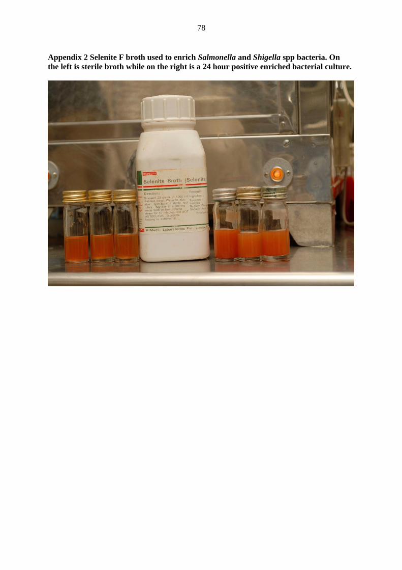
78
Appendix 2 Selenite F broth used to enrich Salmonella and Shigella spp bacteria. On
the left is sterile broth while on the right is a 24 hour positive enriched bacterial culture.
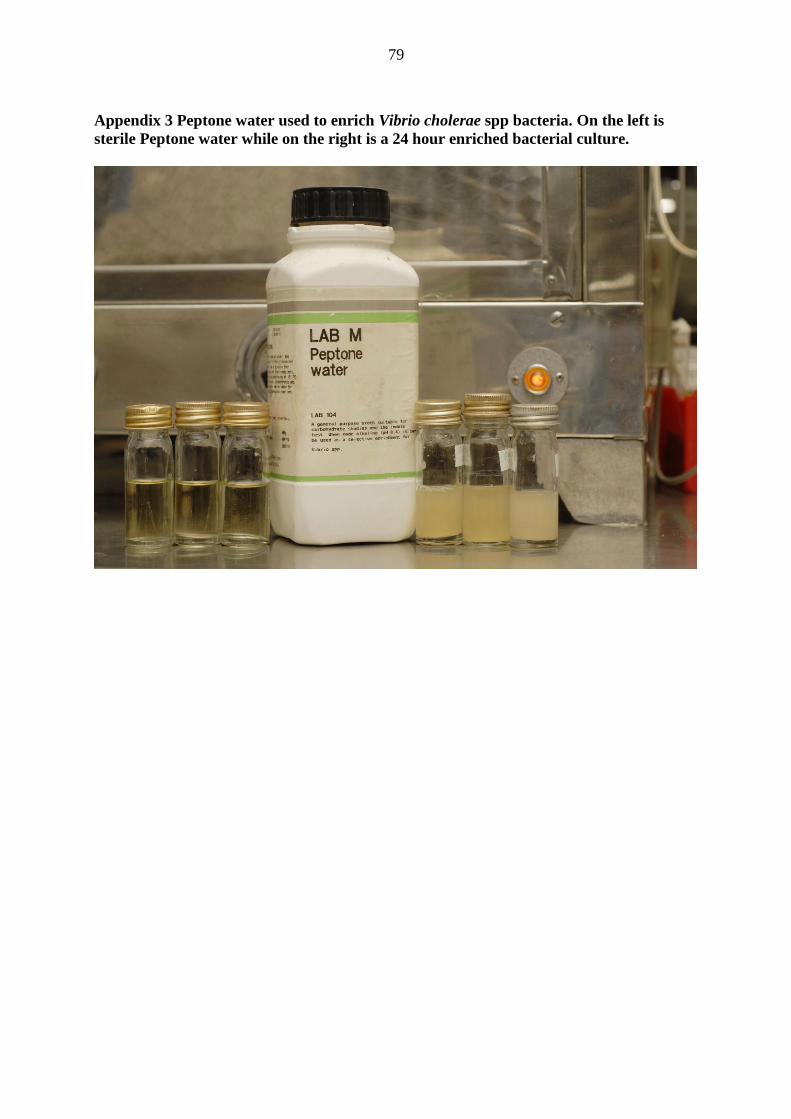
79
Appendix 3 Peptone water used to enrich Vibrio cholerae spp bacteria. On the left is
sterile Peptone water while on the right is a 24 hour enriched bacterial culture.
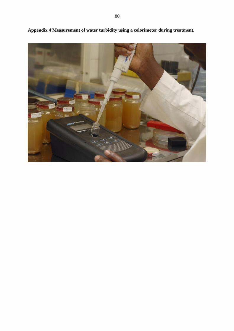
80
Appendix 4 Measurement of water turbidity using a colorimeter during treatment.
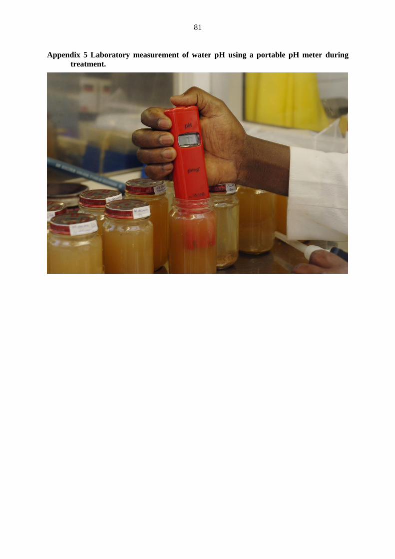
81
Appendix 5 Laboratory measurement of water pH using a portable pH meter during
treatment.
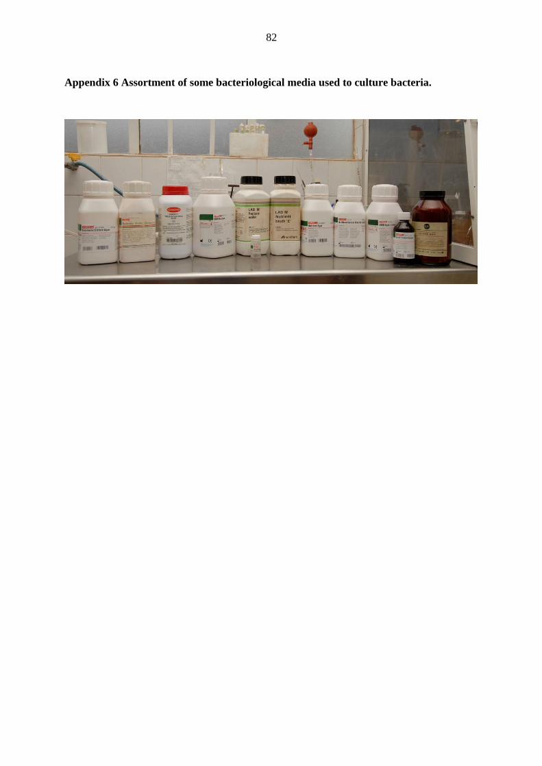
82
Appendix 6 Assortment of some bacteriological media used to culture bacteria.