Embed Size (px)
Citation preview

Supplement
Methods:Animals and Cell IsolationMale WT and Pak1 knockout mice (Pak1-/-; 3-6 month) were used for this study and atrial myocytes were isolated as previously described1. The canine model of ventricular tachypacing-induced atrial fibrillation was created in Dr. Arora’s laboratory and AMs were isolated as previously described2,3. Dogs underwent right ventricular tachypacing (3-4 weeks, 240 beats/min), which resulted in a significant decrease in left ventricular function and a significant increase in left atrial and left ventricular dimensions. While some animals demonstrate evidence of right heart failure (ascites), the majority of animals do not demonstrate symptoms of overt left ventricular heart failure (i.e. shortness of breath, pulmonary edema) 3. Isolated cells from all animals were plated on laminin (1 mg/ml, Sigma Aldrich) coated glass coverslips in standard tyrode solution (in mmol/L: NaCl 130, KCl 5.4, CaCl2 1, MgCl2 1.5, Glucose 10, HEPES 5; pH 7.4). Animal procedures were performed with the approval of the IACUC of Rush University and Northwestern University and in accordance with the National Institute of Health's Guide for the Care and Use of Laboratory Animals.
ECG Recordings and Burst Pacing-Induced Atrial ArrhythmiaECG recordings were performed on mice using the ECGenie (Mouse Specifics, Inc.) recording system. Baseline ECGs were recorded after 10 min adaptation to the recording platform. Recordings were obtained before and after intraperitoneal injection of 150 ng/g of carbachol (CCh). Bipolar left atrial electrograms were recorded from spontaneously beating mouse hearts (WT, Pak1-/-) in the Langendorff configuration, during perfusion with Ctrl Krebs-Henseleit solution (in mmol/L: NaCl 119, NaHCO3 25, KH2PO4 1.2, MgSO4 2, glucose 10, KCl 4.0, CaCl2 1.8, Na-pyruvate 2, gassed with 95 % O2/5 % CO2). Sinus rhythm (SR) was interrupted by 10 episodes of burst pacing (1 s, 50 Hz) applied to the right atria in 10 s intervals. After burst pacing recovery to SR or occurrence and duration of arrhythmic activity was quantified.
Subcellular Fractionation of HL-1 cells.HL-1 cells were maintained for 48 h after infection with adenovirus encoding for

either Pak1-RNAi or LacZ and harvested as previously described4. Cell lysate samples were separated using pre-cast 4-20 % Novex tris-glycine gels (Invitrogen) following standard electrophoresis protocols for SDS-PAGE and immunoblotting with 45 µg of protein loaded per well. Primary antibodies were directed against Rac1 (Cytoskeleton, #ARC03) and Na+/K+-ATPase (NKA: ThermoSci, PA5-17251). Densitometric analysis of was performed using ImageJ. The data are presented as relative density adjusted to the respective loading control (NKA).
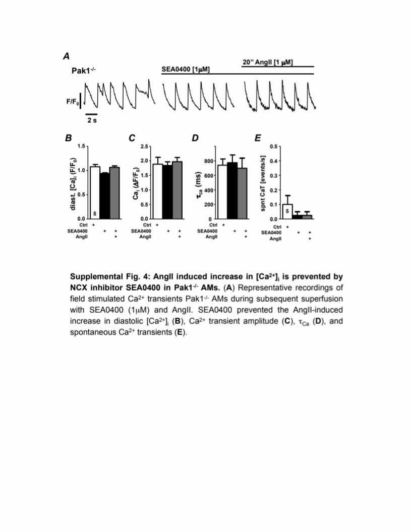
Intracellular Ca2+ and ROS MeasurementsTo visualize changes of the intracellular Ca2+ concentration ([Ca2+]i), AMs were incubated (15 min) at room temperature with fluo-4 acetoxymethyl ester (10 µmol/L; fluo-4/AM; Invitrogen) for ROS measurements cells were loaded with DCFH (10 mmol/L for 30 min at 37oC). [Ca2+]i and ROS measurements were performed and analyzed described1,4. Ca2+-transients are presented as background-subtracted fluorescence normalized to the diastolic fluorescence (F0) at the beginning of the recording (ΔF/F0). AMs were field stimulated at 0.5 Hz for the duration of the experiment. All averaged data are presented as means ± S.E.M., the number of experiments (n) refers to the number of cells examined and is indicated in the text. For each experimental group, cells from at least 2 different cell isolations/animals were used. Significance was evaluated by paired and unpaired t-test.
Quantitative Polymerase Chain Reaction (qPCR)To rule out that changes in excitation-contraction coupling are based on changes in protein expression we quantified mRNA levels of the Ca2+ handling proteins NCX, SERCA, phospholamban (PLN), and RYR by qPCR. Total RNA was extracted from mouse atrial tissue using Trizol (Thermo Fisher Scientific)/chloroform and a Beadbug homogenizer. Extracted RNA was dissolved in DEPC-treated water, stored at -80 °C and used as a template for cDNA synthesis within 24 h. Total RNA (1 μg) was used for cDNA synthesis with the iScript gDNA Clear, cDNA Synthesis Kit (Bio-Rad). The qRT-PCR was performed using a Bio-Rad CFX96 qPCR Instrument. Primers were designed and tested for efficiency prior to quantitation experiments. Primer sequences are provided in Supplemental Figure 2C.

PCR reactions consisted of first-strand cDNA template, forward and reverse primers (100 nM final concentration) and iQ SYBR Green Supermix (BIO-RAD) in a total volume of 10 μl. Glyceraldehyde 3-phosphate dehydrogenase (GAPDH), hypoxanthine guanine phosphoribosyl transferase 1 (HPRT-1), Ribosomal Protein L32 (Rpl32) and Peptidylprolyl Isomerase A (PPIA) transcript levels were used as housekeeping genes. For every mRNA quantitation triplicates were obtained as well as a technical repeat. Standard control PCR reactions were carried out to test for contaminations. Data analysis was performed using the relative expression software tool (REST 2009 ©) for group-wise comparison and statistical analysis of relative expression levels5.


References
1. Desantiago J, Bare DJ, Xiao L, Ke Y, Solaro RJ, Banach K: p21-Activated kinase1 (Pak1) is a negative regulator of NADPH-oxidase 2 in ventricular myocytes. J Mol Cell Cardiol 2014; 67:77–85.
2. Yue L, Feng J, Li GR, Nattel S. Transient outward and delayed rectifier currents in canine atrium: properties and role of isolation methods. Am J Physiol Heart Circ Physiol 1996;270:H2157-2168.
3. Ng J, Villuendas R, Cokic I, et al.: Autonomic remodeling in the left atrium and pulmonary veins in heart failure: creation of a dynamic substrate for atrial fibrillation. Circ Arrhythm Electrophysiol 2011; 4:388–396.
4. Desantiago J, Bare DJ, Ke Y, Sheehan KA, Solaro RJ, Banach K: Functional integrity of the T-tubular system in cardiomyocytes depends on p21-activated kinase 1. J Mol Cell Cardiol 2013; 60:121–128.
5. Pfaffl MW, Horgan GW, Dempfle L: Relative expression software tool (REST) for group-wise comparison and statistical analysis of relative expression results in real-time PCR. Nucleic Acids Res Oxford University Press, 2002; 30:e36.