Embed Size (px)
Citation preview
8/12/2019 VIGS a Tool for a Billie Gould
http://slidepdf.com/reader/full/vigs-a-tool-for-a-billie-gould 1/12
Bio Med Central
Page 1 of 12(page number not for citation purposes)
Plant Methods
Open AccessMethodology Virus-induced gene silencing as a tool for functional analyses in theemerging model plant Aquilegia (columbine, Ranunculaceae)
Billie Gould and Elena M Kramer* Address: Department of Organismic and Evolutionary Biology, Harvard University, 16 Divinity Ave, Cambridge, MA, 02138 USA
Email: Billie Gould - [email protected]; Elena M Kramer* - [email protected]* Corresponding author
AbstractBackground: The lower eudicot genus Aquilegia, commonly known as columbine, is currently thesubject of extensive genetic and genomic research aimed at developing this taxon as a new modelfor the study of ecology and evolution. The ability to perform functional genetic analyses is a criticalcomponent of this development process and ultimately has the potential to provide insight into thegenetic basis for the evolution of a wide array of traits that differentiate flowering plants. Aquilegiais of particular interest due to both its recent evolutionary history, which involves a rapid adaptiveradiation, and its intermediate phylogenetic position between core eudicot (e.g., Arabidopsis) andgrass (e.g., Oryza) model species.
Results: Here we demonstrate the effective use of a reverse genetic technique, virus-induced genesilencing (VIGS), to study gene function in this emerging model plant. Using Agrobacteriummediatedtransfer of tobacco rattle virus (TRV) based vectors, we induce silencing of PHYTOENEDESATURASE( AqPDS) in Aquilegia vulgarisseedlings, and ANTHOCYANIDIN SYNTHASE( AqANS) andthe B-class floral organ identity gene PISTILLATAin A. vulgarisflowers. For all of these genes, silencingphenotypes are associated with consistent reduction in endogenous transcript levels. In addition,we show that silencing of AqANShas no effect on overall floral morphology and is therefore asuitable marker for the identification of silenced flowers in dual-locus silencing experiments.
Conclusion: Our results show that TRV-VIGS in Aquilegia vulgarisallows data to be rapidlyobtained and can be reproduced with effective survival and silencing rates. Furthermore, thismethod can successfully be used to evaluate the function of early-acting developmental genes. Inthe future, data derived from VIGS analyses will be combined with large-scale sequencing andmicroarray experiments already underway in order to address both recent and ancientevolutionary questions.
Background The genus Aquilegia is comprised of approximately 70 spe-cies distributed across temperate areas of North America,Europe, and Asia, with several ornamental varieties soldcommercially [ 1]. These species have undergone a very recent and rapid adaptive radiation in response to biotic
and abiotic factors, resulting in low sequence variationamong species [ 2-4]. Thus, they are ideal for evolutionary studies in that they display a wide range of ecological andmorphological diversity but retain high levels of cross-compatibility between species, allowing for genetic dissec-tion of traits [ 5]. Aquilegia possesses a small diploid
Published: 12 April 2007
Plant Methods 2007, 3:6 doi:10.1186/1746-4811-3-6
Received: 12 March 2007Accepted: 12 April 2007
This article is available from: http://www.plantmethods.com/content/3/1/6
© 2007 Gould and Kramer; licensee BioMed Central Ltd.This is an Open Access article distributed under the terms of the Creative Commons Attribution License ( http://creativecommons.org/licenses/by/2.0 ),which permits unrestricted use, distribution, and reproduction in any medium, provided the original work is properly cited.
8/12/2019 VIGS a Tool for a Billie Gould
http://slidepdf.com/reader/full/vigs-a-tool-for-a-billie-gould 2/12
Plant Methods 2007, 3 :6 http://www.plantmethods.com/content/3/1/6
Page 2 of 12(page number not for citation purposes)
genome (n = 7, 1C = 320–400 Mbp, S. Hodges, pers.comm.), is self-fertile and reproduces regularly with highfecundity. In addition, Aquilegia occupies an intermediatephylogenetic position between core eudicot (e.g., Arabi-dopsis) and grass (e.g., Oryza) model species. Thus func-
tional genetic analyses of physiological andmorphological adaptation in Aquilegia will be valuable inmaking evolutionary comparisons across divergent angiosperms. Many resources already exist to facilitateresearch in this genus, including genetic maps of severalmajor quantitative trait loci [ 5], a fingerprinted BAClibrary [ 6], and an annotated expressed sequence tag (EST) database [ 7]. Most significantly, sequencing of theentire Aquilegia genome will commence at the Joint Genome Institute in 2007 [ 8]. To add to this growing body of research, here we demonstrate effective use of arapid reverse genetic technique, virus-induced gene silenc-ing (VIGS), in this emerging model plant.
VIGS is a method that utilizes the RNAi pathway in plantsto induce transient gene knock-down [ 9]. This processbegins with the Agrobacterium-mediated introduction of modified virus-based cDNA constructs that also containfragments of endogenous gene sequences. Once expressedin vivo, dsRNAs are generated from an encoded viralpolymerase as the virus replicates and spreads through theplant (reviewed [ 10 ]). These dsRNAs are then targeted by DICER-like enzymes and degraded into siRNA. In turn,the siRNA molecules provide a template for degradationof complimentary RNAs, including complimentary endogenous mRNAs, by the RNA-induced silencing com-
plex (RISC). Silencing persists until proliferation of viralRNAs is overcome by the silencing response.
Induction of VIGS is a useful alternative to the often diffi-cult and laborious process of generating stably trans-formed plants, and offers the ability to overcomefunctional redundancy by suppressing all or most mem-bers of a gene family [ 11 ]. VIGS vectors have been devel-oped from viral systems that affect many plant hostsincluding many dicots (reviewed [ 10 -12 ]) and severalmonocots (barley [ 13 ] and wheat [ 14 ]). Here we demon-strate VIGS in the species Aquilegia vulgaris using vectorsbased on the bipartite genome of the tobacco rattle virus
(TRV) (reviewed [ 9]). This system uses two vectors,derived from binary transformation plasmids, which havecDNAs encoding the TRV RNA1 (TRV1) and TRV RNA2(TRV2) inserted into the T-DNA region [ 15 ]. Both vectorscontain a duplicated 35S promoter and a self-cleaving ribozyme sequence to enable rapid generation of intact
viral transcripts (Figure 1). Genes essential for plant toplant transmission of TRV through its nematode vector [16 ] have been deleted from TRV2 [ 15 ]. The TRV systemhas the advantage of being able to penetrate meristematic cells [11 ] and overall causes mild viral symptoms in a
wide range of susceptible hosts [ 17 ]. Although TRV-VIGShas primarily been used in the Solanaceae (tobacco [ 18 ]),it has also been successfully applied in Arabidopsis [19 ]and the lower eudicot Papaver (poppy [ 20 ]).
Here we have successfully produced TRV-VIGS of two phe-notypic marker genes in Aquilegia: PHYTOENE DESATU-RASE ( AqPDS) and ANTHOCYANIDIN SYNTHASE( AqANS). Silencing of AqPDS results in decreased produc-tion of photoprotective carotenoid proteins and the sub-sequent breakdown of chlorophyll pigments [ 21 ,22]. Thisis easily detectable as a photobleaching phenotype inchlorophyll-containing tissues. Silencing of AqANSreduces conversion of colorless leucoanthocyanidins tocolored anthocyanidin [ 23] inhibiting development of the purple wild-type flower color. We have compared
AqPDS silencing at two different developmental stages,using RT-PCR to detect viral transcripts in silenced plants
and the simultaneous decrease in the relative expressionlevel of endogenous AqPDS transcript. Similarly we havemeasured endogenous AqANS levels in silenced and unsi-lenced Aquilegia flowers. Finally, we have used TRV-VIGSto simultaneously knock-down AqANS and a non-marker locus, the floral developmental gene PISTILLATA[24 ,25 ],in order to generate a homeotic floral phenotype. Thisstudy demonstrates that VIGS will be a useful tool for ana-lyzing a wide range of gene function in Aquilegia that cannow be combined with other genetic tools for functionalstudies in this genus.
Results and Discussion
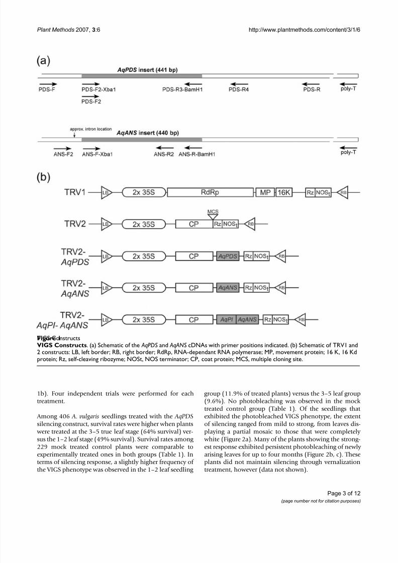
TRV- AqPDS -VIGS treatment of Aquilegia vulgarisseedlingsIn order to test the ability of TRV-based VIGS to promotegene silencing in Aquilegia, we prepared a TRV2 construct [9] containing a 441 bp fragment of AqPDS (Figure 1a).
This fragment was initially isolated from A. vulgaris cDNA using degenerate primers and then re-amplified using locus-specific primers for insertion into the TRV2 plas-mid. As discussed above, VIGS-induced silencing of theendogenous AqPDS locus should result in an easily recog-nizable photobleached phenotype. Although we testedseveral methods for introducing the TRV1/2 plasmids,including direct injection of roots and vegetative parts
(data not shown), only vacuum infiltration of seedlings yielded strong and consistent silencing of AqPDS. Thus far we have been successful in inducing TRV-VIGS in threespecies, A. vulgaris, A. caerulea , and A. alpina , but for thisstudy we focused on A. vulgaris . TRV-VIGS treatment wascarried out on seedlings in two developmental stages:those with 1–2 true leaves and those with 3–5 true leaves.Cotyledon-stage seedlings were not used because initialtrials showed high rates of mortality. Groups of seedlings
were treated with the TRV1 and TRV2- AqPDS constructs or with TRV1 and TRV2 (unmodified) as controls (Figure
8/12/2019 VIGS a Tool for a Billie Gould
http://slidepdf.com/reader/full/vigs-a-tool-for-a-billie-gould 3/12
Plant Methods 2007, 3 :6 http://www.plantmethods.com/content/3/1/6
Page 3 of 12(page number not for citation purposes)
1b). Four independent trials were performed for eachtreatment.
Among 406 A. vulgaris seedlings treated with the AqPDSsilencing construct, survival rates were higher when plants
were treated at the 3–5 true leaf stage (64% survival) ver-sus the 1–2 leaf stage (49% survival). Survival rates among 229 mock treated control plants were comparable toexperimentally treated ones in both groups (Table 1). Interms of silencing response, a slightly higher frequency of the VIGS phenotype was observed in the 1–2 leaf seedling
group (11.9% of treated plants) versus the 3–5 leaf group(9.6%). No photobleaching was observed in the mock treated control group (Table 1). Of the seedlings that exhibited the photobleached VIGS phenotype, the extent of silencing ranged from mild to strong, from leaves dis-playing a partial mosaic to those that were completely
white (Figure 2a). Many of the plants showing the strong-est response exhibited persistent photobleaching of newly arising leaves for up to four months (Figure 2b, c). Theseplants did not maintain silencing through vernalizationtreatment, however (data not shown).
VIGS ConstructsFigure 1VIGS Constructs . (a) Schematic of the AqPDSand AqANScDNAs with primer positions indicated. (b) Schematic of TRV1 and2 constructs: LB, left border; RB, right border; RdRp, RNA-dependant RNA polymerase; MP, movement protein; 16 K, 16 Kdprotein; Rz, self-cleaving ribozyme; NOSt, NOS terminator; CP, coat protein; MCS, multiple cloning site.
8/12/2019 VIGS a Tool for a Billie Gould
http://slidepdf.com/reader/full/vigs-a-tool-for-a-billie-gould 4/12
Plant Methods 2007, 3 :6 http://www.plantmethods.com/content/3/1/6
Page 4 of 12(page number not for citation purposes)
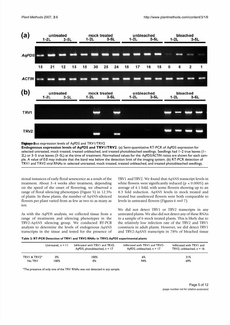
In order to confirm that observed photobleaching wasdue to infection with TRV1 and TRV2 as well as silencing of the endogenous AqPDS locus, we performed semi-quantitative RT-PCR on all treatment groups, comparing bleached and unbleached individuals (Figure 3 and Table2). The presence of both TRV RNAs was detected in 100%of sampled plants exhibiting a bleached phenotype and in0% of untreated plants. Among sampled plants that were
treated with the silencing construct but that did not exhibit a bleached phenotype, 96% showed no presenceof either TRV1 or TRV2. One plant in this group was pos-itive for both RNAs indicating that infection had occurredbut did not produce silencing. In the mock treated controlgroup, 69% of plants showed no presence of either TRV1or TRV2, and 31% showed the presence of both, which isindicative of the approximate overall rate of uptake of these constructs in A. vulgaris seedlings. We did not detect only one of the viral RNAs in any of the samples.
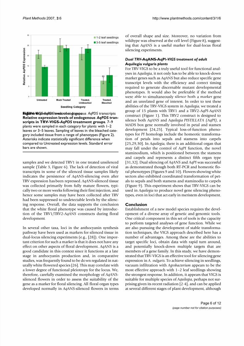
Semi-quantitative RT-PCR supported the conclusion that the observed photobleached phenotype was caused by areduction in the levels of endogenous AqPDS transcripts.Lower relative levels of expression were detected in pho-tobleached plants compared with both untreated plantsand mock treated plants from both seedling stages. In the3–5 leaf seedling group, we detected an average 2.0 foldreduction in AqPDS levels between TRV-VIGS treated
bleached seedlings and untreated seedlings. This differ-ence was not statistically significant due to the high levelsof variation in the degree of photobleaching. Reduction of
AqPDS transcript levels in the 3–5 leaf group ranged fromno detectable reduction up to a 15-fold decrease in expres-sion as compared with the average AqPDS level inuntreated seedlings. In the 1–2 leaf developmental stagegroup there was an average 2.7 fold reduction in the
AqPDS level (Figure 4), which was much more consistent and highly significant at p < 0.0005.
TRV- AqANS -VIGS treatment of adult Aquilegia vulgaris plants
In order to test if VIGS can be used to create silencing phe-notypes in Aquilegia flowers, we targeted the ANTHOCYA- NIDIN SYNTHASE (AqANS) gene in post-vernalization(cold treated) plants. AqANS appears to be a single copy locus in Aquilegia (S. Hodges, pers. comm., [ 26 ]). The
AqANS sequence was obtained from the recently com-piled Aquilegia EST database and used to amplify andclone a 440 bp fragment into the TRV2 vector (see Exper-imental Procedures). Adult plants with 15 or more leaves
were vernalized for 8 weeks at 4°C to induce flowering. Two weeks after transfer to the greenhouse at 20°C, we wounded each plant at the basal rosette and injected themix of Agrobacterium solutions into the wound with a nee-
dle-less syringe at weekly intervals. We also tested floraldip-based methods [ 27 ] and vacuum infiltration of inflo-rescences, but, unlike the experiments with the seedlings,
we had more consistent success with the injection method(data not shown). Two separate groups of plants weretreated with the injection technique for a total of 48 plantstreated with TRV1/TRV2- AqANS, 6 control plants treated
with TRV1/TRV2 (unmodified), and 4 plants left untreated. Survival rates were 100% for both TRV treat-ment groups (one untreated plant expired of naturalcauses during the experiment) and there were only occa-
Table 1: Survival and silencing rates among seedlings
Seedling size, Construct N Treated % Survival % Phenotype (of total) % Phenotype (of survivors)
1–2 La, TRV2- AqPDS 218 48.6% 11.9% 24.5%1–2 L, TRV2 97 52.6% 0.0% 0.0%
3–5 Lb, TRV2- AqPDS 188 64.4% 9.6% 14.9%3–5 L, TRV2 132 59.1% 0.0% 0.0%
a1–2 L = 1–2 leaves at infiltration; b3–5 L = 3–5 leaves at infiltration.
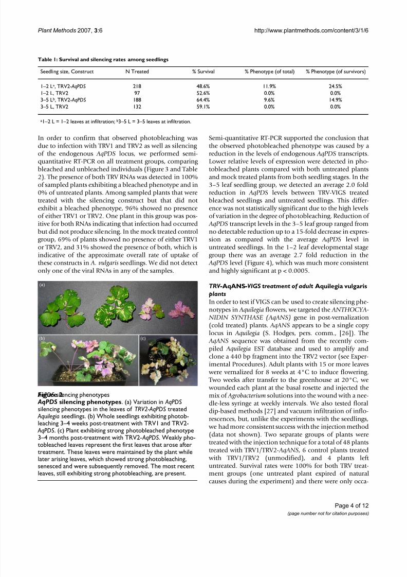
AqPDSsilencing phenotypesFigure 2
AqPDS silencing phenotypes . (a) Variation in AqPDSsilencing phenotypes in the leaves of TRV2-AqPDStreated Aquilegiaseedlings. (b) Whole seedlings exhibiting photob-leaching 3–4 weeks post-treatment with TRV1 and TRV2-
AqPDS. (c) Plant exhibiting strong photobleached phenotype3–4 months post-treatment with TRV2- AqPDS. Weakly pho-tobleached leaves represent the first leaves that arose aftertreatment. These leaves were maintained by the plant whilelater arising leaves, which showed strong photobleaching,senesced and were subsequently removed. The most recentleaves, still exhibiting strong photobleaching, are present.
8/12/2019 VIGS a Tool for a Billie Gould
http://slidepdf.com/reader/full/vigs-a-tool-for-a-billie-gould 5/12
Plant Methods 2007, 3 :6 http://www.plantmethods.com/content/3/1/6
Page 5 of 12(page number not for citation purposes)
sional instances of early floral senescence as a result of thetreatment. About 3–4 weeks after treatment, depending on the speed of the onset of flowering, we observed arange of floral silencing phenotypes (Figure 5) in 12.5%of plants. In these plants, the number of AqANS-silencedflowers per plant varied from as few as two to as many asten.
As with the AqPDS analysis, we collected tissue from arange of treatments and silencing phenotypes in the
TRV2- AqANS silencing group. We conducted RT-PCR
analysis to determine the levels of endogenous AqANStranscripts in the tissue and tested for the presence of
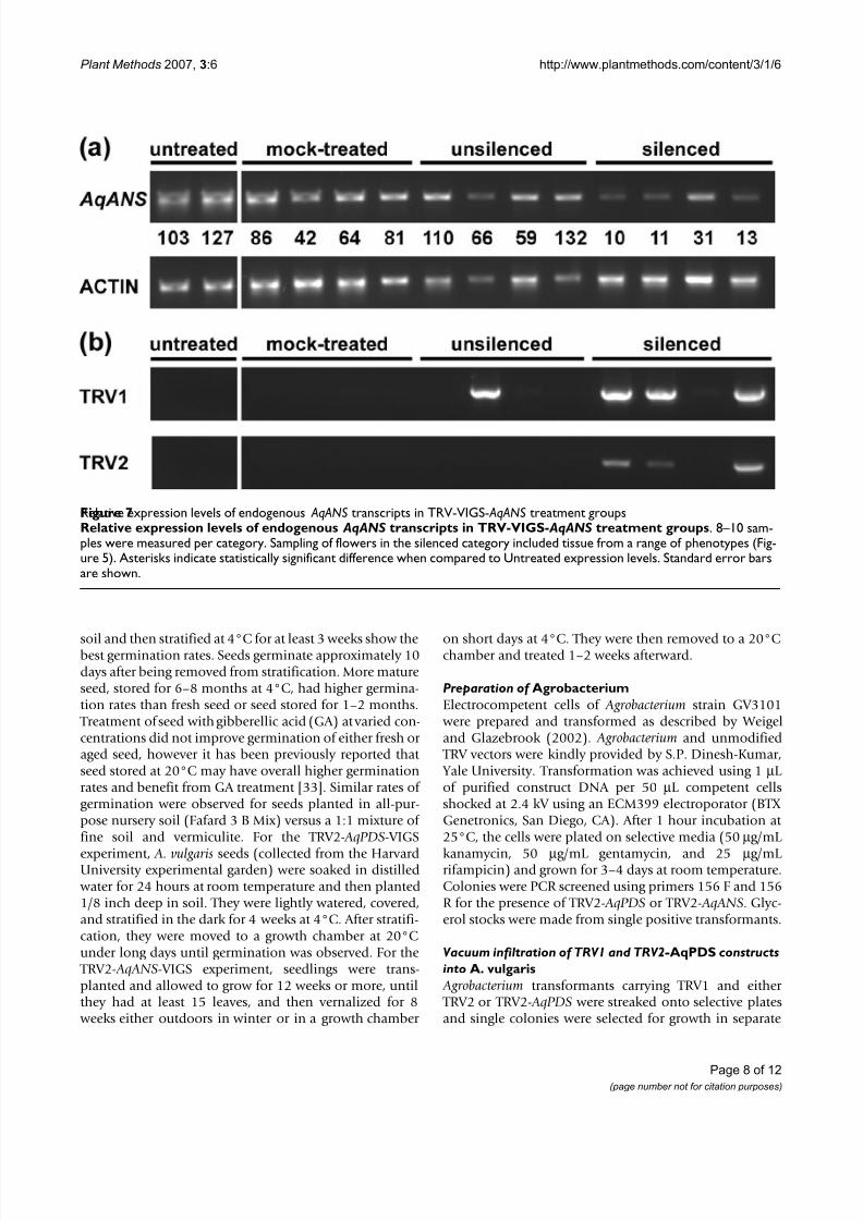
TRV1 and TRV2. We found that AqANS transcript levels in white flowers were significantly reduced (p < 0.0005) anaverage of 4.1 fold, with some flowers showing up to an8.3 fold reduction. AqANS levels in mock treated andtreated but unsilenced flowers were both comparable tolevels in untreated flowers (Figures 6 and 7).
We did not detect TRV1 or TRV2 transcripts in any untreated plants. We also did not detect any of these RNAsin a sample of 6 mock treated plants. This is likely due tothe relatively low infection rate of the TRV2 and TRV1
constructs in adult plants. However, we did detect TRV1and TRV2- AqANS transcripts in 78% of bleached tissue
Table 2: RT-PCR Detection of TRV1 and TRV2 RNAs in TRV2- AqPDS experimental plants
Untreated, n = 11 Infiltrated with TRV1 and TRV2- AqPDS; photobleached, n = 17
Infiltrated with TRV1 and TRV2- AqPDS; unbleached, n = 17
Infiltrated with TRV1 andTRV2; unbleached, n = 16
TRV1 & TRV2* 0% 100% 6% 31%No TRV 100% 0% 94% 69%
*The presence of only one of the TRV RNAs was not detected in any sample.
Endogenous expression levels of AqPDSand TRV1/TRV2Figure 3Endogenous expression levels of AqPDS and TRV1/TRV2 . (a) Semi-quantitative RT-PCR of AqPDSexpression forselected untreated, mock treated, treated unbleached, and treated photobleached seedlings. Seedlings had 1–2 true leaves (1– 2L) or 3–5 true leaves (3–5L) at the time of treatment. Normalized values for the AqPDS/ ACTINratios are shown for each sam-ple. A value of 0.0 may indicate that the band was below the detection limit of the imaging system. (b) RT-PCR detection ofTRV1 and TRV2 viral RNAs in selected untreated, mock treated, treated unbleached, and treated photobleached seedlings.
8/12/2019 VIGS a Tool for a Billie Gould
http://slidepdf.com/reader/full/vigs-a-tool-for-a-billie-gould 6/12
Plant Methods 2007, 3 :6 http://www.plantmethods.com/content/3/1/6
Page 6 of 12(page number not for citation purposes)
samples and we detected TRV1 in one treated unsilencedsample (Table 3, Figure 6). The lack of detection of viraltranscripts in some of the silenced tissue samples likely indicates the persistence of AqANS-silencing even after
TRV expression has been repressed. AqANS-silenced tissue was collected primarily from fully mature flowers, typi-cally two or more weeks following their first injection, andhence some samples may have been collected after TRV had been suppressed to undetectable levels by the silenc-ing response. Overall, the data supports the conclusionthat the white floral phenotype was caused by introduc-tion of the TRV1/TRV2- AqANS constructs during floraldevelopment.
In several other taxa, loci in the anthocyanin synthesispathway have been used as markers for silenced tissue indual-locus silencing experiments (e.g., [ 28 ]). One impor-
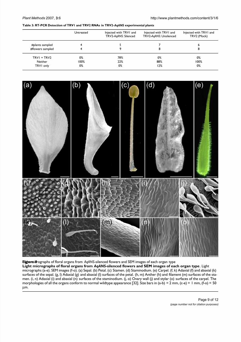
tant criterion for such a marker is that it does not have any effect on other aspects of floral development. AqANS is agood candidate in this context since it functions at a latestage in anthocyanin production and, in comparativestudies, was frequently found to be down regulated in nat-urally white flowered species [ 26 ]. This may correlate witha lower degree of functional pleiotropy for the locus. We,therefore, carefully examined the morphology of AqANS-silenced flowers in order to assess the suitability of thegene as a marker for floral silencing. All floral organ typesdeveloped normally in AqANS-silenced flowers in terms
of overall shape and size. Moreover, no variation from wildtype was observed at the cell level (Figure 8), suggest-ing that AqANS is a useful marker for dual-locus floralsilencing experiments.
Dual TRV- AqANS -AqPI -VIGS treatment of adultAquilegia vulgaris plantsFor TRV-VIGS to be a truly useful tool for functional anal-
yses in Aquilegia, it not only has to be able to knock-downmarker genes such as AqANS but also reduce specific genetranscript levels with the efficiency and correct timing required to generate discernable mutant developmentalphenotypes. It would also be preferable if the method
were able to simultaneously silence both a marker geneand an unrelated gene of interest. In order to test theseabilities of the TRV-VIGS system in Aquilegia, we treated agroup of 15 plants with TRV1 and a TRV2- AqPI-AqANSconstruct (Figure 1). This TRV2 construct is designed to
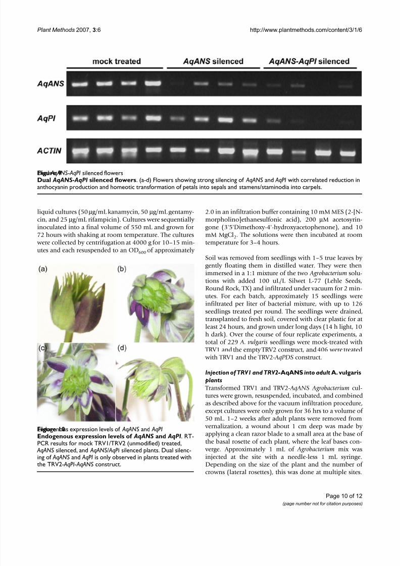
silence both AqANS and Aquilegia PISTILLATA (AqPI) , aMADS box gene normally involved in petal and stamendevelopment [ 24 ,25 ]. Typical loss-of-function pheno-types for PI homologs include the homeotic transforma-tion of petals into sepals and stamens into carpels[25 ,29 ,30 ]. In Aquilegia, there is an additional organ that may fall under the control of AqPI function, the novelstaminodium, which is positioned between the stamensand carpels and represents a distinct fifth organ type[31 ,32]. Dual silencing of AqANS and AqPI was successfulas demonstrated though both RT-PCR and homeotic flo-ral phenotypes (Figures 9 and 10 ). Flowers showing whitesectors also exhibited coordinated transformation of pet-
als to sepals and both stamens and staminodia to carpels(Figure 9). This experiment shows that TRV-VIGS can beused in Aquilegia to produce novel gene silencing pheno-types, even in loci that act early in meristem development.
ConclusionEstablishment of a new model species requires the devel-opment of a diverse array of genetic and genomic tools.One critical component in this set of tools is the capacity to perform targeted analyses of gene function. While weare also pursuing the development of stable transforma-tion techniques, the VIGS approach described here has anumber of advantages. Among these are the abilities to
target specific loci, obtain data with rapid turn around,and potentially knock-down multiple targets that aremembers of a gene family. In this study, we have demon-strated that TRV-VIGS is an effective tool for silencing geneexpression in A. vulgaris. To achieve silencing in seedlings,
vacuum infiltration with Agrobacterium appears to be themost effective approach with 1–2 leaf seedlings showing the strongest response. In addition, it appears that VIGS issuitable for multiple species of Aquilegia, perhaps not sur-prising given its recent radiation [ 2-4], and can be appliedat several different stages of plant development, although
Relative expression levels of endogenous AqPDStranscriptsin TRV-VIGS- AqPDStreatment groupsFigure 4Relative expression levels of endogenous AqPDS tran-scripts in TRV-VIGS- AqPDS treatment groups . 7–9plants were sampled in each category for plants with 1–2leaves or 3–5 leaves. Sampling of leaves in the bleached cate-gory included tissue from a range of phenotypes (Figure 2).Asterisks indicate statistically significant difference whencompared to Untreated expression levels. Standard errorbars are shown.
8/12/2019 VIGS a Tool for a Billie Gould
http://slidepdf.com/reader/full/vigs-a-tool-for-a-billie-gould 7/12
Plant Methods 2007, 3 :6 http://www.plantmethods.com/content/3/1/6
Page 7 of 12(page number not for citation purposes)
at somewhat lower rates of efficiency (data not shown). VIGS in Aquilegia can also be used to generate floral devel-opmental phenotypes with low risk of mortality and effec-
tive rates of gene silencing. Both a marker gene such as
AqANS (useful for rapid identification of plants with thestrongest levels of silencing) and an unrelated gene of interest can be silenced simultaneously in order to inves-tigate developmental function. The addition of VIGS toour growing set of resources for Aquilegia will further
advance its rise as an important new model system for thestudy of evolutionary and ecological questions.
MethodsGeneration of VIGS TRV2- AqPDS and TRV2- AqANSconstructs
Total RNA was extracted from Aquilegia vulgaris leaf tissueusing Plant RNA Isolation Reagent (Invitrogen, Carlsbad,CA) and mRNA was purified using Magnetite Oligo (dT)Particles (Novagen (EMD), San Diego, CA). cDNA wasgenerated from 500 ng of mRNA using Superscript IIReverse Transcriptase™ (Invitrogen). The AqPDS sequence
was isolated through PCR using degenerate primers from
conserved regions of the PDS gene (PDS-F: 5'-TGGAAR-GARCAYTCIATGATWTTTGCWATG-3' and PDS-R: 5'- ACRACATGRTACTTIAVDATYTTWGCTTT-3', Figure 1). The primary AqPDS fragment was then sequenced to con-firm its identity [DQ923721] using BigDye Terminator ®
(Applied Biosystems, Foster City, CA) and re-amplifiedusing locus-specific primers with appended restrictionsites (PDS-F2-XbaI: 5'-GGTCTAGACAGCCGATTTGATT-
TCCCAGAT-3' and PDS-R3-BamH1: 5'-AAGGATCCGA-GAATTGAGTCGGACTTCACC-3', Figure 1). The AqANSsequence was obtained from the Aquilegia EST database[DR946275 and DR946276], and primers were designedto amplify a 440 bp fragment of the gene (ANS-F-Xba1: 5'-
GGTCTAGATTGGGATTGGAAGAAGAAAGGC-3' and ANS-R-BamH1: 5'-AAGGATCCATGTTGAGCAAATGT-GCGA-3', Figure 1).
Both gene products were double digested with XbaI andBamH1, as was the TRV2 vector, and ligated separately using T4 DNA ligase (New England Biolabs, Ipswich,MA). The resulting constructs were transformed into heat-shock competent E.coli (TOP10 cells, Invitrogen) andplated on selective LB media containing 50 µ g/mL kan-amycin. Colonies were PCR screened for the presence of the modified constructs using primers 156 F (5'-TTACT-CAAGGAAGCACGATGAGC-3') and 156 R (5'-GAACCG-
TAGTTTAATGTCTTCGGG-3'), which span the multiplecloning site in TRV2. Both constructs were then purified(Fast-Plasmid Mini kit, Eppendorf, Hamburg, Germany)and the identity of the final constructs was verified by sequencing with the 156 F and 156 R primers and BigDye
Terminator ®
Germination of A. vulgaris seedlings and growth of adult plants When germinating Aquilegia vulgaris seed, we found that seeds soaked in distilled water for 24 hours, planted in
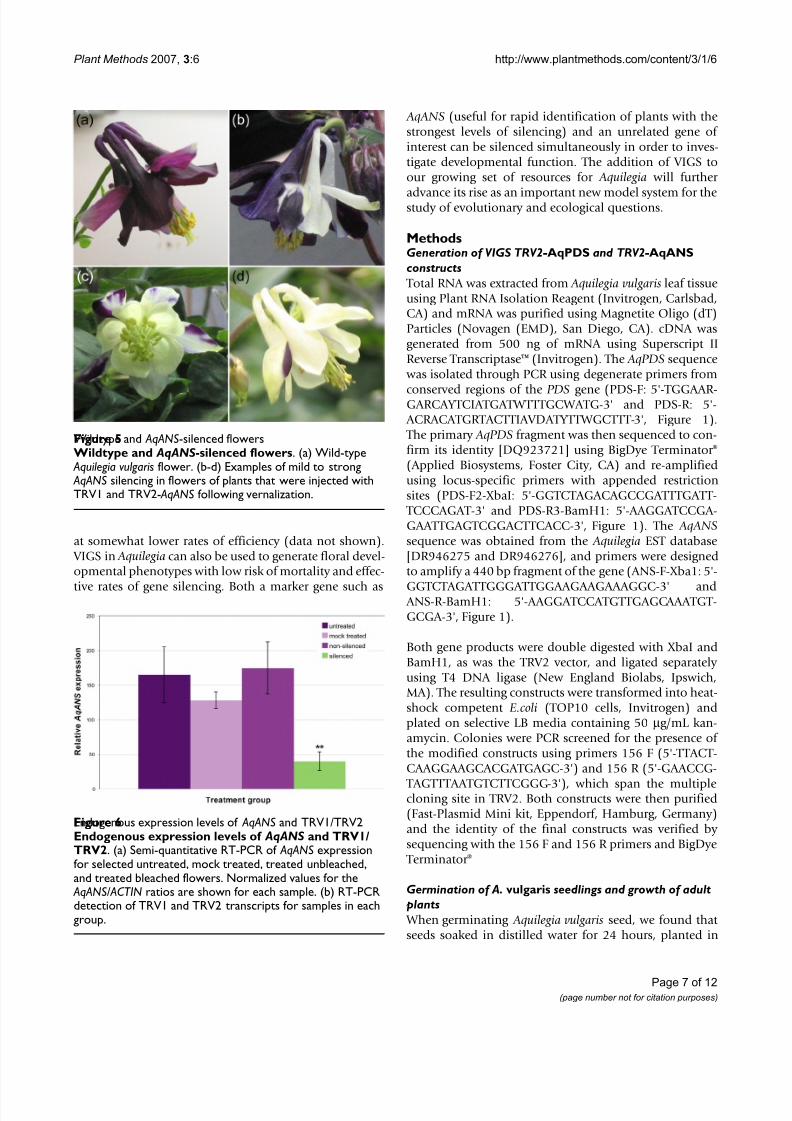
Wildtype and AqANS-silenced flowersFigure 5 Wildtype and AqANS -silenced flowers . (a) Wild-type Aquilegia vulgarisflower. (b-d) Examples of mild to strong AqANSsilencing in flowers of plants that were injected withTRV1 and TRV2- AqANSfollowing vernalization.
Endogenous expression levels of AqANSand TRV1/TRV2Figure 6Endogenous expression levels of AqANS and TRV1/TRV2 . (a) Semi-quantitative RT-PCR of AqANSexpressionfor selected untreated, mock treated, treated unbleached,and treated bleached flowers. Normalized values for the
AqANS/ ACTINratios are shown for each sample. (b) RT-PCRdetection of TRV1 and TRV2 transcripts for samples in eachgroup.
8/12/2019 VIGS a Tool for a Billie Gould
http://slidepdf.com/reader/full/vigs-a-tool-for-a-billie-gould 8/12
Plant Methods 2007, 3 :6 http://www.plantmethods.com/content/3/1/6
Page 8 of 12(page number not for citation purposes)
soil and then stratified at 4°C for at least 3 weeks show thebest germination rates. Seeds germinate approximately 10days after being removed from stratification. More matureseed, stored for 6–8 months at 4°C, had higher germina-tion rates than fresh seed or seed stored for 1–2 months.
Treatment of seed with gibberellic acid (GA) at varied con-centrations did not improve germination of either fresh or aged seed, however it has been previously reported that seed stored at 20°C may have overall higher germinationrates and benefit from GA treatment [ 33 ]. Similar rates of germination were observed for seeds planted in all-pur-pose nursery soil (Fafard 3 B Mix) versus a 1:1 mixture of fine soil and vermiculite. For the TRV2- AqPDS-VIGS
experiment, A. vulgaris seeds (collected from the HarvardUniversity experimental garden) were soaked in distilled water for 24 hours at room temperature and then planted1/8 inch deep in soil. They were lightly watered, covered,and stratified in the dark for 4 weeks at 4°C. After stratifi-cation, they were moved to a growth chamber at 20°Cunder long days until germination was observed. For the
TRV2- AqANS-VIGS experiment, seedlings were trans-planted and allowed to grow for 12 weeks or more, untilthey had at least 15 leaves, and then vernalized for 8
weeks either outdoors in winter or in a growth chamber
on short days at 4°C. They were then removed to a 20°Cchamber and treated 1–2 weeks afterward.
Preparation of AgrobacteriumElectrocompetent cells of Agrobacterium strain GV3101
were prepared and transformed as described by Weigeland Glazebrook (2002). Agrobacterium and unmodified
TRV vectors were kindly provided by S.P. Dinesh-Kumar, Yale University. Transformation was achieved using 1 µ L of purified construct DNA per 50 µ L competent cellsshocked at 2.4 kV using an ECM399 electroporator (BTX Genetronics, San Diego, CA). After 1 hour incubation at 25°C, the cells were plated on selective media (50 µ g/mL
kanamycin, 50 µ g/mL gentamycin, and 25 µ g/mL rifampicin) and grown for 3–4 days at room temperature.Colonies were PCR screened using primers 156 F and 156R for the presence of TRV2- AqPDS or TRV2- AqANS. Glyc-erol stocks were made from single positive transformants.
Vacuum infiltration of TRV1 and TRV2- AqPDS constructsinto A. vulgaris
Agrobacterium transformants carrying TRV1 and either TRV2 or TRV2- AqPDS were streaked onto selective platesand single colonies were selected for growth in separate
Relative expression levels of endogenous AqANStranscripts in TRV-VIGS- AqANStreatment groupsFigure 7Relative expression levels of endogenous AqANS transcripts in TRV-VIGS- AqANS treatment groups . 8–10 sam-ples were measured per category. Sampling of flowers in the silenced category included tissue from a range of phenotypes (Fig-ure 5). Asterisks indicate statistically significant difference when compared to Untreated expression levels. Standard error barsare shown.
8/12/2019 VIGS a Tool for a Billie Gould
http://slidepdf.com/reader/full/vigs-a-tool-for-a-billie-gould 9/12
Plant Methods 2007, 3 :6 http://www.plantmethods.com/content/3/1/6
Page 9 of 12(page number not for citation purposes)
Light micrographs of floral organs from AqANS-silenced flowers and SEM images of each organ typeFigure 8Light micrographs of floral organs from AqANS -silenced flowers and SEM images of each organ type . Lightmicrographs (a-e). SEM images (f-o). (a) Sepal. (b) Petal. (c) Stamen. (d) Staminodium. (e) Carpel. (f, k) Adaxial (f) and abaxial (k)surfaces of the sepal. (g, l) Adaxial (g) and abaxial (l) surfaces of the petal. (h, m) Anther (h) and filament (m) surfaces of the sta-men. (i, n) Adaxial (i) and abaxial (n) surfaces of the staminodium. (j, o) Ovary wall (j) and stylar (o) surfaces of the carpel. Themorphologies of all the organs conform to normal wildtype appearance [32]. Size bars in (a-b) = 2 mm, (c-e) = 1 mm, (f-o) = 50µ m.
Table 3: RT-PCR Detection of TRV1 and TRV2 RNAs in TRV2- AqANS experimental plants
Untreated Injected with TRV1 andTRV2- AqANS: Silenced
Injected with TRV1 andTRV2- AqANS: Unsilenced
Injected with TRV1 andTRV2 (Mock)
#plants sampled 4 5 7 6#flowers sampled 4 9 8 8
TRV1 + TRV2 0% 78% 0% 0%Neither 100% 22% 88% 100%
TRV1 only 0% 0% 12% 0%
8/12/2019 VIGS a Tool for a Billie Gould
http://slidepdf.com/reader/full/vigs-a-tool-for-a-billie-gould 10/12
Plant Methods 2007, 3 :6 http://www.plantmethods.com/content/3/1/6
Page 10 of 12(page number not for citation purposes)
liquid cultures (50 µ g/mL kanamycin, 50 µ g/mL gentamy-cin, and 25 µ g/mL rifampicin). Cultures were sequentially inoculated into a final volume of 550 mL and grown for 72 hours with shaking at room temperature. The cultures
were collected by centrifugation at 4000 g for 10–15 min-utes and each resuspended to an OD 600 of approximately
2.0 in an infiltration buffer containing 10 mM MES (2-[N-morpholino]ethanesulfonic acid), 200 µ M acetosyrin-gone (3'5'Dimethoxy-4'-hydroxyacetophenone), and 10mM MgCl 2. The solutions were then incubated at roomtemperature for 3–4 hours.
Soil was removed from seedlings with 1–5 true leaves by gently floating them in distilled water. They were thenimmersed in a 1:1 mixture of the two Agrobacterium solu-tions with added 100 uL/L Silwet L-77 (Lehle Seeds,Round Rock, TX) and infiltrated under vacuum for 2 min-
utes. For each batch, approximately 15 seedlings wereinfiltrated per liter of bacterial mixture, with up to 126seedlings treated per round. The seedlings were drained,transplanted to fresh soil, covered with clear plastic for at least 24 hours, and grown under long days (14 h light, 10h dark). Over the course of four replicate experiments, atotal of 229 A. vulgaris seedlings were mock-treated with
TRV1 and the empty TRV2 construct, and 406 were treated with TRV1 and the TRV2- AqPDS construct.
Injection of TRV1 and TRV2- AqANS into adult A. vulgaris plants Transformed TRV1 and TRV2- AqANS Agrobacterium cul-
tures were grown, resuspended, incubated, and combinedas described above for the vacuum infiltration procedure,except cultures were only grown for 36 hrs to a volume of 50 mL. 1–2 weeks after adult plants were removed from
vernalization, a wound about 1 cm deep was made by applying a clean razor blade to a small area at the base of the basal rosette of each plant, where the leaf bases con-
verge. Approximately 1 mL of Agrobacterium mix wasinjected at the site with a needle-less 1 mL syringe.Depending on the size of the plant and the number of crowns (lateral rosettes), this was done at multiple sites.
Endogenous expression levels of AqANSand AqPIFigure 10Endogenous expression levels of AqANS and AqPI . RT-PCR results for mock TRV1/TRV2 (unmodified) treated,
AqANSsilenced, and AqANS/ AqPIsilenced plants. Dual silenc-ing of AqANSand AqPIis only observed in plants treated withthe TRV2- AqPI- AqANSconstruct.
Dual AqANS- AqPIsilenced flowersFigure 9Dual AqANS - AqPI silenced flowers . (a-d) Flowers showing strong silencing of AqANSand AqPIwith correlated reduction inanthocyanin production and homeotic transformation of petals into sepals and stamens/staminodia into carpels.
8/12/2019 VIGS a Tool for a Billie Gould
http://slidepdf.com/reader/full/vigs-a-tool-for-a-billie-gould 11/12
Plant Methods 2007, 3 :6 http://www.plantmethods.com/content/3/1/6
Page 11 of 12(page number not for citation purposes)
The procedure was repeated at weekly intervals for 5–6 weeks with new injection sites created with each treat-ment.
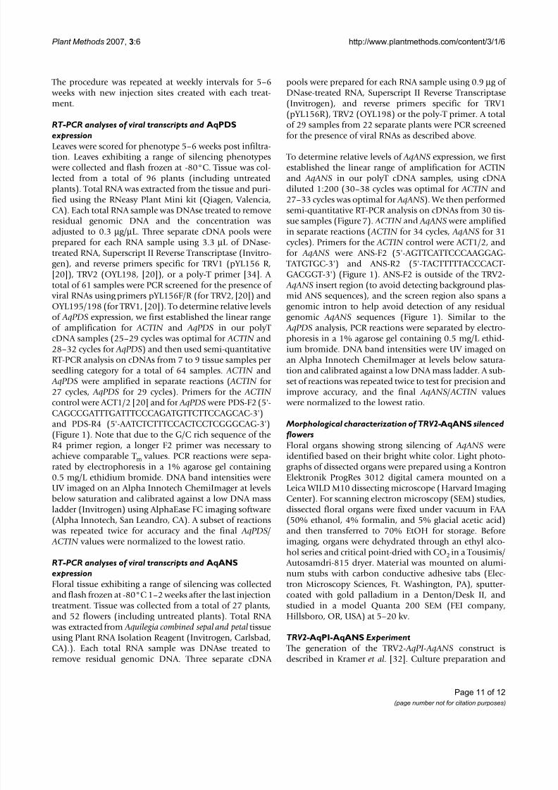
RT-PCR analyses of viral transcripts and AqPDS
expressionLeaves were scored for phenotype 5–6 weeks post infiltra-tion. Leaves exhibiting a range of silencing phenotypes
were collected and flash frozen at -80°C. Tissue was col-lected from a total of 96 plants (including untreatedplants). Total RNA was extracted from the tissue and puri-fied using the RNeasy Plant Mini kit (Qiagen, Valencia,CA). Each total RNA sample was DNAse treated to removeresidual genomic DNA and the concentration wasadjusted to 0.3 µ g/µ L. Three separate cDNA pools wereprepared for each RNA sample using 3.3 µ L of DNase-treated RNA, Superscript II Reverse Transcriptase (Invitro-gen), and reverse primers specific for TRV1 (pYL156 R,
[20 ]), TRV2 (OYL198, [ 20 ]), or a poly-T primer [ 34 ]. A total of 61 samples were PCR screened for the presence of viral RNAs using primers pYL156F/R (for TRV2, [ 20]) andOYL195/198 (for TRV1, [ 20 ]). To determine relative levelsof AqPDS expression, we first established the linear rangeof amplification for ACTIN and AqPDS in our polyT cDNA samples (25–29 cycles was optimal for ACTIN and28–32 cycles for AqPDS) and then used semi-quantitativeRT-PCR analysis on cDNAs from 7 to 9 tissue samples per seedling category for a total of 64 samples. ACTIN and
AqPDS were amplified in separate reactions ( ACTIN for 27 cycles, AqPDS for 29 cycles). Primers for the ACTIN control were ACT1/2 [ 20 ] and for AqPDS were PDS-F2 (5'-
CAGCCGATTTGATTTCCCAGATGTTCTTCCAGCAC-3')and PDS-R4 (5'-AATCTCTTTCCACTCCTCGGGCAG-3')(Figure 1). Note that due to the G/C rich sequence of theR4 primer region, a longer F2 primer was necessary toachieve comparable T m values. PCR reactions were sepa-rated by electrophoresis in a 1% agarose gel containing 0.5 mg/L ethidium bromide. DNA band intensities wereUV imaged on an Alpha Innotech ChemiImager at levelsbelow saturation and calibrated against a low DNA massladder (Invitrogen) using AlphaEase FC imaging software(Alpha Innotech, San Leandro, CA). A subset of reactions
was repeated twice for accuracy and the final AqPDS / ACTIN values were normalized to the lowest ratio.
RT-PCR analyses of viral transcripts and AqANSexpressionFloral tissue exhibiting a range of silencing was collectedand flash frozen at -80°C 1–2 weeks after the last injectiontreatment. Tissue was collected from a total of 27 plants,and 52 flowers (including untreated plants). Total RNA
was extracted from Aquilegia combined sepal and petal tissueusing Plant RNA Isolation Reagent (Invitrogen, Carlsbad,CA).). Each total RNA sample was DNAse treated toremove residual genomic DNA. Three separate cDNA
pools were prepared for each RNA sample using 0.9 µ g of DNase-treated RNA, Superscript II Reverse Transcriptase(Invitrogen), and reverse primers specific for TRV1(pYL156R), TRV2 (OYL198) or the poly-T primer. A totalof 29 samples from 22 separate plants were PCR screened
for the presence of viral RNAs as described above.
To determine relative levels of AqANS expression, we first established the linear range of amplification for ACTINand AqANS in our polyT cDNA samples, using cDNA diluted 1:200 (30–38 cycles was optimal for ACTIN and27–33 cycles was optimal for AqANS). We then performedsemi-quantitative RT-PCR analysis on cDNAs from 30 tis-sue samples (Figure 7). ACTIN and AqANS were amplifiedin separate reactions ( ACTIN for 34 cycles, AqANS for 31cycles). Primers for the ACTIN control were ACT1/2, andfor AqANS were ANS-F2 (5'-AGTTCATTCCCAAGGAG-
TATGTGC-3') and ANS-R2 (5'-TACTTTTTACCCACT-
GACGGT-3') (Figure 1). ANS-F2 is outside of the TRV2- AqANS insert region (to avoid detecting background plas-mid ANS sequences), and the screen region also spans agenomic intron to help avoid detection of any residualgenomic AqANS sequences (Figure 1). Similar to the
AqPDS analysis, PCR reactions were separated by electro-phoresis in a 1% agarose gel containing 0.5 mg/L ethid-ium bromide. DNA band intensities were UV imaged onan Alpha Innotech ChemiImager at levels below satura-tion and calibrated against a low DNA mass ladder. A sub-set of reactions was repeated twice to test for precision andimprove accuracy, and the final AqANS / ACTIN values
were normalized to the lowest ratio.
Morphological characterization of TRV2- AqANS silencedflowersFloral organs showing strong silencing of AqANS wereidentified based on their bright white color. Light photo-graphs of dissected organs were prepared using a KontronElektronik ProgRes 3012 digital camera mounted on aLeica WILD M10 dissecting microscope (Harvard Imaging Center). For scanning electron microscopy (SEM) studies,dissected floral organs were fixed under vacuum in FAA (50% ethanol, 4% formalin, and 5% glacial acetic acid)and then transferred to 70% EtOH for storage. Beforeimaging, organs were dehydrated through an ethyl alco-
hol series and critical point-dried with CO 2 in a Tousimis/ Autosamdri-815 dryer. Material was mounted on alumi-num stubs with carbon conductive adhesive tabs (Elec-tron Microscopy Sciences, Ft. Washington, PA), sputter-coated with gold palladium in a Denton/Desk II, andstudied in a model Quanta 200 SEM (FEI company,Hillsboro, OR, USA) at 5–20 kv.
TRV2-AqPI-AqANS Experiment The generation of the TRV2- AqPI - AqANS construct isdescribed in Kramer et al. [32 ]. Culture preparation and
8/12/2019 VIGS a Tool for a Billie Gould
http://slidepdf.com/reader/full/vigs-a-tool-for-a-billie-gould 12/12
Plant Methods 2007, 3 :6 http://www.plantmethods.com/content/3/1/6
Page 12 of 12
injection was performed as described for TRV2- AqANS.RT-PCR was performed as described for TRV2- AqANS andin Kramer et al . [32 ].
Competing interests
The author(s) declare that they have no competing inter-ests.
Authors' contributionsBG participated in the design of the study, conducted allof the experiments described above and drafted the man-uscript. EMK conceived of the study, participated in itsdesign and coordination, and helped to draft the manu-script. All authors read and approved the final manu-script.
AcknowledgementsWe would like to thank the Dinesh-Kumar lab and Irish lab, both of YaleUniversity, for kindly providing training and materials for this project.Thanks also Katie Fifer for help with seedling treatments and to Sarah Math-ews, Scott Hodges, members of the Kramer lab and two anonymousreviewers for their helpful comments. This work was supported by NSFgrants EF-0412727 and IBN-0319103 to EMK. SEM analysis was conductedat Harvard's Center for Nanoscale Systems supported by NSF Infrastruc-ture Grant 0099916.
References1. Munz PA: Aquilegia: The cultivated and wild columbines.
Gentes Herbarium 1946, 7:1-150.2. Hodges SA: Rapid radiation due to a key innovation in colum-
bines. In Molecular evolution and adaptive radiation Edited by: GivnishTJ, Sytsma KJ. Cambridge , Cambridge University Press;1997:391-405.
3. Hodges SA: Floral nectar spurs and diversification. Int J Plant Sci
1997, 158(6 Suppl.): S81-88.4. Hodges SA, Arnold ML: Columbines: a geographically wide-spread species flock. Proc Nat'l Acad Sci 1994, 91: 5129-5132.
5. Hodges SA, Whittall JB, Fulton M, Yang JY: Genetics of floral traitsinfluencing reproductive isolation between Aquilegia for-mosa and Aquilegia pubescens. Am Nat 2002, 159[Suppl.]: S51-S60.
6. Institute CUG: CUGI Aquilegia Physical Map. [http://www.genome.clemson.edu/projects/aquilegia/ ].
7. TIGR: TIGR Aquilegia Gene Index. [http://compbio.dfci.harvard.edu/tgi/cgi-bin/tgi/gimain.pl?gudb=aquilegia].
8. Institute JG: JGI Community Sequencing Program FY2007.[http://www.jgi.doe.gov/sequencing/why/CSP2007/aquilegia.html ].
9. Dinesh-Kumar SP, Anandalakshmi R, Marathe R, Schiff M, Liu Y:Virus-Induced Gene Silencing. In Plant Functional Genomics Vol-ume 236 . Edited by: Grotewold E. Totowa, NJ , Humana Press;2003:287-293.
10. Robertson D: VIGS vectors for gene silencing: Many targets,
many tools. Annual Review of Plant Biology 2004, 55: 495-519.11. Burch-Smith TM, Anderson JC, Martin GB, Dinesh-Kumar SP: Appli-cations and advantages of virus-induced gene silencing for gene function studies in plants. Plant Journal 2004,39(5): 734-746.
12. Watson JM, Fusaro AF, Wang MB, Waterhouse PM: RNA silencingplatforms in plants. Febs Journal 2005, 579(26): 5962-5987.
13. Hein I, Pacak MB, Hrubikova K, Williamson S, Dinesen M, SoenderbyIE, Sundar S, Jarmolowski A, Shirasu K, Lacomme C: Virus-inducedgene silencing-based functional characterization of genesassociated with powdery mildew resistance in barley. PlantPhysiology 2005, 138(4): 2155-2164.
14. Scofield SR, Huang L, Brandt AS, Gill BS:Development of a virus-induced gene-silencing system for hexaploid wheat and its
use in functional analysis of the Lr21-mediated leaf rustresistance pathway. Plant Physiology 2005, 138(4): 2165-2173.
15. Ratcliff F, Martin-Hernandez AM, Baulcombe DC: Tobacco rattlevirus as a vector for analysis of gene function by silencing.Plant Journal 2001, 25(2): 237-245.
16. Hernandez C, Visser PB, Brown DJF, Bol JF: Transmission of tobacco rattle virus isolate PpK20 by its nematode vector
requires one of the two non-structural genes in the viralRNA 2. Journal of General Virology 1997, 78: 465-467.17. VIDE: VIDE Database. [http://image.fs.uidaho.edu/vide/
descr808.htm ].18. Liu Y, Nakayama N, Schiff M, Litt A, Irish VF, Dinesh-Kumar SP: Virus
induced gene silencing of a DEFICIENS ortholog in Nico-tiana benthamiana. Plant Molecular Biology 2004, 54(5): 701-711.
19. Wang CC, Cai XZ, Wang XM, Zheng Z: Optimisation of tobaccorattle virus-induced gene silencing in Arabidopsis. Functional Plant Biology 2006, 33(4): 347-355.
20. Hileman LC, Drea S, de Martino G, Litt A, Irish VF: Virus-inducedgene silencing is an effective tool for assaying gene functionin the basal eudicot species Papaver somniferum (opiumpoppy). Plant Journal 2005, 44(2): 334-341.
21. Demmigadams B, Adams WW: Photoprotection and Other Responses of Plants to High Light Stress. Annual Review of PlantPhysiology and Plant Molecular Biology 1992, 43: 599-626.
22. Siefermannharms D: The Light-Harvesting and Protective
Functions of Carotenoids in Photosynthetic Membranes.Physiologia Plantarum 1987, 69(3): 561-568.23. Holton TA, Cornish EC: Genetics and biochemistry of anthocy-
anin biosynthesis. Plant Cell 1995, 7(7): 1071-1083.24. Goto K, Meyerowitz EM: Function and regulation of the Arabi-
dopsis floral homeotic gene PISTILLATA. Genes and Develop-ment 1994, 8:1548-1560.
25. Bowman JL, Smyth DR, Meyerowitz EM: Genes directing flower development in Arabidopsis. The Plant Cell 1989, 1:37-52.
26. Whittall JB, Voelckel C, Kliebenstein DJ, Hodges SA: Convergence,constraint and the role of gene expression during adaptiveradiation: floral anthocyanins in Aquilegia. Mol Ecol 2006,15(14): 4645-4657.
27. Clough SJ, Bent AF: Floral dip: a simplified method for Agro-bacterium-mediated transformation of Arabidopsis thal-iana. Plant J 1998, 16: 735-743.
28. Chen JC, Jiang CZ, Gookin TE, Hunter DA, Clark DG, Reid MS:Chalcone synthase as a reporter in virus-induced gene silenc-
ing studies of flower senescence. Plant Molecular Biology 2004,55(4): 521-530.29. Vandenbussche M, Zethof J, Royaert S, Weterings K, Gerats T: The
duplicated B-class heterodimer model: Whorl-specificeffects and complex genetic interactions in Petunia hybridaflower development. Plant Cell 2004, 16(3): 741-754.
30. Trobner W, Ramirez L, Motte P, Hue I, Huijser P, Lonnig WE, SaedlerH, Sommer H, Schwarz-Sommer Z: Globosa - a homeotic gene
which interacts with deficiens in the control of antirrhinumfloral organogenesis. EMBO J 1992, 11: 4693-4704.
31. Tucker SC, Hodges SA: Floral ontogeny of Aquilegia, Semiaqui-legia, and Isopyrum (Ranunculaceae). Int J Plant Sci 2005,166(4): 557-574.
32. Kramer EM, Holappa L, Gould B, Jaramillo MA, Setnikov D, SantiagoP: Elaboration of B gene function to include the identity of novel floral organs in the lower eudicot Aquilegia (Ranuncu-laceae). Plant Cell 2007, Epub ahead of print: .
33. Deno NC: Seed Germination: Theory and Practice, 2nd edi-
tion. for reprints, contact Norman C. Deno, 139 Lenor Dr., StateCollege, PA 16810. , Independently Published; 1993.34. Kramer EM, Dorit RL, Irish VF: Molecular evolution of genes con-
trolling petal and stamen development: Duplication anddivergence within the APETALA3 and PISTILLATA MADS-box gene lineages. Genetics 1998, 149: 765-783.