Embed Size (px)
Citation preview
JournalofEndocrinology
ResearchE GRASSO and others VIP contribution to the
decidualization process221 :1 121–131
VIP contribution to thedecidualization program:regulatory T cell recruitment
Esteban Grasso, Daniel Paparini, Mariana Aguero1, Gil Mor2, Claudia Perez Leiros
and Rosanna Ramhorst
Immunopharmacology Laboratory, School of Sciences, University of Buenos Aires and IQUIBICEN- CONICET
(National Research Council of Science and Technology), Int. Guiraldes 2160, Ciudad Universitaria, Pabellon 2 Piso 4,
Buenos Aires C1428EHA, Argentina1School of Sciences, University of Buenos Aires, Buenos Aires, Argentina2Obstetrics, Gynecology and Reproductive Sciences, School of Medicine, Yale University, New Haven,
Connecticut, USA
http://joe.endocrinology-journals.org � 2014 Society for EndocrinologyDOI: 10.1530/JOE-13-0565 Printed in Great Britain
Published by Bioscientifica Ltd.
Correspondence
should be addressed
to R Ramhorst
Abstract
During early pregnancy, the human uterus undergoes profound tissue remodeling charac-
terized by leukocyte invasion and production of proinflammatory cytokines, followed by tissue
repair and tolerance maintenance induction. Vasoactive intestinal peptide (VIP) is produced by
trophoblast cells and modulates the maternal immune response toward a tolerogenic profile.
Here, we evaluated the contribution of the VIP/VPAC to endometrial renewal, inducing
decidualization and the recruitment of induced regulatory T cells (iTregs) that accompany the
implantation period. For that purpose, we used an in vitro model of decidualization with a
human endometrial stromal cell line (HESC) stimulated with progesterone (P4) and
lipopolysaccharide (LPS) simulating the inflammatory response during implantation and human
iTregs (CD4CCD25CFOXP3C) differentiated from naıve T cells obtained from peripheral blood
mononuclear cells of fertile women. We observed that VIP and its receptor VPAC1 are
constitutively expressed in HESCs and that P4 increased VIP expression. Moreover, in HESC VIP
induced expression of RANTES (CCL5), one of the main chemokines involved in T cell
recruitment, and this effect is enhanced by the presence of P4 and LPS. Finally, assays of the
migration of iTregs toward conditioned media from HESCs revealed that endogenous VIP
production induced by P4 and LPS and RANTES production were involved, as anti-RANTES
neutralizing Ab or VIP antagonist prevented their migration. We conclude that VIP may have an
active role in the decidualization process, thus contributing to recruitment of iTregs toward
endometrial stromal cells by increasing RANTES expression in a P4-dependent manner.
Key Words
" VIP
" early pregnancy
" human endometrial stromalcells
" decidualization
Journal of Endocrinology
(2014) 221, 121–131
Introduction
Endometrial receptivity, embryo implantation and the
maintenance of pregnancy is a sequence of intricate
events that requires a coordinated interaction between
the endometrial epithelial and stromal cells, the maternal
immune system and the blastocyst (Dey et al. 2004,
Gellersen et al. 2007, Stoikos et al. 2008).
In this context, during early pregnancy, the uterus
undergoes profound remodeling and leukocyte invasion
JournalofEndocrinology
Research E GRASSO and others VIP contribution to thedecidualization process
221 :1 122
associated with the production of proinflammatory factors
(Dimitriadis et al. 2010, Perez Leiros & Ramhorst 2013).
Under the influence of progesterone (P4), endometrial
stromal cells differentiate into epithelioid decidual cells
and secrete diverse mediators, which contribute to the
generation of a local immune-privileged site supporting
the nidation of a semiallogenic fetus (Gellersen & Brosens
2003, Mesiano et al. 2011).
The decidualization of the stromal cells also occurs
in the absence of pregnancy and the declining P4 levels
trigger several effects, such as the expression of pro-
inflammatory cytokines, chemokines and matrix metallo-
proteinases, and activate a sequence of events leading
to menstruation (Salamonsen & Woolley 1999, Catalano
et al. 2007). Moreover, decidualization in humans is
apparent 10 days after ovulation, indicating that P4 is
not the primary trigger of this differentiation process.
In fact, initiation of the decidual process is dependent
on elevated levels of cAMP (Teklenburg et al. 2010a),
indicating that local factors could be involved in the
activation of adenylate cyclase in stromal cells.
As menstruation and early pregnancy are inflam-
matory conditions that cause a degree of physiological
tissue injury, the exposure of the uterus to a threatening
stimulus at a dose below the threshold for tissue injury
will provide tolerance against a more severe subsequent
insult (King & Critchley 2010, Teklenburg et al. 2010b).
During the process of tissue renewal associated with
the menstrual cycle, uterine cells undergo apoptosis
necessary for the removal of cellular debris. Implantation
and early placentation represent still another period of
high tissue turnover and renewal. During the early stage
of implantation, trophoblast cells break the epithelial
lining of the uterus in order to adhere, then invade
endometrial tissue and replace endothelial cells, gene-
rating apoptotic bodies of the trophoblast that will
contribute to the induction of a tolerogenic microenvir-
onment (Abrahams et al. 2004a). Therefore, implantation
involves a tight homeostatic control provided by immune
cells selectively recruited and/or expanded depending on
the subpopulation during the early stages of gestation and
the contribution of redundant molecules able to trigger
multiple tolerogenic programs (Gomez-Lopez et al.
2010, Perez Leiros & Ramhorst 2013). In this context,
the modulation of chemokines and their receptors
selectively controls the recruitment of different leukocyte
populations (Bromley et al. 2008, Fraccaroli et al.
2009a). During the implantation period, in particular,
the b-chemokine CCL5 (RANTES) is locally produced by
the human endometrium and, interestingly, it has the
http://joe.endocrinology-journals.org � 2014 Society for EndocrinologyDOI: 10.1530/JOE-13-0565 Printed in Great Britain
potential to act in an autocrine manner by the differential
expression of its receptors CCR1, CCR3 and CCR5
(Ramhorst et al. 2006, 2007). In addition, RANTES is
produced by human endometrial T lymphocytes, CD4C
and CD8C cells, and its production is increased in the
presence of physiological concentrations of P4 (Ramhorst
et al. 2006).
One of the main effects of RANTES is the induction and
the recruitment of regulatory T cells (Tregs; Fraccaroli et al.
2009b). The specialized population of Tregs is essential for
preventing a maternal immune response against paternal
antigens. Basically, natural Tregs (nTregs) (derived from the
thymus) that constitutively express CD25 can be distin-
guished from inducible Tregs, CD4CCD25C FOXP3C cells
that are induced from CD4CCD25K precursors in the
peripheral lymphoid organs (induced regulatory T cells
(iTregs); Guerin et al. 2009). Prior to implantation, the
seminal fluid can drive the expansion of iTregs (Robertson
et al. 2013) and then the continuous release of placental
antigens into the maternal circulation would maintain a
Treg population targeted specifically against paternal
antigens (Aluvihare et al. 2004). Previously, we described
the development of an in vitro differentiation model of
iTregs from naıve CD45RACCCR7C cells obtained from
peripheral blood mononuclear cells (PBMCs) isolated from
fertile women. We observed that trophoblast cells not only
contributed to their differentiation in a TGFb-dependent
pathway, but also secreted chemokines, such as RANTES,
MCP1 (CCL2) and IL8, which were capable of selectively
recruiting them (Ramhorst et al. 2012).
Vasoactive intestinal peptide (VIP) is a pleiotropic
peptide with embryotrophic, smooth-muscle-relaxing,
prosecretory and immunomodulatory effects (Ekstrom
et al. 1983, Spong et al. 1999, Gonzalez-Rey et al. 2007,
Leceta et al. 2007, Couvineau & Laburthe 2012). VIP was
shown to downregulate inflammatory factors and inhibit
antigen-specific Th1-driven immune responses switching
to a tolerogenic profile with the generation or expansion
of Treg cells (Gonzalez-Rey et al. 2007, Leceta et al. 2007).
In addition, among several mediators released locally, we
have proposed a role for VIP at the early maternal–
placental interface with immunosuppressant and trophic
effects (Fraccaroli et al. 2009c, Perez Leiros & Ramhorst
2013. Certainly, using an in vitro model of trophoblast and
maternal leukocyte interaction, VIP showed a Th1-limit-
ing and Treg-promoting response that would favor early
pregnancy outcome. VIP also decreased the production of
inflammatory mediators after culture of PBMCs from
fertile women with trophoblast cells, while it increased
TFGb and IL10 production (Fraccaroli et al. 2009c).
Published by Bioscientifica Ltd.
JournalofEndocrinology
Research E GRASSO and others VIP contribution to thedecidualization process
221 :1 123
Taking into account the findings that endometrial
stromal cells are exposed to an inflammatory response that
preconditions the uterus during the peri-implantation
period and that VIP mediates protolerogenic responses,
we evaluated the contribution of the VIP/VPAC system to
endometrial renewal, induction of decidualization and the
recruitment of iTregs that accompany the implantation
period. In the present study, we used an in vitro model of
decidualization with a human endometrial stromal cell line
(HESC) stimulated with P4 and lipopolysaccharide (LPS)
simulating the inflammatory response during implantation.
Materials and methods
Human endometrial stromal cells
Immortalized HESC described by Krikun et al. (2004) were
maintained in DMEM–F12 media supplemented with 10%
FCS and 2 mM glutamine. For the different assays, HESCs
were cultured in 24-well plates until they reached 70%
confluence. Different combinations of VIP (10K7 M), LPS
(100 ng/ml), P4 (10K6 M, the physiological concentration
reported at the feto-maternal interface) (Van Voorhis
et al. 1989) and VIP antagonist (ANT, 10K5 M) were
added for 24 h.
Conditioned media HESCs were cultured in DMEM–
F12 media supplemented with 10% FCS, overnight
supernatants were collected and maintained at K20 8C
until use.
Decidualization HESCs were cultured in 24-well
plates containing DMEM–F12 media supplemented with
10% FCS in the presence of VIP (10K6–10K8 M), or
medroxyprogesterone acetate (MPA) (10K8 M)–dibutyryl
cAMP (2.5!10K3 M) for 8 days, changing half of the
culture media every 48 h, and then used in the assays
described below.
Peripheral blood mononuclear cells
PBMCs were isolated from fertile, non-pregnant women
who had two or more previous normal pregnancies
without any miscarriage. The ‘Investigation and Ethics
Committee’ from the Argentinean Society of Gyneco-
logical and Reproductive Endocrinology (SAEGRE)
approved this study and all the patients provided their
written consent to participate in it.
PBMCs were isolated from heparinized peripheral
blood by density gradient centrifugation on Ficoll-
http://joe.endocrinology-journals.org � 2014 Society for EndocrinologyDOI: 10.1530/JOE-13-0565 Printed in Great Britain
Hypaque (Amersham Pharmacia Biotech). Cells were
extensively washed and resuspended in RPMI 1640 media
(Life Technologies) supplemented with 10% human AB
serum, 2 mM glutamine and 1% penicillin–streptomycin.
In vitro differentiation of iTregs
In vitrodifferentiation was performed as described previously
(Ramhorst et al. 2012). Briefly, naıve CD4 T cells were
isolated from PBMCs of fertile women by negative depletion
using the Easy Sep Kit and following the manufacturer’s
recommendations. The recovered naıve CD4 T cells were
cultured in plates precoated with anti-CD3 (10 mg/ml, BD-
Pharmigen, Franklin Lakes, NJ, USA)Canti-CD28 (1 mg/ml,
BD-Pharmigen) and maintained in media supplemented
with IL2 (2 ng/ml, Peprotech, Rocky Hill, NJ, USA) and
recombinant TGFb (10 ng/ml, R&D Systems, Minneapolis,
MN, USA). Media were changed every 48 h, and after 5 days
of culture, we obtained 26G4% of CD4CFOXP3C cells.
Real-time PCR
We evaluated the expression of chemokines, IL8, MCP1
and RANTES, as well as VIP and its receptors, VPAC1 and
VPAC2, and the decidualization markers, KLF13 and IGF-
binding protein 1 (IGFBP1, also known as placental
protein 12), in HESCs under different combinations of
stimuli. After 24 h of stimulation, total RNA was isolated
using TRIzol reagent (Life Technologies) following the
manufacturer’s recommendations, cDNAs were generated
from 1 mg RNA using a MMLV reverse transcriptase,
RNasin (RNase inhibitor) and oligo (dT) kit (Clontech)
and stored at K20 8C for batch analysis. Sample volume
was increased to 25 ml with the solution containing 50 mM
KCl, 10 mM Tris (pH 8.3), 1.5 mM MgCl2, 0.1 mM forward
and reverse primers (described in Table 1), 1 U Taq
polymerase in a DNA Thermocycler (PerkinElmer/Cetus,
Boston, MA, USA) and 1:30 000 dilution of SybrGreen. The
PCR programs used were an initial denaturation at 95 8C
for 5 min, followed by 35 cycles at 95 8C for 20 s, 20 s at a
melting temperature specified in Table 1 and 20 s at 72 8C.
SybrGreen fluorescence was measured at the end of each
cycle. A final elongation at 72 8C for 10 min was also
performed. Real-time PCR was performed on a Bio-Rad iQ5
Real-time PCR system. Results were expressed as arbitrary
units normalized to GAPDH expression.
Flow cytometry analysis
Intracellular staining for FOXP3 detection The
flow cytometric analysis was performed according to the
Published by Bioscientifica Ltd.
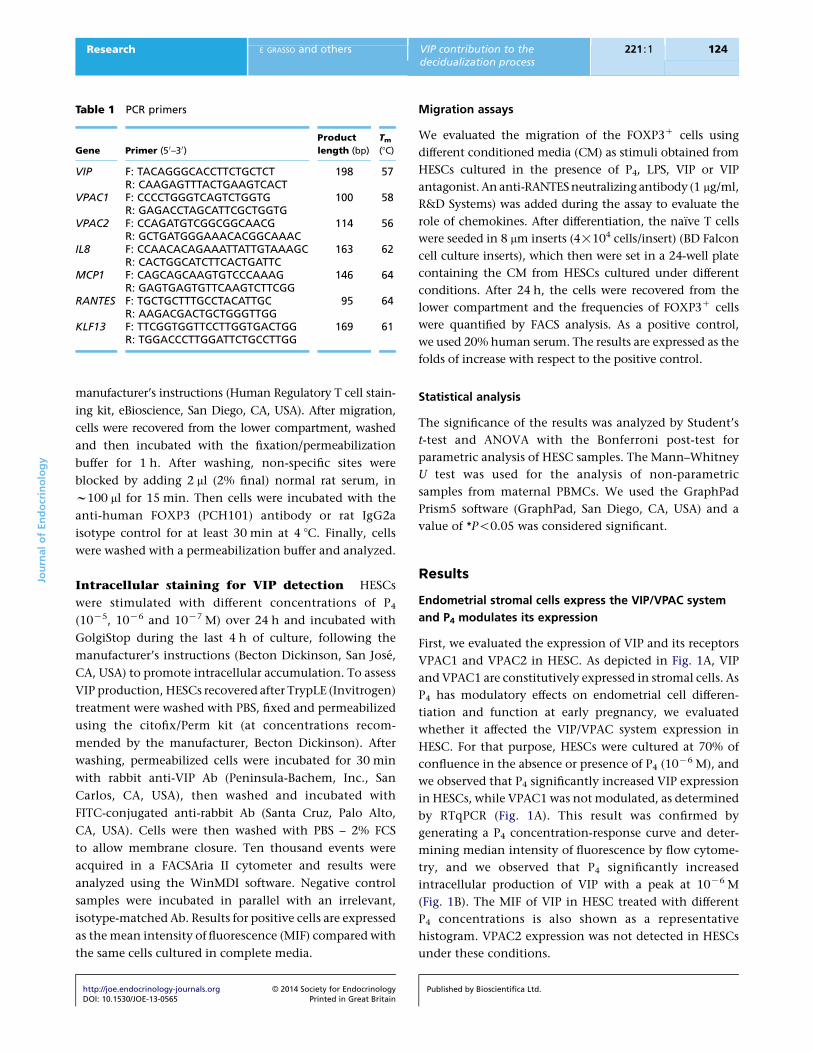
Table 1 PCR primers
Gene Primer (5 0–3 0)
Product
length (bp)
Tm(8C)
VIP F: TACAGGGCACCTTCTGCTCT 198 57R: CAAGAGTTTACTGAAGTCACT
VPAC1 F: CCCCTGGGTCAGTCTGGTG 100 58R: GAGACCTAGCATTCGCTGGTG
VPAC2 F: CCAGATGTCGGCGGCAACG 114 56R: GCTGATGGGAAACACGGCAAAC
IL8 F: CCAACACAGAAATTATTGTAAAGC 163 62R: CACTGGCATCTTCACTGATTC
MCP1 F: CAGCAGCAAGTGTCCCAAAG 146 64R: GAGTGAGTGTTCAAGTCTTCGG
RANTES F: TGCTGCTTTGCCTACATTGC 95 64R: AAGACGACTGCTGGGTTGG
KLF13 F: TTCGGTGGTTCCTTGGTGACTGG 169 61R: TGGACCCTTGGATTCTGCCTTGG
JournalofEndocrinology
Research E GRASSO and others VIP contribution to thedecidualization process
221 :1 124
manufacturer’s instructions (Human Regulatory T cell stain-
ing kit, eBioscience, San Diego, CA, USA). After migration,
cells were recovered from the lower compartment, washed
and then incubated with the fixation/permeabilization
buffer for 1 h. After washing, non-specific sites were
blocked by adding 2 ml (2% final) normal rat serum, in
w100 ml for 15 min. Then cells were incubated with the
anti-human FOXP3 (PCH101) antibody or rat IgG2a
isotype control for at least 30 min at 4 8C. Finally, cells
were washed with a permeabilization buffer and analyzed.
Intracellular staining for VIP detection HESCs
were stimulated with different concentrations of P4
(10K5, 10K6 and 10K7 M) over 24 h and incubated with
GolgiStop during the last 4 h of culture, following the
manufacturer’s instructions (Becton Dickinson, San Jose,
CA, USA) to promote intracellular accumulation. To assess
VIP production, HESCs recovered after TrypLE (Invitrogen)
treatment were washed with PBS, fixed and permeabilized
using the citofix/Perm kit (at concentrations recom-
mended by the manufacturer, Becton Dickinson). After
washing, permeabilized cells were incubated for 30 min
with rabbit anti-VIP Ab (Peninsula-Bachem, Inc., San
Carlos, CA, USA), then washed and incubated with
FITC-conjugated anti-rabbit Ab (Santa Cruz, Palo Alto,
CA, USA). Cells were then washed with PBS – 2% FCS
to allow membrane closure. Ten thousand events were
acquired in a FACSAria II cytometer and results were
analyzed using the WinMDI software. Negative control
samples were incubated in parallel with an irrelevant,
isotype-matched Ab. Results for positive cells are expressed
as the mean intensity of fluorescence (MIF) compared with
the same cells cultured in complete media.
http://joe.endocrinology-journals.org � 2014 Society for EndocrinologyDOI: 10.1530/JOE-13-0565 Printed in Great Britain
Migration assays
We evaluated the migration of the FOXP3C cells using
different conditioned media (CM) as stimuli obtained from
HESCs cultured in the presence of P4, LPS, VIP or VIP
antagonist. An anti-RANTES neutralizing antibody (1 mg/ml,
R&D Systems) was added during the assay to evaluate the
role of chemokines. After differentiation, the naıve T cells
were seeded in 8 mm inserts (4!104 cells/insert) (BD Falcon
cell culture inserts), which then were set in a 24-well plate
containing the CM from HESCs cultured under different
conditions. After 24 h, the cells were recovered from the
lower compartment and the frequencies of FOXP3C cells
were quantified by FACS analysis. As a positive control,
we used 20% human serum. The results are expressed as the
folds of increase with respect to the positive control.
Statistical analysis
The significance of the results was analyzed by Student’s
t-test and ANOVA with the Bonferroni post-test for
parametric analysis of HESC samples. The Mann–Whitney
U test was used for the analysis of non-parametric
samples from maternal PBMCs. We used the GraphPad
Prism5 software (GraphPad, San Diego, CA, USA) and a
value of *P!0.05 was considered significant.
Results
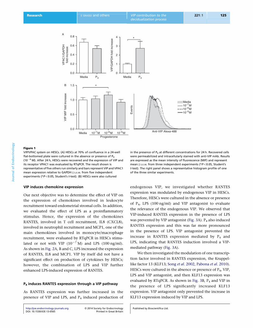
Endometrial stromal cells express the VIP/VPAC system
and P4 modulates its expression
First, we evaluated the expression of VIP and its receptors
VPAC1 and VPAC2 in HESC. As depicted in Fig. 1A, VIP
and VPAC1 are constitutively expressed in stromal cells. As
P4 has modulatory effects on endometrial cell differen-
tiation and function at early pregnancy, we evaluated
whether it affected the VIP/VPAC system expression in
HESC. For that purpose, HESCs were cultured at 70% of
confluence in the absence or presence of P4 (10K6 M), and
we observed that P4 significantly increased VIP expression
in HESCs, while VPAC1 was not modulated, as determined
by RTqPCR (Fig. 1A). This result was confirmed by
generating a P4 concentration-response curve and deter-
mining median intensity of fluorescence by flow cytome-
try, and we observed that P4 significantly increased
intracellular production of VIP with a peak at 10K6 M
(Fig. 1B). The MIF of VIP in HESC treated with different
P4 concentrations is also shown as a representative
histogram. VPAC2 expression was not detected in HESCs
under these conditions.
Published by Bioscientifica Ltd.
0.8 4 *
*
3
2
1
0
0.6
0.4
0.2
0.0
1.5
1.0
0.5
0.0
VIP
MIF
fold
incr
ease
Eve
nts
Media
A
B
Media
Media
Progesterone10–7M
10–7M
10–6M
10–6M
10–5M
10–5M
P4 Media
Anti-VIP Alexa-488
VIP
/GA
PD
H fo
ld in
crea
se
VP
AC
1/G
AP
DH
fold
incr
ease
P4
Figure 1
VIP/VPAC system on HESCs. (A) HESCs at 70% of confluence in a 24-well
flat-bottomed plate were cultured in the absence or presence of P4
(10K6 M). After 24 h, HESCs were recovered and the expression of VIP and
its receptor VPAC1 was evaluated by RTqPCR. The result shown is
representative of five others run similarly and bars represent VIP and VPAC1
mean expression relative to GAPDHGS.E.M. from five independent
experiments (*P!0.05, Student’s t-test). (B) HESCs were also cultured
in the presence of P4 at different concentrations for 24 h. Recovered cells
were permeabilized and intracellularly stained with anti-VIP mAb. Results
are expressed as the mean intensity of fluorescence (MIF) and represent
meanGS.E.M. from three independent experiments (*P!0.05, Student’s
t-test). The right panel shows a representative histogram profile of one
of the three similar experiments.
JournalofEndocrinology
Research E GRASSO and others VIP contribution to thedecidualization process
221 :1 125
VIP induces chemokine expression
Our next objective was to determine the effect of VIP on
the expression of chemokines involved in leukocyte
recruitment toward endometrial stromal cells. In addition,
we evaluated the effect of LPS as a proinflammatory
stimulus. Hence, the expression of the chemokines
RANTES, involved in T cell recruitment, IL8 (CXCL8),
involved in neutrophil recruitment and MCP1, one of the
main chemokines involved in monocyte/macrophage
recruitment, were evaluated by RTqPCR in HESCs stimu-
lated or not with VIP (10K7 M) and LPS (100 ng/ml).
As shown in Fig. 2A, B and C, LPS increased the expression
of RANTES, IL8 and MCP1. VIP by itself did not have a
significant effect on production of cytokines by HESCs;
however, the combination of LPS and VIP further
enhanced LPS-induced expression of RANTES.
P4 induces RANTES expression through a VIP pathway
As RANTES expression was further increased in the
presence of VIP and LPS, and P4 induced production of
http://joe.endocrinology-journals.org � 2014 Society for EndocrinologyDOI: 10.1530/JOE-13-0565 Printed in Great Britain
endogenous VIP, we investigated whether RANTES
expression was modulated by endogenous VIP in HESCs.
Therefore, HESCs were cultured in the absence or presence
of P4, LPS (100 ng/ml) and VIP antagonist to evaluate
the relevance of the endogenous VIP. We observed that
VIP-induced RANTES expression in the presence of LPS
was prevented by VIP antagonist (Fig. 3A). P4 also induced
RANTES expression and this was far more pronounced
in the presence of LPS. VIP antagonist prevented the
increase in RANTES expression mediated by P4 and
LPS, indicating that RANTES induction involved a VIP-
mediated pathway (Fig. 3A).
We then investigated the modulation of one transcrip-
tion factor involved in RANTES expression, the Kruppel-
like factor 13 (KLF13; Song et al. 2002, Pabona et al. 2010).
HESCs were cultured in the absence or presence of P4, VIP,
LPS and VIP antagonist, and then KLF13 expression was
evaluated by RTqPCR. As shown in Fig. 3B, P4 and VIP in
the presence of LPS significantly increased KLF13
expression. VIP antagonist only prevented the increase in
KLF13 expression induced by VIP and LPS.
Published by Bioscientifica Ltd.

2.0
1.5
1.0
0.5
0.0
2.0
1.5
1.0
0.5
0.0
3.0
2.0
1.0
0.0Media VIP LPS LPS + VIP
Media VIP LPS LPS + VIP
Media
RA
NT
ES
/GA
PD
H (
A.U
.)IL
8/G
AP
DH
(A
.U.)
MC
P1/
GA
PD
H (
A.U
.)
VIP
A
B
C
LPS
*
**
**
* *
LPS + VIP
Figure 2
Induction of chemokines by LPS and VIP. HESCs at 70% of confluence in a
24-well flat-bottomed plate were cultured in the absence or presence of
VIP (10K7 M) and LPS (100 ng/ml). After 24 h, HESCs were recovered and the
expression of (A) RANTES (CCL5), (B) IL8 (CXCL8) and (C) MCP1 (CCL2)
was evaluated by RTqPCR. Bars represent mean chemokine expression
relative to GAPDH GS.E.M. from five independent experiments
(*P!0.05, Student’s t-test).
2.5
*
*
**
*
*
* *
**
2.0
1.5
1.0
0.5
0.0
2.0
1.5
1.0
0.5
0.0
Media
B
A
RA
NT
ES
/GA
PD
H fo
ld in
crea
seK
LF13
/GA
PD
H fo
ld in
crea
se
MediaVIP VIP VIP+ANT
LPS
LPS
P4 P4+ANTP4
Media MediaVIP VIP VIP+ANT P4 P4+ANTP4
Figure 3
Progesterone induced RANTES expression through a VIP pathway. HESCs at
70% of confluence in a 24-well flat-bottom plate were stimulated with
different combinations of VIP (10K7 M), P4 (10K6 M), VIP antagonist
(ANT: 10K5 M) and LPS (100 ng/ml). After 24 h, HESCs were recovered and
the expression of RANTES (A) and KLF13 (B) was evaluated by RTqPCR. Bars
represent mean RANTES or KLF13 expression relative to GAPDH GS.E.M.
relativized to LPS stimuli from six independent experiments (*P!0.05,
ANOVA, Bonferroni post-test).
JournalofEndocrinology
Research E GRASSO and others VIP contribution to thedecidualization process
221 :1 126
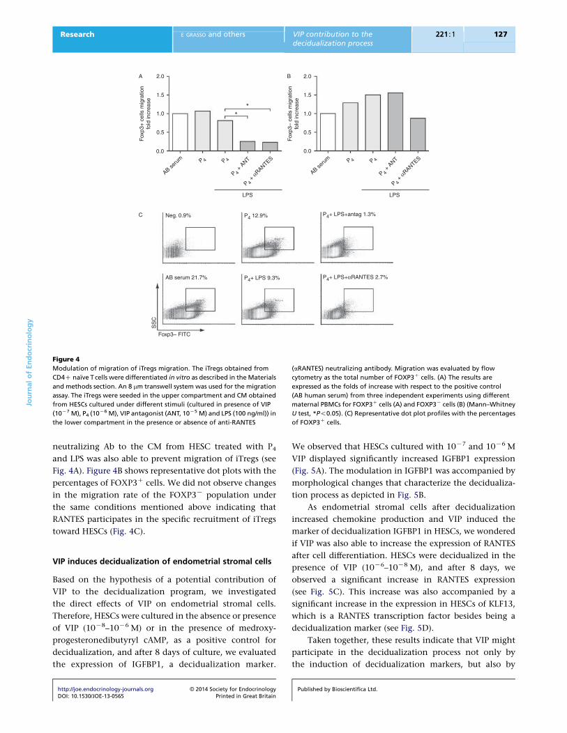
Endometrial stromal cells specifically recruit iTregs
through RANTES production
Our next step was to determine if HESCs have the ability to
attract iTregs. Human Treg cells were differentiated from
naıve CD45RACCCR7C cells obtained from PBMCs
obtained from fertile women and cultured with IL2 and
TGFb over 5 days, as described in the Materials and
http://joe.endocrinology-journals.org � 2014 Society for EndocrinologyDOI: 10.1530/JOE-13-0565 Printed in Great Britain
methods section. We then performed migration assays
using a multi-chamber system. In-vitro-differentiated
iTregs were seeded onto 8 mm pore inserts, allowing cell
migration toward the CM used as a chemotactic stimulus
in the lower compartment. After 24 h, cells were recovered
from the lower compartment and FOXP3 expression was
quantified by FACS analysis. As shown in Fig. 4A, the CM
from HESCs increased the frequency of FOXP3C cells to
levels similar to the migration observed in the presence of
human serum (positive control). However, when the
migration assay was performed in the presence of CM
from HESCs cultured in the presence of VIP antagonist, the
recruitment of iTregs to the lower compartment was
prevented (Fig. 4A). Moreover, addition of anti-RANTES
Published by Bioscientifica Ltd.
2.0A B
C
1.5
1.0
Fox
p3+
cel
ls m
igra
tion
fold
incr
ease
Fox
p3–
cells
mig
ratio
nfo
ld in
crea
se
LPS
Neg. 0.9%
AB serum 21.7%
Foxp3– FITC
SS
C
P4+ LPS 9.3% P4+ LPS+αRANTES 2.7%
P4 12.9% P4+ LPS+antag 1.3%
LPS
**
0.5
0.0
2.0
1.5
1.0
0.5
0.0
AB seru
m P 4 P 4P 4
+ A
NTP 4
+ αRANTES
AB seru
m P 4 P 4P 4
+ A
NTP 4
+ αRANTES
Figure 4
Modulation of migration of iTregs migration. The iTregs obtained from
CD4C naıve T cells were differentiated in vitro as described in the Materials
and methods section. An 8 mm transwell system was used for the migration
assay. The iTregs were seeded in the upper compartment and CM obtained
from HESCs cultured under different stimuli (cultured in presence of VIP
(10K7 M), P4 (10K6 M), VIP antagonist (ANT, 10K5 M) and LPS (100 ng/ml)) in
the lower compartment in the presence or absence of anti-RANTES
(aRANTES) neutralizing antibody. Migration was evaluated by flow
cytometry as the total number of FOXP3C cells. (A) The results are
expressed as the folds of increase with respect to the positive control
(AB human serum) from three independent experiments using different
maternal PBMCs for FOXP3C cells (A) and FOXP3K cells (B) (Mann–Whitney
U test, *P!0.05). (C) Representative dot plot profiles with the percentages
of FOXP3C cells.
JournalofEndocrinology
Research E GRASSO and others VIP contribution to thedecidualization process
221 :1 127
neutralizing Ab to the CM from HESC treated with P4
and LPS was also able to prevent migration of iTregs (see
Fig. 4A). Figure 4B shows representative dot plots with the
percentages of FOXP3C cells. We did not observe changes
in the migration rate of the FOXP3K population under
the same conditions mentioned above indicating that
RANTES participates in the specific recruitment of iTregs
toward HESCs (Fig. 4C).
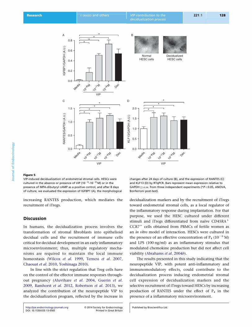
VIP induces decidualization of endometrial stromal cells
Based on the hypothesis of a potential contribution of
VIP to the decidualization program, we investigated
the direct effects of VIP on endometrial stromal cells.
Therefore, HESCs were cultured in the absence or presence
of VIP (10K8–10K6 M) or in the presence of medroxy-
progesteronedibutyryl cAMP, as a positive control for
decidualization, and after 8 days of culture, we evaluated
the expression of IGFBP1, a decidualization marker.
http://joe.endocrinology-journals.org � 2014 Society for EndocrinologyDOI: 10.1530/JOE-13-0565 Printed in Great Britain
We observed that HESCs cultured with 10K7 and 10K6 M
VIP displayed significantly increased IGFBP1 expression
(Fig. 5A). The modulation in IGFBP1 was accompanied by
morphological changes that characterize the decidualiza-
tion process as depicted in Fig. 5B.
As endometrial stromal cells after decidualization
increased chemokine production and VIP induced the
marker of decidualization IGFBP1 in HESCs, we wondered
if VIP was also able to increase the expression of RANTES
after cell differentiation. HESCs were decidualized in the
presence of VIP (10K6–10K8 M), and after 8 days, we
observed a significant increase in RANTES expression
(see Fig. 5C). This increase was also accompanied by a
significant increase in the expression in HESCs of KLF13,
which is a RANTES transcription factor besides being a
decidualization marker (see Fig. 5D).
Taken together, these results indicate that VIP might
participate in the decidualization process not only by
the induction of decidualization markers, but also by
Published by Bioscientifica Ltd.
0.8
0.6
0.4
0.2
0.0
1.5
1.0
0.5
0.0
Med
ia
10–8 M
10–7 M
10–6 M Dec
Med
ia
10–8 M
10–7 M
10–6 M Dec
VIP
A
C D
B
IGF
BP
1/G
AP
DH
(A
.U.)
RA
NT
ES
/GA
PD
H (
A.U
.)
KLF
13/G
AP
DH
(A
.U.)
VIP
NormalHESC cells
DecidualizedHESC cells
*
**
**
2.0
1.5
1.0
0.5
0.0
Med
ia
10–8 M
10–7 M
10–6 M Dec
VIP
**
**
**
*
Figure 5
VIP-induced decidualization of endometrial stromal cells. HESCs were
cultured in the absence or presence of VIP (10K6–10K8 M) or in the
presence of MPA-dibutyryl cAMP as a positive control, and after 8 days
of culture, we evaluated the expression of IGFBP1 (A), the morphological
changes after 24 days of culture (B), and the expression of RANTES (C)
and KLF13 (D) by RTqPCR. Bars represent mean expression relative to
GAPDHGS.E.M. from three independent experiments (*P!0.05, ANOVA,
Bonferroni post-test).
JournalofEndocrinology
Research E GRASSO and others VIP contribution to thedecidualization process
221 :1 128
increasing RANTES production, which mediates the
recruitment of iTregs.
Discussion
In humans, the decidualization process involves the
transformation of stromal fibroblasts into epithelioid
decidual cells and the recruitment of immune cells
critical for decidual development in an early inflammatory
microenvironment; thus, multiple regulatory mecha-
nisms are required to maintain the local immune
homeostasis (Wilcox et al. 1999, Terness et al. 2007,
Chaouat et al. 2010, Yoshinaga 2010).
In line with the strict regulation that Treg cells have
on the control of the effector immune responses through-
out pregnancy (Aluvihare et al. 2004, Guerin et al.
2009, Ramhorst et al. 2012, Robertson et al. 2013), we
analyzed the contribution of the neuropeptide VIP to
the decidualization program, reflected by the increase in
http://joe.endocrinology-journals.org � 2014 Society for EndocrinologyDOI: 10.1530/JOE-13-0565 Printed in Great Britain
decidualization markers and by the recruitment of iTregs
toward endometrial stromal cells, as a local regulator of
the inflammatory response during implantation. For that
purpose, we used the HESC cultured under different
stimuli and iTregs differentiated from naıve CD45RAC
CCR7C cells obtained from PBMCs of fertile women as
an in vitro model of interaction. HESCs were cultured in
the presence of an effective concentration of P4 (10K6 M)
and LPS (100 ng/ml) as an inflammatory stimulus that
modulated chemokine production but did not affect cell
viability (Abrahams et al. 2004b).
The results presented in this study indicating that the
neuropeptide VIP, with potent anti-inflammatory and
immunomodulatory effects, could contribute to the
decidualization process inducing endometrial stromal
cell expression of decidualization markers and the
selective recruitment of iTregs toward HESCs by increasing
production of RANTES under the effect of P4 in the
presence of a inflammatory microenvironment.
Published by Bioscientifica Ltd.

JournalofEndocrinology
Research E GRASSO and others VIP contribution to thedecidualization process
221 :1 129
Our conclusions are based on several observations:
first, HESCs express VIP and its constitutive receptor
VPAC1, and P4 has the ability to increase VIP production.
Secondly, expression of RANTES, one of the main
chemokines involved in T cell recruitment, was induced
by VIP in the presence of LPS, and its induction was
mediated by P4. Finally, the assay of migration of iTregs
toward CM from HESCs revealed that the endogenous VIP
production induced by P4 and LPS stimulation could
selectively attract them through production of RANTES,
as the anti-RANTES neutralizing Ab or VIP antagonist
prevented the migration of iTregs.
VIP might be one of the first mediators that induces
decidualization through its interaction with the VPAC1
receptor and triggering cAMP signaling in HESCs to
increase the expression of IGFBP1 and KLF13, both
markers of decidualization accompanied by morpho-
logical changes characteristic of decidualized cells.
In fact, KLF13 is not only a decidualization marker, but
also a transcription factor that binds to the RANTES
promoter, which is necessary to mediate RANTES tran-
scription (Song et al. 2002). This mechanism could explain
how endogenous VIP regulates RANTES expression in
HESCs, thus contributing to the selective recruitment of
iTregs that might allow the control of tissue damage
during embryo implantation. In this context, Nancy et al.
(2012) recently reported that genes encoding chemokines
are subject to epigenetic silencing in decidual stromal cells
to restrain the attraction of Th1 and T cytotoxic profiles
as a strategy to prevent potential tissue damage. In brief,
the decidualization program involves many regulatory
molecules that play functional roles, such as insulin-like
growth factors, interleukin 1, 6, 10 and TGFb families,
the neuropeptide VIP, chemokines such as RANTES with
their receptors and adhesion molecules that generate a
network to control implantation processes such as
trophoblast adhesion, invasion and the selective recruit-
ment of maternal leukocyte subpopulations (Salamonsen
& Woolley 1999, Dimitriadis et al. 2010, Terness et al.
2007, Yoshinaga 2010, Fraccaroli et al. 2011).
Interestingly, spontaneous decidualization of stromal
cells occurs in the absence of pregnancy. It was proposed
that cyclic endometrial decidualization followed by
menstruation ‘preconditions’ uterine tissues for a hyper-
inflammatory response and oxidative stress that is in turn
accompanied by deep trophoblast invasion during
early pregnancy (Brosens et al. 2009, Teklenburg et al.
2010a,b). Therefore, the ability of the human endome-
trium to generate an adequate decidual response based on
successive inflammatory events might contribute to the
http://joe.endocrinology-journals.org � 2014 Society for EndocrinologyDOI: 10.1530/JOE-13-0565 Printed in Great Britain
sensitization of the uterine tissues. Under this hypothesis
of repeated inflammatory events, it is conceivable that
tight immune homeostatic control prior to implantation
is required (Kim et al. 2009, Weiss et al. 2009). In this
context, the ability of HESCs to selectively recruit iTregs
might contribute to maintenance of immune homeostasis
at early stages of implantation.
Finally, even though research in the past few years
have provided a better understanding of trophoblast–
endometrial interactions during the initial stage of
implantation by means of various human cell experimental
approaches, the identification of biomarkers with clinical
utility for patients with implantation failures is still an
objective to be achieved.
Declaration of interest
The authors declare that they have no financial nor any other potential
conflict of interest.
Funding
This study was supported by grants to R R (Consejo Nacional de
Investigaciones Cientıficas y Tecnicas (CONICET) Proyectos de Investigacion
Plurianuales (PIP) 2659, 200034 from University of Buenos Aires) and to C P L
(Proyectos de Investigacion Cientıfica y Tecnologica (PICT) 2011 0144 from
Agencia Nacional de Promocion Cientıfica y Tecnologica (ANPCyT)).
Author contribution statement
C P L and R R designed the study and wrote the manuscript. E G carried out
all the experiments with HESC cells and decidualization, the differentiation
of iTregs the co-cultures, and the migration assays. D P and M A helped with
RT-PCRs data analyses and interpretation. G M supervised the study and
discussed the results. All authors read and approved the final manuscript.
References
Abrahams VM, Kim YM, Straszewski SL, Romero R & Mor G 2004a
Macrophages and apoptotic cell clearance during pregnancy.
American Journal of Reproductive Immunology 51 275–282. (doi:10.1111/
j.1600-0897.2004.00156.x)
Abrahams VM, Bole-Aldo P, Kim YM, Straszewski-Chavez SL, Chaiwor-
apongsa T, Romero R & Mor G 2004b Divergent trophoblast responses
to bacterial products mediated by TLRs. Journal of Immunology 173
4286–4296.
Aluvihare VR, Kallikourdis M & Betz AG 2004 Regulatory T cells mediate
maternal tolerance to the fetus. Nature Immunology 5 266–271.
(doi:10.1038/ni1037)
Bromley SK, Mempel TR & Luster AD 2008 Orchestrating the orchestrators:
chemokines in control of T cell traffic. Nature Immunology 9 970–980.
(doi:10.1038/ni.f.213)
Brosens JJ, Parker MG, McIndoe A, Pijnenborg R & Brosens IA 2009
A role for menstruation in preconditioning the uterus for successful
pregnancy. American Journal of Obstetrics and Gynecology 200
615.e1–615.e6. (doi:10.1016/j.ajog.2008.11.037)
Published by Bioscientifica Ltd.
JournalofEndocrinology
Research E GRASSO and others VIP contribution to thedecidualization process
221 :1 130
Catalano RD, Critchley HO, Heikinheimo O, Baird DT, Hapangama D,
Sherwin JRA, Charnock-Jones DS, Smith SK & Sharkey AM 2007
Mifepristone induced progesterone withdrawal reveals novel regulatory
pathways in human endometrium. Molecular Human Reproduction 13
641–654. (doi:10.1093/molehr/gam021)
Chaouat G, Petitbarat M, Dubanchet S, Rahmati M & Ledee N 2010
Tolerance to the foetal allograft? American Journal of Reproductive
Immunology 63 624–636. (doi:10.1111/j.1600-0897.2010.00832.x)
Couvineau A & Laburthe M 2012 VPAC receptors: structure, molecular
pharmacology and interaction with accessory proteins. British Journal of
Pharmacology 166 42–50. (doi:10.1111/j.1476-5381.2011.01676.x)
Dey SK, Lim H, Das SK, Reese J, Paria BC, Daikoku T & Wang H 2004
Molecular cues to implantation. Endocrine Reviews 25 341–373.
(doi:10.1210/er.2003-0020)
Dimitriadis E, Nie G, Hannan NJ, Paiva P & Salamonsen LA 2010 Local
regulation of implantation at the human fetal–maternal interface.
International Journal of Developmental Biology 54 313–322. (doi:10.1387/
ijdb.082772ed)
Ekstrom J, Mansson B & Tobin G 1983 Vasoactive intestinal peptide evoked
secretion of fluid and protein from rat salivary glands and the
development of supersensitivity. Acta Physiologica Scandinavica 119
169–175. (doi:10.1111/j.1748-1716.1983.tb07322.x)
Fraccaroli L, Alfieri J, Leiros CP & Ramhorst R 2009a Immunomodulatory
effects of chemokines during the early implantation window. Frontiers
in Bioscience 1 288–298. (doi:10.2741/e28)
Fraccaroli L, Alfieri J, Larocca L, Calafat M, Mor G, Leiros CP & Ramhorst R
2009b A potential tolerogenic immune mechanism in a trophoblast cell
line through the activation of chemokine-induced T cell death and
regulatory T cell modulation. Human Reproduction 24 166–175.
(doi:10.1093/humrep/den344)
Fraccaroli L, Alfieri J, Larocca L, Calafat M, Roca V, Lombardi E, Ramhorst R
& Leiros CP 2009c VIP modulates the pro-inflammatory maternal
response, inducing tolerance to trophoblast cells. British Journal of
Pharmacology 156 116–126. (doi:10.1111/j.1476-5381.2008.00055.x)
Fraccaroli L, Grasso E, Zeitler E, Lombardi E, Gogorza S, Etchepareborda JJ,
Nagle C, Cortelezzi M, Perez Leiros C & Ramhorst R 2011 Modulation of
maternal LIF producers T cells by trophoblast and paternal antigens.
American Journal of Reproductive Immunology 65 133–145. (doi:10.1111/
j.1600-0897.2010.00890.x)
Gellersen B & Brosens J 2003 Cyclic AMP and progesterone receptor cross-
talk in human endometrium: a decidualizing affair. Journal of Endo-
crinology 178 357–372. (doi:10.1677/joe.0.1780357)
Gellersen B, Brosens IA & Brosens JJ 2007 Decidualization of the human
endometrium: mechanisms, functions, and clinical perspectives.
Seminars in Reproductive Medicine 25 445–453. (doi:10.1055/s-2007-
991042)
Gomez-Lopez N, Guilbert LJ & Olson DM 2010 Invasion of the leukocytes
into the fetal–maternal interface during pregnancy. Journal of Leukocyte
Biology 88 625–633. (doi:10.1189/jlb.1209796)
Gonzalez-Rey E, Anderson P & Delgado M 2007 Emerging roles of
vasoactive intestinal peptide: a new approach for autoimmune therapy.
Annals of the Rheumatic Diseases 66(Suppl 3) iii70–iii76. (doi:10.1136/
ard.2007.078519)
Guerin LR, Prins JR & Robertson SA 2009 Regulatory T-cells and immune
tolerance in pregnancy: a new target for infertility treatment? Human
Reproduction Update 15 517–535. (doi:10.1093/humupd/dmp004)
Kim MJ, Romero R, Kim CJ, Tarca AL, Chhauy S, LaJeunesse C, Lee D-C,
Draghici S, Gotsch F, Kusanovic JP et al. 2009 Villitis of unknown
etiology is associated with a distinct pattern of chemokine up-
regulation in the feto-maternal and placental compartments: impli-
cations for conjoint maternal allograft rejection and maternal anti-fetal
graft-versus-host disease. Journal of Immunology 182 3919–3927.
(doi:10.4049/jimmunol.0803834)
King AE & Critchley HOD 2010 Oestrogen and progesterone regulation of
inflammatory processes in the human endometrium. Journal of Steroid
http://joe.endocrinology-journals.org � 2014 Society for EndocrinologyDOI: 10.1530/JOE-13-0565 Printed in Great Britain
Biochemistry and Molecular Biology 120 116–126. (doi:10.1016/j.jsbmb.
2010.01.003)
Krikun G, Mor G, Alvero A, Guller S, Schatz F, Sapi E, Rahman M, Caze R,
Qumsiyeh M & Lockwood CJ 2004 A novel immortalized human
endometrial stromal cell line with normal progestational response.
Endocrinology 145 2291–2296. (doi:10.1210/en.2003-1606)
Leceta J, Gomariz RP, Martinez C, Carrion M, Arranz A & Juarranz Y 2007
Vasoactive intestinal peptide regulates Th17 function in autoimmune
inflammation. Neuroimmunomodulation 14 134–138. (doi:10.1159/
000110636)
Mesiano S, Wang Y & Norwitz ER 2011 Progesterone receptors
in the human pregnancy uterus: do they hold the key to birth
timing? Reproductive Sciences 18 6–19. (doi:10.1177/
1933719110382922)
Nancy P, Tagliani E, Tay C-S, Asp P, Levy DE & Erlebacher A 2012
Chemokine gene silencing in decidual stromal cells limits T cell access
to the maternal–fetal interface. Science 336 1317–1321. (doi:10.1126/
science.1220030)
Pabona JMP, Zeng Z, Simmen FA & Simmen RCM 2010 Functional
differentiation of uterine stromal cells involves cross-regulation
between bone morphogenetic protein 2 and Kruppel-like factor (KLF)
family members KLF9 and KLF13. Endocrinology 151 3396–3406.
(doi:10.1210/en.2009-1370)
Perez Leiros C & Ramhorst R 2013 Tolerance induction at the early
maternal–placental interface through selective cell recruitment and
targeting by immune polypeptides. American Journal of Reproductive
Immunology 69 359–368. (doi:10.1111/aji.12087)
Ramhorst R, Patel R, Corigliano A, Etchepareborda JJ, Fainboim L &
Schust D 2006 Induction of maternal tolerance to fetal alloantigens
by RANTES production. American Journal of Reproductive Immunology
56 302–311. (doi:10.1111/j.1600-0897.2006.00430.x)
Ramhorst R, Gutierrez G, Corigliano A, Junovich G & Fainboim L 2007
Implication of RANTES in the modulation of alloimmune
response by progesterone during pregnancy. American Journal of
Reproductive Immunology 57 147–152. (doi:10.1111/j.1600-0897.2006.
00458.x)
Ramhorst R, Fraccaroli L, Aldo P, Alvero AB, Cardenas I, Leiros CP & Mor G
2012 Modulation and recruitment of inducible regulatory T cells by
first trimester trophoblast cells. American Journal of Reproductive
Immunology 67 17–27. (doi:10.1111/j.1600-0897.2011.01056.x)
Robertson SA, Prins JR, Sharkey DJ & Moldenhauer LM 2013 Seminal fluid
and the generation of regulatory T cells for embryo implantation.
American Journal of Reproductive Immunology 69 315–330. (doi:10.1111/
aji.12107)
Salamonsen LA & Woolley DE 1999 Menstruation: induction by matrix
metalloproteinases and inflammatory cells. Journal of Reproductive
Immunology 44 1–27. (doi:10.1016/S0165-0378(99)00002-9)
Song A, Patel A, Thamatrakoln K, Liu C, Feng D, Clayberger C & Krensky AM
2002 Functional domains and DNA-binding sequences of RFLAT-1/
KLF13, a Kruppel-like transcription factor of activated T lymphocytes.
Journal of Biological Chemistry 277 30055–30065. (doi:10.1074/
jbc.M204278200)
Spong CY, Lee SJ, McCune SK, Gibney G, Abebe DT, Alvero R, Brenneman DE
& Hill JM 1999 Maternal regulation of embryonic growth: the role
ofvasoactive intestinalpeptide.Endocrinology140917–924. (doi:10.1210/
endo.140.2.6481)
Stoikos CJ, Harrison CA, Salamonsen LA & Dimitriadis E 2008 A distinct
cohort of the TGFb superfamily members expressed in human
endometrium regulate decidualization. Human Reproduction 23
1447–1456. (doi:10.1093/humrep/den110)
Teklenburg G, Salker M, Molokhia M, Lavery S, Trew G, Aojanepong T,
Mardon HJ, Lokugamage AU, Rai R, Landles C et al. 2010a Natural
selection of human embryos: decidualizing endometrial stromal cells
serve as sensors of embryo quality upon implantation. PLoS ONE 5
e10258. (doi:10.1371/journal.pone.0010258)
Published by Bioscientifica Ltd.
JournalofEndocrinology
Research E GRASSO and others VIP contribution to thedecidualization process
221 :1 131
Teklenburg G, Salker M, Heijnen C, Macklon NS & Brosens JJ 2010b The
molecular basis of recurrent pregnancy loss: impaired natural embryo
selection. Molecular Human Reproduction 16 886–895. (doi:10.1093/
molehr/gaq079)
Terness P, Kallikourdis M, Betz AG, Rabinovich GA, Saito S & Clark DA 2007
Tolerance signaling molecules and pregnancy: IDO, galectins, and the
renaissance of regulatory T cells. American Journal of
Reproductive Immunology 58 238–254. (doi:10.1111/j.1600-0897.2007.
00510.x)
Van Voorhis BJ, Anderson DJ & Hill JA 1989 The effects of RU 486 on
immune function and steroid-induced immunosuppression in vitro.
http://joe.endocrinology-journals.org � 2014 Society for EndocrinologyDOI: 10.1530/JOE-13-0565 Printed in Great Britain
Journal of Clinical Endocrinology and Metabolism 69 1195–1199.
(doi:10.1210/jcem-69-6-1195)
Weiss G, Goldsmith LT, Taylor RN, Bellet D & Taylor HS 2009
Inflammation in reproductive disorders. Reproductive Sciences 16
216–229. (doi:10.1177/1933719108330087)
Wilcox AJ, Baird DD & Weinberg CR 1999 Time of implantation of the
conceptus and loss of pregnancy. New England Journal of Medicine 340
1796–1799. (doi:10.1056/NEJM199906103402304)
Yoshinaga K 2010 Research on blastocyst implantation essential factors
(BIEFs). American Journal of Reproductive Immunology 63 413–424.
(doi:10.1111/j.1600-0897.2010.00853.x)
Received in final form 21 January 2014Accepted 3 February 2014Accepted Preprint published online 3 February 2014
Published by Bioscientifica Ltd.