Embed Size (px)
Citation preview
Dr. PN SHARMA
Department of Plant Pathology
CSK HP Agricultural University
Palampur-176 062 (HP State) INDIA
Pl Path 502
Viroids
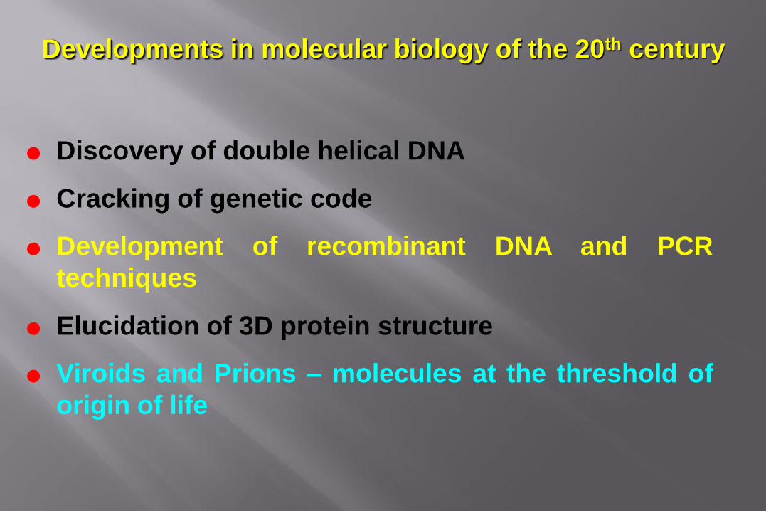
Developments in molecular biology of the 20th century
Discovery of double helical DNA
Cracking of genetic code
Development of recombinant DNA and PCR
techniques
Elucidation of 3D protein structure
Viroids and Prions – molecules at the threshold of
origin of life

THEODOR O. DIENER
Discoverer of the viroid 1971
Yellow green rods denote the first viroid as seen in electron micrograph
Therefore often denoted as subviral particles or agents
Viroids (T.O. Diener, 1971): are small, low mol
wt. RNA units (250-370 bp.), lack protein coat, replicate themselves and cause disease Example: Potato spindle tuber viroid, coconut codang-cadang.
Autonomously replicating Pathogens, unencapsidated Single

Self replicating circular, low molecular weight RNA without protein coat
Infect only plant cells
Produce variable symptoms on different hosts like stunting, bark scaling, proliferation, veinal necrosis and also symptom less carrier (No symptoms)
Vegetatively propagated, highly seed and pollen transmitted

Losses caused byviroid diseases
Potato Spindle 1917 USA 26 - 90% 1971 PSTVd
Tuber USSR 54%
China 60%
Canada 64%
Cadang Cadang 1927 Philippines 20 million 1975 CCCVd
of coconut nuts
Hop Stunt 1952 Japan 17 - 60% 1977 HSVd
DISEASE LOSS VIROID
NAME COUNTRY
FIRST
REPORT
VIROID
ETIOLOGY
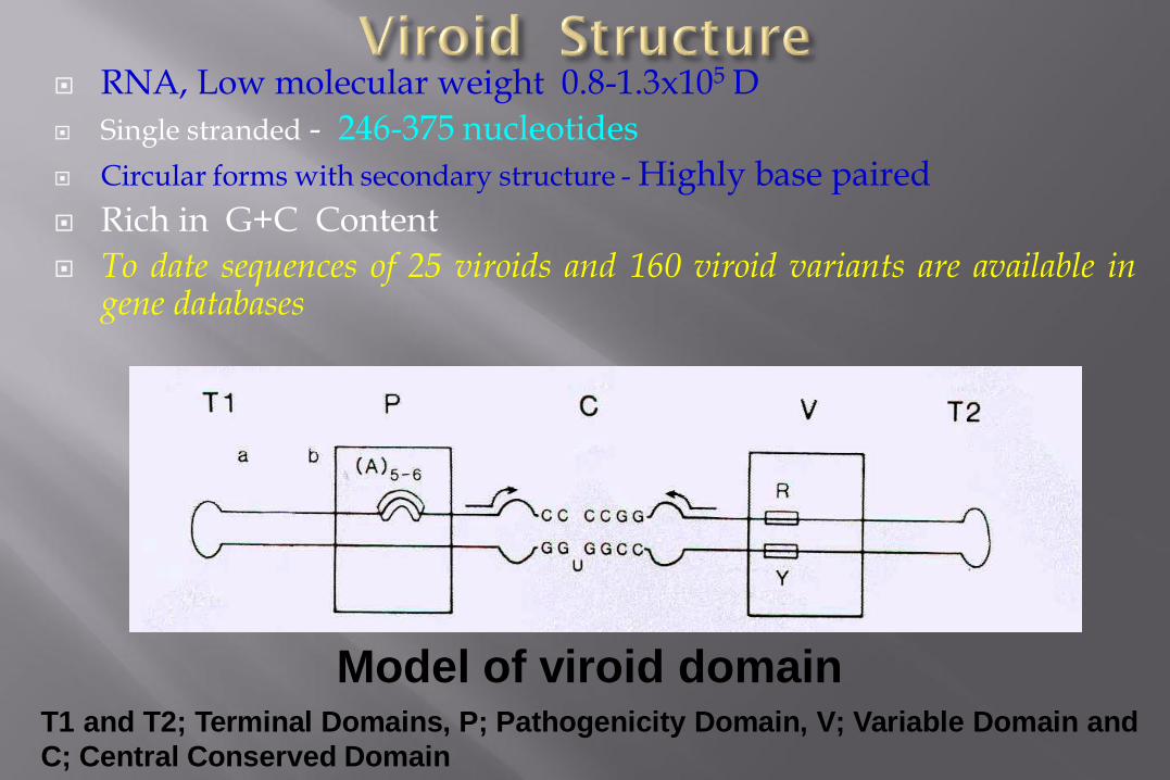
RNA, Low molecular weight 0.8-1.3x10⁵ D
Single stranded - 246-375 nucleotides
Circular forms with secondary structure - Highly base paired
Rich in G+C Content
To date sequences of 25 viroids and 160 viroid variants are available in gene databases
Model of viroid domain T1 and T2; Terminal Domains, P; Pathogenicity Domain, V; Variable Domain and
C; Central Conserved Domain

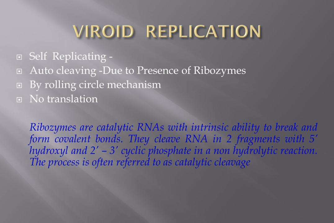
Self Replicating -
Auto cleaving -Due to Presence of Ribozymes
By rolling circle mechanism
No translation
Ribozymes are catalytic RNAs with intrinsic ability to break and form covalent bonds. They cleave RNA in 2 fragments with 5’ hydroxyl and 2’ – 3’ cyclic phosphate in a non hydrolytic reaction. The process is often referred to as catalytic cleavage

ROLLING CIRCLE MECHANISM
Asymmetric model Symmetric model

Common in plasmid or bacteriophage DNA and the circular RNA genome of e.g.
Viroids, and DNA viruses e.g. geminiviruses
Rolling circle DNA replication is initiated by an initiator protein encoded by the plasmid or
bacteriophage DNA, which nicks one strand of the double-stranded, circular DNA
molecule at a site called the double-strand origin, or DSO.
The initiator protein remains bound to the 5' phosphate end of the nicked strand, and the free 3' hydroxyl end
is released to serve as a primer for DNA synthesis by DNA polymerase II.
Using the unnicked strand as a template, replication proceeds around the circular DNA molecule, displacing
the nicked strand as single-stranded DNA. Displacement of the nicked strand is carried out by a host-encoded
helicase called PcrA (plasmid copy reduced) in the presence of the plasmid replication initiation protein.
Continued DNA synthesis can produce multiple single-stranded linear copies of the original
DNA in a continuous head-to-tail series called a concatamer.
These linear copies can be converted to double-stranded circular molecules through the
following process:
First, the initiator protein makes another nick to terminate synthesis of the first (leading) strand. RNA
polymerase and DNA polymerase III then replicate the single-stranded origin (SSO) DNA to make another
double-stranded circle. DNA polymerase I removes the primer, replacing it with DNA, and DNA ligase joins
the ends to make another molecule of double-stranded circular DNA.
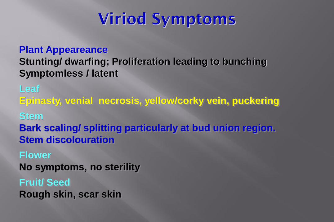
Plant Appeareance
Stunting/ dwarfing; Proliferation leading to bunching
Symptomless / latent
Leaf
Epinasty, venial necrosis, yellow/corky vein, puckering
Stem
Bark scaling/ splitting particularly at bud union region.
Stem discolouration
Flower
No symptoms, no sterility
Fruit/ Seed
Rough skin, scar skin

Sap Tomato bioassay
Graft Citron bioassay, Cucumber bioassay
Vegetative Pruning / Cutting Knives
Seed Very high rate
Pollen High rate
Insect Not yet confirmed universally


Symptoms on Inoculated Tomato Spindle Shaped Tubers


Artificially inoculated seedling (left), 6
years after inoculation, showing stunting,
sterility and disordered pinnae, compared
with a healthy seedling.

Severe infection leading to tree decline

Stunting

CEVd CEVd-t

Intensity of disease known only
after deformation of fruit is
observed
Symptoms on leaves appear as mild
chlorosis
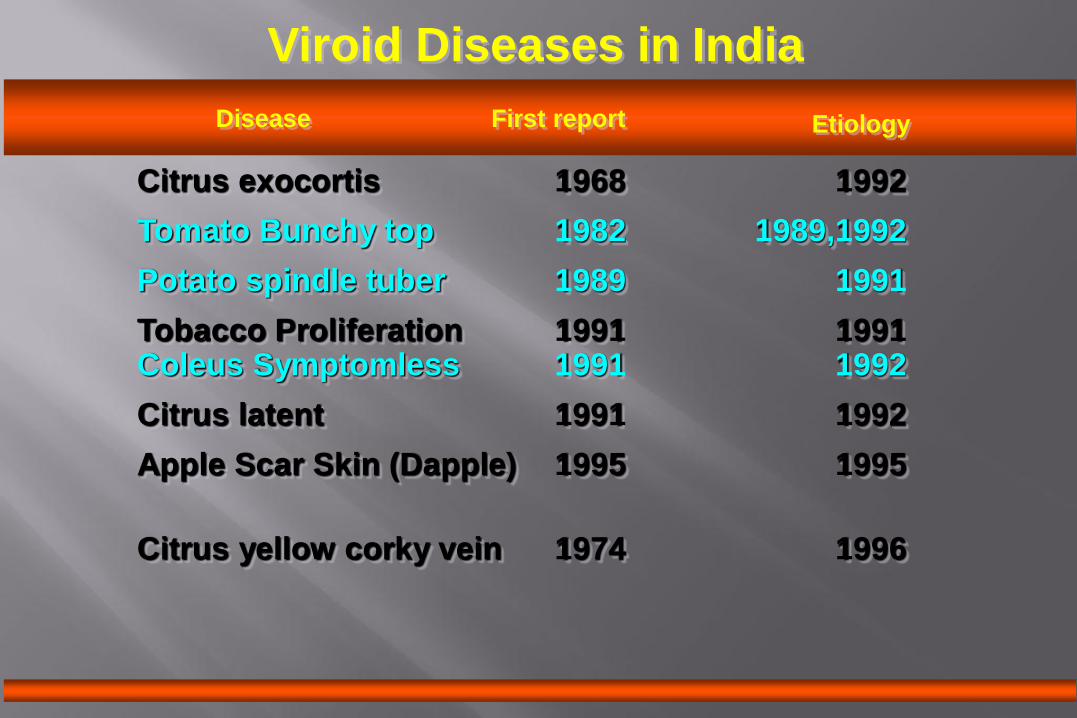
Viroid Diseases in India
Citrus exocortis 1968 1992
Tomato Bunchy top 1982 1989,1992
Potato spindle tuber 1989 1991
Tobacco Proliferation 1991 1991 Coleus Symptomless 1991 1992
Citrus latent 1991 1992
Apple Scar Skin (Dapple) 1995 1995
Citrus yellow corky vein 1974 1996
Disease First report Etiology

VIROID INFECTIONS IN DIFFERENT
PLANT FAMILIES
ASTERACEAE : CCMVd, CSVd (2)
CARYOPHYLLACEAE: CSVd (1)
CUCURBITACEAE: CPFVd (1)
GESNERIACEAE: CLVd (1)
LABIATAE: CYVd, CbVd (2)
LAURACEAE: ASBVd (1)
PALMAE: CCCVd, CTVd, OPFYVd (3)
ROSACEAE: ASSVd, PLMVd, PDVd, PBCVd (4)
RUTACEAE: CEVd, HSVd (citron), CiVVd, CIT.
CACHEXIA (4)
VITACEAE : HSVd (gv), HSVd (ggv), AGVd,
GYSVd, G 1bVd , HSVd (hop), HLVd,
CEVd (gv) (8)
SOLANACEAE: PSTVd, ITBTVd, TASVd,
TAPMVd, NgPVd (5)
THEACEAE: TDDVd (1)

primary sources of inoculum: Seed and pollen
Vegetative propagation
Large scale monoculture
Escape from natural host to commercial crop and
vice-versa
Evolution of natural recombinants
Lack of adequate quarantine check

Detection of Viroids


Detection of Citrus Viroids by PCR
L-R: Marker (100bp), CEVd (2,3), CVd II(5),
CVd gr III (6,7), CYCVVd (9,10)

Management of viroid diseases
Biotechnological Approach
Eradication of Sources of inoculum
Cultural Practices
Quarantine Regulations
Biotechnological Approach