Embed Size (px)
Citation preview
ORIGINAL ARTICLE
Virtual MR arthroscopy of the shoulder: image gallerywith arthroscopic correlation of major pathologiesin shoulder instability
A. Stecco Æ D. Volpe Æ N. Volpe Æ P. Fornara ÆA. Castagna Æ A. Carriero
Received: 8 October 2007 / Accepted: 19 August 2008 / Published online: 7 October 2008
� Springer-Verlag 2008
Abstract
Background The purpose of this study was to compare
virtual MR arthroscopic reconstructions with arthroscopic
images in patients affected by shoulder joint instability.
MR arthrography (MR-AR) of the shoulder is now a well-
assessed technique, based on the injection of a contrast
medium solution, which fills the articular space and finds
its way between the rotator cuff (RC) and the glenohumeral
ligaments. In patients with glenolabral pathology, we used
an additional sequence that provided virtual arthroscopy
(VA) post-processed views, which completed the MR
evaluation of shoulder pathology.
Materials and methods We enrolled 36 patients, from
whom MR arthrographic sequence data (SE T1w and GRE
T1 FAT SAT) were obtained using a GE 0.5 T Signa—
before any surgical or arthroscopic planned treatment; the
protocol included a supplemental 3D, spoiled GE T1w
positioned in the coronal plane. Dedicated software loaded
on a work-station was used to elaborate VAs. Two radi-
ologists evaluated, on a semiquantitative scale, the
visibility of the principal anatomic structures, and then, in
consensus, the pathology emerging from the VA images.
Results These images were reconstructed in all patients,
except one. The visualization of all anatomical structures
was acceptable. VA and MR arthrographic images were
fairly concordant with intraoperative findings.
Conclusions Although in our pilot study the VA find-
ings did not change the surgical planning, the results
showed concordance with the surgical or arthroscopic
images.
Keywords 3D MR imaging � Magnetic resonance �MR arthrography � Virtual MR arthrography
Introduction
MR arthrography (MR-AR) is now considered the gold-
standard of radiology in the evaluation of shoulder
instability.
In patients with such a condition, MR-AR allows an
accurate assessment of: capsulolabral complex, undersur-
face of the rotator cuff (RC), glenohumeral ligaments and
RC interval [1]; moreover, the technique also gives infor-
mation on eventual glenoid and humeral bone loss, for
which computed tomography (CT) is the standard of
reference.
The rationale of this technique is the capsular disten-
tion. This allows distinction of individual structures by
improved soft-tissue contrast and physical separation
obtained by the intra-articular contrast material, and
allows an analysis of the distribution of contrast material
‘‘in’’ and ‘‘around’’ the joint. The combination of
administration of paramagnetic contrast with a fast gra-
dient-echo 3D T1w (SPGR 3D T1) pulse sequence
provides a new method for displaying vascular and
intestinal morphology [2–6]. Based on the same
A. Stecco (&) � D. Volpe � N. Volpe � A. Carriero
Department of Radiology, Maggiore della Carita Hospital,
Eastern Piedmont University, Novara, Italy
e-mail: [email protected]
P. Fornara
Department of Orthopaedic Surgery, Maggiore della Carita
Hospital, Corso Mazzini 18, Novara, Italy
A. Castagna
Department of Orthopaedic Surgery, Istituto Clinico Humanitas,
Milan, Italy
123
J Orthopaed Traumatol (2008) 9:187–193
DOI 10.1007/s10195-008-0027-1
principles, virtual MR arthroscopy of the shoulder can be
performed. Virtual arthroscopy (VA) provides a unique
means for evaluating the complex relationship of various
intra-articular structures, by virtually placing the observer
within the articular space, and creating an ‘‘arthroscopic-
like’’ illusion. The technique has the potential to enhance
the diagnostic performance of MR-AR, especially for
orthopedic surgeons, helping in the evaluation of cross-
sectional images [7].
The purpose of this investigation was to demonstrate the
feasibility of virtual MR arthroscopy in comparison with
the arthroscopic findings that served as the standard of
reference.
Materials and methods
Patients
Between 2002 and 2006, we enrolled 36 patients (24 males,
12 females; age range 24–50 years; mean age 37 years).
All patients were scheduled for arthroscopic treatment
of shoulder instability for labral, ligamentous or bone
lesions. The patients had undergone MR-AR during the
3–6 months before the surgical treatment, but accepted to
repeat the examination, signing an informed consent.
The study was performed according to the Declaration
of Helsinki and did not require consent from the insti-
tutional ethics board, because the new MR examination
had been requested by the orthopedic surgeon for the
planning and had a clinical value when considering that
all scans had been performed within a range of 7 days
before the treatment. The arthroscopy was performed by
the same surgeon in all patients. The original MR-AR
findings, for which they were enlisted for surgical treat-
ment and to be confirmed after that, are shown in
Table 1.
Imaging
MR arthrographic procedure were performed with a
22-gauge needle placed in the glenohumeral joint using
percutaneous anterior palpatory-guided access. Diluted
gadopentate (Magnevist) with a concentration of
0.7/100 ml was injected. MR imaging was performed on a
GE 0.5 T Signa.
All patients were evaluated on a low-field (0.5 T)
superconductive magnet (General Electric Signa Contour,
Milwaukee, IL, USA) with three standard multiplanar MR
arthrographic pulse sequences (Spin Echo T1 and Gradient-
Echo T1 with spectral fat saturation) and a supplemental
3D spoiled gradient-echo T1 positioned in the oblique
coronal plane (TR 22.8–28.6 ms, TE 5.7–9.9 ms, flip angle
45�, 512 9 256 matrix and slice thickness 1 mm).
The 3D MR data sets were post-processed by two
radiologists using dedicated software (Navigator) loaded
on a work-station (ADW 3.1, GE). Virtual views were
constructed based on surface-rendering algorithms. Tech-
nically less demanding, they are ideal for viewing the type
of ‘‘bright/dark’’ data provided by the contrast-enhanced
3D MR image sets. Prior to applying the surface-rendering
algorithm, a threshold value was specified. Specification of
the correct threshold is critical to the quality and accuracy
of object depiction. The threshold was separately adapted
for each individual slice. Surface models were then cal-
culated and displayed, employing an imaginary light
source. As a guideline, the threshold should correspond to
the center of the signal difference between the brightest
pixel found within the contrast-filled, intra-articular space
and the signal of the surrounding hypointense structures.
The wide field of view (FOV) of the navigator has a conic
shape and simulates the geometry of optic fibers used in
endoscopy. The angle of the FOV of the VA varies from
15� to 60� and can be adjusted by the user. With a 15�angle, it will be a smaller FOV obtaining a larger image
(greater zoom). A 60� angle means larger FOV and smaller
zoom.
Analysis
We calculated the time expended at the work-station, the
number of cases successfully reconstructed and the efficacy
in depicting the following six principal anatomical struc-
tures to be looked at from inside the capsule: ligaments,
labrum, glenoid and humeral surfaces, capsule and RC
tendon undersurface. This latter evaluation was performed,
independently, by two radiologists unaware of the clinical
suspicion, or of the MR-AR and surgical findings, by filling
a predefined form listing five subjective levels (semi-
quantitative) of visualization: no visualization, poor,
discrete, good and optimal.
Table 1 List of pathologies of our patients, diagnosed before virtual
arthroscopy examination and in view of the surgical arthroscopic
treatment
Bankart 21
Bony Bankart 3
Perthes 1
Alpsa 2
Haghl 3
Glad 1
Hill-Sachs 26
Slap I 3
Slap II 1
Slap III 2
Slap IV 1
188 J Orthopaed Traumatol (2008) 9:187–193
123
In a second step, the two radiologists evaluated, in
consensus, the standard MR-AR versus the VA, in com-
parison with the arthroscopic findings (Table 3).
To show the results of our study, we chose an image
case gallery of the lesions that we found.
Results
The time required to create virtual MR arthroscopic models
ranged between 10 and 15 min (mean 13 min).
In 35 of 36 patients, it was possible to render a virtual
arthroscopic image set of the shoulder. Only in one case
could VA not be performed due to a movement artefact of a
non-collaborating patient. In all 35 cases, in the VA images,
we were able to recognize the six anatomical districts, with a
resulting mean quality of visualization of ‘‘optimal’’ for
glenoid, humeral head and capsule sites for both radiolo-
gists; and good to optimal for the labrum, discrete to good for
the ligaments, and poor to discrete for the RC undersurface
for the first and second readers, respectively(Table 2).
The correlation among ‘‘standard’’ MR-AR, VA and
arthroscopy (Table 2) showed that VA does not oversize
the lesions, nor does it overestimate the number of lesions
compared with the standard of reference. The MR arthro-
graphic technique, however, overcalled two cases of
ALPSA and bony Bankart, not corresponding to the in-
traoperative findings. VA showed no false positives,
although it appears to be less accurate toward Bankart,
HAGL and SLAP I lesions than MR-AR when compared
with intraoperative findings.
The MR-AR, in our case series, had near the 100% of
correspondence with the intraoperative findings, except for
the already described overestimation (false positives) and
for one case of missed Bankart and two of missed SLAP
type I (false negatives). We show an image gallery of the
correlation with the intraoperative arthroscopic findings
(Figs. 1, 2; 3; 4; 5).
Discussion
VA is a new radiology technique that allows internal 3D
anatomic structures to be to viewed by means of a complex
electronic post-processing [8]. Images appear as a gray
shaded surface seen from a conic point of view.
Structures demonstrated on MR-AR could be visualized
on virtual MR arthroscopy in a way that is most similar to
Table 2 Overall mean of 35 patients of the quality of visualization of the six intra-articular subsites on the virtual arthroscopy images, examined
by the two blinded radiologists (RAD1 and RAD2)
Sites No visualization Poor Discrete Good Optimal
RAD1 RAD2 RAD1 RAD2 RAD1 RAD2 RAD1 RAD2 RAD1 RAD2
Ligaments x x
Labrum x x
Glenoid x x
Humeral head x x
Tendon undersurface x x
Capsule x x
Table 3 Types of lesion ‘‘called’’ by the MR and VA techniques, in comparison with the intraoperative findings
Pathology MR arthrography Virtual arthroscopy Arthroscopy MR/arthroscopy VA/arthroscopy
Bankart 21 19 22 21/22 19/22
Bony Bankart 3 2 2 3/2 2/2
Perthes 1 1 1 1/1 1/1
Alpsa 2 1 1 2/1 1/1
Haghl 3 1 3 3/3 1/3
Glad 1 1 1 1/1 1/1
Hill-Sachs 26 26 26 26/26 26/26
Slap I 3 2 4 3/4 2/4
Slap II 1 1 1 1/1 1/1
Slap III 2 2 2 2/2 2/2
Slap IV 1 1 1 1/1 1/1
J Orthopaed Traumatol (2008) 9:187–193 189
123
operative arthroscopy. These include RC tears, and labral
and articular cartilage abnormalities. Virtual MR shoulder
arthroscopy appears to be a reliable adjunct to standard
MR-AR. Anatomical structures were correctly identified
[9], as seen in our case gallery.
The new technique is useful in the study of
glenoid-labrum and SLAP lesions, with a near 100%
reproducibility of our findings. There have been some cases
of false negatives, but no false positives, for the VA ima-
ges. Such false negatives can be explained by the main
negative side of the technique; that is, visualization of the
surface of the structures, with no possibility of assessing its
internal structures. Arthroscopy is potentially affected by
the same bias, but during this procedure it is possible to
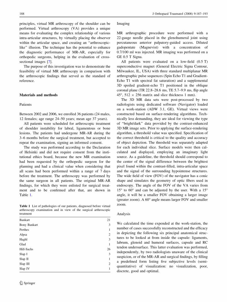
Fig. 1 Bankart lesion as seen
on magnetic resonance image
(MRI) (a), virtual MR
endoscopy (b) and arthroscopic
images (c). Axial GRE MR
shows (yellow arrow) avulsion
and fragmentation of the labrum
with detachment of the antero-
inferior capsulolabral complex
and rupture of the scapular
periosteum
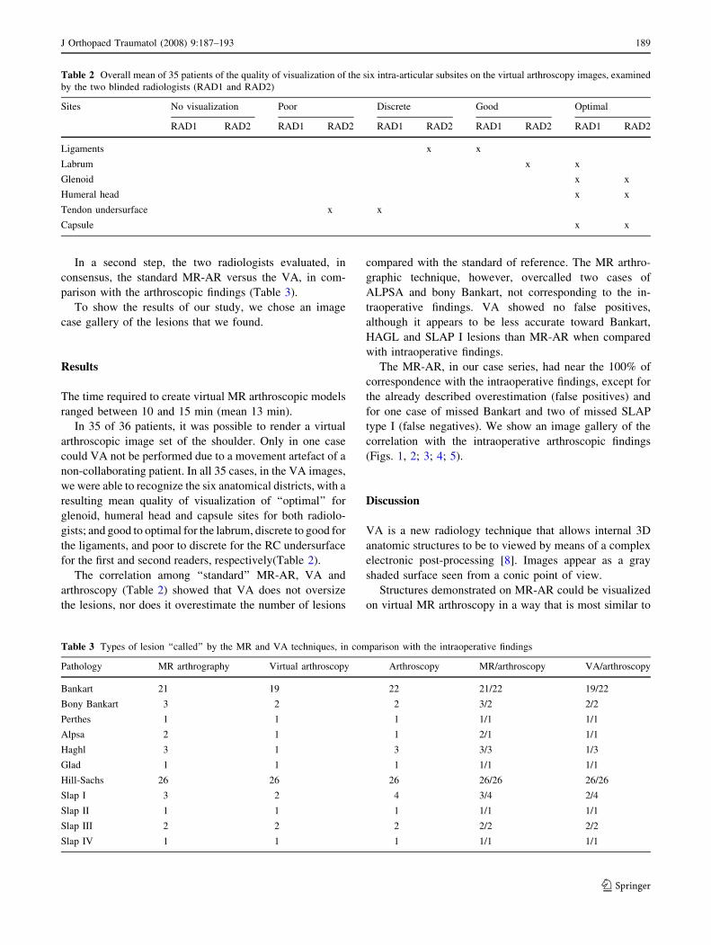
Fig. 2 Bony Bankart lesion as
seen on magnetic resonance
image (MRI) (a), virtual MR
endoscopy (b) and arthroscopic
images (c). Coronal MR images
show avulsion fracture of the
glenoid rim (yellow arrow) that
carries with it the capsulolabral
complex
190 J Orthopaed Traumatol (2008) 9:187–193
123
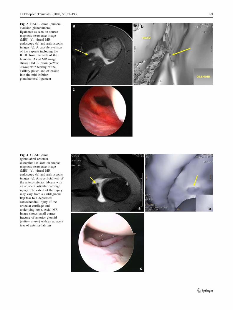
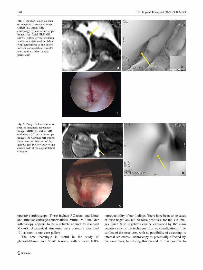
Fig. 3 HAGL lesion (humeral
avulsion glenohumeral
ligament) as seen on source
magnetic resonance image
(MRI) (a), virtual MR
endoscopy (b) and arthroscopic
images (c). A capsule avulsion
of the capsule including the
IGHL from the neck of the
humerus. Axial MR image
shows HAGL lesion (yellowarrow) with tearing of the
axillary pouch and extension
into the mid-inferior
glenohumeral ligament
Fig. 4 GLAD lesion
(glenolabral articular
disruption) as seen on source
magnetic resonance image
(MRI) (a), virtual MR
endoscopy (b) and arthroscopic
images (c). A superficial tear of
the antero-inferior labrum with
an adjacent articular cartilage
injury. The extent of the injury
may vary from a cartilaginous
flap tear to a depressed
osteochondral injury of the
articular cartilage and
underlying bone. Axial MR
image shows small corner
fracture of anterior glenoid
(yellow arrow) with an adjacent
tear of anterior labrum
J Orthopaed Traumatol (2008) 9:187–193 191
123
assess the viability of the structures, their aspect and thus
their status.
Bony Bankart could be overcalled by MR in general,
because an intraspongious signal alteration could be mis-
taken for a fracture; the standard of reference for bone loss
evaluation is CT. VA could identify gaps in the surface or
abrupt glenoid surface irregularities in the case of bony
Bankart, helped by the particular viewpoint from inside.
The VA images of the glenoid should be tested versus CT
to assess accuracy in the qualitative and quantitative
assessment of bone loss, a critical parameter that could
actually switch the treatment to an open procedure.
In our pilot study, VA findings, although in some cases
different with respect to the MR-AR, did not change the
therapeutic planning of the surgeon, because of the
experimental nature of the study. Costs did not differ much
between the two, because only 8 min longer room occu-
pancy was required by the VA patient (the time for the
pulse sequence). However, reconstruction time spent at
the work-station can be the negative side of this technique.
The learning curve associated with this VA reconstruction
is quite fast—it can take 2–3 days—but knowledge of
shoulder anatomy is mandatory to understand the viewing
position inside the articulation.
The additional diagnostic contribution of this method
will have to be estimated in time; today, however, it
remains true that VA can show a good view of a complex
anatomy, such as that of the shoulder. This procedure may
be useful in the future as a diagnostic tool and as an adjunct
to clinical and surgical planning, as well as an interactive
tool for learning arthroscopic anatomy and pathology.
The major bias of the present study was the low field
(0.5 T) of the MR scanner, which resulted in less spatial
resolution in the pulse sequences and bigger voxels in
reconstructions (like VA algorithms); we are currently
testing the protocol on a high-field (1.5 T) MR magnet.
Conflict of interest statement The authors declare that they have
no conflict of interest.
References
1. Faletti C (2007) L’imaging nell’instabilita di spalla. In: Fornara P,
Stecco A, Carriero A, Mordente G, Cisari C (eds) Spalla: clinica,
imaging patologia e riabilitazione. Idelson Gnocchi, Napoli,
pp 223–232
2. Applegate GR (1998) Three-dimensional MR arthrography of the
shoulder: an intraarticular perspective. AJR Am J Roentgenol
171:239–241
3. Davis CP, Ladd ME, Romanowski BJ, Wildermuth S, Knoplioch
JF, Debatin JF (1996) Human aorta: preliminary results with
virtual endoscopy based on three-dimensional MR imaging data
sets. Radiology 199:37–40
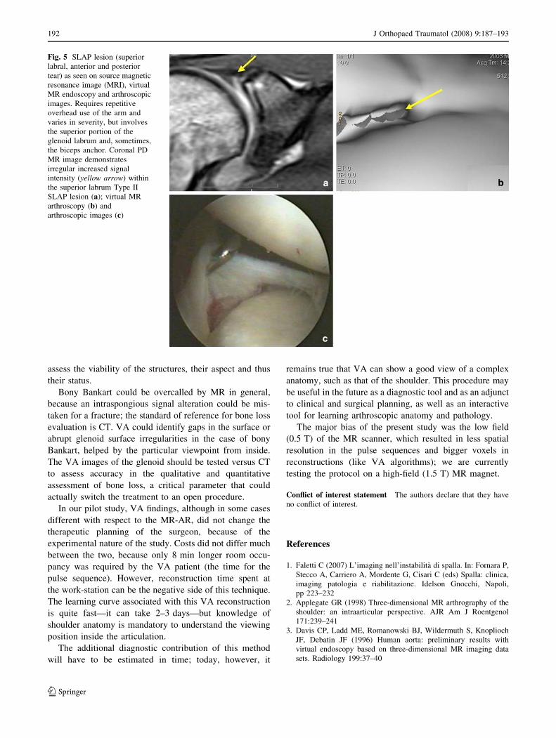
Fig. 5 SLAP lesion (superior
labral, anterior and posterior
tear) as seen on source magnetic
resonance image (MRI), virtual
MR endoscopy and arthroscopic
images. Requires repetitive
overhead use of the arm and
varies in severity, but involves
the superior portion of the
glenoid labrum and, sometimes,
the biceps anchor. Coronal PD
MR image demonstrates
irregular increased signal
intensity (yellow arrow) within
the superior labrum Type II
SLAP lesion (a); virtual MR
arthroscopy (b) and
arthroscopic images (c)
192 J Orthopaed Traumatol (2008) 9:187–193
123

4. Dubno B, Debatin JF, Luboldt W, Schmidt M, Hany TF,
Bauerfeind P (1998) Virtual MR cholangiography. AJR
171:1547–1550
5. Leung DA, McKinnon GC, Davis CP, Pfammater T, Krestin GP,
Debatin JF (1996) Breath-hold contrast-enhanced, three-dimen-
sional MR angiography. Radiology 200:569–571
6. Luboldt W, Bauernfeind P, Steiner P, Fried M, Krestin GF,
Debatin JF (1999) Preliminary assessment of three-dimensional
magnetic resonance imaging for various colonic disorders. Lancet
349:1288–1291
7. Luboldt W, Debatin JF (1998) Virtual endoscopic colonography
based on 3D MRI. Abdom Imaging 23:568–572
8. Neri E, Boraschi P, Caramella D et al (2000) MR virtual
endoscopy of the upper urinary tract. AJR Am J Roentgenol
175:1697–1702
9. Weishaupt D, Wildermuth S, Schmid M, Hilfiker PR, Hodler J,
Debatin JF (1999) Virtual MR arthroscopy: new insights into joint
morphology. J Magn Reson Imaging 9:757–760
J Orthopaed Traumatol (2008) 9:187–193 193
123