Embed Size (px)
Citation preview
G.J.B.B., VOL.3 (1) 2014: 109-114 ISSN 2278 – 9103
VIRULENCE DETERMINATION AMONG VIBRIO HARVEYI HATCHERYISOLATES THROUGH HAEMOLYSIS AND GROWTH CONSTRAINT
K. Ramesh, M. Natarajan, H. Sridhar & S. Umamaheswari*Microbial Biotechnology Laboratory, Department of BiotechnologyManonmaniam Sundaranar University, Tirunelveli – 627 012, India
Corresponding author * E-mail: [email protected]
ABSTRACTBacterial diseases mainly due to vibriosis in Penaeid shrimp culture implicating several species of Vibrios. Vibrio harveyiis considered as an important causative agent of the systemic vibriosis, which occurs in any bio-fields. The virulence ofthis bacterium is due to the production and expression of several virulent factors such as hemolysin, cysteine andmetalloprotease, phospholipases, exotoxins, luciferase and siderophore. The present study intends the virulencedetermination among three strains of V. harveyi VSH3, VSH5 and VSH9 isolated from shrimp hatchery along the coastlineof Tuticorin, Tamil Nadu during vibriosis outbreaks through their haemolysis and growth performance. Haemolyticactivity examined against sheep blood resulted minor or no variations among the organisms in agar plate assay. However,in microtitre assay, Extra Cellular Products (ECP) of all isolates rendered increased activity with increase in dose and timeof exposure. The bacterium VSH5 exhibited more haemolytic effect (at 500 µl concentration 72% haemolysis) than that ofVSH9 and VSH3 reavled that the bacterium is highly virulent than the others. The bacterium VSH5 showed well growthin the optimum temperature (33°C), NaCl (2%) and pH (7.3). The maximum luminescence was expressed in 37°C, 2.5-3%NaCl and pH 7-9 in 18 to 48 hrs. Diverse colony morphology was observed in VSH5 on solid medium incubated for 3 to 5days or longer. Growth curve experiment revealed that the bacterim VSH5 is a fast grower, completed its log phase within7 hrs. Pathogenic strain like V. harveyi VSH5 causes high mortality and affects aquaculture production in hatchery as wellas pond level. Hence, control measure against this kind of bacterium is urgently needed for maintain the sustainability ofshrimp aquaculture in India and other Asian country.
KEYWORDS: Vibrio harveyi VSH5, Haemolysis, Growth Parameters, Colony Variation, Growth Curve.
INTRODUCTIONShrimp aquaculture is a most important industry in Indiaand other Asian countries. Shrimp hatcheries along thecoastline involved in shrimp seed production often sufferenormous economic losses due to luminescent bacterialdisease, commonly called as vibriosis. Vibriosis outbreaksare being increasingly recognized as a significantconstraint to aquaculture production and trade inworldwide. The genus Vibrio belongs to the gamma-proteobacteria and is Gram-negative, usually motile rods(Thompson et al., 2004). Vibrio disease in aquaculture isdescribed as vibriosis or bacterial disease, Penaeidbacterial septicaemia, Penaeid vibriosis, luminescentvibriosis or red-leg disease (Aguirre-Guzmán et al., 2004).Among the Vibrios, Vibrio harveyi (luminous Vibrio) isthe main cause of shrimp death infecting larva in thehatchery as well as in the cultivation pond (Won & Park,2008). V. harveyi is one of the important etiological agentsof mass mortalities of Penaeus monodon larvae rearingsystems. The virulence of V. harveyi causes 100% lossesat a time in shrimp production (Chythanya et al., 2002;Musa et al., 2008). Luminescent strains of V. harveyi havebeen reported to cause major losses in the shrimplarviculture in the Phillippine (Lavilla-Pitogo et al., 1990),Australia (Pizzutto & Hirst, 1995), South America(Alvarez et al., 1998; Robertson et al., 1998) and Mexico(Vandenberghe et al., 1999). Although almost all types of
cultured crustaceans can be affected by these bacteria, themost serious problems have been reported in Penaeidshrimp culturing (Austin & Zhang, 2006). Adult shrimpssuffering vibriosis may appear hypoxic, shows reddeningof the body with red to brown gills, reduce feeding andmay be observed swimming lethargically at the edges andsurface of ponds (Anderson et al., 1988; Nash et al.,1992). Vibriosis infected post larvae (PL) exhibits reducedmotility, reduced phototaxis and empty guts (Chen, 1992).Vibriosis is expressed by a way of number of syndromeswhich include oral and enteric vibriosis, appendage andcuticular vibriosis, localised vibriosis of wounds, shelldisease, systemic vibriosis and septic hepatopancreatitis(Lightner, 1996). Vibriosis Infected animals shows signsof lethargy, tissue and appendage necrosis, slow growth,slow metamorphosis, body malformation,bioluminescence, muscle opacity and melanisation(Aguirre-Guzmán et al., 2004). Despite its role as aserious pathogen of cultured marine animals, thepathogenic mechanisms of V. harveyi yet have to be fullyelucidated (Austin & Zhang, 2006), although severaldifferent virulent factors have been identified. Some ofthe hatcheries along the coastal regions of Tuticorin, SouthTamil Nadu showed the symptoms of luminous vibriosisduring the middle of 2012. The usage of medication andother treatments could not prevent the disease prevalencein the hatcheries. In our previous study we isolated several
109
Vibrio harveyi hatchery isolates through haemolysis and growth constraint
110
vibrio pathogens during Vibriosis outbreak from the abovesaid regions. Among the isolates, three (VSH3, VSH5 andVSH9) were phenotypically and genotypically identifiedas V. harveyi. Hence, the present study was performed toselect the highly virulent strain among them based on theirhemolytic and growth ability.
MATERIALS & METHODSBacterial strainsThree strains of V. harveyi VSH3, VSH5 and VSH9 wereisolated and identified from hatchery water duringvibriosis outbreak. The stock cultures maintained inMicrobial Biotechnology Laboratory, ManonmaniamSundaranar University, Alwarkurichi were used as targetpathogens throughout the study.Hemolysin test on agar platesHemolysin test was carried out according to the methoddescribed by Austin et al. (2005) with slight modification.Bacterial strains were grown overnight in marine broth at25ºC in an incubator shaker. A drop of each of the isolatewas spotted onto freshly prepared blood agar (marine agarcontaining 1% de-fibrinated sheep blood) plates. Finally,the plates were covered with parafilm and kept in anincubator at 30ºC for 48 hrs. The test was repeated threetimes.Haemolytic activity of ECPExtra cellular products (ECP) of the Vibrios were obtainedby centrifuging overnight culture in a concentration of1.5×108 cells/ml and tested for haemolytic activity.Haemolytic titrations were conducted in 96-well microtitreplates (Tarsons, Kolkata). Five ml of blood was collectedfrom a healthy universal donor individual (O+ve) anderythrocytes were collected after repeated washing insterile normal saline (0.85% w/v NaCl, pH 7.2) andresuspended in normal saline to 0.5%. A volume of 0.5mlof the cell suspension was mixed with variousconcentrations of ECP (50, 100, 150, 200……450 and500µl). Total volume of each well was made up to one mlwith normal saline. The mixtures were incubated for 1 hrat 37ºC. Haemolytic activity was determined by theappearance of lytic erythrocytes visibly ormicroscopically. Haemolytic activity was also performedby spectroscopic method (Yang et al., 2005). Followingincubation at 37ºC for 1 hr, the mixture was centrifuged at1500 rpm for 10 min in a cooling centrifuge. The freehemoglobin in the supernatants was measured in UV-Visible spectrophotometer at 540 nm. Drabkin’s solution(500 µl) and Saline (500 µl) were used as positive(maximal) and negative (minimal) haemolytic controls.Each experiment was performed in triplicates for eachconcentration.The percentage haemolysis was calculated according tothe following formula:
% Haemolysis = [At–An/Ac–An] ×100
Where, At is the absorbance of test sample, An isabsorbance of the negative control, Ac is the absorbance ofthe positive control.Optimal condition for culturing highly virulent strainof V. harveyiThe bacterium was cultured in both solid and liquidmedium of TSA and TSB. Colony morphology wasstudied on TCBS and marine agar. The bacterium wasincubated at various temperatures (from room temperatureto 37°C), in media of various percentages of NaCl(between 0.5-7%) and various alkalinity (pH 5 -12). Underthese physical conditions growth, colony morphology andluminescence production were investigated.Growth curve of virulent strainAbout 200 ml of the marine broth was inoculated with theovernight culture of test organism and kept on Orbitalshaker at room temperature. Observations for growth ODwas taken using a spectrophotometer at 600nm ofabsorbance after every 15 min till the log phase wasachieved, after which readings were taken every 30minutes. Zero reading was calibrated with un-inoculatedbroth (control). Growth curve was plotted as OD 600readings against time and different phases of growth werehence determined.
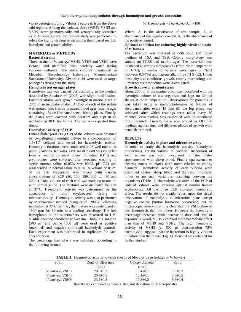
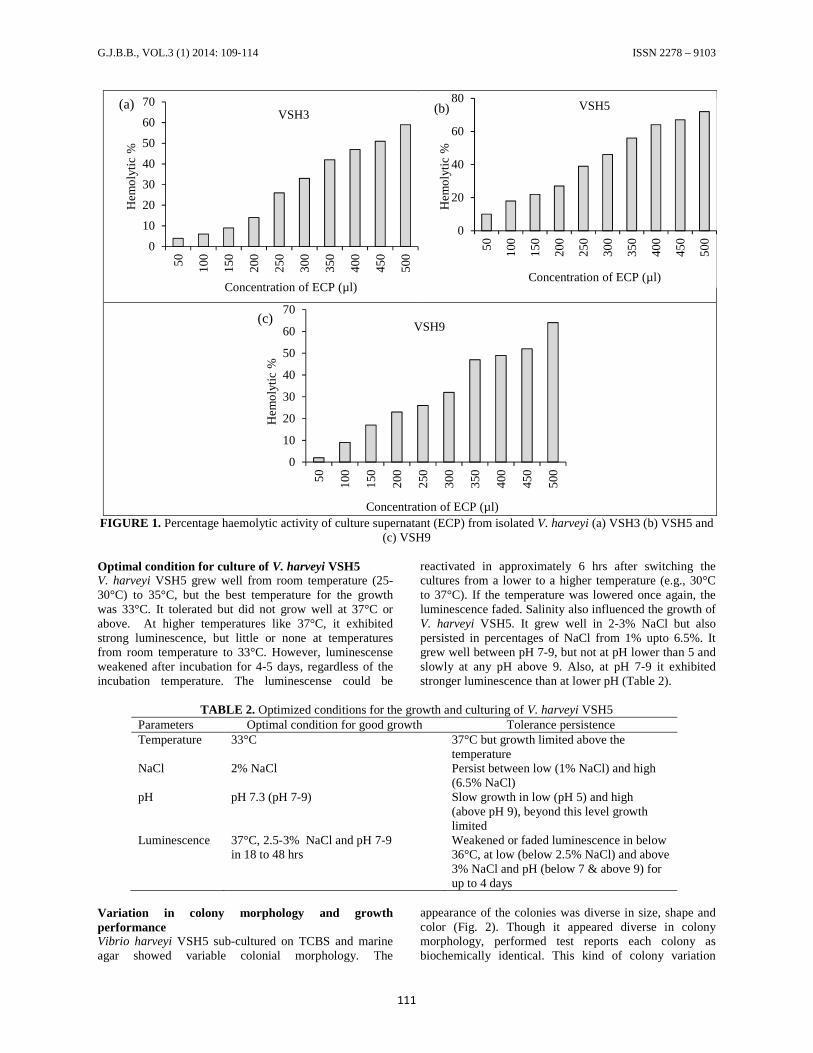
RESULTSHaemolytic activity in plate and microtitre assayIn order to study the haemolytic activity (hemolysinproduction), certain volume of bacterial suspension ofeach isolate was spot inoculated on the platessupplemented with sheep blood. Finally opalescence orclearing zones on plates were noted relative to colonydiameter. Haemolytic activity of each Vibrios wereexamined against sheep blood and the result indicatedminor or no such variations occurring between theorganisms (Table 1). Haemolytic activities of the ECP ofisolated Vibrios were screened against normal humanerythrocytes. All the three ECP indicated haemolyticeffect. The results do not clearly report upon the visualobservation of haemolysis in microtitre plate exceptnegative control (button formation occurrence) but onmicroscopic observation it is clear that the VSH5 showedbest haemolysis than the others. However the haemolyticpercentage increased with increase in dose and time ofexposure. Overall, VSH5 exhibited more haemolytic effectthan that of VSH9 and VSH3. The high haemolyticactivity of VSH5 (at 500 µl concentration 72%haemolysis) suggests that the bacterium is highly virulentin nature than the others (Fig. 1). Hence it was selected forfurther studies.
TABLE 1. Haemolytic activity towards sheep red blood of three isolates of V. harveyiStrain Zone of Clearance
(mm)Colony diameter
(mm)Ratio
V. harveyi VSH3 20.8±0.2 15.4±0.1 5.3±0.2V. harveyi VSH5 20.9±0.5 15.1±0.1 5.8±0.5V. harveyi VSH9 23.1±0.2 17.5±0.2 5.6±0.4
Results are expressed as mean ± standard deviation of three replicates
G.J.B.B., VOL.3 (1) 2014: 109-114 ISSN 2278 – 9103
111
FIGURE 1. Percentage haemolytic activity of culture supernatant (ECP) from isolated V. harveyi (a) VSH3 (b) VSH5 and(c) VSH9
Optimal condition for culture of V. harveyi VSH5V. harveyi VSH5 grew well from room temperature (25-30°C) to 35°C, but the best temperature for the growthwas 33°C. It tolerated but did not grow well at 37°C orabove. At higher temperatures like 37°C, it exhibitedstrong luminescence, but little or none at temperaturesfrom room temperature to 33°C. However, luminescenseweakened after incubation for 4-5 days, regardless of theincubation temperature. The luminescense could be
reactivated in approximately 6 hrs after switching thecultures from a lower to a higher temperature (e.g., 30°Cto 37°C). If the temperature was lowered once again, theluminescence faded. Salinity also influenced the growth ofV. harveyi VSH5. It grew well in 2-3% NaCl but alsopersisted in percentages of NaCl from 1% upto 6.5%. Itgrew well between pH 7-9, but not at pH lower than 5 andslowly at any pH above 9. Also, at pH 7-9 it exhibitedstronger luminescence than at lower pH (Table 2).
TABLE 2. Optimized conditions for the growth and culturing of V. harveyi VSH5Parameters Optimal condition for good growth Tolerance persistenceTemperature 33°C 37°C but growth limited above the
temperatureNaCl 2% NaCl Persist between low (1% NaCl) and high
(6.5% NaCl)pH pH 7.3 (pH 7-9) Slow growth in low (pH 5) and high
(above pH 9), beyond this level growthlimited
Luminescence 37°C, 2.5-3% NaCl and pH 7-9in 18 to 48 hrs
Weakened or faded luminescence in below36°C, at low (below 2.5% NaCl) and above3% NaCl and pH (below 7 & above 9) forup to 4 days
Variation in colony morphology and growthperformanceVibrio harveyi VSH5 sub-cultured on TCBS and marineagar showed variable colonial morphology. The
appearance of the colonies was diverse in size, shape andcolor (Fig. 2). Though it appeared diverse in colonymorphology, performed test reports each colony asbiochemically identical. This kind of colony variation
0
10
20
30
40
50
60
70
50 100
150
200
250
300
350
400
450
500
Hem
olyt
ic %
Concentration of ECP (µl)
VSH3(a)
0
20
40
60
80
50 100
150
200
250
300
350
400
450
500
Hem
olyt
ic %
Concentration of ECP (µl)
VSH5(b)
0
10
20
30
40
50
60
7050 10
0
150
200
250
300
350
400
450
500
Hem
olyt
ic %
Concentration of ECP (µl)
VSH9(c)

Vibrio harveyi hatchery isolates through haemolysis and growth constraint
appeared only on solid medium incubated for 3 to 5 daysor longer. The variation was not evident upon dailysubculture. Growth curve experiments revealed that VSH5is a fast growing bacterial isolate that reached its log phase
after 30 min of incubation at room temperature withcontinuous shaking of 150 rpm. The log phase continuesup to 7.0 hrs after which the stationary phase seemed tostart (Fig. 3).
FIGURE 2. Various colony types of VSH5: (A) clouded green small rough colonies on TCBS (B) Smooth colonies withblack pigments on TCBS (C) Small and big margin colonies on marine agar (D) Small dark centered yellow halo aroundcolonies (E) Clear dark centered green colonies (F) Rough medium sized green unshaped colonies (G) large colonies withdark green round (H) Large elevated dark green to yellow colonies and (I) Colonies partially digested.
FIGURE 3. Growth curve of V. harveyi VSH5 in Marine broth
DISCUSSIONNakayama et al. (2006) observed that the ECP of V.harveyi VP1 showed higher haemolytic activity comparedwith VT2 and other strains. A similar trend was also
observed with the strain of V. harveyi VSH5 isolated in thepresent study. Several reports have evaluated thepathogenicity of environmental isolates of V. harveyi tolarva and juvenile penaeid shrimps (Harris & Owen, 1999;
0
0.2
0.4
0.6
0.8
1
1.2
1.4
1.6
0 30 60 90 120
150
180
210
240
270
300
330
360
390
420
450
480
510
540
570
Abs
orba
nce
(OD
at
600
nm)
Time in minutesAbsorbance (O.D at 600 nm)
112

G.J.B.B., VOL.3 (1) 2014: 109-114 ISSN 2278 – 9103
113
Pizzutto & Hirst, 1995; Ruangpan et al., 1999). One of thereasons for variation in the virulence levels of Vibriosreported in this study revealed that infectivity of V.harveyi is dependent on the virulence factors of the strainsemployed (Gomez-Gill et al., 1998). Some studies indicatethat the virulence factors produced by V. harveyi can becontributed from toxins (either protease or hemolysin)(Liu & Lee, 1999; Zhang & Austin, 2000; Zhang et al.,2001). However, other studies represented that thepathogenicity of V. harveyi is derived from phage in whichgenes coding for toxin production are acquired by genetransduction (Morris & Robert, 1995). The toxinproduction in bacteria may be controlled by genetransduction but some bacteria have been found to expresstoxin by a process called quorum sensing (Bernd et al.,2001; Costi et al., 2002). It is reported that the bacterialluminescense is produced by different autoinducer in eachgenus or species. The major autoinducer of V. harveyi hasbeen reported to be a long chain aliphatic aldehyde. Luxgene expression triggers the synthesis and accumulation ofautoinducer during the growth of bacteria. The electrontransport proceeds by the catalytic reaction of luciferaseamong the reduced flavin mononucleotide (FMNH2), O2
and a long chain aliphatic aldehyde produces flavinmononucleotide (FMN) and an aliphatic carboxylic acidwhich emits the light (Fisher et al., 1995). The results ofthe present study suggested that the temperature may alsoinfluence the expression of luminescence which is inaccordance with the study of Pasharawipas et al., (1998).The temperature may either stimulate luciferase activity orthe production or function of the autoinducer. Thebacterium VSH5 does not grow at higher temperaturessuch as 37°C and above as the temperature may affect theproduction and activities of luciferase or autoinducer. Thepresent study also reports that the luminescence expressionwas affected by pH. Optimum pH (7-9) resulted in strongluminescence which also correlates with the study ofPasharawipas et al. (1998). The combination oftemperature and alkalinity might find some application inmanipulation of V. harveyi for higher production of poly-3-hydroxybutyrate (PHB), a raw material in plasticindustry due to its properties like thermoplasticity, waterresistance and biodegradability. It was previously reportedthat the production of PHB is related to luminousexpression controlled by the lux autoinducer (Sun et al.,1994).The variation in colony morphology of VSH5 is ofsomewhat interest. Similar variation was obtained byPasharawipas et al.(1998) during sub-culturing of V.harveyi VH1039 on TSA and TCBS. The variability ofVSH5 might involve the fact that it is a lysogenic host oftemperate phages which are rarely found in cultureenvironment. The variability of other bacteria has alsobeen reported to be due to transposon like behaviour ofbacteriophages (Reidl & Makalanos, 1995; Belas et al.,1984). This may be the best explanation for variablemorphology of bacterial colonies since the biochemicaltests did not change for each colony. The growth curveresult of strain VSH5 is in corroboration with the findingsof Aisha & Nuzhat (2011) who recorded the growth curveof V. harveyi N6. Mortality among the cultured shrimp inhatcheries is due to the presence of highly virulent strainlike V. harveyi VSH5 with different colony morphology
and growth performance. This proves that there is anurgent need for a new eco-friendly preventive measureagainst the Vibrio pathogens in shrimp aquaculture toovercome quality seeds (larvae) exports.Study to determine the virulence levels of the bacterialpathogen in aquatic animals is a key to prevent vibriosis inmarine aquaculture. The virulence of V. harveyi isreported dependent on host species (Vera et al., 1992),doses, time exposure and age of host species (Jun &Huaishu, 1998) and pathogenic factors of the bacterialstrains (Gomez-Gill et al., 1998). This paper describes thevirulence of three strains of V. harveyi isolated fromshrimp hatchery water based on their haemolytic activityand growth performance. Highly virulent strain like VSH5causes mortality on animals and affects aquacultureproduction in hatchery level. Hence, control measureagainst this kind of bacterial pathogen is urgently neededfor sustainability of shrimp aquaculture in India and otherAsian country.
REFERENCESAguirre-Guzmán, G., Meija Ruíz, H. and Ascencio, F.(2004) A review of extracellular virulence product of Vibriosp. important in disease of cultivated shrimp. AquacultureRes. 35, 1395-1404.
Aisha, N. and Nuzhat, A. (2011) Isolation andcharacterization of indigenous luminescent marine bacteriafrom Karachi coast. Acad. Res. Int. 1, 74-83.
Alvarez, J. D., Austin, B., Alvarez, A. M. and Reyes, H.(1998) Vibrio harveyi: a pathogen of penaeid shrimps andfish in Venezuela. J. Fish Dis. 21, 313-316.
Anderson, I. G., Shamsudin, M. N. and Shariff, M. (1988)Bacterial septicemia in juvenile tiger shrimp, Penaeusmonodon, cultured in Malaysian brackish water ponds. AsianFis. Sci. 2, 93-108.
Austin, B. and Zhang, X. H. (2006) Vibrio harveyi: asignificant pathogen of marine vertebrates and invertebrates.Lett. Appl. Microbiol. 43, 119-124.
Austin, B., Austin, D., Sutherland, R., Thompson, F. andSwings, J. (2005) Pathogenicity of vibrios to rainbow trout(Oncorhynchus mykiss, Walbaum) and Artemianauplii. Environ. Microbiol. 7, 1488-1495.
Belas, R., Mileham, A., Simon, M. and Silverman, M. (1984)Transposon mutagenesis of marine Vibrio sp. J.Bacteriol. 158, 890-896.
Bernd, K., Michael, D. P. B., Bettina, A. B., Markus, H. andAndreas, P. (2001) Group A Streptococcal growth phase-associated virulence factor regulation by a novel operon(Fas) with homologies to two-component-type regulatorsrequires a small RNA molecule. Mol. Microbiol. 39, 392-406.
Chen, D. (1992) An overview of the disease situation,diagnostic techniques, treatments and preventatives used onshrimp farms in China. In: Fuls, W. and K. L. Main (Ed.):Diseases of cultured penaeid shrimp in Asia and the UnitesStates. Hawaii, pp. 47-55.
Vibrio harveyi hatchery isolates through haemolysis and growth constraint
114
Chythanya, R., Karunasagar, I. and Karunasagar, I. (2002)Inhibition of shrimp pathogenic vibrios by a marinePseudomonas I-2 strain. Aquaculture 208, 1-10.
Costi, D. S., Eleftherios, M., Kavindra, V. S., Xiang, Q.,Danielle, A. G., Murray, B. E., Ausubel, F. M. andCalderwood, S. B. (2002) Virulence effect of Enterococcusfaecalis protease genes and the quorum-sensing locus fsr inCaenorhabditis elegans and mice. Infect. Immunol. 70, 5647-5650.
Fisher, A. J., Raushel, F. M., Baldwin, T. O. and Rayment, I.(1995)Three dimensional structure of bacterial luciferasefrom Vibrio harveyi at 2.4 A° resolution. Biochem. 34, 6581-6586.
Gomez-Gill, B., Herrera-Vega, M. A., Abreu-Gobois, F. A.and Roque, A. (1998) Bioencapsulation of two diferent Vibriospecies in nauplii of the brine shrimp (Artemia franciscana).Appl. Environ. Microbiol. 64, 2318-2322.
Harris, L. J. and Owens, L. (1999) Production of exotoxins bytwo luminous Vibrio harveyi strains known to be primarypathogens of Penaeus monodon larvae. Dis. Aquat. Org. 38,11-22.
Jun, L. I. and Huai-Shu, X. (1998) Isolation and biologicalcharacteristics of Vibrio harveyi affecting hatchery-rearedPenaeus chinensis larvae. Chin. J. Oceanol.Limnol. 29, 353–361.
Lavilla-Pitogo, C. R. Baticados, M., C. L., Cruz Lacierda, E.R. and de La Peña L. D. (1990) Occurrence of luminousbacterial disease of Penaeus monodon larvae in thePhilippines. Aquaculture 91, 1-13.
Lightner, D. V. (1996) A Handbook of pathology anddiagnostic procedures for diseases of Penaeid shrimp. WorldAquaculture Society, Baton Rouge, Louisiana, USA.
Liu, P.C. and Lee, K. K. (1999) Cysteine protease is a majorexotoxin of pathogenic luminous Vibrio harveyi in the tigerprawn, Penaeus monodon. Lett. Appl .Microbiol. 28, 428-430.
Morris, P. and Robert, G. H. (1995) Classification of isolatesof Vibrio harveyi virulent to Penaeus monodon larvae byprotein profile analysis and M13 DNA fingerprinting. Dis.Aquat. Org. 21, 61-68.
Musa, N., Wei, L. S. and Wee, W. (2008) Phenotypic andgenotypic characteristics of Vibrio harveyi isolated fromblack tiger shimp (Penaeus monodon). World J. Appl. Sci. 3,885-902.
Nakayama, T., Nomura, N. and Matsumura, M. (2006) Studyon the relationship of protease production and luminescencein Vibrio harveyi. J. Appl. Microbiol. 101, 200-205.
Nash, G., Nithimathachoke, C., Tungmandi, C.,Arkarjarmorin, A., Prathampipat, P. and Ruamthaveesub P.(1992) Vibriosis and its control in pond-reared Penaeusmonodon in Thailand. In: Shariff, I.M., R. P. Subasinghe and
J. R. Arthur (Ed.): Diseases in Asian Aquaculture. ThePhillippines, pp. 143-155.
Pasharawipas, T., Sriurairatana, S., Direkbusarakom, S.,Donayadol, Y., Thaikua, S., Ruangpan, L. and Flegel, T. W.(1998) Luminous Vibrio harveyi associated with tea browngill syndrome in black tiger shrimp. In: Flegel, T. W. (Ed.):Advances in shrimp biotechnology, National Center forGenetic Engineering and Biotechnology, Bangkok.
Pizzutto, M. and Hirst, R. G. (1995) Classification of isolatesof Vibrio harveyi virulent to Penaeus monodon larvae byprotein profile analysis and M13 DNA fingerprinting. Dis.Aquat. Org. 21, 61-68.
Reidl, J. and Mekalanos, J. (1995) Characterization of Vibriocholera bacteriophage K139 and use of a novel mini-transposon to identify a phage-encoded virulence factor. Mol.Microbiol. 18, 685-701.
Robertson, P. A. W., Calderon, J., Carrera, L., Stark, J. R.,Zherdmant, M. and Austin, B. (1998) Experimental Vibrioharveyi infections in Penaeus vannamei larvae. Dis. Aquat.Org. 32, 151-155.
Ruangpan, L., Danayadol, Y., Direkbusarakom, S.,Siurairatana, S. and Flegel, T. W. (1999) Lethal toxicity ofVibrio harveyi to cultivated Penaeus monodon induced by abacteriophage. Dis. Aquat. Org. 35, 195–201.
Sun, W., Cao, J. G., Teng, K. and Meighen, E. A. (1994)Biosynthesis of poly 3-hydroxybutyrate in the luminescentbacterium, Vibrio harveyi and regulation by the luxautoinducer, N-(3-hydroxybutanoyl) homoserine lactone. J.Biol. Chem. 269, 20785-20790.
Thompson, F. L., Iida, T. and Swings, J. (2004) Biodiversityof vibrios. Microbiol. Mol. Biol. Rev. 68, 403-431.
Vandenberghe, J., Verdonck, L., Robles Arozarena, R.,Rivera, G., Bolland, A., Balladares, M., Gomez-Gil, B.,Calderon, J., Sorgeloos, P. and Swings, J. (1999) Vibriosassociated with Litopenaeus vannamei larvae, postlarvae,broodstock, and hatchery probionts. Appl. Environ.Microbiol. 65, 2592-2597.
Vera, P., Navas, J. I. and Quintero, M. C. (1992)Experimental study of the virulence of three species ofVibrio bacteria in Penaeus japonicus (Bate 1881) juveniles.Aquaculture 107, 119-123.
Won, K. M., Park, S. (2008) Pathogenicity of Vibrio harveyito cultured marine fishes in Korea. Aquaculture 285, 8-13.
Yang, Z. G., Sun, H. X. and Fang, W. H. (2005) Haemolyticactivities and adjuvant effect of Astragalus membranaceussaponins (AMS) on the immune responses to ovalbumin inmice. Vaccine 23, 5196-5203.
Zhang, X. H, and Austin, B. (2000) Pathogenicity of Vibrioharveyi to Salmonids. J. Fish Dis. 23, 93-102.Zhang, X. H., Meaden, P. G. and Austin, B. (2001)Duplication of hemolysin genes in a virulent isolate of Vibrioharveyi. Appl. Environ. Microbiol. 67, 3161-3167.