Embed Size (px)
Citation preview
Visceral and Parietal Anomalies
Arlet G. Kurkchubasche Francois I. Luks

Role of fetal diagnosis
Postnatal diagnoses
Do we need to intervene prenatally? Do we need to deliver this baby early? What do we expect for this baby postnatally?
Prenatal findings
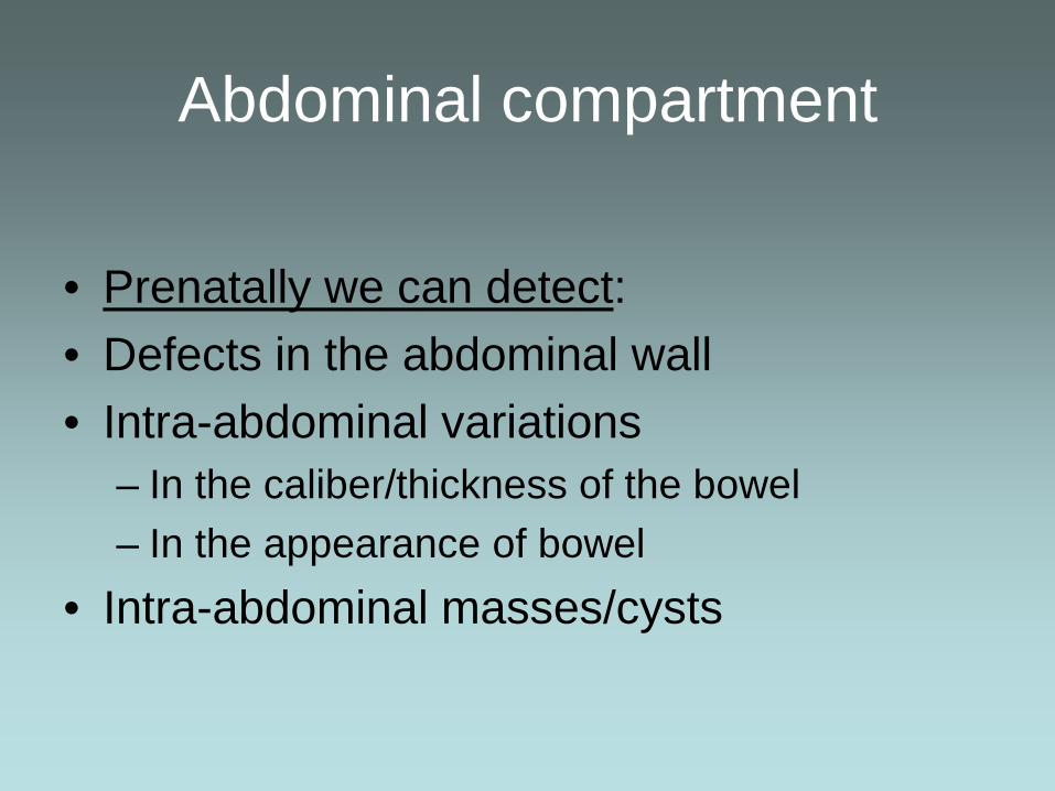
Abdominal compartment
• Prenatally we can detect: • Defects in the abdominal wall • Intra-abdominal variations
– In the caliber/thickness of the bowel – In the appearance of bowel
• Intra-abdominal masses/cysts

Parietal defects
– Defects of the abdominal wall
• Gastroschisis – full-thickness/off center
• Omphalocele – covered muscular defect/central
• Prunebelly syndrome – Laxity of intact wall
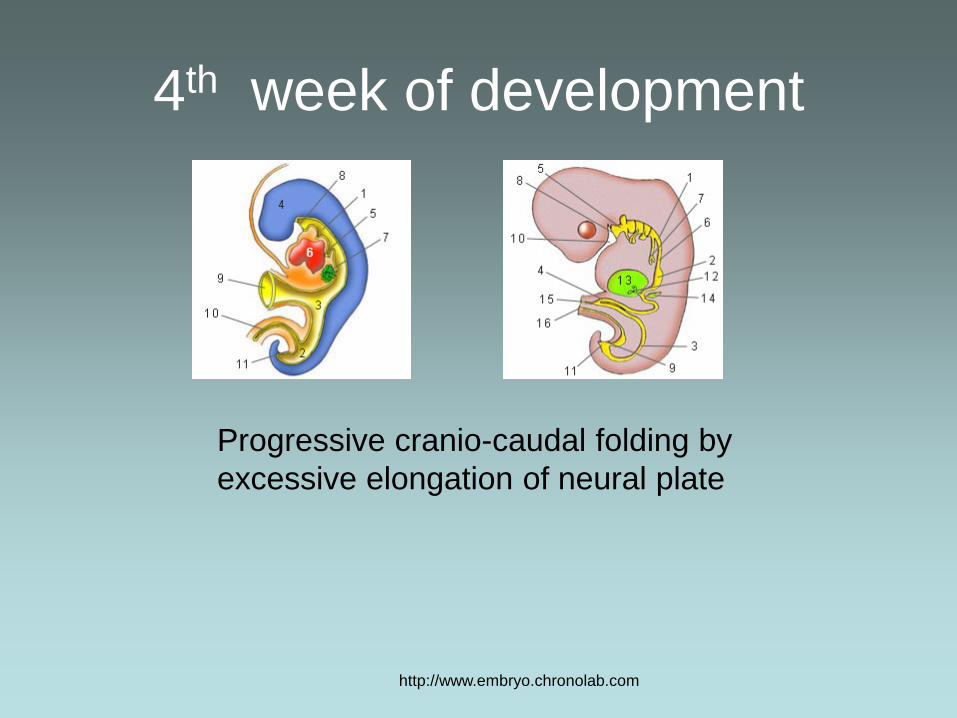
4th week of development
http://www.embryo.chronolab.com
Progressive cranio-caudal folding by excessive elongation of neural plate

4th week of development
http://www.embryo.chronolab.com
Lateral edges of germ disk Fold and fuse on midline
Gut tube
Abdominal wall
Amnion

Clinical impact
• Two very different entities
Omphalocele Gastroschisis

Omphalocele • Newborn Characteristics
– Incidence 1/4000 – 1/7000 Live births – Midline defect covered with membrane – Liver typically within sac – Associated anomalies: - 50% none
• with chromosomal (trisomy 13 and 18) 10-20% • cardiac anomalies 30-35% • overgrowth syndromes – Beckwith Wiedeman
• Maternal Characteristics – Advanced age, – MS-AFP elevated

Fetal diagnosis • Fetal U/S Characteristics
– Contour change of abdominal wall • Liver extends beyond confines of
abdominal cavity – Membrane covers AWD and cord
attaches to it. • Cord insertion site appears distant from
abdominal wall. – May be difficult to determine size of
defect • Hernia of cord, omphalocele, giant
omphalocele

Omphalocele • Postnatal management
– Protection of viscera – Evaluation for associated anomalies
• Genetic eval, Echo, hypoglycemia
– Closure abdominal wall – • Primary or staged operations
• Outcomes – Related to morbidity of associated
anomalies – When isolated defect – outcome related
to size of defect

Omphalocele - spectrum
Hernia of the cord Large Omphalocele Pentalogy of Cantrell
Giant omphalocele Open diaphragm & pericardium

Prenatal discussion
• Geneticist, Neonatologist, Cardiologist and Pediatric surgeon – Diagnosis can be virtually certain
• MSAFP elevated, fetal U/S findings
– Associated anomalies need to be determined • Role of amniocentesis, fetal echo
– Elevated risk for IUFD, IUGR, preterm labor • Incidence of omphalocele in live and stillborns 1/300- 1/4000
i.e. high IUFD rate
– Delivery at tertiary care center • Assure specialists available, possible C/S for giant omph.

Gastroschisis- the other AWD • Full thickness defect in
the abdominal wall – to the right of the
umbilicus • Embryology
– Weakness of abdominal wall?
– Consequence of involution of right umbilical vein?

Gastroschisis • Newborn characteristics
– Abd wall defect to right of umbilical cord – Variable size (<5 mm to 3 cm) – variable amount intestine +/- stomach,
• Maternal characteristics – Young – under age 20 4x increased risk – Elevated MS AFP – better predictor than for
omphalocele – Predisposing factors
• Smokers – 1.6 fold risk • ? Use of vasoactive medicines – pseudoephedrine increased
risk 3 fold

Prenatal issues
• Fetal diagnosis – Diagnosis can be
certain – Fetal intestine
adjacent to umbilical cord and external to abdominal wall
only pitfall = ruptured omphalocele - rare

Prenatal issues • Prenatal discussion
– Neonatologist / Pediatric surgeon – Focus on uni-system issues
• Only associated anomaly is within same system = atresia ( <30% cases)
• Significant impact only if SBS – Importance of delivery at tertiary care center
• Optimal premature infant care • Prenatal management
Monitor growth Monitor condition of intestine

Fetal U/S: gastroschisis
Dilation of intra-abdominal bowel suggests obstruction at fascial level
Marked small bowel dilatation
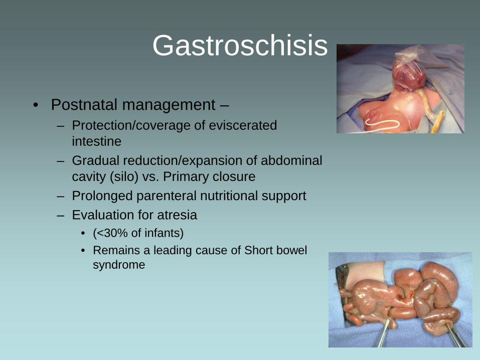
Gastroschisis
• Postnatal management – – Protection/coverage of eviscerated
intestine – Gradual reduction/expansion of abdominal
cavity (silo) vs. Primary closure – Prolonged parenteral nutritional support – Evaluation for atresia
• (<30% of infants) • Remains a leading cause of Short bowel
syndrome

Gastroschisis - outcomes
Survival approaches 100% Hospitalization 4-8 weeks unless complicated by atresia/SBS

Prenatal issues
• Fetal intervention? – Defect life-threatening?– NO – Intervene to improve condition/function of
bowel at birth? • Is exposure to amniotic fluid toxic? • Early delivery? • C/S delivery?

Fetal intervention for gastroschisis
– Alter place of delivery • Yes: deliver in center with neonatal/surgical access
– Alter mode of delivery • C/Section: promoted to “protect” the intestine and
shorted hospital stay due to more rapid advance to enteral feeding
• Evidence does not support these assertions

Fetal intervention for gastroschisis
– Alter timing of delivery • If amniotic fluid is caustic, early delivery makes
sense • Prospective series at Brown:
– No recommendations of early delivery – Parameters: age at closure, age at first and full feeds – Results: No rationale for early delivery

Gastroschisis
Gestational age (weeks)
Age
at d
efin
itive
clo
sure
38 39 40 37 34 35 36 33
1 week

Abdominal wall defects
• Prenatal counseling – Excellent overall prognosis in absence of
associated defects
– Often prolonged neonatal ICU stay – No long-term sequelae

Visceral abnormalities
• Intestinal obstructions
– mechanical and functional • Duplications • Disorders of rotation • Internal hernias

Visceral anomalies
• Prenatal diagnosis depends on: – Alterations in amniotic fluid volume
• polyhydramnios – Alterations in appearance of intestine:
• dilated, edematous, or smaller than expected – Alterations in content of intestine
• Echogenic material – Alterations within abdominal cavity
• Calcifications, ascites

Intestinal Obstruction
• Postnatal classification – • Proximal
– Esophageal atresia, duodenal atresia, prox jejunal atresia
• Distal – Jejuno-ileal atresia, meconium ileus, colon
atresia, imperforate anus, Hirschsprung’s
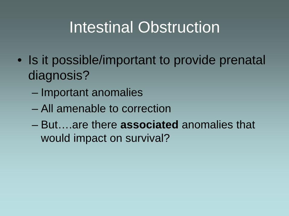
Intestinal Obstruction
• Is it possible/important to provide prenatal diagnosis? – Important anomalies – All amenable to correction – But….are there associated anomalies that
would impact on survival?

Possible Prenatal U/S findings
Esophageal atresia Duodenal atresia Jejunoileal atresia
Small stomach Double Bubble Dilated loops of intestine
Polyhydramnios with

Possible Prenatal U/S findings
Esophageal atresia Duodenal atresia Jejunoileal atresia
VACTERL association Vertebral, anorectal, cardiac tracheo-esophageal, renal, limb
Chromosomal Cardiac Other atresia
Generally isolated to intestine
Look for other anomalies

Distal intestinal obstruction • Not as easily evident
– May not have abnormal amniotic fluid volume – Dilation may be diffuse
• Content of intestine may be best clue – Higher density content = echogenic – Must consider anatomic and functional problems
• Examples: – Distal small bowel obstruction, colon atresia,
Hirschsprung’s Disease, imperforate anus • Prenatal Dx at this time is unreliable
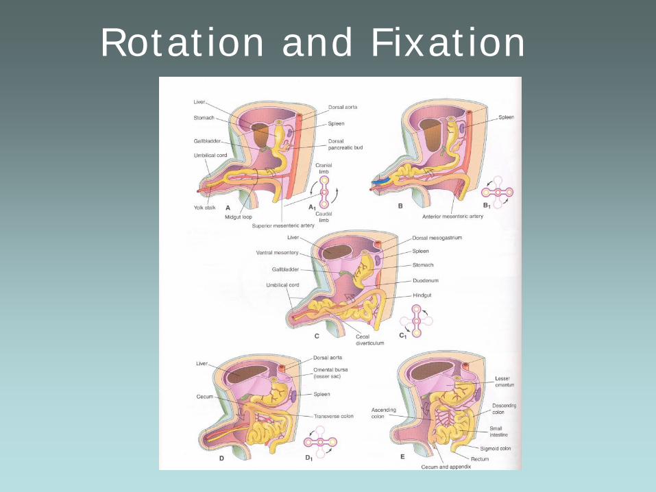
Rotation and Fixation

Disorders of rotation
• Can these be identified prenatally?

Disorders of rotation
• Generally – no! • Postnatal U/S Dx depends on:
– Abnormal relation of SMA/SMV – Spiraling of bowel with volvulus
• Only if there are prenatal complications – Paucity of fluid filled intestine, echogenic
content

Malrotation predisposes to volvulus – prenatal or postnatal
Intestinal obstruction and echogenic bowel

Abnormality in intestinal content
– “Echogenic bowel”
• Echogenicity = Brightness of fetal bowel with transducer frequency of 5MHz or less
• Term that applies in 2nd trimester only • 3 gradations
– 1 close to normal – 2 about as bright as liver – 3 as bright as bone (iliac crest)
• Incidence – estimated 0.2-2% in 2nd trimester

Echogenic bowel
• In most cases this finding is TRANSIENT and has no adverse sequelae.
• It may be associated with: – Swallowed blood/maternal bleeding – Cystic fibrosis (meconium ileus variants) – Aneuploidy (13,18, 21) – Infection (CMV, parvo) – GI obstruction, volvulus – IUGR, fetal alcohol syndrome, twin gestation

Echogenic bowel
– Association with aneuploidy
• In studies of pts at risk for aneuploidy incidence of this finding varies between 5.5% - 14%
• 75% of these pts had other suspicious findings on U/S, only 3% had this as an isolated finding
• Limited conclusions to be reached in normal risk population with this finding
• In 9067 pregnancies identified 56 pt with EB – 47 agreed to genetic counseling- 22 agreed to amnio 3 cases tri21 one case tri 18 one case cmv. 12 with adverse outcomes only 3 had EB as only finding

Echogenic bowel – Association with cystic fibrosis – About 3% will be diagnosed with CF
• 10 studies of 309 fetuses w/ echogenic bowel eleven (3.6%) had CF
• Largest series 244 cases of EB – at least 2 cases of CF = 20x expected incidence
• Incidence of CF depends on ethnic background of sample population
• Other findings which enhance dx: – polyhydramnios, ascites, calcifications, other
structural anomalies – clinical postnatal correlate – thick, inspissated
meconium
Echogenic bowel
– If echogenic bowel resolves – outcome is usually normal
– In 182 pregnancies with EB – 13% resulted in IUFD, 5% IUGR and 5% required GI surgery.
– In the majority of cases – the isolated finding of echogenic bowel will have no clinical consequence.

Newborn diagnoses related to the abdomen
• Intra-abdominal masses • Cystic and solid • Fetal tumors • Hepatobiliary anomalies
Fetal cystic abdominal masses • Differential diagnosis often difficult
– Ovarian cyst (female fetus) – Distended bladder? Urachal cyst – Choledochal cyst, liver cyst – Mesenteric cyst (rare) – Cystic kidney disease (see urology) – Adrenal cyst (neuroblastoma, hemorrhage, sequestration) – Teratoma – Neurenteric cyst (rare) – Rarely important to establish definitive dx!
• Standard: treatment after birth

Ovarian cyst – Ovary under maternal hormonal
stimulation – Cyst size may be large: >4-5 cm – Risk of torsion ?related to size? – Value of antenatal intervention?
• Needle aspiration • Risk to fetus/mother outweighs
benefit • Potential for spontaneous
resolution after birth

Ovarian cyst
Potential for spontaneous resolution
Consider aspiration postnatally if concerned for torsion based on size

Fetal abdominal masses
• Adrenal masses – Location is subdiaphragmatic – Adrenal hemorrhage – Neuroblastoma – Intra-abdominal pulmonary
sequestration • Prenatal treatment not indicated

Sacrococcygeal teratoma • These tumors can present primarily within the abdominal
cavity as cystic or solid lesions • …Or they can be exophytic lesions • …Or anywhere in between!

Sacrococcygeal teratoma – Most often benign
• May contain immature elements: evolve to malignant • Good prognosis if operated before 2 months of age
– If early prenatal diagnosis & large tumor: • Risk of hydrops and fetal demise • Risk to mother (“mirror” syndrome)
– Intervention to save mother (and fetus): • Attempt at open fetal surgery – few successful cases • Radiofrequency ablation technique- catastrophic • Only fetal operation for mother’s sake? or termination

Prenatal diagnosis of abdominal abnormalities
• Gastroschisis and Omphalocele
– Diagnosis and implications • Proximal intestinal obstruction/ Malrotation
– Diagnosis and outcomes • Echogenic bowel-
– considerations in prenatal counseling

Prenatal diagnosis of abdominal abnormalities
• screen for associated anomalies
– chromosomal disorders or potentially life-threatening structural disorders
• prepare parents for postnatal event – Counsel regarding length of gestation, mode
and site of delivery • rarely require fetal intervention

Questions?