Embed Size (px)
Citation preview

1
Visual development in babies and infants
Marko Nardini
UCL Institute of Ophthalmology
Vision
• a major function of the primate brain
• vision develops rapidly in early life and serves as a base for development of action, cognition, communication, social interactions
VISION
Spatial information Temporal change Colour
Orientation Motion Depth
RecognitionObjects
FacesVisual action
Reaching
Locomotion
Navigation
Visual cognition
Physics/causality
Social
cognition
MEMORY MOTOR CONTROL ATTENTION
Global orientation Global motion
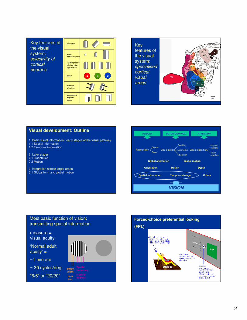
Key features of the visual system:
the retina-geniculostriate pathway
Key features of the visual system:
subcortical & cortical pathways
SC = superior colliculus
OMN = oculomotor nuclei
2
R G B
orientation
Scale/
spatial frequency
"spatial phase" =
left/right edge
light/dark bar
colour
direction
of motion
stereoscopic
disparity
(depth)
Key features of
the visual
system:
selectivity of
cortical
neurons
Key
features of
the visual
system:
specialised
cortical
visual
areas
Visual development: Outline
1. Basic visual information - early stages of the visual pathway1.1 Spatial information1.2 Temporal information
2. Later stages 2.1 Orientation2.2 Motion
3. Integration across larger areas3.1 Global form and global motion
VISION
Spatial information Temporal change Colour
Orientation Motion Depth
RecognitionObjects
FacesVisual action
Reaching
Locomotion
Navigation
Visual cognition
Physics/causality
Social
cognition
MEMORY MOTOR CONTROL ATTENTION
Global orientation Global motion
Most basic function of vision:
transmitting spatial information
measure =
visual acuity
‘Normal adult
acuity’ =
~1 min arc
~ 30 cycles/deg
“6/6” or “20/20”
Stripewidth
(minarc)
Spatial
frequency
(cycles/
degree)
Forced-choice preferential looking
(FPL)
3
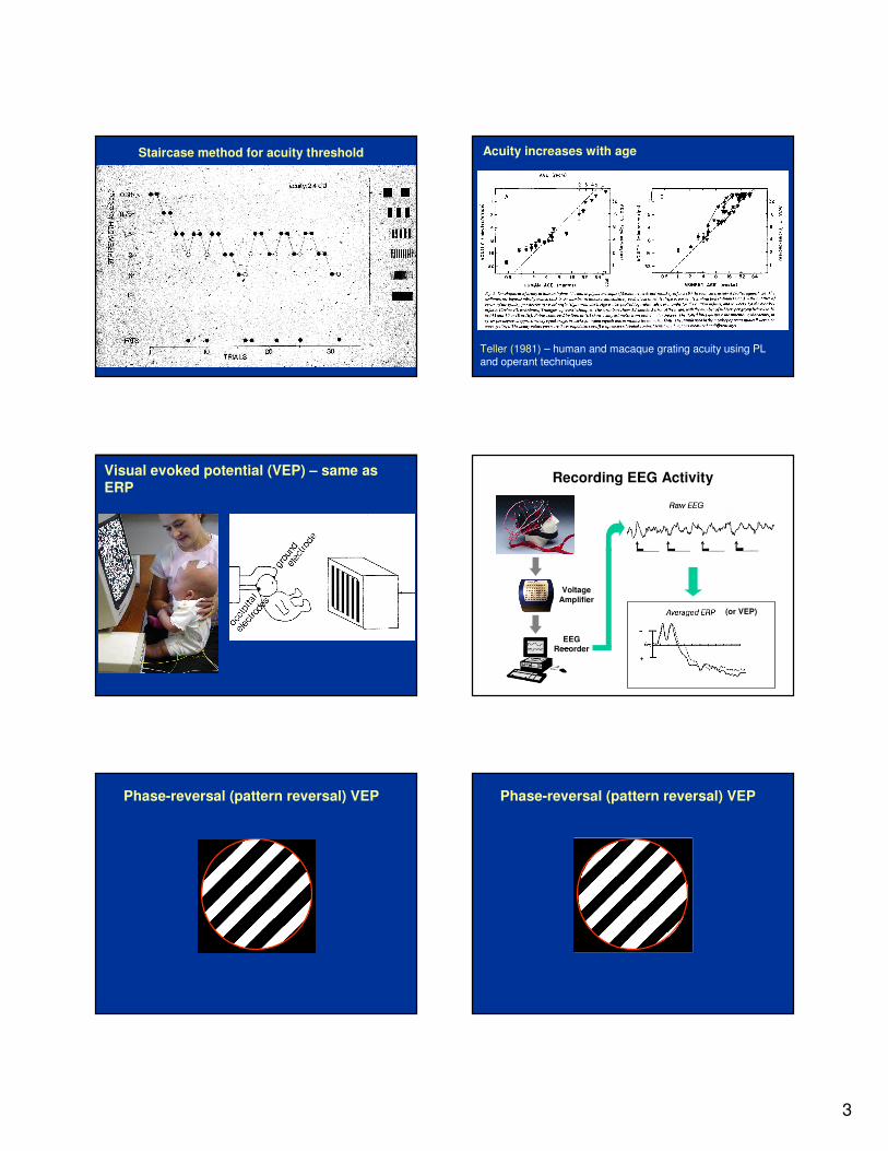
Staircase method for acuity threshold
Teller (1981) – human and macaque grating acuity using PL and operant techniques
Acuity increases with age
Visual evoked potential (VEP) – same as ERP
Recording EEG Activity
Voltage
Amplifier
EEG Recorder
Stimuli Presentations
(or VEP)
Phase-reversal (pattern reversal) VEP Phase-reversal (pattern reversal) VEP
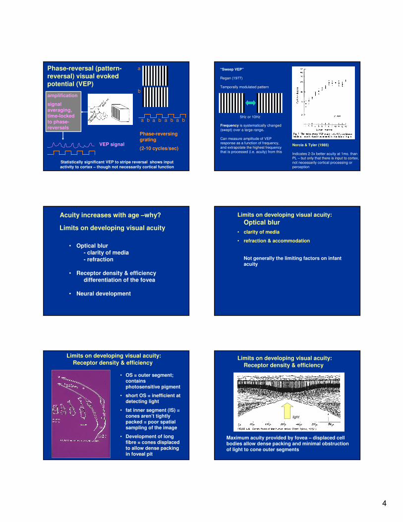
4
a
b
ba ba ba ba
Phase-reversinggrating
(2-10 cycles/sec)
amplification
signal averaging, time-locked to phase-reversals
Phase-reversal (pattern-
reversal) visual evoked potential (VEP)
VEP signal
Statistically significant VEP to stripe reversal shows input
activity to cortex – though not necessarily cortical function
“Sweep VEP”
Regan (1977)
Temporally modulated pattern
5Hz or 10Hz
Frequency is systematically changed
(swept) over a large range.
Can measure amplitude of VEP response as a function of frequency,
and extrapolate the highest frequency that is processed (i.e. acuity) from this
Norcia & Tyler (1985)
Indicates 2-3x better acuity at 1mo. than PL – but only that there is input to cortex,
not necessarily cortical processing or perception
Limits on developing visual acuity
• Optical blur
- clarity of media- refraction
• Receptor density & efficiencydifferentiation of the fovea
• Neural development
Acuity increases with age –why? Limits on developing visual acuity:
Optical blur
• clarity of media
• refraction & accommodation
Not generally the limiting factors on infant acuity
Limits on developing visual acuity:
Receptor density & efficiency
• OS = outer segment; contains photosensitive pigment
• short OS = inefficient at detecting light
• fat inner segment (IS) = cones aren’t tightly packed = poor spatial sampling of the image
• Development of long fibre = cones displaced to allow dense packing in foveal pit
Limits on developing visual acuity:
Receptor density & efficiency
Maximum acuity provided by fovea – displaced cell bodies allow dense packing and minimal obstruction of light to cone outer segments
(fovea picture)
light

5
coarse spacing (newborn) – pattern under-sampled
fine spacing (adult)– patternadequately sampled
photoreceptor density, image sampling & development of acuity
Banks & Bennett (1988)
• Sampling argument combined with poor efficiency due to short outer segments
• Calculated in comparison with adult and ‘ideal observer’ model
• May account for overall change, but
- both adult and infant fall far short of ideal observer – little idea of factors
- poor account of acuity changes during infancy, especially first 3 months
• Other, more central, changes are going on
Limits on developing visual acuity:Neural development
• myelination of visual pathways- what are functional consequences?
• development and distribution of cortical neurons
• developing connectivity in cortex (and elsewhere)
- increasingly complex dendritic and axonal processes
Development and distribution of cortical neurons
• cell proliferation
• cell migration
• cell differentiation into different structural types
All three processes are complete before birth
Although all cells are born before birth, the mass of the brain increases postnatally from 350 g –1350 g (approx x 4)
This increase must include
• myelin
• fibres and synapses associated with increased connectivity
Connectivity determines function
Total synapses =
volume
x
synapses/cm3
Synapse numbers increase, then decrease (Huttenlocher)
6
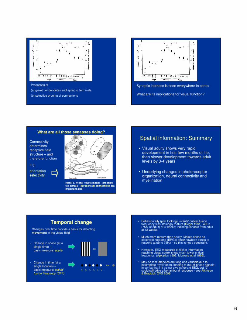
Processes of
Processes of
(a) growth of dendrites and synaptic terminals
(b) selective pruning of connections
Synaptic increase is seen everywhere in cortex.
What are its implications for visual function?
What are all those synapses doing?
+
+
+
+
+
+
–
–
–
–
–
–
l.g.n. cells cortical cell
++
+ ++
+
Connectivity determines receptive field
structure – and therefore function
e.g.
orientation
selectivity
Hubel & Wiesel 1960’s model – probably
too simple – intracortical connections are
important also!
Spatial information: Summary
• Visual acuity shows very rapid development in first few months of life, then slower development towards adult levels by 3-4 years
• Underlying changes in photoreceptor organization, neural connectivity and myelination
Temporal change
• Change in space (at a single time) –basic measure: acuity
• Change in time (at a single location) –basic measure: critical fusion frequency (CFF)
t1 t2 t3 t4 t5 t6 …
Changes over time provide a basis for detecting movement in the visual field
vs.
vs.
• Behaviourally (pref looking), infants’ critical fusion frequency was strikingly mature (Regal 1981): 40Hz (75% of adult) at 4 weeks; indistinguishable from adult at 12 weeks.
• Much more mature than acuity. Makes sense as electroretinograms (ERGs) show newborn cones to respond at up to 75Hz – so this is not a constraint.
• However, EEG measures of flicker information reaching visual cortex show much lower critical frequency. (Apkarian 1993, Morrone et al 1996).
• May be that latencies are long and variable due to incomplete myelination, leading to out-of-phase signals in cortex that (1) do not give coherent EEG, but (2) could still drive a behavioural response - see Atkinson & Braddick OVS 2009

7
Review
• We have seen how sensitivity to spatial and temporal changes in luminance (and wavelength / colour) develops in the first few months of life
• These provide the building blocks for detecting the orientation, motion and depth of visual patterns
• This requires increasingly sophisticated neural information processing – dependent on cortex (V1) VISION
Spatial information Temporal change Colour
Orientation Motion Depth
RecognitionObjects
FacesVisual action
Reaching
Locomotion
Navigation
Visual cognition
Physics/causality
Social
cognition
MEMORY MOTOR CONTROL ATTENTION
Global orientation Global motion
ORIENTATION processing
Two kinds of ‘subcortical’..
LGN is ‘precortical’– on the route to striate cortex
Superior colliculusis part of a distinct subcortical pathway (but also interconnected with cortex)
Neither LGN or SC show orientation selectivity independent of cortex
cortical neurons – and not
precortical - show selective
sensitivity to :
• orientation
• direction of motion
• binocular disparity (stereopsis)
Development of orientation selective cortical neurons – orientation-reversal VEP
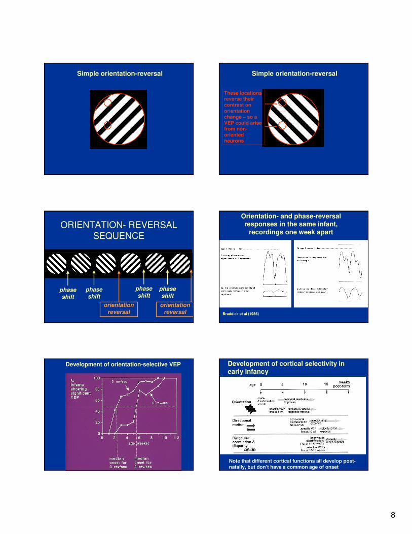
8
Simple orientation-reversal Simple orientation-reversal
These locations reverse their contrast on orientation change – so a VEP could arise from non-oriented neurons
ORIENTATION- REVERSAL SEQUENCE
phaseshift
phaseshift
phase
shiftphaseshift
orientation reversal
orientation reversal
Orientation- and phase-reversal responses in the same infant,
recordings one week apart
Braddick et al (1986)
Development of orientation-selective VEP Development of cortical selectivity in early infancy
Note that different cortical functions all develop post-natally, but don’t have a common age of onset

9
MOTION processing motion processing – a pervasive
aspect of vision
????
Volkmann & Dobson (1976) – 2 month infants preferential looking:
moving > static stimuli
Does this mean that young infants process visual motion?
Consider:
Infants also show high flicker sensitivity in preferential looking (Regal, 1981)
early stages of visual pathway (e.g. retinal ganglion cells) respond to temporal change but not to direction of motion
From flicker to direction of motion
t1 t2 t3 t4 t5 t6 …single location
on retina
is consistent with…
…
e.g. Adelson & Bergen 1985 - spatio-temporal receptive fields
To determine direction of motion, need to compare
- across retinal locations (as for resolving orientation)
- and also across time
10
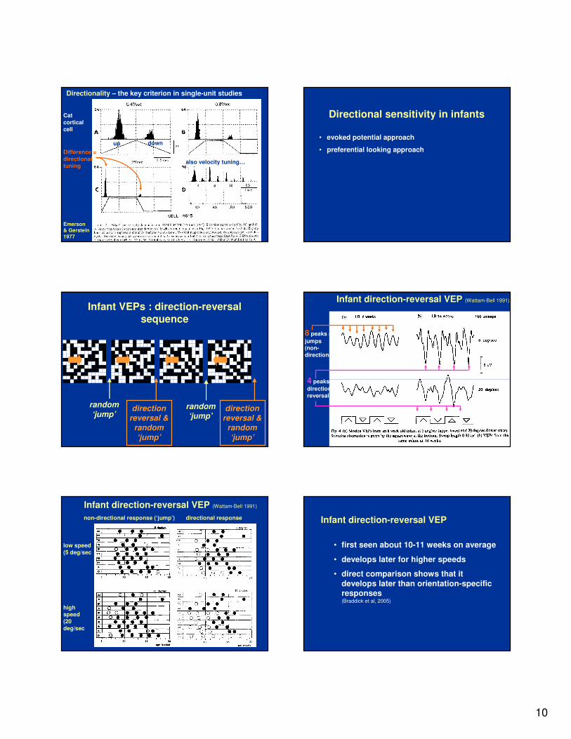
Directionality – the key criterion in single-unit studies
Cat
cortical
cell
Difference =
directional tuning
also velocity tuning…
up down
Emerson & Gerstein
1977
Directional sensitivity in infants
• evoked potential approach
• preferential looking approach
random‘jump’
direction reversal &
random
‘jump’
direction reversal &
random
‘jump’
random‘jump’
Infant VEPs : direction-reversal
sequence
Infant direction-reversal VEP (Wattam-Bell 1991)
8 peaks =
jumps(non-
directional)
4 peaks =
direction
reversals
low speed
(5 deg/sec
non-directional response (‘jump’) directional response
Infant direction-reversal VEP (Wattam-Bell 1991)
high speed
(20
deg/sec
Infant direction-reversal VEP
• first seen about 10-11 weeks on average
• develops later for higher speeds
• direct comparison shows that it
develops later than orientation-specific responses (Braddick et al, 2005)

11
Preferential looking for directional motion Preferential looking for directional motion
with age, sensitivity extends to both higher and lower speeds
Preferential looking for directional motionextension to higher velocities
t1 t2
∆x
t1 t2
∆x
• higher velocities with age - primarily a spatial rather than temporal change
• a ‘fine to coarse progression’ (not expected from acuity changes)
• extension of horizontal connectivity in cortex?
• compare with extending disparity range for stereopsis
All specific functions of primary visual cortex – but they don’t have a common onset. Specific aspects of cortical connectivity each have to develop.
Plasticity of motion processing
• kittens reared in stroboscopic illumination –absence of directional cells in visual cortex (Cynader, Berman & Hein, 1973; Pasternak et al, 1981)
• kittens reared with directional bias show biased distribution of directional selectivity in cortical cells (Daw & Wyatt, 1976)
• separate periods of directional bias and monocular deprivation – show distinct critical periods for motion sensitivity (1st) and binocularity (2nd) (Daw, Berman & Ariel, 1978)

12
•••• recognise objects recognise objects recognise objects recognise objects & events by & events by & events by & events by dynamic dynamic dynamic dynamic characteristics characteristics characteristics characteristics (e.g. biological (e.g. biological (e.g. biological (e.g. biological motion)motion)motion)motion)
•••• register register register register trajectorytrajectorytrajectorytrajectory
•••• selfselfselfself----motion motion motion motion from optic from optic from optic from optic flowflowflowflow
•••• segmentation segmentation segmentation segmentation from relative from relative from relative from relative motionmotionmotionmotion
•••• 3333----D structure D structure D structure D structure from motionfrom motionfrom motionfrom motion
uses of visual motion informationuses of visual motion informationuses of visual motion informationuses of visual motion information
…so directional information can be used for sophisticated perceptual analysis, soon after it first becomes available to the infant
Use of motion for perceptual tasks by
3-5 month-old infants
Berthenthal et al, 1985
• discriminate biological motion
Kaufman-Hayoz et al (1986)
• discriminate motion-defined forms
Kellman & Spelke(1983)
• group parts of occluded object by common motion
E J Gibson et al (1979)
• distinguish rigid motion from non-rigid deformation
Arterberry & Yonas(1988)
• 3-D structure from motion
VISION
Spatial information Temporal change Colour
Orientation Motion Depth
RecognitionObjects
FacesVisual action
Reaching
Locomotion
Navigation
Visual cognition
Physics/causality
Social
cognition
MEMORY MOTOR CONTROL ATTENTION
Global orientation Global motion
GLOBAL form and motion processing
Coherent organization Random organization
To perceive shapes, we need to look for useful changes in orientation over a large area – e.g. those indicating a contour.
Similarly with motion…
Expanding optic flow Contracting optic flow(moving forwards) (moving backwards)
The same local motions are present in both cases, but the global organization is very different
To perceive global motion we need to integrate local motions over a large area

13
macaque V4 (ventral)
response to concentric or radial configurations
(Gallant Braun & Van Essen,
Science, 1993):
macaque V5/MT (dorsal)
response to coherence level of random dot motion
(Britten et al, J Neurosci, 1992; Vis
Neurosci, 1993)
Measure of sensitivity to global form or
motion: coherence threshold
What % of elements needs to be coherently organised in order for the global organisation to be perceived?
i.e.
Lowest % at which global organisation is detected = observer’s coherence threshold
100% coherence
Coherence = 100%
Form coherence
60% coherence
coherence = 60%
Form coherence
6%6%6%6%
13%13%13%13%
50%50%50%50%
motion coherenceDevelopment of global form and
motion processing
Preferential looking and VEP measures:
• Local form emerges earlier than local motion
• Evidence for sensitivity to global motion and
global form by 4-6 months
(Braddick & Atkinson, 2007)
• Global form thresholds reach adult levels later than global motion (Gunn et al, 2002)
14
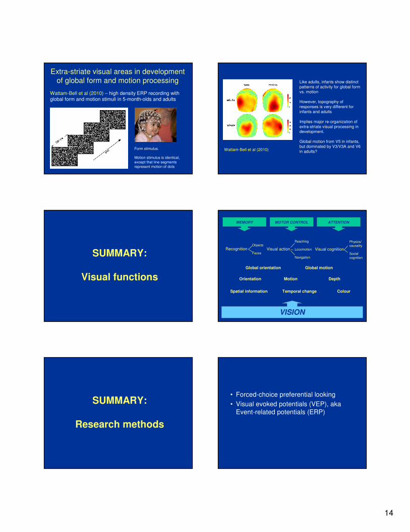
Extra-striate visual areas in development
of global form and motion processing
Wattam-Bell et al (2010) – high density ERP recording with global form and motion stimuli in 5-month-olds and adults
Form stimulus.
Motion stimulus is identical,
except that line segments
represent motion of dots
Wattam-Bell et al (2010)
Like adults, infants show distinct
patterns of activity for global form vs. motion
However, topography of
responses is very different for
infants and adults
Implies major re-organization of
extra-striate visual processing in development.
Global motion from V5 in infants,
but dominated by V3/V3A and V6
in adults?
SUMMARY:
Visual functions
VISION
Spatial information Temporal change Colour
Orientation Motion Depth
RecognitionObjects
FacesVisual action
Reaching
Locomotion
Navigation
Visual cognition
Physics/causality
Social
cognition
MEMORY MOTOR CONTROL ATTENTION
Global orientation Global motion
SUMMARY:
Research methods
• Forced-choice preferential looking
• Visual evoked potentials (VEP), aka
Event-related potentials (ERP)

15
MODEL
• Atkinson & Braddickcortical
control of eye/head
movements
BIRTH: limited orienting to
single targets
3 MO:
integration for
attention switching
visual control of
reach/grasp
5-6 MO:
integration of manual action
& near visual space
visual control of
locomotion
~12 MO:
integration of locomotoraction, attention control,
and near/far visual space
object recognition
attribute binding and
segmentation of objects
faces
DORSAL
VENTRAL
KEY
subcortical orienting
CORTICAL SELECTIVE MODULES
orientation
motion
colour
disparity
SELECTIVE
ATTENTION(local/global)
END
Reading list (p. 1 of 4)
Overview:
Atkinson, J & Braddick, O (in press). Visual development (Chapter 12). In Zelazo,
P.D. (Ed.) Oxford Handbook of Developmental Psychology. OUP
Specific studies:
Teller, DY (1981). The development of visual acuity in human and monkey infants.
Trends in Neurosciences 4: 21-24.
Regan, D (1977). Speedy assessment of visual acuity in amblyopia by the evoked potential method. Ophthalmologica 175(3): 159-64.
Norcia, AM & Tyler, CW (1985). Spatial frequency sweep VEP: Visual acuity during
the first year of life. Vision Research. 25: 1399-1408.
Banks, MS & Bennett, PJ (1988). Optical and photoreceptor immaturities limit the spatial and chromatic vision of human neonates. J Opt Soc America A, 12(5): 2059-
2079.
Reading list (p. 2 of 4)Banks MS & Salapatek P.(1978) Acuity and contrast sensitivity in 1-, 2-, and 3-month-old human infants. Invest Ophthalmol Vis Sci. 17: 361-5.
Adams RJ & Courage ML. (1998) Human newborn color vision: measurement with
chromatic stimuli varying in excitation purity. J Exp Child Psychol. 68(1): 22-34.
Regal, DM. (1981) Development of critical flicker frequency in human infants. Vision Research 21:549-555.
Apkarian, P (1993) Temporal frequency responsivity shows multiple maturational
phases: state-dependent visual evoked potential luminance flicker fusion from birth to
9 months. Vis Neurosci 10: 1007–18.
Morrone MC, Fiorentini A, Burr DC (1996) Development of the temporal properties of visual evoked potentials to luminance and colour contrast in infants. Vision Res 36:
3141–55.
Braddick O, Atkinson J (2009) Infants’ sensitivity to motion and temporal change. Optometry & Vision Science 86(6), 577–582.
Shatz CJ (1996) Emergence of order in visual system development. Journal of
Physiology-Paris 90(3-4): 141-150
Reading list (p. 3 of 4)Braddick, OJ, Atkinson, J, Julesz, B, Kropfl, W, Bodis-Wollner, I, & Raab, E. (1980).
Cortical binocularity in infants. Nature 288: 363-365.
Braddick, OJ, & Atkinson J (1983). Some recent findings on the development of human binocularity: A review. Behavioural Brain Research 10: 141-150.
Fox, R, Aslin, RN, Shea, SL, & Dumais, ST (1980). Stereopsis in human infants.
Science, 207: 323–324.
Held, R, Birch, EE, & Gwiazda J (1980). Stereoacuity of human infants. Proceedings of the National Academy of Sciences of the USA, 77: 5572-5574.
Birch, EE, Gwiazda, J, & Held, R (1982). Stereoacuity development for crossed and
uncrossed disparities in human infants. Vision Research, 22: 507-513.
Volkmann FC & Dobson, V (1976). Infant responses of ocular fixation to moving
visual stimuli. J Exp Child Psychol 22: 86-99.
Adelson EH & Bergen JR (1985). Spatiotemporal energy models for the perception of motion. J. Opt. Soc. Am. A 2(2): 284-299.

16
Reading list (p. 4 of 4)
Emerson RC, Gerstein GL (1977). Simple striate neurons in the cat. II. Mechanisms underlying directional asymmetry and directional selectivity. J Neurophysiol 40: 136-
55.
Wattam-Bell J. (1991) The development of motion-specific cortical responses in
infants. Vision Res 31:287-297.
Braddick, O, Birtles, D, Wattam-Bell, J & Atkinson, J (2005). Motion- and orientation-specific cortical responses in infancy. Vision Research 45: 3169-3179.
Braddick, O, & Atkinson, J (2007). Development of brain mechanisms for visual
global processing and object segmentation. In C. von Hofsten & K. Rosander (Eds.), From action to cognition (Progress in Brain Research, Vol. 164) Amsterdam:
Elsevier.
Gunn, A et al (2002). Dorsal and ventral stream sensitivity in normal development and hemiplegia. Neuroreport 13(6): 843-847.
Wattam-Bell, J et al (2010). Reorganization of Global Form and Motion Processing
during Human Visual Development. Current Biology 20(5): 411-415.