Embed Size (px)
Citation preview
LUND UNIVERSITY
PO Box 117221 00 Lund+46 46-222 00 00
Visualization and manipulation of microRNA in neural cells
Sachdeva, Rohit
Published: 2013-01-01
Link to publication
Citation for published version (APA):Sachdeva, R. (2013). Visualization and manipulation of microRNA in neural cells Molecular Neurogenetics,Faculty of Medicine, Lund University
General rightsCopyright and moral rights for the publications made accessible in the public portal are retained by the authorsand/or other copyright owners and it is a condition of accessing publications that users recognise and abide by thelegal requirements associated with these rights.
• Users may download and print one copy of any publication from the public portal for the purpose of privatestudy or research. • You may not further distribute the material or use it for any profit-making activity or commercial gain • You may freely distribute the URL identifying the publication in the public portalTake down policyIf you believe that this document breaches copyright please contact us providing details, and we will removeaccess to the work immediately and investigate your claim.

Download date: 03. Jun. 2018
1
Academic Dissertation
Visualization and manipulation of microRNA in neural cells
Rohit Sachdeva
2013
With approval of the Faculty of Medicine of Lund University, this thesis will be defended at 09:00 on September 6th, 2013 in Segerfalksalen,
Wallenberg Neuroscience Center, Lund, Sweden.
Faculty Opponent:
Associate Professor Eva Hedlund, PhD,Department of Neuroscience,
Karolinska Institute,Stockholm, Sweden.
2
Molecular Neurogenetics,Department of Experimental Medical Science,Lund University
Rohit Sachdeva
2013-09-06
Visualization and manipulation of microRNA in neural cells
microRNA (miRNA) are small non-coding RNA, 21-23 nucleotides long. miRNA provide a new layer of regulatory control over gene expression programs. It is increasingly evident that miRNA are cell type and tissue specific. Many miRNAs have been identified to be highly abundant in certain regions of the brain. It has also been shown that alterations in levels of specific miRNAs have implications in various neurodegen-erative disorders. However, the role of individual miRNA remains poorly understood.
There are various methods that are used to visualize miRNA, depending on type of sample, resolution and throughput. There is currently no gold standard for transcriptional profiling of miRNA and the use of in-dependent techniques to verify results is preferable. This thesis takes a look at the various techniques avail-able to look at miRNA expression profile, providing insight into advantages, disadvantages, complexities and challenges with each technique. To study the function of miRNA it is also essential to regulate its expression. The thesis highlights various methods used by researchers to downregulate or overexpress miRNA.
This thesis comprises of three papers where I have tried to evolve tools to visualize distinct miRNA profile to differentiate between cells, and manipulate miRNA expression in vitro and in vivo. In paper I, I have used miRNA-regulated vectors to differentiate between embryonic stem cells and brain specific cells. This enabled me to sort out neural cells and transplant them in a Parkinson disease mouse model. Using this method I was able to reduce the frequency of tumor formation post transplantation. [1]
It has been shown that miRNA-124 (miR-124) plays a crucial role in establishing and maintaining a neuronal transcription network. In paper II, I used a similar miRNA-regulated vector to look specifically at miR-124 expression in the brain. Here I also used tools to manipulate the expression of miR-124. I was able to demonstrate that miR-124 is a neuronal fate determinant in the subventricular zone [2]. Finally in paper III, I showed, using miR-9 regulated vectors that miR-9 is not expressed in microglia. Thereby I developed a novel system by which one can specifically target microglia. This system could be used to target genetic modifications to resident microglia in the rodent brain [3].
Overall, further understanding of biogenesis and functionality of this exceptional gene regulator will in turn enhance techniques used to study them. Hopefully, in the future this leads to opportunities to safely pursue miRNA as therapeutic strategies.
microRNA, miRNA, miR-124, miR-9, embryonic stem cells, neural stem cell, cell transplantation, neurogenesis, microglia
1652-8220
English
978-91-87449-55-0
2013-07-27
128

3
Visualization and manipulation of microRNA in neural cells
Rohit Sachdeva
2013

4
Cover art: The cover is an abstract art representation of the theme of the thesis, which is to “visualize” and “manipulate” the neural system.
To make my vision into a beautiful piece of art full credit goes to Liana Fasching.
ISSN 1652-8220ISBN 978-91-87449-55-0Lund University, Faculty of Medicine Doctoral Dissertation Series 2013:83
Printed at Exakta, Malmö, Sweden

5
To my grandparents
In the Gita, Krishna speaks to Arjuna of the importance of finding a guru:
Acquire the transcendental knowledge from a Self-realized master by humble reverence, by sincere inquiry, and by service. The wise ones who have realized
the Truth will impart the Knowledge to you.

6
7
TABLE OF CONTENTS
1) Original papers........................................................................................................................ ...............2) Abstract......................................................................................................................................................3) Layman summary...................................................................................................................................4) Svensk populärvetenskaplig sammanfattning...........................................................................5) Abbreviations ..........................................................................................................................................6) Introduction.............................................................................................................................................
6.1) Introduction to miRNA...........................................................................................................6.2) miRNA in the brain...................................................................................................................6.3) miRNA in neurological disorders.......................................................................................
7) Techniques for studying miRNA.......................................................................................................7.1) Visualization of miRNA...........................................................................................................
miRNA profiling using bulk RNA..............................................................................................Histological approach....................................................................................................................miRNA reporter vectors................................................................................................................
7.2) Manipulation of miRNA..........................................................................................................Loss of function studies of miRNAs.........................................................................................miRNA KO...........................................................................................................................................miRNA knock down.......................................................................................................................miRNA overexpression..................................................................................................................
8) Aims of the thesis...................................................................................................................................9) Results and Discussion.........................................................................................................................
Paper I:...................................................................................................................................................miRNA regulated vectors report GFP expression in neural progenitor cells........FACS sorting of progenitors followed by transplantation..............................................Conclusion...........................................................................................................................................
Paper II:..................................................................................................................................................miR-124T transgenic mice..........................................................................................................miR-124 activity in the adult mouse brain...........................................................................miR-124 activity in neurogenic niches in the adult brain..............................................Inhibition of miR-124 activity in the SVZ.............................................................................Overexpression of miR-124 in the SVZ..................................................................................Conclusion..........................................................................................................................................
Paper III:................................................................................................................................................miR-9 regulated lentiviral vectors...........................................................................................miR-9 activity in neurons in sensor transgenic mice......................................................miR-9 activity in glial cells...........................................................................................................Targeting resident microglia in vivo........................................................................................Monitoring resident microglia activation.............................................................................Conclusion..........................................................................................................................................
10) Concluding remarks and prospects.............................................................................................11) Materials and methods.....................................................................................................................
1115192327313134353939404041424242444549535353545556565758585860606061626263636771

8
Lentiviral vectors................................................................................................................................Lentivirus production......................................................................................................................Quantitative real time–PCR (qRT-PCR).....................................................................................Locked nucleic acid qRT-PCR (LNA-qRT-PCR).......................................................................Luciferase reporter assay................................................................................................................Cell culture............................................................................................................................................Animal work.........................................................................................................................................Immunochemistry..............................................................................................................................
12) References..............................................................................................................................................13) Acknowledgements............................................................................................................................14) Appendix.................................................................................................................................................
7171737474747576819195

9
1) ORIGINAL PAPERS

10

11
1) ORIGINAL PAPERS
Paper I:Sachdeva R, Jönsson ME, Nelander J, Kirkeby A, Gubentif C, Gentner B, Naldini L, Björklund A, Parmar M, Jakobsson J
Tracking differentiating neural progenitors in pluripotent cultures using miroRNA-reg-ulated lentiviral vectorsProceedings of National Academy of Sciences USA. 2010 Jun22; 107(25): 11602-7
Paper II:Åkerblom M, Sachdeva R, Barde I, Verp S, Gentner B, Trono D, Jakobsson J
MicroRNA-124 is a subventricular zone neuronal fate determinantJournal of Neuroscience. 2012 Jun 27; 32 (26): 8879-89.
Paper III:Åkerblom M*, Sachdeva R*, Quintino L*, Wettergrenn EE, Chapman KZ, Manfre G, Lindvall O, Lundberg C, Jakobsson J
Visualisation and genetic modification of resident brain microglia using lentiviral vec-tors regulated by microRNA-9Nature Communication. 2013 Apr 23;4:1770.
*These authors contributed equally.

12

13
2) ABSTRACT

14
15
2) ABSTRACT
microRNA (miRNA) are small non-coding RNA, 21-23 nucleotides long. miRNA provide a new layer of regulatory control over gene expression programs. It is increasingly evident that miRNA are cell type and tissue specific. Many miRNAs have been identified to be highly abundant in certain regions of the brain. It has also been shown that alterations in levels of specific miRNAs have implications in various neurodegenerative disorders. However, the role of individual miRNA remains poorly understood.
There are various methods that are used to visualize miRNA, depending on type of sample, resolution and throughput. There is currently no gold standard for transcriptional profiling of miRNA and the use of independent techniques to verify results is preferable. This thesis takes a look at the various techniques available to look at miRNA expression profile, providing insight into advantages, disadvantages, complexities and challenges with each technique. To study the function of miRNA it is also essential to regulate its expression. The thesis highlights various methods used by researchers to downregulate or overexpress miRNA.
This thesis comprises of three papers where I have tried to evolve tools to visualize dis-tinct miRNA profile to differentiate between cells, and manipulate miRNA expression in vitro and in vivo. In paper I, I have used miRNA-regulated vectors to differentiate between embry-onic stem cells and brain specific cells. This enabled me to sort out neural cells and trans-plant them in a Parkinson disease mouse model. Using this method I was able to reduce the frequency of tumor formation post transplantation. [1]
It has been shown that miRNA-124 (miR-124) plays a crucial role in establishing and maintaining a neuronal transcription network. In paper II, I used a similar miRNA-regulated vector to look specifically at miR-124 expression in the brain. Here I also used tools to ma-nipulate the expression of miR-124. I was able to demonstrate that miR-124 is a neuronal fate determinant in the subventricular zone [2]. Finally in paper III, I showed, using miR-9 regulated vectors that miR-9 is not expressed in microglia. Thereby I developed a novel system by which one can specifically target microglia. This system could be used to target genetic modifications to resident microglia in the rodent brain [3].
Overall, further understanding of biogenesis and functionality of this exceptional gene regulator will in turn enhance techniques used to study them. Hopefully, in the future this leads to opportunities to safely pursue miRNA as therapeutic strategies.

16

17
3) LAYMAN SUMMARY

18
19
3) LAYMAN SUMMARY
The cell, the building block of life, contains deoxyribonucleic acid (DNA), which en-codes the long-term biological information of the organism. Gene is the name given to some stretches of DNA that code for a protein. Living beings depend on proteins to perform a vast array of functions. Levels of proteins vary depending on the type of cell and environment. To orchestrate the right levels of protein in a cell, organisms have evolved a small molecule called microRNA (miRNA).
No more than a couple of decades ago, in 1993, scientists identified miRNA. Over the years, miRNA were shown to carry immense power to regulate genes and thereby the func-tion of the cell. Since its discovery, the number of miRNAs identified in humans is now above 1000. Just like proteins, the presence of a miRNA in a cell depends on the specific cell type, at a given time and particular environmental condition.
There is an increasing amount of literature describing various new miRNAs along with their features, function and mechanism, thus illustrating the importance of these key regu-lators. Many miRNAs have been identified to be highly abundant in certain regions of the brain. It has also been shown that alterations in levels of specific miRNAs have implications in various neurodegenerative disorders such as Parkinson’s and Huntington’s disease. Fur-thermore many studies demonstrate that miRNA are essential for proper neuronal develop-ment and function.
In this thesis I have presented three papers where I have tried to evolve tools to visualize distinct miRNAs within the cell, thereby being able to differentiate between cell types. Fur-thermore, I have used tools to manipulate miRNA levels in cells to look for the consequences. In the first paper I have used miRNA-based tools to differentiate between mouse embryonic stem cells and brain specific cells. Here I was able to sort out neural cells and transplant them in a Parkinson’s disease mouse model. Using this method I was able to reduce the fre-quency of tumor formation post transplantation, which is a common problem.
In the second paper I used similar tools to look specifically at miR-124 in the rodent brain. Here I also used tools to manipulate the expression of miR-124. In this paper I was able to demonstrate that miR-124 is essential for stem cells in the brain to make the decision to develop into neurons. Finally in the last paper I used miR-9 to make it possible to specifi-cally look at microglia, another type of cell in the rodent brain. These cells are known to act as the first and main form of active immune defense in the brain. This novel tool makes it possible to target exclusively microglia; therefore it could be used in studies to provide ge-netic modification to these cells in the rodent brain.
This thesis has enhanced the techniques to study miRNA. This will allow for further un-derstanding of the function of this exceptional gene regulator. Further improvement in sen-sitivity, specificity and robustness of techniques will be required to unravel the full role of miRNAs in the brain and hopefully provide the opportunity to safely use them for therapeu-tic strategies.

20

21
4) SVENSK POPULÄRVETENSKAPLIG SAMMANFATTNING

22
23
4) SVENSK POPULÄRVETENSKAPLIG SAMMANFATTNING
Cellen, livets byggsten, innehåller deoxiribonukleinsyra (DNA), som kodar för organis-mens långsiktiga biologiska information. Gen är namnet på vissa sträckor av DNA som kodar för ett protein. Levande varelser är beroende av proteiner för att kunna fungera. Protein-nivåerna varierar beroende på typ av cell och miljön runtomkring. För att kunna reglera proteinnivåerna i en cell har organismer utvecklat en liten molekyl som kallas mikroRNA (miRNA).
För inte mer än två decennier sedan, 1993, upptäckte forskare miRNA. Under årens lopp har miRNA visat sig ha enorm makt att reglera gener och därmed även cellens funktion. Sedan dess upptäckt har över 1000 olika miRNA identifierats hos människan. Precis som med proteiner beror närvaron av en miRNA i en cell på den specifika celltyp, vid en given tidpunkt och ett särskilt miljöförhållande.
Det finns en ökande mängd litteratur som beskriver olika nya miRNA tillsammans med dess egenskaper, funktioner och mekanismer, vilket illustrerar hur viktiga dessa regulatorer är. Många miRNA har visat sig vara rikligt förekommande i vissa regioner av hjärnan. Det har också visat sig att förändringar i nivåer av specifika miRNA får konsekvenser i form av olika neurodegenerativa sjukdomar såsom Parkinsons och Huntingtons sjukdom. Dessutom visar många studier att miRNA är viktiga för korrekt neural utveckling och funktion.
I denna avhandling presenterar jag tre studier där jag har försökt att utveckla verk-tyg för att visualisera olika miRNA i cellen och därmed kunna skilja mellan olika celltyper. Dessutom har jag använt verktyg för att manipulera miRNA-nivåer i cellen för att titta på konsekvenserna. I den första studien har jag använt miRNA-baserade verktyg för att skilja mellan embryonala stamceller från mus och hjärnspecifika celler. Här kunde jag sortera ut neurala celler och transplantera dem i en musmodell för Parkinsons sjukdom. Med denna metod kunde jag minska frekvensen av tumörbildning efter transplantation, något som är ett vanligt förekommande problem.
I den andra studien använde jag liknande verktyg för att titta specifikt på miR-124 i gna-garens hjärna. Här använde jag också verktyg för att manipulera uttrycket av miR-124. I den-na studie kunde jag visa att miR-124 är nödvändig för stamceller i hjärnan för att kunna ut-vecklas till nervceller. Slutligen i den sista studien använde jag miR-9 för att göra det möjligt att specifikt titta på mikroglia, en annan typ av cell i gnagarens hjärna. Dessa celler är kända för att fungera som den första och viktigaste formen av aktivt immunförsvar i hjärnan. Dessa nya verktyg gör det möjligt att titta uteslutande på mikroglia. Därmed kan det användas i studier för att genetiskt modifiera dessa celler i gnagarens hjärna.
Denna avhandling har förbättrat teknikerna för att studera miRNA. Detta möjliggör för ytterligare förståelse av funktionen hos den exceptionella genregulatorn miRNA. Ytterligare förbättringar i känslighet, specificitet och robusthet av dessa tekniker krävs för att avslöja miRNAs roll i hjärnan och förhoppningsvis ge möjlighet att på ett säkert sätt använda dem för terapeutiska strategier.

24

25
5) ABBREVIATIONS

26
27
5) ABBREVIATIONS
AMO anti miRNA oligonucleotide CNS central nervous system ES cells embryonic stem cells FACS fluorescent associated cell sorting GFP green fluorescent protein iPS cells induced pluripotent stem cells ISH in situ hybridizationLNA locked nucleic acid miRNA microRNA mRNA messenger RNA NSC neural stem cell OB olfactory bulb pre-miRNA precursor miRNApri-miRNA primary miRNARISC RNA induced silencing complex RMS rostral migratory stream SVZ subventricular zone UTR untranslated region

28

29
6) INTRODUCTION

30

31
6) INTRODUCTION
6.1) Introduction to miRNA
microRNA (miRNA) are small non-coding RNA, 21-23 nucleotides long, transcribed from both introns, exons and at multiple places throughout the genome [4]. They are involved in a wide range of biological events leading to inhibition or degradation of the complimentary messenger RNA (mRNA). Founding members of miRNA family lin-4 and let-7 were identi-fied while studying genes that control the timing of larval development in Caenorhabditis elegance (C. elegance) [5-7]. The subsequent discovery of let-7 in other organisms including mammals, resembling similar temporal expression as in C. elegance, suggested that let-7 and possibly other small noncoding RNAs could be playing an important role [8]. This was quickly followed up by various publications which showed that lin-4 and let-7 were part of a large family of small non-coding RNAs found in the various multicellular organisms, of which some expressed temporally whereas most did not [9-11].
For a long time miRNAs were considered to be present only in multicellular organisms. The hypothesis at that time was that miRNA could be essential for a more complex organism design. However in 2007, miRNAs were identified in the unicellular alge Chlamydomonas re-inhardtii indicating that miRNA are evolutionarily older [12]. It is now known that miRNA are evolutionary conserved and are present in the most eukaryotic organisms. Since the discov-ery of miRNA in 1993, the number of miRNA identified in humans is now above 1000 (miR-Base; http://microrna.sanger.ac.uk). It is estimated that miRNAs may regulate over 60% of transcripts in humans [13]. These numbers keep increasing day by day as high throughput sequencing techniques continue to be applied to miRNA discovery and validation.
Most mammalian miRNAs are encoded in introns [14]. An illustration of miRNA biogen-esis is presented in Figure 1. Majority of the miRNA genes are transcribed by RNA polymer-ase II, while miRNA lying within Alu repetitive elements can be transcribed by polymerase III [15]. The polymerase generates a stem loop that contains the primary miRNA (pri-miR-NA) sequence [16, 17]. This stem loop can range in size from hundreds of nucleotides to a few kilobases. The pri-miRNA is further processed by a protein complex, which includes two main components; the RNase II enzyme Drosha, and the double stranded RNA binding domain (dsRBD) protein DiGeorge syndrome critical region gene 8 (DGCR8) [18-22]. The protein complex measures the distance from the single/double strand RNA junction and it then cleaves the pri-miRNA stem loop, resulting in a shorter 70-nucleotide hairpin precur-sor miRNA (pre-miRNA) [23].
Exprotin-5 recognizes the pre-miRNA and thereafter transports it into the cytoplasm [24-26]. The pre-miRNA is cleaved by the protein complex which includes RNAse III enzyme Dicer, that interacts with the dsRBD proteins TRBP (the HIV transactivating response RNA-binding protein) and PACT (protein activator of the interferon induced protein kinase), to form a mature 22 nucleotide miRNA:miRNA* duplex [27-32].
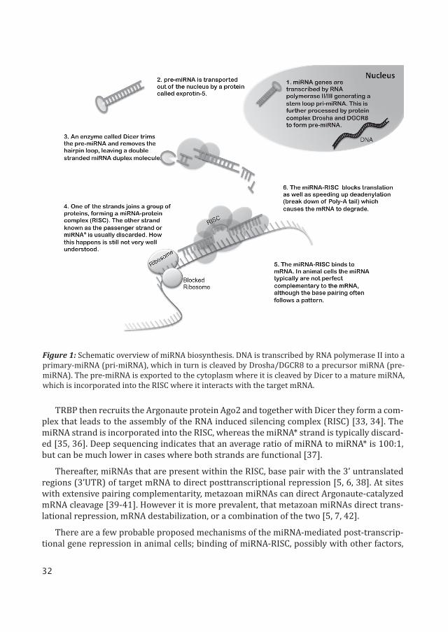
32
TRBP then recruits the Argonaute protein Ago2 and together with Dicer they form a com-plex that leads to the assembly of the RNA induced silencing complex (RISC) [33, 34]. The miRNA strand is incorporated into the RISC, whereas the miRNA* strand is typically discard-ed [35, 36]. Deep sequencing indicates that an average ratio of miRNA to miRNA* is 100:1, but can be much lower in cases where both strands are functional [37].
Thereafter, miRNAs that are present within the RISC, base pair with the 3’ untranslated regions (3’UTR) of target mRNA to direct posttranscriptional repression [5, 6, 38]. At sites with extensive pairing complementarity, metazoan miRNAs can direct Argonaute-catalyzed mRNA cleavage [39-41]. However it is more prevalent, that metazoan miRNAs direct trans-lational repression, mRNA destabilization, or a combination of the two [5, 7, 42].
There are a few probable proposed mechanisms of the miRNA-mediated post-transcrip-tional gene repression in animal cells; binding of miRNA-RISC, possibly with other factors,
Figure 1: Schematic overview of miRNA biosynthesis. DNA is transcribed by RNA polymerase II into a primary-miRNA (pri-miRNA), which in turn is cleaved by Drosha/DGCR8 to a precursor miRNA (pre-miRNA). The pre-miRNA is exported to the cytoplasm where it is cleaved by Dicer to a mature miRNA, which is incorporated into the RISC where it interacts with the target mRNA.
33
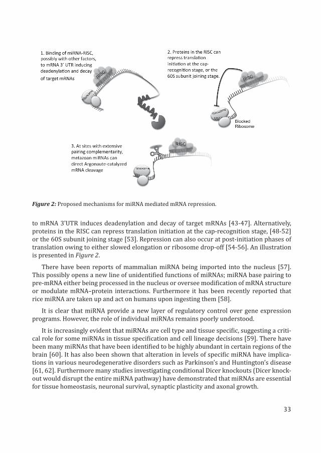
to mRNA 3’UTR induces deadenylation and decay of target mRNAs [43-47]. Alternatively, proteins in the RISC can repress translation initiation at the cap-recognition stage, [48-52] or the 60S subunit joining stage [53]. Repression can also occur at post-initiation phases of translation owing to either slowed elongation or ribosome drop-off [54-56]. An illustration is presented in Figure 2.
There have been reports of mammalian miRNA being imported into the nucleus [57]. This possibly opens a new line of unidentified functions of miRNAs; miRNA base pairing to pre-mRNA either being processed in the nucleus or oversee modification of mRNA structure or modulate mRNA–protein interactions. Furthermore it has been recently reported that rice miRNA are taken up and act on humans upon ingesting them [58].
It is clear that miRNA provide a new layer of regulatory control over gene expression programs. However, the role of individual miRNAs remains poorly understood.
It is increasingly evident that miRNAs are cell type and tissue specific, suggesting a criti-cal role for some miRNAs in tissue specification and cell lineage decisions [59]. There have been many miRNAs that have been identified to be highly abundant in certain regions of the brain [60]. It has also been shown that alteration in levels of specific miRNA have implica-tions in various neurodegenerative disorders such as Parkinson’s and Huntington’s disease [61, 62]. Furthermore many studies investigating conditional Dicer knockouts (Dicer knock-out would disrupt the entire miRNA pathway) have demonstrated that miRNAs are essential for tissue homeostasis, neuronal survival, synaptic plasticity and axonal growth.
Figure 2: Proposed mechanisms for miRNA mediated mRNA repression.
34
This thesis comprises of three papers where I have tried to evolve tools to visualize dis-tinct miRNA profile to differentiate between cells, and manipulate miRNA expression in vitro and in vivo. In the first paper we have used miRNA regulated vectors to differentiate between embryonic stem cells (ES cells) and brain specific cells, thereby making it possible to sort out these cells and transplant them in a Parkinson’s disease mouse model [1]. Using this method we were able to reduce the frequency of tumor formation post transplantation. In the second paper we used similar tools to look specifically at miRNA-124 (miR-124) expres-sion in the brain [2]. Here we also used tools to manipulate the expression of miR-124. We were able to demonstrate that miR-124 is a neuronal fate determinant in the subventricular zone (SVZ). Finally in the last paper we showed that miR-9 regulated vectors could be used to target genetic modifications to resident microglia in the rodent brain [3].
6.2) miRNA in the brain
Expression of many miRNAs is dynamically regulated during brain development, neuro-genesis, and neural maturation [63]. There is a large body of evidence to support a key role for miRNAs in these processes [64, 65]. Disruption of the function of Dicer, leads to loss of all miRNAs, during brain development resulting in gross anatomical changes and in some cases it is embryonically lethal [66-69]. In vitro, neural stem cells (NSCs) can be cultured in the absence of Dicer but are unable to generate neurons or astrocytes upon differentiation [70].
Several specific miRNAs have been implicated in the neuronal differentiation and main-tenance of neuronal phenotype. Some examples would be; miR-9, miR-124, miR-137, miR-184, and let-7 in NSC proliferation and differentiation, miR-125b and miR-128 in neuronal differentiation and maturation, and miR-132, miR-134, and miR-138 in dendritic spine mor-phogenesis. [71-82]
miR-124 is perhaps the most highly expressed brain specific miRNA [59]. It is highly ex-pressed in neurons and is known to promote neuronal identity [67, 83-87]. Knock out (KO) mouse for miR-124-1 display several major developmental phenotypes, however compensa-tion by mir-124-2 and miR-124-3 influence the phenotype of the miR-124-1 KO mouse [80]. miR-124 is not expressed in NSCs but is suggested to regulate the temporal progression of neurogenesis in the SVZ. It is upregulated in the transition between type-C and type-A cells and is further upregulated as the neuroblasts exit the cell cycle [64].
A variety of studies have shown numerous miR-124 target genes. miR-124 may act by silencing Repressor Element 1 Silencing Transcription factor (REST), a master regulator of neuronal phenotype, by switching on neuron specific silencing by targeting SCP1/Ctdsp1 and PTBP-1 [86, 88-91]. Other interesting miR-124 targets are Jagged1 in the Notch signal-ing pathway, a cascade controlling neurogenesis, and Sox9 which controls adult neurogen-esis and is a target in NSCs [64, 83, 84, 92, 93].
It is clear that miR-124 plays a crucial role in establishing and maintaining a neuronal transcription network. In this thesis, as part of paper II, we have tried to analyze the role of miR-124 in neural stem cells in vivo in both loss of function and gain of function experiments.

35
miR-9 was found to have an important role in migration and proliferation of NSCs [94-96]. Generation of a miR-9-2 and miR-9-3 double KO mouse resulted in major phenotypic brain defects. Cortical layers and ventral zone were reduced, lateral ventricles expanded, the proliferative zones hyperplastic and differentiated structures reduced. In addition, mice suffered from growth retardation and died within one week, demonstrating the importance for miR-9 in neurogenesis [97, 98].
Just as miR-124, miR-9 has reciprocal actions with REST. REST is shown to inhibit miR-9-2; REST and cAMP response element-binding protein (CREB) inactivation triggers miR-9-2 activation [99]. Other miR-9 targets include; Stathmin that increases microtubule instability, Tlx that regulates stem cell fate, also Hairy1 that has been suggested to mediate neuronal proliferation [94-96].
In this thesis as part of paper III we have used the fact that miR-9 is not expressed in mi-croglia to develop a system by which we can specifically target microglia.
6.3) miRNA in neurological disorders
Several miRNAs have been implicated in neurological disorders. miR-125 and miR-132 have been linked to several intellectual disability syndromes such as fragile X syndrome, Rett syndrome and Down syndrome [100]. miR-133b is significantly downregulated in midbrain tissue from patients with Parkinson’s disease [61]. miR-7 reduces alpha-synuclein protein, the major component of Lewy bodies in sporadic Parkinson’s disease [101, 102]. miR-107 malfunction in Alzheimer’s disease [103].
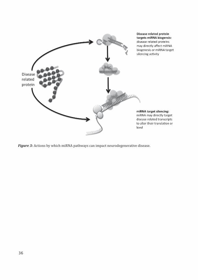
There are a couple of ways by which miRNA pathways can impact neurodegenerative dis-ease. miRNAs can be altered in disease, and may directly target disease related transcripts to alter their translation or level. Evidence also suggests that some disease related proteins such as Huntingtin (Huntington’s disease) and TDP-43 (Amyotrophic lateral sclerosis), may directly affect miRNA biogenesis or miRNA target silencing activity. An illustration of this is presented in Figure 3.
A CAG repeat expansion in the human Huntingtin (Htt) protein causes Huntington’s dis-ease. The Htt protein is shown to interact with Ago2 in the cellular P-bodies, where mRNA is degraded [104]. Downregulation in Htt protein levels impairs miRNA target silencing and it is shown that many miRNAs are misregulated [104-107]. miR- 9 is one of the miRNAs that is shown to be misregulated in Huntington’s disease patients [109]. It is possible that there are several miRNA pathways that impact Huntington’s disease, and they could be interlinked.
Amyotrophic lateral sclerosis is associated with misregulation in TDP-43, caused by a mutation and mislocation in the protein [110, 111]. TDP-43 has been shown to interact with miRNA biogenesis proteins Drosha (nuclear) and Dicer (cytoplasmic) [112].
36
Figure 3: Actions by which miRNA pathways can impact neurodegenerative disease.

37
7) TECHNIQUES FOR STUDYING miRNA

38
39
7) TECHNIQUES FOR STUDYING miRNA
7.1) Visualization of miRNA
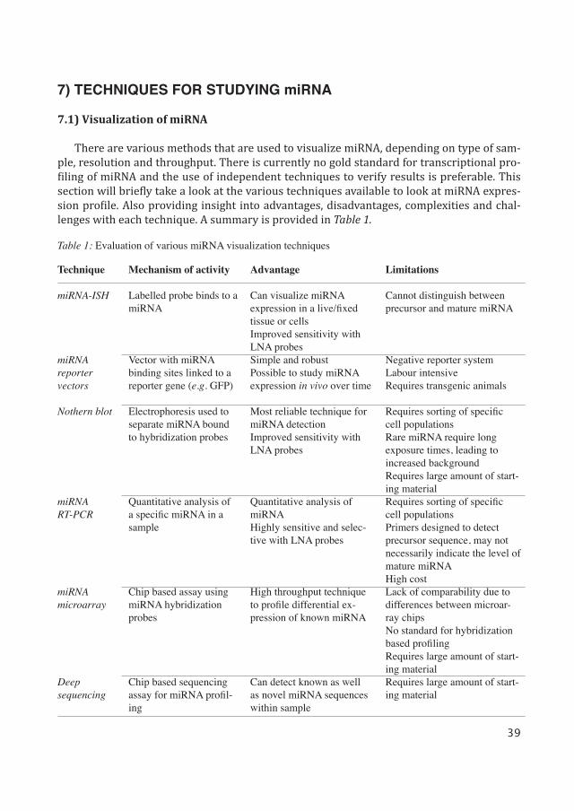
There are various methods that are used to visualize miRNA, depending on type of sam-ple, resolution and throughput. There is currently no gold standard for transcriptional pro-filing of miRNA and the use of independent techniques to verify results is preferable. This section will briefly take a look at the various techniques available to look at miRNA expres-sion profile. Also providing insight into advantages, disadvantages, complexities and chal-lenges with each technique. A summary is provided in Table 1.
Table 1: Evaluation of various miRNA visualization techniques
Technique
miRNA-ISH
miRNA reporter vectors
Nothern blot
miRNA RT-PCR
miRNA microarray
Deepsequencing
Mechanism of activity
Labelled probe binds to a miRNA
Vector with miRNA binding sites linked to a reporter gene (e.g. GFP)
Electrophoresis used to separate miRNA bound to hybridization probes
Quantitative analysis of a specific miRNA in a sample
Chip based assay using miRNA hybridization probes
Chip based sequencing assay for miRNA profil-ing
Advantage
Can visualize miRNA expression in a live/fixed tissue or cellsImproved sensitivity with LNA probesSimple and robustPossible to study miRNA expression in vivo over time
Most reliable technique for miRNA detectionImproved sensitivity with LNA probes
Quantitative analysis of miRNAHighly sensitive and selec-tive with LNA probes
High throughput technique to profile differential ex-pression of known miRNA
Can detect known as well as novel miRNA sequences within sample
Limitations
Cannot distinguish between precursor and mature miRNA
Negative reporter systemLabour intensiveRequires transgenic animals
Requires sorting of specific cell populationsRare miRNA require long exposure times, leading to increased backgroundRequires large amount of start-ing materialRequires sorting of specific cell populationsPrimers designed to detect precursor sequence, may not necessarily indicate the level of mature miRNAHigh costLack of comparability due to differences between microar-ray chipsNo standard for hybridization based profilingRequires large amount of start-ing materialRequires large amount of start-ing material
40
miRNA profiling using bulk RNA
The most widely used method to profile miRNA is northern blot analysis combined with polyacrylamide gels to examine the expression of both the mature and precursor miRNA [9]. This method allows quantification of the expression as well as miRNA size determination [6, 11, 113]. However a major drawback of this method is its poor sensitivity, especially when monitoring expression of low abundant miRNA. Moreover a large amount of total RNA per sample is required for northern blot analysis, which is not feasible when cell or tissue source is limited. An advancement in this technique has been to use locked nucleic acid (LNA) modi-fied probes. This has provided improved sensitivity and high specificity [114].
LNA based primers can also be used to quantify mature miRNA expression using qRT-PCR. This technique has been used extensively in papers II and III. It is also possible to quan-tify expression of precursor miRNA using primers designed to target the stem loop sequence [115].
Recently it has become possible to look at miRNA expression at a global level using miRNA-arrays, PCR-array, deep sequencing of small RNAs or other more specialized plat-forms. These approaches have revealed the complexity of miRNA expression patterns among different cell types and have allowed identification of a number of candidate miRNAs that appear to be enriched in specific populations of cells. However, the technical difficulties of purifying specific populations of cells from in vivo material, using for example fluorescence activated cell sorting, make these approaches problematic to transfer to the in vivo setting.
Histological approach
To analyze miRNA expression in tissue samples, histological approaches have been used. These have greatly relied on in situ hybridization (ISH) techniques. However standard ISH protocols cannot be used to detect miRNA. Additional fixation steps of the miRNA are needed and probe hybridization must be optimal [116]. As with northern blot and qRT-PCR techniques the advent of LNA modified oligonucleotides has allowed the use of LNA in situ hybridization probes to bind to the miRNA. This increases the melting temperature, thereby resulting in stringent hybridization conditions, which in turn increases specificity and sensi-tivity [117, 118]. miRNA-ISH using LNA probes has been used in paper II to look at miR-124 expression in the SVZ in adult mouse tissue.
However like all other techniques, miRNA-ISH has its drawbacks. Firstly, discriminat-ing between precursor and mature miRNA is difficult when using ISH. To do so, additional probes that target all the various precursor transcripts need to be used [119]. This can be a technical challenge when analyzing miRNAs with multiple precursor transcripts, such as miR-9 or miR-124. Furthermore, the results from this method are of limited resolution, thereby making it difficult to distinguish between two adjacent cells. In addition, due to the stringent treatment of the tissue, miRNA-ISH is also problematic to use in combination with other routine labeling techniques.
41
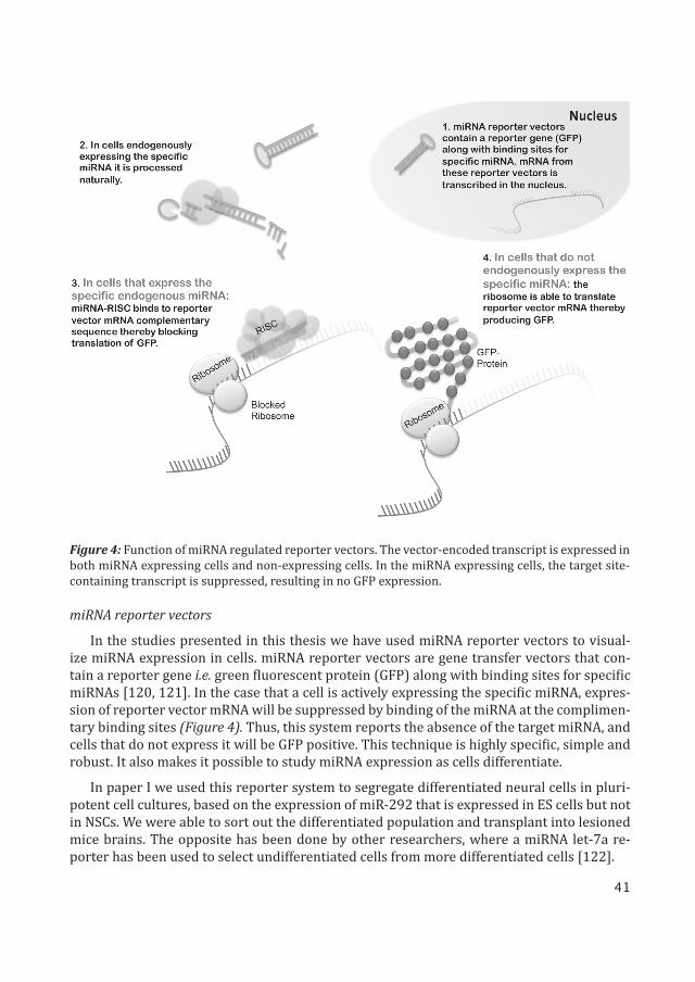
miRNA reporter vectors
In the studies presented in this thesis we have used miRNA reporter vectors to visual-ize miRNA expression in cells. miRNA reporter vectors are gene transfer vectors that con-tain a reporter gene i.e. green fluorescent protein (GFP) along with binding sites for specific miRNAs [120, 121]. In the case that a cell is actively expressing the specific miRNA, expres-sion of reporter vector mRNA will be suppressed by binding of the miRNA at the complimen-tary binding sites (Figure 4). Thus, this system reports the absence of the target miRNA, and cells that do not express it will be GFP positive. This technique is highly specific, simple and robust. It also makes it possible to study miRNA expression as cells differentiate.
In paper I we used this reporter system to segregate differentiated neural cells in pluri-potent cell cultures, based on the expression of miR-292 that is expressed in ES cells but not in NSCs. We were able to sort out the differentiated population and transplant into lesioned mice brains. The opposite has been done by other researchers, where a miRNA let-7a re-porter has been used to select undifferentiated cells from more differentiated cells [122].
Figure 4: Function of miRNA regulated reporter vectors. The vector-encoded transcript is expressed in both miRNA expressing cells and non-expressing cells. In the miRNA expressing cells, the target site-containing transcript is suppressed, resulting in no GFP expression.
42
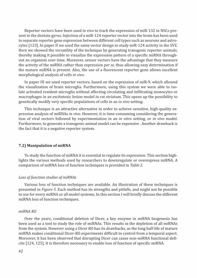
Reporter vectors have been used in vivo to track the expression of miR-132 in NSCs pre-sent in the dentate gyrus. Injection of a miR-124 reporter vector into the brain has been used to separate reporter gene expression between different cell types such as neurons and astro-cytes [123]. In paper II we used the same vector design to study miR-124 activity in the SVZ. Here we showed the versatility of the technique by generating transgenic reporter animals, thereby making it possible to visualize the expression pattern of a specific miRNA through-out an organism over time. Moreover, sensor vectors have the advantage that they measure the activity of the miRNA rather than expression per se, thus allowing easy determination if the mature miRNA is present. Also, the use of a fluorescent reporter gene allows excellent morphological analysis of cells in vivo.
In paper III we used reporter vectors, based on the expression of miR-9, which allowed the visualization of brain microglia. Furthermore, using this system we were able to iso-late activated resident microglia without affecting circulating and infiltrating monocytes or macrophages in an excitotoxic lesion model in rat striatum. This opens up the possibility to genetically modify very specific populations of cells in an in vivo setting.
This technique is an attractive alternative in order to achieve sensitive, high-quality ex-pression analysis of miRNAs in vivo. However, it is time-consuming considering the genera-tion of viral vectors followed by experimentation in an in vitro setting, or in vivo model. Furthermore, to generate a transgenic animal model can be expensive . Another drawback is the fact that it is a negative reporter system.
7.2) Manipulation of miRNA
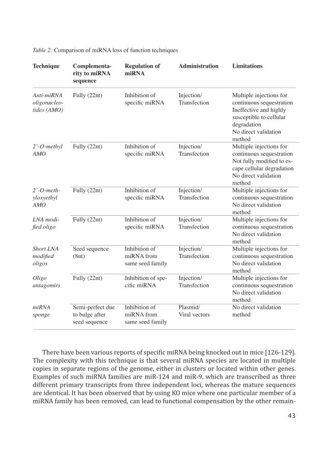
To study the function of miRNA it is essential to regulate its expression. This section high-lights the various methods used by researchers to downregulate or overexpress miRNA. A comparison of miRNA loss of function techniques is provided in Table 2.
Loss of function studies of miRNAs
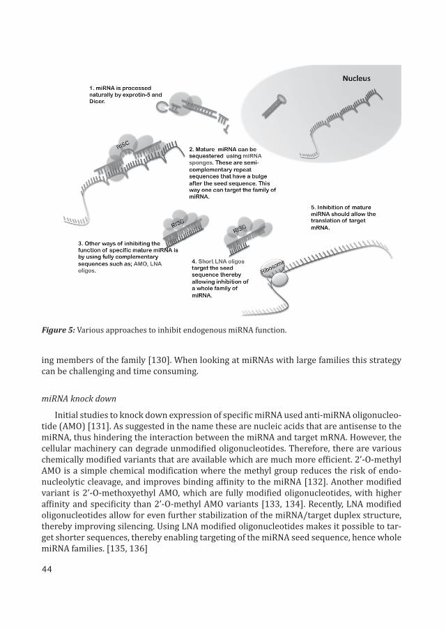
Various loss of function techniques are available. An illustration of these techniques is presented in Figure 5. Each method has its strengths and pitfalls, and might not be possible to use for every miRNA or all model systems. In this section I will briefly discuss the different miRNA loss of function techniques.
miRNA KO
Over the years, conditional deletion of Dicer, a key enzyme in miRNA biogenesis has been used as a tool to study the role of miRNAs. This results in the depletion of all miRNAs from the system. However using a Dicer KO has its drawbacks, as the long half-life of mature miRNA makes conditional Dicer-KO experiments difficult to control from a temporal aspect. Moreover, it has been observed that disrupting Dicer can cause non-miRNA functional defi-cits [124, 125]. It is therefore necessary to enable loss of function of specific miRNA.
43
Technique
Anti-miRNA oligonucleo-tides (AMO)
2’-O-methyl AMO
2’-O-meth-yloxyethyl AMO
LNA modi-fied oligo
Short LNA modified oligos
Oligo antagomirs
miRNA sponge
Complementa-rity to miRNA sequence
Fully (22nt)
Fully (22nt)
Fully (22nt)
Fully (22nt)
Seed sequence (8nt)
Fully (22nt)
Semi-perfect due to bulge after seed sequence
Regulation of miRNA
Inhibition of specific miRNA
Inhibition of specific miRNA
Inhibition of specific miRNA
Inhibition of specific miRNA
Inhibition of miRNA from same seed family
Inhibition of spe-cific miRNA
Inhibition of miRNA from same seed family
Administration
Injection/Transfection
Injection/Transfection
Injection/Transfection
Injection/Transfection
Injection/Transfection
Injection/Transfection
Plasmid/Viral vectors
Limitations
Multiple injections for continuous sequestrationIneffective and highly susceptible to cellular degradationNo direct validation methodMultiple injections for continuous sequestrationNot fully modified to es-cape cellular degradationNo direct validation methodMultiple injections for continuous sequestrationNo direct validation methodMultiple injections for continuous sequestrationNo direct validation methodMultiple injections for continuous sequestrationNo direct validation methodMultiple injections for continuous sequestrationNo direct validation methodNo direct validation method
Table 2: Comparison of miRNA loss of function techniques
There have been various reports of specific miRNA being knocked out in mice [126-129]. The complexity with this technique is that several miRNA species are located in multiple copies in separate regions of the genome, either in clusters or located within other genes. Examples of such miRNA families are miR-124 and miR-9, which are transcribed as three different primary transcripts from three independent loci, whereas the mature sequences are identical. It has been observed that by using KO mice where one particular member of a miRNA family has been removed, can lead to functional compensation by the other remain-
44
ing members of the family [130]. When looking at miRNAs with large families this strategy can be challenging and time consuming.
miRNA knock down
Initial studies to knock down expression of specific miRNA used anti-miRNA oligonucleo-tide (AMO) [131]. As suggested in the name these are nucleic acids that are antisense to the miRNA, thus hindering the interaction between the miRNA and target mRNA. However, the cellular machinery can degrade unmodified oligonucleotides. Therefore, there are various chemically modified variants that are available which are much more efficient. 2’-O-methyl AMO is a simple chemical modification where the methyl group reduces the risk of endo-nucleolytic cleavage, and improves binding affinity to the miRNA [132]. Another modified variant is 2’-O-methoxyethyl AMO, which are fully modified oligonucleotides, with higher affinity and specificity than 2’-O-methyl AMO variants [133, 134]. Recently, LNA modified oligonucleotides allow for even further stabilization of the miRNA/target duplex structure, thereby improving silencing. Using LNA modified oligonucleotides makes it possible to tar-get shorter sequences, thereby enabling targeting of the miRNA seed sequence, hence whole miRNA families. [135, 136]
Figure 5: Various approaches to inhibit endogenous miRNA function.
45
Despite the successful knockdown of miRNA in vitro and in vivo using these AMO’s, it has several limitations. Firstly, it is not possible to measure the down regulation of the miRNA as the AMO binds to the miRNA and sequesters it but does not degrade it. [137, 138] The only possible way to confirm the decrease of miRNA is by using indirect methods. Here one can analyze up-regulation of endogenous miRNA target genes. Secondly it is not possible to identify the cells in which the AMO’s are active, as they do not carry a reporter. Finally, the level of AMO’s has to be kept constant to allow a continuous sequestration of the miRNA.
Oligonucleotide antagomirs are chemically modified; they are cholesterol conjugated single stranded RNA analogues complementary to a target miRNA [138]. The modification includes a partial phosphorothioate backbone in addition to 2’-O-methoxyethyl to inhibit Ago2-mediated cleavage. In vivo, antagomirs have been given through intravenous injection where they appear to efficiently target miRNA in various tissues. However, antagomirs do not cross the blood brain barrier, which means that in the brain, antagomirs have the same limitations as AMO’s have.
miRNA sponges are transcripts expressed from strong promoters, containing multiple, tandem binding sites to a selected member of the miRNA seed family [139]. We have used miRNA sponge vectors against miR-124 in paper II. The binding site is imperfect, containing a bulge, to prevent RNA interference cleavage and degradation of the sponge RNA through endonucleolytic cleavage by Ago2. The main advantage of miRNA sponges is the possibility to achieve stable expression from integrated transgenes in vivo [140]. This can be used to study long-term effects of miRNA loss of function and also allows for stably expressing cell lines to be generated. The use of vector-mediated delivery also enables the incorporation of a reporter gene in order to identify the modified cells. Another advantage is that sponges complementarily bind to the seed sequence of the miRNA, which means that one sponge can target an entire family of miRNAs. In summary, these features make sponge vectors an attractive approach to study miRNA function in vivo in NSCs. As with the use of AMO’s, the sponge vectors have limitations, including the difficulty of validating the down regulation of a specific miRNA and the only possible way to confirm the decrease of miRNA is to use indirect methods.
Taken together, it is evident that it is technically challenging to perform loss of function studies of miRNAs. The problem of validating the inhibition, the use of transient systems, to-gether with the appearance of potential off target effects makes the interpretation of several studies challenging.
miRNA overexpression
Overexpression studies of miRNAs are quite simple to perform. Direct delivery of miRNA-duplexes or the use of various plasmid based approaches and viral vectors have been ef-fectively used to overexpress miRNA. There are several relatively simple designs of vectors enabling stable expression that all appear to work efficiently [141].
However miRNA overexpression experiments should be looked at with a degree of skep-ticism. This is because overexpression studies have a potential to generate false positive

46
results due to the supraphysiological increase in miRNA levels that are generally achieved. Such exaggerated miRNA overexpression can potentially saturate RISC complexes and dis-place other endogenous miRNAs. Another consideration is that miRNA overexpression stud-ies are usually performed in a cell environment that is artificial to the chosen miRNA, in which case cell specific natural targets may be missed and other superficial targets detected. These problems of overexpression can be avoided by combing overexpression studies with loss of function studies.
We have used miRNA overexpression vectors for miR-124 in paper II. The fact that gain of function studies are easier to perform than loss of function studies is reflected in the litera-ture and a lot of the insight gained in the miRNA field has come from overexpression studies.

47
8) AIMS OF THE THESIS

48

49
8) AIMS OF THE THESIS
To understand the role of miRNA mediated gene regulation in the brain, would require high-resolution maps of miRNA expression patterns, and tools that allow us to study the func-tional role of individual miRNAs. The overall goal of this thesis was to address these issues by providing novel techniques that can be used to visualize and manipulate miRNA in neu-ral cells, in vitro as well as in vivo. The following specific aims were addressed through the course of this thesis:
1. To develop miRNA based reporter vectors that allow the identification of miRNA ac-tivity at a cellular resolution. This would make it possible to identify different cell populations such as neural progenitor cells and microglia.
2. To establish miRNA reporter transgenic animal models that allow simple and robust visualization of miRNA.
3. To optimize techniques to inhibit miRNA or overexpress miRNA in the brain, which would provide the necessary resources to study the role and function of specific
miRNA.

50

51
9) RESULTS AND DISCUSSION

52
53
9) RESULTS AND DISCUSSION
Paper I: Tracking differentiating neural progenitors in pluripotent cultures using miroRNA-regulated lentiviral vectors. (2010) PNAS
Although numerous protocols describe efficient neutralization of pluripotent cells, a common feature is that they result in a heterogeneous cell population. This limits detailed molecular characterization of specific populations of differentiating cells, and is particularly problematic in transplantation paradigms in which contamination of undifferentiated cells causes outgrowth or rejection of the graft. In this paper, we have used a miRNA-regulated lentiviral reporter system to visualize and segregate differentiating neuronal cells in pluri-potent cultures.
miRNA regulated vectors report GFP expression in neural progenitor cells
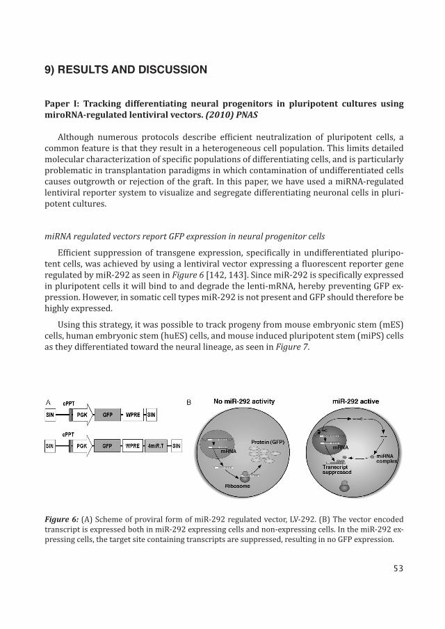
Efficient suppression of transgene expression, specifically in undifferentiated pluripo-tent cells, was achieved by using a lentiviral vector expressing a fluorescent reporter gene regulated by miR-292 as seen in Figure 6 [142, 143]. Since miR-292 is specifically expressed in pluripotent cells it will bind to and degrade the lenti-mRNA, hereby preventing GFP ex-pression. However, in somatic cell types miR-292 is not present and GFP should therefore be highly expressed.
Using this strategy, it was possible to track progeny from mouse embryonic stem (mES) cells, human embryonic stem (huES) cells, and mouse induced pluripotent stem (miPS) cells as they differentiated toward the neural lineage, as seen in Figure 7.
Figure 6: (A) Scheme of proviral form of miR-292 regulated vector, LV-292. (B) The vector encoded transcript is expressed both in miR-292 expressing cells and non-expressing cells. In the miR-292 ex-pressing cells, the target site containing transcripts are suppressed, resulting in no GFP expression.
54
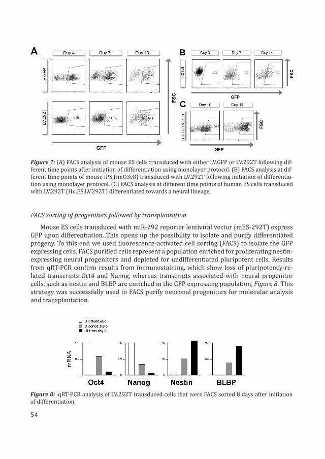
FACS sorting of progenitors followed by transplantation
Mouse ES cells transduced with miR-292 reporter lentiviral vector (mES-292T) express GFP upon differentiation. This opens up the possibility to isolate and purify differentiated progeny. To this end we used fluorescence-activated cell sorting (FACS) to isolate the GFP expressing cells. FACS purified cells represent a population enriched for proliferating nestin-expressing neural progenitors and depleted for undifferentiated pluripotent cells. Results from qRT-PCR confirm results from immunostaining, which show loss of pluripotency-re-lated transcripts Oct4 and Nanog, whereas transcripts associated with neural progenitor cells, such as nestin and BLBP are enriched in the GFP expressing population, Figure 8. This strategy was successfully used to FACS purify neuronal progenitors for molecular analysis and transplantation.
Figure 7: (A) FACS analysis of mouse ES cells transduced with either LV.GFP or LV.292T following dif-ferent time points after initiation of differentiation using monolayer protocol. (B) FACS analysis at dif-ferent time points of mouse iPS (imO3c8) transduced with LV.292T following initiation of differentia-tion using monolayer protocol. (C) FACS analysis at different time points of human ES cells transduced with LV.292T (Hu.ES.LV.292T) differentiated towards a neural lineage.
Figure 8: qRT-PCR analysis of LV.292T transduced cells that were FACS sorted 8 days after initiation of differentiation.
55
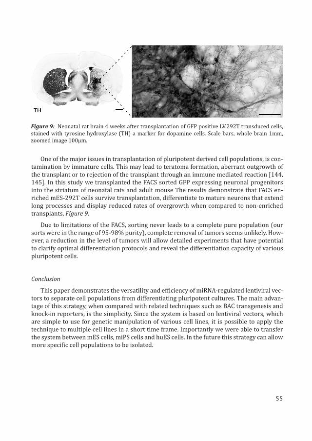
Figure 9: Neonatal rat brain 4 weeks after transplantation of GFP positive LV.292T transduced cells, stained with tyrosine hydroxylase (TH) a marker for dopamine cells. Scale bars, whole brain 1mm, zoomed image 100μm.
One of the major issues in transplantation of pluripotent derived cell populations, is con-tamination by immature cells. This may lead to teratoma formation, aberrant outgrowth of the transplant or to rejection of the transplant through an immune mediated reaction [144, 145]. In this study we transplanted the FACS sorted GFP expressing neuronal progenitors into the striatum of neonatal rats and adult mouse The results demonstrate that FACS en-riched mES-292T cells survive transplantation, differentiate to mature neurons that extend long processes and display reduced rates of overgrowth when compared to non-enriched transplants, Figure 9.
Due to limitations of the FACS, sorting never leads to a complete pure population (our sorts were in the range of 95-98% purity), complete removal of tumors seems unlikely. How-ever, a reduction in the level of tumors will allow detailed experiments that have potential to clarify optimal differentiation protocols and reveal the differentiation capacity of various pluripotent cells.
Conclusion
This paper demonstrates the versatility and efficiency of miRNA-regulated lentiviral vec-tors to separate cell populations from differentiating pluripotent cultures. The main advan-tage of this strategy, when compared with related techniques such as BAC transgenesis and knock-in reporters, is the simplicity. Since the system is based on lentiviral vectors, which are simple to use for genetic manipulation of various cell lines, it is possible to apply the technique to multiple cell lines in a short time frame. Importantly we were able to transfer the system between mES cells, miPS cells and huES cells. In the future this strategy can allow more specific cell populations to be isolated.
56
Paper II: MicroRNA-124 is a subventricular zone neuronal fate determinant. (2012) J. Neurosci.
In adults, new neurons are continuously formed in a few discrete regions of the brain. NSCs reside in the SVZ. The NSCs are termed as type-B cells, which are of astrocytic lineage. They undergo asymmetric cell division to form rapidly amplifying progenitors, type-C cells that then differentiate to neurons. These newly generated neurons migrate via the rostral migratory stream (RMS) to the olfactory bulb (OB). In this article we found that miRNA play a crucial role in adult neurogenesis by regulating the differentiation of progenitor cells into neurons.
miR-124T transgenic mice
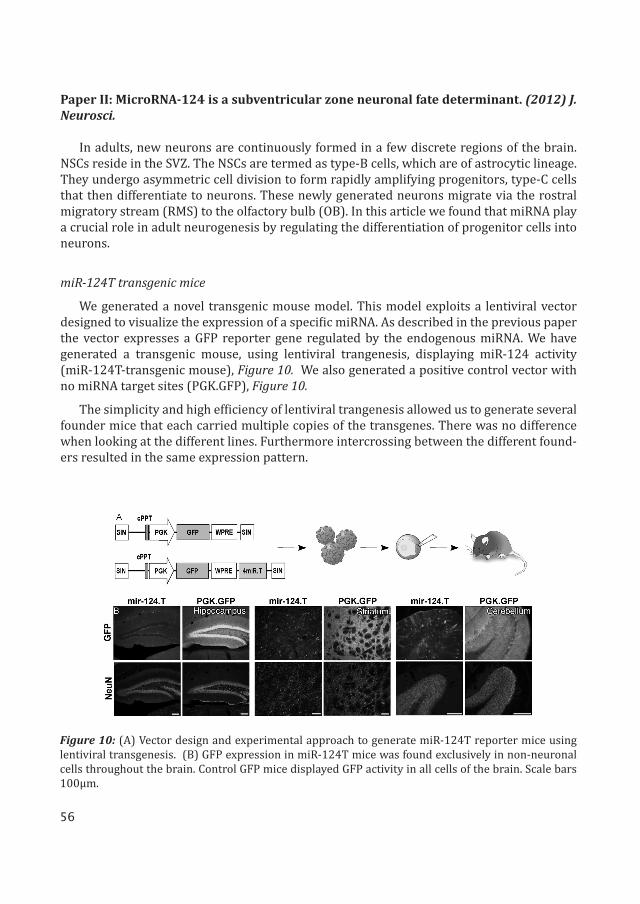
We generated a novel transgenic mouse model. This model exploits a lentiviral vector designed to visualize the expression of a specific miRNA. As described in the previous paper the vector expresses a GFP reporter gene regulated by the endogenous miRNA. We have generated a transgenic mouse, using lentiviral trangenesis, displaying miR-124 activity (miR-124T-transgenic mouse), Figure 10. We also generated a positive control vector with no miRNA target sites (PGK.GFP), Figure 10.
The simplicity and high efficiency of lentiviral trangenesis allowed us to generate several founder mice that each carried multiple copies of the transgenes. There was no difference when looking at the different lines. Furthermore intercrossing between the different found-ers resulted in the same expression pattern.
Figure 10: (A) Vector design and experimental approach to generate miR-124T reporter mice using lentiviral transgenesis. (B) GFP expression in miR-124T mice was found exclusively in non-neuronal cells throughout the brain. Control GFP mice displayed GFP activity in all cells of the brain. Scale bars 100μm.
57
miR-124 activity in the adult mouse brain
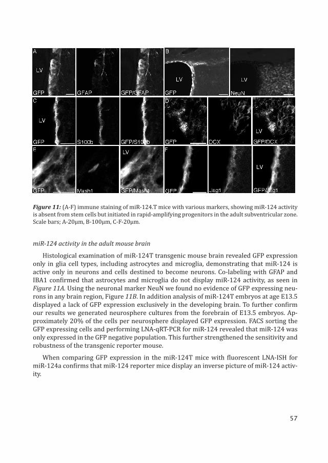
Histological examination of miR-124T transgenic mouse brain revealed GFP expression only in glia cell types, including astrocytes and microglia, demonstrating that miR-124 is active only in neurons and cells destined to become neurons. Co-labeling with GFAP and IBA1 confirmed that astrocytes and microglia do not display miR-124 activity, as seen in Figure 11A. Using the neuronal marker NeuN we found no evidence of GFP expressing neu-rons in any brain region, Figure 11B. In addition analysis of miR-124T embryos at age E13.5 displayed a lack of GFP expression exclusively in the developing brain. To further confirm our results we generated neurosphere cultures from the forebrain of E13.5 embryos. Ap-proximately 20% of the cells per neurosphere displayed GFP expression. FACS sorting the GFP expressing cells and performing LNA-qRT-PCR for miR-124 revealed that miR-124 was only expressed in the GFP negative population. This further strengthened the sensitivity and robustness of the transgenic reporter mouse.
When comparing GFP expression in the miR-124T mice with fluorescent LNA-ISH for miR-124a confirms that miR-124 reporter mice display an inverse picture of miR-124 activ-ity.
Figure 11: (A-F) immune staining of miR-124.T mice with various markers, showing miR-124 activity is absent from stem cells but initiated in rapid-amplifying progenitors in the adult subventricular zone. Scale bars; A-20μm, B-100μm, C-F-20μm.
58
miR-124 activity in neurogenic niches in the adult brain
Examining the SVZ stem cell niche, GFP expressing cells were detected lining the lateral ventricle, and co-labeling with S100B confirmed that ependymal cells express GFP, therefore not exhibiting miR-124 activity, Figure 11C. Following this we wanted to examine whether miR-124 was initiated upon differentiation of the ependymal cells.
Immunostaining for type-B astrocytic cells with GFAP, type-C cells with MASH1 and type-A cells, which represent early migrating neuroblasts with doublecortin (DCX), revealed only type-B cells co-labeled with GFP, Figure 11A,D,E. This means that miR-124 activity is absent from the actual NSC population in the SVZ.
Using BrdU pulse-chase experiment in vivo we found that miR-124 is absent from NSCs in the SVZ and the dentate gyrus of the hippocampus (second neurogenic niche in the adult brain) but it is rapidly induced as the progenitor cells begin to develop into neurons.
In the miR-124T reporter mice, notch ligand receptor Jagged1 (Jag1) expression was de-tected in cells lining the ventricle and in the SVZ. These cells co-labeled with GFP, confirming that miR-124 activity and Jag1 expression do not overlap, thus providing in vivo evidence of miR-124 suppression of Jag1 in the SVZ niche, Figure 11F.
Inhibition of miR-124 activity in the SVZ
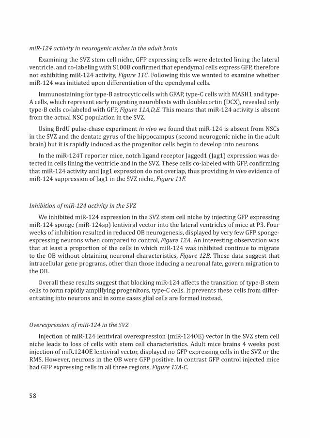
We inhibited miR-124 expression in the SVZ stem cell niche by injecting GFP expressing miR-124 sponge (miR-124sp) lentiviral vector into the lateral ventricles of mice at P3. Four weeks of inhibition resulted in reduced OB neurogenesis, displayed by very few GFP sponge-expressing neurons when compared to control, Figure 12A. An interesting observation was that at least a proportion of the cells in which miR-124 was inhibited continue to migrate to the OB without obtaining neuronal characteristics, Figure 12B. These data suggest that intracellular gene programs, other than those inducing a neuronal fate, govern migration to the OB.
Overall these results suggest that blocking miR-124 affects the transition of type-B stem cells to form rapidly amplifying progenitors, type-C cells. It prevents these cells from differ-entiating into neurons and in some cases glial cells are formed instead.
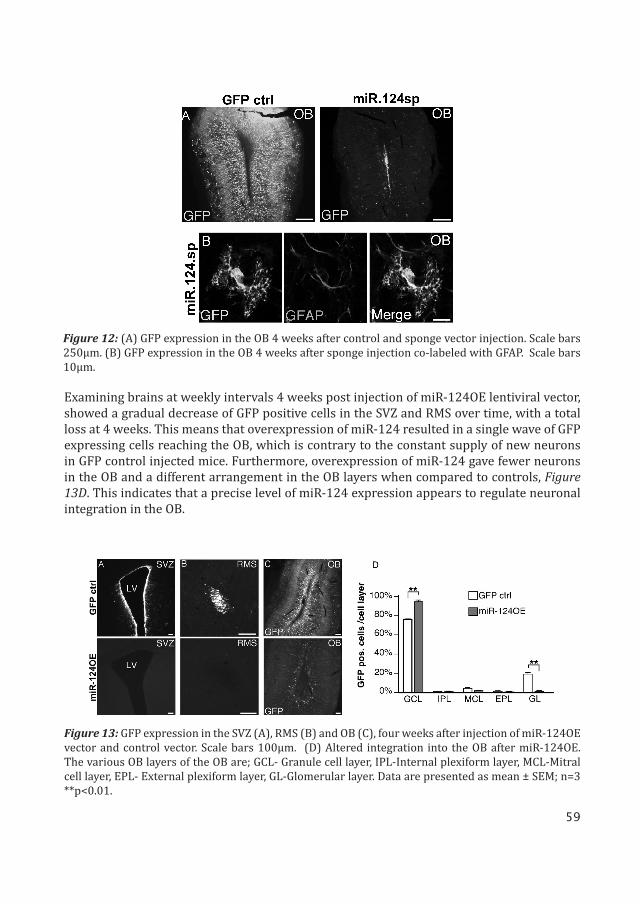
Overexpression of miR-124 in the SVZ
Injection of miR-124 lentiviral overexpression (miR-124OE) vector in the SVZ stem cell niche leads to loss of cells with stem cell characteristics. Adult mice brains 4 weeks post injection of miR.124OE lentiviral vector, displayed no GFP expressing cells in the SVZ or the RMS. However, neurons in the OB were GFP positive. In contrast GFP control injected mice had GFP expressing cells in all three regions, Figure 13A-C.
59
Figure 12: (A) GFP expression in the OB 4 weeks after control and sponge vector injection. Scale bars 250μm. (B) GFP expression in the OB 4 weeks after sponge injection co-labeled with GFAP. Scale bars 10μm.
Figure 13: GFP expression in the SVZ (A), RMS (B) and OB (C), four weeks after injection of miR-124OE vector and control vector. Scale bars 100μm. (D) Altered integration into the OB after miR-124OE. The various OB layers of the OB are; GCL- Granule cell layer, IPL-Internal plexiform layer, MCL-Mitral cell layer, EPL- External plexiform layer, GL-Glomerular layer. Data are presented as mean ± SEM; n=3 **p<0.01.
Examining brains at weekly intervals 4 weeks post injection of miR-124OE lentiviral vector, showed a gradual decrease of GFP positive cells in the SVZ and RMS over time, with a total loss at 4 weeks. This means that overexpression of miR-124 resulted in a single wave of GFP expressing cells reaching the OB, which is contrary to the constant supply of new neurons in GFP control injected mice. Furthermore, overexpression of miR-124 gave fewer neurons in the OB and a different arrangement in the OB layers when compared to controls, Figure 13D. This indicates that a precise level of miR-124 expression appears to regulate neuronal integration in the OB.
60
Conclusion
The expression pattern of miR-124 was previously well characterized; it is brain specific, expressed in all neurons, but not expressed in neuronal stem cells, astrocytes or in micro-glia. The expression is initiated as stem cells differentiate towards neurons. In this work we confirm and extend this expression pattern. Our results showed that miR-124 is essential for adult neurogenesis. In short, inhibition of miR-124 blocked neurogenesis, whereas overex-pression of miR-124 forced progenitor cells to undergo neuronal differentiation.
Another interesting finding is the identification of the notch ligand receptor Jag1 as a miR-124 target. Recent studies demonstrate a critical role of the notch pathway not only in regulating NSCs, but also in maintaining ependymal cells in a non-proliferating state [146]. The consequences of miR-124 overexpression in the SVZ are very similar to notch inhibition, suggesting that miR-124 is a crucial player in this network.
Adult neurogenesis has been implicated in numerous brain disorders, including Alz-heimer’s disease and various psychiatric conditions. Therefore, the finding that a miRNA governs this process opens up new possibilities to understand and treat these disorders. Furthermore, the simplicity of lentiviral transgenesis allows for the generation of a large number of this type of miRNA reporter mice, which would greatly enhance our understand-ing of miRNA expression patterns in the brain.
Paper III: Visualisation and genetic modification of resident brain microglia using len-tiviral vectors regulated by microRNA-9. (2013) Nature communications
Microglia are a resident population of immune cells in the brain that have classically been associated with canonical central nervous system (CNS) responses against pathogens and brain injuries [147]. However, recent studies have indicated that microglia play a more com-plex role in maintaining CNS homeostasis than what was initially believed [148-150].
The exact role of microglia in the healthy and diseased brain remains largely elusive. This is because it has been difficult to genetically modify the resident microglia population in a cell-type specific manner [151]. Here, we investigate if miR-9 regulated lentiviral vectors can be used to specifically target genetic modification to resident microglia in the rodent brain.
miR-9 regulated lentiviral vectors
Efficient transgene expression, specifically in microglia, was achieved by using a lentivi-ral vector expressing a fluorescent reporter gene regulated by miR-9. We first used the lenti-viral miR-9 reporter vector (LV.miR-9.T) on mES and mouse neural progenitor cells, since it is well established that miR-9 expression is initiated during neural development but absent from embryonic stem cells. LV.miR-9.T was efficiently inhibited in mouse neural progenitor cells but not in mES cells confirming that miR-9 is active in neural progenitor cells.
61
miR-9 activity in neurons in sensor transgenic mice
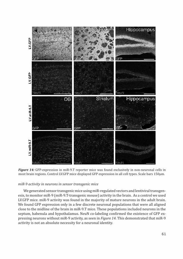
We generated sensor transgenic mice using miR-regulated vectors and lentiviral transgen-esis, to monitor miR-9 (miR-9.T-transgenic mouse) activity in the brain. As a control we used LV.GFP mice. miR-9 activity was found in the majority of mature neurons in the adult brain. We found GFP expression only in a few discrete neuronal populations that were all aligned close to the midline of the brain in miR-9.T mice. These populations included neurons in the septum, habenula and hypothalamus. NeuN co-labeling confirmed the existence of GFP ex-pressing neurons without miR-9 activity, as seen in Figure 14. This demonstrated that miR-9 activity is not an absolute necessity for a neuronal identity.
Figure 14: GFP-expression in miR-9.T reporter mice was found exclusively in non-neuronal cells in most brain regions. Control LV.GFP mice displayed GFP expression in all cell types. Scale bars 150μm.
62
miR-9 activity in glial cells
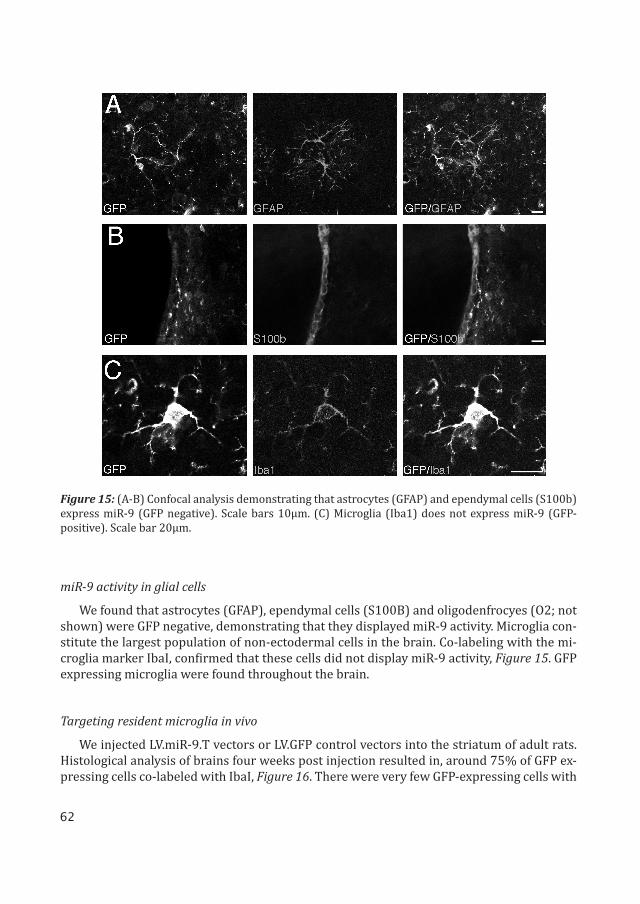
We found that astrocytes (GFAP), ependymal cells (S100B) and oligodenfrocyes (O2; not shown) were GFP negative, demonstrating that they displayed miR-9 activity. Microglia con-stitute the largest population of non-ectodermal cells in the brain. Co-labeling with the mi-croglia marker IbaI, confirmed that these cells did not display miR-9 activity, Figure 15. GFP expressing microglia were found throughout the brain.
Targeting resident microglia in vivo
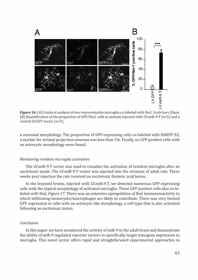
We injected LV.miR-9.T vectors or LV.GFP control vectors into the striatum of adult rats. Histological analysis of brains four weeks post injection resulted in, around 75% of GFP ex-pressing cells co-labeled with IbaI, Figure 16. There were very few GFP-expressing cells with
Figure 15: (A-B) Confocal analysis demonstrating that astrocytes (GFAP) and ependymal cells (S100b) express miR-9 (GFP negative). Scale bars 10μm. (C) Microglia (Iba1) does not express miR-9 (GFP-positive). Scale bar 20μm.
63
Figure 16: (A) Confocal analysis of two representative microglia co-labeled with Iba1. Scale bars 20μm. (B) Quantification of the proportion of GFP/Iba1 cells in animals injected with LV.miR-9.T (n=5) and a control LV.GFP vector (n=5).
a neuronal morphology. The proportion of GFP-expressing cells co-labeled with DARPP-32, a marker for striatal projection neurons was less than 1%. Finally, no GFP positive cells with an astrocytic morphology were found.
Monitoring resident microglia activation
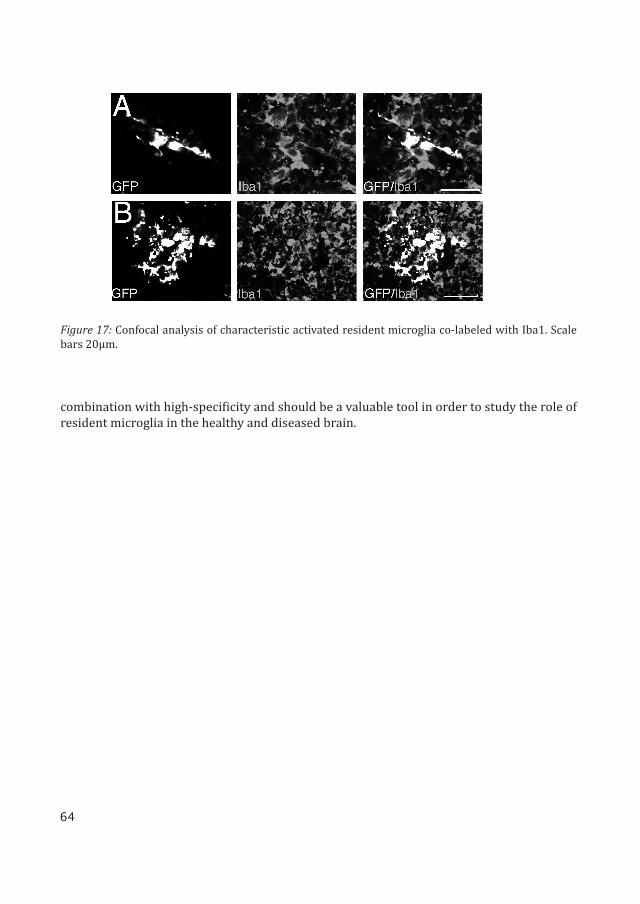
The LV.miR-9.T vector was used to visualize the activation of resident microglia after an excitotoxic insult. The LV.miR-9.T vector was injected into the striatum of adult rats. Three weeks post injection the rats received an excitotoxic ibotenic acid lesion.
In the lesioned brains, injected with LV.miR-9.T, we detected numerous GFP expressing cells with the typical morphology of activated microglia. These GFP positive cells also co-la-beled with IbaI, Figure 17. There was an extensive upregulation of IbaI immunoreactivity to which infiltrating monocytes/macrophages are likely to contribute. There was very limited GFP expression in cells with an astrocyte-like morphology, a cell-type that is also activated following an excitotoxic lesion.
Conclusion
In this paper we have monitored the activity of miR-9 in the adult brain and demonstrate the ability of miR-9 regulated reporter vectors to specifically target transgene expression to microglia. This novel vector offers rapid and straightforward experimental approaches in
64
Figure 17: Confocal analysis of characteristic activated resident microglia co-labeled with Iba1. Scale bars 20μm.
combination with high-specificity and should be a valuable tool in order to study the role of resident microglia in the healthy and diseased brain.

65
10) CONCLUDING REMARKS AND PROSPECTS

66

67
10) CONCLUDING REMARKS AND PROSPECTS
miRNAs are no more than a couple of decades old, but are now being hailed as a fresh promise for biological discovery and therapeutics. These small molecules, consisting of a short string of nucleic acids, carry immense power to regulate genes and thereby large-scale cellular function. The rapidly increasing amount of literature describing various new miRNAs along with their features, function and mechanism is an illustration of the impor-tance of these key regulators. However, there are numerous basic questions that are yet to be addressed. Therefore constant advances in the techniques to study miRNA are required.
The advances reported in this thesis; miRNA reporters, miRNA sponges and transgenic reporter systems will enable new types of studies that will clarify the functional properties of individual miRNAs. It will also make it possible to specifically target transgene expression to a particular cell type in the brain.
So far we have only begun to realize the complexity of miRNA-mediated regulation in the brain. Since miRNA do not function through a single target gene, but rather through a com-bined regulation of many different genes. This together with the fact that the expressional regulation of genes varies due to the level of miRNA, makes understanding them complex. Manipulating miRNA by inhibiting or overexpressing miRNA should therefore be done in vivo since the biogenesis might be different from what occurs in vitro. Furthermore, the syn-thetic changes in levels of miRNA should reflect endogenous interactions in order to get the correct biological picture. Therefore using miRNA sponges might be beneficial over miRNA knockdowns. However, applying multiple methods in parallel will increase the likelihood of proper reflection of the presence and regulation of a miRNA.
Further understanding of biogenesis and functionality of this exceptional gene regulator will in turn enhance the techniques used to study them. Improvement in sensitivity, specific-ity and robustness of assays will be required. It will be very interesting to follow this field as it matures and unravels the full role of miRNAs in the brain and hopefully provide the op-portunity to safely pursue them as therapeutic strategy.

68

69
11) MATERIALS AND METHODS

70
71
11) MATERIALS AND METHODS
Lentiviral vectors
All vectors used during this thesis have been third generation self-inactivating (SIN) len-tiviral vectors. The miRNA target, sponge and overexpression sequences were cloned into the 3’UTR of the transgene expression cassette.
miRNA reporter vector constructs were cloned by adding four complementary miRNA target sites immediately downstream of the woodchuck hepatitis virus posttranscriptional response element (WPRE) of the SIN lentiviral vector expressing GFP driven by the phosho-glycerate kinase 1 (PGK) promoter derived from the phosphoglycerol kinase housekeeping gene. Target sequences for the miRNAs used are as follows miR-292: ACACTCAAAACCTGGCG-GCACTT, miR-124: TTAAGGCACGCGGTGAATGCCA, miR-9: TCATACAGCTAGATAACCAAAG.
Similarly miRNA sponge vectors were synthesized by adding eight tandem repeats of im-perfectly complementary miRNA sequences, thereby forming a central bulge when binding to the target miRNA. A stronger promoter derived from cytomegalovirus (CMV) was required for these vectors to drive the overexpression of the sponge sequence to provide a successful miRNA knockdown. The miR-124 sponge sequence is as follows: TTAAGGCACG-TA-TGAAT-GCCA. The miR-125b sponge sequence is as follows: TCACAAGTT-TA-GTCTCAGGGA.
miRNA overexpression vectors were synthesized by PCR amplification of genomic frag-ments containing miRNA sequences flanked on either side by 200bp. The fragment was then cloned in between the GFP and WPRE sequence in the pFUGW vector. Primers for PCR of the miR-124 genomic sequence from mouse DNA are as follows, forward: ATGAATTCTCGC-CAGCTTTTTCTTTCTC and reverse: ATGAATTCATTTGCATCTGCACAAACCC. Primers for PCR of the miR-125b genomic sequence from mouse DNA are as follows, forward: ATTCTA-GAGTTGCGCTCCCCTCAGTC and reverse: ATGGTACCGCAGCTCCCAAGAGCCTAAC.
Lentivirus production
As mentioned previously all vectors used in this thesis are third generation lentiviral vectors. These have been produced using transient co-transfection of the four plasmids; two packaging plasmids (pMDL, pRSV-REV) an envelope plasmid (pMD2.G) and a transfer plasmid. The main differences between a third generation system and the previously used second generation system are as follows: 1) The tat protein has been eliminated from the packaging completely. 2) The rev protein is expressed on an independent plasmid. 3) The 5’LTR of the transfer plasmid has been modified to include a conventional promoter and delete a portion of the LTR. The production protocol used is as follows:
72
Day 1: The cell line used for lentiviral production is human 293T or often referred to as Hu-man Embryonic Kidney (HEK) cells. These cells are easy to grow and transfect. In the evening of the first day, 24 hours prior to transfection, cells are plated in two T175 flasks (per batch) at a density of 6x10⁴ cells/cm2 with 20 ml standard DMEM culture media supplemented with 10% fetal bovine serum (FBS) and antibiotics (penicillin/streptomycin 1000 U/ml).
Day 2: In the afternoon, 2 hours prior to transfection, media is changed to 20 ml IMDM, sup-plemented with 10% antibiotics (penicillin/streptomycin 1000 U/ml).For transfection, the plasmid DNA mix per batch is prepared by adding: pMD2.G – 21.6 μg, pMDL – 30 μg, pRSV-REV – 15 μg and transfer vector plasmid 76.8 μg. The plasmid solu-tion is made up to a final volume of 2,700 μl with 0.1x TE. Finally 300 μl of 2.5 M CaCl2 is added. The mix is left at room temperature for 5 min. The precipitate is formed by drop wise addition of 3,000 μl of 2x HBS solution (Na2HPO4 solution with pH 7) to the 3,000 μl DNA-TE-CaCl2 mixture as mentioned above, while vortexing at full speed. The precipitate is im-mediately added to the 293T cells. High magnification microscopy should reveal a very small granular precipitate of the CaPi-precipitated plasmid DNA, initially above the cell monolayer and after incubation in the 37°C incubator overnight, at the bottom of the plate in the large spaces between cells.
Day 3: 14 hours after transfection, media is replaced with 20 ml of fresh IMDM, supplement-ed with 10% antibiotics (penicillin/streptomycin 1000 U/ml).
Day 4: 30 hours after change to fresh IMDM media the supernatant is harvested into 50 ml falcon tubes. Thereafter it is spun down for 10 min at 2,500 rpm. The supernatant is then filtered using a syringe and 0.2 μm filter into a new 50 ml falcon tube. The supernatant can now be stored in the fridge overnight.
Day 5: The supernatant is transferred to ultracentrifuge tubes. Thereafter it is spun down in an ultracentrifuge for 1.5 hours at 25,000 rpm. After the spin the supernatant is removed and 80 μl of cold PBS is added to the bottom of the tube. The tube is covered with parafilm and kept in the fridge for 2 hours to let the virus particles dissolve. Two hours later the len-tivirus can be aliquoted into desired volumes and stored at -80°C.
To determine the concentration of the viral vector, commonly know as the virus titer, one can use either FACS, in the case a fluorescent transgene such as GFP is present, or by qRT-PCR. When doing the titration by FACS 100,000 293T cells are transduced with the lentivirus in a serial dilution of 3 μl, 1 μl, 0.3 μl, 0.1 μl, 0.03 μl and 0.01 μl. Seventy-two hours later the cells are harvested and FACS analyzed, gating for fluorescence. The titer can be calculated by the following equation: (percentage fluorescent cells x 100,000 (total number of cells transduced) x dilution factor)/ 1,000. This will provide the titer in the units TU/ml.
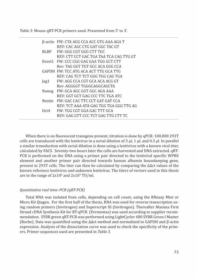
73
When there is no fluorescent transgene present, titration is done by qPCR: 100,000 293T cells are transduced with the lentivirus in a serial dilution of 3 μl, 1 μl, and 0.3 μl. In parallel a similar transduction with serial dilution is done using a lentivirus with a known viral titer, calculated by FACS. Seventy-two hours later the cells are harvested and DNA extracted. qRT-PCR is performed on the DNA using a primer pair directed to the lentiviral specific WPRE element and another primer pair directed towards human albumin housekeeping gene, present in 293T cells. The titer can then be calculated by comparing the ΔΔct values of the known reference lentivirus and unknown lentivirus. The titers of vectors used in this thesis are in the range of 2x10⁸ and 2x10⁹ TU/ml.
Quantitative real time–PCR (qRT-PCR)
Total RNA was isolated from cells, depending on cell count, using the RNeasy Mini or Micro Kit Qiagen. For the first half of the thesis, RNA was used for reverse transcription us-ing random primers (Invitrogen) and Superscript III (Invitrogen). Thereafter Maxima First Strand cDNA Synthesis Kit for RT-qPCR (Fermentas) was used according to supplier recom-mendation. SYBR green qRT-PCR was performed using LightCycler 480 SYBR Green I Master (Roche). Data was quantified using the ΔΔct method and normalized to GAPDH and β-actin expression. Analysis of the dissociation curve was used to check the specificity of the prim-ers. Primer sequences used are presented in Table 3.
β-actin FW: CTA AGG CCA ACC GTG AAA AGA T REV: CAC AGC CTG GAT GGC TAC GTBLBP FW: GGG CGT GGG CTT TGC REV: CTT CCT GAC TGA TAA TCA CAG TTG GTEvovl1 FW: CCC CGG GAG GAA TGG GCT CTT Rev: TAG GGT TGT GCC ACA GGG CCAGAPDH FW: TCC ATG ACA ACT TTG GCA TTG REV: CAG TCT TCT GGG TGG CAG TGAJag1 FW: AGG CCA CGT GCA ACA ACG GT Rev: AGGGGT TGGGCAGGCAGCTANanog FW: GCA AGC GGT GGC AGA AAA REV: GGT GCT GAG CCC TTC TGA ATCNestin FW: GAC CAC TTC CCT GAT GAT CCA REV: TCT AAA ATA GAG TGG TGA GGG TTG AGOct4 FW: TGG CGT GGA GAC TTT GCA REV: GAG GTT CCC TCT GAG TTG CTT TC
Table 3: Mouse qRT-PCR primers used. Presented from 5’ to 3’.
74
Locked nucleic acid qRT-PCR (LNA-qRT-PCR)
To validate miRNA overexpression vectors, qRT-PCR was used along with LNA primers (Exiqon). Total RNA was isolated from cells using the miRNeasy Mini/Micro Kit (Qiagen). RNA was used for reverse transcription using miRCURY LNA Universal RT cDNA Synthesis Kit (Exiqon), according to supplier recommendation. SYBR green qRT-PCR was performed using LightCycler 480 SYBR Green I Master (Roche). Data was quantified using the ΔΔct method and normalized to miR-103 expression.
Luciferase reporter assay
To validate miRNA sponge vectors ability to inhibit mature miRNA luciferase reporter as-say was used. The luciferase reporter construct was designed by incorporating miRNA-bind-ing sites on the target gene from genomic DNA into the luciferase reporter vector pSICHECK-2 (Promega). In our case putative miR-124 binding sites of Jag1 3’UTR were amplified from mouse genomic DNA using the following primers: Jag1utr forward: CTCGAGCGCTCTCACA-GCTATGCAAAA and Jag1utr reverse: GCGGCCGCTGGCTTACAGGCAACACGTTA.
Sixty thousand Hela cells per well were seeded in a 6-well plate. The luciferase reporter construct was co-transfected with the lentiviral miR-124 overexpression construct and the miR-124sp construct 24 hours after plating using TurboFect Transfection Reagent (Fermen-tas) according to supplier recommendation. Forty-eight hours after transfection, cells were assayed for luciferase activity using a Dual-Luciferase Reporter Assay Kit (Promega) accord-ing to supplier recommendation.
Cell culture
mES cells were cultured on 0.1% gelatin (Sigma G2500-100G) coated plastics in mES expansion medium (Table 4), plated at a density of 4x10⁴ cells/cm2. To maintain cells in an undifferentiated state, the media was supplemented with leukaemia inhibitory factor (LIF), which was produced in house.
Monolayer differentiation of mNS cells was carried out on PA6 stromal cells as previously described [152]. A day prior to start of differentiation, PA6 cells were irradiated at 50 Gy and plated at a density sufficient to cover the surface of the flask in mES expansion medium. Twenty-four hours later the media was removed from the attached PA6 cells and the cells were washed with PBS. Thereafter mES cells were added at a density of 60 cells/cm2 in mES differentiation medium. Four days later half the medium was renewed. At day 8, the medium was replaced with N2B27 medium (Stem cell Science) for the remainder of the 14 day dif-ferentiation period.
Cells were fixed for immunocytochemistry by removing medium and applying cold PBS containing 4% PFA for 15 min. This was followed by three rinses with PBS. FACS sorting for differentiated mES cells and preparation of cells for transplantation was performed as previ-ously described [152].
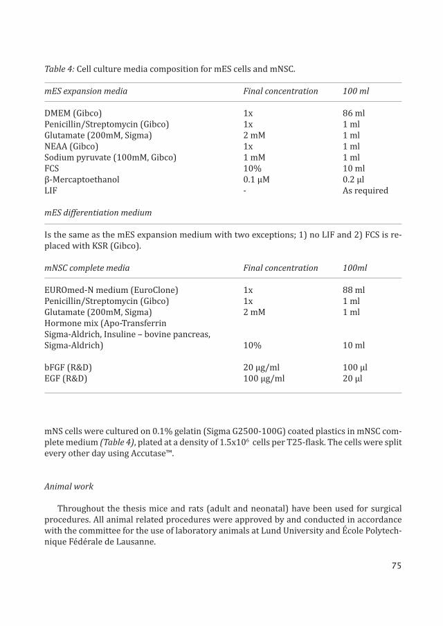
75
mNS cells were cultured on 0.1% gelatin (Sigma G2500-100G) coated plastics in mNSC com-plete medium (Table 4), plated at a density of 1.5x10⁶ cells per T25-flask. The cells were split every other day using Accutase™.
Animal work
Throughout the thesis mice and rats (adult and neonatal) have been used for surgical procedures. All animal related procedures were approved by and conducted in accordance with the committee for the use of laboratory animals at Lund University and École Polytech-nique Fédérale de Lausanne.
Table 4: Cell culture media composition for mES cells and mNSC.
mES expansion media Final concentration 100 ml
DMEM (Gibco) 1x 86 mlPenicillin/Streptomycin (Gibco) 1x 1 mlGlutamate (200mM, Sigma) 2 mM 1 mlNEAA (Gibco) 1x 1 mlSodium pyruvate (100mM, Gibco) 1 mM 1 mlFCS 10% 10 mlβ-Mercaptoethanol 0.1 μM 0.2 μlLIF - As required
mES differentiation medium
Is the same as the mES expansion medium with two exceptions; 1) no LIF and 2) FCS is re-placed with KSR (Gibco).
mNSC complete media Final concentration 100ml
EUROmed-N medium (EuroClone) 1x 88 mlPenicillin/Streptomycin (Gibco) 1x 1 mlGlutamate (200mM, Sigma) 2 mM 1 mlHormone mix (Apo-Transferrin Sigma-Aldrich, Insuline – bovine pancreas, Sigma-Aldrich) 10% 10 ml
bFGF (R&D) 20 μg/ml 100 μlEGF (R&D) 100 μg/ml 20 μl

76
MicroRNA transgenic reporter animals were made by lentiviral transgenesis, as pre-viously described. Offspring were genotyped and the number of integrated transgenes was estimated using qRT-PCR. The following primers were used: WPRE fw: 5’ GGCACT-GACAATTCCGTGGT 3’, rev: 5’ AGGGACGTAGCAGAAGGACG 3’. Probe WPRE: 5’ ACGTCCTTTC-CATGGCTGCTCGC 3’. Titine fw: 5’ AAAACGAGCAGTGACGTGAGC 3’, rev: 5’ TTCAGTCATGCT-GCTAGCGC 3’. Probe Titine: 5’ TCGACGGAAGCGTCTCGTCTCAGTC 3’. The data was quantified using the ΔΔct method. The transgene data (WPRE primer) was normalized with the Titine data to give the number of transgenes in the genome of each animal.
In the first paper, one day old neonatal rats were unilaterally injected with 6-OHDA into the right ventricle to lesion them. Two days later 1 μl cell suspension (100,000 cells/μl) was injected unilaterally into the dorsal lesioned striatum. The pups were placed on ice for 5 min before positioning them in an ice-cold stereotaxic frame. Adult mice (Charles River) were in-jected with 6-OHDA into the right substantia nigra at 6–8 weeks of age. Then 4–6 weeks later, 1 μl cell suspension (100,000 cells/μl) was unilaterally injected into the lesioned striatum.
For vector injections of miRNA sponge and overexpression lentivirus, 1 μl of concentrat-ed lentiviral vector was unilaterally injected into the right ventricles of NMRI mice at post-natal day 3 (Charles River). The pups were placed on ice for 5 min before positioning them in an ice-cold stereotaxic frame.
For surgical procedures on adult mice, anesthesia was induced and maintained using 2% isofluorane, delivered in a mix of air and N2O at a 4:1 ratio. All surgical procedures were performed using Kopf stereotaxic frames (Kopf instruments, Tujunga, CA). Intracranial in-jections were performed using a 5 μl Hamilton syringe with a 22-gauge needle fitted with a glass capillary.
Immunochemistry
Similar procedures were followed for both fixed sections and cells. A brief summary of the protocol; start with a preincubation for 1 hour in blocking solution (5% normal serum and 0.25% Triton-X in 0.1 M potassium phosphate buffered saline (KPBS)). The primary antibody was diluted in the blocking solution and applied overnight at room temperature. Following rinses in KPBS, either biotinylated (Vector Labs) or fluorophore-conjugated sec-ondary antibodies (Molecular Probes or Jackson Laboratories) were diluted in blocking so-lution and applied for 2 hours. Biotinylated secondary antibodies were followed by 1 hour incubation with fluorophore-conjugated streptavidin. Following fluorescent labeling, the free-floating sections were mounted on coated glass slides, and all sections and cells were cover-slipped with the anti-bleaching reagent PVA-DABCO. A list of primary antibodies used in this procedure has been provided in Table 5, together with dilution factors.
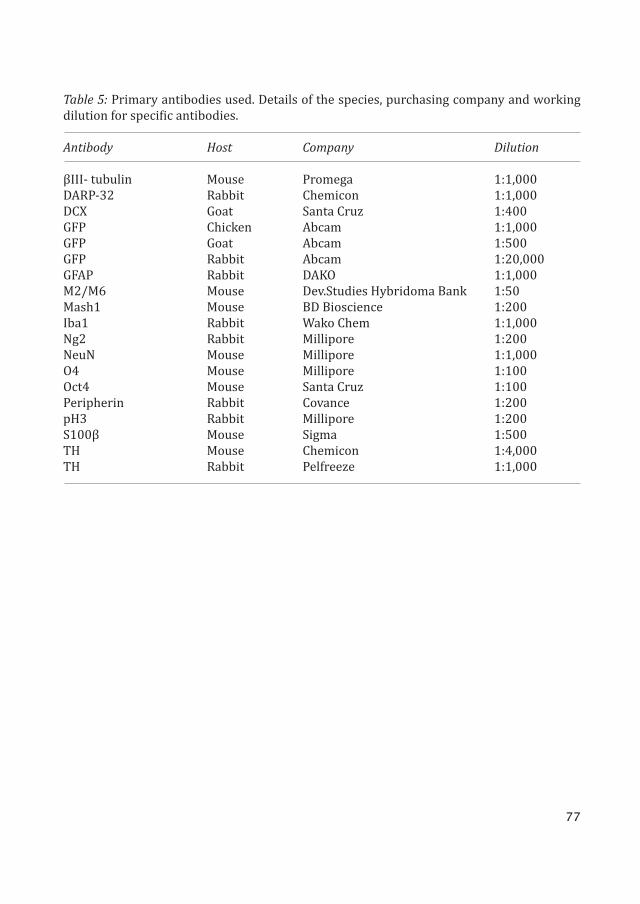
77
Table 5: Primary antibodies used. Details of the species, purchasing company and working dilution for specific antibodies.
Antibody Host Company Dilution
βIII- tubulin Mouse Promega 1:1,000DARP-32 Rabbit Chemicon 1:1,000DCX Goat Santa Cruz 1:400GFP Chicken Abcam 1:1,000GFP Goat Abcam 1:500GFP Rabbit Abcam 1:20,000GFAP Rabbit DAKO 1:1,000M2/M6 Mouse Dev.Studies Hybridoma Bank 1:50Mash1 Mouse BD Bioscience 1:200Iba1 Rabbit Wako Chem 1:1,000Ng2 Rabbit Millipore 1:200NeuN Mouse Millipore 1:1,000O4 Mouse Millipore 1:100Oct4 Mouse Santa Cruz 1:100Peripherin Rabbit Covance 1:200pH3 Rabbit Millipore 1:200S100β Mouse Sigma 1:500TH Mouse Chemicon 1:4,000TH Rabbit Pelfreeze 1:1,000

78

79
12) REFERENCES

80
81
12) REFERENCES
1. Sachdeva, R., et al., Tracking differentiating neural progenitors in pluripotent cultures using microRNA-regulated lentiviral vec-tors. Proceedings of the National Academy of Sciences of the United States of America, 2010. 107(25): p. 11602-7.
2. Akerblom, M., et al., MicroRNA-124 is a sub-ventricular zone neuronal fate determinant. The Journal of neuroscience : the official journal of the Society for Neuroscience, 2012. 32(26): p. 8879-89.
3. Akerblom, M., et al., Visualization and genet-ic modification of resident brain microglia using lentiviral vectors regulated by micro-RNA-9. Nature communications, 2013. 4: p. 1770.
4. Bartel, D.P., MicroRNAs: genomics, biogen-esis, mechanism, and function. Cell, 2004. 116(2): p. 281-97.
5. Lee, R.C., R.L. Feinbaum, and V. Ambros, The C. elegans heterochronic gene lin-4 encodes small RNAs with antisense complementarity to lin-14. Cell, 1993. 75(5): p. 843-54.
6. Reinhart, B.J., et al., The 21-nucleotide let-7 RNA regulates developmental timing in Caenorhabditis elegans. Nature, 2000. 403(6772): p. 901-6.
7. Wightman, B., I. Ha, and G. Ruvkun, Posttran-scriptional regulation of the heterochro-nic gene lin-14 by lin-4 mediates temporal pattern formation in C. elegans. Cell, 1993. 75(5): p. 855-62.
8. Pasquinelli, A.E., et al., Conservation of the sequence and temporal expression of let-7 heterochronic regulatory RNA. Nature, 2000. 408(6808): p. 86-9.
9. Lagos-Quintana, M., et al., Identification of novel genes coding for small expressed RNAs. Science, 2001. 294(5543): p. 853-8.
10. Lau, N.C., et al., An abundant class of tiny RNAs with probable regulatory roles in Caenorhabditis elegans. Science, 2001. 294(5543): p. 858-62.
11. Lee, R.C. and V. Ambros, An extensive class of small RNAs in Caenorhabditis elegans.
Science, 2001. 294(5543): p. 862-4.12. Molnar, A., et al., miRNAs control gene
expression in the single-cell alga Chla-mydomonas reinhardtii. Nature, 2007. 447(7148): p. 1126-9.
13. Friedman, R.C., et al., Most mammalian mR-NAs are conserved targets of microRNAs. Genome research, 2009. 19(1): p. 92-105.
14. Kim, Y.K. and V.N. Kim, Processing of intronic microRNAs. The EMBO journal, 2007. 26(3): p. 775-83.
15. Borchert, G.M., W. Lanier, and B.L. Davidson, RNA polymerase III transcribes human mi-croRNAs. Nature structural & molecular bi-ology, 2006. 13(12): p. 1097-101.
16. Cai, X., C.H. Hagedorn, and B.R. Cullen, Hu-man microRNAs are processed from capped, polyadenylated transcripts that can also function as mRNAs. RNA, 2004. 10(12): p. 1957-66.
17. Lee, Y., et al., MicroRNA genes are tran-scribed by RNA polymerase II. The EMBO journal, 2004. 23(20): p. 4051-60.
18. Denli, A.M., et al., Processing of primary mi-croRNAs by the Microprocessor complex. Nature, 2004. 432(7014): p. 231-5.
19. Gregory, R.I., et al., The Microprocessor com-plex mediates the genesis of microRNAs. Na-ture, 2004. 432(7014): p. 235-40.
20. Han, J., et al., The Drosha-DGCR8 complex in primary microRNA processing. Genes & de-velopment, 2004. 18(24): p. 3016-27.
21. Landthaler, M., A. Yalcin, and T. Tuschl, The human DiGeorge syndrome critical region gene 8 and Its D. melanogaster homolog are required for miRNA biogenesis. Current bi-ology : CB, 2004. 14(23): p. 2162-7.
22. Lee, Y., et al., The nuclear RNase III Dro-sha initiates microRNA processing. Nature, 2003. 425(6956): p. 415-9.
23. Han, J., et al., Molecular basis for the recogni-tion of primary microRNAs by the Drosha-DGCR8 complex. Cell, 2006. 125(5): p. 887-901.
24. Bohnsack, M.T., K. Czaplinski, and D. Gorlich,
82
Exportin 5 is a RanGTP-dependent dsRNA-binding protein that mediates nuclear ex-port of pre-miRNAs. RNA, 2004. 10(2): p. 185-91.
25. Lund, E., et al., Nuclear export of microRNA precursors. Science, 2004. 303(5654): p. 95-8.
26. Yi, R., et al., Exportin-5 mediates the nuclear export of pre-microRNAs and short hairpin RNAs. Genes & development, 2003. 17(24): p. 3011-6.
27. Chendrimada, T.P., et al., TRBP recruits the Dicer complex to Ago2 for microRNA pro-cessing and gene silencing. Nature, 2005. 436(7051): p. 740-4.
28. Forstemann, K., et al., Normal microRNA maturation and germ-line stem cell main-tenance requires Loquacious, a double-stranded RNA-binding domain protein. PLoS biology, 2005. 3(7): p. e236.
29. Hutvagner, G., et al., A cellular function for the RNA-interference enzyme Dicer in the maturation of the let-7 small temporal RNA. Science, 2001. 293(5531): p. 834-8.
30. Jiang, F., et al., Dicer-1 and R3D1-L catalyze microRNA maturation in Drosophila. Genes & development, 2005. 19(14): p. 1674-9.
31. Ketting, R.F., et al., Dicer functions in RNA interference and in synthesis of small RNA involved in developmental timing in C. el-egans. Genes & development, 2001. 15(20): p. 2654-9.
32. Lee, Y., et al., The role of PACT in the RNA silencing pathway. The EMBO journal, 2006. 25(3): p. 522-32.
33. Gregory, R.I., et al., Human RISC couples mi-croRNA biogenesis and posttranscriptional gene silencing. Cell, 2005. 123(4): p. 631-40.
34. Maniataki, E. and Z. Mourelatos, A human, ATP-independent, RISC assembly machine fueled by pre-miRNA. Genes & development, 2005. 19(24): p. 2979-90.
35. Schwarz, D.S., et al., Asymmetry in the as-sembly of the RNAi enzyme complex. Cell, 2003. 115(2): p. 199-208.
36. Du, T. and P.D. Zamore, microPrimer: the biogenesis and function of microRNA. De-velopment, 2005. 132(21): p. 4645-52.
37. Ruby, J.G., et al., Large-scale sequencing re-
veals 21U-RNAs and additional microRNAs and endogenous siRNAs in C. elegans. Cell, 2006. 127(6): p. 1193-207.
38. Slack, F.J., et al., The lin-41 RBCC gene acts in the C. elegans heterochronic pathway be-tween the let-7 regulatory RNA and the LIN-29 transcription factor. Molecular cell, 2000. 5(4): p. 659-69.
39. Hutvagner, G. and P.D. Zamore, A microRNA in a multiple-turnover RNAi enzyme com-plex. Science, 2002. 297(5589): p. 2056-60.
40. Song, J.J., et al., Crystal structure of Argo-naute and its implications for RISC slicer ac-tivity. Science, 2004. 305(5689): p. 1434-7.
41. Yekta, S., I.H. Shih, and D.P. Bartel, MicroR-NA-directed cleavage of HOXB8 mRNA. Sci-ence, 2004. 304(5670): p. 594-6.
42. Lim, L.P., et al., Microarray analysis shows that some microRNAs downregulate large numbers of target mRNAs. Nature, 2005. 433(7027): p. 769-73.
43. Wu, L., J. Fan, and J.G. Belasco, MicroRNAs di-rect rapid deadenylation of mRNA. Proceed-ings of the National Academy of Sciences of the United States of America, 2006. 103(11): p. 4034-9.
44. Behm-Ansmant, I., et al., mRNA degrada-tion by miRNAs and GW182 requires both CCR4:NOT deadenylase and DCP1:DCP2 de-capping complexes. Genes & development, 2006. 20(14): p. 1885-98.
45. Giraldez, A.J., et al., Zebrafish MiR-430 pro-motes deadenylation and clearance of ma-ternal mRNAs. Science, 2006. 312(5770): p. 75-9.
46. Eulalio, A., et al., Target-specific require-ments for enhancers of decapping in miR-NA-mediated gene silencing. Genes & devel-opment, 2007. 21(20): p. 2558-70.
47. Eulalio, A., et al., Deadenylation is a wide-spread effect of miRNA regulation. RNA, 2009. 15(1): p. 21-32.
48. Pillai, R.S., et al., Inhibition of translational initiation by Let-7 MicroRNA in human cells. Science, 2005. 309(5740): p. 1573-6.
49. Humphreys, D.T., et al., MicroRNAs control translation initiation by inhibiting eukary-otic initiation factor 4E/cap and poly(A) tail function. Proceedings of the National
83
Academy of Sciences of the United States of America, 2005. 102(47): p. 16961-6.
50. Thermann, R. and M.W. Hentze, Drosophila miR2 induces pseudo-polysomes and in-hibits translation initiation. Nature, 2007. 447(7146): p. 875-8.
51. Mathonnet, G., et al., MicroRNA inhibition of translation initiation in vitro by target-ing the cap-binding complex eIF4F. Science, 2007. 317(5845): p. 1764-7.
52. Wakiyama, M., et al., Let-7 microRNA-medi-ated mRNA deadenylation and translational repression in a mammalian cell-free sys-tem. Genes & development, 2007. 21(15): p. 1857-62.
53. Chendrimada, T.P., et al., MicroRNA silencing through RISC recruitment of eIF6. Nature, 2007. 447(7146): p. 823-8.
54. Petersen, C.P., et al., Short RNAs repress translation after initiation in mammalian cells. Molecular cell, 2006. 21(4): p. 533-42.
55. Nottrott, S., M.J. Simard, and J.D. Richter, Hu-man let-7a miRNA blocks protein produc-tion on actively translating polyribosomes. Nature structural & molecular biology, 2006. 13(12): p. 1108-14.
56. Maroney, P.A., et al., Evidence that microR-NAs are associated with translating messen-ger RNAs in human cells. Nature structural & molecular biology, 2006. 13(12): p. 1102-7.
57. Hwang, H.W., E.A. Wentzel, and J.T. Mendell, A hexanucleotide element directs microRNA nuclear import. Science, 2007. 315(5808): p. 97-100.
58. Zhang, L., et al., Exogenous plant MIR168a specifically targets mammalian LDLRAP1: evidence of cross-kingdom regulation by microRNA. Cell research, 2012. 22(1): p. 107-26.
59. Lagos-Quintana, M., et al., Identification of tissue-specific microRNAs from mouse. Cur-rent biology : CB, 2002. 12(9): p. 735-9.
60. McNeill, E. and D. Van Vactor, MicroRNAs shape the neuronal landscape. Neuron, 2012. 75(3): p. 363-79.
61. Kim, J., et al., A MicroRNA feedback circuit in midbrain dopamine neurons. Science, 2007. 317(5842): p. 1220-4.
62. Buckley, N.J., et al., The role of REST in tran-scriptional and epigenetic dysregulation in Huntington’s disease. Neurobiology of dis-ease, 2010. 39(1): p. 28-39.
63. Kapsimali, M., et al., MicroRNAs show a wide diversity of expression profiles in the devel-oping and mature central nervous system. Genome biology, 2007. 8(8): p. R173.
64. Cheng, L.C., et al., miR-124 regulates adult neurogenesis in the subventricular zone stem cell niche. Nature neuroscience, 2009. 12(4): p. 399-408.
65. Liu, C., et al., Epigenetic regulation of miR-184 by MBD1 governs neural stem cell pro-liferation and differentiation. Cell stem cell, 2010. 6(5): p. 433-44.
66. Davis, T.H., et al., Conditional loss of Dicer disrupts cellular and tissue morphogenesis in the cortex and hippocampus. J Neurosci, 2008. 28(17): p. 4322-30.
67. De Pietri Tonelli, D., et al., miRNAs are es-sential for survival and differentiation of newborn neurons but not for expansion of neural progenitors during early neurogen-esis in the mouse embryonic neocortex. De-velopment, 2008. 135(23): p. 3911-21.
68. Giraldez, A.J., et al., MicroRNAs regulate brain morphogenesis in zebrafish. Science, 2005. 308(5723): p. 833-8.
69. Kawase-Koga, Y., G. Otaegi, and T. Sun, Dif-ferent timings of Dicer deletion affect neu-rogenesis and gliogenesis in the developing mouse central nervous system. Dev Dyn, 2009. 238(11): p. 2800-12.
70. Andersson, T., et al., Reversible block of mouse neural stem cell differentiation in the absence of dicer and microRNAs. PloS one, 2010. 5(10): p. e13453.
71. Martino, S., et al., MicroRNA implications across neurodevelopment and neuropathol-ogy. Journal of biomedicine & biotechnology, 2009. 2009: p. 654346.
72. Vo, N.K., X.A. Cambronne, and R.H. Goodman, MicroRNA pathways in neural development and plasticity. Current opinion in neurobiol-ogy, 2010. 20(4): p. 457-65.
73. Abdelmohsen, K., et al., miR-375 inhibits differentiation of neurites by lowering HuD levels. Molecular and cellular biology, 2010.
84
30(17): p. 4197-210.74. Smrt, R.D., et al., MicroRNA miR-137 regu-
lates neuronal maturation by targeting ubiquitin ligase mind bomb-1. Stem cells, 2010. 28(6): p. 1060-70.
75. Kole, A.J., et al., miR-29b is activated during neuronal maturation and targets BH3-only genes to restrict apoptosis. Genes & devel-opment, 2011. 25(2): p. 125-30.
76. Gaughwin, P., et al., Stage-specific modu-lation of cortical neuronal development by Mmu-miR-134. Cerebral cortex, 2011. 21(8): p. 1857-69.
77. Chen, H., et al., miR-7 and miR-214 are spe-cifically expressed during neuroblastoma differentiation, cortical development and embryonic stem cells differentiation, and control neurite outgrowth in vitro. Bio-chemical and biophysical research commu-nications, 2010. 394(4): p. 921-7.
78. Balzer, E., et al., LIN28 alters cell fate suc-cession and acts independently of the let-7 microRNA during neurogliogenesis in vitro. Development, 2010. 137(6): p. 891-900.
79. Coolen, M. and L. Bally-Cuif, MicroRNAs in brain development and physiology. Current opinion in neurobiology, 2009. 19(5): p. 461-70.
80. Sanuki, R., et al., miR-124a is required for hippocampal axogenesis and retinal cone survival through Lhx2 suppression. Nature neuroscience, 2011. 14(9): p. 1125-34.
81. Mellios, N., et al., miR-132, an experience-dependent microRNA, is essential for visual cortex plasticity. Nature neuroscience, 2011. 14(10): p. 1240-2.
82. Tognini, P., et al., Experience-dependent ex-pression of miR-132 regulates ocular domi-nance plasticity. Nature neuroscience, 2011. 14(10): p. 1237-9.
83. Farrell, B.C., E.M. Power, and K.W. Mc Der-mott, Developmentally regulated expression of Sox9 and microRNAs 124, 128 and 23 in neuroepithelial stem cells in the developing spinal cord. International journal of devel-opmental neuroscience : the official journal of the International Society for Develop-mental Neuroscience, 2011. 29(1): p. 31-6.
84. Liu, K., et al., MiR-124 regulates early neu-
rogenesis in the optic vesicle and forebrain, targeting NeuroD1. Nucleic acids research, 2011. 39(7): p. 2869-79.
85. Yoo, A.S., et al., MicroRNA-mediated conver-sion of human fibroblasts to neurons. Na-ture, 2011. 476(7359): p. 228-31.
86. Visvanathan, J., et al., The microRNA miR-124 antagonizes the anti-neural REST/SCP1 pathway during embryonic CNS develop-ment. Genes & development, 2007. 21(7): p. 744-9.
87. Maiorano, N.A. and A. Mallamaci, Promotion of embryonic cortico-cerebral neuronogen-esis by miR-124. Neural development, 2009. 4: p. 40.
88. Conaco, C., et al., Reciprocal actions of REST and a microRNA promote neuronal identity. Proceedings of the National Academy of Sci-ences of the United States of America, 2006. 103(7): p. 2422-7.
89. Wu, J. and X. Xie, Comparative sequence analysis reveals an intricate network among REST, CREB and miRNA in mediating neu-ronal gene expression. Genome biology, 2006. 7(9): p. R85.
90. Lunyak, V.V. and M.G. Rosenfeld, No rest for REST: REST/NRSF regulation of neurogen-esis. Cell, 2005. 121(4): p. 499-501.
91. Makeyev, E.V., et al., The MicroRNA miR-124 promotes neuronal differentiation by trig-gering brain-specific alternative pre-mRNA splicing. Molecular cell, 2007. 27(3): p. 435-48.
92. Artavanis-Tsakonas, S., M.D. Rand, and R.J. Lake, Notch signaling: cell fate control and signal integration in development. Science, 1999. 284(5415): p. 770-6.
93. Grandjean, V., et al., The miR-124-Sox9 para-mutation: RNA-mediated epigenetic control of embryonic and adult growth. Develop-ment, 2009. 136(21): p. 3647-55.
94. Delaloy, C., et al., MicroRNA-9 coordinates proliferation and migration of human em-bryonic stem cell-derived neural progeni-tors. Cell stem cell, 2010. 6(4): p. 323-35.
95. Zhao, C., et al., A feedback regulatory loop involving microRNA-9 and nuclear recep-tor TLX in neural stem cell fate determina-tion. Nature structural & molecular biology,
85
2009. 16(4): p. 365-71.96. Bonev, B., A. Pisco, and N. Papalopulu, Mi-
croRNA-9 reveals regional diversity of neu-ral progenitors along the anterior-posterior axis. Developmental cell, 2011. 20(1): p. 19-32.
97. Shibata, M., et al., MicroRNA-9 modulates Cajal-Retzius cell differentiation by sup-pressing Foxg1 expression in mouse medial pallium. The Journal of neuroscience : the official journal of the Society for Neurosci-ence, 2008. 28(41): p. 10415-21.
98. Shibata, M., et al., MicroRNA-9 regulates neurogenesis in mouse telencephalon by targeting multiple transcription factors. The Journal of neuroscience : the official journal of the Society for Neuroscience, 2011. 31(9): p. 3407-22.
99. Laneve, P., et al., A minicircuitry involving REST and CREB controls miR-9-2 expres-sion during human neuronal differentia-tion. Nucleic acids research, 2010. 38(20): p. 6895-905.
100. Fineberg, S.K., K.S. Kosik, and B.L. Davidson, MicroRNAs potentiate neural development. Neuron, 2009. 64(3): p. 303-9.
101. Junn, E., et al., Repression of alpha-synuclein expression and toxicity by microRNA-7. Proceedings of the National Academy of Sci-ences of the United States of America, 2009. 106(31): p. 13052-7.
102. Doxakis, E., Post-transcriptional regulation of alpha-synuclein expression by mir-7 and mir-153. The Journal of biological chemis-try, 2010. 285(17): p. 12726-34.
103. Wang, W.X., et al., The expression of microR-NA miR-107 decreases early in Alzheimer’s disease and may accelerate disease progres-sion through regulation of beta-site amyloid precursor protein-cleaving enzyme 1. The Journal of neuroscience : the official journal of the Society for Neuroscience, 2008. 28(5): p. 1213-23.
104. Savas, J.N., et al., Huntington’s disease pro-tein contributes to RNA-mediated gene si-lencing through association with Argonaute and P bodies. Proceedings of the National Academy of Sciences of the United States of America, 2008. 105(31): p. 10820-5.
105. Lee, S.T., et al., Altered microRNA regulation in Huntington’s disease models. Experimen-tal neurology, 2011. 227(1): p. 172-9.
106. Johnson, R., et al., Regulation of neural mac-roRNAs by the transcriptional repressor REST. RNA, 2009. 15(1): p. 85-96.
107. Zuccato, C., et al., Widespread disruption of repressor element-1 silencing transcription factor/neuron-restrictive silencer factor oc-cupancy at its target genes in Huntington’s disease. The Journal of neuroscience : the of-ficial journal of the Society for Neuroscience, 2007. 27(26): p. 6972-83.
108. Johnson, R., et al., A microRNA-based gene dysregulation pathway in Huntington’s dis-ease. Neurobiology of disease, 2008. 29(3): p. 438-45.
109. Marti, E., et al., A myriad of miRNA variants in control and Huntington’s disease brain regions detected by massively parallel se-quencing. Nucleic acids research, 2010. 38(20): p. 7219-35.
110. Ferraiuolo, L., et al., Molecular pathways of motor neuron injury in amyotrophic lateral sclerosis. Nature reviews. Neurology, 2011. 7(11): p. 616-30.
111. Cohen, T.J., V.M. Lee, and J.Q. Trojanowski, TDP-43 functions and pathogenic mecha-nisms implicated in TDP-43 proteinopa-thies. Trends in molecular medicine, 2011. 17(11): p. 659-67.
112. Kawahara, H., T. Imai, and H. Okano, MicroR-NAs in Neural Stem Cells and Neurogenesis. Frontiers in neuroscience, 2012. 6: p. 30.
113. Calin, G.A., et al., Frequent deletions and down-regulation of micro- RNA genes miR15 and miR16 at 13q14 in chronic lym-phocytic leukemia. Proceedings of the Na-tional Academy of Sciences of the United States of America, 2002. 99(24): p. 15524-9.
114. Valoczi, A., et al., Sensitive and specific detec-tion of microRNAs by northern blot analysis using LNA-modified oligonucleotide probes. Nucleic acids research, 2004. 32(22): p. e175.
115. Chen, C., et al., Real-time quantification of microRNAs by stem-loop RT-PCR. Nucleic acids research, 2005. 33(20): p. e179.
116. Pena, J.T., et al., miRNA in situ hybridization
86
in formaldehyde and EDC-fixed tissues. Nat Methods, 2009. 6(2): p. 139-41.
117. Obernosterer, G., J. Martinez, and M. Alenius, Locked nucleic acid-based in situ detection of microRNAs in mouse tissue sections. Nat Protoc, 2007. 2(6): p. 1508-14.
118. Wheeler, G., et al., In situ detection of animal and plant microRNAs. DNA Cell Biol, 2007. 26(4): p. 251-5.
119. Obernosterer, G., et al., Post-transcriptional regulation of microRNA expression. RNA, 2006. 12(7): p. 1161-7.
120. Brown, B.D., et al., Endogenous micro-RNA can be broadly exploited to regulate transgene expression according to tissue, lineage and differentiation state. Nat Bio-technol, 2007. 25(12): p. 1457-67.
121. Mansfield, J.H., et al., MicroRNA-responsive ‘sensor’ transgenes uncover Hox-like and other developmentally regulated patterns of vertebrate microRNA expression. Nat Genet, 2004. 36(10): p. 1079-83.
122. Di Stefano, B., et al., A microRNA-based sys-tem for selecting and maintaining the pluri-potent state in human induced pluripotent stem cells. Stem cells, 2011. 29(11): p. 1684-95.
123. Colin, A., et al., Engineered lentiviral vec-tor targeting astrocytes in vivo. Glia, 2009. 57(6): p. 667-79.
124. Kaneko, H., et al., DICER1 deficit induces Alu RNA toxicity in age-related macular degen-eration. Nature, 2011. 471(7338): p. 325-30.
125. Konopka, W., et al., MicroRNA loss enhances learning and memory in mice. The Journal of neuroscience : the official journal of the Society for Neuroscience, 2010. 30(44): p. 14835-42.
126. Rodriguez, A., et al., Requirement of bic/mi-croRNA-155 for normal immune function. Science, 2007. 316(5824): p. 608-11.
127. Thai, T.H., et al., Regulation of the germinal center response by microRNA-155. Science, 2007. 316(5824): p. 604-8.
128. van Rooij, E., et al., Control of stress-depend-ent cardiac growth and gene expression by a microRNA. Science, 2007. 316(5824): p. 575-9.
129. Zhao, Y., et al., Dysregulation of cardiogen-esis, cardiac conduction, and cell cycle in mice lacking miRNA-1-2. Cell, 2007. 129(2): p. 303-17.
130. Sanuki, R., et al., miR-124a is required for hippocampal axogenesis and retinal cone survival through Lhx2 suppression. Nat Neurosci, 2011. 14(9): p. 1125-34.
131. Boutla, A., C. Delidakis, and M. Tabler, De-velopmental defects by antisense-mediated inactivation of micro-RNAs 2 and 13 in Drosophila and the identification of putative target genes. Nucleic acids research, 2003. 31(17): p. 4973-80.
132. Weiler, J., J. Hunziker, and J. Hall, Anti-miR-NA oligonucleotides (AMOs): ammunition to target miRNAs implicated in human dis-ease? Gene Ther, 2006. 13(6): p. 496-502.
133. Davis, S., et al., Improved targeting of miRNA with antisense oligonucleotides. Nucleic Ac-ids Res, 2006. 34(8): p. 2294-304.
134. Esau, C., et al., miR-122 regulation of lipid metabolism revealed by in vivo antisense targeting. Cell Metab, 2006. 3(2): p. 87-98.
135. Obad, S., et al., Silencing of microRNA fami-lies by seed-targeting tiny LNAs. Nat Genet. 43(4): p. 371-8.
136. Vester, B. and J. Wengel, LNA (locked nucleic acid): high-affinity targeting of complemen-tary RNA and DNA. Biochemistry, 2004. 43(42): p. 13233-41.
137. Davis, S., et al., Potent inhibition of micro-RNA in vivo without degradation. Nucleic Acids Res, 2009. 37(1): p. 70-7.
138. Krutzfeldt, J., et al., Silencing of microRNAs in vivo with ‘antagomirs’. Nature, 2005. 438(7068): p. 685-9.
139. Ebert, M.S., J.R. Neilson, and P.A. Sharp, Mi-croRNA sponges: competitive inhibitors of small RNAs in mammalian cells. Nat Meth-ods, 2007. 4(9): p. 721-6.
140. Gentner, B., et al., Stable knockdown of mi-croRNA in vivo by lentiviral vectors. Nat Methods, 2009. 6(1): p. 63-6.
141. Krutzfeldt, J., M.N. Poy, and M. Stoffel, Strate-gies to determine the biological function of microRNAs. Nat Genet, 2006. 38 Suppl: p. S14-9.
142. Brown, B.D., et al., Endogenous micro-
87
RNA can be broadly exploited to regulate transgene expression according to tissue, lineage and differentiation state. Nature bio-technology, 2007. 25(12): p. 1457-67.
143. Brown, B.D., et al., Endogenous microRNA regulation suppresses transgene expres-sion in hematopoietic lineages and enables stable gene transfer. Nature medicine, 2006. 12(5): p. 585-91.
144. Master, Z., M. McLeod, and I. Mendez, Ben-efits, risks and ethical considerations in translation of stem cell research to clinical applications in Parkinson’s disease. Journal of medical ethics, 2007. 33(3): p. 169-73.
145. Deleidi, M., et al., Development of histocom-patible primate-induced pluripotent stem cells for neural transplantation. Stem cells, 2011. 29(7): p. 1052-63.
146. Carlen, M., et al., Forebrain ependymal cells are Notch-dependent and generate neuro-blasts and astrocytes after stroke. Nature neuroscience, 2009. 12(3): p. 259-67.
147. Graeber, M.B., Changing face of microglia. Science, 2010. 330(6005): p. 783-8.
148. Ekdahl, C.T., Microglial activation - tuning and pruning adult neurogenesis. Frontiers in pharmacology, 2012. 3: p. 41.
149. Schafer, D.P., et al., Microglia sculpt postna-tal neural circuits in an activity and com-plement-dependent manner. Neuron, 2012. 74(4): p. 691-705.
150. Derecki, N.C., et al., Wild-type microglia ar-rest pathology in a mouse model of Rett syn-drome. Nature, 2012. 484(7392): p. 105-9.
151. Chow, A., B.D. Brown, and M. Merad, Study-ing the mononuclear phagocyte system in the molecular age. Nature reviews. Immu-nology, 2011. 11(11): p. 788-98.
152. Jonsson, M.E., et al., Identification of trans-plantable dopamine neuron precursors at different stages of midbrain neurogenesis. Experimental neurology, 2009. 219(1): p. 341-54.


![Flow Simulation and Visualization - KIT · 2015. 5. 24. · Flow Simulation and Visualization Marco Ament (a) (b) Fig. 1: GPU-based Navier-Stokes simulation [3] with interactive manipulation](https://img.pdfslide.net/doc/110x75/61301adb1ecc51586943e1b4/flow-simulation-and-visualization-kit-2015-5-24-flow-simulation-and-visualization.jpg)