Embed Size (px)
Citation preview
Vitamin A Is a Negative Regulator of OsteoblastMineralizationThomas Lind1*., Anders Sundqvist1.¤, Lijuan Hu1, Gunnar Pejler2, Goran Andersson3, Annica Jacobson1,
Hakan Melhus1
1 Department of Medical Sciences, Section of Clinical Pharmacology, Uppsala University, Uppsala, Sweden, 2 Department of Anatomy, Physiology and Biochemistry,
Swedish University of Agricultural Sciences, Uppsala, Sweden, 3 Division of Pathology, Department of Laboratory Medicine, Karolinska Institute, Karolinska University
Hospital, Huddinge, Sweden
Abstract
An excessive intake of vitamin A has been associated with an increased risk of fractures in humans. In animals, a highvitamin A intake leads to a reduction of long bone diameter and spontaneous fractures. Studies in rodents indicate that thebone thinning is due to increased periosteal bone resorption and reduced radial growth. Whether the latter is aconsequence of direct effects on bone or indirect effects on appetite and general growth is unknown. In this study wetherefore used pair-feeding and dynamic histomorphometry to investigate the direct effect of a high intake of vitamin A onbone formation in rats. Although there were no differences in body weight or femur length compared to controls, there wasan approximately halved bone formation and mineral apposition rate at the femur diaphysis of rats fed vitamin A. To try toclarify the mechanism(s) behind this reduction, we treated primary human osteoblasts and a murine preosteoblastic cell line(MC3T3-E1) with the active metabolite of vitamin A; retinoic acid (RA), a retinoic acid receptor (RAR) antagonist(AGN194310), and a Cyp26 inhibitor (R115866) which blocks endogenous RA catabolism. We found that RA, via RARs,suppressed in vitro mineralization. This was independent of a negative effect on osteoblast proliferation. Alkalinephosphatase and bone gamma carboxyglutamate protein (Bglap, Osteocalcin) were drastically reduced in RA treated cellsand RA also reduced the protein levels of Runx2 and Osterix, key transcription factors for progression to a matureosteoblast. Normal osteoblast differentiation involved up regulation of Cyp26b1, the major enzyme responsible for RAdegradation, suggesting that a drop in RA signaling is required for osteogenesis analogous to what has been found forchondrogenesis. In addition, RA decreased Phex, an osteoblast/osteocyte protein necessary for mineralization. Takentogether, our data indicate that vitamin A is a negative regulator of osteoblast mineralization.
Citation: Lind T, Sundqvist A, Hu L, Pejler G, Andersson G, et al. (2013) Vitamin A Is a Negative Regulator of Osteoblast Mineralization. PLoS ONE 8(12): e82388.doi:10.1371/journal.pone.0082388
Editor: Luc Malaval, INSERM U1059/LBTO, Universite Jean Monnet, France
Received August 11, 2013; Accepted October 26, 2013; Published December 10, 2013
Copyright: � 2013 Lind et al. This is an open-access article distributed under the terms of the Creative Commons Attribution License, which permits unrestricteduse, distribution, and reproduction in any medium, provided the original author and source are credited.
Funding: This work was supported by The Swedish Medical Research Council. The funders had no role in study design, data collection and analysis, decision topublish, or preparation of the manuscript.
Competing Interests: The authors have declared that no competing interests exist.
* E-mail: [email protected]
. These authors contributed equally to this work.
¤ Current address: Ludwig Institute for Cancer Research, Science for Life Laboratory, Uppsala University, Biomedical Centre, Uppsala, Sweden
Introduction
Excessive vitamin A (retinol) intake is a risk factor for fracture in
humans and the vitamin is the only known compound that can
induce spontaneous fractures of long bones in animals [1-4].
Studies in rodents have shown that these spontaneous fractures are
caused by a reduced bone diameter, whereas there is little or no
effect on bone mineral density [5]. This bone thinning, in turn,
appears to be caused by increased periosteal bone resorption and
reduced diaphyseal radial growth [6,7]. However, since a high
vitamin A intake also results in anorexia and reduced weight gain,
it is unclear whether the observed reduction of bone formation is a
direct effect of vitamin A on bone or a consequence of indirect,
systemic effects on appetite and general growth. To date, there are
no studies that have controlled for these indirect effects, nor are
there studies that have included dynamic histomorphometry at the
diaphyseal site of the long bones.
Except in the eye, retinol is converted to retinal and then to
retinoic acid (RA) in target cells, where RA binds to specific
nuclear RA receptors (RARs). RAR expression has been shown in
both primary human osteoblasts and in the murine preosteoblastic
cell line (MC3T3-E1) [8,9]. The intracellular RA concentration is
determined by the balance between the activity of aldehyde
dehydrogenase driven RA synthesis and the RA-specific inactiva-
tion by the oxidizing P450 enzymes (CYP26 A, B and C).
CYP26B1 expression has been shown to be increased by RA and
reduced by a pan-RAR antagonist, indicating that this gene is a
direct target of RA [10–12]. Human null and hypomorphic
mutations in this major regulator of RA concentration in
osteoblastic cells, CYP26B1, lead to severe skeletal anomalies,
demonstrating the importance of strict regulation of intracellular
RA levels also for human bone health [13].
Osteoblast differentiation is initiated by the expression of a key
transcription factor named Runx2 in progenitor cells, leading to
the generation of preosteoblasts. Runx2-deficient mice show a
PLOS ONE | www.plosone.org 1 December 2013 | Volume 8 | Issue 12 | e82388
complete lack of ossified bones and, hence, Runx2 has been
implicated as the master gene of osteoblast differentiation [14]. In
preosteoblasts, Runx2 induces Sp7 (Osterix), which is an essential
transcription factor for the initiation of extracellular matrix
production and mineralization. The mature osteoblast is typically
characterized by high bone gamma carboxyglutamate protein
(Bglap, Osteocalcin) expression. As the osteoblasts become
surrounded by mineralized bone they reach their differentiation
endpoint, switching to an osteocyte phenotype, which is charac-
terized by dentin matrix protein 1 (Dmp1), sclerostin (Sost) and
fibroblast growth factor 23 (Fgf23) expression. Osteocytes, which
make up more than 90% of all bone cells in the adult skeleton,
were recently shown to be of major importance in regulating bone
homeostasis by being the main source of the cytokine receptor
activator of nuclear factor-kB ligand (RANKL, Tnfsf11) [15,16].
The membrane-bound, full-length RANKL protein is considered
the pivotal form, inducing osteoclastogenesis by binding to RANK
on osteoclast progenitors [16–18].
Although there are numerous studies of RA effects in
osteoblasts, information on the effects on bone formation in vitro
is still very sparse and in a recent review it was concluded that ‘‘it is
not possible to reach a firm conclusion regarding vitamin A action
at this time.’’ [7]. The aim of this study was therefore to clarify the
direct effect of vitamin A and its active metabolite RA on
osteoblast mineralization, both in vivo and in vitro.
Materials and Methods
AnimalsThis study was carried out in strict accordance with the
recommendations in the Guide for the Care and Use of
Laboratory Animals of Sweden. The protocol was approved by
the Committee on the Ethics of Animal Experiments of the
University of Uppsala (Permit Number: C254/7). Male Sprague-
Dawley rats, 5 weeks of age, were obtained from Mollegaards
Breeding Centre, Ltd. (Skensved, Denmark). They were fed a
standard diet (Lactamin R36, Stockholm, Sweden) containing 12
IU vitamin A/g pellet (‘‘Control’’), or a standard diet supple-
mented with 1700 IU vitamin A/g pellet (‘‘Vitamin A’’) for 7 days
[19]. The control group is a pair-fed group, i.e. the control animals
were fed the same amount of chow as consumed by the vitamin A
group to keep food intake and body weight gain of the groups the
same (n = 10/group). Vitamin A was added to the pellets in the
form of retinyl palmitate and retinyl acetate. At the end of the
experiment, the rats were sacrificed by exsanguinations from the
abdominal aorta under Eqvitesin anesthesia (chloral hydrate
182 mg/kg, pentobarbital 41.7 g/kg) and all efforts were made
to minimize suffering.
Serum analysesSerum analyses of vitamin A were done by AS Vitas (Oslo,
Norway) on samples (n = 10/group) shielded from light. Briefly,
200 mL of serum or standard solutions was mixed with 600 mL of
2-propanol and centrifuged at 4000 g. The supernatant was
analyzed by liquid chromatography on an HP-1100 HPLC system
furnished with a Supersphere 100 RP-18 column (Agilent
Technologies, Palo Alto, CA) and detected at 325 nm with an
ultraviolet detector. Fgf23 (n = 10/group) was measured using the
ELISA kit for rat FGF23 (Cusabio, Cat no. CSB-E12170r) and
phosphate (n = 10/group) was quantified with the Quantichrome
Phosphate Assay Kit (DIPI-500, Hayward, CA).
HistomorphometryHistomorphometric parameters were measured from the
diaphysis (cortical bone) of the femur of four randomly selected
animals per group at Pharmatest, Finland, as recommended by the
American Society for Bone and Mineral Research Histomor-
phometry Nomenclature Committee [20]. The analysis of cortical
bone was done using BioQuant Osteo II software version 8.12
(BioQuant Image Analysis Corporation, Nashville, TN). Bones
were double-labeled with calcein at day 0 and at day 6 prior to the
scheduled terminal necropsy at day 7 to measure dynamic
parameters. The following parameters were determined at the
periosteum: Total cross-sectional area of the bone (mm2),
Mineralizing surface (%), Mineral apposition rate (mm/day) and
Bone formation rate/bone surface (mm3/mm2/day).
ReagentsAlizarin Red S, cetylpyridinium chloride and all-trans-retinoic
acid (RA) were from Sigma-Aldrich, Sweden. RA was dissolved in
95% ethanol in a dark room under the flow of nitrogen. The
2.0 mM stock solution was shielded from light and stored at –70uCuntil use. The Cyp26 specific inhibitor, R115866 (talarozole, a gift
from Barrier Therapeutics, Geel, Belgium) and the high affinity
pan-RAR-antagonist (AGN194310, a gift from Dr. Chandraratna,
Allergan Inc, Irvine, CA) were dissolved in dimethyl sulfoxide
(DMSO). Primary antibodies were: mouse monoclonal anti-Runx2
(D130-3) (MBL, Japan), mouse monoclonal anti-ActB (sc47778),
rabbit polyclonal anti-Osterix (sc-22536-R), goat polyclonal anti-
RANKL (sc-7628), goat polyclonal anti-Phex (sc-47324) from
Santa Cruz Biotech. (Santa Cruz, CA), rabbit polyclonal anti-
Dmp1 (M176) (Takara Bio Inc., Japan) and a rabbit polyclonal
anti-Cathepsin K [21].
Cell culturePrimary human osteoblasts were isolated from bone obtained
from male donors undergoing knee replacement surgery and had
no reported bone-related pathologies other than osteoarthritis.
The osteoblastic phenotype of cells was verified by use of
biochemical markers as previously described [22]. Uppsala
University Hospital ethics committee approved this study (Permit
Number: Dnr Ups 03-561) and waived the need for consent from
these de-identified donors. The primary human osteoblasts and
the mouse preosteoblast cell line, MC3T3-E1 subclone 4 (from
ATCC) were cultured in a-MEM supplemented with 10% heat
inactivated fetal bovine serum, 2 mM L-glutamine, 100 mg/ml
streptomycin and 100 U/ml penicillin. To induce osteoblastogen-
esis, cells were switched to osteogenic media (control) containing
25 mg/ml ascorbic acid and 10 mM b-glycerophosphate. Change
of media was done every 2nd or 3rd day. At the end of the
experiment total RNA was extracted using TRI ReagentH (Sigma-
Aldrich) or protein was extracted using 2 x SDS-PAGE sample
buffer according to the BioRad protocol.
Cell proliferationMouse MC3T3-E1 cells were seeded in control medium with or
without 400 nM RA for 10 days and with 400 nM RA or 1 mRAR-antagonist (AGN) for 14 days. Cell proliferation was
measured with a MTT kit (Sigma-Aldrich) in a 96-well plate
and by cell number counting in a 12-well plate using Nucleo-
CounterTM (Chemometec, Allerød, Denmark) according to the
manufacturer’s instructions. Each experiment was performed at
least three times using triplicates.
Vitamin A Inhibits Osteoblasts
PLOS ONE | www.plosone.org 2 December 2013 | Volume 8 | Issue 12 | e82388

Quantitative RT-PCRFour hundred ng of total RNA was transcribed to cDNA using
the TaqMan system (Applied Biosystems, USA). Quantitative real
time PCR was performed using inventoried TaqManH Gene
Expression Assays for Cyp26b1 ENSMUSG00000063415,
(Mm00558507_m1), Alpl ENSMUSG00000028766,
(Mm01187117_m1), Bglap (Osteocalcin) ENSMUSG00000074483
(Mm03413826_mH), Runx2 ENSMUSG00000039153
(Mm00501580_m1), Sp7 (Osterix) ENSMUSG00000060284
(Mm00504574_m1), Phex ENSMUSG00000057457
(Mm00448119_m1), Dmp1 ENSMUSG00000029307
(Mm01208363_m1), Sost ENSMUSG00000001494
(Mm00470479_m1), Tnfsf11 (RANKL) ENSMUSG00000022015
(Mm00441908_m1) and Fgf23 ENSMUSG00000000182
(Mm00445621_m1) according to the manufacturer’s protocol,
on a TaqMan 7000 apparatus. Cycling protocol: 50uC for 2 min,
followed by 95uC for 10 min and then 40 cycles of 95uC 15 sec
followed by 60uC for 1 min. For standardization, expression levels
were divided by expression level for Actb ENSMUSG00000029580
(Mm00607939_s1), derived from dilution standard curves of Ct
values for each gene. Each experiment was performed at least
three times using triplicates.
Alizarin-S Red stainingCells were rinsed 2 times with PBS, fixed in ice-cold 70%
ethanol for 1h and then stained with 40 mM Alizarin-S Red
(pH 4.2), for 10 min with shaking. The amount of stain was
quantified by solubilization with 10% cetylpyridinium chloride
followed by reading the absorbance at 560 nm. Each experiment
was performed at least three times using quadruplicates.
ImmunoblottingCell lysates were boiled for 5 minutes and DNA was sheared
with a 21G needle followed by protein determination using the
BCA protein reagent (Pierce, Rockford, IL). An equal amount of
protein was separated by SDS-PAGE as described previously [23].
The primary antibody was detected with a horseradish peroxidase
conjugated secondary antibody (DAKO, Denmark), which was
diluted 1:20,000 and then processed by chemiluminescence with
ECL reagents (Millipore, Billerica MA). The pixel density of the
bands was assessed using ImageJ software (U.S. National Institutes
of Health, Bethesda, MD, USA).
ImmunohistochemistryThe bone (humeri) preparation and immunohistochemistry
have previously been described in detail [24]. The bones from all
animals were sectioned in the same orientation in order to make
comparable sections. Visualization of the primary antibodies
where achieved by incubation with secondary biotinylated
antibody at a dilution of 1:200 in 10% serum and PBS followed
by an avidin-biotin-peroxidase complex incubation using the
Vectastain ABC-kit (Vector Laboratories) and the substrate
diaminobenzidine tetrahydrochloride (DAB, DAKO).
Statistical AnalysesThe data were analyzed by the Students t-test or, for variables
with deviations from the normal distribution, the Mann-Whitney
U test. In every case, p,0.05 was considered statistically
significant.
Results
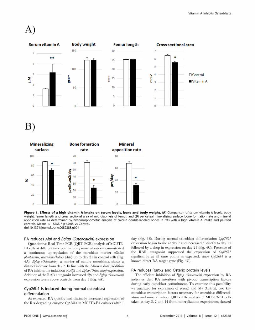
A high dietary vitamin A intake leads to a reducedmineral apposition rate
To control for indirect effects of vitamin A on appetite and
general growth, we applied pair-feeding, i. e. the control rats were
fed the same amount of food as that consumed by the vitamin A
group. The vitamin A group acquired an approximately doubling
of vitamin A levels in serum (Fig. 1A). Although there were no
differences in final body weight or femur length, rats with
hypervitaminosis A had thinner bones as they evinced a
diminished total cross-sectional bone area (12%) at the mid
diaphysis of the femur (Fig.1A). Dynamic histomorphometric
analysis of these bones clearly showed that a high intake of vitamin
A reduced the mineralizing surface (20%), bone formation rate
(60%) and the mineral apposition rate (54%) (Fig. 1B). These
results indicate that a high vitamin A intake has direct and
inhibiting effects on bone formation.
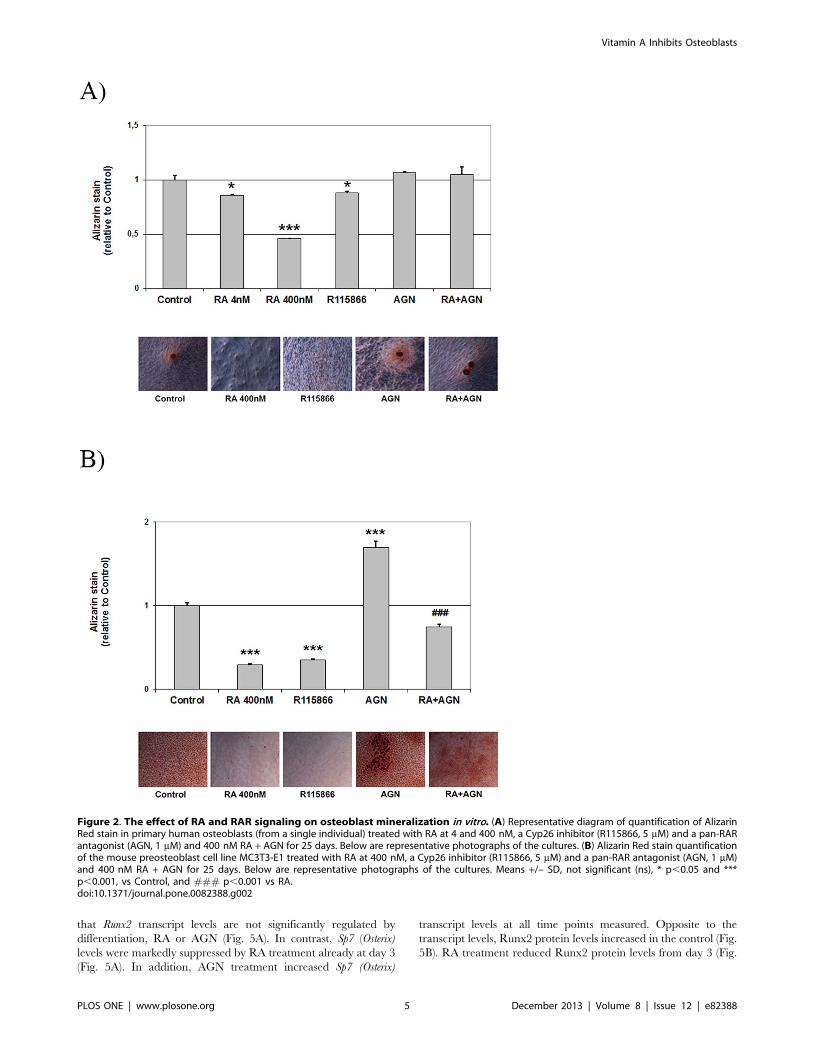
Retinoic acid reduces osteoblast mineralization in vitroTo clarify the specific effect of vitamin A on osteoblasts, we first
added RA to cultures of primary human osteoblasts. As seen in
Figure 2A, addition of 4 nM or 400 nM RA reduced calcium
deposition by 14 and 54%, respectively, as quantified by Alizarin
Red staining. Increasing endogenous RA concentrations using an
inhibitor of the intracellular RA-degrading Cyp26 enzymes
(R115866), reduced osteoblast mineralization by 12%. Addition
of a pan-RA receptor (RAR)-antagonist (AGN) induced a small,
non-significant increase (7%) in Alizarin Red staining. As
preparations of primary cells from bone may contain other cells
types, we then performed mineralization experiments using the
mouse osteoblast cell line MC3T3-E1. In these cells, the effects of
RA, the Cyp26 inhibitor and the RAR antagonist were even more
pronounced (Fig. 2B). Thus, addition of 400 nM RA or R115866
reduced Alizarin staining by 70 and 65%, respectively. AGN
addition alone distinctly increased Alizarin staining to 170% of
control. The significant increase in mineralization after adding the
RAR antagonist indicates that the RA effect is RAR-dependent
and that blocking endogenous RA is sufficient to increase
mineralization. Concomitant addition of AGN completely re-
versed the effect of RA in the human cells and partly reversed the
negative effect of RA on mineralization in the mouse cell line (Fig.
2A,B).
The reduced mineralization is not only a consequence ofinhibited cell proliferation
Next, we measured cell proliferation during MC3T3-E1
mineralization experiments using an MTT-based proliferation
assay. As seen in Fig. 3A, RA inhibited MC3T3-E1 proliferation
and the RAR-antagonist (AGN) did not produce any detectable
difference compared to control cells. Control cell proliferation
appears to level off after day 10. The inhibition of cell proliferation
by RA was confirmed by counting cells grown with or without
400 nM RA for 10 days (Fig. 3B). The number of non-viable cells
did not differ from control cultures (Fig. 3B). Next, to determine
whether RA:s inhibitory effect on mineralization was dependent
on its reduction of proliferation, we added RA or AGN at different
time points. The results show that RA reduced mineralization
even when added as late as day 14 (21%), after completion of the
proliferation phase. Moreover, the RAR antagonist increased
mineralization (16%) also when added after completion of the
proliferation phase (Fig. 3C). Together, these in vitro findings are in
agreement with the dynamic histomorphometric results described
above.
Vitamin A Inhibits Osteoblasts
PLOS ONE | www.plosone.org 3 December 2013 | Volume 8 | Issue 12 | e82388
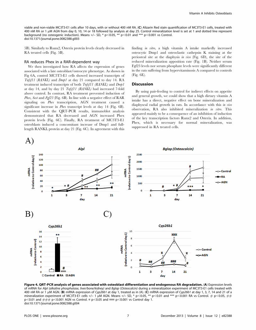
RA reduces Alpl and Bglap (Osteocalcin) expressionQuantitative Real Time-PCR (QRT-PCR) analysis of MC3T3-
E1 cells at different time points during mineralization demonstrated
a continuous up-regulation of the osteoblast marker alkaline
phosphatase, liver/bone/kidney (Alpl) up to day 21 in control cells (Fig.
4A). Bglap (Osteocalcin), a marker of mature osteoblasts, shows a
distinct increase from day 7. In line with the Alizarin data, addition
of RA inhibits the induction of Alpl and Bglap (Osteocalcin) expression.
Addition of the RAR antagonist increased Alpl and Bglap (Osteocalcin)
expression levels above controls from day 3 (Fig. 4A).
Cyp26b1 is induced during normal osteoblastdifferentiation
As expected RA quickly and distinctly increased expression of
the RA degrading enzyme Cyp26b1 in MC3T3-E1 cultures after 1
day (Fig. 4B). During normal osteoblast differentiation Cyp26b1
expression began to rise at day 7 and increased distinctly to day 14
followed by a drop in expression on day 21 (Fig. 4C). Presence of
the RAR antagonist suppressed the expression of Cyp26b1
significantly at all time points as expected, since Cyp26b1 is a
known direct RA target gene (Fig. 4C).
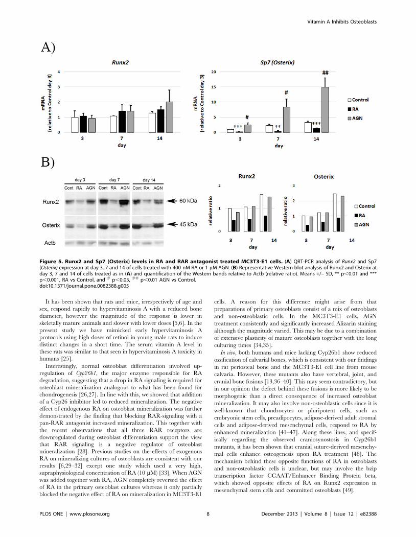
RA reduces Runx2 and Osterix protein levelsThe efficient inhibition of Bglap (Osteocalcin) expression by RA
indicates that RA interferes with pivotal transcription factors
during early osteoblast commitment. To examine this possibility
we analyzed for expression of Runx2 and Sp7 (Osterix), two key
osteoblast transcription factors necessary for osteoblast differenti-
ation and mineralization. QRT-PCR analysis of MC3T3-E1 cells
taken at day 3, 7 and 14 from mineralization experiments showed
Figure 1. Effects of a high vitamin A intake on serum levels, bone and body weight. (A) Comparison of serum vitamin A levels, bodyweight, femur length and cross sectional area of mid diaphysis of femur, and (B) periosteal mineralizing surface, bone formation rate and mineralapposition rate as determined by histomorphometric analysis of calcein double-labeled bones in rats with a high vitamin A intake and pair-fedcontrols. Means +/– SEM, * p,0.05 vs Control.doi:10.1371/journal.pone.0082388.g001
Vitamin A Inhibits Osteoblasts
PLOS ONE | www.plosone.org 4 December 2013 | Volume 8 | Issue 12 | e82388
that Runx2 transcript levels are not significantly regulated by
differentiation, RA or AGN (Fig. 5A). In contrast, Sp7 (Osterix)
levels were markedly suppressed by RA treatment already at day 3
(Fig. 5A). In addition, AGN treatment increased Sp7 (Osterix)
transcript levels at all time points measured. Opposite to the
transcript levels, Runx2 protein levels increased in the control (Fig.
5B). RA treatment reduced Runx2 protein levels from day 3 (Fig.
Figure 2. The effect of RA and RAR signaling on osteoblast mineralization in vitro. (A) Representative diagram of quantification of AlizarinRed stain in primary human osteoblasts (from a single individual) treated with RA at 4 and 400 nM, a Cyp26 inhibitor (R115866, 5 mM) and a pan-RARantagonist (AGN, 1 mM) and 400 nM RA + AGN for 25 days. Below are representative photographs of the cultures. (B) Alizarin Red stain quantificationof the mouse preosteoblast cell line MC3T3-E1 treated with RA at 400 nM, a Cyp26 inhibitor (R115866, 5 mM) and a pan-RAR antagonist (AGN, 1 mM)and 400 nM RA + AGN for 25 days. Below are representative photographs of the cultures. Means +/– SD, not significant (ns), * p,0.05 and ***p,0.001, vs Control, and ### p,0.001 vs RA.doi:10.1371/journal.pone.0082388.g002
Vitamin A Inhibits Osteoblasts
PLOS ONE | www.plosone.org 5 December 2013 | Volume 8 | Issue 12 | e82388
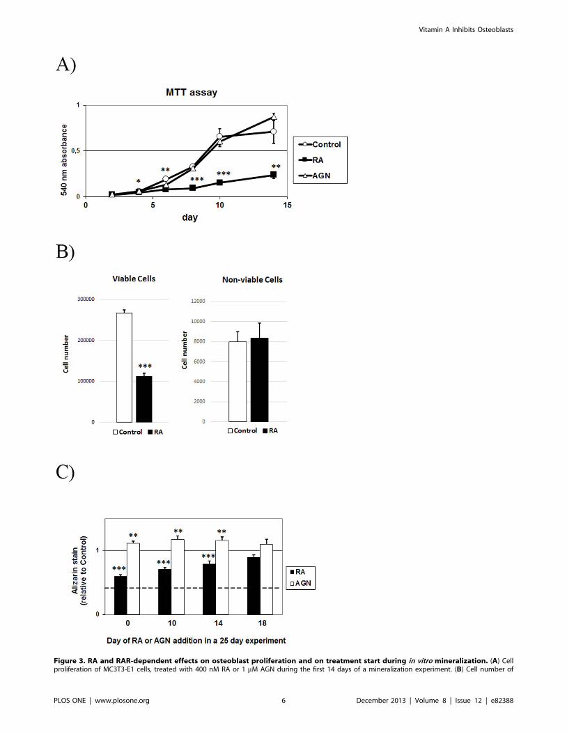
Figure 3. RA and RAR-dependent effects on osteoblast proliferation and on treatment start during in vitro mineralization. (A) Cellproliferation of MC3T3-E1 cells, treated with 400 nM RA or 1 mM AGN during the first 14 days of a mineralization experiment. (B) Cell number of
Vitamin A Inhibits Osteoblasts
PLOS ONE | www.plosone.org 6 December 2013 | Volume 8 | Issue 12 | e82388
5B). Similarly to Runx2, Osterix protein levels clearly decreased in
RA treated cells (Fig. 5B).
RA reduces Phex in a RAR-dependent wayWe then investigated how RA affects the expression of genes
associated with a late osteoblast/osteocyte phenotype. As shown in
Fig 6A, control MC3T3-E1 cells showed increased transcripts of
Tnfsf11 (RANKL) and Dmp1 at day 21 compared to day 14. RA
treatment induced transcripts of both Tnfsf11 (RANKL) and Dmp1
at day 14, and by day 21 Tnfsf11 (RANKL) had increased 7-fold
above control. In contrast, RA treatment prevented induction of
Phex, Sost and Fgf23 (Fig. 6B). In line with a negative effect of RAR
signaling on Phex transcription, AGN treatment caused a
significant increase in Phex transcript levels at day 14 (Fig. 6B).
Consistent with the QRT-PCR results, immunoblot analysis
demonstrated that RA decreased and AGN increased Phex
protein levels (Fig. 6C). Finally, RA treatment of MC3T3-E1
osteoblasts induced a concomitant increase of Dmp1 and full-
length RANKL protein at day 21 (Fig. 6C). In agreement with this
finding in vitro, a high vitamin A intake markedly increased
osteocytic Dmp1 and osteoclastic cathepsin K staining at the
periosteal site at the diaphysis in vivo (Fig. 6D), the site of the
reduced mineralization apposition rate (Fig. 1B). Neither serum
Fgf23 levels nor serum phosphate levels were significantly different
in the rats suffering from hypervitaminosis A compared to controls
(Fig. 6E).
Discussion
By using pair-feeding to control for indirect effects on appetite
and general growth, we could show that a high dietary vitamin A
intake has a direct, negative effect on bone mineralization and
diaphyseal radial growth in rats. In accordance with this in vivo
observation, RA also inhibited mineralization in vitro. This
appeared mainly to be a consequence of an inhibition of induction
of the key transcription factors Runx2 and Osterix. In addition,
Phex, which is necessary for normal mineralization, was
suppressed in RA treated cells.
viable and non-viable MC3T3-E1 cells after 10 days, with or without 400 nM RA. (C) Alizarin Red stain quantification of MC3T3-E1 cells, treated with400 nM RA or 1 mM AGN from day 0, 10, 14 or 18 followed by analysis at day 25. Control mineralization level is set at 1 and dotted line representbackground (no osteogenic induction). Means +/– SD, * p,0.05, ** p,0.01 and *** p,0.001 vs Control.doi:10.1371/journal.pone.0082388.g003
Figure 4. QRT-PCR analysis of genes associated with osteoblast differentiation and endogenous RA degradation. (A) Expression levelsof mRNA for Alpl (alkaline phosphatase, liver/bone/kidney) and Bglap (Osteocalcin) during a mineralization experiment of MC3T3-E1 cells treated with400 nM RA or 1 mM AGN. (B) mRNA expression of Cyp26b1 at day 1, treated as in (A). (C) mRNA expression of Cyp26b1 at day 1, 3, 7, 14 and 21 of amineralization experiment of MC3T3-E1 cells +/– 1 mM AGN. Means +/– SD, * p,0.05, ** p,0.01 and *** p,0.001 RA vs Control. # p,0.05, ##p,0.01 and ### p,0.001 AGN vs Control. ¤ p,0.05 and ¤¤¤ p,0.001 vs Control day 1.doi:10.1371/journal.pone.0082388.g004
Vitamin A Inhibits Osteoblasts
PLOS ONE | www.plosone.org 7 December 2013 | Volume 8 | Issue 12 | e82388
It has been shown that rats and mice, irrespectively of age and
sex, respond rapidly to hypervitaminosis A with a reduced bone
diameter, however the magnitude of the response is lower in
skeletally mature animals and slower with lower doses [5,6]. In the
present study we have mimicked early hypervitaminosis A
protocols using high doses of retinol in young male rats to induce
distinct changes in a short time. The serum vitamin A level in
these rats was similar to that seen in hypervitaminosis A toxicity in
humans [25].
Interestingly, normal osteoblast differentiation involved up-
regulation of Cyp26b1, the major enzyme responsible for RA
degradation, suggesting that a drop in RA signaling is required for
osteoblast mineralization analogous to what has been found for
chondrogenesis [26,27]. In line with this, we showed that addition
of a Cyp26 inhibitor led to reduced mineralization. The negative
effect of endogenous RA on osteoblast mineralization was further
demonstrated by the finding that blocking RAR-signaling with a
pan-RAR antagonist increased mineralization. This together with
the recent observations that all three RAR receptors are
downregulated during osteoblast differentiation support the view
that RAR signaling is a negative regulator of osteoblast
mineralization [28]. Previous studies on the effects of exogenous
RA on mineralizing cultures of osteoblasts are consistent with our
results [6,29–32] except one study which used a very high,
supraphysiological concentration of RA (10 mM) [33]. When AGN
was added together with RA, AGN completely reversed the effect
of RA in the primary osteoblast cultures whereas it only partially
blocked the negative effect of RA on mineralization in MC3T3-E1
cells. A reason for this difference might arise from that
preparations of primary osteoblasts consist of a mix of osteoblasts
and non-osteoblastic cells. In the MC3T3-E1 cells, AGN
treatment consistently and significantly increased Alizarin staining
although the magnitude varied. This may be due to a combination
of extensive plasticity of mature osteoblasts together with the long
culturing times [34,35].
In vivo, both humans and mice lacking Cyp26b1 show reduced
ossification of calvarial bones, which is consistent with our findings
in rat periosteal bone and the MC3T3-E1 cell line from mouse
calvaria. However, these mutants also have vertebral, joint, and
cranial bone fusions [13,36–40]. This may seem contradictory, but
in our opinion the defect behind these fusions is more likely to be
morphogenic than a direct consequence of increased osteoblast
mineralization. It may also involve non-osteoblastic cells since it is
well-known that chondrocytes or pluripotent cells, such as
embryonic stem cells, preadipocytes, adipose-derived adult stromal
cells and adipose-derived mesenchymal cells, respond to RA by
enhanced mineralization [41–47]. Along these lines, and specif-
ically regarding the observed craniosynostosis in Cyp26b1
mutants, it has been shown that cranial suture-derived mesenchy-
mal cells enhance osteogenesis upon RA treatment [48]. The
mechanism behind these opposite functions of RA in osteoblasts
and non-osteoblastic cells is unclear, but may involve the bzip
transcription factor CCAAT/Enhancer Binding Protein beta,
which showed opposite effects of RA on Runx2 expression in
mesenchymal stem cells and committed osteoblasts [49].
Figure 5. Runx2 and Sp7 (Osterix) levels in RA and RAR antagonist treated MC3T3-E1 cells. (A) QRT-PCR analysis of Runx2 and Sp7(Osterix) expression at day 3, 7 and 14 of cells treated with 400 nM RA or 1 mM AGN. (B) Representative Western blot analysis of Runx2 and Osterix atday 3, 7 and 14 of cells treated as in (A) and quantification of the Western bands relative to Actb (relative ratio). Means +/– SD, ** p,0.01 and ***p,0.001, RA vs Control, and # p,0.05, ## p,0.01 AGN vs Control.doi:10.1371/journal.pone.0082388.g005
Vitamin A Inhibits Osteoblasts
PLOS ONE | www.plosone.org 8 December 2013 | Volume 8 | Issue 12 | e82388
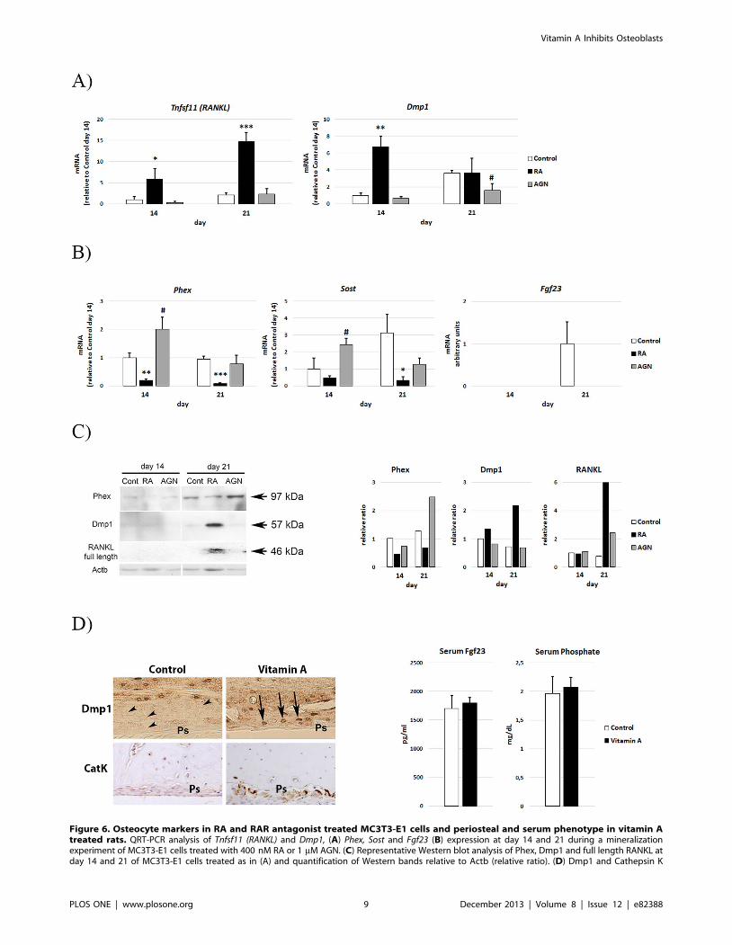
Figure 6. Osteocyte markers in RA and RAR antagonist treated MC3T3-E1 cells and periosteal and serum phenotype in vitamin Atreated rats. QRT-PCR analysis of Tnfsf11 (RANKL) and Dmp1, (A) Phex, Sost and Fgf23 (B) expression at day 14 and 21 during a mineralizationexperiment of MC3T3-E1 cells treated with 400 nM RA or 1 mM AGN. (C) Representative Western blot analysis of Phex, Dmp1 and full length RANKL atday 14 and 21 of MC3T3-E1 cells treated as in (A) and quantification of Western bands relative to Actb (relative ratio). (D) Dmp1 and Cathepsin K
Vitamin A Inhibits Osteoblasts
PLOS ONE | www.plosone.org 9 December 2013 | Volume 8 | Issue 12 | e82388
Our study showed a distinct reduction of Runx2 protein upon
RA treatment, whereas Runx2 transcript levels were unchanged,
consistent with several studies that have indicated that the Runx2
transcript levels are less clearly regulated [50–53]. It is not yet
known how RA alters Runx2 protein levels, but one possible
mechanism could be via RA-induced proteasomal degradation, in
analogy to how RA induces degradation of phosphorylated Smad1
[54]. Alternatively, the decreased Osterix protein levels may result
in reduced Runx2 protein stability, as it was recently shown that
Runx2 and Osterix protein co-expression synergistically increases
their stability [55].
That vitamin A is a negative regulator of bone mineralization is
further strengthen by the recent observation that a retinol-free diet
is more efficient than a high phosphate diet in rescuing the
mineralization defects in the Phex mutant Hyp mouse [56]. The
underlying cause of the reduced mineralization in the Hyp mouse
is the inactivation of the Phex gene. Phex has been shown to
function in concert with Dmp1 and Fgf23 in regulating phosphate
metabolism [57]. Mice lacking both Phex and Dmp1 have non-
additive effects, suggesting a common pathway for these proteins
[58]. Notably, a Dmp1 transgene does not rescue but instead
further reduces bone mineralization in Hyp mice. This indicates
that in the presence of low Phex levels, Dmp1 has a negative effect
of bone mineralization [58]. Since RA downregulated Phex and
upregulated Dmp1 levels in our in vitro cultures, this may, in part,
have contributed to the reduced mineralization. Furthermore, RA
prevented the induction of Fgf23 at the osteoblast level which was
not reflected at the serum level, but is in line with the fact that
retinol deprivation appears to impact the cell-autonomous
mineralization defect of Hyp osteoblasts but not serum parameters
[56].
Previously published data suggest that RA can induce an
osteocytic phenotype in short- and long-term cultures [13,59].
Here, using 400 nM of RA during long-term culture (weeks), we
observed that the expression of the osteocyte-associated genes
RANKL and Dmp1 increased (which has also been observed
earlier by us and others [13,60]). In contrast, other osteocyte-
associated genes such as Phex, Sost and Fgf23 were downregulated,
which is opposite to recent studies [13,59]. The explanation for
these discrepancies is probably that Laue et al. [13] used a higher
concentration of RA, fasting MCT3T-E1 cells and much shorter
treatment time (48 h), and that Mattinzoli el al. [59] used a very
high (10 mM) concentration of RA. In this context it is worth
pointing out that induction of osteocyte markers in cell culture
takes quite a long time, e.g. it takes 14 days to produce detectable
amounts of the osteocyte-specific Sost protein in the osteocyte cell
line IDG-SW3 [61]. We showed here that the RANKL and Dmp1
proteins first appeared clearly on day 21 in the RA treated
preosteoblastic cell line, MC3T3-E1. The concomitant expression
of Dmp1 and full-length RANKL induced by RA in vitro is in
agreement with our present and previous [19] in vivo observations
of increased periosteal Dmp1 staining and the number of
cathepsin K-positive osteoclasts in hypervitaminosis A. Although
we have not been able to stain for full-length RANKL in these
bone sections, it is tempting to speculate that vitamin A
concomitantly induces Dmp1 and RANKL also in vivo, and that
the full-length RANKL in these periosteal cells stimulates the
formation of periosteal osteoclasts. Further studies will be needed
to clarify this.
Acknowledgments
We thank Barrier Therapeutics, Geel, Belgium, for the Cyp26 inhibitor
and Dr. Chandraratna, Allergan, for the high affinity pan-RAR-antagonist,
Olle Nilsson for providing human osteoblasts and Valeria Giandomenico
for insightful suggestions on the manuscript.
Author Contributions
Conceived and designed the experiments: TL AS AJ HM. Performed the
experiments: TL AS LH GA AJ. Analyzed the data: TL AS LH GA AJ
HM. Contributed reagents/materials/analysis tools: GP GA. Wrote the
paper: TL HM.
References
1. Melhus H, Michaelsson K, Kindmark A, Bergstrom R, Holmberg L, et al. (1998)
Excessive dietary intake of vitamin A is associated with reduced bone mineral
density and increased risk for hip fracture. Ann Intern Med 129: 770–778.
2. Michaelsson K, Lithell H, Vessby B, Melhus H (2003) Serum retinol levels and
the risk of fracture. N Engl J Med 348: 287–294.
3. Feskanich D, Singh V, Willett WC, Colditz GA (2002) Vitamin A intake and hip
fractures among postmenopausal women. JAMA 287: 47–54.
4. Binkley N, Krueger D (2000) Hypervitaminosis A and bone. Nutr Rev 58: 138–
144.
5. Johansson S, Lind PM, Hakansson H, Oxlund H, Orberg J, et al. (2002)
Subclinical hypervitaminosis A causes fragile bones in rats. Bone 31: 685–689.
6. Kneissel M, Studer A, Cortesi R, Susa M (2005) Retinoid-induced bone thinning
is caused by subperiosteal osteoclast activity in adult rodents. Bone 36: 202–214.
7. Conaway HH, Henning P, Lerner UH (2013) Vitamin A metabolism, action,
and role in skeletal homeostasis. Endocr Rev May 29 [Epub ahead of print].
8. Kindmark A, Torma H, Johansson A, Ljunghall S, Melhus H (1993) Reverse
transcription-polymerase chain reaction assay demonstrates that the 9-cis
retinoic acid receptor alpha is expressed in human osteoblasts. Biochem Biophys
Res Commun 192: 1367–1372.
9. Inoue A, Otsuka E, Hiruma Y, Hirose S, Furuya M, et al. (1996) Stimulation by
retinoids of the natriuretic peptide system of osteoblastic MC3T3-E1 cells.
Biochem Biophys Res Commun 228: 182–186.
10. Reijntjes S, Gale E, Maden M (2003) Expression of the retinoic acid catabolising
enzyme CYP26B1 in the chick embryo and its regulation by retinoic acid. Gene
Expr Patterns 3: 621–627.
11. Lee LM, Leung CY, Tang WW, Choi HL, Leung YC, et al. (2012) A
paradoxical teratogenic mechanism for retinoic acid. Proc Natl Acad Sci U S A
109: 13668–13673.
12. Gericke J, Ittensohn J, Mihaly J, Alvarez S, Alvarez R, et al. (2013) Regulation of
retinoid-mediated signaling involved in skin homeostasis by RAR and RXR
agonists/antagonists in mouse skin. PLoS One 8: e62643.
13. Laue K, Pogoda HM, Daniel PB, van Haeringen A, Alanay Y, et al. (2011)
Craniosynostosis and multiple skeletal anomalies in humans and zebrafish result
from a defect in the localized degradation of retinoic acid. Am J Hum Genet 89:
595–606.
14. Karsenty G (2008) Transcriptional control of skeletogenesis. Annu Rev
Genomics Hum Genet 9: 183–196.
15. Nakashima T, Hayashi M, Fukunaga T, Kurata K, Oh-Hora M, et al. (2011)
Evidence for osteocyte regulation of bone homeostasis through RANKL
expression. Nat Med 17: 1231–1234.
16. Jimi E, Nakamura I, Amano H, Taguchi Y, Tsurukai T, et al. (1996) Osteoclast
function is activated by osteoblastic cells through a mechanism involving cell-to-
cell contact. Endocrinology 137: 2187–2190.
17. Mbalaviele G, Nishimura R, Myoi A, Niewolna M, Reddy SV, et al. (1998)
Cadherin-6 mediates the heterotypic interactions between the hemopoietic
osteoclast cell lineage and stromal cells in a murine model of osteoclast
differentiation. J Cell Biol 141: 1467–1476.
18. Xiong J, Onal M, Jilka RL, Weinstein RS, Manolagas SC, et al. (2011) Matrix-
embedded cells control osteoclast formation. Nat Med 17: 1235–1241.
(CatK) immunohistochemical staining at the diaphyseal periosteal site in rats suffering from hypervitaminosis A and in control rats. Upper panel:Arrow heads indicate Dmp1 negative osteocytes close to the periosteum (Ps) in control rat bone and arrows indicate Dmp1 positive osteocytes closeto the periosteum in hypervitaminosis A rat bone. Lower panel: only Vitamin A animals show clear CatK staining at the Ps site. (E) Serum Fgf23 andphosphate levels in rats from (D). Means +/– SD, * p,0.05, ** p,0.01 and *** p,0.001 RA vs Control. # p,0.05, ## p,0.01 AGN vs Control.doi:10.1371/journal.pone.0082388.g006
Vitamin A Inhibits Osteoblasts
PLOS ONE | www.plosone.org 10 December 2013 | Volume 8 | Issue 12 | e82388
19. Lind T, Lind PM, Jacobson A, Hu L, Sundqvist A, et al. (2011) High dietary
intake of retinol leads to bone marrow hypoxia and diaphyseal endostealmineralization in rats. Bone 48: 496–506.
20. Parfitt AM, Drezner MK, Glorieux FH, Kanis JA, Malluche H, et al. (1987)
Bone histomorphometry: standardization of nomenclature, symbols, and units.Report of the ASBMR Histomorphometry Nomenclature Committee. J Bone
Miner Res 2: 595–610.21. Hollberg K, Marsell R, Norgard M, Larsson T, Jonsson KB, et al. (2008)
Osteoclast polarization is not required for degradation of bone matrix in rachitic
FGF23 transgenic mice. Bone 42: 1111–1121.22. Jonsson KB, Frost A, Nilsson O, Ljunghall S, Ljunggren O (1999) Three
isolation techniques for primary culture of human osteoblast-like cells: acomparison. Acta Orthop Scand 70: 365–373.
23. Hu L, Lind T, Sundqvist A, Jacobson A, Melhus H (2010) Retinoic acidincreases proliferation of human osteoclast progenitors and inhibits RANKL-
stimulated osteoclast differentiation by suppressing RANK. PLoS One 5:
e13305.24. Hollberg K, Hultenby K, Hayman A, Cox T, Andersson G (2002) Osteoclasts
from mice deficient in tartrate-resistant acid phosphatase have altered ruffledborders and disturbed intracellular vesicular transport. Exp Cell Res 279: 227–
238.
25. Smith FR, Goodman DS (1976) Vitamin A transport in human vitamin Atoxicity. N Engl J Med 294: 805–808.
26. Pacifici M, Cossu G, Molinaro M, Tato F (1980) Vitamin A inhibitschondrogenesis but not myogenesis. Exp Cell Res 129: 469–474.
27. Hoffman LM, Garcha K, Karamboulas K, Cowan MF, Drysdale LM, et al.(2006) BMP action in skeletogenesis involves attenuation of retinoid signaling. J
Cell Biol 174: 101–113.
28. Roforth MM, Liu G, Khosla S, Monroe DG (2012) Examination of nuclearreceptor expression in osteoblasts reveals Rorb as an important regulator of
osteogenesis. J Bone Miner Res 27: 891–901.29. Ohishi K, Nishikawa S, Nagata T, Yamauchi N, Shinohara H, et al. (1995)
Physiological concentrations of retinoic acid suppress the osteoblastic differen-
tiation of fetal rat calvaria cells in vitro. Eur J Endocrinol 133: 335–341.30. Nuka S, Sawada N, Iba K, Chiba H, Ishii S, et al. (1997) All-trans retinoic acid
inhibits dexamethasone-induced ALP activity and mineralization in humanosteoblastic cell line SV HFO. Cell Struct Funct 22: 27–32.
31. Cohen-Tanugi A, Forest N (1998) Retinoic acid suppresses the osteogenicdifferentiation capacity of murine osteoblast-like 3/A/1D-1M cell cultures.
Differentiation 63: 115–123.
32. Iba K, Chiba H, Yamashita T, Ishii S, Sawada N (2001) Phase-independentinhibition by retinoic acid of mineralization correlated with loss of tetranectin
expression in a human osteoblastic cell line. Cell Struct Funct 26: 227–233.33. Song HM, Nacamuli RP, Xia W, Bari AS, Shi YY, et al. (2005) High-dose
retinoic acid modulates rat calvarial osteoblast biology. J Cell Physiol 202: 255–
262.34. Liu F, Malaval L, Aubin JE (1997) The mature osteoblast phenotype is
characterized by extensive plasticity. Exp Cell Res 232: 97–105.35. Candeliere GA, Liu F, Aubin JE (2001) Individual osteoblasts in the developing
calvaria express different gene repertoires. Bone 28: 351–361.36. Maclean G, Dolle P, Petkovich M (2009) Genetic disruption of CYP26B1
severely affects development of neural crest derived head structures, but does not
compromise hindbrain patterning. Dev Dyn 238: 732–745.37. Yashiro K, Zhao X, Uehara M, Yamashita K, Nishijima M, et al. (2004)
Regulation of retinoic acid distribution is required for proximodistal patterningand outgrowth of the developing mouse limb. Dev Cell 6: 411–422.
38. Wiley MJ (1983) The pathogenesis of retinoic acid-induced vertebral
abnormalities in golden Syrian hamster fetuses. Teratology 28: 341–353.39. Laue K, Janicke M, Plaster N, Sonntag C, Hammerschmidt M (2008)
Restriction of retinoic acid activity by Cyp26b1 is required for proper timingand patterning of osteogenesis during zebrafish development. Development 135:
3775–3787.
40. Spoorendonk KM, Peterson-Maduro J, Renn J, Trowe T, Kranenbarg S, et al.(2008) Retinoic acid and Cyp26b1 are critical regulators of osteogenesis in the
axial skeleton. Development 135: 3765–3774.
41. Iwamoto M, Shapiro IM, Yagami K, Boskey AL, Leboy PS, et al. (1993)Retinoic acid induces rapid mineralization and expression of mineralization-
related genes in chondrocytes. Exp Cell Res 207: 413–420.
42. Iwamoto M, Yagami K, Shapiro IM, Leboy PS, Adams SL, et al. (1994)
Retinoic acid is a major regulator of chondrocyte maturation and matrixmineralization. Microsc Res Tech 28: 483–491.
43. Wang W, Kirsch T (2002) Retinoic acid stimulates annexin-mediated growthplate chondrocyte mineralization. J Cell Biol 157: 1061–1069.
44. Skillington J, Choy L, Derynck R (2002) Bone morphogenetic protein andretinoic acid signaling cooperate to induce osteoblast differentiation of
preadipocytes. J Cell Biol 159: 135–146.
45. Yamashita A, Takada T, Narita J, Yamamoto G, Torii R (2005) Osteoblasticdifferentiation of monkey embryonic stem cells in vitro. Cloning Stem Cells 7:
232–237.
46. Malladi P, Xu Y, Yang GP, Longaker MT (2006) Functions of vitamin D,
retinoic acid, and dexamethasone in mouse adipose-derived mesenchymal cells.Tissue Eng 12: 2031–2040.
47. Wan DC, Siedhoff MT, Kwan MD, Nacamuli RP, Wu BM, et al. (2007)Refining retinoic acid stimulation for osteogenic differentiation of murine
adipose-derived adult stromal cells. Tissue Eng 13: 1623–1631.
48. James AW, Levi B, Xu Y, Carre AL, Longaker MT (2010) Retinoic acid
enhances osteogenesis in cranial suture-derived mesenchymal cells: potentialmechanisms of retinoid-induced craniosynostosis. Plast Reconstr Surg125:
1352–1361.
49. Wiper-Bergeron N, St-Louis C, Lee JM (2007) CCAAT/Enhancer bindingprotein beta abrogates retinoic acid-induced osteoblast differentiation via
repression of Runx2 transcription. Mol Endocrinol 21: 2124–2135.
50. Prince M, Banerjee C, Javed A, Green J, Lian JB, et al. (2001) Expression and
regulation of Runx2/Cbfa1 and osteoblast phenotypic markers during thegrowth and differentiation of human osteoblasts. J Cell Biochem 80: 424–440.
51. Sudhakar S, Li Y, Katz MS, Elango N (2001) Translational regulation is acontrol point in RUNX2/Cbfa1 gene expression. Biochem Biophys Res
Commun 289: 616–622.
52. Jones DC, Wein MN, Oukka M, Hofstaetter JG, Glimcher MJ, et al. (2006)
Regulation of adult bone mass by the zinc finger adapter protein Schnurri-3.Science 312: 1223–1227.
53. Li X, Huang M, Zheng H, Wang Y, Ren F, et al. (2008) CHIP promotes Runx2
degradation and negatively regulates osteoblast differentiation. J Cell Biol 181:959–972.
54. Sheng N, Xie Z, Wang C, Bai G, Zhang K, et al. (2010) Retinoic acid regulatesbone morphogenic protein signal duration by promoting the degradation of
phosphorylated Smad1. Proc Natl Acad Sci U S A 107: 18886–18891.
55. Rashid H, Chen H, Ma C, Sinha K, DeCrombrugghe B, et al. (2012) Runx2
and Osterix Molecular Complex Synergistically Regulate Osteogenic Genes,ASBMR Annual Meeting presentation #FR0218.
56. Seitz S, Rendenbach C, Barvencik F, Streichert T, Jeschke A, et al. (2013)Retinol deprivation partially rescues the skeletal mineralization defects of Phex-
deficient Hyp mice. Bone 53: 231–238.
57. Martin A, Liu S, David V, Li H, Karydis A, et al. (2011) Bone proteins PHEX
and DMP1 regulate fibroblastic growth factor Fgf23 expression in osteocytes
through a common pathway involving FGF receptor (FGFR) signaling. FASEB J25: 2551–2562.
58. Martin A, David V, Li H, Dai B, Feng JQ, et al. (2012) Overexpression of theDMP1 C-terminal fragment stimulates FGF23 and exacerbates the hypophos-
phatemic rickets phenotype in Hyp mice. Mol Endocrinol 26: 1883–1895.
59. Mattinzoli D, Messa P, Corbelli A, Ikehata M, Zennaro C, et al. (2012) A novel
model of in vitro osteocytogenesis induced by retinoic acid treatment. Eur CellMater 24: 403–425.
60. Jacobson A, Johansson S, Branting M, Melhus H (2004) Vitamin A differentiallyregulates RANKL and OPG expression in human osteoblasts. Biochem Biophys
Res Commun 322: 162–167.
61. Woo SM, Rosser J, Dusevich V, Kalajzic I, Bonewald LF (2011) Cell line IDG-
SW3 replicates osteoblast-to-late-osteocyte differentiation in vitro and acceler-
ates bone formation in vivo. J Bone Miner Res 26: 2634–2646.
Vitamin A Inhibits Osteoblasts
PLOS ONE | www.plosone.org 11 December 2013 | Volume 8 | Issue 12 | e82388