Embed Size (px)
Citation preview
Review ArticleVitamin C versus Cancer: Ascorbic Acid Radical andImpairment of Mitochondrial Respiration?
Rumiana Bakalova ,1,2 Zhivko Zhelev,3,4 Thomas Miller,5 Ichio Aoki,1,2
and Tatsuya Higashi 1
1Department of Molecular Imaging and Theranostics, National Institute of Radiological Sciences (NIRS), National Institutes forQuantum and Radiological Science and Technology (QST), Chiba, Japan2Group of Quantum-State Controlled MRI, Institute for Quantum Life Science, National Institutes for Quantum and RadiologicalScience and Technology (QST), Chiba, Japan3Institute of Biophysics and Biomedical Engineering, Bulgarian Academy of Sciences, Sofia, Bulgaria4Medical Faculty, Trakia University, Stara Zagora, Bulgaria5IC-MedTech Co., San Diego, CA, USA
Correspondence should be addressed to Rumiana Bakalova; [email protected]
Received 21 July 2019; Revised 14 September 2019; Accepted 28 September 2019; Published 10 January 2020
Academic Editor: Francisco J. Romero
Copyright © 2020 Rumiana Bakalova et al. This is an open access article distributed under the Creative Commons AttributionLicense, which permits unrestricted use, distribution, and reproduction in any medium, provided the original work is properly cited.
Vitamin C as a cancer therapy has a controversial history. Much of the controversy arises from the lack of predictive biomarkers forstratification of patients, as well as a clear understanding of the mechanism of action and its multiple targets underlying theanticancer effect. Our review expands the analysis of cancer vulnerabilities for high-dose vitamin C, based on several facts,illustrating the cytotoxic potential of the ascorbyl free radical (AFR) via impairment of mitochondrial respiration and themechanisms of its elimination in mammals by the membrane-bound NADH:cytochrome b5 oxidoreductase 3 (Cyb5R3). Thisenzyme catalyzes rapid conversion of AFR to ascorbate, as well as reduction of other redox-active compounds, using NADH asan electron donor. We propose that vitamin C can function in “protective mode” or “destructive mode” affecting cellularhomeostasis, depending on the intracellular “steady-state” concentration of AFR and differential expression/activity of Cyb5R3in cancerous and normal cells. Thus, a specific anticancer effect can be achieved at high doses of vitamin C therapy. The reviewis intended for a wide audience of readers—from students to specialists in the field.
1. Introduction
In the April 2019 issue of Nature Reviews Cancer, Ngo et al.have described cancer vulnerabilities for high-dose vitaminC therapy [1]. The authors note that vitamin C as a cancertherapy has a controversial history, and much of the contro-versy arises from the lack of predictive biomarkers for strati-fication of patients, as well as a clear understanding of themechanism of action and its multiple targets underlying theanticancer effect [2].
Our article links empirical and experimental observationsfrom clinical practice that may, at first glance, seem dis-jointed. However, common characteristics of the pathologiesdiscussed herein include mitochondrial dysfunction and
severe inflammation. These are important targets for vitaminC and hallmarks of cancer. In particular, we expand the anal-ysis of cancer vulnerabilities for high-dose vitamin C, basedon several significant and indicative facts, illustrating thecytotoxic potential of the ascorbyl free radical (AFR) viaimpairment of mitochondrial respiration and the mecha-nisms of its elimination in mammals:
(i) AFR is an intermediate product from the oxidationof ascorbate by one-electron reduction of transitionmetal ions and/or interaction with free radicals [1].AFR does not readily react with oxygen or othermolecules to generate more reactive radicals andmay temporarily accumulate in cells [3]
HindawiOxidative Medicine and Cellular LongevityVolume 2020, Article ID 1504048, 12 pageshttps://doi.org/10.1155/2020/1504048
(ii) AFR is a by-product of some vital biochemical reac-tions. Ascorbate participates in the recovery ofhemoglobin from methemoglobin in erythrocytes,as well as in the enzymatic hydroxylation (e.g., dur-ing the synthesis of noradrenalin in the chromaffingranules of dopamine-synthesizing cells) [4–6]. Bothprocesses are accompanied by AFR production
(iii) NADH:cytochrome b5 oxidoreductase 3 (Cyb5R3)catalyzes rapid conversion of AFR to ascorbate, aswell as reduction of other redox-active compounds,using NADH as an electron donor [7, 8]. Thus,Cyb5R3 eliminates AFR, restores the ascorbate“pool,” and helps maintain the NAD+/NADH ratioin cells. This enzyme is ubiquitously expressed inall mammalian cells, implying an important role incell homeostasis
(iv) Lack or downregulation of Cyb5R3 leads to develop-ment of recessive hereditary methemoglobinemia(RHM), accompanied by neuropathies and impair-ment of mitochondrial function [7, 9]. Ascorbatetreatment has beneficial effects on methemoglobine-mia, especially in patients with ascorbate deficiency.However, ascorbate deficiency does not explain theneuropathy and mitochondrial impairment associ-ated with this pathology
(v) Downregulation of membrane-bound Cyb5R3 incells leads to lower NAD+/NADH ratio, impairedmitochondrial respiration rate, and decreased ATPproduction, which is associated with higher sensitiv-ity to oxidative stress [9]
(vi) We propose that the accumulation of high concen-trations of AFR in cells during oxidative stressand/or some specific metabolic activity is responsi-ble for the impairment of mitochondrial respirationin the absence or downregulation of Cyb5R3, partic-ularly the enzyme located on the outer mitochon-drial membrane (OMM Cyb5R3). This can inducecytotoxicity in mammalian cells. Perhaps this iswhy enzymes found in plants that convert ascorbateto AFR (ascorbate oxidase and ascorbate peroxidase)did not evolve in animals and humans [3]
Is AFR harmful formitochondria and can pharmacologicaldoses of vitamin C attack cancer cells by inhibiting membrane-bound Cyb5R3 and impairing mitochondrial respiration?
2. Cyb5R3/VDAC1 Is a Redox-Cycling SystemThat Converts AFR to Ascorbate
The discovery of enzyme activity of Cyb5R3 dates backto 1951, when Lehninger reported that isolated rat livermitochondria catalyze rapid oxidation of NADH in thepresence of added cytochrome c. This mitochondrialNADH:cytochrome c oxidoreductase activity was foundto be independent of the respiratory chain. Thirty yearslater, it was found that this enzyme is NADH:semidehy-
droascorbic acid reductase, which uses a cytochrome b5-likehemoprotein of the outer mitochondrial membrane (OMMcytochrome b5), which is distinct from microsomalcytochrome b5 [10]. A similar enzyme was also found inSaccharomyces cerevisiae—mitochondrial NADH:cyto-chrome b5 reductase, which catalyzes the reduction ofD-erythroascorbic acid radical [11].
Recently, OMM NADH:cytochrome b5 reductase wasclassified as OMM Cyb5R3 (NADH:ferricytochrome b5 oxi-doreductase 3; EC 1.6.2.2.). Cyb5R3 belongs to the class ofcancer-related and disease-related genes. Cyb5R3 enzymebelongs to the class of potential drug targets and predictedmembrane proteins (according to The Human Protein Atlas).The Cyb5R3 protein is encoded in two isoforms: (i) soluble,exclusively expressed in erythrocytes, and (ii) membrane-bound, expressed in all other cells and anchored to the outermitochondrial membrane, endoplasmic reticulum, and plas-matic membrane [8]. Deficiency of cytosolic Cyb5R3 causestype I RHM, which is a benign condition with mild cyanosis,fatigue, and shortness of breath upon exertion [9, 12]. Defi-ciency of membrane-bound Cyb5R3 causes incurable typeII RHM, accompanied by severe neuropathy, permanent cya-nosis, and impairment of mitochondrial respiration [9, 12].
OMM Cyb5R3 is functionally connected to the voltage-dependent anion channel 1 (VDAC1), the most abundantprotein of the outer mitochondrial membrane [8]. Initially,it was believed that VDAC1 possesses NADH-dependentoxidoreductase activity. However, VDAC1 does not containflavin, heme, and/or metal ions in its active center, nor anyother catalytic motif of the known NAD(P)H-dependent oxi-doreductases. Recombinant VDAC1 and VDAC1 purifiedfrom comigrating proteins either lack oxidoreductase activityor exhibit very low activity [8, 13, 14]. The purity of VDAC1preparations studied usually reaches only 90-95%. Inall cases, other proteins of equal molecular weights(32-35 kDa) are present, which affect the efficiency of separa-tion [15–18]. It was reported that Cyb5R3 consists of 5-10%of the purified VDAC1 [15, 18]. The oxidoreductase activityascribing to VDAC1 is inhibited by nonspecific VDACblockers [15, 16, 19], as well as by specific Cyb5R3 inhibitors[19]. This is direct evidence that oxidoreductase activity ofVDAC1 is due to the presence of Cyb5R3 in the samples. Itseems that both proteins cofunction and could be consideredas one “redox-cycling system” of the outer mitochondrialmembrane. Recent studies on cells treated with paraquat(considered to be carcinogenic) indicate that VDAC1 main-tains membrane integrity and keeps mitochondria intact,while Cyb5R3 acts as an oxidoreductase [8, 20].
3. The Cyb5R3/VDAC1 Redox-Cycling SystemReduces AFR and Helps MaintainMitochondrial Homeostasis
The crucial role of Cyb5R3 in regeneration of AFR to ascor-bate and mitochondrial homeostasis has been demonstratedby Shirabe et al. on human dermal fibroblasts (HDFs),derived from a patient with type II RHM with a pointmutation in the Cyb5R3 gene [21]. This mutation results
2 Oxidative Medicine and Cellular Longevity
in a complete absence of membrane-bound Cyb5R3 in thecells. The authors have reported severely decreased cellularNADH:semidehydroascorbic acid reductase activity androtenone-insensitive NADH:cytochrome c reductase activ-ity, compared to neonatal HDFs. These data suggest thatOMM Cyb5R3 and its NADH:semidehydroascorbic acidreductase activity might contribute to the protection andnormal functioning of the cell, as well as protection ofits mitochondrial electron transport chain (ETC).
Siendones et al. have found that membrane-boundCyb5R3-deficient HDFs, derived from two patients with sim-ilar mutations, are characterized by the decrease of severalparameters: NAD+/NADH ratio, mitochondrial respirationrate, activities of mitochondrial Complexes I-IV, and ATPproduction [22]. This is associated with higher sensitivity ofCyb5R3-deficient HDFs to oxidative stress and acceleratedsenescence, compared to neonatal HDFs. The role of Cyb5R3in the modulation of mitochondrial function has also beenevaluated in Cyb5R3-siRNA-silenced MRC-5 normal fibro-blasts [22]. Downregulation of Cyb5R3 is accompanied by asignificant decrease of the NAD+/NADH ratio and mito-chondrial respiration rate. Cyb5R3-siRNA-silenced MRC-5cells and type II RHMHDFs displayed low proliferation ratesand increased cell death. This is consistent with impairmentof mitochondrial metabolism and reduction of oxidativephosphorylation, based on analysis of oxygen consumption.The authors suggest that impairment of Cyb5R3 expressionand/or its AFR reductase activity may result not only inNADH accumulation but also in defects of mitochondrialETC due to increased oxidative damage, ultimately leadingto senescence.
Recently, it was reported that Cyb5R3 is essential for car-diomyocyte function [23]. Cardiomyocyte-specific inducibleCyb5R3-knockout mice (Myh6-CreERT2-flox/flox) show aloss of mitochondrial biogenesis, associated with 30% lossof total ATP, 50% loss of Complex IV activity, and 25% lossof Complex IV protein quantity [23]. RNAseq pathwayanalysis indicated significantly decreased expression of Com-plexes I, III, and IV after downregulation of membrane-bound Cyb5R3 in cardiomyocytes. This study shows thatCyb5R3 is essential for normal mitochondrial function andcell survival.
The studies described above suggest that the presence ofOMMCyb5R3 is essential for the following parameters at nor-mal conditions: activity of mitochondrial ETC, oxygen con-sumption, ATP production, and resistance to oxidative stress.
In vitro studies on isolated mitochondria indicate thatthe OMM Cyb5R3/VDAC1 complex is responsible forthe transfer of electrons from cytosolic NADH into mito-chondria and that the process is dependent to Complex IV[24, 25]. This is accompanied by oxygen uptake, protonpumping, and generation of mitochondrial potential, sup-ported by small catalytic amounts of external (mitochondrial)cytochrome c. Their data also demonstrate that cytochromec-dependent NADH oxidation is strongly inhibited by dex-tran sulfate (500 kDa)—an inhibitor of VDAC1. They havesuggested that in physiological conditions, cytochrome cmight be transferred in a very limited amount outside themitochondria so as to promote the activation of the
Cyb5R3-dependent electron transport pathway. The processis highly dependent on induction of contact sites in the outerand inner mitochondrial membranes (membrane remodel-ing), as well as on inhibitors of VDAC1. The activity of thisCyb5R3/VDAC1-dependent system becomes functional inremoving the excess cytosolic NADH and is essential for cellsurvival under impairment of the ETC at the level of one ofthe first respiratory complexes. Since the Cyb5R3-catalyzedelectron transfer generates an electrochemical membranepotential associated with the activity of the cytochromeoxidase [24, 26, 27], it represents an additional pathway forproviding energy to cells. This aspect is essential for the cellsin which oxidative phosphorylation activity is not properlysupported by the respiratory chain.
The same authors also demonstrate a nonenzymaticinduction of this alternative pathway for ATP synthesis inmitochondria [24, 25]. They added ascorbate (instead ofNADH) and small catalytic amounts of external cytochromec to intact mitochondria and detected oxygen uptake, accom-panied by cytochrome c reduction and ascorbate oxidation[24, 25]. These processes are dependent on Complex IVand the induction of contact sites in mitochondrial mem-brane and are sensitive to inhibitors of VDAC1. It can beassumed that oxidation of ascorbate by small catalyticamounts of external cytochrome c leads also to AFR produc-tion. AFR may enter the mitochondria via VDAC1 and affectmitochondrial respiration at Complexes III-IV—a hypothe-sis, which is described below in the context of the anticancereffect of high doses of vitamin C.
4. The Cyb5R3/VDAC1 Redox-CyclingSystem in Cancer
Studies discussed above suggest that OMM Cyb5R3/VDAC1is vital for mitochondrial homeostasis, protection againstoxidative stress, prevention of cell senescence, and cellularlongevity. These events clearly relate to AFR eliminationand maintenance of the cytosolic NAD+/NADH ratio—-crucial factors for cell survival.
It has been shown that VDAC1 is highly expressed in allcells as a consequence of exposure to various toxic substancesand plays a crucial role in the protection against intoxication[8, 28, 29]. VDAC1 interacts with both proapoptotic andantiapoptotic factors, which makes it a gatekeeper formitochondria-mediated cell death or survival signaling path-ways [30, 31]. This illustrates the complexity of VDAC func-tions in normal and cancer cells. It has been observed thatoverexpression of VDAC1 in cancer cells is associated withhigh metastatic potential, low therapeutic efficiency, andpoor prognosis [30, 31]. In this case, VDAC1 appears to beinvolved in protecting mitochondria from reactive oxygenspecies (ROS), functioning as a prosurvival pathway [28–30].
Cyb5R3 is also recognized as a carcinogen detoxificationgene [32, 33], although its role in carcinogenesis is not yetwell studied. Single reports on the role of OMM Cyb5R3 incancer have appeared over the past 10 years [32, 34, 35]. Itwas found that the enzyme is overexpressed in cancer cells,protecting them against oxidative stress and induction ofapoptosis [32, 34, 35].
3Oxidative Medicine and Cellular Longevity
Rajcevic et al. have analyzed the proteomic profile of met-abolic proteins in the invasive glioblastoma phenotype byapplying a functional analysis, using isobaric peptide taggingchemistry (iTRAQ) combined with bioinformatics analysis(Ingenuity® Pathway Analysis) [35]. Cyb5R3 is identified asa key protein in a canonical pathway of the cancerous pheno-type associated with mitochondrial dysfunction.
Two groups have reported that membrane-boundCyb5R3 overexpression and polymorphism are associatedwith breast cancer risk in women [32, 34]. Blanke et al.have found that one variant of Cyb5R3 is significantlyoverexpressed in African American women with breastcancer, which is accompanied by impaired detoxificationof aromatic and heterocyclic amine mammary carcino-gens in cigarette smoke [32]. The authors suggest thatoverexpression of membrane-bound Cyb5R3 should beconsidered as a valuable marker in etiologic studies ofprostate, bladder, and colon cancers, which are also asso-ciated with exposure to arylamine and heterocyclic aminecarcinogens.
A large cohort study on patients with estrogen receptor-negative (ERneg) and progesterone receptor-negative (PRneg)breast tumors reveals a central role of membrane-boundCyb5R3 in extravasation/colonization of cancer cells andmetastasis formation at distinct sites (e.g., lung) [34]. Immu-nohistochemical analysis has identified a significant correla-tion between high Cyb5R3 expression and poor disease-free(DFS; p = 0:02) and overall survival (OS: p = 0:04). Multivari-ate analysis revealed about 2-fold higher risk of poor outcomefor ERneg/PRneg patients exhibiting strong staining ofCyb5R3,compared to those expressing weak staining. Cyb5R3 geneknockdown using siRNA in metastatic cells leads to a sig-nificantly decreased tumor burden in lungs when injectedintravenously in immunodeficient mice. The cellular effectsof Cyb5R3-siRNA knockdown showed signaling alterationsassociated with extravasation, such as transforming growthfactor beta (TGFβ) and hypoxia-inducible factor alpha(HIFα) pathways, and strong suppression of proliferation.It is interesting to note that Cyb5R3-siRNA-silencedcancer cells stop proliferation and metastasis, but theycontinue to survive [34].
5. AFR in Mitochondrial Homeostasis at Lowand High Doses of Vitamin C
Our hypothesis is focused on the intracellular generationof AFR at low and high doses of vitamin C, together withits effect on mitochondrial respiration, intracellular redoxbalance, and energy metabolism mediated by the OMMCyb5R3/VDAC1.
We propose that vitamin C can function in “protectivemode” or “destructive mode” affecting cellular homeostasis,depending on the OMM Cyb5R3 expression/activity andintracellular “steady-state” concentration of AFR. The hypoth-esis is focused on the effects of AFR in the mitochondrialintermembrane space and the cytosol.
Membrane-bound Cyb5R3 has several characteristicsthat could be crucial to mitochondrial homeostasis:
(i) The redox potential of the OMM-bound cyto-chrome b5 varies from -160mV to -272mV,depending on the NAD+/NADH ratio [8, 13].Therefore, OMMCyb5R3 can catalyze the reductionof a variety of redox cyclers in addition to AFR, suchas quinone-like compounds, bioreductive xenobio-tics/drug, and nitrocompounds [8, 17]. These redoxcyclers can influence the intracellular redox state
(ii) Membrane-bound Cyb5R3 helps maintain thecytosolic NAD+/NADH ratio, supplying cells withreducing equivalents by transferring electronsfrom cytosolic NADH to the respective membranecomponents (electron acceptors and carriers)[7, 8, 10, 11, 22]
(iii) The membrane-bound Cyb5R3 is considered to beone of the major enzymes that maintain the NAD+/-NADH ratio, by using the coenzyme Q (CoQ)“pools” of the outer mitochondrial membrane andendoplasmic reticulum [8, 10, 11]
CoQ is most likely the main acceptor of excess electrons,as it is the most abundant membrane-bound redox-activecompound in the cell. CoQ is also a key electron carrier inmitochondria. Therefore, outside the inner mitochondrialmembrane, CoQ can serve as a “buffer” for excess-reducingequivalents, accepting electrons from excess cytosolic NADHvia Cyb5R3.
We also assume that the ascorbate could serve as a“buffer” of excess-reducing equivalents in the intracellularaqueous phase of cancer cells due to their oxidative environ-ment, because ascorbate is one of the most abundantcytosolic redox-active compounds. Steady-state levels ofascorbate are maintained by Cyb5R3 [6–8], and they aresignificantly higher in cancer cells compared to normalcells [1, 36].
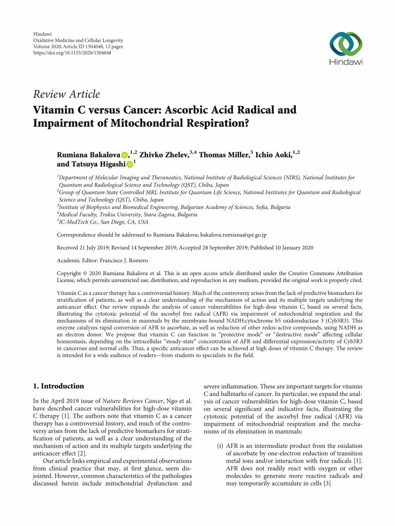
Figures 1 and 2 represent two modes of action of vitamin Con mitochondrial homeostasis via generation of AFR and itselimination by the OMM Cyb5R3/VDAC1: (i) “protectivemode” at low/normal (steady-state) doses and (ii) “destructivemode” at high doses of vitamin C.
Ascorbate and AFR exist as anions, which imply theirpenetration from the cytosol into the mitochondrial inter-membrane space and back through the VDAC1 (Figure 1).
In normal cells (Figure 1), low concentrations of AFRshould be generated due to the low (steady-state) intracellu-lar levels of vitamin C, as a result of (i) relatively lowexpression of vitamin C transport proteins [37] and (ii) lowsteady-state levels of ROS and normal levels of reducingequivalents [38]. Both factors are responsible for the limitedproduction of AFR, which is a product of one-electron oxida-tion of ascorbate. Since AFR is relatively stable, it could berapidly eliminated by the OMM Cyb5R3/VDAC1. This pre-vents effects on mitochondrial respiration, and all complexeswill operate in a “normal mode.” Electrons are transferredfrom Complex III’s Qo site of the CoQ “pool” to cytochromec, creating a proton gradient and leading to normal ATPsynthesis. In this case, the CoQ “pool” is balanced andthe Qo site of Complex III transfers one electron to
4 Oxidative Medicine and Cellular Longevity
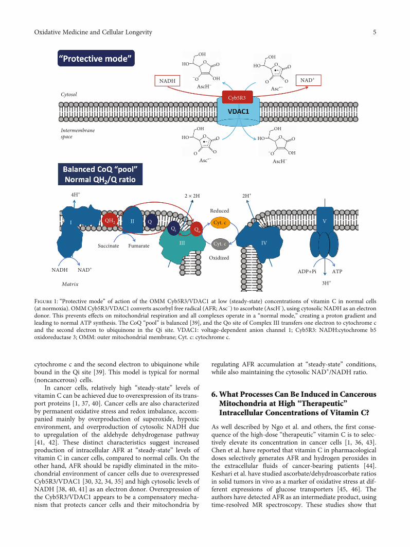
cytochrome c and the second electron to ubiquinone whilebound in the Qi site [39]. This model is typical for normal(noncancerous) cells.
In cancer cells, relatively high “steady-state” levels ofvitamin C can be achieved due to overexpression of its trans-port proteins [1, 37, 40]. Cancer cells are also characterizedby permanent oxidative stress and redox imbalance, accom-panied mainly by overproduction of superoxide, hypoxicenvironment, and overproduction of cytosolic NADH dueto upregulation of the aldehyde dehydrogenase pathway[41, 42]. These distinct characteristics suggest increasedproduction of intracellular AFR at “steady-state” levels ofvitamin C in cancer cells, compared to normal cells. On theother hand, AFR should be rapidly eliminated in the mito-chondrial environment of cancer cells due to overexpressedCyb5R3/VDAC1 [30, 32, 34, 35] and high cytosolic levels ofNADH [38, 40, 41] as an electron donor. Overexpression ofthe Cyb5R3/VDAC1 appears to be a compensatory mecha-nism that protects cancer cells and their mitochondria by
regulating AFR accumulation at “steady-state” conditions,while also maintaining the cytosolic NAD+/NADH ratio.
6. What Processes Can Be Induced in CancerousMitochondria at High “Therapeutic”Intracellular Concentrations of Vitamin C?
As well described by Ngo et al. and others, the first conse-quence of the high-dose “therapeutic” vitamin C is to selec-tively elevate its concentration in cancer cells [1, 36, 43].Chen et al. have reported that vitamin C in pharmacologicaldoses selectively generates AFR and hydrogen peroxides inthe extracellular fluids of cancer-bearing patients [44].Keshari et al. have studied ascorbate/dehydroascorbate ratiosin solid tumors in vivo as a marker of oxidative stress at dif-ferent expressions of glucose transporters [45, 46]. Theauthors have detected AFR as an intermediate product, usingtime-resolved MR spectroscopy. These studies show that
OH OH
OH
HO HOO O O
OO
O
−OAscH−
OH
OH
HO O O
−OAscH−
Asc·−
OH
HO O O
OOAsc·−
2 × 2H 2H+4H+
Reduced
Oxidized
Qo
QH2
Succinate
NADH NAD+
3H+
Fumarate
Qi
QII
III IV
V
ADP+Pi ATP
Cyt. c
Cyt. cI
NADH NAD+
Cyb5R3Cytosol
Intermembranespace
Matrix
Figure 1: “Protective mode” of action of the OMM Cyb5R3/VDAC1 at low (steady-state) concentrations of vitamin C in normal cells(at normoxia). OMMCyb5R3/VDAC1 converts ascorbyl free radical (AFR; Asc⋅-) to ascorbate (AscH-), using cytosolic NADH as an electrondonor. This prevents effects on mitochondrial respiration and all complexes operate in a “normal mode,” creating a proton gradient andleading to normal ATP synthesis. The CoQ “pool” is balanced [39], and the Qo site of Complex III transfers one electron to cytochrome cand the second electron to ubiquinone in the Qi site. VDAC1: voltage-dependent anion channel 1; Cyb5R3: NADH:cytochrome b5oxidoreductase 3; OMM: outer mitochondrial membrane; Cyt. c: cytochrome c.
5Oxidative Medicine and Cellular Longevity
AFR is also generated inside the cells. It should be clari-fied that high intracellular levels of AFR can only beachieved with intravenous vitamin C administration,because ascorbate concentrations in the plasma and tis-sues are tightly controlled as a function of the oral dose[47].
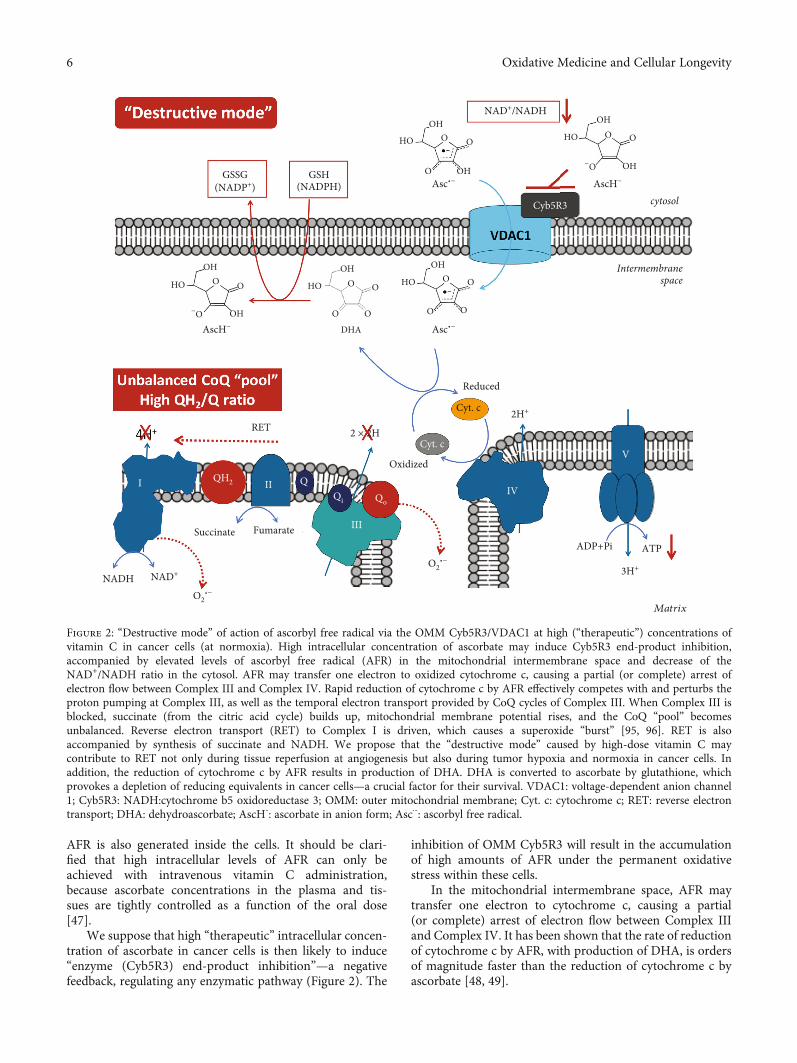
We suppose that high “therapeutic” intracellular concen-tration of ascorbate in cancer cells is then likely to induce“enzyme (Cyb5R3) end-product inhibition”—a negativefeedback, regulating any enzymatic pathway (Figure 2). The
inhibition of OMM Cyb5R3 will result in the accumulationof high amounts of AFR under the permanent oxidativestress within these cells.
In the mitochondrial intermembrane space, AFR maytransfer one electron to cytochrome c, causing a partial(or complete) arrest of electron flow between Complex IIIand Complex IV. It has been shown that the rate of reductionof cytochrome c by AFR, with production of DHA, is ordersof magnitude faster than the reduction of cytochrome c byascorbate [48, 49].
OH OH
OH
HO HOO O O
OH
O
O−O
AscH−
AscH−
OH OH
OH
HO HOO O
OO
DHA
−O
Asc·−
OH
HO O O
OO
OO
Asc·−
2 × 2H
2H+
Reduced
Oxidized
Qo
QH2
Succinate
NADH NAD+ 3H+
Fumarate
Qi
Q
RET
II
III
IV
V
ADP+Pi ATP
Cyt. c
Cyt. c
I
GSH(NADPH)
GSSG(NADP+)
NAD+/NADH
Cyb5R3
Intermembranespace
cytosol
Matrix
O2·−
O2·−
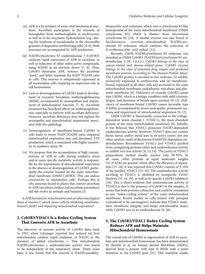
Figure 2: “Destructive mode” of action of ascorbyl free radical via the OMM Cyb5R3/VDAC1 at high (“therapeutic”) concentrations ofvitamin C in cancer cells (at normoxia). High intracellular concentration of ascorbate may induce Cyb5R3 end-product inhibition,accompanied by elevated levels of ascorbyl free radical (AFR) in the mitochondrial intermembrane space and decrease of theNAD+/NADH ratio in the cytosol. AFR may transfer one electron to oxidized cytochrome c, causing a partial (or complete) arrest ofelectron flow between Complex III and Complex IV. Rapid reduction of cytochrome c by AFR effectively competes with and perturbs theproton pumping at Complex III, as well as the temporal electron transport provided by CoQ cycles of Complex III. When Complex III isblocked, succinate (from the citric acid cycle) builds up, mitochondrial membrane potential rises, and the CoQ “pool” becomesunbalanced. Reverse electron transport (RET) to Complex I is driven, which causes a superoxide “burst” [95, 96]. RET is alsoaccompanied by synthesis of succinate and NADH. We propose that the “destructive mode” caused by high-dose vitamin C maycontribute to RET not only during tissue reperfusion at angiogenesis but also during tumor hypoxia and normoxia in cancer cells. Inaddition, the reduction of cytochrome c by AFR results in production of DHA. DHA is converted to ascorbate by glutathione, whichprovokes a depletion of reducing equivalents in cancer cells—a crucial factor for their survival. VDAC1: voltage-dependent anion channel1; Cyb5R3: NADH:cytochrome b5 oxidoreductase 3; OMM: outer mitochondrial membrane; Cyt. c: cytochrome c; RET: reverse electrontransport; DHA: dehydroascorbate; AscH-: ascorbate in anion form; Asc⋅-: ascorbyl free radical.
6 Oxidative Medicine and Cellular Longevity
Rapid reduction of cytochrome c by AFR effectivelycompetes with the electron transport from the CoQ cycle ofComplex III to the cytochrome c, which perturbs the protonpumping. Competitive inhibition by AFR, bypassing Com-plex III to cytochrome c, drives the CoQ “pool” to a morereduced (overcharged) state. If this overcharging phenome-non occurs, there will be insufficient ubiquinone, availablefor binding at the Qi site. This unbalances the Qo/Qi cycle.It also increases the lifetime of semiubiquinone in the Qopocket, allowing oxygen to accept the second electron fromubiquinol during the second step of the CoQ cycle, yieldingsuperoxide (Figure 2) [50].
Briefly, we propose that a high level of AFR in the inter-membrane space of mitochondria blocks Complex III, atleast partially, via unbalancing the CoQ “pool” and impairingmitochondrial respiration in vitamin C-treated cancer cells.Precisely timed redox sequences responsible for the efficientflow of electrons from Complex III to oxidized cytochromec are vulnerable to AFR flux. This drives the CoQ “pool” toa more reduced (overcharged) form. This also causes the life-time of semiubiquinone in the Qo pocket to lengthen, allow-ing oxygen to accept the second electron from ubiquinolduring the second step of the CoQ cycle, yielding superoxide.This can interfere with the timing of the Qo/Qi cycle andconsequently the CoQ “pool” redox balance, impairing mito-chondrial respiration. A highly reduced CoQ “pool” inhibitsproton pumping, yielding up to 80% lower ATP plus higherlevels of superoxide. We further propose that the damagingeffect of high-dose vitamin C on cancer cells is similar tothe mechanism of ischemia-reperfusion injury (IRI) or reox-ygenation damage [51]. In IRI, the mitochondrial CoQ“pool” becomes highly reduced by stopped flow throughComplex III.
It is interesting to note that this mechanism can influencethe mitochondrial ATP production in different directionsdepending on the cell type. In the case of cancer cells, thismechanism should result in a significant reduction of mito-chondrial ATP and increase of superoxide production(Figure 2).
It has been generally accepted that cancer cells use glycol-ysis as a source of ATP (Warburg effect) and do not rely onmitochondrial respiration for ATP production. However,recent studies demonstrate that the majority of ATP in can-cer cells is produced by mitochondria and some tumors showheavy dependence on oxidative phosphorylation [52–56].Many solid tumors are poorly perfused and have a limitedsupply of glucose, but enough oxygen to generate mitochon-drial ATP [57]. The ETC is able to function optimally at oxy-gen levels as low as 0.5% [58]. Therefore, blockingmitochondrial ATP production will induce cell death inpoorly perfused tumors.
Even ignoring the dependence of cancer cells on mito-chondrial ATP, a function of the Krebs cycle (at ComplexII) is necessary for the supply of metabolites for synthesis ofnucleic acids, fatty acids, etc. Blocking the ETC will stop thissupply and suppress proliferation. Krebs cycle metabolites(such as succinate, fumarate, and itaconate) are also coupledwith nonmetabolic signaling in cancer and immune cells,which is crucial for cancer progression and invasion [59, 60].
The key consideration in targeting mitochondria byhigh-dose vitamin C (via AFR) is that normal cells relymostly on mitochondrial respiration for ATP production.As was mentioned above, high concentrations of AFRcannot be reached in normal cells, especially in vivo inthe absence of oxidative stress, due to lower expressionof glucose/ascorbate transport proteins [36, 47] and lowamounts and normal functioning of Cyb5R3 [32, 33].However, normal cells should also be vulnerable to highconcentrations of AFR generated under oxidative stress.Perhaps, this is a part of the dual effect of vitamin C,which can act as an antioxidant or prooxidant, dependingon the environment [61].
It is shown that in the case of noncancer cells withmitochondrial deficiency, high-dose vitamin C could evenincrease ATP production. Eleff et al. reported that adminis-tration of vitamin C and quinone-like provitamin menadioneon the skeletal muscles of a 17-year-old patient with a severedefect in Complex III of the mitochondrial ETC increased therecovery rate, compared to the recovery rate of the youngfemale controls [62]. This rare defect included a stop-codonmutation (G15242A) in the mtDNA-encoded cytochromeb gene, which was accompanied by an effective preventionof aerobic metabolism and oxidative phosphorylation. Theauthors suppose that both substances bypass the deficientComplex III as electron transfer mediators to carry theelectrons from CoQ to cytochrome c. Thus, they increasedATP production from mitochondria compared to the ini-tial level. It is interesting to note that redox cycles of vita-min C and menadione are mediated by Cyb5R3/VDAC1[8, 17] and the enzyme activity could be important for thisbypass. In this patient, the combination of vitamin C andmenadione resulted in the production of more ATP thanin the case of vitamin C applied alone. The authors sug-gest that both substances can directly reduce cytochromec, since the reduction potential of cytochrome c is over+200mV—more positive than those of ascorbate andmenadione [62–65].
7. What Could Be the Consequences ofAFR-Mediated Electron Transfer inCancerous Mitochondria from High-DoseVitamin C Therapy?
(i) Induction of reverse electron transport in the respi-ratory chain and acceleration of superoxide produc-tion at Complex I, as well as Complex III (Figure 2)[50, 65–67]. Degradation of Complex I when theCoQ “pool” is unbalanced, and Complex III is dys-functional [67]. In hypoxic conditions (common tomany types of cancer cells), these redox reactionsoccur even faster, exponentially increasing superox-ide production [50, 65–67].
(ii) Superoxide-mediated destruction of Fe/S clustersfrom the complexes and induction of Fenton’s reac-tions in mitochondria [68–70], in the presence ofAFR and/or ascorbate
7Oxidative Medicine and Cellular Longevity
(iii) Mitochondria within cancer cells are vulnerable to a“destructive mode” of action from high-dose vita-min C, which leads to their collapse
In addition, the conversion of AFR to DHA and subse-quent reduction of DHA to ascorbate by glutathione- andNADPH-dependent pathways will promote a depletion ofglutathione and reducing equivalents in the cancer cells lead-ing to severe oxidative stress—crucial factors for induction ofapoptosis and cell death [38].
An important consequence of the reduction of cyto-chrome c by AFR is the transfer of electrons from cytoplas-mic NADH to Complex IV, which is accompanied byproduction of 1mol ATP per 1mol NADH—suggesting thelikelihood of thermogenesis [71, 72]. Cancer cells oftenexpress heat shock proteins and are very sensitive to the ele-vated temperature of their environment [38, 73]. This mayalso contribute to converting “cold” tumors into “hot”tumors, an important cell death-facilitating factor potentiallyfrom combining high-dose vitamin C therapy with applica-tions in immunotherapy [74].
It is also reported that NADH reduces conduction ofVDAC by partial channel block and/or modulation of itsactivity [75, 76]. As ATP and many other vital substancespass from the mitochondria into the cytosol through VDAC[77, 78], partial inhibition of the VDAC channel would alsocontribute to the impairment of mitochondrial respirationand decrease of cell viability.
Recent studies show that Cyb5R3/VDAC1 can activatebioreductive antitumor drugs, which in combination withvitamin C may affect mitochondrial activity, exhibiting addi-tive or synergistic anticancer effects [17, 79, 80]. For example,vitamin C can sensitize cancer cells to redox-responsivechemotherapeutics (e.g., furanonaphtoquinones) after theirbioreductive activation via Cyb5R3/VDAC1 [81, 82]. In thiscase, the “therapeutic” intravenous dose of vitamin C couldbe decreased to more tolerant values. It has been reportedthat high doses of vitamin C exhibit selective antitumoreffects in combination with menadione—an analogue of1,4-naphoquinone [83–85]. A recent study demonstrates thathigh/tolerable concentrations of menadione inhibit Cyb5R3[86]. It can be assumed that menadione-mediated inhibitionof Cyb5R3 is involved in potentiation of the anticancer effectof high-dose vitamin C via overproduction of AFR and sub-sequent impairment of mitochondrial respiration (as weassume with our “model”). Moreover, vitamin C/menadionecould achieve a selective cytotoxicity against cancer cells dueto the rapid UBIAD1-mediated conversion of menadione tovitamin K2 in normal cells and downregulation of UBIAD1in the majority of cancers leading to strong inhibition of thisconversion [87, 88]. These assumptions could be a prerequi-site/basis for the design of future studies aimed to elucidatethe molecular mechanisms of the synergistic anticancereffects of vitamin C and vitamin C/menadione with conven-tional chemotherapeutics, described in the literature [89].Clarifying the effects of conventional anticancer drugs andhigh-dose vitamin C on Cyb5R3 may also be a prerequisitefor predicting the effectiveness of vitamin C in adjuvant set-tings and stratification of patients with good/poor prognosis.
8. Concluding Remarks andOutstanding Questions
The high concentrations of cytochrome c in mitochondria[90], as well as the possible existence of dedicated pools ofCoQ and cytochrome c (as supported by the “plasticitymodel” of mitochondria) [90, 91], could limit the proposeddirect effects of AFR on oxidative phosphorylation, asdescribed in Figure 2. In fact, reported concentrations of vita-min C in cells under normal conditions are significantlyhigher than those of cytochrome c (~10-30 nmol/mg proteinversus ~0.1-1 nmol/mg protein) [90, 92]. In this case, ascor-bate and AFR concentrations in cancer cells after treatmentwith high doses of vitamin C should be much higher thancytochrome c concentrations [93]. This supports the possi-bility of direct electron transfer from AFR to cytochrome cat Complex IV. Remodeling of the mitochondrial membraneand, in particular, the contact sites of Cyb5R3/VDAC1/-Complex IV to optimize performance can explain the differ-ent efficiency and selectivity of high-dose vitamin C therapywhen combining with different anticancer drugs (dependingon their mechanisms). In addition, the abovementioned con-sequences of AFR-mediated electron transfer in cancerousmitochondria at high-dose vitamin C therapy may explainthe induction of apoptosis even without strong inhibition ofoxidative phosphorylation and ATP production. The mecha-nism, described in Figure 2, may not cause strong cancer celldeath, but it can damage the mitochondria, stop prolifera-tion, and keep cancer cells latent, so they become vulnerableto the immune system of the intact organism.
The analysis of the literature raises several outstandingquestions that need experimental verification:
(1) Does AFR impair mitochondrial respiration byreducing cytochrome c, causing an arrest of electronflow between Complexes III and IV, a reverse elec-tron transport, and an induction of oxidative stressin the absence or downregulation of Cyb5R3/VDAC1
(2) Does vitamin C in low/normal doses work in “protec-tive mode” in normal and cancer cells? If yes, is thisrelated to maintaining the balance of the CoQ “pool”and cytosolic reducing equivalents
(3) Does vitamin C in high doses work in “destructivemode” in cancer cells, but not in normal cells? Ifyes, is this related to maintaining the imbalance ofthe CoQ “pool” and cytosolic reducing equivalents
(4) What are the levels of mitochondrial superoxide, suc-cinate, and ATP in normal and cancer cells of thesame origin, treated with low, moderate, and highdoses of vitamin C? Is there a difference in the levelsof mitochondrial superoxide, succinate, and ATP incells with different proliferative index, treated withvitamin C
(5) Does the effect of pharmacological vitamin C relate tothe expression and activity of membrane-boundCyb5R3/VDAC1? Can high doses of vitamin C attack
8 Oxidative Medicine and Cellular Longevity
cancer cells only by inhibiting Cyb5R3/VDAC1 andspecific destruction of cancer mitochondria
(6) What is the impact/significance of Cyb5R3/VDAC1as a predictive biomarker for stratification of cancer-bearing patients for high-dose vitamin C therapy
(7) Does pharmacological vitamin C have a potential asadjuvant cancer therapy in combination with redox-responsive drugs
Our article is intended to provoke the design of newexperiments (in vitro and in vivo) that can provide evi-dence Pro and Con for the assumptions made, to changethe “status quo” in the controversial field of “pharmacologicalvitamin C.”
If vitamin C can clinically exploit the pathway presentedhere, we suggest it offers a new, exciting rationale for cancerstudies. Perhaps this orthogonal mechanism of action cansafely and more effectively compliment current cancertreatments in the adjuvant setting and help to let the phoe-nix fly [94].
Abbreviations
AFR: Ascorbyl free radicalCoQ: Coenzyme QCyb5R3: NADH:cytochrome b5 oxidoreductase 3DHA: Dehydroascorbic acidHDF: Human dermal fibroblastOMM: Outer mitochondrial membraneRHM: Recessive hereditary methemoglobinemiaVDAC1: Voltage-dependent anion channel 1.
Conflicts of Interest
No potential conflicts of interest are disclosed.
Acknowledgments
This study was partially supported by the IC-MedTech Co.(San Diego, CA, USA) and the Japan Agency for MedicalResearch and Development (AMED) (Project for CancerResearch and Therapeutic Evolution, P-CREATE, no. 16cm0106202h0001).
References
[1] B. Ngo, J. van Riper, L. C. Cantley, and J. Yun, “Targeting can-cer vulnerabilities with high-dose vitamin C,” Nature ReviewsCancer, vol. 19, no. 5, pp. 271–282, 2019.
[2] A. Unly, O. Kirca, andM. Ozdogan, “High-dose vitamin C andcancer,” Journal of Oncological Sciences, vol. 1, pp. 10–12,2015.
[3] N. Smirnoff, “Ascorbic acid metabolism and functions: acomparison of plants and mammals,” Free Radical Biology &Medicine, vol. 122, pp. 116–129, 2018.
[4] A. Allen, C. Fisher, A. Premawardhena et al., “Methemoglobi-nemia and ascorbate deficiency in hemoglobin E β-thalasse-mia: metabolic and clinical implications,” Blood, vol. 120,no. 15, pp. 2939–2944, 2012.
[5] E. J. Diliberto Jr. and P. L. Allen, “Mechanism of dopamineβ-hydroxylation: semidehydroascobate as the enzymic oxi-dation product of ascorbate,” The Journal of BiologicalChemistry, vol. 256, 1981.
[6] F. S. Menniti, J. Knoth, and Diliberto EJ Jr, “Role of ascorbicacid in dopamine beta-hydroxylation. The endogenousenzyme cofactor and putative electron donor for cofactorregeneration,” The Journal of Biological Chemistry, vol. 261,no. 36, pp. 16901–16908, 1986.
[7] R. de Cabo, E. Siendones, R. Minor, and P. Navas, “Cyb5R3: akey player in aerobic metabolism and aging?,” Aging, vol. 2,no. 1, pp. 63–68, 2009.
[8] A. B. Nikiforova, N.-E. L. Saris, and A. G. Kruglov, “Externalmitochondrial NADH-dependent reductase of redox cyclers:VDAC1 or Cyb5R3?,” Free Radical Biology & Medicine,vol. 74, pp. 74–84, 2014.
[9] E. Siendones, M. Ballesteros, and P. Navas, “Cellular andmolecular mechanisms of recessive hereditary Methaemoglo-binaemia type II,” Journal of Clinical Medicine, vol. 7, no. 10,p. 341, 2018.
[10] A. Ito, S. Hayashi, and T. Yoshida, “Participation of a cyto-chrome b5-like hemoprotein of outer mitochondrial membrane(OM cytochrome b) in NADH-semidehydroascorbic acidreductase activity of rat liver,” Biochemical and BiophysicalResearch Communications, vol. 101, no. 2, pp. 591–598, 1981.
[11] J. S. Lee, W. K. Huh, B. H. Lee et al., “Mitochondrial NADH-cytochrome b5 reductase plays a crucial role in the reductionof d-erythroascorbyl free radical in Saccharomyces cerevisiae,”Biochimica et Biophysica Acta (BBA) - General Subjects,vol. 1527, no. 1-2, pp. 31–38, 2001.
[12] M. J. Percy and T. R. Lappin, “Recessive congenital methaemo-globinaemia: cytochrome b5 reductase deficiency,” BritishJournal of Haematology, vol. 141, no. 3, pp. 298–308, 2008.
[13] S. Kimura, H. Nishida, and T. Iyanagi, “Effects of flavin-bindingmotif amino acid mutations in the NADH-cytochrome b5reductase catalytic domain on protein stability and catalysis,”Journal of Biochemistry, vol. 130, no. 4, pp. 481–490, 2001.
[14] M. Gonzalez-Gronow, R. Ray, F. Wang, and S. V. Pizzo, “Thevoltage-dependent anion channel (VDAC) binds tissue-typeplasminogen activator and promotes activation of plasmino-gen on the cell surface,” The Journal of Biological Chemistry,vol. 288, no. 1, pp. 498–509, 2013.
[15] A. Deniaud, C. Rossi, A. Berquand et al., “Voltage-dependentanion channel transports calcium ions through biomimeticmembranes,” Langmuir, vol. 23, no. 7, pp. 3898–3905, 2007.
[16] V. Shoshan-Barmatz, D. Ben-Hail, L. Admoni, Y. Krelin, andS. S. Tripathi, “The mitochondrial voltage-dependent anionchannel 1 in tumor cells,” Biochimica et Biophysica Acta(BBA) - Biomembranes, vol. 1848, no. 10, pp. 2547–2575, 2015.
[17] E. Simamura, H. Shimada, Y. Ishigaki, T. Hatta, N. Higashi,and K. Hirai, “Bio-reductive activation of quinone anti-tumor drugs by mitochondrial voltage-dependent anion chan-nel 1,” Anatomical Science International, vol. 83, no. 4,pp. 261–266, 2008.
[18] M. Tamura, T. Yubisui, and M. Takeshita, “MicrosomalNADH-cytochrome b5 reductase of bovine brain: purificationand properties,” Journal of Biochemistry, vol. 94, no. 5,pp. 1547–1555, 1983.
[19] M. Baker, J. D. Ly, and A. Lawen, “Characterization of VDAC1as a plasma membrane NADH-oxidoreductase,” BioFactors,vol. 21, no. 1-4, pp. 215–221, 2004.
9Oxidative Medicine and Cellular Longevity
[20] H. Shimada, K. Hirai, E. Simamura et al., “Paraquat toxicityinduced by voltage-dependent anion channel 1 acts as anNADH-dependent oxidoreductase,” The Journal of BiologicalChemistry, vol. 284, no. 42, pp. 28642–28649, 2009.
[21] K. Shirabe, M. T. Landi, M. Takeshita, G. Uziel, E. Fedrizzi,and N. Borgese, “A novel point mutation in a 3′ splice site ofthe NADH-cytochrome b5 reductase gene results in immuno-logically undetectable enzyme and impaired NADH-dependent ascorbate regeneration in cultured fibroblasts of apatient with type II hereditary methemoglobinemia,” Ameri-can Journal of Human Genetics, vol. 57, no. 2, pp. 302–310,1995.
[22] E. Siendones, S. SantaCruz-Calvo, A. Martín-Montalvo et al.,“Membrane-bound Cyb5R3 is a common effector of nutri-tional and oxidative stress response through FOXO3a andNrf2,” Antioxidants & Redox Signaling, vol. 21, no. 12,pp. 1708–1725, 2014.
[23] N. T. Carew, H. M. Altmann, J. C. Galley et al., “Abstract20733: cytochrome b5 reductase 3 is essential for cardiomyo-cyte function,” Circulation, vol. 136, Supplementary 1, 2017.
[24] D. Marzulli, G. la Piana, E. Fransvea, and N. E. Lofrumento,“Modulation of cytochrome c -mediated extramitochondrialNADH oxidation by contact site density,” Biochemical andBiophysical Research Communications, vol. 259, no. 2,pp. 325–330, 1999.
[25] G. La Piana, D. Marzulli, V. Gorgoglione, and N. E.Lofrumento, “Porin and cytochrome oxidase containing con-tact sites involved in the oxidation of cytosolic NADH,”Archives of Biochemistry and Biophysics, vol. 436, no. 1,pp. 91–100, 2005.
[26] M. E. Bodrova, V. I. Dedukhova, E. N. Mokhova, and V. P.Skulachev, “Membrane potential generation coupled to oxida-tion of external NADH in liver mitochondria,” FEBS Letters,vol. 435, no. 2-3, pp. 269–274, 1998.
[27] G. La Piana, D. Marzulli, M. I. Consalvo, and N. E.Lofrumento, “Cytochrome c-induced cytosolic nicotinamideadenine dinucleotide oxidation, mitochondrial permeabilitytransition, and apoptosis,” Archives of Biochemistry and Bio-physics, vol. 410, no. 2, pp. 201–211, 2003.
[28] V. Shoshan-Barmatz, Y. Krelin, A. Shteinfer-Kuzmine, andT. Arif, “Voltage-dependent anion channel 1 as an emergingdrug target for novel anti-cancer therapeutics,” Frontiers inOncology, vol. 7, 2017.
[29] A. K. S. Camara, Y. F. Zhou, P. C. Wen, E. Tajkhorshid, andW. M. Kwok, “Mitochondrial VDAC1: a key gatekeeper aspotential therapeutic target,” Frontiers in Physiology, vol. 8,2017.
[30] L. Leanza, M. Zoratti, E. Gulbins, and I. Szabo, “Mitochondrialion channels as oncological targets,” Oncogene, vol. 33, no. 49,pp. 5569–5581, 2014.
[31] A. Magri, S. Reina, and V. De Pinto, “VDAC1 as pharmacolog-ical target in cancer and neurodegeneration: focus on its role inapoptosis,” Frontiers in Chemistry, vol. 6, 2018.
[32] K. L. Blanke, J. C. Sacco, R. C. Millikan, A. F. Olshan, J. Luo,and L. A. Trepanier, “Polymorphisms in the carcinogen detox-ification genes Cyb5A and Cyb5R3 and breast cancer risk inAfrican American women,” Cancer Causes & Control,vol. 25, no. 11, pp. 1513–1521, 2014.
[33] A. Martin-Montalvo, Y. Sun, A. Diaz-Ruiz et al., “Cytochromeb5 reductase and the control of lipid metabolism and health-span,” Aging and Mechanisms of Disease, vol. 2, 2016.
[34] R. R. Lund, R. Leth-Larsen, T. D. Caterino et al., “NADH-cyto-chrome b5 reductase 3 promotes colonization and metastasisformation and is a prognostic marker of disease-free and overallsurvival in estrogen receptor-negative breast cancer,”Molecular& Cellular Proteomics, vol. 14, no. 11, pp. 2988–2999, 2015.
[35] U. Rajcevic, K. Petersen, J. C. Knol et al., “iTRAQ-based prote-omics profiling reveals increased metabolic activity and cellu-lar cross-talk in angiogenic compared with invasiveglioblastoma phenotype,” Molecular & Cellular Proteomics,vol. 8, no. 11, pp. 2595–2612, 2009.
[36] M. Levine, S. J. Padayatty, and M. G. Espey, “Vitamin C: aconcentration-function approach yields pharmacology andtherapeutic discoveries,” Advances in Nutrition, vol. 2, no. 2,pp. 78–88, 2011.
[37] K. C. Sagun, J. M. Cárcamo, and D.W. Golde, “Vitamin C entersmitochondria via facilitative glucose transporter 1 (Glut1) andconfers mitochondrial protection against oxidative injury,” TheFASEB Journal, vol. 19, no. 12, pp. 1657–1667, 2005.
[38] D. Trachootham, J. Alexandre, and P. Huang, “Targeting can-cer cells by ROS-mediated mechanisms: a radical therapeuticapproach?,” Nature Reviews Drug Discovery, vol. 8, no. 7,pp. 579–591, 2009.
[39] M. Sarewicz and A. Osyczka, “Electronic connection betweenthe quinone and cytochrome c redox pools and its role in reg-ulation of mitochondrial electron transport and redox signal-ing,” Physiological Reviews, vol. 95, no. 1, pp. 219–243, 2015.
[40] A. A. Chen, C. J. Marsit, B. C. Christensen et al., “Genetic var-iation in the vitamin C transporter, SLC23A2, modifies the riskof HPV16-associated head and neck cancer,” Carcinogenesis,vol. 30, no. 6, pp. 977–981, 2009.
[41] J. Y. Kim-Muller, J. Fan, Y. J. R. Kim et al., “Aldehyde dehydro-genase 1a3 defines a subset of failing pancreatic β cells in dia-betic mice,” Nature Communications, vol. 7, 2016.
[42] J. H. Kang, S. H. Lee, D. Hong et al., “Aldehyde dehydrogenaseis used by cancer cells for energy metabolism,” Experimental &Molecular Medicine, vol. 48, no. 11, article e272, 2016.
[43] G. Nauman, J. Gray, R. Parkinson, M. Levine, and C. Paller,“Systematic review of intravenous ascorbate in cancer clinicaltrials,” Antioxidants, vol. 7, no. 7, p. 89, 2018.
[44] Q. Chen, M. G. Espey, A. Y. Sun et al., “Ascorbate in pharma-cologic concentrations selectively generates ascorbate radicaland hydrogen peroxide in extracellular fluid in vivo,” Proceed-ings of the National Academy of Sciences of the United States ofAmerica, vol. 104, no. 21, pp. 8749–8754, 2007.
[45] K. R. Keshari, J. Kurhanewicz, R. Bok, P. E. Larson, D. B.Vigneron, and D. M. Wilson, “Hyperpolarized 13C dehydroas-corbate as an endogenous redox sensor for in vivo metabolicimaging,” Proceedings of the National Academy of Sciences ofthe United States of America, vol. 108, no. 46, pp. 18606–18611, 2011.
[46] K. R. Keshari, V. Sai, Z. J. Wang, H. F. VanBrocklin,J. Kurhanewicz, and D. M. Wilson, “Hyperpolarized[1-13C]dehydroascorbate MR spectroscopy in a murine modelof prostate cancer: comparison with 18F-FDG PET,” Journal ofNuclear Medicine, vol. 54, no. 6, pp. 922–928, 2013.
[47] S. J. Padayatty, H. Sun, Y. Wang et al., “Vitamin C pharmaco-kinetics: implications for oral and intravenous use,” Annals ofInternal Medicine, vol. 140, no. 7, pp. 533–537, 2004.
[48] I. Yamazaki, “The reduction of cytochrome c by enzyme-generated ascorbic free radical,” The Journal of BiologicalChemistry, vol. 237, pp. 224–229, 1962.
10 Oxidative Medicine and Cellular Longevity
[49] C. Paciolla and L. De Gara, “Reduction of cytochrome c byascorbic free radical,” Bollettino della Società Italiana di Biolo-gia Sperimentale, vol. 67, no. 2, pp. 137–144, 1991.
[50] L. Bleier and S. Drose, “Superoxide generation by complex III:from mechanistic rationales to functional consequences,” Bio-chimica et Biophysica Acta (BBA) - Bioenergetics, vol. 1827,no. 11-12, pp. 1320–1331, 2013.
[51] E. T. Chouchani, V. R. Pell, E. Gaude et al., “Ischaemic accumu-lation of succinate controls reperfusion injury through mito-chondrial ROS,” Nature, vol. 515, no. 7527, pp. 431–435, 2014.
[52] P. Caro, A. U. Kishan, E. Norberg et al., “Metabolic signaturesuncover distinct targets in molecular subsets of diffuse large Bcell lymphoma,” Cancer Cell, vol. 22, no. 4, pp. 547–560, 2012.
[53] J. Fan, J. J. Kamphorst, R. Mathew et al., “Glutamine-drivenoxidative phosphorylation is a major ATP source in trans-formed mammalian cells in both normoxia and hypoxia,”Molecular Systems Biology, vol. 9, no. 1, p. 712, 2013.
[54] R. Haq, J. Shoag, P. Andreu-Perez et al., “Oncogenic BRAFregulates oxidative metabolism via PGC1α and MITF,” CancerCell, vol. 23, no. 3, pp. 302–315, 2013.
[55] F. Vazquez, J.-H. Lim, H. Chim et al., “PGC1α expressiondefines a subset of human melanoma tumors with increasedmitochondrial capacity and resistance to oxidative stress,”Cancer Cell, vol. 23, no. 3, pp. 287–301, 2013.
[56] S. E. Weinberg and N. S. Chandel, “Targeting mitochondriametabolism for cancer therapy,” Nature Chemical Biology,vol. 11, no. 1, pp. 9–15, 2015.
[57] R. K. Jain, L. L. Munn, and D. Fukumura, “Dissecting tumourpathophysiology using intravital microscopy,” Nature ReviewsCancer, vol. 2, no. 4, pp. 266–276, 2002.
[58] W. L. Rumsey, C. Schlosser, E. M. Nuutinen, M. Robiolio, andD. F. Wilson, “Cellular energetics and the oxygen dependenceof respiration in cardiac myocytes isolated from adult rat,” TheJournal of Biological Chemistry, vol. 265, no. 26, pp. 15392–15402, 1990.
[59] A. G. M. Tielens, C. Rotte, J. J. van Hellemond, andW. Martin,“Mitochondria as we don't know them,” Trends in BiochemicalSciences, vol. 27, no. 11, pp. 564–572, 2002.
[60] D. G. Ryan, M. P. Murphy, C. Frezza et al., “Coupling Krebscycle metabolites to signalling in immunity and cancer,”Nature Metabolism, vol. 1, pp. 16–33, 2019.
[61] J. Du, J. J. Cullen, and G. R. Buettner, “Ascorbic acid: chemis-try, biology and the treatment of cancer,” Biochimica et Bio-physica Acta (BBA) - Reviews on Cancer, vol. 1826, no. 2,pp. 443–457, 2012.
[62] S. Eleff, N. G. Kennaway, N. R. Buist et al., “31P NMR study ofimprovement in oxidative phosphorylation by vitamins K3and C in a patient with a defect in electron transport atcomplex III in skeletal muscle,” Proceedings of the NationalAcademy of Sciences of the United States of America, vol. 81,no. 11, pp. 3529–3533, 1984.
[63] G. C. Wagner, R. J. Kassner, and M. D. Kamen, “Redox poten-tials of certain vitamins K: implications for a role in sulfitereduction by obligately anaerobic bacteria,” Proceedings ofthe National Academy of Sciences of the United States ofAmerica, vol. 71, no. 2, pp. 253–256, 1974.
[64] T. Matsui, Y. Kitagawa, M. Okumura, and Y. Shigeta, “Accu-rate standard hydrogen electrode potential and applicationsto the redox potentials of vitamin C and NAD/NADH,” TheJournal of Physical Chemistry A, vol. 119, no. 2, pp. 369–376,2015.
[65] A. P. West, G. S. Shadel, and S. Ghosh, “Mitochondria ininnate immune responses,” Nature Reviews Immunology,vol. 11, no. 6, pp. 389–402, 2011.
[66] E. L. Robb, A. R. Hall, T. A. Prime et al., “Control of mitochon-drial superoxide production by reverse electron transport atcomplex I,” The Journal of Biological Chemistry, vol. 293,no. 25, pp. 9869–9879, 2018.
[67] A. Guarás, E. Perales-Clemente, E. Calvo et al., “The CoQH2/-CoQ ratio serves as a sensor of respiratory chain efficiency,”Cell Reports, vol. 15, no. 1, pp. 197–209, 2016.
[68] J. C. Crack, J. Green, M. R. Cheesman, N. E. le Brun, and A. J.Thomson, “Superoxide-mediated amplification of the oxygen-induced switch from [4Fe-4S] to [2Fe-2S] clusters in the tran-scriptional regulator FNR,” Proceedings of the National Acad-emy of Sciences of the United States of America, vol. 104,no. 7, pp. 2092–2097, 2007.
[69] S. J. Dixon and B. R. Stockwell, “The role of iron and reactiveoxygen species in cell death,” Nature Chemical Biology,vol. 10, no. 1, pp. 9–17, 2014.
[70] D. B. Zorov, M. Juhaszova, and S. J. Sollott, “Mitochondrialreactive oxygen species (ROS) and ROS-induced ROS release,”Physiological Reviews, vol. 94, no. 3, pp. 909–950, 2014.
[71] J. E. Silva, “Thermogenic mechanisms and their hormonal reg-ulation,” Physiological Reviews, vol. 86, no. 2, pp. 435–464,2006.
[72] C. Bell, N. R. Stob, and D. R. Seals, “Thermogenic responsive-ness to β-adrenergic stimulation is augmented in exercisingversus sedentary adults: role of oxidative stress,” The Journalof Physiology, vol. 570, Part 3, pp. 629–635, 2006.
[73] S. K. Calderwood and J. Gong, “Heat shock proteins promotecancer: it’s a protection racket,” Trends in BiochemicalSciences, vol. 41, no. 4, pp. 311–323, 2016.
[74] J. B. A. G. Haanen, “Converting cold into hot tumors by com-bining immunotherapies,” Cell, vol. 170, no. 6, pp. 1055-1056,2017.
[75] M. Zizi, M. Forte, E. Blachly-Dyson, and M. Colombini,“NADH regulates the gating of VDAC, the mitochondrialouter membrane channel,” The Journal of Biological Chemis-try, vol. 269, no. 3, pp. 1614–1616, 1994.
[76] R. Diaz-Ruiz, M. Rigoulet, and A. Devin, “The Warburg andCrabtree effects: on the origin of cancer cell energy metabolismand of yeast glucose repression,” Biochimica et Biophysica Acta(BBA) - Bioenergetics, vol. 1807, no. 6, pp. 568–576, 2011.
[77] S. F. Okada, W. K. O'Neal, P. Huang et al., “Voltage-dependentanion channel-1 (VDAC-1) contributes to ATP release andcell volume regulation in murine cells,” The Journal of GeneralPhysiology, vol. 124, no. 5, pp. 513–526, 2004.
[78] D. Ben-Hail, R. Begas-Shvartz, M. Shalev et al., “Novel com-pounds targeting the mitochondrial protein VDAC1 inhibitapoptosis and protect against mitochondrial dysfunction,”The Journal of Biological Chemistry, vol. 291, no. 48,pp. 24986–25003, 2016.
[79] E. Guerriero, A. Sorice, F. Capone et al., “Vitamin C effect onmitoxantrone-induced cytotoxicity in human breast cancercell lines,” PLoS One, vol. 9, no. 12, article e115287, 2014.
[80] E. Hatem, S. Azzi, N. el Banna et al., “Auranofin/vitamin C: anovel drug combination targeting triple-negative breast can-cer,” JNCI: Journal of the National Cancer Institute, vol. 111,no. 6, pp. 597–608, 2019.
[81] E. Simamura, K. Hirai, H. Shimada, J. Koyama, Y. Niwa, andS. Shimizu, “Furanonaphthoquinones cause apoptosis of
11Oxidative Medicine and Cellular Longevity
cancer cells by inducing the production of reactive oxygen spe-cies by the mitochondrial voltage-dependent anion channel,”Cancer Biology & Therapy, vol. 5, no. 11, pp. 1523–1529, 2006.
[82] S. Vatolin, T. Radivoyevitch, and J. P. Maciejewski, “Newdrugs for pharmacological extension of replicative life spanin normal and progeroid cells,” npj Aging and Mechanisms ofDisease, vol. 5, no. 1, 2019.
[83] J. Verrax, J. Stockis, A. Tison, H. S. Taper, and P. B. Calderon,“Oxidative stress by ascorbate/menadione association killsK562 human chronic myelogenous leukaemia cells andinhibits its tumour growth in nude mice,” Biochemical Phar-macology, vol. 72, no. 6, pp. 671–680, 2006.
[84] M. Tomasetti, E. Strafella, S. Staffolani, L. Santarelli, J. Neuzil,and R. Guerrieri, “α -Tocopheryl succinate promotes selectivecell death induced by vitamin K3 in combination withascorbate,” British Journal of Cancer, vol. 102, no. 8,pp. 1224–1234, 2010.
[85] X. Ren, S. M. Santhosh, L. Coppo, F. T. Ogata, J. Lu, andA. Holmgren, “The combination of ascorbate and menadionecauses cancer cell death by oxidative stress and replicativestress,” Free Radical Biology & Medicine, vol. 134, pp. 350–358, 2019.
[86] J. T. Szilagyi, K. C. Fussell, Y. Wang et al., “Quinone andnitrofurantoin redox cycling by recombinant cytochrome b5reductase,” Toxicology and Applied Pharmacology, vol. 359,pp. 102–107, 2018.
[87] K. Nakagawa, Y. Hirota, N. Sawada et al., “Identification ofUBIAD1 as a novel human menaquinone-4 biosyntheticenzyme,” Nature, vol. 468, no. 7320, pp. 117–121, 2010.
[88] W. J. Fredericks, J. Sepulveda, P. Lal et al., “The tumorsuppressor TERE1 (UBIAD1) prenyltransferase regulates theelevated cholesterol phenotype in castration resistant prostatecancer by controlling a program of ligand dependent SXRtarget genes,” Oncotarget, vol. 4, no. 7, pp. 1075–1092, 2013.
[89] D. Ivanova, Z. Zhelev, D. Lazarova, P. Getsov, R. Bakalova, andI. Aoki, “Vitamins C and K3: a powerful redox system for sen-sitizing leukemia lymphocytes to everolimus and barasertib,”Anticancer Research, vol. 38, no. 3, pp. 1407–1414, 2018.
[90] G. Lenaz and M. Genova, “Structural and functional organiza-tion of the mitochondrial respiratory chain: a dynamic super-assembly,” The International Journal of Biochemistry & CellBiology, vol. 41, no. 10, pp. 1750–1772, 2009.
[91] R. Acin-Perez and J. A. Enriquez, “The function of therespiratory supercomplexes: the plasticity model,” Biochimicaet Biophysica Acta (BBA) - Bioenergetics, vol. 1837, no. 4,pp. 444–450, 2014.
[92] K. J. Lenton, H. Therriault, A. M. Cantin, T. Fülöp, H. Payette,and J. R. Wagner, “Direct correlation of glutathione and ascor-bate and their dependence on age and season in human lym-phocytes,” The American Journal of Clinical Nutrition,vol. 71, no. 5, pp. 1194–1200, 2000.
[93] A. C. Carr and J. Cook, “Intravenous vitamin C for cancertherapy – identifying the current gaps in our knowledge,”Frontiers in Physiology, vol. 9, p. 1182, 2018.
[94] N. Shenoy, E. Creagan, T. Witzig, and M. Levine, “Ascorbicacid in cancer treatment: let the phoenix fly,” Cancer Cell,vol. 34, no. 5, pp. 700–706, 2018.
[95] D. Speijer, “Oxygen radicals shaping evolution: why fatty acidcatabolism leads to peroxisomes while neurons do without it,”BioEssays, vol. 33, no. 2, pp. 88–94, 2011.
[96] H. Dubouchaud, L. Walter, M. Rigoulet, and C. Batandier,“Mitochondrial NADH redox potential impacts the reactiveoxygen species production of reverse electron transfer throughcomplex I,” Journal of Bioenergetics and Biomembranes,vol. 50, no. 5, pp. 367–377, 2018.
12 Oxidative Medicine and Cellular Longevity

Stem Cells International
Hindawiwww.hindawi.com Volume 2018
Hindawiwww.hindawi.com Volume 2018
MEDIATORSINFLAMMATION
of
EndocrinologyInternational Journal of
Hindawiwww.hindawi.com Volume 2018
Hindawiwww.hindawi.com Volume 2018
Disease Markers
Hindawiwww.hindawi.com Volume 2018
BioMed Research International
OncologyJournal of
Hindawiwww.hindawi.com Volume 2013
Hindawiwww.hindawi.com Volume 2018
Oxidative Medicine and Cellular Longevity
Hindawiwww.hindawi.com Volume 2018
PPAR Research
Hindawi Publishing Corporation http://www.hindawi.com Volume 2013Hindawiwww.hindawi.com
The Scientific World Journal
Volume 2018
Immunology ResearchHindawiwww.hindawi.com Volume 2018
Journal of
ObesityJournal of
Hindawiwww.hindawi.com Volume 2018
Hindawiwww.hindawi.com Volume 2018
Computational and Mathematical Methods in Medicine
Hindawiwww.hindawi.com Volume 2018
Behavioural Neurology
OphthalmologyJournal of
Hindawiwww.hindawi.com Volume 2018
Diabetes ResearchJournal of
Hindawiwww.hindawi.com Volume 2018
Hindawiwww.hindawi.com Volume 2018
Research and TreatmentAIDS
Hindawiwww.hindawi.com Volume 2018
Gastroenterology Research and Practice
Hindawiwww.hindawi.com Volume 2018
Parkinson’s Disease
Evidence-Based Complementary andAlternative Medicine
Volume 2018Hindawiwww.hindawi.com
Submit your manuscripts atwww.hindawi.com