Embed Size (px)
Citation preview
Vivid iq product datasheet – August 2016 – DOC 1887675 Page 1 of 15
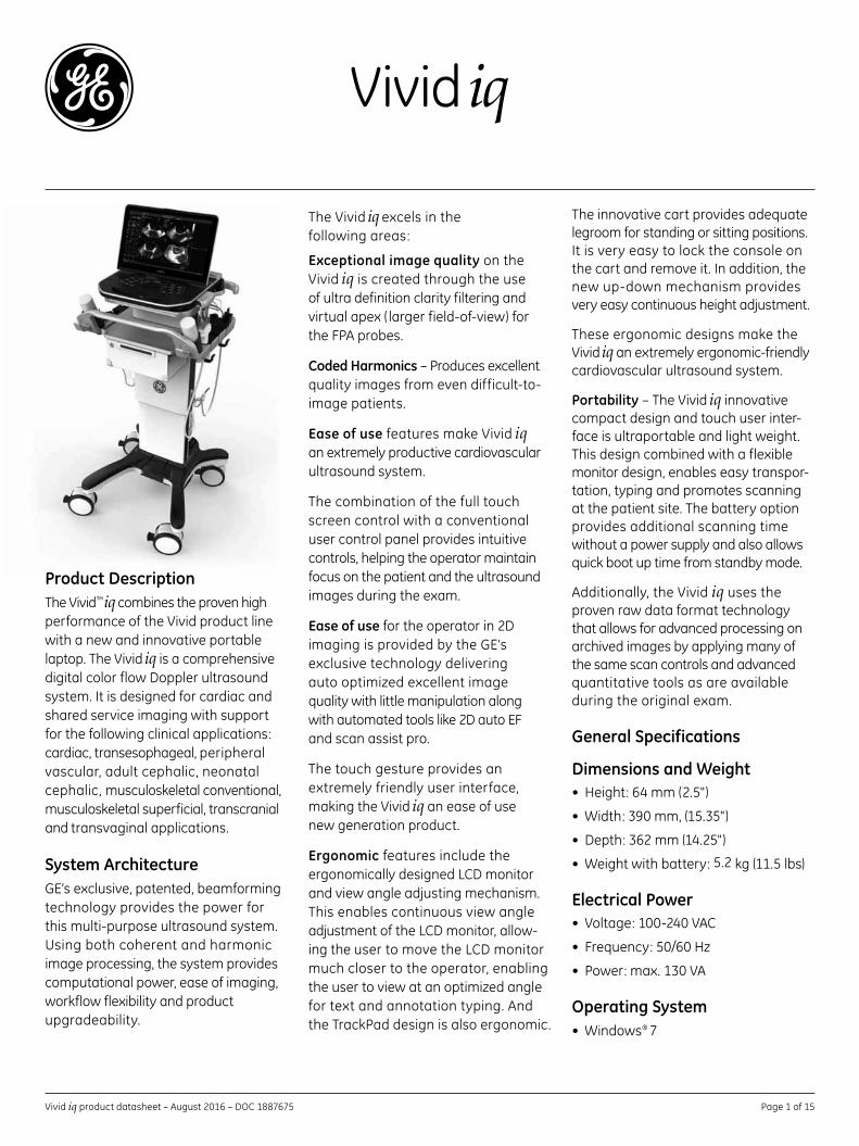
The innovative cart provides adequate legroom for standing or sitting positions. It is very easy to lock the console on the cart and remove it. In addition, the new up-down mechanism provides very easy continuous height adjustment.
These ergonomic designs make the Vivid iq an extremely ergonomic-friendly cardiovascular ultrasound system.
Portability – The Vivid iq innovative compact design and touch user inter-face is ultraportable and light weight. This design combined with a flexible monitor design, enables easy transpor-tation, typing and promotes scanning at the patient site. The battery option provides additional scanning time without a power supply and also allows quick boot up time from standby mode.
Additionally, the Vivid iq uses the proven raw data format technology that allows for advanced processing on archived images by applying many of the same scan controls and advanced quantitative tools as are available during the original exam.
General Specifications
Dimensions and Weight• Height:64mm(2.5")
• Width:390mm,(15.35")
• Depth:362mm(14.25")
• Weightwithbattery:5.2kg(11.5lbs)
Electrical Power• Voltage:100-240VAC
• Frequency:50/60Hz
• Power:max.130VA
Operating System• Windows® 7
The Vivid iq excels in the followingareas:
Exceptional image quality on the Vivid iq is created through the use of ultra definition clarity filtering and virtualapex( largerfield-of-view)fortheFPAprobes.
Coded Harmonics – Produces excellent quality images from even difficult-to-image patients.
Ease of use features make Vivid iq an extremely productive cardiovascular ultrasound system.
The combination of the full touch screen control with a conventional user control panel provides intuitive controls, helping the operator maintain focus on the patient and the ultrasound images during the exam.
Ease of use for the operator in 2D imaging is provided by the GE’s exclusive technology delivering autooptimizedexcellentimage quality with little manipulation along withautomatedtoolslike2DautoEF and scan assist pro.
The touch gesture provides an extremely friendly user interface, making the Vivid iq an ease of use new generation product.
Ergonomic features include the ergonomically designed LCD monitor and view angle adjusting mechanism. This enables continuous view angle adjustment of the LCD monitor, allow-ing the user to move the LCD monitor much closer to the operator, enabling theusertoviewatanoptimizedangle for text and annotation typing. And the TrackPad design is also ergonomic.
Vivid iq
Product DescriptionThe Vivid™ iq combines the proven high performance of the Vivid product line with a new and innovative portable laptop. The Vivid iq is a comprehensive digital color flow Doppler ultrasound system. It is designed for cardiac and shared service imaging with support forthefollowingclinicalapplications:cardiac, transesophageal, peripheral vascular, adult cephalic, neonatal cephalic, musculoskeletal conventional, musculoskeletal superficial, transcranial and transvaginal applications.
System Architecture GE’s exclusive, patented, beamforming technology provides the power for this multi-purpose ultrasound system. Using both coherent and harmonic image processing, the system provides computational power, ease of imaging, workflow flexibility and product upgradeability.
Vivid iq product datasheet – August 2016 – DOC 1887675 Page 2 of 15
Console Design• Laptopstyle
• ECGport
• Integratedsolidstatedrive
• MultipleUSBports(front/back)
• Integratedspeakersfor premium sound
• CPU–Intelduocore
• DCpowerinput
• USBinterface(5)
• HDMIinterface
• ECG
• LAN10/100/1000base
Cart Dimension• Height:835-1115mm(32.9"-43.9")
• Width:524.9mm(20.7")
• Depth:552.3mm(21.7")
• Weight:41kg(90Ibs.)
Cart Design• ThreeUSBports
• Sixprobeholders
• Fourprobecablehooks
• Chargebox(optional ) – to charge up to three batteries and to scan more than 180 min with four fully charged batteries
• Multi-probebox(optional ) –threeRS,one DLP to support 6VT-D
User Interface
Operator Panel• Innovativetrackpaddesign–same
intuitive functionality as track ball
• Ergonomicsimplifiedhardkeylayoutwith ergonomic design around the track pad
• Interactiveback-lightingof application-specific push buttons – adjustable back-light intensity
• Easy-to-learnuserinterfacewithintelligent touch keyboard
• Imagemanageronthetouch screen for quick review of image clipboard contents
• Transcranial
• Transrectal
• Transvaginal
Operating Modes• 2Dtissue
• 2Dcolorflow
• 2Dangioflow
• ColorM-mode
• TissuevelocityM-mode
• ContinuouswaveDoppler
• TissueM-mode
• PulsedwaveDoppler
• AnatomicalM-mode
• Tissuevelocityimaging
• Tissuetracking
• TissuevelocityDoppler
• Bloodflowimaging
• B-flow
• 2Dstress
• AutoEF
• Virtualconvex
• Virtualapex
• Codedphaseinversion
• Compoundimaging
Scanning Methods• Electronicsector
• Electronicconvex
• Electroniclinear
• CWpencil
Transducer Types• Sectorphasedarray
• Convexarray
• Lineararray
Peripheral Options• DVDRW
• Colorvideoprinter
• B/Wvideoprinter
• EightGBmemorystick
• OneTBUSBharddrive
• HDMIcable
Touch Screen• Fulltouchability
• 15.6"ultra-high-resolution,widescreen format, color, multi-touch LCD screen
• Interactiveuser-configurable short-cut software menu
• Application-specificoperatortouchmenu controls operated by finger and swiping
• Application-specificsidebartouchmenu controls operated by finger and swiping
• Overallgain,depthandzoomcontrolbar on the touch for easy adjustment
• Touch-screencontrolofTGCsliders
Display Monitor• 15.6"widescreenfullHigh-Definition(HD)flicker-freeLCDdisplaywithfulltouch ability
• ErgonomicFlexFitdesignwith adjustable typing angle and flexible view angle
• Resolution:1920x1080pixels,fullHD
• Folddownandlockmechanism for transportation
• Screencanbeadjustedindifferentangles for scanning mode, typing modeandclosing,allowingtooptimize the viewing angle in each position
• Backlightadjustable
System Overview
Applications (probe dependent)
• Cardiac
• Transesophageal
• Peripheralvascular
• Fetal/OB
• Abdominaladults
• Pediatric
• Smallorgan
• Neonatalcephalic
• Adultcephalic
• Musculoskeletalconventional
• Musculoskeletalsuperficial
Vivid iq product datasheet – August 2016–DOC1887675 Page3of15
• Three-pedalconfigurablefootswitch
• Rollingbag
Accessories (options)
• InterfacecableforexternalECG
• ECGadapterforDIN-typepediatricselectrode leads
Display Modes• Liveandstoreddisplayformat: fullsizeandsplitscreen,both with thumbnails, for still and cine
• Instant-reviewscreendisplays12 simultaneousloops/imagesforaquick study review
• Selectabledisplayconfigurationofduplexandtriplexmodes:side-by-side or top-bottom during live, digital replay and clipboard image recall
• Single-,dual-andquad-screenview
• Simultaneouscapability
- 2D+PW/CW
- 2D+CFM/TVI+PW
- 2D+CFM+CW
- 2D+CFM/Angio/TVI/SRI/TT/SI/TSI
- 2D+M/AMM/CAMM
- 2D+CFM/Angio/TVI/SRI/TT/SI/TSI+M/AMM/CAMM
- Real-time duplex or triplex mode
- Compound+M/CFM/PW
- 2D + color split screen (simultaneousmode)
• Selectablealternatingmodes
- 2Dorcompound+PW
- 2D+CW
- 2Dorcompound+CFM/PW
- 2D+CFM+CW
• Multi-image(split/quadscreen)
- Liveand/orfrozen
- Independent cine playback
• Timelinedisplay
- Independent2D(orcompound)+PW/CW/Mdisplay
- A choice of display formats with varioussizesof2D+PW/CW/M
• Top/bottomselectableformat
• Side/sideselectableformat
• Spectruminversion
• Acousticframerate
• CINEgauge,imagenumber/framenumber
• Bodymarks:multiplehuman anatomical structures
• Application/presetname
• Measurementresults
• Operatormessage
• Displayedacousticoutput
- TIS:ThermalIndexSoftTissue
- TIC:ThermalIndexCranial(Bone)
- TIB:ThermalIndexBone
• MI:MechanicalIndex
• PoweroutputindB
• Biopsyguidelineandzone
• Heartrate
• TrackPad-drivenannotationarrows
• Activemodedisplay
• Stressprotocolparameters
• Parameterannotationfollow ASEstandard
• Freetextwithwordlibrary
• Scanplanepositionindicatorandprobe temperature are displayed with all TEE probes
• Imageorientationmarker
General System Parameters
System Setup• Pre-programmableM&A and
annotation categories
• Differentuserpresetsper probe/applicationmaybe stored for quick access
• Userprogrammablepresetcapabilitywith administrator preset protection
• Factorydefaultpresetdata,protected against modification
• User-definedannotations
• Bodypatterns
• Customizedcommenthomeposition
Display Annotation• Patientname
• PatientID
• Age,sexandbirthdate
• Hospitalname
• Dateformat:twotypesselectable– MM/DD/YY,DD/MM/YY
• Timeformat:twotypesselectable– 24hours,12hours
• GestationalagefromLMP/EDD/GA
• Probename
• Mapnames
• Probeorientation
• Depthscalemarker
• Focalzonemarkers
• Imagedepth
• Zoomdepth
• B-mode
- Gain
- Imaging frequency
- Frameaveraging
• M-mode
- Gain
- Frequency
- Time scale
• Dopplermode
- Gain
- Angle
- Samplevolumesizeandposition
- Wallfilter
- Velocityand/orfrequencyscale
- Spectruminversion
• Timescale
- PRF
- Doppler frequency
• ColorflowDopplermode
- Framerate
- Samplevolumesize
- Color scale
- Power
- Color baseline
- Color threshold marker
- Color gain
Vivid iq product datasheet – August 2016–DOC1887675 Page4of15
Comprehensive User Manual Available on Board User manual and service manual are included on DVD disk with each system. A printed user manual is provided.
Memory/Image Memory• TwoGBofcinememory
• Selectablecinesequencefor cine review
• Measurements/calculationsand annotations on cine playback
• Scrollingtimelinememory
• Dual-imagecinedisplay
• Quad-imagecinedisplay
• CINEindicatorandcineimage number display
• CINEreviewloop
• CINEreviewspeed
Image Storage• On-boarddatabaseofpatient
information from past exams
• User-selectableECGandtime gated acquisition available on touch panel during live scanning
• User-selectableprospectiveor retrospective capture in config
• Storageformats:
- DICOM®-compressed or uncom-pressed,single/multi-frame, with/withoutrawdata,storageviaclipboardand/orseamlesslydirectly to destination device
- Transfer/“SaveAs”JPEG,MPEG, AVI formats
• Storagedevices(optional ) :
- USBmemorystick:eightGB
- CD-RWstorage:700MB (DVDoptionrequired)
- DVDstorage:-RW(4.7GB)
- Harddriveimagestorage:oneTB
• Comparepreviousimageswith current exam
• Reloadofarchiveddatasets
Image and Data Management• Exceptionalworkflowwithinstant
access data management
• DICOM3.0support–seeDICOM conformance statement for details
• Supportfortransferoftheproprietary rawdatafileswithintheDICOMstandard
• 2D,CFMorTVIdataatmaximumframe rate may be reviewed by scrolling or by running cine loops (cancontainmorethan1000imagesforimagingmodes)
• Imageclipboardforstamp-size storage and review of stored images and loops
• Built-inpatientarchivewith images/loops,patientinformation,measurements and reports
• DICOM-SRStandardstructured reporting mechanism
• Structuredfindingsreporttoolssupport efficient text entries with direct editing of findings text, usability improvements, new configuration options and conclusion section
• Usercanenternormalvalues which are then compared to actual measurements
• ConfigurableHTML-based report function
• Reporttemplatescanbecustomizedon board
• ASE-baseddefaulttextmodules (English),user-customizable
• Internalarchivedatacanbe exported to removable image storagethroughDICOMmedia
• Internalharddisk–forstoring programs, application defaults, ultrasound images and patient archive
• Alldatastorageisbasedon ultrasound raw data, allowing to change gain, baseline, color maps, sweep speeds, etc., for recalled images and loops
Connectivity and DICOM (optional) • Ethernetnetworkconnection
• DICOM3.0
• Verify
• Store
• Modalityworklist
• Storagecommitment
• ModalityPerformedProcedureStep(MPPS)
• Mediaexchange
• DICOMspooler
• DICOMquery/retrieve
• Structuredreporting–compatiblewith adult cardiac and vascular
• Mediastoreofstructuredreporting
• InSite™ ExC capability for remote service/access
• Supportoftwopatients’IDsinDICOM
• SeparateDICOMSRandimage storage destinations
• SimultaneoustransferofDICOM to multiple destinations
Patient Archive
EchoPAC™/Patient Archive• DataformatfullycompatiblewithofflineEchoPACreview/reporting stations of same or newer vintage
• Instantaccesstoultrasoundrawdata provided by the system
• Advancedpost-processinganalysis
• Threeuserlevelshelporganizingdata security requirements
• E-signoffcompatibility,withclearindications in patient management screens and report screen that a report was signed off, and by whom and at what time. The signed off report and exam cannot be changed. The“DiagnosingPhysician”fieldisautomatically assigned to the user that did the sign-off
Vivid iq product datasheet – August 2016 – DOC 1887675 Page 5 of 15
• DICOMmedia–read/writeimages onDICOMformat
• DICOMviewerembeddedonmedia(optionalandselectableinConfig)
• AlphanumericdatacanbeexportedinXMLformat
• JPEGexport(“SaveAs”)forstillframes
• AVIandMPEGexport(“SaveAs”) for cineloops
eVue/MPEGvue (optional)• Allowsinteractiveviewingof
images, loops or full exams
• UsingMPEGvue,examsmaybestored onto removable media or on a remote networked system together with an integrated MPEGvueplayerforviewingon standard PC
Self-contained DICOM Viewer (optional)• ExamscanbetransferredtoCD/DVDorUSBmediawithanintegrated “EZDICOMCDviewer™”
• Self-contained“EZDICOMCDviewer™” allows review of exams from media on a standard PC without installing anything on the host
Insite™ Express Connection (ExC) Enables Remote Service and Training
• Easy,flexibleandsecureconnectivity configuration.The“ContactGE” on-screen button directly generates a real-time service request to the GE online engineering or application specialist. It takes a snapshot (e.g.,errorlogs,setupfiles)ofthe system at the time of the service request to enable analysis of problem before customer contact
• VirtualConsoleObservation(VCO) enables the customer to allow desktop screens to be viewed and controlled remotely over the encrypted tunnel to enable real-time training, device configuration and clinical application support
• OperationofInsiteExpressConnection is dependent on the infrastructure being available – check with your local GE service representative
• Automaticallycalculatedlateralgain
2D Mode
• Sectortiltandwidthcontrol
• Framerateinexcessof1000fps,depending on probe, settings and applications
• Codedoctaveimagingwithcodedphaseinversion–3rdgenerationharmonic tissue imaging providing improved lateral and contrast resolu-tion over conventional fundamental imaging.Featureshelpreducenoise,improve wall definition, and axial resolution, making it well suited for a wide variety of patient groups
• Automatictissueoptimization– singlekeystrokeoptimizesimmedi-ately automatically and dynamically different grayscale settings with the goal of signal independent uniform gain and contrast distribution
• UDclarityandUDspecklereduceimaging – an advanced image processing technique to remove speckle in real-time examining the relative difference between neigh-boring pixel values and determining whether the grayscale variations have a sharp difference, follow a trend, or are random in nature
• Multiple-anglecompoundimaging– multiple co-planar images from dif-ferent angles combined into a single image in real-time to help enhance border definition and contrast resolution, as well as reduce angular dependence of border or edge as compared to no-compound imaging
• Virtualconvexprovidesawider field-of-view with linear probes, effective at certain imaging views where a wide far field may be preferred
• Virtualapexprovidesawider field-of-view with phased array probes, effective at certain imaging views where a wide near field may be preferred
• L/Randup/downinvert,inlive,digitalreplay or image clipboard recall
• Filetransferenablesthecustomer(biomedorclinician)todirectlytransfer systeminformation(e.g.,system logs,images,parametricdata)toGEproductengineeringteams(nopatientdatatransferred)
• Softwarereloadprovidesremote application reconstruction and recovery capabilities in the event of system corruption
Scanning Parameters• Digitalbeamformerwithupto974,026effectivedigitalchannels
• Minimumfield-of-viewrange(depth):1cm(probedependent)
• Maximumfield-of-viewrange(depth):33cm(probedependent)
• Widthrange:10°–168° (probedependent)
• Continuousdynamicreceive focus/continuousdynamic receive aperture
• Adjustabledynamicrange, infinite upper level
• Imagereverse:right/left
• Imagerotationof0,°180°
Tissue ImagingGeneral
• Variabletransmitfrequenciesforresolution/penetrationoptimization
• Displayzoomwithzoomareacontrol
• High-Resolution(HR)zoom– concentrates all image acquisition power into selected Region of Interest(ROI)
• Variablecontourfiltering–foredgeenhancement
• Depthrangeupto30cm– probe specific
• Selectablegrayscaleparameters:gain, reject, DDP, clarity, dynamic range and compress – can be adjusted in live, digital replay and image clipboard recall (probedependent)
• AutomaticallycalculatedTGCcurvesreduce operator interaction
Vivid iq product datasheet – August 2016 – DOC 1887675 Page 6 of 15
• Digitalreplayforretrospective review or automatic looping of images, allowing for adjustment of parameters such as gain, reject, anatomicalM-mode,persistence and replay speed
• Datadependentprocessingperformstemporal processing which helps reduce random noise but leaves motion of significant tissue struc-tures largely unaffected – can be adjusted even in digital replay
• 256shadesofgray
• Colorized2D-mode,user-selectablein real-time, digital replay
M-mode
• TrackPadsteersM-modelineavailable with all imaging probes – max steering angle is probe dependent
• Simultaneousreal-time2D-and M-mode
• M-modePRF1kHz–imagedata acquired is combined to give high-quality recording regardless of display scroll speed
• Digitalreplayforretrospectivereviewof spectral data
• Severaltop-bottomformats,side-by-side format and time-motion-only format – can be adjusted in live or digital replay
• Selectablehorizontalscrollspeed: 1,2,3,4,6,8,12,16seconds across display
• Horizontalscrollcanbeadjusted in live or digital replay
Anatomical M-mode
• M-modecursorcanbeadjusted at any plane
• CurvedanatomicalM-mode– free(curved)drawingofM-modegenerated from the cursor independent from the axial plane
• Canbeactivatedfromlive,digitalreplay or image clipboard recall
• Anatomicalcolorandtissue velocityM-mode
• M&A capability
• VariableROIsizeinwidthanddepth
• User-selectableradialandlateralaveraging to help reduce statistical uncertainty in the color velocity and variance estimates
• DataDependentProcessing(DDP)performs temporal processing and display smoothing to help reduce loss of transient events of hemodynamic significance
• Digitalreplayforretrospectivereviewor automatic looping of color images, allowing for adjustment of parameters such as DDP, encoding principle, baseline shift, color maps, color priority and color gain even on frozen/recalleddata
• Application-dependent,multi-variatemotion discriminator helps reduce flash artifacts
• Dedicatedcoronaryflowapplication
• Multiple-anglecompoundimagingin2D mode is maintained while in color Doppler mode
Color Angio• Angle-independentmodefor visualizationofsmallvesselswithincreased sensitivity compared to standard color flow of previous GE products
Color M-mode• VariableROIlengthandposition–
user-selectable
• User-selectableradialaveragingtohelp reduce statistical uncertainty in the color velocity and variance estimates
• Selectablehorizontalscrollspeed: 1,2,3,4,6,8,12,16seconds across display – can be adjusted during live, digital replay or image clipboard recall
• Real-time2Dimagewhilein colorM-mode
• Samecontrolsandfunctionsavailable as in standard 2D color Doppler
Color Doppler Imaging
General
• SteerablecolorDoppleravailablewith all imaging probes – max steering angle is probe dependent
• TrackPad-controlledROI
• Touchscreen-controlledROI
• Removalofcolormapfromthe tissue during digital replay
• Digitalreplayforretrospective reviewofcolororcolorM-mode data allowing for adjustment of parameters such as encoding principle, color priority and color gain even on stored data
• PRFsettings–user-selectable
• Advancedregressionwallfiltergivesefficient suppression of wall clutter
• Foreachencodingprinciple,multiplecolor maps can be selected in live and digital replay – variance maps available
• Morethan65,000simultaneouscolors processed, providing a smooth display two-dimensional color maps containing a multitude of color hues
• Simultaneousdisplayofgrayscale 2D and 2D with color flow
• Colorinvert–user-selectableinliveand digital replay
• Variablecolorbaseline– user-selectable in live and digital replay
• Multi-variatecolorpriorityfunctiongives delineation of disturbed flows even across bright areas of the 2D-mode image
• ColorDopplerfrequencycanbechanged independently from 2D
Color Flow Imaging• TruSpeedimagingallowseitherultra-
high frame rate or increased lateral resolution as compared to previous generation GE products
• Framerateinexcessof700(itis400on12S-RS)fps,dependingonprobeand settings
Vivid iq product datasheet – August 2016 – DOC 1887675 Page 7 of 15
Anatomical Color M-mode• GE-patented,anyplanecolorM-mode
display derived from color Doppler cine loop
• Alsoapplicabletotissue velocity Imaging
• M&A capability
B-flow• B-flowisadigitalimagingtechniquethatprovidesreal-timevisualization of vascular hemodynamics by directly visualizingbloodreflectorsand presenting this information in a grayscale display
• UseofGE-patentedtechniquestoboost blood echoes, and to help preferentially suppress non-moving tissue signals
• B-flowisavailableformostvascularand shared service applications
Blood Flow Imaging• CombinescolorDopplerwith
grayscale speckle imaging
• Helpsimprovedelineationofbloodflow without bleeding into tissue or vessel wall
Blood Flow Angio Imaging• Combinesangiowithgrayscale
speckle imaging
Tissue Velocity Imaging
Tissue Velocity Imaging Mode
• MyocardialDopplerimagingwithcolor overlay on tissue image
• TissueDopplerdatacanbeacquired in background during regular 2D imaging
• Thevelocityofmyocardialsegmentsafter entire heart cycle can be displayed in one single image
• Tissuecoloroverlaycanberemovedtoshow just the 2D image, still retaining the tissue velocity information
• QuantitativeprofilesforTVI,tissuetracking, strain and strain rate can be derived
• Severaltop-bottomformats, side-by-side format and time- motion-only format – can be adjusted in live or digital replay
• Selectablehorizontalscrollspeed:1,2,3,4,6,8,12,16secondsacrossdisplay – can be adjusted in live or digital replay
• AdjustablespectralDopplerdisplayparameters:gain,reject,compress,color maps – can be adjusted in live or digital replay
• User-adjustablebaselineshift– in live, digital replay and image clipboard recall
• Adjustablevelocityscale
• Wallfilterswithrange10-2000Hz(velocityscaledependent)
• Anglecorrectionwithautomaticadjustment of velocity scale – in live, digital replay and image clipboard recall
• AutoDopplerangle
• Stereospeakersmountedinthe front panel
• Displayannotationsoffrequency,mode,scales,Nyquistlimit,wallfiltersetting, angle correction, acoustic power indices
• Compoundinduplex
PW/HPRF Doppler
• AutomaticHPRFDopplermaintainsits sensitivity even for shallow depths andwiththehighestPRF’s
• DigitalvelocitytrackingDoppler employs processing in range and time for high-quality spectral displays
• Adjustablesamplevolumesizeof1-16mm(probedependent)
• Maximumsamplevolumedepth30cm
CW Doppler
• HighlysensitivesteerableCWavailable with all phased array probes
• TissuevelocityDoppler
• Timemarkersforvalveeventsderived fromanyTMmodehelpsimplifyunderstanding of signals in velocity tracesorcurvedanatomicalM-mode
Tissue Tracking Mode
• Real-timedisplayofthetimeintegral of TVI for quantitative display of myocardial systolic displacement
• Myocardialdisplacementiscalculated and displayed as a color-coded overlayonthegrayscaleandM-mode image – different colors represent different displacement ranges
Spectral DopplerGeneral
• OperatesinPW,HPRFandCWmodes
• TrackPadsteerableDoppleravailablewith all imaging probes – max steering angle is probe dependent
• SelectableDopplerfrequencyforenhancedoptimization
• High-quality,real-timeduplexor triplex operation in all Doppler modes,CWandPW,andfor all velocity settings
• Frameratecontrolforoptimized use of acquisition power between spectrum, 2D and color Doppler modes in duplex or triplex modes
• VeryfastandflexiblespectrumanalysiswithanequivalentDFT rate of 0.2 ms
• AutomaticSpectrumOptimization(ASO)providesasinglepress, automatic,real-timeoptimization ofPWorCWspectrumscale,andbaseline display
• Dynamicgaincompensationfordisplay of flows with varying signal strengths over the cardiac cycle to help improve ease of use
• Dynamicrejectgivesconsistent suppression of background – user-selectable in real-time, digital replay or image clipboard recall
• Digitalreplayforretrospective review of spectral Doppler data
Vivid iq product datasheet – August 2016 – DOC 1887675 Page 8 of 15
Physiological Traces• Integratedthree-leadECGmodule
• AutomaticQRScomplexdetection
• ExternalECGleadinput
• Internallygeneratedrespiratory trace using ECG leads
• ECGleadselection
• AdjustableECGQRSmarkers
Automatic Optimization• DynamicoptimizationofB-mode
image to improve contrast resolution, TGCandgrayscale(softorsharp,user-selectable)
• Auto-spectraloptimize–dynamicadjustmentsofbaseline,andPRF (onliveimage)andanglecorrection
Measurement and Analysis (M&A)• Personalizedmeasurement
protocols allow individual set andorderofM&A items
• Measurementscanbelabeled seamlessly by using protocols or post assignments
• Measurementsassignableto protocol capability
• Parameterannotationfollow ASEstandard
• Seamlessdatastorageand report creation
• User-assignableparameters
• Comprehensivesetofcardiac measurements and calculations to help assess dimensions, flow properties and other functional parameters of the heart
• Comprehensivesetofsharedservicemeasurements and calculations covering vascular, abdominal, obstetrics and other application areas
• Configurationpackagetosetup acustomizedsetandsequence of measurements to use, defining user-defined measurements and changing settings for the factory-defined measurements
• Sample-areapointsmaybedynami-cally anchored to move with the tissue when running the cineloop
• Cinecompounddisplayscineloopsgenerated from a temporal averaging of multiple consecutive heart cycles
Automated Ejection-Fraction Calculation (AutoEF) (optional )
• AutomatedEFmeasurementtoolbased on 2D speckle tracking algorithmandonSimpson
• IntegratedintoM&A package with worksheet summary
Generic Measurements
• BSA(BodySurfaceArea)
• MaxPG(MaximumPressureGradient)
• MeanPG(MeanPressureGradient)
• %Stenosis(StenosisRatio)
• PI(PulsatilityIndex)
• RI(ResistivityIndex)
• HR(HeartRate)–beats/minute
• A/BRatio(VelocitiesRatio)
• TAMAX(TimeAveragedMaximumVelocity)–TracemethodisPeak orManual
• TAMIN(TimeAveragedMinimumVelocity)–TracemethodisFloor
• TAMEAN(TimeAveragedMean Velocity)–TracemethodisMean
• Volume
OB/GYN Application Module• OBpackageforfetalgrowth
analysis containing more than 100 biometry tables
• DedicatedOB/GYNreports
• Fetalgraphicalgrowthcharts
• Growthpercentiles
• Multi-gestationalcalculations (uptofour)
• ProgrammableOBtables
• Expandedworksheets
• User-selectablefetalgrowth parameters based on European, American or Asian methods charts
• GYNpackageforovaryanduterusmeasurements and reporting
• Stressechosupportallowingwallmotion scoring and automatic stress level labeling of measurements
• Supportformeasuringon DICOMimages
• AutomaticDopplertracefunctionality for use in non-cardiac applications in both live and replay
• Worksheetforreview,editand deletion of performed measurements
• Reportingsupportallowinga configurable set of measurements to be shown in the exam report
• DICOMSRexportofmeasurementdata
Intima Media Thickness (IMT) Measurements
• Automaticmeasurements (patentpending)ofcarotidarteryIntima-MediaThickness(IMT)on any acquired frame
• On-boardIMTpackagefacilitatesnon-interrupted workflow – fully integratedwithM&A, worksheet, archiving and reporting functions
• Algorithmprovidesrobust,quick, reliable measurements which can be stored to the on-board archive for review and reporting
• IMTmeasurementcanbemadefromfrozenimagesorimagesretrievedfrom archive
• IMTpackagesupportsmeasure-ments of different regions of the intima in the carotid vessel (e.g.,Lt./Rt./CCA/ICAetc.)
• FrameforIMTmeasurementcan be selected in relation to the ECG waveform
Z-Scores
• Limitedimplementationofz-scores for a set of predefined pediatric dimension measurements
Quantitative Analysis Package (Q-Analysis) (optional )
• Tracesforvelocityorderived parameters(displacement)insidedefined regions of interest as function of time
Vivid iq product datasheet – August 2016–DOC1887675 Page9of15
OB Measurements/Calculations
• Gestationalageby:
- GS(GestationalSac)
- CRL(CrownRumpLength)
- FL(FemurLength)
- BPD(Bi-ParietalDiameter)
- AC(AbdominalCircumference)
- HC(HeadCircumference)
- APTDxTTD(Anterior/PosteriorTrunk Diameter by Transverse TrunkDiameter)
- LV(LengthofVertebra)
- FTA (FetalTrunkCross-sectionalArea)
- HL(HumerusLength)
- BD(BinocularDistance)
- FT(FootLength)
- OFD(OccipitalFrontalDiameter)
- TAD (TransverseAbdominalDiameter)
- TCD (TransverseCerebellumDiameter)
- THD(ThoraxTransverseDiameter)
- TIB(TibiaLength)
- ULNA(UlnaLength)
• EstimatedFetalWeight(EFW)by:
- AC,BPD
- AC,BPD,FL
- AC,BPD,FL,HC
- AC,FL
- AC,FL,HC
- AC,HC
- EFBW
• CalculationsandRatios
- FL/BPD
- FL/AC
- FL/HC
- HC/AC
- CI(CephalicIndex)
- AFI(AmnioticFluidIndex)
- CTAR(Cardio-ThoracicAreaRatio)
• Measurements/calculationsby:ASUM,ASUM2001,Berkowitz,Bertagnoli,Brenner,Campbell,CFEF,Chitty,Eik-Nes,Ericksen,Goldstein,Hadlock,Hansmann,Hellman,Hill,Hohler,Jeanty,JSUM,Kurtz,Mayden,Mercer,Merz,Moore,Nelson,OsakaUniversity, Paris, Rempen, Robinson, Shepard,Shepard/Warsoff,TokyoUniversity,Tokyo/Shinozuka,Yarkoni
Cardiac Measurements• %FS(LVFractionalShortening)
• %IVSThck(IVSFractionalShortening)
• %LVPWThck(LVPosterior WallFractionalShortening)
• AoArchDiam(AorticArchDiameter)
• AoAsc(AscendingAorticDiameter)
• AoDescDiam (DescendingAorticDiameter)
• AoIsthmus(AorticIsthmus)
• AoRootDiam(AorticRootDiameter)
• ARERO (PISA:RegurgitantOrificeArea)
• ARFlow(PISA:RegurgitantFlow)
• ARPHT(AVInsuf.PressureHalfTime)
• ARRad(PISA:RadiusofAliasedPoint)
• ARRF(RegurgitantFraction overtheAorticValve)
• ARRV (PISA:RegurgitantVolumeFlow)
• ARVel(PISA:AliasedVelocity)
• ARVmax(AorticInsuf.PeakVelocity)
• ARVTI (AorticInsuf.VelocityTimeIntegral)
• ARedmaxPG(AorticInsuf. End-DiastolePressureGradient)
• ARedVmax(AorticInsuf. End-DiastolicVelocity)
• AVAccSlope (AorticValveFlowAcceleration)
• AVAccTime (AorticValveAccelerationTime)
• AVAccT/ET(AVAcceleration toEjectionTimeRatio)
• AVEOAI(VTI)(AorticValve Effective Orifice Area Index byContinuityEquationVTI)
• AVEOAIVmax(AorticValve Effective Orifice Area Index byContinuityEquationPeakV)
• AVCO(CardiacOutputbyAorticFlow)
• AVCusp (AorticValveCuspSeparation,2D)
• AVDecTime (AorticValveDecelerationTime)
• AVDiam(AorticDiameter,2D)
• Fetalgraphicaltrending
• Growthpercentiles
• Multi-gestationalcalculations(four)
• Fetalqualitativedescription (anatomicalsurvey)
• Fetalenvironmentaldescription (biophysicalprofile)
• ProgrammableOBtables
• Over20selectableOBcalculations
• Expandedworksheets
GYN Measurements/Calculations
• Rightovarylength,width,height
• Leftovarylength,width,height
• Uteruslength,width,height
• Cervixlength,trace
• Ovarianvolume
• ENDO(endometrialthickness)
• OvarianRI
• UterineRI
• Follicularmeasurements
• Summaryreports
Vascular Calculations• RTECA(RightExternalCarotid ArteryVelocity)
• RTCCA(RightCommonCarotid ArteryVelocity)
• RTBIFURC(RightCarotid BifurcationVelocity)
• RTICA(RightInternalCarotid ArteryVelocity)
• RTICA/CCA(RightInternalCarotid ArteryVelocity/CommonCarotidArteryVelocityRatio)
• LTECA,LTCCA,LTBIFURC,LTICA, LTICA/CCA(sameasabove,forLeftCarotidArtery)
• A/BRatio(VelocitiesRatio)
• %Stenosis(StenosisRatio)
• S/DRatio(SystolicVelocity/DiastolicVelocitiesRatio)
• PI(PulsatilityIndex)
• RI(ResistivityIndex)
• HR(HeartRate)–beats/minute
Vivid iq product datasheet – August 2016 – DOC 1887675 Page 10 of 15
• AVmaxPG (AorticValvePeakPressureGradient)
• AVmeanPG (AorticValveMeanPressureGradient)
• AVSV(StrokeVolumebyAorticFlow)
• AVVmax(AorticValvePeakVelocity)
• AVVmean(AVMeanVelocity)
• AVVTI (AorticValveVelocityTimeIntegral)
• AVA(Vmax)(AVAreabyContinuityEquationbyPeakV)
• AVA(VTI)(AVAreabyContinuity EquationVTI)
• AVAPlanimetry(AorticValveArea)
• AVET(AorticValveEjectionTime)
• CO(Teich) (CardiacOutput,M-mode,Teicholtz)
• D-EExcursion (MVAnteriorLeafletExcursion)
• E’Avg(AveragedEarlyDiastolic MitralValveAnnularVelocity)
• E’Lat(EarlyDiastolicMitralValveLateralAnnularVelocity)
• E’Sept(EarlyDiastolicMitralValveSeptalAnnularVelocity)
• E/E’Avg (MitralInflowEVelocitytoE’AvgRatio)
• E/E’Lat (MitralInflowEVelocitytoE’LatRatio)
• E/E’Sept (MitralInflowEVelocitytoE’SeptRatio)
• EDV(Cube)(LeftVentricleVolume,Diastolic,2D,Cubic)
• EF(A-LA2C)(EjectionFraction2CH,SinglePlane,Area-Length)
• E-FSlope(MitralValveE-FSlope)
• EPSS(E-Point-to-SeptumSeparation,M-mode)
• ERO(EffectiveRegurgitantOrifice)
• ESV(Cube)(LeftVentricleVolume,Systolic,2D,Cubic)
• HR(HeartRate,2D,Teicholtz)
• IVC(InferiorVenaCava)
• IVCT(IsovolumicContractionTime)
• IVRT(IsovolumicRelaxationTime)
• IVSd(InterventricularSeptum Thickness,Diastolic,2D)
• LVLd(Apical) (LeftVentricularLength,Diastolic,2D)
• LVLs(Apical) (LeftVentricularLength,Systolic,2D)
• LVOTArea (LeftVentricleOutflowTractArea)
• LVOTCO (CardiacOutputbyAorticFlow)
• LVOTDiam(LeftVentricularOutflowTractDiameter)
• LVOTMaxPG (LVOTPeakPressureGradient)
• LVOTMeanPG (LVOTMeanPressureGradient)
• LVOTSI (StrokeVolumeIndexbyAorticFlow)
• LVOTSV (StrokeVolumebyAorticFlow)
• LVOTVmax(LVOTPeakVelocity)
• LVOTVmean(LVOTMeanVelocity)
• LVOTVTI(LVOTVelocityTimeIntegral)
• LVPWd(LeftVentricularPosteriorWallThickness,Diastolic,2D)
• LVPWs(LeftVentricularPosteriorWallThickness,Systolic,2D)
• LVsMass(LVMass,Systolic,2D)
• LVsMassIndex (LVMassIndex,Systolic,2D)
• LAAd(A2C)(LeftAtriumArea,Apical2C)
• MCO(MitralValveClosuretoOpening)
• MPArea(MitralValveProsthesis)
• MRAccTime (MVRegurg.FlowAcceleration)
• MRERO (PISA:RegurgitantOrificeArea)
• MRFlow(PISA:RegurgitantFlow)
• MRMaxPG (MitralRegurg.PeakPressureGradient)
• MRRad(PISA:RadiusofAliasedPoint)
• MRRF(RegurgitantFraction OvertheMitralValve)
• MRRV(PISA:RegurgitantVolumeFlow)
• MRVel(PISA:AliasedVelocity)
• MRVmax(MitralRegurg.PeakVelocity)
• VSs(InterventricularSeptum Thickness,Systolic,2D)
• LADiam(LeftAtriumDiameter,2D)
• LAMajor(LeftAtriumMajor)
• LAMinor(LeftAtriumMinor)
• LA/Ao(LADiameterto AoRootDiameterRatio,2D)
• LAAd(A2C)(LeftAtriumArea,Apical2C)
• LAEDV(A-L) (LAEndDiastolicVolume,Area-Length)
• LAEDVIndex(A-L)(LAEndDiastolicVolumeIndex,Area-Length)
• LAESV(A-L)(LAEndSystolicVolume,Area-Length)
• LAESVIndex(A-L)(LAEndSystolicVolumeIndex,Area-Length)
• LAEDVMOD (LAEndDiastolicVolumeMOD)
• LAESVMOD (LAEndSystolicVolumeMOD)
• LIMP(LeftIndexof MyocardialPerformance)
• LVA(s)(LeftVentricularArea, Systolic,2CH)
• LVAd(A2C)(LeftVentricularArea,Diastolic,2CH)
• LVAd(SAX)(LVArea,SAX,Diastolic)
• LVAend(d)(LVEndocardialArea,SAX)
• LVAepi(d)(LVEpicardialArea,SAX)
• LVAs(A4C) (LeftVentricularArea,Systolic,4CH)
• LVAs(SAX)(LVarea,SAX,Systolic)
• LVdMass(LVMass,Diastolic,2D)
• LVdMass(LVMass,Diastolic,M-mode)
• LVdMassIndex (LVMassIndex,Diastolic,2D)
• LVEDV(A-LA2C)(LVVolume,Diastolic,2CH,Area-Length)
• LVESV(A-LA2C)(LVVolume,Systolic,2CH,Area-Length)
• LVET(LeftVentricleEjectionTime)
• LVIDd (LVInternalDimension,Diastolic,2D)
• LVIDs (LVInternalDimension,Systolic,2D)
Vivid iq product datasheet – August 2016 – DOC 1887675 Page 11 of 15
• MRVmean (MitralRegurg.MeanVelocity)
• MRVTI (MitralRegurg.VelocityTimeIntegral)
• MVADur (MitralValveA-WaveDuration)
• MVAVelocity(MVVelocityPeakA)
• MVAccSlope (MitralValveFlowAcceleration)
• MVAccTime (MitralValveAccelerationTime)
• MVAcc/DecTime (MV:Acc.Time/Decel.TimeRatio)
• MVAnDiam (MitralValveAnnulusDiameter,2D)
• MVCO(CardiacOutputbyMitralFlow)
• MVDecSlope (MitralValveFlowDeceleration)
• MVDecTime (MitralValveDecelerationTime)
• MVEVelocity(MVVelocityPeakE)
• MVE/ARatio (MitralValveE-PeaktoA-PeakRatio)
• MVMaxPG (MitralValvePeakPressureGradient)
• MVMeanPG (MitralValveMeanPressureGradient)
• MVPHT (MitralValvePressureHalfTime)
• MVRegFrac (MitralValveRegurgitantFraction)
• MVSI (StrokeVolumeIndexbyMitralFlow)
• MVSV(StrokeVolumebyMitralFlow)
• MVTimetoPeak (MitralValveTimetoPeak)
• MVVmax(MitralValvePeakVelocity)
• MVVmean(MVMeanVelocity)
• MVVTI (MitralValveVelocityTimeIntegral)
• MVA(MitralValveArea)
• MVAByPHT (MitralValveAreaaccordingtoPHT)
• MVAbyPlan(MitralValveArea,2D)
• MVET(MitralValveEjectionTime)
• PVeinA(PulmonaryVeinVelocityPeakA)–Reverse
• PVVTI (PulmonicValveVelocityTimeIntegral)
• PVA(VTI)(PulmonaryArtery VelocityTimeIntegral)
• PVeinS/DRatio (PulmonaryVeinSDRatio)
• PVET(PulmonicValveEjectionTime)
• PVPEP (PulmonicValvePre-EjectionPeriod)
• PVPEP/ETRatio(PVPre-Ejection toEjectionTimeRatio)
• Qp/Qs (Pulmonic-to-SystemicFlowRatio)
• RAMajor(RightAtriumMajor,2D)
• RAMinor(RightAtriumMinor,2D)
• RAA(d) (RightAtriumArea,2D,Diastole)
• RAA(s) (RightAtriumArea,2D,Systole)
• RAEDVA2C(RightAtriumEnd DiastolicVolume,Apical2Chamber)
• RAESVA-L (RAEndSystoleVolume[A-L])
• RALd(RightAtriumLength,Diastole)
• RALs(RALength,Systole)
• RIMP(RightIndexof MyocardialPerformance)
• RJA(A4C)(RegurgitantJetArea)
• RJA/LAA (RegurgitantJetArearatioRJA/LAA)
• RVMajor(RightVentricleMajor)
• RVMinor(RightVentricleMinor)
• RVS’(TricuspidAnnulusSystolic ExcursionVelocity)
• RVAWd(RightVentricle WallThickness,Diastolic,2D)
• RVAWs(RightVentricle WallThickness,Systolic,2D)
• RVET(RightVentricleEjectionTime)
• RVIDd(RightVentricleDiameter, Diastolic,2D)
• RVIDs (RightVentricleDiameter,Systolic,2D)
• RVOTArea (RightVentricleOutflowTractArea)
• PVeinADur (PulmonaryVeinA-WaveDuration)
• PVeinD(PulmonaryVein End-DiastolicPeakVelocity)
• PVeinS(PulmonaryVein SystolicPeakVelocity)
• PAEDP (PulmonaryArteryDiastolicPressure)
• PE(d)(PericardEffusion,M-mode)
• PEs(PericardEffusion,2D)
• PRmaxPG(PulmonicInsuf. PeakPressureGradient)
• PRmeanPG(PulmonicInsuf. MeanPressureGradient)
• PRPHT (PulmonicInsuf.PressureHalfTime)
• PRVmax (PulmonicInsuf.PeakVelocity)
• PRVTI(PulmonicInsuf. VelocityTimeIntegral)
• PRendMaxPG(PulmonicInsuf. End-DiastolePressureGradient)
• PRendVmax(PulmonicInsuf. End-DiastolicVelocity)
• PulmonicDiam (PulmonaryArteryDiameter,2D)
• PVAccSlope (PulmonicValveFlowAcceleration)
• PVAccTime (PulmonicValveAccelerationTime)
• PVAccTime/ETRatio(PVAcceleration toEjectionTimeRatio)
• PVAnDiam (PulmonicValveAnnulusDiameter,2D)
• PVAnnArea(PulmonicValveArea)
• PVCO (CardiacOutputbyPulmonicFlow)
• PVMaxPG(PulmonicValve PeakPressureGradient)
• PVMeanPG(PulmonicValve MeanPressureGradient)
• PVSV (StrokeVolumebyPulmonicFlow)
• PVVmax (PulmonaryArteryPeakVelocity)
• PVVmean(PVMeanVelocity)

Vivid iq product datasheet – August 2016 – DOC 1887675 Page 12 of 15
• RVOTDiam (RVOutputTractDiameter,2D)
• RVOTDiam(RVOutput TractDiameter,M-mode)
• RVOTMaxPG (RVOTPeakPressureGradient)
• RVOTMeanPG (RVOTMeanPressureGradient)
• RVOTSI(LVStrokeVolumeIndex byPulmonicFlow)
• RVOTSV(StrokeVolume byPulmonicFlow)
• RVOTVmax(RVOTPeakVelocity)
• RVOTVmean(RVOTMeanVelocity)
• RVOTVTI(RVOTVelocityTimeIntegral)
• RVSP (RightVentricleSystolicPressure)
• RVWd(RightVentricleWallThickness,Diastolic,M-mode)
• RVWs(RightVentricleWallThickness,Systolic,M-mode)
• RAA(d) (RightAtriumArea,2D,Diastole)
• RAA(s) (RightAtriumArea,2D,Systole)
• SI(A-LA2C)(LVStrokeIndex, SinglePlane,2CH,Area-Length)
• SI(A-LA4C)(LVStrokeIndex, SinglePlane,4CH,Area-Length)
• SI(Bi-plane) (LVStrokeIndex,Bi-plane,MOD)
• SI(bullet) (LVStrokeIndex,Bi-plane,Bullet)
• SI(MODA2C)(LVStrokeIndex, SinglePlane,2CH,MOD)
• SI(MODA4C)(LVStrokeIndex, SinglePlane,4CH,MOD)
• SI(Teich)(LVStrokeIndex, Teicholtz,2D)
• SI(Teich)(LVStrokeIndex, Teicholtz,M-mode)
• SV(A-LA2C)(LVStrokeVolume, SinglePlane,2CH,Area-Length)
• SV(A-LA4C)(LVStrokeVolume, SinglePlane,4CH,Area-Length)
• SV(Bi-plane)(LVStrokeVolume, Bi-plane,MOD)
• TVEVelocity(TricuspidValveEVelocity)
• TVE/ARatio(TricuspidValve E-PeaktoA-PeakRatio)
• TVMaxPG(TricuspidValve PeakPressureGradient)
• TVMeanPG(TricuspidValve MeanPressureGradient)
• TVMeanPG(TricuspidValve MeanPressureGradient)
• TVPHT (TricuspidValvePressureHalfTime)
• TVSV (StrokeVolumebyTricuspidFlow)
• TVVmean(TVMeanVelocity)
• TVVTI (TricuspidValveVelocityTimeIntegral)
• VSDMaxPG (VSDPeakPressureGradient)
• VSDVmax(VSDPeakVelocity)
Please refer to the reference manual for the full list of measurements and calculations for all applications.
AnnotationsBody Marks
• Bodymarkiconsforlocationandposition of probe
• Easyselectionofbodymarksfromtouch panel
Text Annotations
• Easyselectionoftextannotationsfrom touch panel
Scan Assist Pro• Customizableautomationsthat
assist the user through each step of the scan
• Facilitatesconsistencyand reduced keystrokes
• Ultrasoundimage,anatomicalpicture, step by step training through a pre-defined protocol
• Supportsselectionofallmodes,allmeasurements and dual annotations
• SV(Bullet)(LVStrokeVolume, Bi-plane,Bullet)
• SV(MODA2C)(LVStrokeVolume,Single-plane,2CH,MOD)–Simpson
• SV(MODA4C)(LVStrokeVolume,Single-plane,4CH,MOD)–Simpson
• SV(Cube)(LVStrokeVolume,2D,Cubic)
• SV(Cube) (LVStrokeVolume,M-mode,Cubic)
• SV(Teich) (LVStrokeVolume,2D,Teicholtz)
• SV(Teich) (LVStrokeVolume,M-mode,Teicholtz)
• SystemicDiam (SystemicVeinDiameter,2D)
• SystemicVmax (SystemicVeinPeakVelocity)
• SystemicVTI (SystemicVeinVelocityTimeIntegral)
• TAPSE(TricuspidAnnularPlane SystolicExcursion)
• TCO (TricuspidValveClosuretoOpening)
• TRMaxPG(TricuspidRegurg. PeakPressureGradient)
• TRMeanPG(TricuspidRegurg. MeanPressureGradient)
• TRVmax (TricuspidRegurg.PeakVelocity)
• TRVmean (TricuspidRegurg.MeanVelocity)
• TRVTI(TricuspidRegurgitation VelocityTimeIntegral)
• TVADur (TricuspidValveA-WaveDuration)
• TVAVelocity(TricuspidValveAVelocity)
• TVAccTime (TricuspidValveTimetoPeak)
• TVAnnArea(TricuspidValveArea)
• TVAnnDiam(TricuspidValve AnnulusDiameter,2D)
• TVArea(TricuspidValveArea,2D)
• TVCO (CardiacOutputbyTricuspidFlow)
• TVDecSlope (TricuspidValveFlowDeceleration)
Vivid iq product datasheet – August 2016–DOC1887675 Page13of15
• Imagingattributes:octave,steer,dual/quadscreen,compound, zoom,depth,scaleandbaseline
• On-lineoroff-lineprotocoleditor
• Imageacquisitionaccordingto predefined protocol templates
• Variousfactoryprotocoltemplates
• User-configurableprotocoltemplates
Smart Stress Echo (optional)Supported Protocol Examinations
• 2Dpharmacologicalstressecho
• 2Dbicyclestressecho
• 2Dcontinuouscapturestressecho(treadmillstressecho)
• Cardiacresynchronizationtherapyprogramming protocols (availablewiththeAdvancedQScanoption)
Protocol Examinations Features(enabled with Smart Stress option)
• Wallmotionscoring:analysis by wall motion in individual myocardial segments
• Showreference:showareferenceimage from baseline or previous level during acquisition
• Smartstress:automaticallysetupvarious scanning parameters (forinstancegeometry,frequency,gain,etc.)accordingtosame projection on previous level
• Scanmodesettings:scanmode may be specified for individual views in the protocol
• Previewofstore:showrunning loops as preview before storing to the examination
Continuous Capture
• Continuouslyacquirelargeamountsof 2D image data, and selection of pro-jection views for analysis afterwards
• Theentirecontinuouscapture recording may be kept in memory while it is possible to store new images outside the protocol template, or the entire recording can be stored to file
• Selectionofprojectionviewsonscanner or EchoPAC when the entire recording is stored to file
Virus ProtectionTo reduce virus vulnerability, Vivid iq is configured with a minimal set of open ports and with all network services not actively used by the system closed down. This helps to significantly reduce the risk of a virus attack on Vivid iq.
GE is continuously judging the need for additional actions to reduce vulnerability of equipment; this includes vulnerability scanning of our products and evaluation ofnewsecuritypatchesforthe3rd-partytechnologyused.Microsoft® (andother)securitypatchesthataddress serious issues with Vivid iq will be made available to customers after GE verification of those patches.
Probes3Sc-RS Phased Array Probe
• Probepresets:cardiac,pediatric,abdominal, fetal, adult cephalic
• Biopsyguide:multi-angledisposablewith a reusable bracket
6S-RS Phased Array Probe
• Probepresets:pediatric,fetal, neonatal cephalic, abdominal
12S-RS Phased Array Probe
• Probepresets:pediatric, neonatal cephalic, abdomen
6Tc-RS TEE Probe
• Probepreset:cardiac
9T-RS TEE Probe
• Probepreset:pediatric
9L-RS Linear Array Probe
• Probepresets:peripheralvascular,abdomen, pediatrics, small organs, neonatal cephalic, musculoskeletal
• Biopsyguide:multi-angledisposablewith a reusable bracket
12L-RS Linear Array Probe
• Probepresets:peripheralvascular,abdomen, pediatrics, small organs, neonatal cephalic, musculoskeletal
• Biopsyguide:multi-angledisposablewith a reusable bracket
Wall Motion Scoring
• Aspartofthemeasurementandanalysis package one can access a wall motion assessment module, providinganalysis/scoringof individual myocardial segments
• Forusewithallstressmodalities
Cardiac Resynchronization Therapy (CRT) Programming Protocols• CRTprotocolsrequireSmartStressandAdvancedQScan
• Tailoredacquisitionprotocolfordataneeded for programming of AV and VV delays in biventricular pacemakers
• Imageacquisitionofasetofprojection views with various scan mode settings
• Templateeditor
• User-configurableprotocoltemplates
• Configureprotocolname,number of levels and views, name of level and views and several other protocol settings(smartstress,showreference, scan mode, preview of store, timer handling,etc.)
Safety Conformance• IEC60601-2-37
• IEC60601-1
• IEC60601-1-2
• IEC60601-1-6
• NEMAUD3
• TheEuropeanMedicalDevices Directive(MDD)93/42/EEC(CEMark)
• Directive2011/65/EUonthe restriction of use of certain hazardoussubstances
• TheVivid iq ultrasound unit is a ClassIdevice,withBF(probes)andCF(ECGleads)andDefibrillation-Proof Type applied parts according to IEC60601-1
• TheVivid iq ultrasound unit meets theEMCrequirementsinIEC60601-1-2:2014asGroup1,ClassAspecifiedbyCISPR11.
Vivid iq product datasheet – August 2016–DOC1887675 Page14of15
4C-RS Curved Array Probe
• Probepresets:abdomen,GYN, fetal/obstetrics,neonatalcephalic,pediatrics, urological
• Biopsyguide:multi-angledisposablewith a reusable bracket
8C-RS Curved Array Probe
• Probepresets:abdomen,pediatrics,neonatal cephalic, peripheral vascular, cardiac
E8Cs-RS Endo Curved Array Probe
• Probepresets:GYN,transvaginal, fetal/obstetrics,urological, transrectal
• Biopsyguide:fixed-angle, disposable, or reusable bracket
P2D Pencil Probe
• Probepreset:cardiac
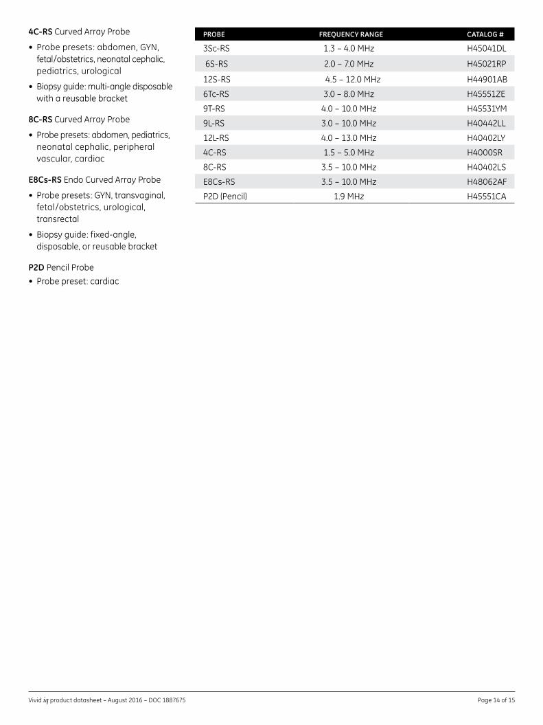
PROBE FREQUENCY RANGE CATALOG #
3Sc-RS 1.3–4.0MHz H45041DL
6S-RS 2.0–7.0MHz H45021RP
12S-RS 4.5–12.0MHz H44901AB
6Tc-RS 3.0–8.0MHz H45551ZE
9T-RS 4.0–10.0MHz H45531YM
9L-RS 3.0–10.0MHz H40442LL
12L-RS 4.0–13.0MHz H40402LY
4C-RS 1.5–5.0MHz H4000SR
8C-RS 3.5–10.0MHz H40402LS
E8Cs-RS 3.5–10.0MHz H48062AF
P2D(Pencil) 1.9MHz H45551CA
DOC 1887675
About GE HealthcareGEHealthcareprovidestransformationalmedicaltechnologies and services to meet the demand for increased access, enhanced quality and more affordable healthcarearoundtheworld.GE(NYSE:GE)worksonthings that matter – great people and technologies takingontoughchallenges.Frommedicalimaging, software & IT, patient monitoring and diagnostics to drug discovery, biopharmaceutical manufacturing technologies and performance improvement solutions, GEHealthcarehelpsmedicalprofessionalsdeliver great healthcare to their patients.
GEHealthcare9900InnovationDriveWauwatosa,WI53226U.S.A.
www.gehealthcare.com
Product may not be available in all countries and regions.Fullproducttechnicalspecificationis availableuponrequest.ContactaGEHealthcare Representative for more information. Please visit www.gehealthcare.com/promotional-locations.
Data subject to change.
© 2016 General Electric Company. DOC 1887675
GE,theGEMonogram,imaginationatwork,Vivid, XDclear,EchoPAC,andInSiteareregisteredtrademarks of General Electric Company.
DICOMistheregisteredtrademarkofthe NationalElectricalManufacturersAssociation.
WindowsandMicrosoftareregisteredtrademarks ofMicrosoftCorporation.
All other third-party trademarks are the property of their respective owners.
Reproduction in any form is forbidden without priorwrittenpermissionfromGE.Nothinginthis material should be used to diagnose or treat any disease or condition. Readers must consult a healthcare professional.