Embed Size (px)
Citation preview

30 Churchill Place ● Canary Wharf ● London E14 5EU ● United Kingdom
An agency of the European Union
Telephone +44 (0)20 3660 6000 Facsimile +44 (0)20 3660 5555 Send a question via our website www.ema.europa.eu/contact
© European Medicines Agency, 2014. Reproduction is authorised provided the source is acknowledged.
26 June 2014 EMA/546752/2014 Committee for Medicinal Products for Human Use (CHMP)
Vizamyl
flutemetamol (18F)
Procedure No. EMEA/H/C/002553
Marketing authorisation holder: GE HEALTHCARE LIMITED
Assessment report for an initial marketing authorisation application
Assessment report as adopted by the CHMP with all commercially confidential information deleted
Vizamyl EMA/546752/2014 Page 2/108
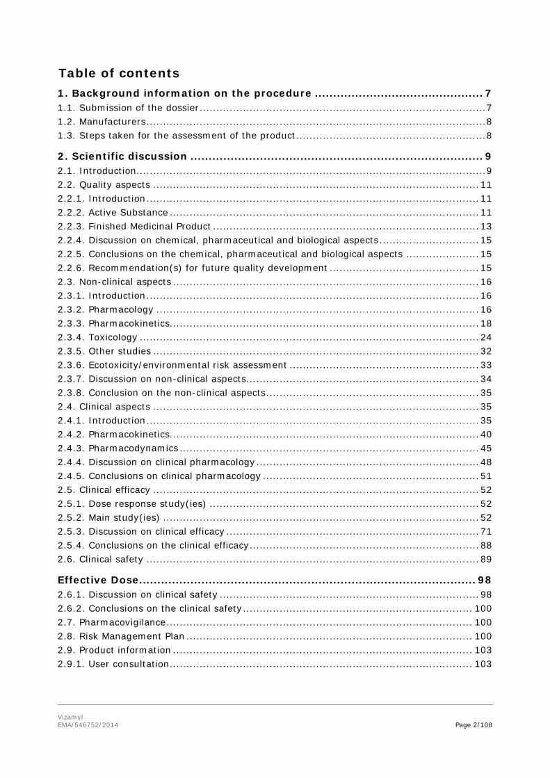
Table of contents 1. Background information on the procedure .............................................. 7 1.1. Submission of the dossier ...................................................................................... 7 1.2. Manufacturers ...................................................................................................... 8 1.3. Steps taken for the assessment of the product ......................................................... 8
2. Scientific discussion ................................................................................ 9 2.1. Introduction......................................................................................................... 9 2.2. Quality aspects .................................................................................................. 11 2.2.1. Introduction .................................................................................................... 11 2.2.2. Active Substance ............................................................................................. 11 2.2.3. Finished Medicinal Product ................................................................................ 13 2.2.4. Discussion on chemical, pharmaceutical and biological aspects .............................. 15 2.2.5. Conclusions on the chemical, pharmaceutical and biological aspects ...................... 15 2.2.6. Recommendation(s) for future quality development ............................................. 15 2.3. Non-clinical aspects ............................................................................................ 16 2.3.1. Introduction .................................................................................................... 16 2.3.2. Pharmacology ................................................................................................. 16 2.3.3. Pharmacokinetics............................................................................................. 18 2.3.4. Toxicology ...................................................................................................... 24 2.3.5. Other studies .................................................................................................. 32 2.3.6. Ecotoxicity/environmental risk assessment ......................................................... 33 2.3.7. Discussion on non-clinical aspects...................................................................... 34 2.3.8. Conclusion on the non-clinical aspects ................................................................ 35 2.4. Clinical aspects .................................................................................................. 35 2.4.1. Introduction .................................................................................................... 35 2.4.2. Pharmacokinetics............................................................................................. 40 2.4.3. Pharmacodynamics .......................................................................................... 45 2.4.4. Discussion on clinical pharmacology ................................................................... 48 2.4.5. Conclusions on clinical pharmacology ................................................................. 51 2.5. Clinical efficacy .................................................................................................. 52 2.5.1. Dose response study(ies) ................................................................................. 52 2.5.2. Main study(ies) ............................................................................................... 52 2.5.3. Discussion on clinical efficacy ............................................................................ 71 2.5.4. Conclusions on the clinical efficacy ..................................................................... 88 2.6. Clinical safety .................................................................................................... 89
Effective Dose............................................................................................ 98 2.6.1. Discussion on clinical safety .............................................................................. 98 2.6.2. Conclusions on the clinical safety ..................................................................... 100 2.7. Pharmacovigilance ............................................................................................ 100 2.8. Risk Management Plan ...................................................................................... 100 2.9. Product information .......................................................................................... 103 2.9.1. User consultation ........................................................................................... 103

Vizamyl EMA/546752/2014 Page 3/108
3. Benefit-Risk Balance............................................................................ 103
4. Recommendations ............................................................................... 106

Vizamyl EMA/546752/2014 Page 4/108
List of abbreviations
Aβ Amyloid β
AD Alzheimer’s disease
AE Adverse event
aMCI Amnestic mild cognitive impairment
BMI Body mass index
AH110690 Non-radioactive analogue of the drug substance
[18F]AH110690 18F-labelled drug substance
AH110690 (18F) Injection
Drug product; the product that is injected containing drug substance and excipients
BSS Bielschowsky silver stain
CDR Clinical Dementia Rating
CER Cerebellum
CERAD Consortium to Establish a Registry for Alzheimer’s Disease
CI Confidence interval
CRF Case Report Form (in paper or electronic format)
CRO Contract research organization
CSR Clinical study report
CT Computed tomography
DMS-IV Diagnostic and Statistical Manual of Mental Disorders, 4th Edition
DVR Distribution volume ratio
E Effective radiation dose (i.e., the sum of risk-weighted organ absorbed radiation dose used as a measure of stochastic radiation risk)
ECG Electrocardiogram
EMA European Medicines Agency
FAS Full analysis set
FDA US Food and Drug Administration
Flutemetamol F 18 Injection
Drug product; the product that is injected containing drug substance and excipients; formerly known as AH110690 F 18 Injection,
Flutemetamol (18F) Drug substance; active component of the investigational medicinal product Flutemetamol F 18 Injection. Formerly known as [18F]AH110690.
FN False negative

Vizamyl EMA/546752/2014 Page 5/108
FP False positive
GC Gas chromatography
GCP Good Clinical Practice
HPLC High performance liquid chromatography
HV Healthy volunteer
IBRI Institute of Biomedical Research and Information
ICH International Conference on Harmonization
IEC Independent ethics committee
IHC Immunohistochemical, immunohistochemistry
IMP Investigational medicinal product
IRB Institutional/independent review board
ISE Integrated Summary of Effectiveness
i.v. Intravenous
Max Maximum
MBq Megabecquerel(s)
mCi Millicurie(s)
MCI Mild cognitive impairment
mGy MilliGray
Min Minimum
MIRD Medical Internal Radiation Dose
mL Milliliter
MMSE Mini-Mental State Examination
MRI Magnetic resonance imaging
MS Mass spectrometry
NIA National Institute on Aging
NINCDS-ADRDA National Institute of Neurological and Communicative Disorders and Stroke; Alzheimer’s Disease and Related Disorders Association
NPH Normal pressure hydrocephalus
OLINDA/EXM Organ Level Internal Dosimetry Assessment/Exponential Modeling
pAD Probable Alzheimer’s disease
PCNS Peripheral and central nervous system
PET Positron emission tomography

Vizamyl EMA/546752/2014 Page 6/108
Ph. Eur. European Pharmacopoeia
p.i. Post injection
PiB Pittsburgh compound B
RAC Radioactive concentration
ROI Region of interest
QC Quality Control
SAE Serious adverse event
SCE Summary of Clinical Efficacy
SD Standard deviation
SOP Standard operating procedure
SoT Standard of truth
SUV Standard uptake value
SUVR
Standardized uptake value ratio. The SUVR is a quantitative measure of amyloid-specific flutemetamol (18F) uptake, normalized for the mean non-specific uptake in a reference region (cerebellum or pons). SUVR is defined as SUVVOI/SUVREF with SUV being the integrated activity over a given time period per unit of injected dose and body weight. When the cerebellum is used as the reference region, cortical regions lacking in amyloid are expected to have SUVR near 1, and cortical regions rich in amyloid are expected to have SUVR greater than 1. When used without qualification, SUVR refers to SUVR-CER (SUVR with the cerebellum as the reference region). A composite SUVR is the simple average of the SUVR in multiple regions
SUVR-CER SUVR using the cerebellum as the reference region
SUVR-PONS SUVR using the pons as the reference region
TLC Thin layer chromatography
TN True negative
TP True positive
UR Uptake ratio
US United States
VOI Volume of interest

Vizamyl EMA/546752/2014 Page 7/108
1. Background information on the procedure
1.1. Submission of the dossier
The applicant GE Healthcare Ltd submitted on 23 November 2012 an application for Marketing Authorisation to the European Medicines Agency (EMA) for VIZAMYL, through the centralised procedure under Article 3 (2) (a) of Regulation (EC) No 726/2004. The eligibility to the centralised procedure was agreed upon by the EMA/CHMP on 14 April 2011.
The applicant applied for the following indication:
“This medicinal product is for diagnostic use only.
VIZAMYL is a radioactive diagnostic agent indicated with positron emission tomography (PET) imaging for the visual detection of amyloid-beta neuritic plaques in the brains of adults who are being evaluated for Alzheimer’s disease (AD).
A normal VIZAMYL scan indicates sparse to no neuritic plaques and is inconsistent with a neuropathological diagnosis of AD at the point of image acquisition: a normal scan reduces the likelihood that a patient’s condition is due to AD. An abnormal VIZAMYL scan is indicative of moderate to frequent amyloid-beta neuritic plaques. Neuropathological examinations have shown this amount of neuritic plaques is present in patients with AD, but also other types of neurologic conditions as well as in older people with normal cognition. VIZAMYL is to be used as an adjunct to other diagnostic evaluations.
An abnormal VIZAMYL scan does not establish a diagnosis of AD.
The safety and efficacy of VIZAMYL have not been established for predicting the development of dementia or other neurological conditions, or for monitoring response to therapies.”
The legal basis for this application refers to:
Article 8.3 of Directive 2001/83/EC - complete and independent application. The applicant indicated that Flutemetamol (18F) was considered to be a new active substance.
The application submitted is composed of administrative information, complete quality data, non-clinical and clinical data based on applicants’ own tests and studies and/or bibliographic literature substituting/supporting certain tests or studies.
Information on Paediatric requirements
Pursuant to Article 7 of Regulation (EC) No 1901/2006, the application included an EMA Decision(s) P/72/2011 on the granting of a (product-specific) waiver.
New active Substance status
The applicant requested the active substance flutemetamol (18F) contained in the above medicinal product to be considered as a new active substance in itself, as the applicant claims that it is not a constituent of a product previously authorised within the Union.
Vizamyl EMA/546752/2014 Page 8/108
Scientific Advice/Protocol Assistance
The applicant did not seek scientific advice at the CHMP.
Licensing status
The product was not licensed in any country at the time of submission of the application.
1.2. Manufacturers
Manufacturers responsible for batch release
AAA, Troyes Advanced Accelerator Applications Technopole de l’Aube 14 rue Gustave Eiffel 10430 Rosières près Troyes France AAA, Forli Advanced Accelerator Applications S.r.l Via Piero Maroncelli 40/42 47014 Meldola (FO) Italy ITP, Madrid Instituto Tecnológico PET, SA. C/Manuel Bartolome Cossio 10 28040 Madrid Spain Seibersdorf Laboratories, Seibersdorf Seibersdorf Labor GmbH Grundstuck Nr. 482/2 EZ98 KG 2444 Seibersdorf Austria
1.3. Steps taken for the assessment of the product
The Rapporteur and Co-Rapporteur appointed by the CHMP were:
Rapporteur: Concepcion Prieto Yerro
Co-Rapporteur: Harald Enzmann
CHMP Peer reviewer: Ian Hudson
PRAC Rapporteur: Julie Williams
PRAC Co-Rapporteur: Miguel-Angel Macia
• The application was received by the EMA on 23 November 2012.
• The procedure started on 26 December 2012.
• The Rapporteur's first Assessment Report was circulated to all CHMP members on 14 March 2013. The Co-Rapporteur's first Assessment Report was circulated to all CHMP members on

Vizamyl EMA/546752/2014 Page 9/108
15 March 2013.
• The PRAC RMP Advice and assessment overview was adopted by PRAC on 11 April 2013.
• During the meeting on 25 April 2013, the CHMP agreed on the consolidated List of Questions to be sent to the applicant. The final consolidated List of Questions was sent to the applicant on 25 April 2013.
• The applicant submitted the responses to the CHMP consolidated List of Questions on 16 October 2013.
• The Rapporteurs circulated the Joint Assessment Report on the applicant’s responses to the List of Questions to all CHMP members on 29 November 2013 .
• During the CHMP meeting on 19 December 2013, the CHMP agreed on a list of outstanding issues to be addressed in writing and/or in an oral explanation by the applicant.
• The PRAC RMP Advice and assessment overview was adopted by PRAC on 5 December 2013.
• The applicant submitted the responses to the CHMP List of Outstanding Issues on 21 May 2014
• The Rapporteurs circulated the Joint Assessment Report on the applicant’s responses to the List of Outstanding Issues to all CHMP members on 4 June 2014.
• The Rapporteurs circulated the Joint Updated Assessment Report to all CHMP members on 18 June 2014.
• During the meeting on 26 June 2014, the CHMP, in the light of the overall data submitted and the scientific discussion within the Committee, issued a positive opinion for granting a Marketing Authorisation to VIZAMYL.
2. Scientific discussion
2.1. Introduction
Problem statement
It is estimated that there are over six million people with dementia in the European Union (Dementia in Europe Yearbook 2006) and it is predicted that this number will double in the next 20 years (Ferri et al. 2005). Alzheimer’s disease (AD) is the most common cause of dementia in the European Union. The definitive diagnosis of AD can be made only post-mortem. The diagnosis of AD during life is based on a thorough clinical and neuropsychiatric examination performed by a clinician experienced in dementia. The best established and still recommended criteria for this purpose are the NINCDS-ADRDA criteria (McKhann et al. 1984), which are handicapped for their limited sensitivity and specificity (i.e. 81% and 70%, respectively (Knopman et al. 2001) to initially diagnose probable AD in subjects with manifest dementia.
The fact is that how AD should be best pre-mortem diagnosed, staged and followed are matters being actively debated in the scientific literature and consensus has not been reached yet. Draft recommendations for updating diagnostic criteria for AD pre-mortem have been published from three independent working groups, incorporating some biomarkers, such as cerebrospinal fluid markers and brain imaging markers, to the clinical and neuropsychiatric evaluation (International Working Group: Dubois 2010, NINCDS-AA: McKhann 2011, DSM-V: Jeste 2010). Nevertheless, neither new diagnostic

Vizamyl EMA/546752/2014 Page 10/108
criteria nor potential biomarkers have still been validated for diagnostic purposes in the context of AD. CHMP published qualification opinions on the use of biomarkers such as cerebrospinal fluid markers, hippocampal magnetic resonance imaging (MRI) and β-amyloid brain positron emission tomography (PET) (EMA/CHMP/SAWP/892998/2011, EMA/CHMP/SAWP/102001/2011, EMA/CHMP/SAWP/893622/2011, EMA/CHMP/SAWP/809208/2011). Their qualification refers solely to the identification of subjects with clinical diagnosis of pre-dementia at increased risk of underlying AD neuropathology or to the identification of patients with a clinical diagnosis of mild to moderate AD, for the purposes of enriching recruitment into clinical trials, aimed at studying drugs potentially slowing the progression/conversion to (severe) AD dementia of the included patients, but not for use as a diagnostic tool or as an outcome or longitudinal measure.
Controversy also exists on the validity of certain diagnosis referring to cognitive impairment in its pre-dementia stages. First of all, the concept of minimal cognitive impairment (as defined by the Petersen Criteria 2003) or the prodromal AD (as defined by Dubois Criteria 2007) reflects a different population. Secondly, it is not settled yet if mild cognitive impairment (MCI) as an episodic memory impaired group is an intermediate stage that a patient with AD will pass through before becoming demented. Only a portion of patients with MCI progresses to clinical AD dementia over 5-10 years (Petersen et al., 1999; Ritchie et al., 2001; Visser et al., 2006; Mitchell et al., 2009) and a recent meta-analysis concluded that most people with MCI will not progress to dementia even after 10 years of follow-up (Klunk et al., 2011).
The idea that brain β-amyloid could be used as an in vivo marker of AD is supported by compelling evidence, since β-amyloid is the main component of the neuritic plaques that are a key diagnostic landmark in the post-mortem definitive diagnosis of the disease (Mirra et al., 1991). However, detection of β-amyloid deposition in the brain does not equal AD for the following reasons:
1. No consensus has still been reached in the scientific community on the β-amyloid hypothesis of AD and the pathological process leading to AD has not been fully elucidated yet. If β-amyloid deposition is demonstrated to be the cause of the disease, this characteristic could contribute to the diagnosis from very early to the full spectrum of clinical AD stages. If not the cause but an effect or consequence, its contribution would be restricted to specific stages of AD in which it actually appears or the deposition may be considered as abnormal.
2. Βeta-amyloid neuritic plaques are not exclusive of AD and may also be present in cognitively normal elderly subjects, patients with MCI, patients with other dementias (dementia of Lewy Body, Parkinson Disease Dementia), Niemann-Pick disease type C and severe brain injury.
3. The capability to visualize β-amyloid deposition in brain tissue is probably not enough for the diagnosis of AD. In fact, pre-specified levels of age-related brain neuritic β-amyloid plaque at autopsy should be integrated with the presence of a clinical history of dementia to arrive at a diagnostic level of certainty with regard to AD (Mirra et al. 1991)
4. And although neuritic plaques are a common factor for the post-mortem definitive diagnosis of the disease, the diagnostic value for AD of different brain β-amyloid plaque types (diffuse plaques with pre-amyloid, neuritic and cored), as well as of different β-amyloid isoforms/species (oligomeric, fibrillar or non-fibrillar) may well be different.
Both the degree of β-amyloid deposition but also its neuroanatomical localization is obviously important for determination of β-amyloid-related pathology in the brain and it is the subject of continuous publication. Braak et al. described the characteristic pattern of deposition for different stages of typical AD (Braak et al. 1994). The regional pattern of β-amyloid accumulation is different in pathologies with beta amyloid different than in typical AD (Edison et al. 2007).

Vizamyl EMA/546752/2014 Page 11/108
There might be several potential diagnostic uses, not confirmed by robust data, of in vivo detection of β-amyloid deposition in the brain in clinical practice worth studying. An obvious one would be excluding the initial diagnosis of AD in difficult cases of dementia, since there is no AD without a particular age-related β-amyloid plaque score in a demented patient (Mirra et al. 1991). The identification and differential diagnosis of AD are especially challenging in its early stages, partly because of the difficulty in distinguish it from the mild decline in memory that can occur with normal aging and from mild cognitive manifestations of other neuropsychiatric conditions, such as depression, as well as other causes of dementia. Other potential clinical uses in the management of AD might be as a staging criterion, marker of progression of the disease, and predictor of response to treatment. It would also be of great value to be able to predict which patients, who upon comprehensive diagnostic testing are found to have cognitive impairment but are not demented and thus do not meet diagnostic criteria for AD (e.g. patients with MCI), are destined to progress to a clinical diagnosis of AD dementia.
Radiopharmaceuticals have been approved by FDA and EMA, and are used in conjunction with a clinical evaluation, for Positron Emission Tomography (PET) imaging of β-amyloid neuritic plaque density in the brains of adult patients with cognitive impairment who are being evaluated for AD and other causes of cognitive impairment. In their context of use it is clearly stated that a negative PET scan indicates sparse or no plaques, which is not consistent with a diagnosis of AD. However, a positive PET scan has important limitations since it does neither independently establish a diagnosis of AD or other cognitive disorder nor allow for predicting development of AD or monitoring response to therapy. In the context of patients with MCI, the limitations these radiopharmaceuticals to show the MCI conversion rate to AD are also acknowledged.
About the product
Flutemetamol (18F) is a small lipophilic new molecular entity designed based on the chemical structure of the amyloid-specific dye, Thioflavin T, and is labelled with fluorine (18F) which emits a positron signal that is detected by a PET scanner. As such Flutemetamol (18F) is a novel radiopharmaceutical agent which has been developed for imaging β-amyloid (Aβ) neuritic plaques in the human brain by PET. Flutemetamol (18F) binds with high affinity and specificity to Aβ aggregates in brain tissue homogenates from patients with AD.
2.2. Quality aspects
2.2.1. Introduction
Vizamyl solution for injection is a novel diagnostic radiopharmaceutical agent which has been developed for imaging β-amyloid neuritic plaques in the human brain by positron emitting tomography (PET).
The finished product is presented as a sterile solution for injection containing 400 MBq/ml of flutemetamol [18F] at reference date and time.
Other ingredients are: sodium chloride, ethanol anhydrous, polysorbate 80, sodium dihydrogen phosphate dihydrate, disodium hydrogen phosphate dodecahydrate and water for injection.
The product is available in Type I glass vials with halobutyl rubber stoppers and aluminium seals.
2.2.2. Active Substance
General information

Vizamyl EMA/546752/2014 Page 12/108
The chemical name of flutemetamol [18F] is 6-benzothiazolol, 2-[3-[18F]fluoro-4-(methylamino)phenyl] and has the following structure:
Flutemetamol [18F] is a small lipophilic organic chemical molecule containing the radioactive isotope fluorine-18 [18F], a positron emitting radionuclide with a physical half-life of 109.8 minutes. Fluorine (18F) decays to stable oxygen (18O) with a half-life of approximately 110 minutes by emitting a positron radiation of 634 keV, followed by photonic annihilation radiation of 511 keV.
Due to the short physical half-life of the active substance the structure elucidation and evaluation was performed on the non-radioactive analogue carrying stable fluorine-19. Its chemical structure was confirmed by elemental analysis (carbon, hydrogen, nitrogen and sulphur), mass spectrometry (MS), UV-Vis, IR and Raman spectroscopy, and 1H, 13C, and 19F NMR spectroscopy. The equivalent structure of the flutemetamol [18F] and the non-radioactive analogue has been confirmed by the nature of the synthetic route used to manufacture flutemetamol [18F], and equivalent retention of the drug substance and the non-radioactive analogue by RP-HPLC and TLC.
Flutemetamol has a non-chiral molecular structure.
The fluorine-19 labelled, non-radioactive analogue of the active substance flutemetamol is a crystalline yellow green to beige non-hygroscopic powder which is freely soluble in dimethylsulfoxide, sparingly soluble in tetrahydrofuran, slightly soluble in ethanol, very slightly soluble in acetonitrile and practically insoluble in water. Since, the radio-labelled flutemetamol [18F] drug substance is not isolated and only exists in solution the crystal structure of the compound is, therefore, not relevant.
Manufacture, characterisation and process controls Due to the short physical half-life of 110 minutes of the radionuclide fluorine-18, the active substance, flutemetamol [18F], is not isolated during the manufacturing process, and it is synthesized in situ during the manufacture of the finished product using a non-radioactive chemical precursor (AH111907) which is radiolabelled using [18F]fluoride in a nucleophilic substitution reaction.
The chemical precursor is manufactured in a six-step process using two commercially available well defined starting materials with acceptable specifications. Process validation data on three commercial scale batches showing that the process is reproducible and capable of consistently producing the chemical precursor of the required quality have been presented.
Active substance and finished product are manufactured in one continuous process. The manufacturing process of the active substance uses a typical fully automated and remote controlled synthesiser unit employed to produce PET radiopharmaceuticals. The chemical precursor, reagents and purification cartridges are supplied as a pre-assembled disposable cassette which is mounted onto the synthesiser. In this regard, the chemical precursor is distributed to the finished product manufacturer dissolved in DMSO in a vial. This is considered acceptable, since it has been demonstrated that it does not have a negative impact on the quality of the medicinal product.
The main steps for the manufacturing process of the active substance are: proton irradiation of 18O-enriched water to obtain fluoride-18, separation of the fluoride-18 from the target water followed by a nucleophilic substitution of the organic chemical precursor AH111907, chemical modification of unreacted precursor to achieve later better separation performance during the chromatographic cartridge

Vizamyl EMA/546752/2014 Page 13/108
purification step from the radiolabelled active substance, deprotection of the radiolabelled intermediate and chromatographic cartridge purification.
The characterisation of the active substance and its impurities are in accordance with the EU guideline on chemistry of new active substances. Potential and actual impurities have been well discussed with regards to their origin and characterised.
Adequate in-process controls are applied during the synthesis. The specifications and control methods for intermediate products, starting materials and reagents have been presented.
Specification The active substance is not isolated during the manufacturing process. Therefore, information on specification is provided in the finished product section.
An appropriate specification for the chemical precursor, has been presented.The analytical methods used have been adequately described and non-compendial methods appropriately validated in accordance with the ICH guidelines.Batch analysis data on five commercial scale batches of the precursor are provided. The results are within the specifications and consistent from batch to batch.
Stability Not applicable since the active substance is not isolated during the manufacturing process. Information on stability is provided in the finished product section.
2.2.3. Finished Medicinal Product
Description of the product and pharmaceutical development The aim of the pharmaceutical development was to obtain a stable formulation for immediate intravenous injection of [18F] flutemetamol.
Throughout the development of flutemetamol [18F] injection, different formulations were used for non-clinical and clinical studies. This, in early clinical trials [11C] flutemetamol was used. However, since [11C] has a half-life of 20.4 minutes which makes it unsuitable for routine manufacture, [18F] with a longer half-life 109.8 minutes was devised and used for subsequent clinical trials.
The sterility of the finished product is assured by aseptic manufacture followed by sterile filtration rather than terminal sterilisation by autoclaving. This has been justified based on the instability of polysorbate 80 upon exposure to heat, and the fast radioactive decay of fluorine-18, and is considered acceptable. The aseptic dispensing process has been validated at the manufacturing sites.
The finished product contains simple and safe European pharmacopoeial excipients commonly used in solutions for injections: ethanol, polysorbate 80, sodium chloride, sodium dihydrogen phosphate dihydrate, disodium hydrogen phosphate dodecahydrate and water for injection. Ethanol is used as solubiliser and stabiliser, polysorbate 80 as solubiliser, sodium chloride as isotonic agent, and the phosphate buffer maintains the pH of the formulation to suitable levels.
All excipients are well known pharmaceutical ingredients and their quality is compliant with Ph. Eur standards. There are no novel excipients used in the finished product formulation.
Compatibility studies of flutemetamol [18F] with physiological saline and water for injections (WFI) showed that the dilution of the drug product with physiological saline or WFI should be avoided because such dilution might decrease the solubility of the active substance leading to precipitation and/or

Vizamyl EMA/546752/2014 Page 14/108
adsorption of the active substance to surfaces. However, physiological saline and WFI are compatible when used to rinse or flush the administration equipment (infusion set and dosing syringe) after administration of flutemetamol [18F] solution for injection.
The primary packaging are type I glass vials with halobutyl rubber stoppers and aluminium seals. The material complies with Ph.Eur. requirements. The choice of the container closure system has been validated by stability data and is adequate for the intended use of the product.
Manufacture of the product and process controls As mentioned above, the active substance and the finished product are manufactured in one automated continuous process which does not allow the isolation of the active substance. Therefore, the manufacturers of the finished product are the same proposed for the manufacture of the active substance. The manufacturing process of the finished product consists of four steps: addition of the excipients and mixing, dilution of the bulk solution, sterile filtration and aseptic dispensing into vials.
One of the dispenser modules proposed by the applicant leads to finished product filled in vials with punctured rubber septum. This is due to the fact that this dispenser fills empty product vials which are already closed with rubber stoppers. The applicant has stated that the vial septa reseals, and in addition has proposed to apply a pre-sterilised cap to each of those vials. This approach has been accepted due to the fact that the vial content will be used within one day. However, the CHMP recommends revising the operation of this dispensing unit so that only vials with non-punctured rubber septa are produced, since this is the usual standard for the delivery of sterile solutions.
The process is considered to be a non- standard manufacturing process. Major steps of the manufacturing process have been validated by a number of studies. It has been demonstrated that the manufacturing process is capable of producing the finished product of intended quality in a reproducible manner. The in-process controls are adequate for this type of manufacturing process.
Product specification The finished product release and shelf-life specifications include appropriate tests for this kind of products and include: appearance, radiochemical identity (HPLC), radionuclidic identity (half-life determination), assay (radioactive concentration), ethanol content (GC), chemical purity (HPLC-UV), residual solvents (GC), pH, sterility (Ph. Eur.), bacterial endotoxins (Ph. Eur.), radiochemical purity (HPLC with radioactivity detector), radionuclidic purity (gamma-ray spectrometry).
Batch analysis results are provided for three representative batches from each of the four proposed manufacturing sites confirming the consistency of the manufacturing process and its ability to manufacture to the intended product specification.
The finished product is released on the market based on the above release specifications, through traditional final product release testing, although sterility test and the radionuclidic purity test for long living radionuclides are conducted after batch release (as usual for this type of products).
Stability of the product Stability data on three commercial scale batches of Vizamyl solution for injection manufactured at each of the four proposed manufacturing sites and stored at 5°C ± 3°C and 50°C ± 2°C with ambient humidity, covering a storage period of 10 hours, have been provided. The batches of Vizamyl are identical to those proposed for marketing and were packed in the primary packaging proposed for marketing. A matrixing approach covering the different container closure systems proposed for commercial supply, minimum and maximum fill volumes, and storage conditions of 5°C ± 3°C and 50°C ± 2°C has been followed.
Vizamyl EMA/546752/2014 Page 15/108
Additionally, supportive stability data on 36 undiluted batches of Vizamyl solution for injection manufactured at another site and stored at 5°C ± 3°C and 50°C ± 2°C have been presented. All batches were tested at end of synthesis and approximately 10 hours post-synthesis.
Although these storage conditions deviate from the standard temperatures stated in the ICH stability guidelines, they have been justified based on the fact that they would represent the worst case scenario to monitor the product performance during handling and transport from the manufacturing site to the final user, according to the experience gained during the clinical phase. Furthermore, the applicant presented additional stability data on three complementary batches manufactured by each of the proposed manufacturing sites intended for commercial supply stored in the temperature range of 20°C to 25°C.
Samples were tested for appearance, radioactive concentration (RAC) at end of synthesis, total content of flutemetamol and related substances radiochemical purity, and [18F]fluoride. The analytical procedures used were stability indicating.Based on available stability data, the shelf-life and storage conditions as stated in the SmPC are acceptable.
Adventitious agents No excipients derived from animal or human origin have been used.
2.2.4. Discussion on chemical, pharmaceutical and biological aspects
Information on development, manufacture and control of the active substance and finished product has been presented in a satisfactory manner. The results of tests carried out indicate consistency and uniformity of important product quality characteristics, and these in turn lead to the conclusion that the product should have a satisfactory and uniform performance in clinical use.
2.2.5. Conclusions on the chemical, pharmaceutical and biological aspects
The quality of this product is considered to be acceptable when used in accordance with the conditions defined in the SmPC. Physicochemical and biological aspects relevant to the uniform clinical performance of the product have been investigated and are controlled in a satisfactory way.
2.2.6. Recommendation(s) for future quality development
In the context of the obligation of the MAHs to take due account of technical and scientific progress, the CHMP recommends the following points for investigation:
The applicant is recommended to revise the operation of the dispensing unit which leads to punctured rubber septa. The use of dispensing units leading to non-punctured rubber septa is the usual standard for the delivery of sterile solutions.
Vizamyl EMA/546752/2014 Page 16/108
2.3. Non-clinical aspects
2.3.1. Introduction
2.3.2. Pharmacology
Amyloid is an abnormal deposit of insoluble protein fibrils in a body tissue or organ. It is characterized by unique staining properties, electron microscopic appearance, and a β-pleated sheet pattern on X-ray diffraction analysis. Amyloid can be formed from many proteins, and it can accumulate in tissue to form visible plaques. It is associated with over 30 human diseases, most notably Alzheimer’s disease (AD). The specific type of amyloid involved in AD is amyloid-β or Aβ which is the main component of neuritic plaques, one of the two hallmarks of AD that can be seen microscopically in brain tissue specimens stained with certain dyes, the other being neurofibrillary tangles of tau protein.
Thioflavin T is one of the histologic dyes used to detect amyloid ex vivo, producing a fluorescent signal that marks the characteristic pattern of amyloid plaques post mortem. However, since Thioflavin T does not penetrate the blood brain barrier, it could not be developed as an agent for in vivo imaging in the brain. Some compounds derived from Thioflavin T have been developed and are designed to be neutral and uncharged molecules to allow them to cross the blood brain barrier. Flutemetamol and Pittsburgh Compound B (PiB) are very similar in chemical structure, differing only by the presence of a fluorine atom in flutemetamol. Clinical data show that they have similar uptake into the human brain and their PET images are similar in their visualization of brain amyloid.
Primary pharmacodynamic studies In vitro affinity assays: demonstration of flutemetamol binding to human β-amyloid protein (B067051, non GLP)
This research study was designed to test the affinity of flutemetamol towards synthetic fibrillar amyloid β1-40 (β-amyloid 1-40 fibrils) in vitro. Although the specific binding site of flutemetamol has not been identified, this study shows that it is associated with the β sheet folds of fibrillar amyloid β.
An in vitro human brain homogenate assay was performed which indicated selectivity for fibrillar amyloid β in the presence of normal brain tissue homogenate. Less non-specific binding in the white matter compared with grey matter was detected.
In vitro human brain autoradiography (B067050, non GLP)
The aim of this study was to investigate uptake of [3H]flutemetamol into senile plaques obtained post-mortem from human brain from Alzheimer’s disease cases. Two different control groups were provided by the inclusion of post-mortem tissue from patients who had suffered from dementia unrelated to Alzheimer’s disease as well as patients with no clinical symptoms of dementia.
Flutemetamol bound to regions of brain associated with Alzheimer’s disease (temporal cortex and the hippocampus) in tissue sections from Alzheimer disease patients but did not bind to similar sections of brain obtained from the two control groups. These results confirm that flutemetamol binds to the amyloid pathology prevalent in dementia related to Alzheimer’s disease.
Characterization of flutemetamol binding in autopsy brains from [11C]PiB imaged subjects (B067078, non GLP)

Vizamyl EMA/546752/2014 Page 17/108
This study was conducted to provide histopathological characterization of flutemetamol binding to AD pathology (including amyloid β plaques, cerebral amyloid angiopathy and neurofibrillary tangles) in post mortem brain tissue sections, to examine the degree to which binding of [3H]flutemetamol correlates with binding of [3H]PiB in brain tissue homogenates from amyloid-positive and amyloid-negative (control) autopsy cases, and to correlate post mortem measures of flutemetamol and PiB with ante mortem PiB PET retention in the same subjects.
The extent of amyloid β aggregates detected post mortem by flutemetamol was similar to that using PiB and correlated with the PET detection of [11C]PiB retention in vivo (Figure 3 below). These results indicate that flutemetamol (18F) is comparable to [11C]PiB in its ability to bind brain fibrillar β amyloid pathology.
These data demonstrate that flutemetamol and PiB have similar patterns of labelling amyloid β plaques and vascular deposits in post mortem fixed neocortical tissue sections. The results suggest that in vivo PET retention of flutemetamol (18F), in brains that contain amyloid β deposits reflects neocortical Aβ plaque load in a manner similar to [11C]PiB imaging.
Secondary pharmacodynamic studies No specific studies have been performed.
Safety pharmacology programme Effects on hERG tail current (B067043, GLP)
Vizamyl EMA/546752/2014 Page 18/108
Flutemetamol did not cause hERG tail current inhibition when compared with vehicle, indicating no inhibition of the hERG channel in this assay. The lowest measured concentration is 60 times higher than the theoretical maximum plasma concentrations of flutemetamol that may be achieved in humans after a single injection of 20 μg flutemetamol into the 3 L plasma volume of the standard man.
Effects of flutemetamol on cardiovascular function in the telemetered dog (B067003, GLP)
This study was conducted to determine the effects of intravenously administered flutemetamol on the cardiovascular system of conscious (telemetered) male Beagle dogs. No treatment related effects were observed on blood pressure, heart rate or ECG parameters.
A single intravenous administration of flutemetamol to male dogs at doses up to 9.3 μg/kg was generally well tolerated and without test item related adverse effects. The NOAEL is considered to be 6.0 μg/kg, equivalent to 10 multiples of the recommended maximum human dose of 20 μg, following adjustment for body surface area.
Effects on the respiratory system in dog (B067040, GLP)
Respiratory function was measured in male and female Beagle dogs on day 10 of the 14-day repeat-dose toxicity study (study B067040, section 4.2). The animals were treated with doses of 7.5 and 15 μg/kg flutemetamol. Respiration rate, tidal volume and minute volume were recorded. There were no effects considered to be treatment related. The NOAEL is 14 μg/kg, equivalent to 23 multiples of the recommended maximum human dose of 20 μg.
Modified Irwin test in the rat (B067004, GLP)
This study was conducted to determine the effects of intravenously administered flutemetamol on the gross behavioural and physiological state of male Sprague Dawley rats. The animals were intravenously administered 1.5, 5 and 16 μg/kg flutemetamol and a positive control (2 mg/kg chlorpromazine). The animals were observed and assessed on 42 behavioural and physiological parameters. No treatment related effects were observed.
The actual highest administered dose may have contained 92% of the nominal value due to possible adsorption, therefore the NOAEL is 14.7 μg/kg flutemetamol, equivalent to 7 multiples of the recommended maximum human dose of 20 μg, following adjustment for body surface area.
Pharmacodynamic drug interactions
No specific studies have been performed to date.
2.3.3. Pharmacokinetics
Pharmacokinetic studies
Biodistribution studies of flutemetamol (18F) in rats have shown that it is rapidly distributed to the brain but not retained in the absence of amyloid β deposits. Distribution and elimination from other tissues is also rapid. Excretion is mainly via the gastrointestinal route. The inclusion of polysorbate 80 in the formulation of flutemetamol (18F) to decrease adsorption and increase solubility did not change the biodistribution profile.
During the flutemetamol (18F) development the purification method changed from high performance liquid chromatography (HPLC) to solid phase extraction (SPE). A bridging biodistribution study was conducted with a test item purified by SPE.

Vizamyl EMA/546752/2014 Page 19/108
Metabolism studies in rat and baboon detected at least two radiolabelled metabolites in the plasma. In vitro metabolism studies showed that the major metabolite was de N-demethylated product. After injection of flutemetamol (18F) in the ALZ103 clinical trail, human plasma samples from the subjects were analysed by HPLC, showing that metabolism of flutemetamol (18F) occurred rapidly and at least two radiolabelled metabolites were detected. Additional studies in the rat showed a rapid metabolism of flutemetamol (18F) and the presence of two radiolabelled metabolites in the plasma that were not detected in the brain.
Protein binding studies using equilibrium dialysis in human, dog and rat plasma indicated that plasma protein binding of [3H]flutemetamol was greater than 95%. However, flutemetamol is rapidly eliminated from blood after intravenous administration to rat and dog.
Vizamyl EMA/546752/2014 Page 20/108
Methods of analysis
Quantification of flutemetamol in plasma samples using liquid chromatography with fluorescence detection (M067003 and V067003)
Flutemetamol in rat, dog an human plasma samples was determined by a HPLC with fluorescence detection. The range of the assay was approximately 0.3 to 30 ng/mL plasma with a lower limit of quantification of 0.3 ng/mL. This method of analysis (M067003) was also used in the toxicokinetic studies. The validation of method V067003 was performed as a non-GLP study in a GLP environment.
Adsorption to dosing equipment (B067058)
Flutemetamol is a lipophilic compound with limited solubility in aqueous solution (<10 μg/mL) and has the potential to adsorb onto dosing equipment used in nonclinical studies. This study investigated the extent that Flutemetamol Solution for Injection can adsorb onto the dosing equipment used in the nonclinical studies.
The recovery values (65 - 92%) were used to represent the maximum adsorption that could occur and they have been applied to estimate the lowest likely doses administered in each of the nonclinical studies that used the specific dosing equipment. Hence, these data impact the NOAEL for each of the studies.
Absorption
No specific absorption studies have been performed.
Distribution
Biodistribution of fluorine-18 after administration of a formulation of flutemetamol (18F) (B067059, B067060 and B067062, non GLP)
After administration of flutemetamol (18F), radioactivity was rapidly distributed throughout the body including the brain. Initial uptake in the brain was 4.4%, but it is not retained in the absence of amyloid β deposits. The initial uptake of radioactivity was predominantly in the muscle (31%), the liver (23%) and the small intestine wall (8%). Uptake was also observed in other organs and tissues such as the skin, kidneys, bone, lung and fat. Data are included in the following table:

Vizamyl EMA/546752/2014 Page 21/108
Between 20 minutes and 4 hours after administration, the amount excreted in urine continued to rise to a final level of 10% at 4 hours. In all other organs and tissues, retention decreased up to 4 hours. The main route of excretion was gastrointestinal with radioactivity in the small intestine contents peaking at 2 hours (72%) and in the large intestine contents at 4 hours (49%). There was no evidence for prolonged retention of significant quantities of the radioactivity in any organ or tissue.
The clinical formulation includes 0.5% polysorbate 80, thus a bridging biodistribution study using a formulation containing 1% polysorbate 80 was conducted in rats (B067060). No biodistribution differences were observed compared with the study that did not include polysorbate 80, including uptake and clearance from the brain.
A study to investigate whether the distribution of radioactivity after administration of [18F]AH-110690 is affected by the presence of radioactive impurities in the formulation (B067062, non GLP)
The synthesis process of flutemetamol (18F) has been scaled up for use in the clinic and this can result in the formation of a significant percentage of radioactive impurities. It was established that the presence of radioactive impurities generated during the high-activity synthesis process of flutemetamol (18F) (at levels of 25 and 17 % total radioactivity) did significantly affect the biodistribution profile of radioactivity post-administration. The presence of these radioactive impurities (thought to be radiolysis products of [18F]flutemetamol) was associated with elevated retention of radioactivity in the blood, reduced initial

Vizamyl EMA/546752/2014 Page 22/108
distribution to the muscle and brain, and a slowed transit of radioactivity through the liver and into the small intestine. Elevated bone retention was also observed in proportion to the amount of [18F] fluoride present.
The decrease in initial distribution of radioactivity to the brain observed with the lower RCP Test Items is an important observation and was proportional to the percentage of radioactive impurities present. This may indicate that the radioactive impurities examined do not cross the blood brain barrier. Consistent with this there was no significant retention of radioactivity in the brain associated with the lower RCP Test Items. Therefore, whilst the presence of such radioactive impurities in a formulation of flutemetamol (18F) should not impair background image quality in the brain (except the contribution from elevated blood retention), reduced delivery of to the brain may be expected in proportion to the amount of flutemetamol (18F) radioactive impurities present.
The other major finding of this study was that the lower RCP Test Items were associated with a slowed transit of radioactivity through the liver and into the small intestine (in parallel with the elevated blood retention observed). It is likely that the change in biodistribution profile associated with these lower RCP Test Items will not detrimentally affect dosimetry (the dose to the intestine wall should be reduced and the increase in bone retention observed is not thought to significantly affect the overall effective dose). It is also important to note that significant levels of radioactive impurities did not accumulate in any radiosensitive organ or tissue.
The formulations used in this study and the previous one were produced using HPLC purification and some, but not all, of the radioactive impurities present in this preparation are expected to be present in the drug product produced using SPE purification. Additional radioactive impurities now known to be present in the SPE purified drug product that were not present in the HPLC purified drug product; therefore an additional study was conducted (B067075).
Biodistribution of flurorine-18 after administration of a formulation of flutemetamol (18F) prepared using the FASTlab with SPE purification (B067075, non GLP)
As mentioned above, the different method of purification changed the impurity profile and non-radiolabelled impurities typical of those in the proposed commercial drug product were present in this formulation. The study was carried out with two different flutemetamol (18F) preparations with RCP of 98.1 and 97.5%.
The biodistribution pattern for the SPE purified flutemetamol (18F) was similar to that reported for an HPLC purified flutemetamol (18F) preparation in male rats (B067059) with no biologically significant differences between the distribution profiles in any of the organs and tissues, including brain.
Plasma protein binding of flutemetamol in rat, dog and human (B067049, B067057, non GLP)
The extent of plasma protein binding by flutemetamol was determined using the equilibrium dialysis methodology. In studies using human plasma and [3H]flutemetamol, >97% binding (1 hour study) was observed following equilibrium. Additional studies with rat and dog plasma were performed using the same methodology, and showed >97% binding to rat plasma proteins, and >95% binding to dog plasma proteins following 3 hours equilibrium dialysis at 37ºC. These results indicate that flutemetamol binds extensively to plasma proteins, and that the measured protein binding is similar in human, dog and rat.
Metabolism
In vitro metabolism flutemetamol (B067013, B067023, B067045, B067046, B067048, non GLP)

Vizamyl EMA/546752/2014 Page 23/108
Several studies were conducted to investigate the in vitro metabolism of flutemetamol by incubation with hepatic S9 fraction obtained from human, dog, mouse and rat. The major metabolite detected after incubation of flutemetamol in hepatic S9 from mouse, dog and human, and from Aroclor 1254 induced rat was the N-demethylated product. No metabolism was observed in control heat inactivated or β-NADPH deficient hepatic S9 fractions. This data indicate that metabolism in the human is similar to that of the rat and dog.
In order to allow correlation with in vitro and in vivo genotoxicity studies, additional rat metabolism studies were performed in the presence of Aroclor 1254 induced hepatic S9 fraction and non-induced rat hepatic S9 fraction (B067013).
After incubation of flutemetamol (18F) with mouse and dog hepatic S9 (B067046) a slower rate of metabolite formation was detected. After 30 minutes incubation, more than 90% of the radioactivity added to dog hepatic S9 fraction and 72% of the radioactivity in mouse hepatic S9 was still associated with flutemetamol. In contrast to the [14C]flutemetamol study B067045, this study shows that flutemetamol (18F) is not significantly metabolised in the presence of either dog or mouse hepatic S9 fraction. As there should be no difference between the metabolism of flutemetamol in the two studies B067045 and B067046, this confirms the hypothesis that the differences in metabolic profiles of [14C] flutemetamol and flutemetamol (18F) are likely to be due to the different synthetic routes, presence of impurities in the [14C]flutemetamol test item and position of radioactive label.
In addition, no metabolism or degradation was observed after incubation of flutemetamol (18F) with rat, dog or human plasma for up to 3 hours. In human whole blood, low amounts of two metabolites were detected after a 3-hour incubation.
In vivo metabolism of [11C]flutemetamol (B067018, B067019, non GLP)
After intravenous administration of [11C]flutemetamol to rat and baboon, [11C]flutemetamol was rapidly metabolised and at least 2 hydrophilic products were observed in both species. In humans (clinical trial ALZ103 Phase 1), it was observed that the metabolism of the intravenously administered flutemetamol (18F) occurred rapidly (only 25% of the parent compound was detected in the circulation 20 minutes post injection and 10% was detected at 180 minutes post injection) and at least two hydrophilic metabolites were detected.
In vivo metabolites of flutemetamol (18F) in plasma, brain and bile (B067071, B067070, non GLP)
Flutemetamol (18F) was intravenously administered to rats and plasma, brain and bile samples were taken at 2, 20 and 60 minutes post-injection. In the plasma, the radioactivity rapidly decreased from 58% at 2 minutes to 13% at 60 minutes post injection. Two major and one minor radiolabelled metabolites were identified, all less lipophilic than the parent compound. In the brain, flutemetamol (18F) accounted for 97, 85 and 75% of the total activity at 2, 20 and 60 minutes post-injection, respectively, with only two minor metabolites observed.
Excretion
The biodistribution study with flutemetamol (18F) in rats (B067059, B067060 and B067062) indicates that radioactivity is predominantly excreted in faeces (approximately 80% by 4 hours post-injection) with minor excretion in urine (approximately 10% by 4 hours post-injection).
Pharmacokinetic drug interactions
Impact of anti-amyloid therapy MAB31 on the distribution of flutemetamol (18F) in Wistar rats (B067064, non GLP)

Vizamyl EMA/546752/2014 Page 24/108
flutemetamol (18F) is likely to be administrated to patients who are receiving anti-amyloid therapies. Therefore, a drug interaction study (in male Wistar rats) was performed to understand the effects of the experimental therapeutic MAB31 on the distribution of flutemetamol (18F) in rats. MAB31 is a monoclonal antibody being developed by Hoffmann-La Roche as a novel anti-β-amyloid therapeutic agent. The study results indicate that MAB31 pre-dosing did not affect the brain delivery and clearance characteristics of [18F]flutemetamol. In addition, MAB31 did not influence the biodistribution of flutemetamol (18F) to any peripheral tissues, and the excretion profile of flutemetamol (18F) was unaffected.
Although the diagnostic target β amyloid plaques were not present in the animals, the initial uptake of activity into normal brain tissue is a good indicator of the potential delivery of activity to the target plaques. In addition, a previous in vitro study has showed that MAB31 and flutemetamol (18F) have different amyloid β binding sites. Therefore, this study shows that pre-dosing with MAB31 (10 mg/kg) 24 hours before the administration of flutemetamol (18F) did not significantly affect the biodistribution of radioactivity observed in rats.
The interactions of flutemetamol with a panel of drugs commonly prescribed to Alzheimer’s patients were assessed in the clinical trials (see clinical section of the assessment report). Taking into account the very low amount of flutemetamol (18F) and its rapid clearance from circulation, no additional pharmacokinetic interaction studies are considered necessary.
Other pharmacokinetic studies
Single-dose intravenous kinetics study with Flutemetamol Solution for Injection in the Wistar rat (B067055, non GLP)
An exploratory kinetic study was conducted to investigate the availability of the test item in peripheral blood and the related exposures after a single dose infusion (60 μg/kg) into the tail vein of male Wistar rats. Cmax was obtained 3 minutes after the end of the infusion, at the first sampling point, with a value of 8.9 ± 0.8 ng/mL. In addition, plasma samples prepared from peripheral blood contained the test item in detectable amounts after a single dose, confirming that test item was administrated to all animals in the study according to the study plan.
2.3.4. Toxicology
Single dose toxicity
Table 1. Single dose toxicity studies. Study ID Species/
Sex/Number/ Group
Dose/Route Approx. lethal dose / observed max non-lethal dose
Major findings
B067001, GLP Sprague Dawley rats, 6/sex/group 2x60 μg/kg, i.v. NOAEL: 2x39 μg/kg
No treatment-related effects.
B067069, GLP Wistar rats, 5/sex/group
2x38, 2x96 μg/kg, i.v. NOAEL: 2x96 μg/kg
No treatment-related effects.
Expanded acute-dose toxicity study in rats (B067001, GLP)
Sprague Dawley rats (6/sex/group) were intravenously administered flutemetamol (nominally 120 μg/kg), vehicle (phosphate buffered saline with 7% ethanol) or control (saline). The maximum volume was 20 mL/kg. The animals were euthanized either 1 or 14 days post-injection.
Vizamyl EMA/546752/2014 Page 25/108
No mortalities were observed in either of the sexes. There was no evidence of any effect related to treatment with flutemetamol in the clinical signs, clinical chemistry, haematology, body weight, body weight changes or gross or microscopic pathology results of this study. There was evidence of an effect of the vehicle on clinical signs. In groups treated with vehicle alone and with flutemetamol, altered behaviour was seen during the first 10 minutes after dosing (ataxia). Females showed reduced liver weight, body weight changes and microscopic observations of prominent glycogen depletion in the liver. Venous/perivenous necrosis at the injection site was observed in both male and female rats. All these results are consistent with the 7% ethanol of the vehicle.
After adjustment for adsorption to the infusion equipment (study B067058), the NOAEL was estimated to be 78 μg/kg, which is equivalent to 38 times the recommended maximum human dose of 20 μg (0.333 μg/kg, assuming a 60 kg human), following adjustment for body surface area.
Expanded acute toxicity study in rats (B067069, GLP)
In this study the effect of dosing Wistar rats (5/sex/group) with an intravenous Flutemetamol Solution for Injection produced using the FASTlab SPE process was evaluated. The test item contained all non-radioactive chemical impurities expected to be present in the clinical drug product, although it was supplemented with added flutemetamol and an impurity AH111832 to increase their concentrations. The maximum single dose was 96 μg/kg, 192 μg/kg flutemetamol after the two doses, whereas low dose groups received a total daily dose of 38 μg/kg.
There were no premature deaths in this study. Decreased activity and ataxia were observed immediately after dosing and considered to be related to the ethanol of the vehicle. Minor changes in some clinical pathology parameters were not considered significant. Although statistically significant differences in lymphocyte counts were observed on Day 2, they were not considered to be of toxicological significance primarily due to the degree of individual variation and the ranges seen within control and treated groups when data from Days 2 and 14 were compared.
There were no test item related macroscopic findings. Microscopic minimal changes in the adrenals and kidney showed no relationship to dose, and were not considered significant. There were no local effects of the test item at the injection site. NOAEL: 192 μg/kg (2 doses of 96 μg/kg), equivalent to 93 times the recommended maximum human dose of 20 μg (0.333 μg/kg), following adjustment for body surface area.
Repeat dose toxicity
Table 2. Repeat dose toxicity studies Study ID Species/Sex/
Number/Group Dose/Route Duration NOEL/ NOAEL
(mg/kg/day) Major findings
B067039, GLP
Wistar rats, 3/sex/group
15, 30, 60 μg/kg/day, i.v. 7 days 60 μg/kg/day
Minimal increase in epididymides weight at high dose.
B067056, GLP
Wistar rats, 10/sex/group
15, 30, 60 (27) μg/kg/day, i.v. 14 days 27 μg/kg/day
Deaths not related to the treatment. No adverse effects.
B067038, GLP
Beagle dogs, 1 male and 2 females
15 μg/kg/day, i.v. 7 days 15 μg/kg/day No adverse
effects.
B067040, GLP
Beagle dogs, 4/sex/group
7.5, 15 μg/kg/day 14 days 14 μg/kg/day No adverse
effects.
Vizamyl EMA/546752/2014 Page 26/108
7-Day dose range finding study in rats (B067039, GLP)
Wistar rats (3/sex/group) were administered flutemetamol once daily for 7 days at three nominal doses (15, 30, 60 μg/kg) by intravenous administration via a tail vein. There were no premature deaths. Signs of dizziness/uncoordinated movements were observed in both the vehicle control group and the high dose group during treatment days 1 to 4. This was attributed to the presence of ethanol in the vehicle. No adverse test item related changes in body weight, food intake, absolute or relative organ weights, or gross findings at necropsy were found, although a minimal increase in the mean weight of epididymides was observed in high dose males.
NOAEL: 60 μg/kg/day. Based on this, the doses used in this study were proposed for the subsequent 14-day repeated dose study with Flutemetamol Solution for Injection (B067056).
14-Day repeated-dose toxicity study in rats (B067056, GLP)
Flutemetamol was intravenously administered to Wistar rats (10/sex/group) at the nominal doses of 15, 30 and 60 μg/kg for 14 days.
Seven animals died immediately after administration. Deaths occurred in groups that had been treated with either vehicle alone or test item, mortalities were not sex or dose-related. Therefore, these deaths were considered to be related to the injection procedure itself.
There were no overt treatment related adverse effects in this study. NOAEL: 27 μg/kg, equivalent to 13 times the recommended maximum human dose of 20 μg (0.333 μg/kg), following adjustment for body surface area.
There were no biologically relevant differences in the TK parameters between days 1 and 14, or between males and females. The increase in plasma concentration was approximately proportional to the increase in dose.
Combined single-dose and 7-day dose range finding study in dogs (B067038, GLP)
Flutemetamol was administered by intravenous injection to Beagle dogs (one male and two females) on 7 consecutive days in order to establish suitable doses for subsequent studies. All animals survived and there were no clinical signs related to the test item in this study. No effects on body weight, food intake, water consumption, biochemistry, hematology, coagulation, electrocardiograms, organ weights or macroscopic findings related to the treatment were found. NOAEL: 15 μg/kg/day.
14-Day repeat-dose toxicity study in dogs (B067040, GLP)
This study was conducted to evaluate the toxicity of flutemetamol when administered by intravenous injection to Beagle dogs for 2 week at a nominal doses of 7.5 and 15 μg/kg/day. No premature deaths occurred. There were no clinical signs of effects on body weight, food intake, ophthalmoscopy, biochemistry, hematology, coagulation, urine, electrocardiograms, respiration, organ weights, macroscopic or microscopic findings related to the treatment. Reddening and histological inflammatory changes at the injection sites were considered to be related to the physical injection procedure and not directly related to treatment with flutemetamol.
After adjustment for adsorption to the infusion equipment the high dose is 14 μg/kg/day, and this dose is considered the NOAEL, which is equivalent to 23 times the recommended maximum human dose of 20 μg (0.333 μg/kg), following adjustment for body surface area.
Vizamyl EMA/546752/2014 Page 27/108
Genotoxicity
Table 3. Genotoxicity studies performed. Type of test/study ID/GLP
Test system Concentrations/ Concentration range/ Metabolizing system
Results Positive/negative/equivocal
Gene mutations in bacteria, B067005, GLP
Salmonella strains TA98, TA100, TA1535, TA1537, TA102
0-50 μg/plate +/- S9
Positive results in strain TA98 after treatment if +S9 (concentration-dependent).
Gene mutations in mammalian cells, B067006, GLP
TK locus of mouse lymphoma L5178Y
1-5 μg/mL +/- S9
-S9: Equivocal. +S9: Positive results (see details in text below).
Chromosomal aberrations in vivo, B067017, GLP
Rat, micronuclei in bone marrow
60, 120, 240 μg/kg +/- S9 Negative
Chromosomal aberrations in vivo, B067056, GLP
Rat, micronuclei in bone marrow (14 days)
Maximum dose 27 μg/kg/day Negative
USD in rat liver, B067016, GLP Rat Maximum dose 39
μg/kg/day Negative
Ames-microbial mutagenesis test (B067005, GLP)
The flutemetamol concentrations used in this test were from 1.5 to 50 μg/plate (the solubility limit of the test article).Reproducible, concentration-related and statistically significant increases in revertant numbers were observed in strain Salmonella typhimurium TA98 after treatment in presence of S9. The increases in revertant numbers were of a sufficient magnitude to be considered clear evidence of flutemetamol mutagenic activity in strain TA98 in the presence of S9. No other concentration-related and reproducible increases in revertant numbers were observed in any other strain.
In addition, statistically significant increases in revertant numbers were also observed following TA102 treatments in the absence and presence of S9. They were considered to be due to chance events and not to be indicative of flutemetamol mutagenic activity.
Mutation at the thymidine kinase (TK) locus of mouse lymphoma L5178Y cells using the microtitre fluctuation technique (B067006, GLP)
The concentration of flutemetamol was in the range of 1 to 5 μg/mL (maximum achievable dose).
In absence of S9, in experiment 1 (3-hour treatment) statistically significant increases in mutant frequency were observed at 3-5 μg/mL and a highly significant concentration-related linear trend was obtained. In experiments 2 and 3 (3-hour treatment) no statistically significant increases in mutant frequency were observed at any concentration. In experiment 3 with 24-hour treatment, a significant increase in mutant frequency was observed at 4.5 μg/mL, but not at the maximum feasible concentration of 5 μg/mL. However, a highly significant concentration-related linear trend was obtained.
In presence of S9, in experiment 1 and 3 (3-hour treatment) an increase in mutant frequency was observed after treatment at the highest concentration tested (5 μg/mL). However, in experiment 2 (3-hour treatment), no significant increases in mutant frequency were found.
It is concluded that flutemetamol shows evidence of mutagenic activity in the presence of S9 and shows equivocal evidence of mutagenic activity in the absence of S9 in this test system.

Vizamyl EMA/546752/2014 Page 28/108
Acute rat bone marrow micronucleus assay (B067017, GLP)
Flutemetamol was intravenously administered to male rats at 60, 120, 240 μg/kg twice on two consecutive days (6 rats/group). Cyclophosphamide was used as a positive control (20 mg/kg as a single dose).
No signs of treatment-related toxicity were observed. After adjustment for adsorption to the infusion equipment the administered dose is considered to have been 156 μg/kg, which is equivalent to 75 times the recommended maximum human dose of 20 μg, following adjustment for body surface area.
14-Day repeat-dose rat micronucleus assay (B067056, GLP)
Flutemetamol was administered by intravenous injection to male and female Wistar rats once a day for 2 weeks at the nominal doses of 15, 30 and 60 μg/kg. In this study the mean number of polychromatic erythrocytes was not increased after treatment with the test item, indicating that flutemetamol was not cytotoxic to the bone marrow under the conditions of this study. Following the adjustment due to adsorption to the infusion equipment, the highest dose administered is 27 μg/kg/day, which is equivalent to 13 times the recommended maximum human dose of 20 μg.
Measurement of unscheduled DNA synthesis (UDS) in rat liver using an in vivo/in vitro procedure (B067016, GLP)
Nominal doses of 30 and 60 μg/kg were administered intravenously to male rats (4/group). Two experiments were carried out, and animals were sacrificed at 2 to 4 hours (experiment 2) or 12 to 14 hours (experiment 1) post injection.
Flutemetamol showed no detectable genotoxic activity in this study. After adjustment for adsorption to the infusion equipment the maximum administered dose was considered to have been 39 μg/kg, which is equivalent to 19 times the recommended maximum human dose of 20 μg.
Carcinogenicity Carcinogenicity studies have not been conducted, because Flutemetamol (18F) Injection is a diagnostic imaging agent intended for infrequent administration with significant intervals between treatments. There is no evidence of pre-neoplastic lesions in repeat-dose toxicity studies and no long-term retention of parent compound or metabolites resulting in local tissue reactions or other pathophysiological responses. Although the in vitro genotoxicity test indicated some potential risk, the three in vivo tests were negative and the risk of genotoxicity, when administered as recommended, is considered to be low. Considering the absence of a significant genotoxic risk and the intended clinical use of flutemetamol, and according to the Guideline on the need of carcinogenicity studies of pharmaceuticals (CPMP/ICH/140/95 S1A), no carcinogenicity studies are required for [18F] flutemetamol.
Reproduction Toxicity Potential adverse effects on fertility of flutemetamol have been assessed by an evaluation of the male and female reproductive organs in the repeat-dose toxicity studies in rats and dogs, showing no adverse treatment related effects. Based on the results from biodistribution studies in rats with the [18F] flutemetamol, radiation dosimetry calculations have been made to estimate exposure of the gonads, and therefore allow an assessment of risk in relation to possible adverse effects on fertility. The absorbed radiation dose in the gonads of rats after administration of [18F] flutemetamol was calculated to be 0.0605 mGy/MBq to the ovaries and 0.0427 mGy/MBq to the testes. No embryonic or fetal toxicity studies have been conducted.

Vizamyl EMA/546752/2014 Page 29/108
Taking into account the intended clinical use of the drug in elder patients and that the drug is going to be administered on few occasions, no reproductive and developmental toxicity studies are considered necessary.
Toxicokinetic data
Table 4 Toxicokinetic data. Study ID Daily Dose
µg/kg b.w. AUC (ng*h/ml)
Cmax (ng/ml)
T½ (min)
B067056 Wistar rats
15 (Day1) 30 (Day1) 60 (Day1) 15 (Day14) 30 (Day14) 60 (Day14)
♂ 15.39 50.00 79.50 20.51 46.08 84.27
♀ 23.09 59.97 102.5 19.89 48.44 89.44
♂ 0.94 2.34 4.57 1.08 2,67 4.67
♀ 1.74 3.63 5.85 1.06 2.66 4.82
♂ 7 11 11 19 17 17
♀ 8 10 12 15 17 16
B067040 Beagle dogs
7.5 (Day1) 15 (Day1) 7.5 (Day14) 15 (Day14)
♂ ND 38.0 16.4 36.2
♀ ND 38.0 ND 42.1
♂ 1.1 2.5 1.1 2.8
♀ 1.1 3.1 1.3 2.7
♂ 8.5 7.8 ND 7.5
♀ ND 8.4 ND 6,5
Study B067040
In the study B067040 toxicokinetic analysis revealed the presence of the active substance even in pre-dose samples. Since the active substance was found in samples taken before the first administration a post-collection contamination is likely.
Due to the small amounts the finding of active substance in pre-dose samples is not considered to impair the results of the study.
The following overview was provided to compare the animal exposure in the toxicity studies with exposure in humans and to provide an estimate on safety factors. This assessment is based on the maximum human dose (MHD).

Vizamyl EMA/546752/2014 Page 30/108
Study Maximum loss by adsorption (%)
NOAEL (µg/kg; nominal)
NOAEL (µg/kg; corrected)
Margin of safety
B067001: acute toxicity study, rat
35 120 78 38
B067039: 7-day repeat dose toxicity study, rat
35 60 39 19
B06705: 14-day repeat dose, rat
No loss 27 NA 13
B067069: Acute toxicity bridging study, rat
No loss 192 NA 93
B067038: acute/7-day repeated dose study, dog
9 15 14 23
B067040: 14-day repeat dose, respiratory safety, dog
9 15 14 23
B067017: Chromosomal aberrations in vivo; bone marrow, rat
35 240 156 75
B067016: Liver UDS, rat
35 60 39 19
B067052: Local tolerance, iv, ia, pv, im, rabbit
35 0.1 ml
3 µg/ml
0.2 µg/ml NA
B067042: Skin,rabbit
8 0.3ml
3 µg/ml
2.8 µg/ml NA
B067044:, Skin, rabbit
8 0.3 ml
3 µg/ml
2.8 µg/ml NA
DRF = dose range finding; MN = micronucleus; N/A = not applicable; UDS = unscheduled DNA synthesis; iv = intravenous; ia = intraarterial; pv = perivenous; im = intramuscular; MHD = recommended maximum human dose of flutemetamol = 20 μg (assuming a human of 60 kg, this = 0.333 μg/kg), Margin of Safety = NOAEL (allometrically scaled)/MHD; Rat: allometric scaling factor to human, based on surface area = 6.2; Dog: allometric scaling factor to human, based on surface area = 1.8.
The presented estimation appears to be justified with respect to the fast elimination of flutemetamol from the circulation and the resulting low AUCs in human and animals. The estimated safety factors range between 19 and 93 times.
Local Tolerance Intravenous, intra-arterial, intramuscular, perivenous tolerance in rabbits (B067052, GLP)

Vizamyl EMA/546752/2014 Page 31/108
No signs of systemic toxicity were observed in the animals (New Zealand White rabbits) during the 8-day observation period. No local signs of irritation were noted after intravenous, intra-arterial or intramuscular administration (concentration of 3 μg/mL and a dose volume of 0.3 mL for each administration route). Perivenous administration caused mild, early onset and transient effects (slight erythema). No microscopic changes were noted in the animals with any route of administration.
After adjustment for adsorption to the infusion equipment, the estimated maximum exposure was 2 μg/mL flutemetamol by the intravenous, perivenous and intra-arterial administration, and 2.8 μg/mL by the intramuscular administration.
Primary skin irritation study in rabbits (B067042, GLP)
Mild, early-onset and transient signs of irritation such as erythema and edema were detected after topical semi-occlusive application of 0.5 mL. These effects were reversible and no longer evident 48 hours after treatment. After adjustment for adsorption to the infusion equipment, the estimated maximum exposure to the skin was approximately 1.4 μg.
Acute eye irritation/corrosion in rabbits (B067044, GLP)
Mild, early-onset and transient ocular changes were observed after application of 0.1 mL (reddening of the conjunctive and sclera). These effects were reversible and were no longer evident 24 hours after treatment. After adjustment for adsorption to the infusion equipment, the estimated maximum exposure to the skin was approximately 0.28 μg.
Other toxicity studies
Antigenicity No antigenicity studies were conducted.
Immunotoxicity No immunotoxicity studies were conducted.
Dependence No dependence studies were conducted.
Metabolites No additional studies on metabolites were conducted.
Studies on impurities No specific studies on impurities have been conducted. There were 5 flutemetamol-related impurities present in the clinical drug product that were not present in the test item used for the majority of the nonclinical studies.
Although an in silico structure-activity assessment showed that only three of the five impurities are likely to pose a similar genotoxic hazard as flutemetamol, a cautious approach was taken by assuming that flutemetamol and related impurities and their metabolites behave in a similar way and that they may all represent a genotoxic hazard.
According to the Guideline on the limits of genotoxic impurities (EMEA/CHMP/QWP/251344/2006) and Questions and answers on the “Guideline on the limits of genotoxic impurities” (EMA/CHMP/SWP/431994/2007 Rev.3) the acceptance criterion for total content of flutemetamol and
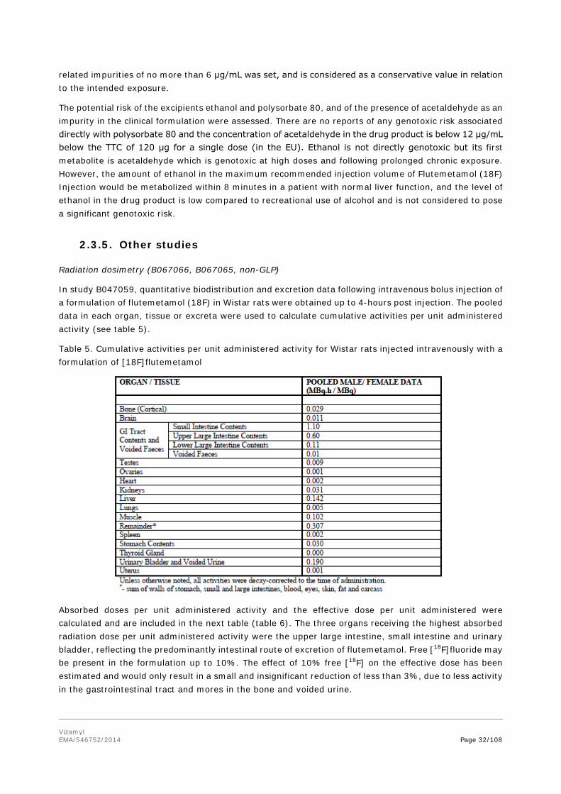
Vizamyl EMA/546752/2014 Page 32/108
related impurities of no more than 6 μg/mL was set, and is considered as a conservative value in relation to the intended exposure.
The potential risk of the excipients ethanol and polysorbate 80, and of the presence of acetaldehyde as an impurity in the clinical formulation were assessed. There are no reports of any genotoxic risk associated directly with polysorbate 80 and the concentration of acetaldehyde in the drug product is below 12 μg/mL below the TTC of 120 μg for a single dose (in the EU). Ethanol is not directly genotoxic but its first metabolite is acetaldehyde which is genotoxic at high doses and following prolonged chronic exposure. However, the amount of ethanol in the maximum recommended injection volume of Flutemetamol (18F) Injection would be metabolized within 8 minutes in a patient with normal liver function, and the level of ethanol in the drug product is low compared to recreational use of alcohol and is not considered to pose a significant genotoxic risk.
2.3.5. Other studies
Radiation dosimetry (B067066, B067065, non-GLP)
In study B047059, quantitative biodistribution and excretion data following intravenous bolus injection of a formulation of flutemetamol (18F) in Wistar rats were obtained up to 4-hours post injection. The pooled data in each organ, tissue or excreta were used to calculate cumulative activities per unit administered activity (see table 5).
Table 5. Cumulative activities per unit administered activity for Wistar rats injected intravenously with a formulation of [18F]flutemetamol
Absorbed doses per unit administered activity and the effective dose per unit administered were calculated and are included in the next table (table 6). The three organs receiving the highest absorbed radiation dose per unit administered activity were the upper large intestine, small intestine and urinary bladder, reflecting the predominantly intestinal route of excretion of flutemetamol. Free [18F]fluoride may be present in the formulation up to 10%. The effect of 10% free [18F] on the effective dose has been estimated and would only result in a small and insignificant reduction of less than 3%, due to less activity in the gastrointestinal tract and mores in the bone and voided urine.

Vizamyl EMA/546752/2014 Page 33/108
Table 6. Organ absorbed dose per unit administered activity for Wistar rats injected intravenously with a formulation of [18F]flutemetamol
Osmolality and haemolytic potential (B067061, non GLP)
This study was conducted with the drug product vehicle used at that time: sodium phosphate 14 mM, 7% ethanol (v/v), 0.5% polysorbate 80 (w/v), pH 7.2. The osmolality was approximately 1250 mOsm/kg. After the incubation of 0.002 to 0.2 mL of the formulation with 2 mL of human blood, haemolysis was not detected. The highest concentration tested in vitro is approximately 50-fold greater than when 10 mL are homogeneously distributed in 5 liters of blood in the human body. Considering that no indications of haemolysis were detected in non-clinical and clinical studies (22 subjects during the Phase 1 clinical trial (ALZ103) and 72 subjects during the Phase 2 clinical trial (ALZ201).), and that sodium chloride 0.9% (w/v) has been added to the formulation during development to raise the osmolality to values close to physiological tonicity, no further studies are required.
2.3.6. Ecotoxicity/environmental risk assessment
A study for the determination of the partition coefficient for flutemetamol provided by the applicant performed as described by OECD 123 Guideline was used. A pH – buffer of 7 was applied to cover the highest logD. The study is considered to be valid and should be used in risk assessment. The measured value logD = 4.27 falls below the trigger value of 4.5 used for PBT screening. Therefore a PBT assessment is not necessary.
Flutemetamol PEC surfacewater value is below the action limit of 0.01 µg/L and is not a PBT substance as log Kow does not exceed 4.5.

Vizamyl EMA/546752/2014 Page 34/108
Table 7. Summary of main study results
Substance (INN/Invented Name): flumetamol (18F)
CAS-number (if available):
PBT screening Result Conclusion
Bioaccumulation potential- log Kow ? 4.27 ± 0.02 Not potential PBT
PBT-assessment
Parameter Result relevant for conclusion
Conclusion
Bioaccumulation log Kow 4.27 ± 0.02 not B
PBT-statement : The compound is not considered as PBT nor vPvB
Phase I
Calculation Value Unit Conclusion
PEC surfacewater , default or refined (e.g. prevalence, literature)
1.64·10-6 µg/L > 0.01 threshold (N)
2.3.7. Discussion on non-clinical aspects
The in vitro pharmacology studies suggest that [3H]flutemetamol is able to label cored and diffuse amyloid β deposits and neuritic plaques. The affinity of flutemetamol (18F) for neurofibrillary tangles is much lower than the affinity for β-amyloid (30-fold lower).
The binding of flutemetamol to 65 central nervous system targets other than amyloid was studied in vitro (B067081). Only weak inhibition (34%) of binding of the reference compound to the non-human P2Y receptor was observed. This class of receptors is expressed in both the periphery and throughout the brain on both neurones and glia. The limited number of studies assessing human AD brain tissues has not demonstrated an increase in the expression of any P2Y subtype. Therefore, while weak binding of flutemetamol to P2Y receptors may contribute to the background of the images, it will not likely have an impact on the imaging efficacy of 18F-flutemetamol.
In the safety pharmacology studies flutemetamol did not show any effect in the cardiovascular, respiratory and behavioural parameters.
Biodistribution studies of flutemetamol (18F) in rats have shown that it is rapidly distributed to the brain but it is not retained in the absence of amyloid β deposits. Distribution to, and elimination from other tissues is also rapid and excretion is primarily via the gastrointestinal route. Significant levels of radioactive impurities did not accumulate in any radiosensitive organ or tissue.
In vitro studies have demonstrated that the metabolic profile of flutemetamol in hepatic S9 obtained from rat (Aroclor 1254 induced), mouse, dog and human are broadly comparable. No unique metabolic species were observed in human.
In the in vivo studies in rat and baboon, a total of 3 radiolabelled products, including [11C]flutemetamol, were detected in plasma. In rats, two major and one minor radiolabelled metabolites were identified, all less lipophilic than the parent compound. Apparently, the Applicant did not identify the chemical structure of any of the metabolites found in the in vitro or in vivo studies, or in the clinical trials. The interactions of flutemetamol with a panel of drugs commonly prescribed to Alzheimer’s patients were assessed in the clinical trials.

Vizamyl EMA/546752/2014 Page 35/108
All general toxicology studies were conducted according to GLP. The toxicity studies in a rodent and a non-rodent species are limited to a maximum duration of 14-days using not radioactively labeled flutemetamol to investigate the toxicological properties of flutemetamol. Although according to the guideline CPMP/ICH/286/95 M3 (R2) the duration of the pivotal toxicity studies to support marketing of treatments of a duration up to 2 weeks should be of 1 month, considering the low dose, the short plasma half-life and the lack of systemic accumulation of flutemetamol, the duration of these studies is justified.
Flutemetamol were tested in a standard battery of genotoxicity tests with conflicting results. Considering the single usage and the low maximum human dose and that the final drug product will be radiolabelled and can therefore be regarded as potentially genotoxic by itself, the genotoxic potential of non-radioactive flutemetamol is regarded as very low compared to the genotoxic potential of the positron emission itself.
The Applicant conducted three local tolerance studies in rabbits with different routes of administration. No specific studies on impurities have been conducted. There were 5 flutemetamol-related impurities present in the clinical drug product that were not present in the test item used for the majority of the nonclinical studies.
The radiation dosimetry estimates were based on the results of a distribution study in rats (B067056). The three organs receiving the highest absorbed radiation dose per unit administered activity were the upper large intestine, small intestine and urinary bladder, reflecting the predominantly intestinal route of excretion of flutemetamol.
Flutemetamol PEC surfacewater value is below the action limit of 0.01 µg/L and is not a PBT substance as log Kow does not exceed 4.5.
2.3.8. Conclusion on the non-clinical aspects
The non-clinical package presented by the Applicant is considered appropriate.
2.4. Clinical aspects
2.4.1. Introduction
The company presented the following dataset to demonstrate the clinical efficacy of PET with flutemetamol (18F): three key studies (phase III studies GE067-007, GE067-015 and GE067-021), supported by another eight clinical studies, and a pooled analyses of all 11 clinical studies composing the flutemetamol (18F) clinical development program (see table 8 below, Study GE067-021 was an electronic reader training study and is not included in the table).
The two Phase 1 studies ([ALZ103] and [GE067-014]) assessed the biodistribution and dosimetry of flutemetamol (18F) in HV (white and Asian, respectively) and in patients with probable AD.
The Phase 2 study [ALZ201] evaluated the ability of flutemetamol (18F) images to differentiate between HV and subjects with AD, and to determine the proportions of aMCI subjects with normal and abnormal flutemetamol (18F) images.
Seven of the 8 Phase 3 studies assessed the diagnostic performance and reproducibility of the blinded visual assessment of flutemetamol (18F) PET images. In all but one study (the electronic reader training study, GE067-021), image readers were trained in person by a consultant nuclear medicine physician who used materials provided by GE Healthcare; in the electronic reader training study (Study GE067-021), readers were training using an electronic training program, also provided by GE Healthcare.

Vizamyl EMA/546752/2014 Page 36/108
The 8th Phase 3 study, [GE067-005], assessed the ability of flutemetamol (18F) images to predict the subsequent development of AD in aMCI subjects.
Populations studied in the clinical development program included European and US healthy volunteers (HVs) subjects with aMCI, pAD, known or suspected normal pressure hydrocephalus (NPH), and subjects with a life expectancy of 1 year or less (regardless of primary diagnosis). In addition, 22 Japanese subjects (14 HVs and 8 AD subjects) were enrolled in one of the 2 Phase 1 studies.
• Tabular overview of clinical studies

Vizamyl EMA/546752/2014 Page 37/108
Table 8. Summary of Studies Included in the Summary of Clinical Efficacy (except for study GE067-21)
ALZ103
ALZ201
GE067-014
GE067-005
GE067-007
GE067-008
GE067-009
GE067-010
GE067-011
GE067-015
Study Type Phase 1
Open-label
Biodistribution
Dosimetry
Phase 2
Open-label
Phase 1 in Japanese
Open-label
Safety
Biodistribution
Dosimetry
Phase 3
Open-label
Phase 3
Open-label
Phase 3
Open-label
Phase 3
Open-label
Phase 3
Open-label
Phase 3
Open-label
Phase 3
Open-label
Efficacy Objectives
Determination of
biodistribution and
dosimetry
Ability to differentiate between HV
and pAD subjects
Determination of
biodistribution and dosimetry
in Japanese
Prediction of conversion
from aMCI to pAD
Estimation of sensitivity for detection of
brain amyloid (neuritic plaque
density)
Correlation between
SUVR and brain amyloid
level
Correlation between
SUVR and brain
amyloid level
Correlation between SUVR
and brain amyloid level
Correlation between SUVR and
brain amyloid
level
Specificity for excluding
amyloid in HV
Population ≥50 y
HV
pAD
HV: young (25-55 y) and older (>55 y)
pAD: ≥55 y
aMCI: ≥55 y
≥50 y
HV
pAD
≥55 y
aMCI
≥55 y with short life
expectancy
≥50 y
NPH
≥50 y
NPH
≥50 y
NPH
≥50 y
NPH
≤40 y
HV

Vizamyl EMA/546752/2014 Page 38/108
ALZ103
ALZ201
GE067-014
GE067-005
GE067-007
GE067-008
GE067-009
GE067-010
GE067-011
GE067-015
Flutemetamol F 18 Injection
1 dose of 100 MBq, 150
MBq or 185 MBq
pAD/aMCI: 1 of
185 MBq; 1 of 333 MBq
[11C]PiB
pAD: 2 of 120 MBq
HV: 1 of 185 MBq
1 dose of 100 MBq, 150 MBq or 185 MBq
1 dose of 185 MBq
1 dose of 185 MBq up to 370 MBq
1 dose 185 MBq
1 dose 185 MBq
1 dose 185 MBq 1 dose 185 MBq
1 dose 185 MBq
Initiation - End Date
24 Oct 2007-05 Mar
2008
03 Sep 2008-23 Mar
2011
21 Feb 2011-11 Oct 2011
11 Nov 2009-16 Jan
2014
22 Jun 2010-23 Nov
2011
21 Dec 2009-02 Jul
2010
11 Mar 2010-05 Jan 2011
15 Jun 2010-17 Nov 2010
31 May 2010-
16 Dec 2010
02 Dec 2010-18 Mar
2011
No. of Subjects Exposed
[Evaluable for
efficacy]
22 (14 HV; 8 pAD)
[22]
72 (25 HV;
20 aMCI; 27 pAD)
[70]
22 (14 HV; 8 pAD)
[22]
232 aMCI
[232]
180 end-of-life subjects
scanned; 69 brain
autopsied [68]
7 NPH
[7]
12 NPH
[10]
15 NPH
[15]
18 NPH
[15]
181 HV
[181]
Age, mean (range)
HV: 63 (51–73)
pAD:69 (55–77)
HV: 56 (25-78)
aMCI: 73 (57, 83)
pAD: 70 (56, 82)
HV: 57 (51–62)
pAD:75 (62–90)
71 (53–91) 80 (47–98) 70 (55–81) 71 (61– 87) 70 (61–75) 68 (53–76)
29 (18–40)

Vizamyl EMA/546752/2014 Page 39/108
ALZ103
ALZ201
GE067-014
GE067-005
GE067-007
GE067-008
GE067-009
GE067-010
GE067-011
GE067-015
Gender, M/F
(%/%)
HV: 10/4 (71/29)
pAD: 6/2 (75/25)
HV: 12/13 (48/52)
aMCI: 11/9 (55/45)
pAD: 12/15 (44/56)
HV: 11/3 (79/21)
pAD: 3/5 (60/40)
114/118 (49/51)
77/103 (43/57)
4/3 (57/43)
7/5 (58/42) 11/4 (73/27) 11/7 (61/39)
78/103 (43/57)
Race, W/B/A/O
HV: 14/0/0/0
pAD: 8/0/0/0
HV: 25/0/0/0
aMCI: 20/0/0/0
pAD: 27/0/0/0
HV: 0/0/14/0
pAD: 0/0/8
/0
225/5/1/1
168/7/1/4
7/0/0/0 10/2/0/0 15/0/0/0 18/0/0/0 156/19/3/3

Vizamyl EMA/546752/2014 Page 40/108
GCP
The clinical trial programme has partly been conducted outside the European Union (United States of America, and Japan). The company stated that each of the clinical studies that enrolled subjects in the flutemetamol (18F) development programme was conducted in accordance with the current revision of the Declaration of Helsinki, the Good Clinical Practice: Consolidated Guideline approved by the ICH, and applicable national and local laws and regulations (e.g., Code of Federal Regulations Parts 50, 54, 56, 312, and 314). The clinical trial GE0067-014 carried out outside the European Union (Japan) meets the ethical requirements of Directive 2001/20/EC. To assist screening and dementia assessment, all subjects diagnosed with probable Alzheimer’s Disease (pAD) were accompanied by a legally acceptable representative. The informed consent form for those subjects with pAD was signed both by the subject and the legally acceptable representative.
2.4.2. Pharmacokinetics
Flutemetamol F 18 Injection (the drug product) contains the drug substance, flutemetamol (18F) - a small lipophilic new molecular entity based on the amyloid-specific dye, Thioflavin T. Following intravenous injection, approximately 7% of administered flutemetamol (18F) activity is distributed to the brain. The radioactive half-life of 18F (approximately 110 minutes; 1.8 hours) necessitates manufacturing, purifying, assaying, and delivering a dose of Flutemetamol F 18 injection to the end user within a single day. The drug substance (flutemetamol (18F)) is 100% bioavailable and hence, no absorption, bioavailability, and bioequivalence studies have been conducted.
For diagnostic radiopharmaceuticals, clinical pharmacology studies per se are not conducted, because no pharmacologic effects are intended or observed owing to the extremely small mass dose of the drug substance. Rather, biodistribution and radiation dosimetry are determined in Phase 1 studies, along with safety. In the Flutemetamol F 18 Injection clinical development program, two Phase 1 studies were conducted: [ALZ103] in White (Caucasian) healthy volunteers (HV) and patients with probable AD (pAD), and [GE067-014] in Japanese HV and patients with pAD (table 9).
Table 9. Phase 1 Studies of Flutemetamol F 18 Injection
Protocol Title Date Subject (population)
Dose, mBq (mCi)
Primary endpoints
Secondary endpoints
ALZ103 A Phase 1, Open-label Study to Assess Safety, Biodistribution, and Radiation Dosimetry and to Optimise the Imaging Protocol of AH110690 (18F) Injection in Healthy Volunteers and
October 24th, 2007-March 05th, 2008
Enrolled and analyzed: 22 (14 HV and 8 pAD)
Age HV: 63 (51-73)
Age pAD: 69 (55-77)
Gender HV: 10/4 (71/29)
pAD: 6/2 (75/25)
Single i.v. injection of 100 MBq
(14 mCi),
150 MBq
(27 mCi),
or 185 MBq
(5 mCi);
Occurrence of AEs; changes in vital signs (BP, heart rate, temperature, respiratory rate, and oxygen saturation), physical examination, clinical
Flutemetamol (18F) brain UR and DVR measured by volume of-interest (VOI) in reference to control tissue in pAD compared to HV.
Dosimetry estimates and
Vizamyl EMA/546752/2014 Page 41/108
Subjects with Probable Alzheimer's Disease
laborator parameters (serum biochemistry, hematology, and urinalysis), ECG
cumulated activity by source and by entire body in HV.
GE067-014 A Phase 1, Open-Label, Study to Assess a) Safety of Flutemetamol (18F) Injection, Biodistribution and Internal Radiation Dosimetry and b) to Optimize the Imaging protocol of Flutemetamol (18F) Injection in Japanese Healthy Volunteers and Alzheimer's Disease Subjects
February 21st, 2011- October 11th, 2011
Enrolled and analyzed: 22 (14 HV and 8 pAD)
Age HV: 57 (51-62)
Age pAD: 75 (62-90)
Gender HV: 11/3 (79/21)
pAD: 3/5 (60/40)
Single i.v. injection of 100 MBq (14 mCi), 150 MBq (27 mCi), or 185 MBq (5 mCi);
Occurrence of AEs;
changes in vital signs (BP, heart rate, temperature, respiratory rate, and oxygen saturation), physical examination, clinical laborator (serum
biochemistry, hematology, and
urinalysis)
ECG
Flutemetamol (18F) brain UR and DVR measured by volume of interest (VOI) in reference to control tissue in pAD compared to HV.
Dosimetry estimates and cumulated activity by source and by entire body in HV.
HV = healthy volunteers; pAD = probable Alzheimer’s disease; N/A = not applicable; ROI = region of interest; PET = positron emission tomography; [18F]AH110690 =[18F]flutemetamol; UR = uptake ratio; DVR = distribution volume ratio; VOI = volume of interest
Study protocol ALZ103 (EudraCT Number: 2007-000784-19)
This was a phase 1, single centre, single dose, open-label, non-randomised study to evaluate the safety, biodistribution, and radiation dosimetry, and also to optimise the imaging procedure of AH110690 (18F) Injection Healthy Volunteers (HV) and Subjects with Probable Alzheimer’s disease (pAD). The study was conducted in a single European study centre: Leuven University Hospital, Belgium between October 24th, 2007-March 05th, 2008.
For study ALZ103, 22 subjects 14 HVs and 8 subjects with pAD) received Flutemetamol (18F) solution for injection and completed the study (no randomization was performed). One subject was withdrawn prior to dosing due to technical problems and was replaced. No withdrawals occurred after administration of the IMP.
Vizamyl EMA/546752/2014 Page 42/108
Study protocol GE-067-014
This was a phase 1, single centre, single dose, open-label, non-randomised study to evaluate the safety, biodistribution, and radiation dosimetry, and also to optimise the imaging protocol of Flutemetamol (18F) Injection in Japanese Healthy Volunteers (HV) and Subjects with Probable Alzheimer’s disease (pAD). The study was conducted at 2 centers in Japan: Institute of Biomedical Research and Innovation (IBRI) and Kobe University between February 21st, 2011-October 11th, 2011.
For study GE067-014, 22 subjects (14 HVs and 8 subjects with pAD) received Flutemetamol F 18 injection and completed the study. All demographic and baseline characteristics data have been submitted for all studies.
The Applicant has stated that trials have been conducted in compliance with GCP requirements.
There was one non-substantial protocol amendment for study ALZ103 (the blood sampling time points were modified to ensure suitable samples for kinetic modelling purposes) and none for study GE067-014. The protocol amendment does not seem to impact on the validity of the studies.
The inclusion and exclusion criteria are considered to be acceptable and the study populations are considered acceptable with regards to demographic characteristics.
The number of planned subjects was considered sufficient to determine estimates of biodistribution parameters.
The study designs and arterial blood, plasma and urine samples collection were acceptable.
There were some protocol deviations that did not affect the safety or efficacy populations. None of the prior/concomitant medications affected entry into the study or affected study results and were submitted.
Absorption
• Bioavailability
No clinical studies investigating bioavailability were conducted. Flutemetamol (18F) is administered via intravenous injection and is therefore fully bioavailable.
• Bioequivalence
N/A
• Influence of food
N/A
Vizamyl EMA/546752/2014 Page 43/108
Distribution
Study protocol ALZ103 (EudraCT Number: 2007-000784-19) Step 1 Summary of Biodistributions Details of biodistributions are provided below:
Organ/Tissue Mean Maximum 18F Activity (range)
Mean Half-life (range)
Brain1 6.7% (5.9% to 8.1%) 0.4h to 1 h Thyroid Gland 0.08% (0.04% to 0.16%) 0.1 h Lung1 7.9% (5.3% to 9.2%) 0.4 h Cardiac Wall 1.5% (0.7% to 2.1%) 0.1 h Spleen 2.0% (0.7% to 3.0%) 0.1 h Liver 25.7% (23.7% to 27.2%) at 0.51 h
p.i. (0.20 h p.i. to 1.17 h p.i.) 2.0 h (range: 0.8 h to 3.0 h).
Gallbladder 13.7% (11.6% to 17.5%) nd Urinary Bladder Contents and Voided Urine
37% (28% to 45%) 2.2 h (1.1 h to 3.6 h)
Remaining Tissues 55% (48% to 61%) nd 1 at the first imaging time point, between about 2 and 4 minutes p.i.; nd = not determined.
Whole Blood and Plasma The mean maximum 18F activity concentrations in whole blood and plasma were 2.27×10⁻⁵ ml-1 (range: 1.30×10⁻⁵ ml-1 to 4.37×10⁻⁵ ml-1) and 2.45×10⁻⁵ ml-1 (range: 1.56×10⁻⁵ ml-1 to 3.73×10⁻⁵ ml-1), respectively. The volume of distribution of 18F was estimated by extrapolating the 18F activity concentration in plasma curve to time 0. The mean value was 3720 mL (range: 2300 mL to 4850 mL) Liver, Gallbladder and Gastrointestinal Tract Contents The 18F activity data provided had a significant non-zero amount of 18F activity present in the GI tract contents (up to about 10%) at very early time points of 2 to 4 minutes p.i. for each subject. This was considered physiologically unlikely for an intravenously administered radiopharmaceutical which would enter the GI tract almost exclusively through hepatobiliary transport. Such a reported rapid uptake of 18F activity in the contents was also inconsistent with reported measured gallbladder biphasic emptying rates in 12 healthy adult volunteers of 0.015 min-1 initially and later at 0.005 min-1 (Lawson et al. 1983). In each subject, the 18F activity in the liver grew to a maximum and then decreased exponentially. The mean maximum measured 18F activity in the liver was 25.7% (range: 23.7% to 27.2%); the mean time of when this maximum occurred was 0.51 h p.i. (range: 0.20 h p.i. to 1.17 h p.i.). The mean half-life of the washout phase was 2.0 h (range: 0.8 h to 3.0 h). Excluding the remaining tissues category, the three organs or tissues with the highest normalised cumulated activities amongst the 6 subjects were the contents of the small intestine, with a mean of 0.429 MBq•h/MBq (range: 0.328 MBq•h/MBq to 0.723 MBq•h/MBq); the liver with a mean of 0.399 MBq•h/MBq (range: 0.335 MBq•h/MBq h to 0.448 MBq•h/MBq); and, the combined contents of the urinary bladder and voided urine, calculated for a 3.5-hour voiding interval, with a mean of 0.287 MBq•h/MBq (range: 0.154 MBq•h/MBq to 0.453 MBq•h/MBq). Internal Radiation Dosimetry The organs/tissues receiving the three highest absorbed doses are the gallbladder wall (mean: 0.287 mGy/MBq; range: 0.035 mGy/MBq to 0.727 mGy/MBq), the urinary bladder wall (mean: 0.145 mGy/MBq; range: 0.082 mGy/MBq to 0.225 mGy/MBq) and the wall of the upper large intestine (mean: 0.117 mGy/MBq; range: 0.096 mGy/MBq to 0.186 mGy/MBq). For all three tissues, the absorbed dose was primarily (over 60%) due to the stopping of positrons. Effective Dose The Effective Doses per unit administered activity for the 6 subjects is presented. The mean value was 0.032 mSv/MBq (range: 0.026 mSv/MBq to 0.045 mSv/MBq.

Vizamyl EMA/546752/2014 Page 44/108
Kinetic Modelling None of the input function-based modelling methods were able to give consistent discrimination between HV and subjects with probable AD in terms of total volume of distribution and binding potential in the cortical regions. Reference region-based modelling presented URs and DVRs with good discrimination between HV and subjects with probable AD in the cortical regions and striatum. From the Step 2 analysis, it was recommended that the PET scan time window for Step 3 should be 80-140 minutes (with an option of continuing imaging up to 170 minutes) post-injection and the recommended method of Step 3 PET data analysis would be reference-ratio modelling using cerebellum cortex as reference region. Study protocol GE-067-014 Flutemetamol (18F) Brain SUVRs Measured by VOI Analysis in Reference and Control Tissue in Subjects with pAD Compared to HVs Normalised Absorbed Doses to Organs and Tissues and Normalized Effective Dose The 3 critical organs / tissues with the highest mean absorbed doses were the urinary bladder wall with 0.114 mGy/MBq (range: 0.0823 mGy/MBq to 0.152 mGy/MBq), kidneys with 0.0751 mGy/MBq (range: 0.044 mGy/MBq to 0.125 mGy/MBq) and liver with 0.0687 mGy/MBq (range: 0.0452 mGy/MBq to 0.107 mGy/MBq). For the example of a 185 MBq administered activity, the mean absorbed doses to the urinary bladder wall, kidneys and liver are 21 mGy, 13.9 mGy and 12.7 mGy, respectively. The mean total body normalised absorbed dose was 0.0118 mGy/MBq (0.0112 mGy/MBq to 0.0133 mGy/MBq). The mean normalised effective dose was 0.0255 mSv/MBq (range: 0.0226 mSv/MBq to 0.0301 mSv/MBq). For the example of a 185 MBq administered activity, the mean effective dose is 4.7 mSv.
Elimination • Excretion
The data from the studies show that following intravenous injection, mean half-life of elimination of activity was 4.5 hours. Extent of excretion ranged from 71% to 100%, with a mean of 89%. By pathway, the mean extent of excretion was 37% renal (range, 28% to 45%) and 52% hepatobiliary (range, 40% to 65%). • Metabolism
In vitro The two major and one minor radiolabelled metabolite were identified in vitro stability, all less lipophilic than the parent compound. The major in vitro metabolite observed following incubation of flutemetamol in hepatic S9 (from mouse, dog and man, and from Aroclor 1254 induced rat) was the N-demethylation product. No metabolism was observed in control heat inactivated or β-NADPH deficient hepatic S9 fractions. This is a preliminary indication that metabolism in the human is similar to that of the rat and dog, and gives support for these two species being appropriate for use in nonclinical safety studies. In Vivo In addition, after injection of flutemetamol (18F) in the clinical ALZ103 Phase 1 trial, human plasma samples from the subjects were analysed by HPLC. These samples were primarily taken to assist with brain kinetic modelling but they also showed that metabolism of the injected flutemetamol (18F) occurred rapidly and at least 2 hydrophilic metabolites were detected.
• Inter-conversion
N/A
• Pharmacokinetics of metabolites
N/A

Vizamyl EMA/546752/2014 Page 45/108
Dose proportionality and time dependencies
N/A
Special populations
The lack of special populations studies is considered acceptable as only tracer quantities of flutemetamol were administered. Studies ALZ-103 and GE067-014 indicated the biodistribution and dosimetry to be comparable in Caucasian and Japanese population.
Pharmacokinetic interaction studies No clinical drug interaction studies were performed as the target of flutemetamol binding (amyloid β) is not a target for concomitant therapies. This is considered to be acceptable since because of the microgram quantities of flutemetamol in the formulation, no drug-drug interactions are expected. A rat drug interaction study performed to assess the effect of an anti-amyloid therapy on the pharmacokinetics of flutemetamol (18F) demonstrated no impact of the therapy.
2.4.3. Pharmacodynamics
Mechanism of action
Flutemetamol (18F) is intended as an in vivo tracer of Aß neuritic plaques in the brain. The fluorine (18F) isotope produces positrons that combine with electrons to form gamma rays that are emitted from the patient and detected by a PET scanner.
The binding target(s) of flutemetamol (18F) has not been clearly elucidated in vivo either in normal subjects or in targeted patients. In the pivotal study GE067-007 in end-of-life patients, whose cognitive impairment status was not determined, correlation between the in vivo flutemetamol (18F) quantitative uptake in cortical grey matter and the total β-amyloid burden averaged from eight particular cortical regions using 4G8 anti-amyloid antibody (that stains β-amyloid found in both neuritic and diffuse plaques) was evaluated. The quantitative PET-autopsy correlation was not significant as analyzed according to the protocol; however, a different statistical approach determined the correlation to be significant. The company acknowledged that in four cases in the autopsy study VIZAMYL positivity was observed in subjects that were close to the positive threshold for neuritic plaques and/or had significant fibrillar amyloid deposits (in diffuse plaques and/or vascular amyloidopathy), indicating that significant fibrillar amyloid-beta deposits other than neuritic plaques may contribute to the VIZAMYL signal. A work [GE-067 Amyloid Phasing White Paper] that encompasses a description of PET positivity in structures with reference to the type of amyloid deposit, namely neuritic and diffuse, in the subject cohort of the 68 cases included in the efficacy analysis of the GE067-007 Phase III clinical trial has been provided. All PET images were negative in the 17 cases with phase 0, 1 or 2 of the 2012 revision to the pathological diagnosis of AD (Hyman et al, 2012). As amyloid appears in the striatum in phase 3, some phase 3 cases may have associated PET images that are positive in the striatum; this occurred in 1 out of 9 cases. In phases 4 and 5, significant amyloid would be expected in the striatum, and 36 of 42 (85%) were positive in the striatum in the read without CT while there was 39 out of 42 (93%) in the read with CT. The majority of deposits in the striatum were found to be in the form of diffuse plaques. In 8 cases, frequent diffuse plaques in the absence of any neuritic plaques were considered positive by PET, demonstrating that flutemetamol (18F) can detect diffuse plaques when they are frequent.
Flutemetamol (18F) binds non-specifically to cerebral white matter both in normal and targeted patients, which has been attributed to solubility of flutemetamol in the lipid content of brain tissues.

Vizamyl EMA/546752/2014 Page 46/108
Primary and Secondary pharmacology Flutemetamol (18F) is administered in doses no higher than 20 µg and does not have any detectable pharmacological activity. No clinical data on secondary pharmacology are available. Brain uptake and distribution of flutemetamol (18F) was not evaluated in a specific study aimed to evaluate pharmacodynamics but in steps 2 and 3 of two similar PK studies (study ALZ103 and GE067-014 in European and Japanese population, respectively). Additionally, quantitative measures of flutemetamol (18F) brain activity (SUVR) to discriminate subjects with pAD and HVs, and pAD and aMCI subjects are reported in study ALZ201. STUDY ALZ103 (Caucasian population) The company considered cerebellum cortex as a suitable reference region in Step 3 and future studies. Uptake ratios (URs) using the cerebellum as reference were calculated and compared for flutemetamol (18F) in 3 HVs and 3 subjects with pAD. Subjects were administered with 185 MBq of flutemetamol (18F). URs were 30-40% higher and DVRs were 20-30% higher in subjects with pAD in the 60-185 minute interval. The recommended PET scan time window for Step 3 was defined as 80-140 min (optionally -170 min) post injection. Step 3 aimed secondarily to optimise the imaging procedure, i.e., investigating how scanning start time and scan duration affected discrimination between subjects with AD and HV. Five subjects with pAD and 5 HVs were recruited. Subjects were administered with 185 MBq of flutemetamol (18F). Image acquisition commenced at 80 minutes post-injection and the scanning window was 80-140 minutes post-injection with 140-170 min post-injection as optional. To maximise the data available for the analysis, Step 2 and Step 3 data were merged into a common time frame. Analysis of the results showed that different scan leongths (5 to 40 minutes) had a negligible effect on discrimination between subjects with pAD and HVs. The same analysis of the effect of different start times also showed a very small difference, but with a minor improvement in discrimination for later scan start times. When comparing URs in subjects with pAD (n=8) and HVs (n=8) for the 85-105 minute time window [150-170 minute time window], significant test results (i.e., p-values <0.05) were obtained in all brain areas except the anterior cingulate cortex, pons and subcortical white matter (Table 10 below). When flutemetamol (18F) URs were examined for individual subjects, 6 out of 8 subjects with pAD showed clear, elevated URs in the cortex when compared to HVs. However, 2 subjects with pAD [MAI1]demonstrated URs in several brain areas which overlapped with the range of URs seen in HVs. Seven out of 8 HVs showed a consistently low cortical uptake. However, 1 HV demonstrated high URs in several brain areas which overlapped with the range of URs seen in subjects with pAD. Table 10. Brain uptake ratios in HV and Subjects with Probable AD

Vizamyl EMA/546752/2014 Page 47/108
Based on this analysis, the recommended parameters for a Phase 2 study were: dose approximately 185 MBq, scanning start time approximately 90 minutes post injection, scan duration approximately 30 minutes and data acquisition in 5-minute bins. STUDY GE067-014 (Japanese population) SUVRs were calculated for flutemetamol (18F) in Step 2 and Step 3 HVs and subjects with pAD. The data show a temporal window (approximately 75 to 135 minutes) during which SUVR values for HVs and the pAD subjects are statistically significantly different in many brain regions (all examined areas of the brain except the pons and medial temporal cortex). When flutemetamol (18F) SUVRs were examined for individual subjects, 5 of 8 subjects with pAD showed clear, elevated SUVRs in the composite cortex when compared to HVs. In these 5 pAD cases the raised cortical SUVR values are likely to reflect in the presence of fibrillar β-amyloid. The other 3 subjects with pAD (subjects[MAI2]from the step 3 cohort) had images consistent with those of the HV cohort and did not have imaging evidence of amyloid deposition to explain their cognitive difficulties. All 8 HVs showed a consistently low cortical uptake. STUDY ALZ201 There was significant discrimination in quantitative PET values between subjects with pAD and HVs in all regions of the cortex and striatal regions. Overall, there was less discrimination between subjects with pAD and aMCI; however, the aMCI quantitative PET values had a bimodal distribution being raised or normal. There was a higher mean quantitative PET value in subjects with pAD in all cortical regions investigated compared with aMCI. Similar findings were noted in quantitative PET value discrimination in the cortex and striatal regions following [11C]PiB administration comparing pAD subjects to aMCI subjects.

Vizamyl EMA/546752/2014 Page 48/108
• Pharmacodynamic interactions with other medicinal products or substances No in vivo pharmacodynamics drug-drug interaction studies have been performed with a number of drugs belonging to classes that may be frequently used by elderly patients.
2.4.4. Discussion on clinical pharmacology
PHARMACOKINETICS
No clinical studies investigating bioavailability were conducted. Flutemetamol (18F) is administered via intravenous injection and is therefore fully bioavailable.
The characteristics of this agent are the rapidity with which it is taken up by the brain, the amount of 18F activity present in the brain and the subsequent washout of 18F activity from background (normal) tissue. The 18F activity uptake by the brain was demonstrated to be rapid and substantial, with rapid washout having already begun prior to the first imaging time point.
Following intravenous injection of the recommended dose of 185 MBq of Flutemetamol F 18 Injection in humans approximately 7% of administered flutemetamol (18F) activities distributed to the brain and approximately 25% of the active compound, [18F]flutemetamol, remains in the circulation 20 minutes post-injection and approximately 10% at 180 minutes.
The brain wash out of the compound starts to be linear after approximately 80 minutes post injection. An imaging window which begins after 80 minutes post-injection shows low and nearly constant levels of uptake in the cortex of healthy volunteers and in the cerebellar cortex for both HV and AD subjects. To give some margin to the 80 to 85 minutes post-injection starting point, the recommendation is to acquire a 20-minute PET scan starting 90 minutes post-injection. This recommendation is acceptable.
When comparing uptake ratios in subjects with probable AD and HV for the 85-105 minute time window [150-170 minute time window], significant test results (i.e., p-values <0.05) were obtained in all brain areas except the anterior cingulate cortex, pons, and subcortical white matter [pons and subcortical white matter]. The results of this statistical comparison support the observation that subjects with probable AD and HV could be differentiated by flutemetamol (18F).
The source organ radiation doses were the highest to the hepatobiliary excretion pathways including the gall bladder, the small intestine, and upper large intestine of the gastrointestinal tract and also to the kidneys and urinary bladder for urinary excretion. The highest absorbed dose values were: gallbladder wall (0.287 mGy/MBq), the urinary bladder wall (0.145 μGy/MBq) and the upper lower intestine wall (0.117 mGy/MBq). These absorbed dose values reflect the high percentage of administered 18F activity that is excreted through the GI tract.
After injection of an activity of100 MBq, the mean effective dose (i.e., the sum of risk-weighted organ absorbed radiation dose used as a measure of stochastic radiation risk) was found to be 32.4 ± 6.5 µSv/MBq (which equals 0.033 mSv/MBq). The levels of radiation dose were considered acceptable and due to the low variability in the measured data it was recommended that the injected activity for subsequent studies could be increased to 185 MBq, corresponding to an Effective dose of 5.9 mSv for the PET scans for each subject to obtain acceptable image quality on the PET camera used in the study.
In several subjects, the transport of 18F activity from the liver to the gallbladder was delayed, not appearing until about at least 30 minutes p.i., reflecting normal biliary transport; in the remaining subject, there was virtually no 18F activity present in the gallbladder contents and the combined rapidity of liver washout and appearance in the GI tract contents reflects the very rapid transfer of 18F activity from the liver into the gut.

Vizamyl EMA/546752/2014 Page 49/108
Excretion was predominantly through the gastrointestinal (GI) tract. The 18F activity excreted through the GI tract was estimated to be 51.7% (± 9.0%). The organ with the highest initial uptake of 18F was the liver at about 20-25% of the administered activity. The organs with the highest cumulated activities were the contents of the small intestine (0.43 ± 0.15 MBq·h/MBq) and the liver (0.40 ± 0.05 MBq·h/MBq). This reflects the large fraction of 18F activity excreted through the GI tract.
The amount of 18F activity excreted through the urinary pathway was estimated to be 37.3% (± 7.2%). The highest initial uptake of 18F was in the combined contents of the urinary bladder and voided urine (0.29 ± 0.10 MBq·h/Mbq). The apparent elimination half-life is 4.5 hours.
Elimination occurs primarily by clearance through the liver and excretion over the intestine which is reflected by the highest dosimetry values found in the liver and small intestine. Possible metabolites possibly are cleared through the urinary system.
In vitro metabolism studies suggest that metabolism of flutemetamol in rat, dog and man are similar. Additional in vivo metabolism studies confirm rapid metabolism of radiolabelled flutemetamol in the periphery, with the presence of two major metabolites that are less lipophilic than the parent compound.
Two major and one minor radiolabelled metabolite were identified, all less lipophilic than the parent compound. In the brain flutemetamol (18F) accounted for 97%, 85% and 75% of the total activity at 2, 20 and 60 min pi respectively, with only two minor metabolites observed. These data compare well with analyses of blood samples that were taken during clinical trial ALZ103 and suggests that metabolites in the brain are unlikely to affect the kinetic modelling that was performed on Phase I clinical data.
The metabolism of [18F]AH110690 is fast and was initially faster in AD than in HV subjects, and then the trend was gradually reversed so that beyond 20 min, more intact tracer was found in AD than in HV subjects. The variation within the HV and AD groups was quite large at early times but small from 20 min.
High performance liquid chromatography (HPLC) analysis of 18F in plasma in the human demonstrated that 18F in circulation at 5 min post-injection (pi) was 6.9% id in the form of the parent flutemetamol (18F) (or about 74.9% of all of the 18F present in the circulation) reducing to 0.1% id (or about 1.7% of all of the 18F present in the circulation) by the time of recommended imaging, 90 min pi. A hydrophilic metabolite represented 2% id (or about 21.7% of the total 18F activity in the circulation) at 5 min pi, increasing to 6.1% id at 90 min (or about 96% of all of the 18F present in the circulation).
In vitro metabolism studies following the incubation of either 14C-labelled or unlabelled flutemetamol in hepatic S9 fraction obtained from man suggest that the major hydrophilic metabolite is the N-demethylation product.
On the basis of these clinical and nonclinical data, there appears to be little likelihood of the hydrophilic metabolite having any demonstrable effect upon the binding of flutemetamol (18F) and, hence, the efficacy of the imaging. This is also supported by data demonstrating that at up to 60 min pi, only 0.1% id or less of the 18F activity present in the rat brain is in the form of 18F-labelled metabolites.
Study ALZ103 was done in subjects with probable Alzheimer’s disease. When flutemetamol (18F) uptake ratios were examined for individual subjects, most subjects with probable AD (6 out of 8) showed clear, elevated uptake ratios in the cortex when compared to healthy volunteers whereas most healthy volunteers (7 out of 8) showed a consistently low cortical uptake.
Based on the data provided by the Applicant, it is extremely unlikely that hepatic or renal impairment would affect the safety, efficacy, or the radiation dosimetry of [18F]flutemetamol. The omission of pharmacokinetics studies in renal or hepatic impairment was considered acceptable.

Vizamyl EMA/546752/2014 Page 50/108
There is no information, if potential drug-drug interactions have been evaluated in vivo which is considered to be overall acceptable.
PHARMACODYNAMICS
The Applicant provided limited data on pharmacodynamics in this submission.
Flutemetamol (18F) does not have any detectable pharmacological activity.
Flutemetamol (18F) is a molecular imaging agent designed for PET imaging of fibrillar β-amyloid in the form of neuritic plaques in the human brain. The correlation of flutemetamol (18F) binding to β-amyloid deposition was investigated in in vitro studies. The binding target(s) of flutemetamol (18F) has not been clearly elucidated in vivo either in normal subjects or in targeted patients. In the pivotal study GE067-007 in end-of-life patients, whose cognitive impairment status was not determined, correlation between the in vivo flutemetamol (18F) quantitative uptake in cortical grey matter and the total β-amyloid burden averaged from eight particular cortical regions using 4G8 anti-amyloid antibody (that stains β-amyloid found in both neuritic and diffuse plaques) was evaluated. The PET-autopsy correlation was not significant as analyzed according to the protocol; however, a different statistical approach shows the correlation to be significant.
Flutemetamol (18F) binds non-specifically to cerebral white matter both in healthy volunteers and targeted patients, which the company attributes to solubility of flutemetamol in the lipid content of brain tissues.
Brain uptake and distribution of flutemetamol (18F) was not evaluated in a specific study aimed to evaluate pharmacodynamics. In two similar PK studies (study ALZ103 and GE067-014 in European and Japanese population, respectively) and a phase 2 clinical study (ALZ201), there were discrimination between the quantitative uptake values in PET images obtained for pAD and HV subjects in most examined areas of the brain.
For Caucasian population (study ALZ103), uptake values of a flutemetamol (18F) PET scan at 80-170 minutes post-injection after administration of 185 MBq of flutemetamol (18F) statistically differed (p-values <0.05) between subjects with pAD (n=8) and HVs (n=8) for the 85-105 minute time window [150-170 minute time window], in all 11 defined brain areas except the anterior cingulate cortex, pons and subcortical white matter. The particular brain regions were selected based on those previously described in post-mortem studies to discriminate HV and AD, plus other cortical regions offering less discrimination but used as reference, and regions with no discrimination capability. Multiplicity of contrasts was not accounted for in the statistical analysis. PET uptake values in 6 out of 8 pAD subjects were clearly high, and it was low in 7 out of 8 HVs. However, it is of concern that the uptake values in 2 pAD subjects overlapped with those of HVs, and the opposite happened to 1 HV. The detected minimal non-specific binding of flutemetamol (18F) in the cerebellum cortex was considered as a suitable reference region.
It was not possible to make any conclusions regarding the optimal scanning window based on trends in the statistical analysis of the data from different scanning windows. However, based on this study, the recommended parameters for a Phase 2 study were: dose approximately 185 MBq, scanning start time approximately 90 minutes post injection, scan duration approximately 30 minutes and data acquisition in 5minute bins.
Significant discrimination in quantitative PET values between subjects with pAD and HVs in all regions of the cortex and striatal regions occurred in phase 2 study ALZ201. Overall, there was less discrimination between subjects with pAD and aMCI; however, the aMCI quantitative PET values had a bimodal distribution, being raised or normal.

Vizamyl EMA/546752/2014 Page 51/108
No in vivo pharmacodynamic drug-drug interaction studies have been performed with a number of drugs belonging to classes that may be frequently used by elderly patients.
2.4.5. Conclusions on clinical pharmacology
PHARMACOKINETICS
It was concluded from study ALZ103 that for all cortical regions of the brain, flutemetamol (18F) was able to differentiate between subjects with probable AD and HV using uptake ratios.
As a result of phase 1 studies the recommended parameters for single scanning in a Phase 2 study would be: dose approximately 185 MBq (corresponding to an effective radiation dose of 5.9 mSv), the PET scan time window should be 80-140 minutes pi, scanning start time around 90 minutes pi, scan duration around 30 minutes, and the recommended method of PET data analysis would be reference-ratio modelling using cerebellum cortex as reference region.
The following pharmacokinetic parameters were derived: Following intravenous injection of the recommended dose of 185 MBq of flutemetamol (18F) in humans, approximately 25% remained in the circulation 20 minutes post-injection and approximately 10% at 180 minutes. The apparent elimination half-life was 4.5 hours. Elimination was approximately 52% hepatobiliary and 37% renal. Information about the identity of metabolites was not provided. The pharmacokinetics in patients with renal or hepatic impairment has not been characterised.
The radiation dosimetry is comparable to approved PET markers like fludeoxyglucose (18F) and is not substantially affected by patient weight. The radioactive half-life of (18F) is approximately 110 minutes, the biological half-life of flutemetamol (18F) in humans is 4.5 hours.
Flutemetamol (18F) is administered in mass doses not higher than 60 µg and does not have any detectable pharmacological activity. Flutemetamol (18F) undergoes radioactive decay releasing a positron which then interacts with a neighbouring electron resulting in the release of two 511-keV gamma rays which can be detected with PET scanners.
PET images after administration of flutemetamol (18F) allowed differentiation between pAD subjects and HVs. Using as the standard of truth each subject’s baseline diagnosis, the majority (defined as agreement between at least 3 of 5 blinded image evaluators) blinded visual assessment of flutemetamol (18F) images showed a sensitivity of 93% and a specificity of 96%. There was a very good agreement between quantitation and visual assessment.
PHARMACODYNAMICS
It was confirmed in vitro that flutemetamol (18F) binds to β-amyloid neuritic plaques in the brain, with negligible binding to neurofibrillary tangles. The in vivo binding of flutemetamol (18F) to other β-amyloid structures or other brain structures or receptors remains unknown.
There is discrimination between pAD and HVs by using quantitative uptake values of flutemetamol (18F) PET scan in some brain regions in phase 1 and 2 studies; however, values were overlapping in some individual cases. Overall, there was less discrimination between subjects with pAD and aMCI. Discrimination between pAD and other non-AD dementias was not assessed.
The quantitative correlation between the in vivo flutemetamol (18F) uptake in cortical grey matter and the β-amyloid burden measured by histopathology might be artifacted and is uncertain.
In conclusion, although the company has not elucidated in vivo how and which of the different types of β-amyloid and other relevant structures are traced by flutemetamol (18F) (which would be important in the context of the proposed indication), the correlation of flutemetamol (18F) binding to β-amyloid deposition was investigated in vitro and together with the totality of the available PD data was considered sufficient to justify the mechanism of action and the intended context of use.
Vizamyl EMA/546752/2014 Page 52/108
2.5. Clinical efficacy
2.5.1. Dose response study(ies)
Neither the optimal dose nor the optimal timing for acquisition of flutemetamol (18F) PET images were determined by specific studies or adequately justified. For clinical practice, the company proposed that images should be acquired starting 90 minutes after of a single intravenous injection of 185 MBq as a bolus over approximately 40 seconds, not based on criteria of efficacy but on the fact that the effective dose (i.e. the absorbed radiation dose in whole-body) is within the level of other PET radiopharmaceuticals approved. The majority of subjects in the clinical development program, but not all, received the recommended dose.
The dose was determined in Phase 1 PK studies ALZ103 and GE067-014 by an activity escalation approach:
• Study ALZ103 was conducted in 3 steps. In step 1 recruiting 6 HVs, the first 2 subjects each received 100 MBq dose of flutemetamol (18F). After a review of safety and internal radiation dosimetry data from these subjects, the dose was increased to 150 MBq for the remaining 4 subjects. A second review of safety and internal radiation dosimetry data was conducted once all 6 evaluable subjects had been completed. Dosimetry estimates in HVs determined that the injected activity for subsequent studies could be increased to 185 MBq. In step 2, 3 pAD subjects and 3 additional HVs were each dosed with 185 MBq and no safety concerns were raised. In step 3, 10 additional subjects (5 pAD and 5 HV) each received 185 MBq. Results were used to define the imaging procedure for the following studies.
• In study GE067-014 a similar activity escalation methodology to that of ALZ103 was used. Results were in agreement with the conclusions of ALZ103.
Therefore, 185 MBq was determined as optimal activity to be administered.
2.5.2. Main study(ies)
The CHMP considers that, from the data provided by the company to support efficacy, there is a single pivotal study to base this application (i.e. GE067-007). Then, CPMP/EMA/2330/99, CPMP/EWP/1119/98/Rev and EMEA/CHMP/EWP/321180/2008 also apply. This was a multicenter, single-dose, open-label Phase III study conducted at 15 centers in the United States and 4 centers in the UK.

Vizamyl EMA/546752/2014 Page 53/108
Study GE067-007
Methods
Study Participants
Subjects aged 55 years or older, who had been diagnosed with a terminal illness and a short life expectancy (approximately 1 year or less) were included. Subjects were excluding if having a known or suspected structural brain abnormalities, such as infarcts or tumors, which might interfere with the interpretation of PET images.
Treatments Patients underwent screening visit before the administration of flutemetamol (18F), including anatomic brain imaging and clinical history. An anatomic image (MRI/CT) had to be performed within the previous 12 months before or simultaneously to PET image. Each subject received a single i.v. dose of Flutemetamol F 18 Injection between 110 to 370 MBq. PET imaging for 30 minutes (or as short as 10 minutes if not possible) was to start approximately 90 minutes after the injection. Flutemetamol (18F) PET images were visually read (as positive or negative) by 5 independent readers, who were previously in-person trained and qualified, and were also blinded to the standard of truth (SoT) results and all other information about the subject. A 10-minute acquisition PET image was interpreted using a Sokoloff colour scale. Five particular brain regions were assessed (frontal lobe/anterior cingulate, posterior cingulate/precuneus, lateral temporal lobe, insula and striatum). Selection of regions was performed to include those investigated in the literature in which the greatest differences exist between HVs and subjects with pAD. The blinded visual image interpretations were carried out first in a session where all study images were evaluated without anatomic brain images for reference, and in randomized order for each reader. In a second session in the same day, PET images were presented in a re-randomized order and were re-reviewed, this time with the concomitant or previous subject’s anatomic images. Visual assessment of the flutemetamol (18F) PET scan was completed in three reading sessions (including both PET only reads and PET+CT reads) with a minimum number of images in one day of 90 and the maximum of 178. PET-only and PET+CT images were read separately but in a similar session. The criteria to interpret images were normal or abnormal as follows (see fig 84-3):
• A region is considered as having a normal pattern if the tracer signal in cortical regions is low (i.e. distinctly lower signal intensity compared with adjacent white matter and similar in intensity to the grey matter-rich regions of the cerebellum). Signal will not be completely absent in grey matter regions of the images, as white matter binding in adjacent regions will bleed into the grey matter regions due to PET partial volume resolution effects.
• A region is considered abnormal if the tracer signal in cortical regions appears high (i.e.,
approximately at the same or higher signal intensity as adjacent white matter and greater than the grey matter-rich regions of the cerebellum).

Vizamyl EMA/546752/2014 Page 54/108
Figure 84-3. Normal (left) and abnormal (right) flutemetamol (18F) images showing equal cerebellar cortex uptake values in both the normal and abnormal scans. The abnormal image shows uptake levels in the grey matter which are similar to or greater than the adjacent white matter intensities. The grey matter in the normal image is at approximately the same level of intensity as the cerebellar cortex.

Vizamyl EMA/546752/2014 Page 55/108
An assignation of any of the cortical regions described above as abnormal (elevated grey matter intensity) indicated that the image was recorded as abnormal. Otherwise it was recorded as normal. PET images were also interpreted quantitatively as SUVRs, using the cerebellum cortex as an optimal reference region, after being co-registered with the subject’s anatomic image, in the 8 regions taken post-mortem for amyloid analysis. The 8 different regions for the quantitative method of flutemetamol (18F) PET interpretation were selected to cover mainly cortical regions with known presence of beta-amyloid. Pathologic evidence of cortical neuritic plaques based on Bielchowsky silver stain (BSS) was defined as the SoT for this study since amendment 1. SoT results were available for those subjects who died during the study and underwent brain autopsy. Brain tissue specimens were taken from the following 8 cortical regions: precuneus, midfrontal lobe, superior and middle temporal, anterior and posterior cingulates, inferior parietal and primary visual cortex. A single neuropathologist conducted postmortem brain assessments, and was blinded to brain PET and anatomic imaging data and results as well as all clinical information about the subject. For each region, a mean regional score of the neuritic plaque density was established by using a modified Consortium to Establish a Registry for Alzheimer’s Disease (CERAD) scoring system. Diffuse plaques are not scored for the CERAD criteria as shown hereinafter.
Each mean regional score was compared to 1.5 and the region was classified as abnormal if the regional score exceeded 1.5, otherwise it was classified as normal. A subject was classified as normal only if all regions were classified as normal.
Objectives
The primary objective was to determine the sensitivity of blinded visual interpretations of flutemetamol (18F) PET images without anatomic brain images for detecting brain fibrillar amyloid beta. Secondary Objectives: • To determine the specificity of the visual interpretations of flutemetamol (18F) PET images without
anatomic brain images for detecting brain fibrillar amyloid beta. • To determine the sensitivity and specificity of blinded visual interpretations of flutemetamol (18F) PET
images with anatomic brain images using CT for detecting brain fibrillar amyloid beta. • To determine the level of association between global and regional estimates of brain uptake (standard
uptake value ratio [SUVR]) of flutemetamol (18F) and corresponding estimates of brain fibrillar amyloid beta levels made post-mortem.
• Inter-reader and intra-reader agreement • To determine the sensitivity and specificity of blinded visual interpretations of flutemetamol (18F) PET
images without anatomic brain images versus the clinical diagnosis.
Outcomes/endpoints The primary efficacy endpoint was the sensitivity of the blinded visual interpretation of each subject’s Flutemetamol F 18 Injection brain PET image as normal or abnormal, without anatomic brain images for
Vizamyl EMA/546752/2014 Page 56/108
reference. The success criterion was the lower bound of the 2-sided 95% exact confidence interval for sensitivity being greater than a priori threshold of 70% for at least 3 of the 5 reader results. Secondary endpoints were the specificity of the blinded interpretation without anatomic brain images, both sensitivity and specificity of blinded reading with anatomic brain images, the correlation between PET uptake and beta-amyloid levels postmortem, and inter- and intra-reader agreement.
Sample size Subjects were planned to be followed until a minimum of 31 evaluable brains that were positive for fibrillar amyloid beta according to the SoT had been obtained for the primary analysis, as determined by an IDMC, which would review the pathology data on an ongoing basis during the study.
Randomisation There was no randomized order to perform both recruitment and the imaging techniques. PET images were randomized in the reading sessions, and randomization was different between the PET-only assessment and the subsequent PET-anatomic session.
Blinding (masking) No blinding of Flutemetamol F 18 Injection was performed. However, certain personnel were blinded to specific data as detailed before under “Treatments” section. Image reviewers were blinded to subject clinical information.
Statistical methods Major changes were performed in the statistical methods during the conduct of the study. At amendment 2, the primary statistical analysis was established as to assess confidence intervals of sensitivity for visual flutemetamol (18F) PET scan reading without anatomic image computed on an individual reader basis (five raters). The primary efficacy endpoint, the sample size and the visual PET reading were established as detailed above. The stopping rule was also introduced at that time. Results
Participant flow
Of the total 203 enrolled subjects, 180 were dosed, 176 completed flutemetamol (18F) PET study, and 69 died during the study and underwent brain autopsy; the brains from 68 of the subjects who died were considered evaluable so these comprised the post-mortem analysis set.

Vizamyl EMA/546752/2014 Page 57/108

Vizamyl EMA/546752/2014 Page 58/108
Recruitment
No data provided.
Conduct of the study The study started 22-Jun-10 and ended 23-Nov-11. There were 2 amendments to the original protocol of study GE067-007. The initial protocol was dated 8 Feb 2010, Amendment 1 on 30-Dec-10 and Amendment 2 on 15-Sep-11. The most significant changes to the protocol resulting from these amendments concerned study participants, treatments, objectives, outcomes/endpoints, sample size and statistical methods.
Baseline data In the post-mortem analysis set, mean age was approximately 80 years, and >90% of subjects were ≥65 years of age. Most subjects were white (94%) and non-Hispanic or Latino (68%). Females comprised 51%. The time from death to autopsy ranged from 1 to 53 hours, with a mean (SD) of 12.3 (9.31) hours and median of 10.8 hours. From the total, 21 (31%) had no history of cognitive impairment, about 44% had AD, 25% had other dementia disorders, and none had MCI. The mean dose administered in the post-mortem analysis set was 348.2 MBq (range of 185-388.5).
Numbers analysed
Of the 180 subjects dosed, 176 underwent PET imaging and 69 died and underwent brain autopsy; however, only 68 were considered evaluable for the standard-of-truth assessment.
Outcomes and estimation
PRIMARY EFFICACY ANALYSIS The results of the primary analysis, determination of the sensitivity of blinded visual interpretation of flutemetamol (18F) PET images, without anatomic brain images, for detecting brain fibrillar amyloid beta, ranged from 81% to 93 % (table 11 below).
Table 11 Sensitivity of Blinded Visual Image Interpretation without Anatomic Images
Blinded Visual Interpretation
Abnormal by
SoTa (N = 43)
Totals
Sensitivity
(95% CI)
Reader 1 Normal, n (%) 8 (19) 30 81% (67%, 92%)
Abnormal, n (%) 35 (81) 38 Totals 43 68
Reader 2 Normal, n (%) 5 (12) 28 88% (74%, 96%)*
Abnormal, n (%) 37 (88) 39 Totals 42 67
Reader 3 Normal, n (%) 3 (7) 14 93% (81%, 99%)*
Abnormal, n (%) 40 (93) 54 Totals 43 68
Reader 4 Normal, n (%) 3 (7) 23 93% (81%, 99%)*
Abnormal, n (%) 39 (93) 44 Totals 42 67
Reader 5 Normal, n (%) 5 (12) 28 88% (75%, 96%)*
Abnormal, n (%) 38 (88) 40 Totals 43 68

Vizamyl EMA/546752/2014 Page 59/108
a Moderate/frequent neuritic (amyloid) plaques, per modified CERAD criteria based on specimens stained with the Bielschowsky silver stain. Sensitivity = True positives / (True positives + False negatives); SoT = standard of truth Note: exact 95% binomial confidence intervals (CI) are presented for sensitivity and are rounded off to the nearest integer. * 2-sided 95% confidence interval with a lower bound >70%
Because the lower bound of the 2-sided 95% exact confidence interval for sensitivity was >70% for at least 3 of the 5 readers (the predefined criterion for study success), the primary objective of the study was met. SECONDARY EFFICACY ANALYSES *Specificity of Blinded Visual PET Image Interpretation without Anatomic Images The specificity of the blinded visual interpretation of PET images without anatomic images ranged from 44% to 92%. Specificity was ≥80% for 4 of the 5 blinded readers (table 12).
Table 12 Specificity of Blinded Visual PET Image Interpretation without Anatomic Images
Blinded Visual Interpretation
Normal by SoTa
(N = 25)
Totals
Specificity
(95% CI)
Reader 1
Normal, n (%) 22 (88) 30 88% (69%, 98%)
Abnormal, n (%) 3 (12) 38
Totals 25 68
Reader 2
Normal, n (%) 23 (92) 28 92% (74%, 99%)
Abnormal, n (%) 2 (8) 39
Totals 25 67
Reader 3
Normal, n (%) 11 (44) 14 44% (24%, 65%)
Abnormal, n (%) 14 (56) 54
Totals 25 68
Reader 4
Normal, n (%) 20 (80) 23 80% (59%, 93%)
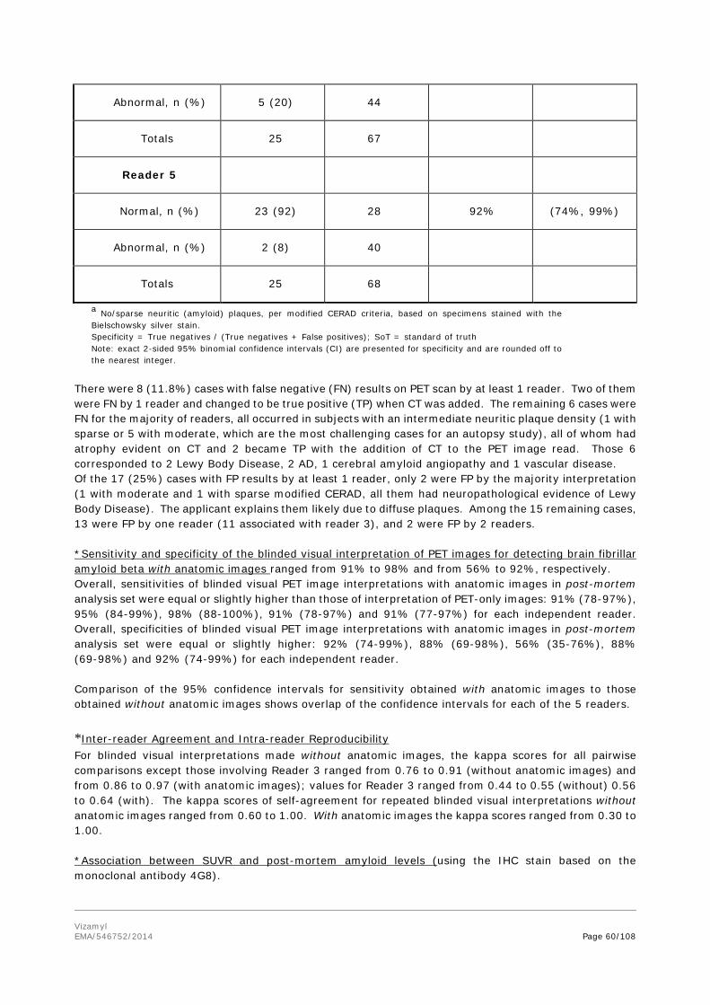
Vizamyl EMA/546752/2014 Page 60/108
Abnormal, n (%) 5 (20) 44
Totals 25 67
Reader 5
Normal, n (%) 23 (92) 28 92% (74%, 99%)
Abnormal, n (%) 2 (8) 40
Totals 25 68
a No/sparse neuritic (amyloid) plaques, per modified CERAD criteria, based on specimens stained with the Bielschowsky silver stain. Specificity = True negatives / (True negatives + False positives); SoT = standard of truth Note: exact 2-sided 95% binomial confidence intervals (CI) are presented for specificity and are rounded off to the nearest integer.
There were 8 (11.8%) cases with false negative (FN) results on PET scan by at least 1 reader. Two of them were FN by 1 reader and changed to be true positive (TP) when CT was added. The remaining 6 cases were FN for the majority of readers, all occurred in subjects with an intermediate neuritic plaque density (1 with sparse or 5 with moderate, which are the most challenging cases for an autopsy study), all of whom had atrophy evident on CT and 2 became TP with the addition of CT to the PET image read. Those 6 corresponded to 2 Lewy Body Disease, 2 AD, 1 cerebral amyloid angiopathy and 1 vascular disease. Of the 17 (25%) cases with FP results by at least 1 reader, only 2 were FP by the majority interpretation (1 with moderate and 1 with sparse modified CERAD, all them had neuropathological evidence of Lewy Body Disease). The applicant explains them likely due to diffuse plaques. Among the 15 remaining cases, 13 were FP by one reader (11 associated with reader 3), and 2 were FP by 2 readers. *Sensitivity and specificity of the blinded visual interpretation of PET images for detecting brain fibrillar amyloid beta with anatomic images ranged from 91% to 98% and from 56% to 92%, respectively. Overall, sensitivities of blinded visual PET image interpretations with anatomic images in post-mortem analysis set were equal or slightly higher than those of interpretation of PET-only images: 91% (78-97%), 95% (84-99%), 98% (88-100%), 91% (78-97%) and 91% (77-97%) for each independent reader. Overall, specificities of blinded visual PET image interpretations with anatomic images in post-mortem analysis set were equal or slightly higher: 92% (74-99%), 88% (69-98%), 56% (35-76%), 88% (69-98%) and 92% (74-99%) for each independent reader. Comparison of the 95% confidence intervals for sensitivity obtained with anatomic images to those obtained without anatomic images shows overlap of the confidence intervals for each of the 5 readers. *Inter-reader Agreement and Intra-reader Reproducibility For blinded visual interpretations made without anatomic images, the kappa scores for all pairwise comparisons except those involving Reader 3 ranged from 0.76 to 0.91 (without anatomic images) and from 0.86 to 0.97 (with anatomic images); values for Reader 3 ranged from 0.44 to 0.55 (without) 0.56 to 0.64 (with). The kappa scores of self-agreement for repeated blinded visual interpretations without anatomic images ranged from 0.60 to 1.00. With anatomic images the kappa scores ranged from 0.30 to 1.00. *Association between SUVR and post-mortem amyloid levels (using the IHC stain based on the monoclonal antibody 4G8).

Vizamyl EMA/546752/2014 Page 61/108
Regional SUVR determinations and IHC β-amyloid measures (4G8) were compared using mixed effect regression models (Table 13). Table 13 Fixed Effects from the Mixed Effects Models used for Comparison of Regional SUVRs with Regional IHC Amyloid Measures
Fixed Effect SUVR-CER SUVR-PONS Estimate (95% CI) p-value Estimate (95% CI) p-value
SUVR PET Measurement Slope 0.8482 (-0.295, 1.992) 0.1452 3.3985
(-0.117, 6.914) 0.0580 Region of the Brain <0.0001 <0.0001
Time from PET scan to death 0.0447 (-1.013, 1.102) 0.9318 0.0861
(-0.938, 1.110) 0.8646 CI = confidence interval; SUVR = standard uptake value ratio; SUVR-CER = SUVR determined using the cerebellar cortex as the reference region; SUVR-PONS = SUVR determined using the pons as the reference region. Fixed effects from mixed model of immunohistochemical estimates of amyloid level (percent amyloid in tissue specimen) = PET measurement of SUVR + region of the brain + time in months from PET scan to death [(date of death – date of PET scan)/30]. Number of subjects in the model = 30. Maximum observations per subject = 8.
With regional SUVR, model fit was good but the slope was not significantly different from 0 with either reference region (p = 0.1452 and p = 0.0580), indicating a lack of statistically significant association between regional SUVR and pathology results based on 4G8 staining. Brain region was a significant factor in both models. Time from PET scan to death was not significant in either model.
To clarify the contribution of the different regions to this highly significant correlation, a post-hoc analysis to assess the correlation between flutemetamol (18F) and amyloid pathology for each of the 8 regions included in the study independently was performed. As seen in Table 14 and Table 15, this new analysis demonstrated a statistically significant correlation in each of the 8 regions (p-value <0.001); it also showed that, for all 8 regions, the variation in SUVR explained 61% or more of the variation in amyloid
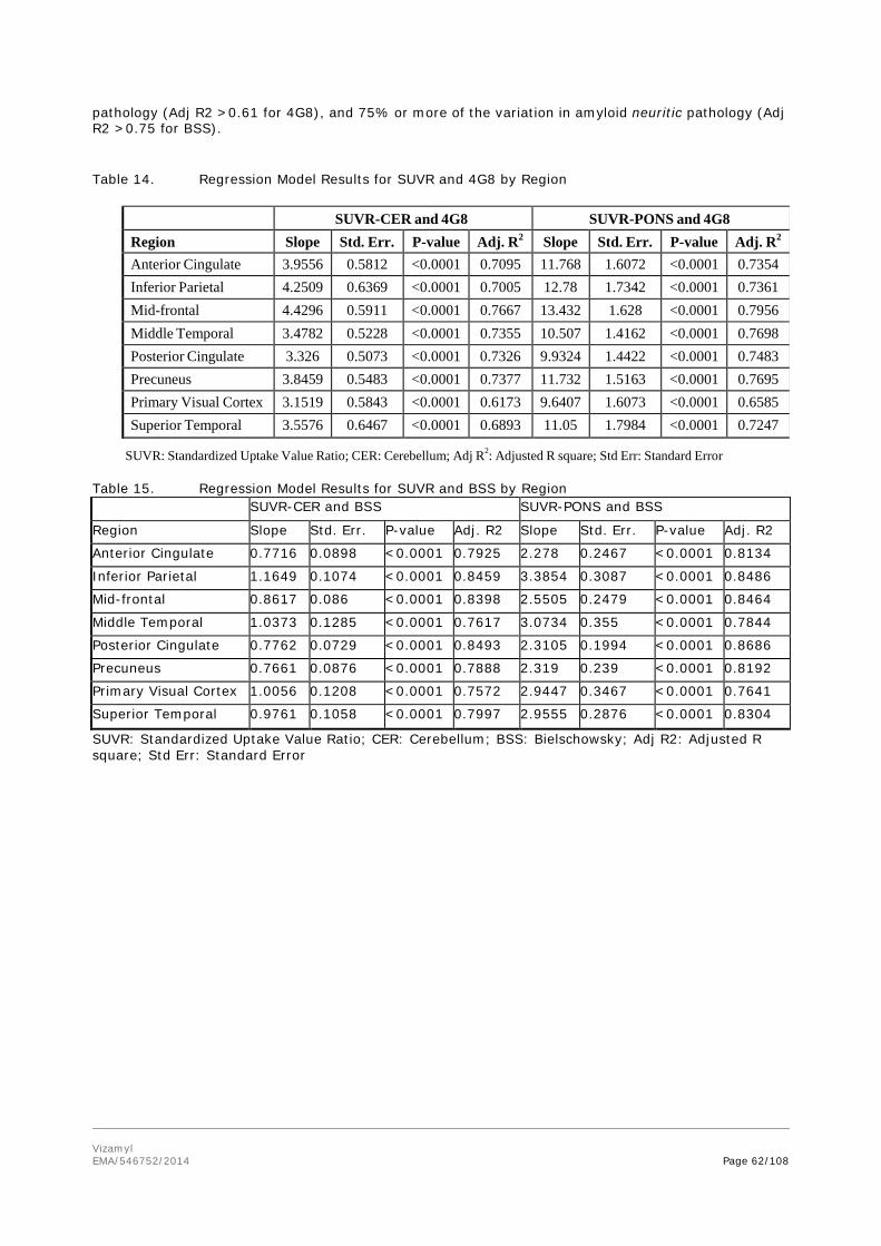
Vizamyl EMA/546752/2014 Page 62/108
pathology (Adj R2 >0.61 for 4G8), and 75% or more of the variation in amyloid neuritic pathology (Adj R2 >0.75 for BSS). Table 14. Regression Model Results for SUVR and 4G8 by Region
SUVR: Standardized Uptake Value Ratio; CER: Cerebellum; Adj R2: Adjusted R square; Std Err: Standard Error Table 15. Regression Model Results for SUVR and BSS by Region
SUVR-CER and BSS SUVR-PONS and BSS
Region Slope Std. Err. P-value Adj. R2 Slope Std. Err. P-value Adj. R2
Anterior Cingulate 0.7716 0.0898 <0.0001 0.7925 2.278 0.2467 <0.0001 0.8134
Inferior Parietal 1.1649 0.1074 <0.0001 0.8459 3.3854 0.3087 <0.0001 0.8486
Mid-frontal 0.8617 0.086 <0.0001 0.8398 2.5505 0.2479 <0.0001 0.8464
Middle Temporal 1.0373 0.1285 <0.0001 0.7617 3.0734 0.355 <0.0001 0.7844
Posterior Cingulate 0.7762 0.0729 <0.0001 0.8493 2.3105 0.1994 <0.0001 0.8686
Precuneus 0.7661 0.0876 <0.0001 0.7888 2.319 0.239 <0.0001 0.8192
Primary Visual Cortex 1.0056 0.1208 <0.0001 0.7572 2.9447 0.3467 <0.0001 0.7641
Superior Temporal 0.9761 0.1058 <0.0001 0.7997 2.9555 0.2876 <0.0001 0.8304
SUVR: Standardized Uptake Value Ratio; CER: Cerebellum; BSS: Bielschowsky; Adj R2: Adjusted R square; Std Err: Standard Error
SUVR-CER and 4G8 SUVR-PONS and 4G8 Region Slope Std. Err. P-value Adj. R2
Slope Std. Err. P-value Adj. R2
Anterior Cingulate 3.9556 0.5812 <0.0001 0.7095 11.768 1.6072 <0.0001 0.7354 Inferior Parietal 4.2509 0.6369 <0.0001 0.7005 12.78 1.7342 <0.0001 0.7361 Mid-frontal 4.4296 0.5911 <0.0001 0.7667 13.432 1.628 <0.0001 0.7956 Middle Temporal 3.4782 0.5228 <0.0001 0.7355 10.507 1.4162 <0.0001 0.7698 Posterior Cingulate 3.326 0.5073 <0.0001 0.7326 9.9324 1.4422 <0.0001 0.7483 Precuneus 3.8459 0.5483 <0.0001 0.7377 11.732 1.5163 <0.0001 0.7695 Primary Visual Cortex 3.1519 0.5843 <0.0001 0.6173 9.6407 1.6073 <0.0001 0.6585 Superior Temporal 3.5576 0.6467 <0.0001 0.6893 11.05 1.7984 <0.0001 0.7247

Vizamyl EMA/546752/2014 Page 63/108
Ancillary analyses The positive predictive value (PPV) and the negative predictive value (NPV) of the blinded visual interpretation of flutemetamol (18F) PET scan were calculated. PPV without anatomic images ranged from 74% to 95% by reader, with a median of 92% and a majority value of 95%. NPV without anatomic images ranged from 73% to 87% by reader, with a median of 82% and a majority value of 79%, and with all five blinded readers having lower limits of the 95% confidence intervals extending below 70% (range 49% to 66%). Results of sensitivity and specificity of blinded visual PET image interpretations without anatomic images in the pivotal study using a majority read of five readers for estimation of beta amyloid deposition were 86% (95% CI: 72% to 95%), and 92% (95% CI: 74% to 99%), respectively. With respect to neuropathological diagnosis (identifying/excluding AD) according to NIA-Reagan criteria, the blinded visual interpretation of flutemetamol (18F) PET images without anatomic images had the following performance metrics: · Sensitivity ranged from 79% to 92% by reader, with a median of 87% and a majority value of 84% · Specificity ranged from 39% to 79% by reader, with a median of 75% and a majority value of 79% · PPV ranged from 67% to 85%, with a median of 81% and a majority value of 84% · NPV ranged from 72% to 86%, with a median of 79% and a majority value of 79%
Summary of main study(ies)
The following table 16 summarises the efficacy results from the main studies supporting the present application. This summary should be read in conjunction with the discussion on clinical efficacy as well as the benefit risk assessment (see later sections).
Table 16. Summary of Efficacy for study GE067-007
TITLE: A Principal Open-Label Study to Compare the Brain Uptake of
Flutemetamol (18F) with Brain Fibrillar Amyloid β Levels Determined Postmortem
Study identifier GE067-007
Design It was designed to determine the sensitivity of blinded visual interpretations of flutemetamol (18F) PET images without anatomic brain images for detecting brain fibrillar β-amyloid
Duration of main phase: After PET imaging, follow-up until the death of the patient when post-mortem brain analyses for tissue amyloid were conducted
Duration of Run-in phase: screening within 35 days before the administration of study drug
Hypotheses The lower bound of the 2-sided 95% exact confidence interval for sensitivity of the blinded visual flutemetamol (18F) PET scan reading without anatomic image being greater than a priori threshold of 70% for at least 3 of the 5 reader results

Vizamyl EMA/546752/2014 Page 64/108
Treatments groups 180 end-of-life patients with an approximate maximal life expectancy of 1 year who consented to autopsy
Participants received 185-370 MBq of flutemetamol (18F), administered intravenously (as a bolus over approximately 40 seconds) at a maximum volume of 10 mL. The images were acquired starting 90 minutes after injection. The scan duration ranged from 10-30 minutes.
Endpoints and definitions
Primary Endpoint:
Sensitivity of the blinded visual flutemetamol (18F) PET Scan reading without anatomic image compared to the Neuropathologist’s assessment based on modified CERAD criteria
Database lock All those recruited patients until at least 31 brains were deemed fibrillar Aβ-positive according to the SoT.
Results and Analysis
Analysis description
Primary Analyses
Analysis population
From 176 subjects who underwent PET imaging, those 68 who died and underwent evaluable brain autopsy (with clinical diagnosis as probable 30 AD subjects, 17 had other dementia disorders, none had MCI and 21 had no history of cognitive impairment), were included in the postmortem analysis set.
Descriptive statistics
Statistical analysis:
Primary analysis was assessing confidence intervals of sensitivity for visual flutemetamol (18F) PET scan reading computed on an individual reader basis (five raters).
Participant flow:
A total of 203 subjects were screened for the study. The study included 180 subjects that were assigned to receive treatment. The primary objective of this study required postmortem brain specimens. A total of 69 subjects died, only 68 brains were available for efficacy analyses, and the brain from 1 of the remaining 68 subjects was not evaluable. Efficacy was assessed in the post-mortem autopsy set consisting of 42 amyloid-positive cases in 67 autopsied cases (observers 2 and 4) or 43 cases amyloid-positive cases in 68 autopsied cases (observers 1, 3, and 5). There were some protocol deviations.
Effect estimate per comparison
Primary Endpoint (Sensitivity analysis)
The sensitivity (point estimate and 95%CI) of the visual binary rating of the flutemetamol (18F) PET scan without anatomic image (positive, negative) for detection of the neuropathologist’s assessment based on modified CERAD criteria (none-sparse vs moderate-frequent neuritic plaque density at the brain) were 81% (67-92%) for reader 1, 88% (74-96%) for reader 2, 93% (81-99%) for reader 3, 93% (81-99%) for reader 4 and 88% (75-96%) for reader 5.
Vizamyl EMA/546752/2014 Page 65/108
Analysis description
For Secondary analyses see clinical assessment section
Analysis performed across trials (pooled analyses AND meta-analysis) The company presented pooled analyses of efficacy of the 11 studies composing the flutemetamol (18F) clinical development program. No meta-analysis was presented. The pooling strategy for analyses each used 1 of 5 levels of data integration based on the type of training of the blinded image readers (in-person or electronic medium), the SoT (or bases of comparison of the PET image), and populations for analysis. Integration levels 1 through 3 were based on data resulting from in-person training of blinded image readers; levels 4 and 5 were based on data resulting from blinded reader training using an electronic program (Study GE067-021). The primary analysis for efficacy was based on the Level 1 integration of data from subjects with known (or presumed) brain β-amyloid status (normal or abnormal) including those at the end of life and young (≤40 years of age) cognitively intact volunteers in whom brain amyloid is known to be extremely rare. The effect of age, gender, race and medications used for treatment of AD symptoms on the efficacy of Flutemetamol F 18 Injection was analysed. Results show that these covariables generally did not affect the efficacy of Flutemetamol F 18 Injection.
SUPPORTIVE STUDIES
Study GE067-015 This was a multi-center, open-label phase 3 study to determine the overall specificity of Flutemetamol F 18 Injection for excluding the presence of brain amyloid based on the visual assessment of a PET scan by 5 independent blinded readers (previously in-person trained in PET scan reading) reviewing images from a population of healthy young adult subjects aged 18 to 40 (presumed to be amyloid negative). The study was prematurely stopped after reassessment of sample size which changed from 300 HVs in the original protocol to be up to 300 after amendment 2. When the study was prematurely stopped, 181 subjects had been dosed with 185 MBq of Flutemetamol F 18 Injection, and completed the study. All scans from this study were blindly and randomly mixed with approximately equal numbers of flutemetamol (18F) scans from the GE-067-005 aMCI study (which was expected to contain some abnormal images) to avoid potential bias if readers saw only (or predominantly) normal scans. The primary objective (i.e. at least 3 of 5 blinded readers demonstrated specificity with a lower bound of the 95% confidence interval that exceeded 80%) was achieved. The results of blinded reads by 5 independent readers showed specificity of 100%, 68%, 99%, 99% and 99%. The lower bounds of the 95% confidence intervals for specificity for the 5 readers were 98%, 61%, 97%, 97% and 96%, exceeding 95% for 4 of the 5 readers The between-reader agreement was >99% for all comparisons except those including Reader 2 (with agreement rates of 68-69%). The within-reader reproducibility in a 10% random sample of images from the full dataset was 100% for Readers 1, 3, 4 and 5 and 75% for Reader 2.
Vizamyl EMA/546752/2014 Page 66/108
Study GE067-008, GE067-009, GE067-010 and GE067-011 These were four Phase 3, open-label, non-randomized single center (-008 and -010) or multi-centre (-009 and -011) biopsy studies. They primarily aimed to assess the level of association between brain uptake of flutemetamol (18F) (as estimated by SUVR) and the level of amyloid (as estimated by IHC with 4G8 or NAB228) in the biopsy samples taken from the cortex of patients with normal pressure hydrocephalus (NPH) while undergoing shunt placement (study GE067-008, -009, -011) or during intracranial pressure measurement (study GE067-010 and -011). In the “retrospective” studies (GE067-008 with n=7 and GE067-010 with n=15), subjects had previously had front lobe cortical biopsy for detection and quantification of amyloid so PET imaging was performed after biopsy. In the “prospective” studies (GE067-009 with n=12 and GE067-011 with n=18), subjects were scheduled for shunt placement surgery and PET imaging was performed prospectively before shunt placement and brain biopsy. Comparisons between SUVR and biopsy estimates were performed for either contralateral to the biopsy site (retrospective studies, in which biopsy preceded imaging) or from the biopsy site (prospective studies, in which biopsy followed imaging). Subjects underwent PET imaging of the brain for 30 minutes starting approximately 90 minutes after receiving 185 MBq of flutemetamol (18F) intravenously. In study GE067-008, a regression model confirmed a high level of direct association between SUVR-CER (i.e., based on the cerebellum as the reference region and the NAB228 IHC estimate of amyloid β level. The full model was significant (R2 =0.834, p = 0.027), as was the model factor for SUVR (slope = 0.631, p = 0.011). The Pearson correlation coefficient was 0.799 (p = 0.0174), 0.858 (p <0.0001) and 0.669 (p = 0.0064) for studies GE067-009, GE067-010 and GE067-011, respectively. Study ALZ201 This was a multi-center, open-label, non-randomised, phase 2 study primarily aimed to examine the efficacy of visual assessment of raised flutemetamol (18F) brain uptake for separating subjects with pAD from healthy volunteers (HVs) and to determine the proportions of aMCI subjects with normal and abnormal flutemetamol (18F) images. Quantitative analysis of PET images were also performed and compared for the three recruited subgroups. Image quality and reproducibility were also assessed. Test-retest variability was assessed in a subgroup of patients with pAD. The study also assessed the correlation between flutemetamol (18F) and [11C]PiB in some of the recruited patients with clinical AD and all the amCI. All subjects received a single i.v. dose of 185 MBq of Flutemetamol F 18 Injection, except 5 subjects with pAD who received 2 administrations of approximately 120 MBq each. There were 70 evaluable subjects: 25 with pAD (based on NINCDS-ADRDA), 20 with aMCI (based on Petersen criteria), and 25 HVs [10 young HVs and 15 older HVs]. During blinded visual assessment of flutemetamol (18F) images by 5 independent readers previously in-person trained, overall all readers reported a greater percentage of pAD subjects (92%) to have raised amyloid beta levels compared to aMCI patients (45%) and HVs (4%) based on the the subject’s clinical diagnosis at study entry.

Vizamyl EMA/546752/2014 Page 67/108
Measures of efficacy were visual assessment as normal or abnormal for each subject and were used to estimate sensitivity for pAD subjects, specificity for HV subjects, and assign aMCI subjects to pAD or HV.There was significant discrimination in quantitative PET values between subjects with pAD and HVs in all regions of the cortex and striatal regions. Overall, there was less discrimination between subjects with pAD and aMCI; however, the aMCI quantitative PET values had a bimodal distribution being raised or normal. There was a higher mean quantitative PET value in subjects with pAD in all cortical regions investigated compared with aMCI. Similar findings were noted in quantitative PET value discrimination in the cortex and striatal regions following [11C]PiB administration comparing pAD subjects to aMCI subjects. There was significant correlation (r2=0.82) between quantitative PET uptake values generated from flutemetamol (18F) and [11C]PiB PET scans in 20 subjects with pAD and 20 with aMCI who also received an i.v. dose of [11C]PiB not exceeding 370 MBq on the same day or within approximately 30 days after receiving Flutemetamol F 18 Injection. PET readers rated the flutemetamol (18F) PET image quality of subjects with pAD as excellent in 21 (84%) and good in 4 (16%) subjects, respectively. In all aMCI subjects and HVs, the images were rated as excellent. Following administration of a second dose of 120 MBq of Flutemetamol F 18 Injection in 5 with pAD (who had also received 120 MBq as the first dose), the images were rated as good in all subjects (100%). Cortical flutemetamol (18F) test-retest quantitative PET estimates had a low inter-subject variability of 1-4% in pAD subjects. The variability was lowest in the occipital cortex, striatum, POC, and the frontal cortex and was highest in the medial temporal cortex, subcortical white matter, and pons. The mean test-retest variability of all subjects and regions for quantitative flutemetamol (18F) PET estimates was excellent. Between-Reader Agreement showed a Fleiss kappa score of 0.96 by the 5 readers in 70 of 74 flutemetamol (18F) images read interpreted visually. In the other 4 images, there was agreement by 4 out of 5 readers. Reassessment of the clinical diagnosis in 67 returning subjects at a 2-year follow-up (all baseline recruited patients except for 3 in the pAD group and 1 each in the aMCI and HV groups) was performed to evaluate the relationship between baseline flutemetamol (18F) PET imaging and changes in diagnostic status within 24 months. There were no changes from the initial diagnosis for any of the returning pAD subjects (n = 22) or HVs (n = 24). Of the returning subjects who had initially been diagnosed with aMCI (n = 19), 9 (47%) were diagnosed to have pAD at the 2-year follow-up; 7 of these had raised flutemetamol (18F) uptake at baseline. The other 10 (53%), eight of whom had negative flutemetamol PET scans, retained at the 2-year follow-up a baseline diagnosis of aMCI. aMCI unchanged aMCI to AD Number
of subjects Month 24 Subjects with baseline PET positive,
No (%) 2 (22.2%) 7 (77.8%) 9
Subjects with baseline PET negative, No (%)
8 (80%) 2 (20%) 10
As calculated by the CHMP assessors, sensitivity of the visual PET scan reading (including 95% CI) to show the aMCI conversion rate from baseline to clinically probable AD at 2-year follow-up in 9 converters was 77.8% (95%CI: 40.0%-97.2%), specificity in 10 non-converters was 80.0% (95%CI: 44.4%-97.5%), positive likelihood ratio was 3.89 (95% CI: 1.07-14.10) and negative likelihood ratio was 0.28 (95% CI: 0.08-0.98). The design of this study did not allow for estimating the risk of MCI progression to clinical AD.
Vizamyl EMA/546752/2014 Page 68/108
Study GE067-005 This was a multi-center, open-label, non-randomised, phase 3 study. This study was primarily aimed to compare the time to conversion to probable AD in aMCI subjects with normal and abnormal patterns of flutemetamol (18F) uptake based on the visual assessment of a PET scan by five independent blinded readers previously in-person trained. The original protocol was amended five times, and the data below corresponds to final protocol after amendment 5. Patients received a single dose of 185 MBq of flutemetamol (18F). The pre-planned primary analysis was a time-to-event analysis (Cox proportional hazards).
At the time of the initial submission, it was an ongoing study and only inter-reader and intra-reader reproducibility of visual assessment of the individual flutemetamol PET images were reported with regard to efficacy (safety was also reported). Recruitment had already been completed at that time, with a total of 232 subjects with amnestic MCI (Petersen criteria) included, and the study was pending of the follow-up period to be concluded. All subjects subsequently attended follow-up visits to determine whether or not conversion to probable AD had occurred. Each subject was to be assessed by the site every 6 months until they convert to AD, or, if they did not convert, until either 3 years had elapsed from imaging or the required number of conversions was reached (whichever occurred later). The follow-up time period for non-converters was amended two times.
The CHMP required that the applicant provide the results of study GE067-005 concerning the conversion rate from aMCI to pAD. However, to avoid breaking the blind before completion of the 36-month follow-up, the company provided an ad hoc analysis with the percentage of aMCI patients with positive flutemetamol (18F) PET scans and rates of conversion from aMCI to clinical probable AD (pAD) as a function of flutemetamol (18F) scan status (positive/negative) for a random sample of 70 (30%) of the 232 subjects in the GE067-005 study. The mean duration of follow-up was 27.1 months (range, 4 to 40; median, 32.4; SD 10.6), derived as time from last dose to date of completion/withdrawal for subjects who completed or withdrew and time from last dose to data cut-off date (15 July 2013) for subjects who were continuing. After completion of the 36-month follow-up period, the applicant provided the full study results. In this study, from the 232 patients clinically diagnosed with mild cognitive impairment (MCI) and were followed for 36 months to evaluate the relationship between flutemetamol (18F) imaging and changes in diagnostic status 98 (42 %) had abnormal (positive) flutemetamol (18F) scans. Of the 232 patients enrolled, 224 had at least one post-scan review by the independent committee and were included in the analysis. At the 36-month follow-up, 81 (36 %) converted to clinical AD. Of the 97 MCI subjects who had a positive PET scan and at least one committee assessment, 52 (53.6 %) were classified clinically as converted to clinical AD after 36 months compared to 29 (22.8 %) of 127 who had a negative scan and at least one committee assessment. At 36 months sensitivity (secondary analysis) of flutemetamol (18F) scan to show the MCI conversion rate to AD in 81 converters was 64.2 % (95% CI: 53 % to 75 %), specificity (secondary analysis) in 143 non-converters was 68.5% (95% CI: 60% to 76%). Based on the majority read, the positive and negative likelihood ratios were 2.04 and 0.52, respectively. The design of this study did not allow for estimating the risk of MCI progression to clinical AD. Kappa scores of inter-reader agreement between pairs of readers ranged from 77% (κ = 0.56) to 98% (κ = 0.96). Agreement between Readers 1, 3, 4 and 5 taken in pairs ranged from 90 to 98% but between Reader 2 and each of the other 4 readers ranged from 77 to 85%. The intra-reader reproducibility of the rereads was good, ranging from 86% to 100%. Study GE067-021 This study evaluated the performance of an electronic program for training five PET scan readers via blinded visual interpretations of 276 flutemetamol (18F) PET image sets selected from previous

Vizamyl EMA/546752/2014 Page 69/108
flutemetamol clinical trials. Performance was assessed in terms of the sensitivity and specificity of each reader. Seven analysis populations were defined to address the study objectives:
• Analysis Population 1 (n = 135) consisted of subjects with any SoT type. • Analysis Population 2 (n = 104) consisted of all subjects from analysis population 1 except the 31
subjects from study GE067-015. • Analysis Population 3 (n = 68) consisted of those 68 subjects from analysis population 1 of the
autopsy study • Analysis Population 4 (n = 276) consisted of subjects with a variety of diagnoses. • Analysis Population 5 (n = 29) consisted of subjects whose images were drawn randomly without
replacement from Analysis Population 4. • Analysis Population 6 (n = 80) consisted of subjects with aMCI • Analysis Population 7 (n = 8) consisted of subjects with aMCI from Analysis Population 5.
The primary analyses consisted of determining sensitivity and specificity of the blinded visual interpretation of flutemetamol (18F) PET images without anatomic images, which were determined from subjects with any SoT type (Analysis Population 1). Secondarily, the diagnostic performance of the blinded visual interpretation of flutemetamol (18F) PET images of the brain without anatomic images was determined in subjects with autopsy (Analysis Population 3) (table 17).
Table 17. Summary of Sensitivity and Specificity without Anatomic Images – Analysis Population 3a
Standard of Truthb Sensitivity and Specificity
Empirical Predictive Value
Reader Blinded Visual Interpretation
Normal Abnormal Totals Sensitivity % (95% CI)
Specificity % (95% CI)
PPV % (95% CI)
NPV % (95% CI)
1 Normal 18 3 21 93% (81%, 99%) 72% (51%, 88%) 85% (72%, 94%) 86% (64%, Abnormal 7 40 47
Totals 25 43 68 2 Normal 21 3 24 93% (81%, 99%) 84% (64%, 96%) 91% (78%, 98%) 88% (68%,
Abnormal 4 40 44 Totals 25 43 68
3 Normal 22 4 26 91% (78%, 97%) 88% (69%, 98%) 93% (81%, 99%) 85% (65%, Abnormal 3 39 42
Totals 25 43 68 4 Normal 15 3 18 93% (81%, 99%) 60% (39%, 79%) 80% (66%, 90%) 83% (59%,
Abnormal 10 40 50 Totals 25 43 68
5 Normal 23 6 29 86% (72%, 95%) 92% (74%, 99%) 95% (83%, 99%) 79% (60%, Abnormal 2 37 39
Totals 25 43 68 Majority Read
Normal 21 3 24 93% (81%, 99%) 84% (64%, 96%) 91% (78%, 98%) 88% (68%, Abnormal 4 40 44
Totals 25 43 68 Sensitivity = True positives / (True positives + False negatives); Specificity = True negatives / (True negatives + False positives); Empirical positive predictive value (PPV) = True positives / (True positives + False positives); Empirical negative predictive value (NPV) = True negatives / (True negatives + False negatives) a Includes subjects with autopsy-based histopathological SoT results (i.e., brain amyloid status confirmed histopathologically from specimens collected at autopsy (study GE067-007)).

Vizamyl EMA/546752/2014 Page 70/108
b SoT is based on interpretation of brain tissue with regards to amyloid levels using Bielschowsky silver stain (brain autopsy for GE067-007). The results from these analyses in Analysis Population 3 were compared to the results from study GE067-007 (which used in-person training of readers). There were no meaningful differences between the two methods of reader training (electronic vs. in-person) in sensitivity and specificity observed for the five readers. In fact, those readers showing higher sensitivities were at the same time those showing lower specificities. The Spearman correlation coefficient of the five pairs of accuracy estimates (analysis conducted by the CHMP) was strong (Spearman’s rho= -0.68). In Analysis Population 3:
• There were 3 FN cases for the majority of readers, all occurred in subjects with sparse or moderate neuritic plaque density (2 had a neuropathology diagnosis of Lewy Body Disease (LBD), and 1 of Cerebral Amyloid Angiopathy (CAA)).
• 4 were FP by the majority interpretation (all had a neuropathological diagnosis of Lewy Body Disease (Dementia with Lewy Body or Parkinson’s disease). The applicant explains them likely due to diffuse plaques.
There were no significant differences between the results obtained without anatomic images and those obtained with anatomic images for any reader. Inter-reader agreement (Analysis Population 4, table 18) across all 5 readers was 81%; this corresponded to a kappa score of 0.83. The lower bound of the 2-sided 95% CI for kappa exceeded the pre-specified criterion (0.6) for study success. All other kappa scores (for pairwise comparisons of reader interpretations) also met the success criterion. The percentage of self-agreement (in analysis population 5) for repeated blinded visual interpretations without anatomic images was 100% for Reader 1, 97% for Readers 2, and 4, and was 93% for Readers 3 and 5. Table 18. Image Selection for Analyses of Reproducibility (Inter-Reader Agreement and Intra-Reader Reproducibility) – Analysis Population 4 Study
Phase
Number of Subjects
Population
How Selected
GE067-007 3 68 EOL All 68 evaluable subjects with autopsy data in the study GE067-009 GE067-010 GE067-011
3
36
NPH
All 36 evaluable subjects in the 3 studies
GE067-015 3 31 YHV Randomly selected from all 181 evaluable subjects in the study
GE067-005 3 60 aMCI Randomly selected from all 232 evaluable subjects in the study
ALZ201
2
55
20 aMCI, 15 EHV, 20 pAD
All 20 aMCI and all 15 EHV subjects in the study were selected. Of 27 pAD subjects in the study, the 20 who received 185-MBq doses were selected
ALZ103
1
10
5 EHV, 5 pAD Of 14 HV in the study, 5 EHV who received 185-MBq doses were selected. Of 8 pAD in the study, 5 who received 185 MBq doses were selected
GE067-014
1
16
8 EHV, 8 pAD Of 14 HV in the study, 8 EHV who had each received a 185-MBq dose were selected. All 8 pAD in the study were selected.
Totals
276
68 EOL, 36 NPH, 31 YHV, 80 aMCI, 28 EHV, 33 pAD
aMCI = amnestic mild cognitive impairment; EHV = elderly healthy volunteer; EOL = end-of-life; NPH = normal pressure hydrocephalus; pAD = probable Alzheimer’s disease; YHV = young healthy volunteers
Regarding inter-reader agreement for PET with anatomic imaging, comparison of the 95% confidence interval limits for each reader pair with and without anatomic images reveals overlapping confidence intervals in every case, indicating that there are no statistically significant differences in inter-reader

Vizamyl EMA/546752/2014 Page 71/108
agreements for PET images read with and without anatomic images. The same happens for the intra-reader agreement. Reader confidence without anatomic images for Analysis Population 4 ranged from 61-97%, depending on the reader. Those with low reader confidence ranged from <1 to 20%. If PET images were assessed combined with anatomic images, the corresponding levels of reader confidence were in similar ranges.
2.5.3. Discussion on clinical efficacy
The company contended that the clinical efficacy of PET with flutemetamol (18F) had been demonstrated by the findings in three key studies (phase III studies GE067-007, -015 and -021), supported by four phase III biopsy studies (GE067-008, -009, -010 and -011), one phase III ongoing study (GE067-005), one phase II study (ALZ201) and a pooled analysis of data. The phase III study in healthy volunteers (GE067-015) and the study GE067-021 which did not recruit patients were not considered as pivotal by the CHMP. Therefore, the CHMP considered that there was only one pivotal study (i.e. GE067-007) to base this application on, and for that reason the points to consider for submitting a single pivotal study in support of marketing authorization (CPMP/EMA/2330/99), as well as the “Guideline on Clinical Evaluation of Diagnostic Agents” (CPMP/EWP/1119/98/Rev 1) and Appendix 1 on Imaging Agents (EMEA/CHMP/EWP/321180/2008) were considered to apply in this case. The proposed indication was as follows: “This medicinal product is for diagnostic use only. VIZAMYL is a radioactive diagnostic agent indicated with positron emission tomography (PET) imaging for the visual detection of amyloid-beta neuritic plaques in the brains of adults who are being evaluated for Alzheimer’s disease (AD). A normal VIZAMYL scan indicates sparse to no neuritic plaques and is inconsistent with a neuropathological diagnosis of AD at the point of image acquisition: a normal scan reduces the likelihood that a patient’s condition is due to AD. An abnormal VIZAMYL scan is indicative of moderate to frequent amyloid-beta neuritic plaques. Neuropathological examinations have shown this amount of neuritic plaques is present in patients with AD, but also other types of neurologic conditions as well as in older people with normal cognition. VIZAMYL is to be used as an adjunct to other diagnostic evaluations. An abnormal VIZAMYL scan does not establish a diagnosis of AD. The safety and efficacy of VIZAMYL have not been established for predicting the development of dementia or other neurological conditions, or for monitoring response to therapies.” The recommended dose was of 185 MBq of flutemetamol (18F), administered intravenously (as a bolus over approximately 40 seconds) at a maximum volume of 10 mL. The images should be acquired starting 90 minutes after injection. The scan duration should typically be 20 minutes. Flutemetamol (18F) is intended for registration for a pathological indication (i.e. with PET imaging for the visual detection of amyloid-beta neuritic plaques in the brain). The company has also focused on the use of flutemetamol (18F) for a concrete diagnostic paradigm (i.e. as an adjunct to other diagnostic evaluations in adults who are being evaluated for AD for refutation of a neuropathological diagnosis of AD, reducing the likelihood that the patient’s condition is due to AD). Approval has not been requested for the use of this radiopharmaceutical for establishing a diagnosis of AD, predicting the development of dementia or other neurological conditions, or for monitoring response to therapies. For the validation of flutemetamol (18F) as an imaging PET agent, two stages have to be considered: a) An initial phase in which it is established how well (the relevant types of) brain β–amyloid deposition
can be visualized and quantified by flutemetamol (18F) in the relevant areas b) Further phase(s) where the efforts are aimed at demonstrating which particular practical purpose(s)
the imaging is useful for and how this is achieved, i.e. for a particular use in clinical practice.

Vizamyl EMA/546752/2014 Page 72/108
Design and conduct of clinical studies PIVOTAL STUDY GE067-007 Both the abovementioned phases were attempted in the pivotal study GE067-007, in which the visual detection of amyloid-beta neuritic plaques in the brain on PET images was assessed versus histopathology of autopsied samples as a Standard of Truth (SOT). The approach of using autopsy data as the SOT is justified as it can demonstrate that the results obtained with the investigational diagnostic agent are valid for estimation of plaque detection (even if not sufficient for the diagnosis of a particular disease). The clinical use of PET with flutemetamol (18F) will rely on its accurate quantitative and topographic assessment of beta amyloid accumulation in the brain. Relevant quantitative correlation in quantity and topography of PET images with autopsy specimens should be demonstrated. Study GE067-007 attempted to demonstrate this as a secondary objective. The clinical value of the estimation (quantity and topography) of beta amyloid deposition in brain is still a matter of discussion in the scientific community. If there is to be such a value (i.e. for designation of a patient as beta amyloid positive/negative or for a definitive diagnosis/exclusion of a specific beta amyloid pathology) any in vivo reliable method of estimation of beta amyloid deposition has to demonstrate its sensitivity/specificity for the particular clinical setting and population in which it is intended to be used. In this sense, the pivotal study GE067-007 primarily aimed at demonstrating sensitivity of the amyloid burden in the whole brain on flutemetamol (18F) PET images without anatomic brain images (evaluated by five blinded readers on a binary visual scale as normal/abnormal) versus the quantitative measurement by BSS at pathology. As a secondary objective, specificity was also assessed. It refers to sensitivity and specificity of PET to classify a patient as “beta-amyloid present” or “beta-amyloid absent”, based on the patient’s neuropathological diagnosis. A secondary analysis of the pivotal study was performed to demonstrate sensitivity and specificity of PET for the diagnosis of AD based on the patient’s neuropathological diagnosis using NIA-Reagan diagnostic criteria for AD. The likelihood of AD using these criteria is based solely on histopathological assessment (both neuritic plaque density and neurofibrillary tangle score). However, pre-specified levels of age-related brain neuritic β-amyloid plaque at autopsy should be integrated with the presence of a clinical history of dementia to arrive at a diagnostic level of certainty with regard to AD (Mirra et al. 1991). Therefore, sensitivity and specificity for the clinical diagnosis of AD cannot be concluded from the pivotal study results. In an attempt to assess clinical utility of the flutemetamol (18F) PET imaging, the predictive values of the test for detection of beta amyloid accumulation were included as a post-hoc analysis in the pivotal study. However, this approach does not comply with the relevant guideline to evaluate the clinical usefulness of a medicinal diagnostic product. According to the requirements for this it would be needed to address the impact of flutemetamol (18F) PET imaging on diagnostic thinking or on patient management in the intended population and clinical context. Such impact was not assessed. Participants underwent a MRI/CT prior or simultaneous to PET to allow appropriate anatomic location of the PET signal, which is of paramount importance in patients with cerebral atrophy which makes PET image interpretation difficult. Additional secondary endpoint(s) in the pivotal study included review of anatomic brain images along with the PET images to compare their validity versus PET-only images. Inter-reader variability was also calculated. Intra-reader variability was designed to be assessed in only 10% of patients which, in this study, is a very small sample size for any conclusions to be considered as solid.
Vizamyl EMA/546752/2014 Page 73/108
Recruited participants in the autopsy study had as a prerequisite to be suffering from a terminal illness and to have a short anticipated life expectancy of approximately 1 year or less (resulting in patients with very different pathologies -cancer, heart failure, as well as pre-defined dementia and other non-dementia medical conditions). These patients were not considered to be the best reflection neither of the actual range/distribution of brain β–amyloid deposits nor of the cognitive status expected in the intended population for routine clinical use of flutemetamol (18F) (i.e. “adults who are being evaluated for AD”). Indeed, their clinical diagnosis of dementia subtype (if present) was likely to be inaccurate as it was determined only on the basis of previous clinical history and a very brief neurological examination, without performing other mandatory laboratory tests needed to exclude the presence of either significant white matter disease or other non-neurodegenerative dementias. Moreover, such dementia status referred to the time of PET imaging and it might have changed within the time interval between PET and autopsy. Although the choice of this “end-of-life” population for the primary analysis has an impact on the external validity of this pivotal study, and the diagnostic utility in the intended clinical population has to be established, such population indeed allows for the assessment of the correlation of PET versus histopathology within a reasonable timeframe. A visual PET reading method was used in the pivotal study since amendment 1. However, the method was not previously optimized for in-vivo detection of brain beta amyloid deposition.
PET images were visually interpreted by independent readers, who were in-person trained in the interpretation of such images and blinded to the SOT results and any other information about the subject. The reader had to complete both PET-only and PET plus anatomic reference review with criteria of “normal, “abnormal” or “non-evaluable”. Quantificaation of PET images was also performed. Such quantitative assessment of flutemetamol (18F) PET images was conducted at 15 recruiting sites in the United States and 4 in the UK. It is known from the literature (Boellaard 2011) that the quantification e.g. of whole-body fludeoxyglucose (18F) PET studies is affected by many physiological and physical factors. It was outlined that the variability seen in quantification of fludeoxyglucose (18F) uptake using SUV will not be present in the quantification of flutemetamol (18F) uptake, mainly as a result of the use of a reference region for the estimation of the SUVR, but also as a result of the differences in tracer characteristics. Substantial variability in image quality or accuracy due to the multiple-site study design was also avoided through carefully conducted site initiation and qualifications of imaging equipment. Autopsied samples were assessed by BSS and IHC with 48G. Both techniques can be considered well-accepted for evaluating and measuring beta amyloid deposition in the brain post-mortem. It is known that IHC with 48G measures both neuritic and diffuse plaques combined. Estimation by BSS was used for the SOT since the first protocol amendment; however it was not previously optimized as to detect the relevant types/quantity/neuroanatomical location of beta amyloid accumulation. The interpretation criterion for the post-mortem estimates used as SoT in study GE067-007 was not validated. The company modified the standardized widely used post-mortem CERAD criteria (Mirra et al. 1991) to convert the semiquantitative assessment of neuritic plaques (none, sparse, moderate and frequent) of 8 particular cortical areas directly into a neuropathologic diagnosis. For diagnosis of AD, this approach is not valid, since: 1) neither the patient’s age nor the clinical information regarding the presence or absence of dementia -as estipulated in the original CERAD criteria- were taken into consideration, and 2) more than 3 particular neocortical regions were included for assessment. This way, the final neuropathologic diagnosis refers simply to the presence of higher than 1.5 of mean score of neuritic plaque density in at least one out of 8 particular brain regions and not to the definitive diagnosis or exclusion of AD. The choice of those eight particular brain regions to be analysed has not been justified as relevant for the clinical context in which this radiopharmaceutical is intended. The interpretation criterion for the post-mortem estimates of amyloid beta deposition is not validated criterion of
1 Methods Mol Biol. 2011;727:335‐49

Vizamyl EMA/546752/2014 Page 74/108
abnormality. On the other hand, a mean regional score not exceeding 1.5 in all those 8 particular regions is not a validated criterion for normality. The final diagnosis of neuritic plaque density (as none-sparse or moderate-frequent) should ideally match the CERAD criteria (Mirra et al. 1991) [i.e. be established based on the maximal neuritic plaque density measured on sections of middle frontal gyrus, superior and middle temporal gyri, and inferior parietal lobe].
The original protocol was amended twice during pivotal trial, changing several critical aspects of the trial such as the main objectives, primary efficacy endpoint, primary efficacy analysis, sample size estimation and interim analysis. Several of the changes concern directly the efficacy analysis and they might have impacted on the validity of the results. The original primary endpoint was the region-specific estimate of cortical tracer uptake determined from quantitative analysis of the flutemetamol (18F) PET images. The calculated sample size was 20 sample brains and a well-designed interim analysis was planned to be conducted when 12 sample brains were collected. Statistical considerations addressed adequately the impact of this interim analysis on Type I error rate using O’Brian-Fleming spending function. The first amendment switched primary efficacy analyses to computing exact 95% binomial confidence interval for the sensitivity of 3 independent blinded visual assessments of flutemetamol (18F) PET images with anatomic images for detecting brain fibrillar Aβ. The protocol considered a simple majority to be the criteria to classify each subject’s PET image as normal or abnormal. Sample size was calculated to have 90% power to demonstrate that the lower limit of the 95% exact confidence interval of sensitivity of the majority read was greater than 70%. The sample size calculation gave a minimun of 22 evaluable brains that are fibrillar Aβ-positive according to the SOT (post-mortem examination of tissue specimens from cortical brain regions). This first amendment also eliminated the interim analysis although an Independent Data Monitoring Committee (IDMC) monitored accrual and prompted study termination when the sample size of 22 abnormal brains was reached. The second amendment further changed the primary efficacy analysis. After the amendment, the primary efficacy endpoint was the blinded visual assessment of brain PET images as normal or abnormal without anatomic brain images for reference performed by 5 independent blinded readers, individually trained, in the evaluation of PET fibrillar Aβ imaging. The primary analysis was changed from assessing 95% exact confidence interval of sensitivity computed for a majority read (two of three raters) to assessing exact confidence intervals of sensitivity computed on an individual reader basis (five raters). In this way, the primary objective was changed to demonstrate that at least three of the 5 readers show lower limits of their sensitivity 95% exact CI greater than 70%. The Primary Efficacy analysis was planned to be performed when at least 31 brains were deemed fibrillar Aβ-positive according to the SOT. This new sample size estimation was based on a different assumption than the one used in the first amendment sample size calculation. In fact, the true sensitivity was estimated to be lower (92%) than in the first amendment (95%). This prompted the sample size to rise from 22 to 31 subjects.
The second amendment also stipulated that an IDMC would review the pathological data resulting from brain tissue analyses on an ongoing basis during the study. When the IDMC determined that the study had accrued at least 31 brains that are pathologically abnormal for fibrillar Ab ("amyloid-positive”), or 22 amyloid-negative brains, and they had a corresponding assessable PET image, the IDMC was to inform the applicant of this finding. When the applicant had confirmed such results, it would declare that the study stopping rule had been met. The charter document of the IDMC stated that after 31 amyloid-positive brains and every three new amyloid-positive brain were accrued, the IDMC would communicate to the sponsor that a new milestone had been reached.
The brains of all subjects who died after the declaration that the stopping rule was met were planned to be preserved and stored for possible future full analysis if requested by a regulatory authority, or if the company determined that such analysis would be useful. Collection of brains after the stopping rule was

Vizamyl EMA/546752/2014 Page 75/108
set up to continue until either 1) the brains of all scanned subjects had been collected, or 2) flutemetamol was approved, whichever occurred first. The primary efficacy analysis set up at amendment 2 has the following drawbacks: 1. It relegates specificity as secondary outcome. The guideline on the evaluation of diagnostic products
(CPMP/EWP/1119/98/Rev.1) requires both sensitivity and specificity as co-primary endpoints. Both sensitivity and specificity are negatively correlated and a slight change on a rater’s threshold for classifying an image as positive impacts on both sensitivity and specificity.
2. The success criterion (the lower bound of the 2-sided 95% exact confidence interval for sensitivity being greater than a priori threshold of 70% for at least 3 of the 5 reader results) seemed a little demanding and was not justified in terms of clinical relevance.
Despite the fact that the trial was designed to stop after the company had confirmed that there was recruitment of 31 amyloid-positive brains, the study further continued including 12 extra amyloid-positive subjects, to allow at least 22 amyloid-negative brains to accrue. In a total of 68 autopsied patients with evaluable images, there were only 2 cases (1 for reader 2 and 1 for reader 4) considered unevaluable by those readers. Every reader’s assessments were independent of the milestones reported by the IDMC or the Sponsor’s decision to stop the study.
During the conduct of the study, the following other issues were changed raising some concerns: • Before amendment 1, it was possible to administer a dose as low as 111 MBq with similar acquisition
time (30 min) although with a less than optimal image quality. No subjects were dosed with less than 185 MBq. Since some subjects could not tolerate lying still for 30 minutes because of the severity of their illness, they were allowed to receive a higher activity (up to 370 MBq) to shorten the imaging time. Inclusion of a 370 MBq dose in posology would affect radiation dosimetry but not efficacy. For subjects who could not tolerate lying still for 20 minutes application of a 370 MBq activity for 10 minutes could be a more feasible alternative if the higher radiation exposure is justifiable.The anatomic image for appropriate PET signal location for some secondary endpoints was originally either MRI or CT; however, at amendment 2 was exclusively CT. The company stated that, in the pivotal autopsy study in 68 patients, there were 54 patients who had a CT scan, 6 with MRI and 8 with both CT and MRI. There were very few patients in the MRI subgroup. It is unknown how the efficacy results of PET with flutemetamol (18F) in the autopsy study might have been affected by the change of the type of anatomic image to localize PET signal (the option of MRI/CT was changed exclusively for CT at amendment 2).
• Some protocol deviations were noted that related to the volume of radiopharmaceutical administered in 8 patients in a single center (the site was diluting dose to a more convenient volume); this raised concerns whether the clinical sites conducted the trial following the standards. Six patients in the pivotal study were identified as having protocol deviations which potentially might have interfered with the efficacy evaluation. They were classified as relating to radiopharmaceutical’s activity, to the time when PET image was acquired and to the time when CT was performed in relation to PET. It was concluded that they were unlikely to have affected efficacy results since only 2 incorrect interpretations were obtained from those patient’s images, which did not affect the majority result.
RE-READING STUDY GE067-026 As collection of brains had been set up to continue, the CHMP requested data obtained from all recruited patients that had already been autopsied to be presented by means of a well-designed re-reading study. The company presented the protocol for the required study (GE067-026), to assess sensitivity and specificity for detection of neuritic plaque density of the majority read of the visual subject-level PET reading vs histopathology in all autopsied patients from the pivotal study who had already died and be autopsied by June 10th, 2013. It is well accepted that sensitivity and specificity are considered as co-primary endpoints, that the primary analysis relies on the majority reading with anatomic images being used at the discretion of the reader as
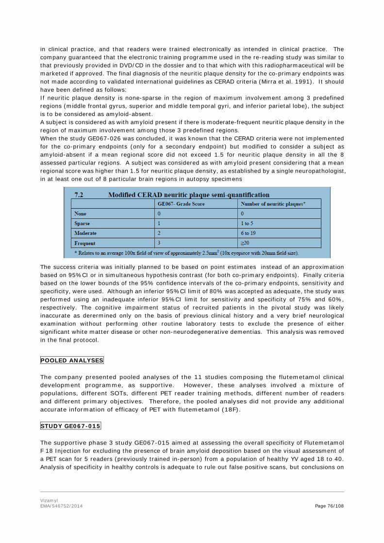
Vizamyl EMA/546752/2014 Page 76/108
in clinical practice, and that readers were trained electronically as intended in clinical practice. The company guaranteed that the electronic training programme used in the re-reading study was similar to that previously provided in DVD/CD in the dossier and to that which with this radiopharmaceutical will be marketed if approved. The final diagnosis of the neuritic plaque density for the co-primary endpoints was not made according to validated international guidelines as CERAD criteria (Mirra et al. 1991). It should have been defined as follows: If neuritic plaque density is none-sparse in the region of maximum involvement among 3 predefined regions (middle frontal gyrus, superior and middle temporal gyri, and inferior parietal lobe), the subject is to be considered as amyloid-absent. A subject is considered as with amyloid present if there is moderate-frequent neuritic plaque density in the region of maximum involvement among those 3 predefined regions. When the study GE067-026 was concluded, it was known that the CERAD criteria were not implemented for the co-primary endpoints (only for a secondary endpoint) but modified to consider a subject as amyloid-absent if a mean regional score did not exceed 1.5 for neuritic plaque density in all the 8 assessed particular regions. A subject was considered as with amyloid present considering that a mean regional score was higher than 1.5 for neuritic plaque density, as established by a single neuropathologist, in at least one out of 8 particular brain regions in autopsy specimens
The success criteria was initially planned to be based on point estimates instead of an approximation based on 95%CI or in simultaneous hypothesis contrast (for both co-primary endpoints). Finally criteria based on the lower bounds of the 95% confidence intervals of the co-primary endpoints, sensitivity and specificity, were used. Although an inferior 95%CI limit of 80% was accepted as adequate, the study was performed using an inadequate inferior 95%CI limit for sensitivity and specificity of 75% and 60%, respectively. The cognitive impairment status of recruited patients in the pivotal study was likely inaccurate as derermined only on the basis of previous clinical history and a very brief neurological examination without performing other routine laboratory tests to exclude the presence of either significant white matter disease or other non-neurodegenerative dementias. This analysis was removed in the final protocol.
POOLED ANALYSES The company presented pooled analyses of the 11 studies composing the flutemetamol clinical development programme, as supportive. However, these analyses involved a mixture of populations, different SOTs, different PET reader training methods, different number of readers and different primary objectives. Therefore, the pooled analyses did not provide any additional accurate information of efficacy of PET with flutemetamol (18F). STUDY GE067-015 The supportive phase 3 study GE067-015 aimed at assessing the overall specificity of Flutemetamol F 18 Injection for excluding the presence of brain amyloid deposition based on the visual assessment of a PET scan for 5 readers (previously trained in-person) from a population of healthy YV aged 18 to 40. Analysis of specificity in healthy controls is adequate to rule out false positive scans, but conclusions on

Vizamyl EMA/546752/2014 Page 77/108
external validity are limited since the enrolled population for this analysis does not reflect real life population. The study involved reading of PET images of HV mixed with PET images of all aMCI patients recruited in another study of flutemetamol (18F) (study GE067-005) to avoid biases. The inter- and intra-observer concordance was established by assessing the percent in agreement without considering the agreement by chance. Such assessment is not appropriate. In this study, agreement by chance is very likely high due to the disbalance between the frequency of normal image sets (from HVs) and from abnormal image sets (from study GE067-005). In this sense, it would have been important to analyze the criteria using images from study GE067-005, particularly because in case-control studies the diagnostic performance of the test might have been higher since it is easy to distinguish between normal and abnormal cases. Studies GE067-008, -009, -010 and -011 Additionally, four small-sized Phase 3 biopsy studies (GE067-008, -009, -010 and -011) aimed at demonstrating the correlation between the estimation of beta amyloid deposition by histopathological methods from biopsied cortical samples in patients with normal pressure hydrocephalus (NPH) and quantitative analysis of PET images. Each of these four supportive studies lack enough precision to draw firm conclusions on the correlation between quantitative PET analysis and histopathology, and the diagnostic performance of visual PET scan reading in the recruited population. This is due to the fact biopsy did not allow for a reliable SOT estimate, since the area in which PET signal was quantitated and the biopsied area did not fully match topographically, and in patients with asymmetric patterns of beta amyloid deposition the assumption of similar depositions in different areas is not valid. Additionally the company did not demonstrate that the presence or absence of amyloid in a relatively circumscribed sample of brain tissue can reliably be extrapolated to a global assessment of cerebral amyloid load. Study ALZ201 The intended role of flutemetamol (18F) was to exclude the diagnosis of AD in adults who are being evaluated for AD. There are limited clinical data addressing this issue coming from the supportive ALZ201 trial. The study was primarily aimed at differentiating AD from normal cognition, by a visual interpretation of flutemetamol (18F) PET images, and secondarily to differentiate them from MCI. Quantitative analysis of PET image, image quality and reproducibility were also assessed. Test-retest variability was assessed in a subgroup of patients with pAD. The study also attempted to provide with comparative data between flutemetamol (18F) and [11C]PiB in some of the recruited patients with clinical AD and all with aMCI. All those previous analyses were also done in all recruited sample at 2-year follow-up. The sensitivity of the visual reading of baseline PET scan was determined for subjects diagnosed as pAD at the follow-up, and the specificity for subjects diagnosed as HVs. Then, there was a 2-year gap between the PET scan and the clinical diagnosis, which introduces uncertainty into the calculated values of sensitivity and specificity. The design of this study did not allow for estimating the risk of MCI progression to clinical AD. Enrolled subjects in this supportive study did not encompass the overall anticipated population in which flutemetamol (18F) is to be used. Indeed, not all tests standardized for management of patients suspected of AD were followed to confirm the absence of systemic disorders or other brain diseases that in and of themselves could account for the progressive cognitive impairment. Moreover, the clinical diagnosis is not an appropriate SOT to support claims for AD diagnosis, due to its well-known lack of diagnostic performance. For these reasons, results from study ALZ201 cannot reliably be extrapolated to the proposed indication for use.

Vizamyl EMA/546752/2014 Page 78/108
Study GE067-005 The patients in this supportive study were all with aMCI based on the Petersen criteria. Conclusions regarding the relative progression to AD over a 3-year period of normal PET and abnormal PET populations of aMCI might attempt to support a prognosis indication, which is not the intended use requested for flutemetamol (18F) at the moment. It has to be kept in mind that the study results might have been biased since the protocol had been amendment 5 times involving crucial issues such as efficacy endpoints, main objective, primary and secondary endpoints and sample size. The applicant justified the 3-year study follow-up period based on a published meta-analysis of studies reporting the rate of conversion from aMCI to AD. However, there are published reports of conversion rate with a much longer follow-up period. Study GE067-021 The Applicant had chosen the electronic training programme, and not in-person training, to be available to physicians post-approval. The clinical development of flutemetamol (18F) included a phase 3 trial attempting to evaluate efficacy of flutemetamol (18F) PET images after electronic training of PET readers: study GE067-021. In this study no patients were recruited and flutemetamol (18F) PET image sets were selected from 9 previous flutemetamol clinical trials in subjects with histopathological confirmation of their brain amyloid status, young HVs, elderly HVs, subjects with clinically pAD and subjects with aMCI. Validation of the electronic programme in study GE067-021 was attempted in different population subgroups using different statistical analyses and different reference/truth standards. Regarding analysis of sensitivity and specificity of visual interpretation of PET images, results from the autopsied population subgroup (i.e. analysis population 3), for the estimation of brain beta amyloid neuritic plaque density could be considered as supportive. The analysis population 3 in this study matches the post-mortem analysis population of study GE067-007, which accounts for comparative purposes. Sensitivity and specificity results, if analysed in the patients with aMCI, for confirming or refuting of clinical diagnosis of aMCI, can also be considered of interest. However, such population subgroups were composed of some mixed patients of both study GE057-005 and ALZ201, and therefore no direct comparison to those individual studies is possible. Regarding evaluation of sensitivity and specificity, the mixing of different populations diagnosed by different SOTs raises concerns about potential bias of the results since the prevalence of beta amyloid deposition will differ among the different populations. Other issues related to the clinical development programme Concerning interpretation of PET images, two distinct methods have been used during the development programme: a binary visual and a quantitative one. However, the applicant did not justify why the visual method of PET scan reading as a composite measure for the whole brain was chosen as principal for the pivotal and most supportive studies, instead of the quantitative one which was also performed in most trials, if not all. There are differences in the visual interpretation of PET images among the pivotal study, supportive phase II-III clinical studies, the method recommended in the SmPC and the one to be provided by an electronic training programme. The electronic version is the methodology proposed in clinical practice.
Table 19. The differences in the BIE visual interpretation through the flutemetamol (18F) programme
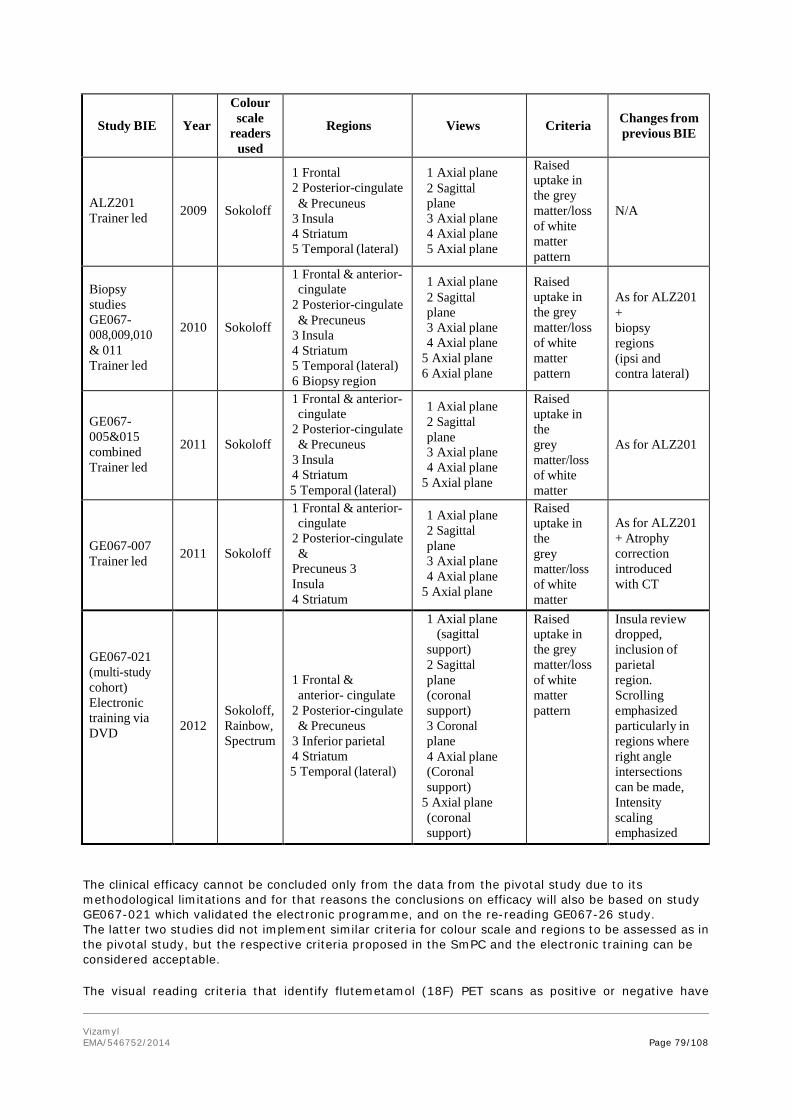
Vizamyl EMA/546752/2014 Page 79/108
Study BIE
Year
Colour scale
readers used
Regions
Views
Criteria
Changes from previous BIE
ALZ201 Trainer led
2009
Sokoloff
1 Frontal 2 Posterior-cingulate & Precuneus
3 Insula 4 Striatum 5 Temporal (lateral)
1 Axial plane 2 Sagittal plane 3 Axial plane 4 Axial plane 5 Axial plane
Raised uptake in the grey matter/loss of white matter pattern
N/A
Biopsy studies GE067- 008,009,010 & 011 Trainer led
2010
Sokoloff
1 Frontal & anterior- cingulate
2 Posterior-cingulate & Precuneus
3 Insula 4 Striatum 5 Temporal (lateral) 6 Biopsy region
1 Axial plane 2 Sagittal plane 3 Axial plane 4 Axial plane
5 Axial plane 6 Axial plane
Raised uptake in the grey matter/loss of white matter pattern
As for ALZ201 + biopsy regions (ipsi and contra lateral)
GE067- 005&015 combined Trainer led
2011
Sokoloff
1 Frontal & anterior- cingulate
2 Posterior-cingulate & Precuneus
3 Insula 4 Striatum 5 Temporal (lateral)
1 Axial plane 2 Sagittal plane 3 Axial plane 4 Axial plane
5 Axial plane
Raised uptake in the grey matter/loss of white matter
As for ALZ201
GE067-007 Trainer led
2011
Sokoloff
1 Frontal & anterior- cingulate
2 Posterior-cingulate &
Precuneus 3 Insula 4 Striatum
1 Axial plane 2 Sagittal plane 3 Axial plane 4 Axial plane
5 Axial plane
Raised uptake in the grey matter/loss of white matter
As for ALZ201 + Atrophy correction introduced with CT
GE067-021 (multi-study cohort) Electronic training via DVD
2012
Sokoloff, Rainbow, Spectrum
1 Frontal & anterior- cingulate
2 Posterior-cingulate & Precuneus
3 Inferior parietal 4 Striatum 5 Temporal (lateral)
1 Axial plane (sagittal
support) 2 Sagittal plane (coronal support) 3 Coronal plane 4 Axial plane (Coronal support)
5 Axial plane (coronal support)
Raised uptake in the grey matter/loss of white matter pattern
Insula review dropped, inclusion of parietal region. Scrolling emphasized particularly in regions where right angle intersections can be made, Intensity scaling emphasized A h
The clinical efficacy cannot be concluded only from the data from the pivotal study due to its methodological limitations and for that reasons the conclusions on efficacy will also be based on study GE067-021 which validated the electronic programme, and on the re-reading GE067-26 study. The latter two studies did not implement similar criteria for colour scale and regions to be assessed as in the pivotal study, but the respective criteria proposed in the SmPC and the electronic training can be considered acceptable. The visual reading criteria that identify flutemetamol (18F) PET scans as positive or negative have

Vizamyl EMA/546752/2014 Page 80/108
inherent difficulties and represent a challenge. PET images of the brain are known to be difficult to read, and the difficulty increases also from the intrinsic characteristics of a PET amyloid tracer. The criterion for positivity in the visual method is looking at the loss of reduction of radioactive signal intensity between white matter (with invariably high uptake) and grey matter (with either no radiopharmaceutical uptake (if normal) or some level of uptake (if abnormal)). Traditional scanners in use today often lack the fine volumetric resolution and high-contrast ratio required to precisely differentiate between grey and white matter. Because grey and white matter are interlaced in such a compact way, and also due to the size of the grey matter (about 5 mm), distinguishing both can be challenging. Additionally, in cases where the uptake in grey matter is insufficient (as for example in borderline cases with insufficient intensity of amyloid deposition, or in cases with reduction of grey matter width as in brain atrophy) this challenge is increasing. Statistics can improve the accuracy of diagnosis beyond that attainable by a human observer. The strength of this approach is that, no a priori hypothesis is required about the locations that may be affected and the whole volume is automatically analyzed. Comprehensive packages are available for statistical comparison of brain perfusion SPECT. The differences between the normal database and the test subjects are expressed and, to help to interpretate the differences, functional images are displayed. They may allow for a single subject diagnosis. A methodology to help distinguish between white and grey matter in PET scans and help quantify the intensity of amyloid uptake in grey matter would be an important tool and is potentially achievable nowadays in clinical practice. The applicant has already used a quantitative PET reading methodology in the clinical programme, and that is why it is recommended that the company should develop and validate a quantitative reading PET methodology based on their product. Technical problems in the scan itself (noise or oversmoothing) or in brain anatomy (levels of atrophy) can affect the anatomical location of the gray matter/white matter border and are important to be considered in the interpretation of a flutemetamol (18F) PET scan. MRI or CT scans may be helpful for discerning anatomy in cases in which atrophy or a low quality scan complicate the PET image interpretation. The CHMP highly encouraged the use of a co-registered recent MRI image or CT scan for the visual interpretation of flutemetamol (18F) PET scans, particularly in those cases in which there is uncertainty about the location of grey matter and grey/white matter border on the PET scan. For these reasons the following paragraphs were recommended to be included in sections 4.2. and 4.4. of the SmPC, respectively: “VIZAMYL images should only be interpreted by readers trained in the interpretation of PET images with flutemetamol (18F). A recent co-registered computed tomography (CT) scan or magnetic resonance (MR) imaging of the patient to get a fused PET-CT or PET-MR image is recommended in cases of uncertainty about the location of grey matter and of the grey/white matter border in the PET scan (see section 4.4 Interpretation of VIZAMYL images).” “Some scans may be difficult to interpret due to image noise, atrophy with a thinned cortical ribbon, or image blur, which could lead to interpretation errors. For cases in which there is uncertainty about the location of grey matter and of the grey/white matter border on the PET scan, and a co-registered recent CT or MR image is available, the interpreter should examine the fused PET-CT or PET-MR image to clarify the relationship of the PET radioactivity and the grey matter anatomy.” Flutemetamol (18F) will be prescribed by physicians skilled in the clinical management of neurodegenerative disorders. Another requirement is that PET scans be read blinded to clinical data as done in the autopsy studies. This is acceptable since the clinical status of the patient might influence the image interpretation if atrophy is suspected. This is included in the SmPC although it will be hard to be fulfilled considering that Nuclear Medicine physicians usually review clinical data to interpret brain SPECT/PET images. Due to the difficulties for visual qualitative interpretation of flutemetamol (18F) PET scans, it is mandatory to complete an appropriate reader training prior to routine clinical image interpretation. Two distinct

Vizamyl EMA/546752/2014 Page 81/108
methods have been used during the clinical development: an in-person training (in studies GE067-007, -015, -005 and ALZ201) and the electronic training (in study 021), but the proposed PET reading method as described in the SmPC and in the electronic training programme can be considered acceptable.
Efficacy data and additional analyses DIAGNOSTIC PERFORMANCE
The pivotal study has some important methodological drawbacks and limitations which prevent from concluding that adequate diagnostic performance of PET wit flutemetamol (18F) has been demonstrated. From 176 subjects in the pivotal study GE067-007 who underwent PET imaging, 68 who died and underwent brain autopsy were considered evaluable for the SOT assessment (Postmortem Analysis set). The age range of the population 60-95, and the mean value ~ 80 years are not representative ot the target population in clinical practice. The same is true for the levels of cognitive impairment which may be different from that of the intended population considering that only 31% patients had no history of cognitive impairment, about 44% had AD, 25% had other dementias, and none had MCI.
Although the success criterion of the study was met (see Table 16. Summary of Efficacy for study GE067-007), it is important to note that this result critically depended on a very low number of participants, making the trial lack robustness. In fact, a minor perturbation such as changing the result of just two participants, will impact significantly on the whole trial result which will be regarded as unsuccessful. Furthermore, reader 2 and 4 did not rate the images of one subject [MAI3]which in turn was doubtfully classified as abnormal whenever readers 1, 3 and 5 rated this subject as normal, abnormal and abnormal respectively.
The values of specificity of blinded visual PET image interpretations without anatomic images for estimation of a non-sparse neuritic plaque densitiy in post-mortem analysis set were 88% (69-98%), 92% (74-99%), 44% (24-65%), 80% (59-93%) and 92% (74-99%) for each independent reader. The company hypothesized that the very poor specificity observed with reader 3 for the blinded visual interpretation of flutemetamol (18F) PET scans without anatomic images might be due to the reader’s difficulty in differentiating between grey matter and white matter, to having spent too little time in reading images, or to unsuccessful training.
Sensitivity and specificity of the blinded visual interpretation of PET images for detecting brain fibrillar amyloid beta with anatomic images ranged from 91% to 98% and from 56% to 92%, respectively.
Comparison of the 95% confidence intervals for sensitivity obtained with anatomic images to those obtained without anatomic images shows overlap of the confidence intervals for each of the 5 readers.
In the re-reading study (study GE067-026), the primary objective was to estimate the sensitivity and specificity (as co-primary endpoints) of the majority (i.e., the image interpretation reached by at least 3 of the 5 readers) interpretation of flutemetamol (18F) images for imaging amyloid neuritic plaque density versus histopathology at 8 particular brain regions. Sensitivity was 90.8% with a 95% confidence interval of (81.9%, 96.2%); the lower bound (81.9%) exceeded the pre-specified value of 75%. Specificity was 90.0% with a 95% confidence interval of (73.5%, 97.9%); the lower bound (73.5%) exceeded the pre-specified value of 60%.
Sensitivity and specificity were also determined as a secondary analysis using a SoT based on the 3 neocortical regions originally recommended by CERAD [Mirra et al. 1991]. Based on the majority read and considering SoT as positive if moderate-frequent beta-amyloid neuritic plaque density in the region of maximum involvement and negative if none-sparse, sensitivity for detection of moderate-frequent
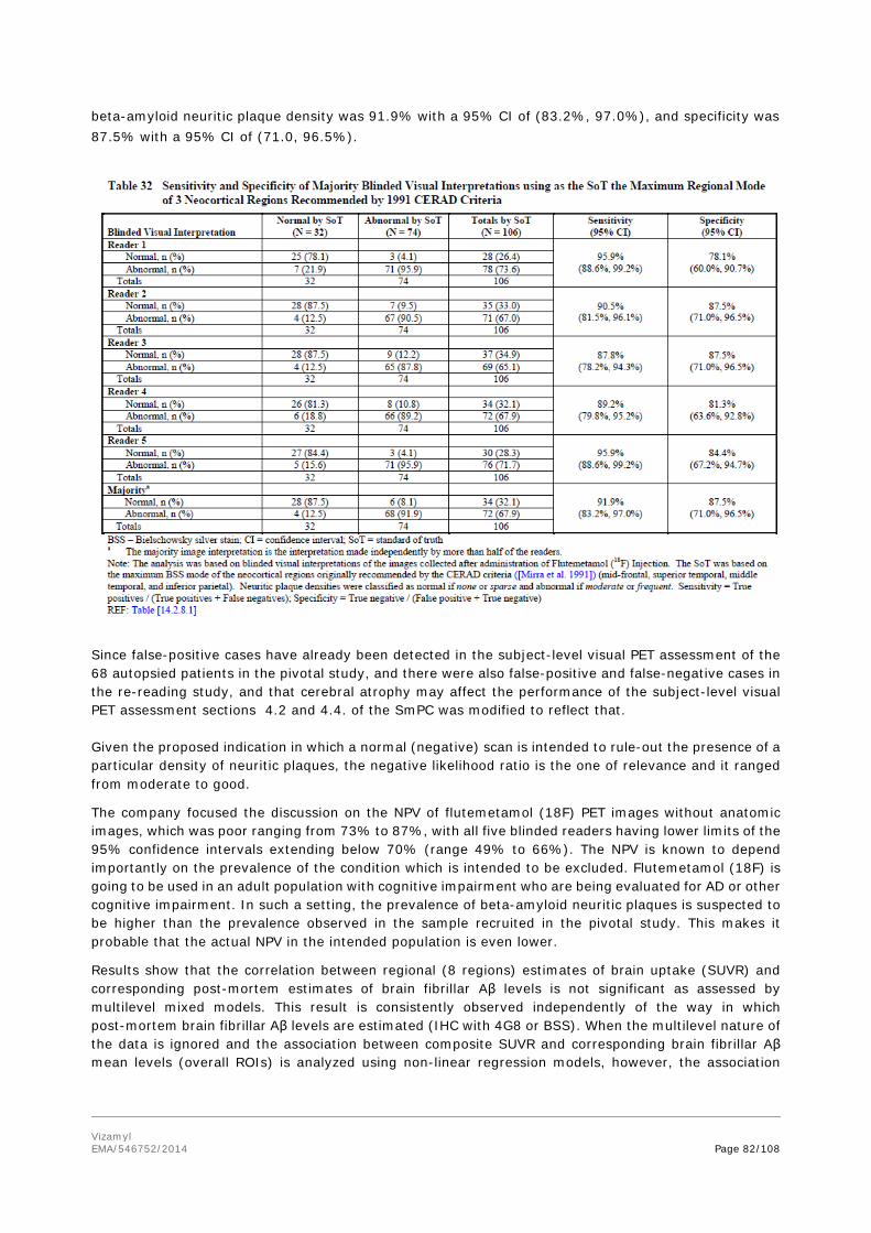
Vizamyl EMA/546752/2014 Page 82/108
beta-amyloid neuritic plaque density was 91.9% with a 95% CI of (83.2%, 97.0%), and specificity was 87.5% with a 95% CI of (71.0, 96.5%).
Since false-positive cases have already been detected in the subject-level visual PET assessment of the 68 autopsied patients in the pivotal study, and there were also false-positive and false-negative cases in the re-reading study, and that cerebral atrophy may affect the performance of the subject-level visual PET assessment sections 4.2 and 4.4. of the SmPC was modified to reflect that. Given the proposed indication in which a normal (negative) scan is intended to rule-out the presence of a particular density of neuritic plaques, the negative likelihood ratio is the one of relevance and it ranged from moderate to good.
The company focused the discussion on the NPV of flutemetamol (18F) PET images without anatomic images, which was poor ranging from 73% to 87%, with all five blinded readers having lower limits of the 95% confidence intervals extending below 70% (range 49% to 66%). The NPV is known to depend importantly on the prevalence of the condition which is intended to be excluded. Flutemetamol (18F) is going to be used in an adult population with cognitive impairment who are being evaluated for AD or other cognitive impairment. In such a setting, the prevalence of beta-amyloid neuritic plaques is suspected to be higher than the prevalence observed in the sample recruited in the pivotal study. This makes it probable that the actual NPV in the intended population is even lower.
Results show that the correlation between regional (8 regions) estimates of brain uptake (SUVR) and corresponding post-mortem estimates of brain fibrillar Aβ levels is not significant as assessed by multilevel mixed models. This result is consistently observed independently of the way in which post-mortem brain fibrillar Aβ levels are estimated (IHC with 4G8 or BSS). When the multilevel nature of the data is ignored and the association between composite SUVR and corresponding brain fibrillar Aβ mean levels (overall ROIs) is analyzed using non-linear regression models, however, the association
Vizamyl EMA/546752/2014 Page 83/108
becomes statistically significant. These latter models were not planned in the protocol but included in the Statistical Analysis Plan.
In summary, correlation PET-autopsy was not significant as analyzed according to the protocol; however, using a different statistical approach revealed the correlation to be significantwhich could be also as a result of statistical artifacts.
An additional issue was the fact that in the GE067-007 post-mortem analysis set, 28 of the 68 subjects were found to have non-AD neurodegenerative pathology consisting of synucleinopathies (n=20; 13 were PET positive and 7 were PET negative for the majority), tauopathies (n=1, with negative PET), and vasculopathy (n=7; 1 with PET positive and 6 with PET negative), once again confirming the differences between the study population and the one for which the product will be intended in the clinic. In study GE067-015, all 181 HVs who received 185 MBq of Flutemetamol F 18 Injection were available for blinded visual read of PET images. The true histopathologic status of these subjects is however unclear as it is possible to have beta amyloid deposition in the brain without any symptoms regarding cognition. The images were read by 4 out of 5 independent readers (except reader 2) as normal in 99-100% of the cases, with the lower bounds of the 95% confidence intervals exceeding 95%. Therefore the success criterion was met. In this study the results of Reader 2 raise a concern: a normalilty rate as low as 68% with a lower limit of the 95% confidence interval of 67%. When comparing results from reader 2 and the remaining four readers, doubts about the accuracy of the visual reading of flutemetamol (18F) PET scans in healthy volunteers can arise. The high failure rate of reader 2 in Study GE067-015 illustrated the need for an appropriate reader training program. To address that, the applicant included several improvements in the electronic image training programme intended for clinical use, including more challenging cases added to the training and to the training test, especially images which borderline levels of tracer uptake in the grey matter. Study GE067-008, -009, -010 and -011 lack enough precision to draw firm conclusions on the correlation between quantitative PET analysis and histopathology, and the diagnostic performance of visual PET scan reading in the recruited population, as discussed previously. Study ALZ201 represents a clinical scenario where amyloid imaging (both visual and quantitative PET scan reading) is used to confirm the clinical diagnosis at baseline. 92% out of 25 recruited subjects with clinical diagnosis of probable AD (according to NINCS-ADRDA criteria), 45% out of 20 recruited patients with aMCI (based on Petersen criteria) and 4% out of 25 HVs (10 young HVs and 15 older HVs subjects) were considered positive after visual PET scan reading by the majority of 5 independent blinded readers. All them received a single i.v. dose of 185 MBq of Flutemetamol F 18 Injection, except 5 subjects with pAD who received 2 administrations of approximately 120 MBq each. The numbers for AD are consistent with the expected prevalence of amyloid-negative individuals in a clinically diagnosed AD population based on some literature reports of the false positive rate for the clinical diagnosis of AD versus autopsy. However, these numbers are inconsistent with other literature reports showing that 20% of clinically-diagnosed AD subjects did not have AD at autopsy and lacked amyloid pathology (Lim et al. 1999, Pearl et al. 1997), and with findings from another PET amyloid tracer as florbetapir (18F) as shown in the EPAR. The observation in aMCI subjects is consistent with the autopsy literature that shows 33% to 62% of MCI subjects are positive at post-mortem examination (Bennett, 2005; Petersen, 2006). Finally, the observation in HVs is inconsistent with literature reports that 13% to 45% of apparently cognitively HVs subjects have significant beta amyloid pathology at autopsy (Hulette et al. 1998, Davis et al. 1999, Price et al. 1999, Schimitt et al. 2000; knopman et al. 2003, Aizenstein et al. 2008) and also inconsistent with findings from other PET amyloid tracers as shown in the EPAR. Diagnostic performance values of flutemetamol (18F) PET are tabulated below considering either a combined set of aMCI plus HVs when assessing the diagnosis of pAD, or a combined set of pAD plus HVs when assessing the diagnosis of aMCI:

Vizamyl EMA/546752/2014 Page 84/108
Agreement with baseline
diagnosis of MCI (n=20)
Agreement with baseline diagnosis of clinical AD (n=25)
Sensitivity 9/20 = 45% 23/25 = 92% (95% CI: 23.2-66.8%) (95% CI: 81.4-100%)
Specificity Using non-MCI cases Using non-AD cases (cognitively normal & clinical AD) (cognitively normal & MCI) 26/50 = 52% 35/45 = 77.8% (95% CI: 38.2-65.8%) (95% CI: 65.6-89.9%)
Positive likelihood ratio 0.94 (95% CI: 0.53-1.65)
4.14 (95% CI: 2.37-7.24)
Negative likelihood ratio 1.06 (95% CI: 0.66-1.71)
0.10 (95% CI: 0.03-0398)
There was significant correlation (r2=0.82) between quantitative PET uptake values generated from flutemetamol (18F) and [11C]PiB PET scans in 20 subjects with pAD and 20 with aMCI who also received an i.v. dose of [11C]PiB not exceeding 370 MBq on the same day or within approximately 30 days after receiving Flutemetamol F 18 Injection. This comparative data can be considered only as informative since [11C]PiB is not an approved radiopharmaceutical and has not been validated up to now. Reassessment of the clinical diagnosis in 67 returning subjects at a 2-year follow-up showed no changes from the initial diagnosis for any of the returning pAD subjects (n = 22) or HVs (n = 24). Of the returning subjects who had initially been diagnosed with aMCI (n = 19), 9 (47%) were diagnosed to have pAD at the 2-year follow-up; 7 of these had raised flutemetamol (18F) uptake at baseline. The other 10 (53%), eight of whom had negative flutemetamol PET scans, retained at the 2-year follow-up a baseline diagnosis of aMCI. Sensitivity of the visual PET scan reading (including 95% CI) to show the aMCI conversion rate from baseline to clinically probable AD at 2-year follow-up in 9 converters was 77.8% (95%CI: 40.0%-97.2%), specificity in 10 non-converters was 80.0% (95%CI: 44.4%-97.5%), positive likelihood ratio was 3.89 (95% CI: 1.07-14.10) and negative likelihood ratio was 0.28 (95% CI: 0.08-0.98). The design of this did not allow for estimating the risk of MCI progression to clinical AD. In study GE067-005, a total of 232 subjects with amnestic MCI (Petersen criteria) received an i.v. dose of approximately 185 MBq ofFlutemetamol (18F) and were submitted for a 3-year follow-up to determine whether or not conversion to probable AD had occurred. At baseline, by the majority image interpretation, 134 (58%) of the 232 subjects had normal (negative) flutemetamol (18F) scans and 98 (42%) had abnormal (positive) scans. Of the 232 patients enrolled, 224 had at least one post-scan review by the independent committee and were included in the analysis. At the 36-month follow-up, 81 (36 %) converted to clinical AD. Of the 97 MCI subjects who had a positive PET scan and at least one committee assessment, 52 (54 %) were classified clinically as converted to clinical AD after 36 months compared to 29 (23 %) of 127 who had a negative scan and at least one committee assessment. At 36 months sensitivity of flutemetamol (18F) scan to show the MCI conversion rate to AD in 81 converters was 64 % (95% CI: 53 % to 75 %), specificity in 143 non-converters was 69% (95% CI: 60% to 76%). Based on the majority read, the positive and negative likelihood ratios were 2.04 and 0.52, respectively. In the primary analysis the hazard ratio based on majority image read was 2.5. In study GE067-021 the sensitivity and specificity for detecting or excluding brain amyloid of the blinded visual interpretation of flutemetamol (18F) PET images of the brain without anatomic images, assessed in the subgroup analysis population 3 (brain autopsy study population, n = 68) after electronic

Vizamyl EMA/546752/2014 Page 85/108
training of 5 independent blinded PET readers, was counterbalanced as it was observed in the same sample in study GE067-007 using an in-person training of another 5 different individual readers. In fact, those readers showing higher sensitivities were at the same time those showing lower specificities. When interpreting PET images with anatomic images, the sensitivity and specificity for any individual reader were not significantly different than those for the PET-only image interpretation. It seems that focal atrophy in the key assessment regions as found in study GE067-021 may contribute to false negative and a substantial number of diffuse plaques to false positive assessments. This is sufficiently covered in the SmPC as well as the updated training program. In the autopsied population, for the majority read, there were 3 FN cases for the majority of readers (2 had a neuropathology diagnosis of Lewy Body Disease (LBD), and 1 of Cerebral Amyloid Angiopathy (CAA)) and 4 FP (all had a neuropathological diagnosis of Lewy Body Disease (Dementia with Lewy Body or Parkinson’s disease). Therefore, it is possible that a FN result occurs if there is sparse neuritic plaque density; so is a FP result when there is moderate plaque density. TECHNICAL PERFORMANCE AND PRACTICABILITY Image quality
Following administration of the first dose of Vizamyl in study ALZ201, PET images of subjects with pAD were rated as excellent in 21 (84%) and good in 4 (16%) subjects, respectively. In all 20 aMCI subjects and 5 HVs the images were rated as excellent. Following administration of a second dose of 120 MBq of Vizamyl in 5 with pAD (who had also received 120 MBq as the first dose), the images were rated as good in all subjects (100%).
Reader confidence
Only data on reader confidence of the visual blinded interpretation of PET scans (both PET-only images and PET with anatomic images) was obtained in study GE067-021. The percentage of PET-only images with a high reader confidence ranged from 61-97%, depending on the reader. Those with low reader confidence ranged from <1 to 20%. If PET images were assessed combined with anatomic images, the corresponding levels of reader confidence were in similar ranges. The applicant has assessed post-hoc the reader confidence in PET images by each of the 5 readers of pivotal GE067-007. All readers had most of the PET readings categorised as the highest confidence rates 4 or 5 (92.6% for reader 1, 88.1% for reader 2, 100% for reader 3, 80.6% for reader 4 and 80.9% for reader 5). Inter and intra-reader concordance
In pivotal autopsy study GE067-007, results show high inter-reader agreement (kappa scores ranging from 0.76 to 0.91) of PET readers of PET-only images for all pairwise comparisons except those involving Reader 3, which ranged from 0.44 to 0.55 in kappa score. Inter-reader agreement behaved similarly for the interpretation of PET images with anatomic images by those 5 blinded individual readers, and the absolute values were slightly higher than those obtained in PET-only images. In the study GE067-015, the inter-reader and intra-reader agreements for blinded visual PET scan reading of HVs were >99% and 100%, respectively. Reader 2 showed values of 68% and 75%, respectively. Having in mind the results from reader 3 in study GE067-007 and reader 2, doubts about the reproducibility of the visual reading of flutemetamol (18F) PET scans could arise. In study ALZ201 there was complete agreement among 5 readers (94.6%) in HVs and patients with aMCI and pAD (with a Fleiss kappa score of 0.96) in 70 of 74 flutemetamol (18F) images read interpreted visually.

Vizamyl EMA/546752/2014 Page 86/108
In study GE067-005 (aMCI subjects), the inter-reader agreements for blinded visual PET scan reading ranged from 77% (κ = 0.56) to 98% (κ = 0.96) between pairs of readers. The intra-reader reproducibility of the rereads was good, ranging from 86% to 100%. In the electronic training study GE067-021 recruiting different populations using different reference/truth standards (analysis population 4), results show high inter-reader agreement reproducibility of blinded visual interpretation of flutemetamol (18F) PET images of the brain without anatomic images for all readers: kappa=0.83. In a subset of patients (analysis population 5), there was high percentage of intra-reader agreement (93-100%). Neither population 4 nor population 5 matched the population of the autopsy pivotal study. Inter-reader agreement (kappa) was slightly lower for the PET+ anatomic image set than for PET-only set; however, the confidence intervals overlap. Intra-reader agreement was quite similar between both sets. Test-retest reproducibility
In study ALZ201, cortical flutemetamol (18F) test-retest quantitative PET estimates had a low inter-subject variability of 1-4% in pAD subjects. The variability was lowest in the occipital cortex, striatum, POC, and the frontal cortex and was highest in the medial temporal cortex, subcortical white matter, and pons. The mean test-retest variability of all subjects and regions was excellent. IMPACT ON DIAGNOSTIC THINKING AND/OR PATIENT MANAGEMENT
The guideline on clinical evaluation of diagnostic agents (CPMP/EWP/1119/98/Rev. 1) that applies to flutemetamol (18F), establishes as a requirement for new diagnostic agents, that “……relevant impact on diagnostic thinking and/or patient management in the appropriate clinical context should be demonstrated, if therapeutic consequences of the diagnosis obtained with a new agent are not obvious, or the benefit/risk balance is unclear…..”. Since this has not been demonstrated for this diagnostic agent the company was recommended to perform a study to assess the impact on diagnostic thinking and patient management. INDICATION
The original wording of the indication was proposed as: “This medicinal product is for diagnostic use only. VIZAMYL is a radioactive diagnostic agent indicated with positron emission tomography (PET) imaging for the visual detection of amyloid-beta neuritic plaques in the brains of adults who are being evaluated for Alzheimer’s disease (AD). A normal VIZAMYL scan indicates sparse to no neuritic plaques and is inconsistent with a neuropathological diagnosis of AD at the point of image acquisition: a normal scan reduces the likelihood that a patient’s condition is due to AD. An abnormal VIZAMYL scan is indicative of moderate to frequent amyloid-beta neuritic plaques. Neuropathological examinations have shown this amount of neuritic plaques is present in patients with AD, but also other types of neurologic conditions as well as in older people with normal cognition. VIZAMYL is to be used as an adjunct to other diagnostic evaluations. An abnormal VIZAMYL scan does not establish a diagnosis of AD. The safety and efficacy of VIZAMYL have not been established for predicting the development of dementia or other neurological conditions, or for monitoring response to therapies.” The first statement (i.e. “… indicated for the visual detection of amyloid-beta neuritic plaques in the brains”) should be referred to in correspondence with the results on the technical and diagnostic performances of flutemetamol (18F) PET scan reading to estimate the amyloid neuritic plaque density, as assessed in a pivotal study, the re-reading study and various supportive phase II-III studies, which were previously discussed in this report. Further on the indication wording continues to say that a normal flutemetamol (18F) PET scan indicates sparse to no neuritic plaques. Such criterion of normality is in line with the criteria to establish normality

Vizamyl EMA/546752/2014 Page 87/108
of visual PET reading in the pivotal, re-reading and supportive phase II-III studies. EMA recently acknowledged in regulatory opinions that none to sparse neuritic plaque density in the area with maximal neuritic plaque density measured on sections of frontal, temporal or parietal cortex, is incompatible with a definitive diagnosis of AD. The statement that an abnormal flutemetamol (18F) PET scan is indicative of moderate to frequent amyloid-beta neuritic plaques is in line with the criteria to establish normality of visual PET reading in the pivotal, re-reading and supportive phase II-III studies. The wording continues stating that neuropathological examinations have shown that moderate to frequent amount of neuritic plaques is present in patients with AD, but also other types of neurologic conditions as well as in older people with normal cognition. It is also stated that abnormal VIZAMYL scan does not establish a diagnosis of AD. To this regard, the amyloid cascade hypothesis suggests that accumulation of Aβ is the key pathological step in the pathogenesis of AD, however, Aβ deposition is not the only factor of AD. For that reason, pre-specified levels of age-related brain neuritic β-amyloid plaque at autopsy should be integrated with the presence of a clinical history of dementia to arrive at a diagnostic level of certainty with regard to AD (Mirra et al. 1991). Beta-amyloid plaques may also be present in elderly with normal cognition, patients with MCI, with other dementias (dementia of Lewy Body, Parkinson disease dementia), Niemann-Pick disease type C and severe brain injury. VIZAMYL is to be used as an adjunct to other diagnostic evaluations, and not as a stand-alone test, which is acceptable. As this product is not to replace to clinical assessment of patients, the indication was recommended to be reworded to state from the very beginning that flutemetamol (18F) should be used in conjunction with clinical assessment. The last statement of the indication (“The safety and efficacy of VIZAMYL have not been established for predicting the development of dementia or other neurological conditions, or for monitoring response to therapies”) is crucial since it may trigger decisions that patients with clinical features of AD may not be treated on the basis of these findings. Since this radiopharmaceutical has not demonstrated efficacy for any particular diagnostic objective of a validated internationally-recognised disease, limitations of the use of the radiopharmaceutical are of major interest and were included in the indication wording as a cross-reference to the full information stated in section 4.4. of the SmPC. Taking all this into account the following revised indication has been accepted by the CHMP: “This medicinal product is for diagnostic use only. Vizamyl is a radiopharmaceutical medicinal product indicated for Positron Emission Tomography (PET) imaging of β-amyloid neuritic plaque density in the brains of adult patients with cognitive impairment who are being evaluated for Alzheimer’s disease (AD) and other causes of cognitive impairment. Vizamyl should be used in conjunction with a clinical evaluation. A negative scan indicates sparse or no plaques, which is not consistent with a diagnosis of AD. For the limitations in the interpretation of a positive scan, see sections 4.4 and 5.1.” with the following additional wording in section 4.4. of the SmPC: “Limitations of use A positive scan does not independently establish a diagnosis of AD or other cognitive disorder since neuritic plaque deposition in grey matter may be present in asymptomatic elderly and some neurodegenerative dementias (Alzheimer’s disease, Lewy body dementia, Parkinson’s disease dementia). For the limitations of use in patients with mild cognitive impairment (MCI), see section 5.1.

Vizamyl EMA/546752/2014 Page 88/108
The efficacy of Vizamyl for predicting development of AD or monitoring response to therapy has not been established (see section 5.1).” and including in section 5.1. the results of 2 longitudinal studies recruiting subjects clinically diagnosed as aMCI, AD or cognitively normal (study ALZ201), and subjects with aMCI (study GE067-005) who were followed for some years to evaluate the relationship between flutemetamol (18F) imaging and changes in diagnostic status.
EFFICACY IN SUBPOPULATIONS
No efficacy subanalysis has been presented for the use of flutemetamol (18F) in patients with atypical presentations of AD (asymmetric, frontal variants, posterior cortical degeneration and a single positive abnormal region). The paediatric use of this radiopharmaceutical has neither been assessed nor expected. A product-specific waiver for paediatric studies was granted. Studies intended to evaluate dose adjustments in special risk patients in which the product is intended to be used (including at least renal impaired subjects, elderly, and, if appropriate, in those clinical settings excluded from the pivotal study) were not conducted. The use of flutemetamol (18F) in renal impaired subjects (n=18), cardiac impaired subjects (n=9) and hepatic impaired subjects (n=1) is documented by the applicant. In the pivotal autopsy study, there were 13 subjects with renal impairment and 1 patient with hepatic impairment. The available data did not indicate that clinical efficacy in patients with renal impairment would be different than the overall efficacy; therefore, no dose adjustment will be required.
2.5.4. Conclusions on the clinical efficacy
The optimal dose and the optimal time window of flutemetamol (18F) PET images, as proposed in the SmPC, have not been established based on dedicated studies but the methodology used to determine them was considered acceptable. PET image quality and reader confidence of the visual PET scan interpretation was high in the studied populations. The single pivotal study allowed, by comparison to the histopathology of autopsied samples in end-of-life patients, conclusions to be made on correlation and diagnostic performance of the visual PET scan reading regarding neuritic plaque density. Recruitment of end-of-life patients, although a different population than the one intended for in clinical practice, permits autopsied samples of the brain to be available within a reasonable timeframe. The single pivotal trial failed to demonstrate adequate diagnostic performance since specificity was not analysed as a co-primary endpoint and the success criterion (i.e. a lower limit of the 95% CI of sensitivity being higher than 70%) was not justified as of clinical relevance. Moreover, the actual correlation of PET versus histopathology might be artifacted and is unknown. For that reason the CHMP requested a well-designed re-reading study to assess all autopsied patients recruited for the pivotal autopsy study to support this application. The clinical efficacy of the visual reading of (18F) flutemetamol PET images was in the end sufficiently supported by the results from pivotal study GE067-007, supportive study GE067-021, and the re-reading study GE067-026, in which the pivotal study has been expanded to include a total of 106 patients. A consistent high sensitivity (>80%), and a specificity exceeding 84% for at least 3 out of 5 readers and exceeding 78% for all 5 readers, was demonstrated in a larger patient population group when analysed in the re-reading study using the appropriate CERAD criteria. Results of the re-reading study of the pivotal autopsy study demonstrated an acceptable lower limit of sensitivity (i.e. 83.2%) for the visual

Vizamyl EMA/546752/2014 Page 89/108
assessment of flutemetamol (18F) PET images on a subject-basis for excluding moderate-frequent β-amyloid neuritic plaque density in the most heavily affected neocortical region (based on section with the maximum involvement among middle frontal gyrus, inferior parietal lobule and superior and middle temporal gyri). In the supportive study ALZ201 recruiting HVs and a subgroup of the intended population (subjects with aMCI and pAD), validation of visual PET scan reading was attempted versus the clinical diagnosis at baseline and at a short follow-up. In the supportive study GE067-005, recruiting subgroup of the intended population (subjects with aMCI), validation of visual PET scan reading was attempted versus the clinical diagnosis at baseline and after a short follow-up. It should be noted that results in both studies were not obtained versus a (reference) SoT and no assessment was done for the value of flutemetamol (18F) in the differential diagnosis of clinically probable AD vs other dementia subtypes (non-neurodegenerative and non-AD degenerative dementias) and of MCI vs other causes of cognitive impairment. The training programme for visual PET readers in clinical practice will be electronic. In a supportive study, the electronic training programme was attempted to be validated in patients with different diagnoses from previous clinical studies of flutemetamol (18F). Diagnostic performance in the subgroup population from the autopsy study was similar to that in the pivotal study. High inter-reader agreement was observed for different populations; so was for aMCI when assessing intra-reader concordance. Relevant impact on diagnostic thinking and/or patient management in the appropriate clinical context has not been demonstrated for this diagnostic product which itself may have immediate therapeutic implications. No efficacy subanalysis has been presented for the use of flutemetamol (18F) in patients with atypical presentations of AD (asymmetric, frontal variants, posterior cortical degeneration and a single positive abnormal region). In conclusion, although the impact of flutemetamol (18F) PET on diagnostic thinking and/or patient management was not adequately assessed, the sensitivity and specificity of visual flutemetamol (18F) PET reading (the reading method proposed for clinical use), for estimation of beta amyloid moderate to frequent neuritic plaque density in the brain of end-of-life patients, could be concluded upon from the results of the re-reading study, the data from the pivotal study and the supportive studies. The limitations of the pivotal study were acknowledged but the re-reading data have shown an acceptable lower limit of sensitivity (of the visual assessment of flutemetamol (18F) PET images on a subject-basis) for excluding β-amyloid neuritic plaque deposition. Inter- and intra-reader reproducibility of visual PET reading after electronic reader training in a mixed population, including the intended population, was high. The following post approval measures were recommended to address abovementioned issues:
1. The company should continue to develop and validate a quantitative PET reading methodology based on their product. Such a methodology will help distinguish between white and grey matter in PET scans and help quantify the intensity of amyloid uptake in grey matter, and as such would be an important tool in the practical use of the product.
2. The company is encouraged to perform a study to assess the impact on diagnostic thinking and patient management since the therapeutic consequences of the diagnosis of labelling brain beta-amyloid are not obvious. For the design of this study, parallel HTA/scientific advice is recommended.
2.6. Clinical safety
The primary safety analysis is based on the comprehensive integration of data from all subjects exposed to any amount of flutemetamol (18F) in all studies of the clinical development programme with some

Vizamyl EMA/546752/2014 Page 90/108
exceptions. The investigator-sponsored phase 1 study (Report A42404) is not included in the pooled safety analysis due to the study design.
Patient exposure
The safety database for flutemetamol (18F) contained safety data from 10 clinical studies sponsored by GE Healthcare and conducted to evaluate the safety and efficacy of Vizamyl in PET imaging to detect fibrillar amyloid β, in the form of neuritic plaques, in the brain. The primary safety analysis is based on the comprehensive integration of data from all subjects exposed to any amount of Vizamyl in the 10 studies (see table 20). Table 20 Studies Included in the Integrated Summary of Safety

Vizamyl EMA/546752/2014 Page 91/108
Study Phase Location Treatments Number of Subjects
End of life a
pAD aMCI NPH HV Total
[ALZ103] 1 1 center in Belgium
One 100-, 150-, or 185-MBq dose of flutemetamolb
8 14 22
[GE067-014] 1 1 center in Japan
One 100-, 150-, or 185-MBq dose of flutemetamolb
8 14 22
[ALZ201] 2 8 centers in Europe
• One 185-MBq dose of flutemetamolb or
• Two 120-MBq doses of flutemetamolb or
• One 185-MBq dose of flutemetamolb and one 333-MBq dose of [11C]PiB
27 20 25 72
[GE067-005] 3 28 centers in US and Europe
One 185-MBq dose of flutemetamolb
232 232
[GE067-007] 3 19 centers in US and Europe
One 185- to 370-MBq dose of flutemetamolb
180 180
[GE067-008] 3 1 center in US
One 185-MBq dose of flutemetamolb
7 7
[GE067-009] 3 1 center in US
One 185-MBq dose of flutemetamolb
12 12
[GE067-010] 3 1 center in Finland
One 185-MBq dose of flutemetamolb
15 15
[GE067-011] 3 4 centers in Finland
One 185-MBq dose of flutemetamolb
18 18
[GE067-015] 3
10 centers in US and Europe
One 185-MBq dose of flutemetamolb
181 181
Total Number of Subjects in Safety Analysis Set 180 43 252 52 234 761

Vizamyl EMA/546752/2014 Page 92/108
aMCI = amnestic MCI; HV = healthy volunteer; MCI = mild cognitive impairment; NPH = normal pressure hydrocephalus; pAD = probable Alzheimer’s disease.
a Life expectancy <1 year.
b Flutemetamol F 18 Injection.
A total of 761 subjects received Flutemetamol F 18 Injection. The recommended dose of Flutemetamol F 18 Injection is a single intravenous injection of 185 MBq. The majority of subjects in the clinical development program received this regimen. However, different nominal doses were allowed in 4 studies. Across all 761 subjects in the Safety Analysis Set, the mean (SD) net activity administered was 219.3 (76.2) MBq, ranging from 93.4 to 403.3 MBq, with a median of 183.5 MBq. Most subjects (530, 70%) received a dose of Flutemetamol F 18 Injection of ≥166.5 to 203.5 MBq (185 MBq ±10%); 154 (20%) received ≥333 to 407 MBq (370 MBq ± 10%), 32 (4%) received ≥203.5 to 333 MBq, and the remaining 45 (6%) of subjects received a dose <166.5 MBq. Mean (SD) age overall was 62 (21.24) years, ranging from 18 to 98 years. Of the 761 subjects in clinical studies of flutemetamol (18F), 447 (59%) were 65 and over, while 246 (32%) were 75 and over. In total, 367 subjects (48%) were male and 394 (52%) were female. Most were White (693, 91%); 33 (4%) were Black and 27 (4%) were Asian. Mean (SD) height overall was 167.3 (10.31) cm, ranging from 137 to 197 cm. Mean (SD) weight overall was 73.1 (17.38) kg, ranging from 26.8 to 156.0 kg. Mean (SD) body mass index (BMI) overall was 26.1 (5.30) kg/m2, ranging from 12 to 58 kg/m2. A total of 113 subjects (15%) took at least 1 prior medication. The most common ATC Level 1 category of drugs taken as prior medication was nervous system medications (45 subjects, 6%), followed by alimentary tract and metabolism (27 subjects, 4%) and musculoskeletal system medications (25 subjects, 3%). A total of 509 subjects (67%) took concomitant medications. The most common ATC primary classification levels of drugs taken as concomitant medication were: nervous system medications (343 subjects, 45%), alimentary tract and metabolism medications (321 subjects, 42%), and cardiovascular system (298 subjects, 39%). Few subjects took prior or concomitant sedatives. Lorazepam was the most common prior (4 subjects, 1%) and concomitant (50 subjects, 7%) sedative taken. The next most common concomitant sedatives taken were alprazolam (23 subjects, 3%), temazepam (20 subjects, 3%), and zolpidem tartrate (13 subjects, 2%). All other sedatives were taken by ≤ 1% of subjects.
Adverse events
In total, 151 adverse events (AEs) were reported for 76 subjects (10%); AEs considered by the investigator to be at least possibly related to Flutemetamol F 18 Injection were reported for 44 subjects (6%) (table 21).
Table 21. Summary of Treatment-Emergent Adverse Events Related to Treatment

Vizamyl EMA/546752/2014 Page 93/108
System Organ Class
Preferred Term a,b
Number (%) of
Subjects (N = 761) c
Frequency Classificationd
Subjects with any treatment-emergent adverse event at least possibly related to treatment
44 (6)
Cardiac disorders 4 (1) Arrhythmiae
1 (<0.5) Uncommon Palpitations 3 (<0.5) Uncommon
Ear and labyrinth disorders 1 (<0.5) Vertigo 1 (<0.5) Uncommon
Eye disorders 1 (<0.5) Eye swelling 1 (<0.5) Uncommon
Gastrointestinal disorders 11 (1) Abdominal discomfort 1 (<0.5) Uncommon Dyspepsia 2 (<0.5) Uncommon Nausea 8 (1) Common Oral discomfort 1 (<0.5) Uncommon Vomiting 1 (<0.5) Uncommon
General disorders and administration site conditions 14 (2) Asthenia 1 (<0.5) Uncommon Chest discomfort 7 (1) Uncommon Fatigue 1 (<0.5) Uncommon Feeling abnormal 1 (<0.5) Uncommon Feeling cold 1 (<0.5) Uncommon Feeling hot 2 (<0.5) Uncommon Infusion site pain 1 (<0.5) Uncommon Edema 1 (<0.5) Uncommon Pyrexia 1 (<0.5) Uncommon
Immune system disorders 1 (<0.5) Anaphylactic reaction 1 (<0.5) Uncommon
Investigations 9 (1) Blood glucose decreased 1 (<0.5) Uncommon Blood lactate dehydrogenase increased 1 (<0.5) Uncommon Blood pressure increased 7 (1) Uncommon Neutrophil count increased 1 (<0.5) Uncommon Respiratory rate increased 1 (<0.5) Uncommon
Musculoskeletal and connective tissue disorders 5 (1) Back pain 4 (1) Uncommon Muscle tightness 1 (<0.5) Uncommon Musculoskeletal pain 1 (<0.5) Uncommon
Nervous system disorders 14 (2) Dizziness 6 (1) Uncommon Dysgeusia 1 (<0.5) Uncommon Headache 5 (1) Uncommon Hypoesthesia 2 (<0.5) Uncommon Hypotonia 2 (<0.5) Uncommon Tremor 1 (<0.5) Uncommon
Psychiatric disorders 3 (<0.5) Anxiety 3 (<0.5) Uncommon
Reproductive system and breast disorders 1 (<0.5) Erectile dysfunction 1 (<0.5) Uncommon
Respiratory, thoracic and mediastinal disorders 5 (1) Dyspnea 3 (<0.5) Uncommon Hyperventilation 1 (<0.5) Uncommon Throat irritation 1 (<0.5) Uncommon

Vizamyl EMA/546752/2014 Page 94/108
Skin and subcutaneous tissue disorders 5 (1) Hypoesthesia facial 1 (<0.5) Uncommon Pruritus 1 (<0.5) Uncommon Rash 1 (<0.5) Uncommon Skin tightness 1 (<0.5) Uncommon Swelling face 1 (<0.5) Uncommon
Vascular disorders 17 (2) Flushing 16 (2) Common Hypertension 1 (<0.5) Uncommon Pallor 1 (<0.5) Uncommon
N = Safety Analysis Set; number of subjects dosed (used as the denominator for all percentages); % = 100% X number/N. a Subjects reporting more than 1 event in a category are counted only once for that category at the strongest relationship reported.
Only events at least possibly related are included. b Medical Dictionary for Regulatory Activities (MedDRA) coding dictionary version 13.1. c Includes subjects from ALZ103, ALZ201, GE067-005, GE067-007, GE067-008, GE067-009, GE067-010, GE067-011, GE067-014, and
GE067-015. For subjects in ALZ201, events within 24 hours of either dose are considered. dThe frequencies of adverse reactions are defined as follows: Very common (>1/10), common (>1/100 to <1/10), uncommon (>1/1,000 to
<1/100), rare (>1/10,000 to <1/1,000), very rare (<1/10,000) and not known (cannot be estimated from the available data). Percentages may appear inconsistent with the frequency classification because of rounding.
eThis event was reported as “feeling of arrhythmia” and thus should have been coded as “palpitations”. It is treated as such in the SmPC.
Most of the non-serious AEs were mild to moderate and resolved without treatment. AEs that led to discontinuation of the study drug were reported for 3 subjects (<0.5%). One subject (in study ALZ201) was discontinued due to mild extravasation not related to Flutemetamol F 18 Injection. The two other subjects presented the following AEs of moderate intensity and resolved in ≤12 minutes: chest discomfort, abdominal discomfort, arrhythmia (reported as feeling of arrhythmia; see below), and hypotonia were reported for 1 subject (in study GE067-015), and dyspepsia, dyspnea, and hypotonia were reported for the other (in study GE067-015). The system organ most frequently affected by AEs were nervous system disorders (22 subjects, 3%), vascular disorders (21 subjects, 3%), and general disorders and administration site conditions (19 subjects, 2%). AE types reported for ≥8 subjects (1%) were: flushing (16 subjects, 2%), blood pressure increased (10 subjects, 1%), headache (10 subjects, 1%), dizziness (8 subjects, 1%), and nausea (8 subjects, 1%).
• Flushing was considered at least possibly related to Flutemetamol F 18 Injection for all 16 subjects; it was of mild intensity for 14 subjects and moderate for 2 subjects.
• Blood pressure increased was considered at least possibly related to Flutemetamol F 18 Injection for 7 of the 10 subjects; it was mild for 8 subjects and moderate for 2.
• Headache was considered at least possibly related to Flutemetamol F 18 Injection for 5 of the 10 subjects; it was mild for 8, moderate for 1, and severe for 1 subject.
• Dizziness was considered at least possibly related for 6 of the 8 subjects; it was mild for 6 subjects and moderate for 2.
• Nausea was considered at least possibly related for 6 of the 8 subjects; it was mild for 7 subjects and moderate for 1.
A total of 13 subjects had AEs of blood pressure increased or hypertension. Of the 13 cases, 5 were male and 8 were female. Nine cases were mild and 4 were moderate in intensity. Five were related, 3 were possibly related, 4 were unlikely related, and 1 was not related. The subject had a history of hypertension in 7 cases. In no case was treatment given. Most AEs of flushing occurred shortly after injection, and most cases occurred in women. No other risk factors for predicting flushing were identified. Based on other AEs that occurred along with flushing in some cases, the flushing may have been related to histamine release, possibly related to polysorbate 80.
Vizamyl EMA/546752/2014 Page 95/108
Serious adverse event/deaths/other significant events
SAEs were reported for 5 subjects (1%): anemia (Study GE067-007), anaphylactic reaction (Study GE067-005), back pain and headache (Study GE067-005), prostate cancer (Study GE067-007), and senile dementia (Study GE067-007). The SAE anaphylactic reaction was considered related to investigational medicinal product (IMP); no other SAE was considered to be related to Flutemetamol F 18 Injection. All other SAEs resolved and the subjects recovered. Prostate cancer and senile dementia had fatal outcomes but were not considered related to Flutemetamol F 18 Injection. The other severe AEs resolved and the subjects all recovered.
Laboratory findings
Clinical laboratory investigations, vital signs, ECG and physicial and neurological examinations were evaluated. The changes from baseline in those parameters were not considered to be of clinical concern or to warrant any Warnings, Precautions, or Contraindications for Flutemetamol F 18 Injection. The following cases of AEs were considered related to flutemetamol (18F):
• One subject (Study GE067-008) had a mild AE of blood glucose decreased; the event resolved without treatment and the subject recovered.
• One subject (Study GE067-005) had a moderately intense AE of blood lactate dehydrogenase increased; no resolution date or time was reported. No treatment was given.
• One subject in Study GE067-015 had mild neutrophil count increased. This subject also had nausea and swelling face.
• 13 subjects had AEs of blood pressure increased or hypertension. • Three subjects (one in Study Ge067-005 and two in Study GE067-015) had mild AEs of
palpitations, all events resolved, and all subjects recovered. • One subject in Stutdy GE067-015 had a mild AE of hyperventilation that began 1 minute after
dosing and lasted 1 minute. This subject also had the AE of feeling hot. • One subject in Study GE067-005 had a moderate AE of respiratory rate increased that began
immediately after dosing and lasted 3 hours 18 minutes. It was not treated. • One subject in Study GE067-015 had mild dyspnea that began 1 minute after injection and lasted
one minute without treatment. This subject also had back pain. Another subject in GE067-015 had moderate dyspnea that started 2 minutes after injection and resolved in 10 minutes with oxygen
Safety in special populations
Subpopulation analyses were conducted to look for any differential safety effects related to the NPH status, gender, age, race, ethnicity, sedative use, baseline serum creatinine level and for the status of normal pressure hydrocephalus. The percentages of subjects with AEs and related AEs among NPH subjects are compared with the overall population. The differences in safety are not considered to be of clinical significance; however, caution should be exercised because of the small number of NPH subjects recruited. AEs reported for ≥8 (1%) of subjects (flushing, blood pressure increased/hypertension, headache, dizziness, and nausea) are discussed for the overall safety population by demographic subgroups (Age, Gender, Race, and Ethnicity). Caution should be exercised in interpreting these results because of the small number of NPH subjects.

Vizamyl EMA/546752/2014 Page 96/108
AE data by sedative use showed that the headache rate was not related to sedative use. Dizziness was less frequent among sedative users (0% vs. 3%), as was flushing (1% vs. 2%). Nausea and blood pressure increased/hypertension were more frequent among sedative users (3% vs. 2% for each). Only nausea had a higher frequency among subjects with high baseline levels of serum creatinine (6% vs. 1% and 0%). Caution should be exercised in interpreting these results because of the small numbers of events (only 18 subjects had elevated serum creatinine at baseline).
Of the 761 subjects in clinical studies of flutemetamol (18F), 447 (59%) were 65 and over, while 246 (32%) were 75 and over. No overall differences in safety were observed between these subjects and younger subjects, and other reported clinical experience has not identified differences in responses between the elderly and younger patients. Selective vulnerability in the likely target population of older individuals is not expected.
No analysis of subjects with altered hepatic function (i.e. elevated serum liver enzymes) has been performed to check any discernible differences in AEs or lab values versus those with normal-range hepatic parameters. The rate of the brain uptake of flutemetamol (18F) (which is a necessary step for efficacy) is rapid compared to the rate of elimination by either the hepatic or renal routes. Therefore, there is no effect on dose and no need for dose adjustment for renal or hepatic impairment. There is no evidence of a safety concern in renal and hepatic impairment. Flutemetamol (18F) is not expected to be used in women of child-bearing potential.
Immunological events The following adverse reactions may occur as symptoms and signs of a hypersensitivity reaction to VIZAMYL or any of its excipients: eye swelling, pallor, dyspnoea, throat irritation, vomiting, rash, pruritus, skin tightness, swelling face. One anaphylactoid reaction was observed in the clinical development program of flutemetamol (18F). It was possibly related to the polysorbate 80 contained in the formulation. With 761 subjects exposed to flutemetamol (18F) in the development program to date, the anaphylactoid reaction rate was approximately 0.13%. The risk of this reaction was managed in the clinical development program by excluding subjects with known or suspected hypersensitivity to any component of Flutemetamol F 18 Injection. Following approval of the product, the risk will be managed by appropriate labeling text that will contraindicate use of the product in patients with known/suspected hypersensitivity to any component of the product. In addition, appropriate text will be included in the Warnings and Precautions sections of the labeling to advise users of the possibility of this reaction and to recommended immediate availability of emergency equipment and personnel trained in its use should a similar reaction occur.
Safety related to drug-drug interactions and other interactions
No clinical studies to investigate interactions that may affect safety have been performed for Flutemetamol F 18 Injection. Nevertheless, a logistic regression analysis was conducted to determine if use of an AD medication was associated with an increased risk of having an adverse event. AE occurrence (yes/no) was the dependent variable and AD concomitant medication use (yes/no) was the independent variable. Of 761 subjects who received any dose of Flutemetamol (18F) Injection, 152 (20%) reported use of one or more AD concomitant medications (donepezil, donepezil hydrochloride, galantamine, galantamine hydrobromide, memantine, memantine hydrochloride, or rivastigmine). The AE rate among

Vizamyl EMA/546752/2014 Page 97/108
patients on AD medications was 21/152 (14%), and the AE rate among patients not on those medications was 55/609 (9%). Although the AE rate was numerically higher among AD medication users, the use of one or more AD medications was not a significant factor in the logistic regression analysis (p = 0.0897).
Discontinuation due to adverse events
Adverse events that led to discontinuation of the study drug were reported for 3 subjects (<0.5%). One subject (Study ALZ201) was discontinued due to mild extravasation not related to flutemetamol (18F). Two other subjects for whom the study drug was discontinued due to TEAEs considered related to flutemetamol (18F); chest discomfort, abdominal discomfort, arrhythmia (reported as feeling of arrhythmia; see below), and hypotonia were reported for 1 subject (Study GE067-015), and dyspepsia, dyspnea, and hypotonia were reported for the other (Study GE067-015). Both of these TEAEs were moderate in intensity, and both resolved in ≤12 minutes. A subject in Study GE067-015 presented with symptomatology which was not clearly indicative for an anaphylactoid reaction.
Dosimetry and radiation protection
The human radiation dosimetry of flutemetamol (18F) has been studied in two PK studies (the European study with Caucasian population ALZ103 and the Japanese study GE067-014 which were detailed in section ”Pharmacokinetics”). The estimated absorbed radiation doses for adults following intravenous injection of flutemetamol (18F) from study ALZ103 are shown in the table 22 below. Values were calculated assuming emptying of the urinary bladder at 3.5-hour intervals and human biodistribution data using OLINDA/EXM software.
Table 22: Estimated Radiation Absorbed Doses from VIZAMYL (Adults)

Vizamyl EMA/546752/2014 Page 98/108
Target Organ/Tissue Absorbed Radiation Dose
µGy/MBq Adrenals 13 Brain 11 Breasts 5 Gallbladder wall 287 Heart wall 14 Kidneys 31 Liver 57 Lower large intestine wall 42 Lungs 16 Muscle 9 Osteogenic Cells 11 Ovaries 25 Pancreas 15 Red marrow 13 Skin 5 Small intestine wall 102 Spleen 15 Stomach wall 12 Testes 8 Thymus 6 Thyroid 6 Upper large intestine wall 117 Urinary bladder wall 145 Uterus Total body
25 12
Effective Dose 32 µSv/MBq The SmPC includes specifications related to radiation protection in the context of manipulation and elimination of the radiopharmaceutical by healthcare professionals, and radiation protection for the family. Pregnancy and breast feeding were exclusion criteria in all clinical trials so Flutemetamol F 18 Injection has not been studied in pregnant women. No embryonic or fetal toxicity studies to assess radioactive exposure have been conducted. No cases of pregnancy following exposure to Flutemetamol F 18 Injection have been reported to the Sponsor as of 31 July 2012. It is anticipated that a large fraction, if not the majority, of women in whom Flutemetamol F 18 Injection may be used after approval will not be of childbearing potential; therefore, the probability of a pregnant women receiving Flutemetamol F 18 injection is considered low.
Post marketing experience
Not applicable.
2.6.1. Discussion on clinical safety
The overall number of patients exposed to flutemetamol (18F) in the clinical trials sponsored by the company is small (n= 761), particularly considering the prevalence of AD in the general population. No post-marketing data is available since no marketing authorization had been issued in the world until the start date of this application procedure.
Vizamyl EMA/546752/2014 Page 99/108
The majority of subjects in the clinical development program received the dose recommended in clinical practice (i.e. 185 MBq) except the subjects in 4 studies: started at 100 MBq and increased to 185 MBq in the 2 Phase 1 studies in pAD and HVs (ALZ103 and GE067-014), 7 pAD subjects in the Phase 2 study [ALZ201] who received 2 doses of 120 MBq (3.2 mCi) of Flutemetamol F 18 Injection, and in the Phase 3 brain autopsy study [GE067-007], which enrolled subjects ≥55 years of age who were at the end-of-life, that the dose range was increased to allow up to 370 MBq. Overall, the sample exposed to a single administration of flutemetamol (18F) in the safety database is 295 with cognitive impairment (pAD and aMCI). Flutemetamol (18F) was generally well tolerated. Related AEs were reported in 44 subjects (6%). Flushing was a common related AEs (n=16) and occurring mostly in women. Other related AES occurring in less than 1% subjects were blood pressure increased (n=7), headache (n=5), dizziness (n=6) and nausea (n=6) and hypertension (n=3). Hypersensitivity reaction to Vizamyl or any of its excipients has also been reported. Only few serious adverse events have been reported (n=5), and only one severe anaphylactic reaction was considered related to the study drug. No deaths were attributed to flutemetamol (18F). The changes from baseline in clinical laboratory investigations, vital signs, ECG and physicial and neurological examinations were not considered to be of clinical concern or to warrant any Warnings, Precautions, or Contraindications for Flutemetamol F 18 Injection. The differences in safety in any subpopulation related to NPH, gender, race, ethnicity and sedative use are not considered to be of clinical significance. No clinical studies were performed in subpopulations related to the concomitant use of AD medications. Although the AE rate was numerically higher among AD medication users, the use of one or more AD medications was not a significant factor. Selective vulnerability in the likely target population of older individuals is not expected. There is no effect on dose and no need for dose adjustment for renal or hepatic impairment. There is no evidence of a safety concern in renal and hepatic impairment. There were 2 cases of discontinuation due to AEs related to flutemetamol (18F) The human radiation dosimetry of flutemetamol (18F) yields an effective dose of 0.032 mSv/MBq. It is in the range of other approved radiopharmaceuticals. Specifications related to radiation protection in the context of manipulation and elimination of the radiopharmaceutical by healthcare professionals, and radiation protection for the family, are appropriate to be included in the SmPC and in accordance with those approved for other fluorine (18F) radiopharmaceuticals. The safety for repeated injections cannot be concluded upon due to the low number of patients exposed within a short time period. There are no safety effects concerning age as well as patients with impaired hepatic or renal function. The specifications of use of this radiopharmaceutical in pregnancy were drafted in line with the EMA core SmPC for radiopharmaceuticals. Flutemetamol (18F) is not expected to be used in women of child-bearing potential. The paediatric use of flutemetamol (18F) cannot be recommended, and is not expected. A full waiver to perform paediatric investigations was granted.
Vizamyl EMA/546752/2014 Page 100/108
2.6.2. Conclusions on the clinical safety
Although, flutemetamol (18F) has been studied in a limited number of patients (safety population of completed clinical trials n=761), overall, there were no significant safety signals identified with flutemetamol (18F) PET imaging. The paediatric use of flutemetamol (18F) is not recommended. There are no safety effects concerning age as well as patients with impaired hepatic or renal function.
2.7. Pharmacovigilance
Detailed description of the pharmacovigilance system
The CHMP considered that the Pharmacovigilance system as described by the applicant fulfils the legislative requirements and the statement indicated that the applicant had the services of a qualified person responsible for pharmacovigilance and had the necessary means for the notification of any adverse reaction suspected of occurring either in the Community or outside of it.
2.8. Risk Management Plan
The CHMP received the following PRAC Advice on the submitted Risk Management Plan:
The PRAC considered that the risk management plan version 7.0 is acceptable. In addition, minor revisions were recommended to be taken into account with the next RMP update. The PRAC endorsed PRAC Rapporteur assessment report is attached.
The CHMP endorsed this advice without changes.
The CHMP endorsed the Risk Management Plan version 7.0 with the following content:
Safety concerns
The following summary of identified risks, potential risks and missing information with regard to use of flutemetamol (18F) is provided in the RMP:
Important identified risk • Hypersensitivity
Important potential risk • PET imaging interpretation errors
• Carcinogenicity and hereditary defects
• Off-label usage
Missing information • Safety in patients with renal impairment
• Safety in patients with hepatic impairment
• Clinical experience in patients receiving more than one dose
Pharmacovigilance plan
The following table presents the on-going and planned additional pharmacovigilance studies/activities in the Pharmacovigilance Plan:

Vizamyl EMA/546752/2014 Page 101/108
Study/activity Type, title and category (1-3)
Objectives Safety concerns addressed
Status (planned, started)
Date for submission of interim or final reports (planned or actual)
Post-authorisation safety study GE067-027 (Category 3)
• Assess compliance with completion of the educational programme;
• Assess the effectiveness of the educational programme;
• Assess the understanding and compliance of readers with the approved indication;
• Assess the frequency of reading errors in routine clinical practice;
PET imaging interpretation errors (important potential risk)
Study protocol Synopsis (planned)
Final report: Approx. April 2018 (7 months after last image read)
Post-authorisation safety study GE067-028 (Category 3)
Characterise off-label usage;
Off-label usage Study protocol Synopsis (planned)
Final report: Approx. 4 years post- authorisation
Risk minimisation measures
The following table presents the summary of risk minimisation measures:
Safety concern Routine risk minimisation measures
Additional risk minimisation measures
Important identified risks Hypersensitivity Summary of Product
Characteristics and package leaflet to include a contraindication (4.3), warnings and precautions (4.4), information on undesirable effects (4.8).
None.
Important potential risks PET imaging interpretation errors
Summary of product characteristics and package leaflet to include detailed information on posology and method of administration (4.2). In particular, referring physicians should be experienced in the management of dementia, and guidance on image acquisition, image display and image interpretation including pictures/images is provided.
Educational programme for healthcare professionals to ensure accurate and reliable interpretation of the PET images: a self-directed physician training course for evaluating [18F]flutemetamol PET images (RMP Annex 10).
Carcinogenicity and Summary of product characteristics and package leaflet to include
None.
Vizamyl EMA/546752/2014 Page 102/108
Safety concern Routine risk minimisation measures
Additional risk minimisation measures
hereditary defects detailed information on posology and method of administration (4.2). In particular, Vizamyl should be used by authorised personnel only. Special warnings and precautions (4.4) include use of the lowest possible dose and alert about the possibility of increased radiation to patients with renal and/or hepatic impairment. Pregnancy and lactation are addressed in 4.6. The possibility of cancer induction and hereditary defects due to exposure to ionising radiation is included in section 4.8 on undesirable effects. Preclinical safety data (5.3) is given.
Off-label usage None. None. Missing information Safety in patients with renal impairment
Summary of product characteristics and package leaflet to include detailed information on posology and method of administration (4.2). In particular, users are requested to consider a possibly increased radiation exposure; Special warnings and precautions (4.4) reiterate the information above. The possibility of cancer induction and hereditary defects due to exposure to ionising radiation is included in section 4.8 on undesirable effects. Relevant pharmacokinetic properties are presented in section 5.2.;
None.
Safety in patients with hepatic impairment
Summary of product characteristics and package leaflet to include detailed information on posology and method of administration (4.2). In particular, users are requested to consider a possibly increased radiation exposure; Special warnings and precautions (4.4) reiterate the information above. The possibility of cancer induction and hereditary defects due to exposure to ionising radiation is included in section 4.8 on undesirable effects. Relevant
None.

Vizamyl EMA/546752/2014 Page 103/108
Safety concern Routine risk minimisation measures
Additional risk minimisation measures
pharmacokinetic properties are presented in section 5.2.;
Clinical experience in patients receiving more than one dose
None. None.
2.9. Product information
2.9.1. User consultation
The methodology of the user consultation survey is considered satisfactory and the results show that the package leaflet meets the criteria for readability as set out in the ‘Guideline on the readability of the label and package leaflet of medicinal products for human use’ and comply with the requirements and recommendations of articles 59(3) and 61(1) of Directive 2001/83/EC as amended by Directive 2004/27/EC.
3. Benefit-Risk Balance
Benefits
Beneficial effects
In clinical practice, the presence of pre-specified levels of age-related brain β-amyloid neuritic plaque density at autopsy, in the presence of clinical history of dementia, confirms the diagnosis of AD. However, this definitive diagnosis can only be achieved post-mortem.The ante-mortem diagnosis of AD in demented patients relies solely on well-accepted standardised clinical criteria with limited sensitivity and specificity. This clearly shows the high need for better diagnostic procedures for AD in vivo. In the diagnostic process in AD radiopharmaceuticals are used for imaging β-amyloid neuritic plaques in the context that a negative PET scan is not consistent with a diagnosis of AD, while a positive PET scan does not independently establish a diagnosis of AD or other cognitive disorder, since neuritic plaque deposition in grey matter may be present in asymptomatic elderly and some other neurodegenerative dementias. Flutemetamol (18F) is a novel radiopharmaceutical which has been developed for imaging of β-amyloid neuritic plaques by PET in the brain of adults who are being evaluated for AD. For flutemetamol (18F), the diagnostic performance was evaluated versus the histopathological diagnosis at autopsy of the β-amyloid deposition in the pivotal study GE067-007, and versus the established clinical diagnosis in two supportive studies. Diagnostic performance was the primary focus of the pivotal efficacy study GE067-007. Results from the primary analysis achieved the pre-specified study objective: sensitivity was 81% (67-92%) for reader 1, 88% (74-96%) for reader 2, 93% (81-99%) for reader 3, 93% (81-99%) for reader 4 and 88% (75-96%) for reader 5. The ability to correctly differentiate between brain regions with and without β-amyloid deposition by using visual assessments of regional flutemetamol (18F) uptake in co-registered PET based on autopsy data thus clearly exceeded the respective target value of 70% for the majority of the readers. In the re-reading study of the pivotal study (which expanded studied population up to 106 patients), the visual subject-level assessment of flutemetamol (18F) PET images (which is the one recommended in clinical practice) showed sensitivity of 91.9% (95%CI: 83.2 – 97%) of the majority read versus the

Vizamyl EMA/546752/2014 Page 104/108
neuropathological diagnosis according to CERAD criteria, clearly exceeding the target value for sensitivity in the pivotal study. Flutemetamol (18F) estimates with a high likelihood the beta-amyloid deposition, and therefore provides valuable additional information that although not yet included in the current clinical diagnostic standard of AD, can be useful. In the current status of lack of reliable and validated biomarkers and the non-feasibility of biopsies, this kind of accurate information on amyloid burden is not available by using any other approach apart from the use of PET radiopharmaceuticals for beta-amyloid imaging (which, when achieving a reasonable level of sensitivity on a subject-level basis, can exclude a diagnosis of AD). Moreover, information on amyloid burden is made available for the physician at a time when this information may still be useful for making decisions about patient management.
Uncertainty in the knowledge about the beneficial effects. It has not been clearly elucidated in vivo how and which of the different types of beta-amyloid and other relevant structures are traced or not by flutemetamol (18F) in the brain. This will help assessing how useful/misleading tracing them might be for efficacy of the radiopharmaceutical in the proposed indication. In the pivotal autopsy study, correlation between in vivo flutemetamol (18F) uptake values and the quantitative levels of amyloid deposition at autopsy might be artifacted and is uncertain. Flutemetamol (18F) is not a stand-alone diagnostic tool, and needs to be used in conjunction with a clinical evaluation. Flutemetamol (18F) is proposed not only as a marker of β-amyloid deposition in the brain, but also for diagnostic purposes of AD in patients with cognitive impairment. The data on sensitivity and specificity of the visual reading of flutemetamol (18F) PET uptake in pAD and aMCI subjects in supportive studies ALZ201 and GE067-005 are handicapped by the limitations of the clinical diagnostic criteria used as standard. Moreover, no assessment was done for the value of flutemetamol (18F) in the differential diagnosis of clinical pAD versus other dementia subtypes, and of aMCI versus other causes of cognitive impairment. In addition, there is no compelling evidence from longitudinal long-term phase III trials in the intended population to confirm that flutemetamol (18F) allows either for confirmation of AD pathology or for exclusion of AD in combination with other diagnostic evaluations. This involves differential diagnosis between AD and the most common non-AD dementias likely to be confused with AD (including Lewy body dementia, vascular dementia and some cases of frontotemporal dementia), and diagnosis of AD in those circumstances of particular uncertainty such as early stages of the disease or atypical presentations (asymmetric, frontal variants, posterior cortical degeneration, and a single positive abnormal region). For flutemetamol (18F), very limited data were submitted in MCI patients where the diagnostic test would be the most useful (only 19 MCI patients from study ALZ201, and 232 in study GE067-005 assessed for their conversion rate to probable AD over a short 3-year follow-up period). No relevant data for predicting development of AD or monitoring response to therapy have been provided for flutemetamol (18F). Actual impact of flutemetamol (18F) PET on diagnostic thinking and/or patient management has not been demonstrated. Further to this it remains unclear whether a tangible benefit for the patients can be expected from a change in patient management that follows a positive result. Nowadays, in the absence of disease modifying treatments for AD:
• The clinical benefit to the patient brought by early AD diagnosis is unclear • In false negative cases, omitting/delaying treatment is not crucial, and flutemetamol (18F) would
not avoid other diagnostic tests (eg. MRI, CT, blood tests, etc.) since they are generally performed before PET to exclude non-neurodegenerative dementias when a suspicion of AD exists.
As therapeutic consequences of the diagnosis of labeling brain β-amyloid are not obvious, it is recommended to perform a study to assess the impact on diagnostic thinking and patient management.

Vizamyl EMA/546752/2014 Page 105/108
Risks
Unfavourable effects
Although the exposure to flutemetamol (18F) is limited, there were no significant safety signals identified with flutemetamol (18F) PET imaging. From clinical studies, the most frequently reported treatment related adverse events were flushing, increased blood pressure, headache, dizziness and nausea. Exposure to flutemetamol (18F) implies radiation exposure of the patient, but it is in the range of other approved radiopharmaceuticals, and is acceptable.
Uncertainty in the knowledge about the unfavourable effects
False positive findings in the detection of beta amyloid deposition, with the possible consequence of a wrong diagnosis of MCI/AD (and its consequences), cannot be excluded. The impact of reader’s subjectivity on the subjective interpretation of flutemetamol (18F) PET images was obvious even in the controlled setting of the clinical trials, and could not be completely eliminated by training. It is unknown what extent of either inter-reader variability or of individual readers with a high rate of wrong readings must be expected in a “real world setting” regardless of any training provided. Similarly, it is unclear to what extent any success reached of the reader’s training in the clinical trials may be representative for the effect of a training of users post-marketing. Whereas the radiation exposure appears acceptable, a futher minimization might be possible. However, as dose-finding studies of flutemetamol (18F) were not performed, it is not clear whether a lower dose might have been used with the same imaging quality.
Benefit-risk balance
Importance of favourable and unfavourable effects Disease modifying treatment in any non-degenerative dementia syndrome and treatments for early intervention to prevent widespread and irreversible neuropathological changes are still not available, and that is why the applicanion of radiopharmaceuticals in the diagnostic context for these diseases raises questions on their practical usage. Still, Vizamyl is a non-invasive method for visualization and neuroanatomical localization of β-amyloid deposition in the brain ante-mortem, and as such its potential value in the diagnostic approach of AD (for differential diagnosis, early diagnosis, other diagnostic purposes) is of paramount importance.
Benefit-risk balance
Discussion on the benefit-risk balance
Until recently, histopathological methods in autopsied samples were the only available tool to estimate β-amyloid neuritic plaque density in the brain. The ability of PET radiopharmaceuticals to estimate such density while the patient is still alive is considered a significant improvement in the diagnostic procedures for adult patients who are being evaluated for AD and other causes of cognitive impairment. In this sense, the presence of scarce to no β-amyloid neuritic plaques in the brain is not compatible with a diagnosis of AD. A positive scan result, however, does not confirm the diagnosis of AD since there might be β-amyloid deposition in the brain in asymptomatic elderly subjects and patients with other neurodegenerative dementias. The availability of a product that can identify with a high sensitivity and specificity the presence and absence of β -amyloid neuritic plaques in the brain is of paramount importance.

Vizamyl EMA/546752/2014 Page 106/108
The safety profile of flutemetamol (18F) is reassuring, although the risk of false positive readings remains and these in theory may result in the wrong diagnosis of AD. Appropriate tools to minimize false positive readings are proposed and they include: an appropriate training programme, a restriction of the use of flutemetamol (18F) to its intended use, ensuring that flutemetamol (18F) PET scans are perceived as an additional diagnostic tool rather than the source of an all-overruling medical truth, and tools for addressing the inter-reader variability and possible user non-compliance. There are still a few issues that remain open like the impact of flutemetamol (18F) PET use on diagnostic behaviour, the quantitative correlation between in vivo quantitative values of brain uptake of flutemetamol (18F) and levels of amyloid deposition at autopsy, and the fact that in the submitted supportive trials in the intended population, sensitivity and specificity of flutemetamol (18F) has not been firmly established for the initial diagnosis of AD or exclusion of AD pathology vs accepted gold standard. Despite the abovementioned issues, the data on the sensitivity and specificity of flutemetamol (18F) for detecting the presence of moderate to frequent β-amyloid neuritic plaque density in the brain are convincing. The additional data submitted from the re-reading of the pivotal autopsy study have convincingly shown an acceptable lower limit of sensitivity (of the visual assessment of flutemetamol (18F) PET images on a subject-basis) for excluding β-amyloid deposition. Flutemetamol (18F) estimates with a high likelihood the beta-amyloid deposition, and therefore provides valuable additional information that although not yet included in the current clinical diagnostic standard of AD, is made available for the physician at a time when this information may still be useful for making decisions on patient management.
4. Recommendations
Outcome
Based on the CHMP review of data on quality, safety and efficacy, the CHMP considers by consensus that the risk-benefit balance of flutemetamol (18F) in the indication: “This medicinal product is for diagnostic use only. Vizamyl is a radiopharmaceutical medicinal product indicated for Positron Emission Tomography (PET) imaging of β-amyloid neuritic plaque density in the brains of adult patients with cognitive impairment who are being evaluated for Alzheimer’s disease (AD) and other causes of cognitive impairment. Vizamyl should be used in conjunction with a clinical evaluation. A negative scan indicates sparse or no plaques, which is not consistent with a diagnosis of AD. For the limitations in the interpretation of a positive scan, see sections 4.4 and 5.1.”
is favourable and therefore recommends the granting of the marketing authorisation subject to the following conditions:
Conditions or restrictions regarding supply and use
Medicinal product subject to restricted medical prescription.
Conditions and requirements of the Marketing Authorisation
• Periodic Safety Update Reports The marketing authorisation holder shall submit the first periodic safety update report for this product within six months following authorisation. Subsequently, the marketing authorisation holder shall submit

Vizamyl EMA/546752/2014 Page 107/108
periodic safety update reports for this product in accordance with the requirements set out in the list of Union reference dates (EURD list) provided for under Article 107c(7) of Directive 2001/83/EC and published on the European medicines web-portal.
Conditions or restrictions with regard to the safe and effective use of the medicinal product
• Risk Management Plan (RMP)
The MAH shall perform the required pharmacovigilance activities and interventions detailed in the agreed RMP presented in Module 1.8.2 of the Marketing Authorisation and any agreed subsequent updates of the RMP.
An updated RMP should be submitted:
• At the request of the European Medicines Agency;
• Whenever the risk management system is modified, especially as the result of new information being received that may lead to a significant change to the benefit/risk profile or as the result of an important (pharmacovigilance or risk minimisation) milestone being reached.
If the submission of a PSUR and the update of a RMP coincide, they can be submitted at the same time.
• Additional risk minimisation measures Prior to launch in each Member State the Marketing Authorisation Holder (MAH) shall agree the final educational programme with the National Competent Authority.
The MAH shall ensure that, following discussions and agreement with the National Competent Authorities in each Member State where Vizamyl is marketed, at launch and after launch, all physicians who are expected to use Viazmyl have access to a training course in order to ensure accurate and reliable interpretation of the PET images.
The training course for healthcare professionals should contain the following key elements:
• Information on amyloid pathology in Alzheimer Disease; relevant information on Vizamyl as an β-amyloid PET tracer, including the approved indication according to the SmPC, limitations of Vizamyl use, interpretation errors, safety information and the results of clinical trials informing on the diagnostic use of Vizamyl.
• Review of the PET reading criteria, including method of image review, criteria for interpretation, and images demonstrating the binary read methodology.
• The material should include Vizamyl PET demonstration cases with correct PET scan interpretation by an experienced reader; Vizamyl-PET scans for self-assessment; and a self-qualification procedure to be offered to each trainee. Training should include a sufficient number of clearly positive and negative cases as well as intermediate level cases. Cases should be histopathologically confirmed, if possible.
• Expertise and qualification of trainers in both electronic and in-person training should be ensured.

Vizamyl EMA/546752/2014 Page 108/108
Conditions or restrictions with regard to the safe and effective use of the medicinal product to be implemented by the Member States.
Not applicable.
New Active Substance Status
Based on the CHMP review of data on the quality properties of the active substance, the CHMP considers that flutemetamol (18F) is qualified as a new active substance.

![Sulfur - fluorine bond in PET radiochemistry...Sulfur-[18F] fluorine radiolabelled reagents and compounds [18F]Sulfonyl fluorides The first account of the sulfur-[18F] fluorine bond](https://img.pdfslide.net/doc/110x75/6132f51ddfd10f4dd73ac7b8/sulfur-fluorine-bond-in-pet-radiochemistry-sulfur-18f-fluorine-radiolabelled.jpg)


![[18F]CFT synthesis and binding to monoamine transporters ... · 18F directly into the phenyl ring of [18F] ... Analytical HPLC was conducted using a Merck-Hitachi L-7100 HPLC pump,](https://img.pdfslide.net/doc/110x75/5ea6c4bc848da70a83657d94/18fcft-synthesis-and-binding-to-monoamine-transporters-18f-directly-into-the.jpg)
![Post-mortem histopathology underlying β-amyloid PET ...€¦ · A recent phase III trial of [18F]flutemetamol positron emission tomography (PET) imaging in 106 end-of-life subjects](https://img.pdfslide.net/doc/110x75/6128f3e918ccd57368713d6a/post-mortem-histopathology-underlying-amyloid-pet-a-recent-phase-iii-trial.jpg)