Embed Size (px)
Citation preview
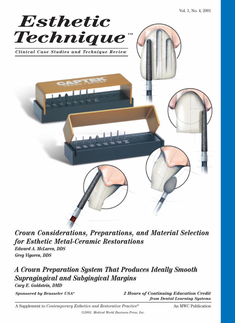
Crown Considerations, Preparations, and Material Selectionfor Esthetic Metal-Ceramic RestorationsEdward A. McLaren, DDSGreg Vigoren, DDS
A Crown Preparation System That Produces Ideally SmoothSupragingival and Subgingival MarginsCary E. Goldstein, DMD
Sponsored by Brasseler USA® 2 Hours of Continuing Education Credit
from Dental Learning Systems
A Supplement to Contemporary Esthetics and Restorative Practice® An MWC Publication
©2001. Medical World Business Press, Inc.
Esthetic Technique™
Clinical Case Studies and Technique Review
Vol. 1, No. 4, 2001
Dear Reader:There has been a continuous increase in demand for more esthet-
ic, or “tooth-colored,” restorations by the dental profession and thepublic over the past few decades. This demand has come aboutbecause of an increased public awareness of and desire for estheticmaterials over metallic-based restorations, the introduction of newtechnologies by industry and the scientific community, and a growingpopulation of clinicians who have elected a preference for enhancingthe esthetics of their patients. It would be safe to assume that all clini-cians would favor an esthetic restorative procedure as long as therewas no significant compromise in health care delivery to theirpatients.
Dr. McLaren and Dr. Vigoren have presented a comprehensive dis-cussion of improving esthetics without compromising basic principlesof ceramic crown restorations. Specifically, they focus their article onthe proper use of the Captek™ technique of a metal-ceramic crown.Using a balance of science and personal experience, they are able todiscuss in depth many of the basic guidelines and principles that canassist clinicians with achieving successful results when using thistechnology. They discuss important factors such as depth of prepara-tions, indications for specifically designed burs or diamonds, type ofcore materials present, and single vs multiple units. The end result is a three-dimensional, integrated outline of the principles of ceramiccrown restorations. Both the experienced and novice clinician willbenefit from the authors’ experiences and knowledge.
Dr. Goldstein reviews the use and significance of a series of dia-mond instruments that should assist clinicians when performing vari-ous esthtic procedures. Behind each procedural step are principlesrelated to optimizing the final esthetic procedure. Dentists should find this information useful in developing a consistent approach totooth preparation rather than relying on a history of inconsistent preparation routines that may not meet the demands of today’s newer technologies.
Dental Learning Systems would like to thank Brasseler USA® forsponsoring this clinical series for the dental profession.
Sincerely,
E. Steven Duke, DDS, MSDProfessor and ChairmanDepartment of Restorative DentistryIndiana University School of Dentistry
The Esthetic Technique™ series is made possible through an educational grant from Brasseler USA®.
To order additional copies call 800-926-7636, x180. D452
Dental Learning Systems 241 Forsgate Drive, Jamesburg, NJ 08831-1676 • (800) 926-7636 • Fax (732) 656-1148
Bruce Crispin,DDS, MS
Nasser Barghi,DDS
Lee Culp, CDT
John Kois,DMD, MSD
Gerard Kugel,DMD, MS
Edward A.McLaren, DDS
Larry Rosenthal,DDS
Howard Strassler, DMD
Douglas A.Terry, DDS
Thomas F.Trinkner, DDS
ADVISORY BOARD
Dental Learning Systems is an ADA Recognized Provider
Academy of General Dentistry Approved National Sponsor. FAGD/MAGD Credit
7/18/1990 to 12/31/2002
Publisher and President, Daniel W. Perkins; Director of Publishing Operations, Ken Senerth; Vice President of Sales and Associate Publisher, Anthony Angelini; EditorialDirector, Allison W. Walker; Associate Projects Editor, Lisa M. Neuman; Projects Director, Eileen R. Henry-Lewis; Copy Editors, Barbara Marino and Susan Costello; DesignDirector, Jennifer Kmenta; Design Director, Special Projects, Liz Arendt; Circulation Director, Jackie Hubler; Northeast Regional Sales Manager, Jeffery E. Gordon; West CoastRegional Sales Manager, Michael Gee; Executive and Advertising Offices, Dental Learning Systems, 241 Forsgate Drive, Jamesburg, NJ 08831-1676, Phone (732) 656-1143, Fax(732) 656-1148.
Postmaster: Send address changes to Contemporary Esthetics and Restorative Practice®, Attn: Data Control, One Broad Avenue, Fairview, NJ 07022-1570. Send correspond-ence regarding subscriptions or address changes to Data Control, One Broad Avenue, Fairview, NJ 07022-1570, or call (800) 603-3512. Periodicals postage paid at Monroe
Township, NJ 08831, and at additional mailing entries.
Contemporary Esthetics and Restorative Practice® (ISSN 1523-2581, USPS 017-212) is published 12 times a year by Dental Learning Systems, 241 Forsgate Drive, Jamesburg,NJ 08831-0505. Copyright © 2001. Medical World Business Press, Inc./A division of Medical World Communications, Inc. Printed in the USA. All rights reserved. No part of thisissue may be reproduced in any form without written permission from the publisher.
Contemporary Esthetics and Restorative Practice® is a registered trademark of Medical World Business Press, Inc. Medical World Communications Corporate Officers:Chairman/CEO, John J. Hennessy; President, Curtis Pickelle; Chief Financial Officer, Steven J. Resnick; Vice President of Business Development, RobertIssler; Vice President of Manufacturing, Frank A. Lake.
D452
Duke sig
3ESTHETIC TECHNIQUE VOL. 1, NO. 4, 2001
LEARNING OBJECTIVESAfter reading this article, the reader should be able to:
• describe the preparation techniques for single- andmultiple-crown situations.
• identify situations in which a core system should be used.
• discuss the clinical situations in which full-coveragecrowns supported by a high-strength core should beconsidered.
• explain the difficulties in controlling proper toothreduction in clinical situations where multiplecrowns are necessary.
Esthetic metal ceramics have seemed almost oxy-moronic to many dentists and patients. Theesthetic push in recent years has been for the
metal-free restoration under the guise that superioresthetics can be obtained only without using metal sub-strates in crown restorations. Also, concerns overpotential metal toxicity have led the public and manydentists to choose metal-free restorations. Many ofthese metal-free systems, marketed as alternatives to
conventional porcelain-fused-to-metal (PFM) restora-tions, have little or no clinical or toxicity data to sup-port their use. High-gold alloys have proven safe andare tolerated well by patients; it has been only the useof nonprecious alloys that have created toxic reactionsin patients.1 Porcelains have proven to be highly bio-compatible, which favors a healthy gingival response,and are frequently used in restorations for veneers,metal-ceramic, or high-strength ceramic copings.2
However, it is possible to fabricate PFM restorationsthat rival any all-ceramic restoration esthetically3 withthe proper use of new-generation porcelains and alloysystems.4 Specifically, the use of a Captek™,a substratewith Vita® Omega 900b, a new-generation, lower-fusingporcelain, has demonstrated excellent esthetic results(Figure 1). The specific framework design parametersfor the esthetic fabrication of porcelain-veneeredCaptek™ crowns is dealt with elsewhere.4,5 As critical asthe design parameters are for the restoration to obtain anesthetic result, also critical is the proper tooth prepara-tion to allow room for the esthetic material.
CROWN CONSIDERATIONSIdeally, the least amount of tooth structure possible
should be removed when preparing teeth for crownrestorations. In situations where minimal tooth structureremoval is required, a porcelain-bonded restoration isindicated, assuming it will satisfy the functional andesthetic requirements of the patient (Figures 2 and 3).6
Excess tooth structure should never be mutilated to sat-isfy the requirements of a certain material when a moreconservative approach will satisfy functional, biologic,and esthetic requirements. Clinical situations thatrequire a conventional PFM are the same for a Captek™-supported restoration.
ABSTRACTIn recent years, many dentists and patients
have opted for metal-free restorations amid con-
cerns regarding esthetics and potential metal
toxicity. The use of a Captek™ substrate with a
new-generation, lower-fusing porcelain has
demonstrated excellent esthetic results, proving
that it is possible to fabricate porcelain-fused-
to-metal restorations that rival any all-ceramic
restoration. This article will discuss the clinical
considerations in the use of crown systems, and
preparation guidelines for esthetic metal-ceram-
ic restorations using Captek™ cores.
Crown Considerations,Preparations, and Material Selection for Esthetic Metal- Ceramic RestorationsEdward A. McLaren, DDS
Greg Vigoren, DDS
Edward A. McLaren, DDSDirector, Center for Esthetic
Dentistry
Director, School for Esthetic Dental
Design
School of Dentistry
University of California at
Los Angeles
Los Angeles, California
Greg Vigoren, DDSAdjunct Faculty
Center for Esthetic Dentistry
School of Dentistry
University of California at
Los Angeles
Los Angeles, California
Private Practice
Newport Beach, California
aCaptek, Altamonte Springs, FL 32714; 800-921-2227bVita Zahnfabrik, Germany distributed in US by Vident™, Brea, CA 92621; 800-848-2726

4 VOL. 1, NO. 4, 2001 ESTHETIC TECHNIQUE
Full-coverage crowns support-ed by a high-strength core should beconsidered in the following clinicalsituations: to replace an existingPFM restoration; when there is acompromised substrate (tooth); andwhen there is need for support forthe porcelain. It is important tounderstand that porcelain gets itsstrength from being bonded to ahigh-strength substrate—eitherenamel, metal, or ceramic core. Ifthe substrate has only dentin orcomposite—both of which are lowmodulus (flexible) materials—thenthe less flexible and more brittleceramic will absorb a disproportion-ate amount of stress under load,which increases the chance for brit-tle failure.7 In a study that looked atthe failure load of bonded, pressedceramics to materials of differentelastic moduli, it was concludedthat the failure load was proportion-al to the flexibility of the substrate.8
The more flexible the substrate, thelower the failure load. Thus, thinteeth or extremely broken-downteeth, especially those with a large(and flexible) composite buildup,should not be considered for bond-ed porcelain restorations that haveno core support. In these clinical sit-uations, a crown should be placedthat uses a high-strength core.
A core system should also beused in situations where porcelainwould be subjected to high shearand tensile stresses. Porcelain can
extend several millimeters occlusal-ly or incisally in situations wherethe stresses placed on it will be pri-marily compressive in nature andthe substrate is stiff.
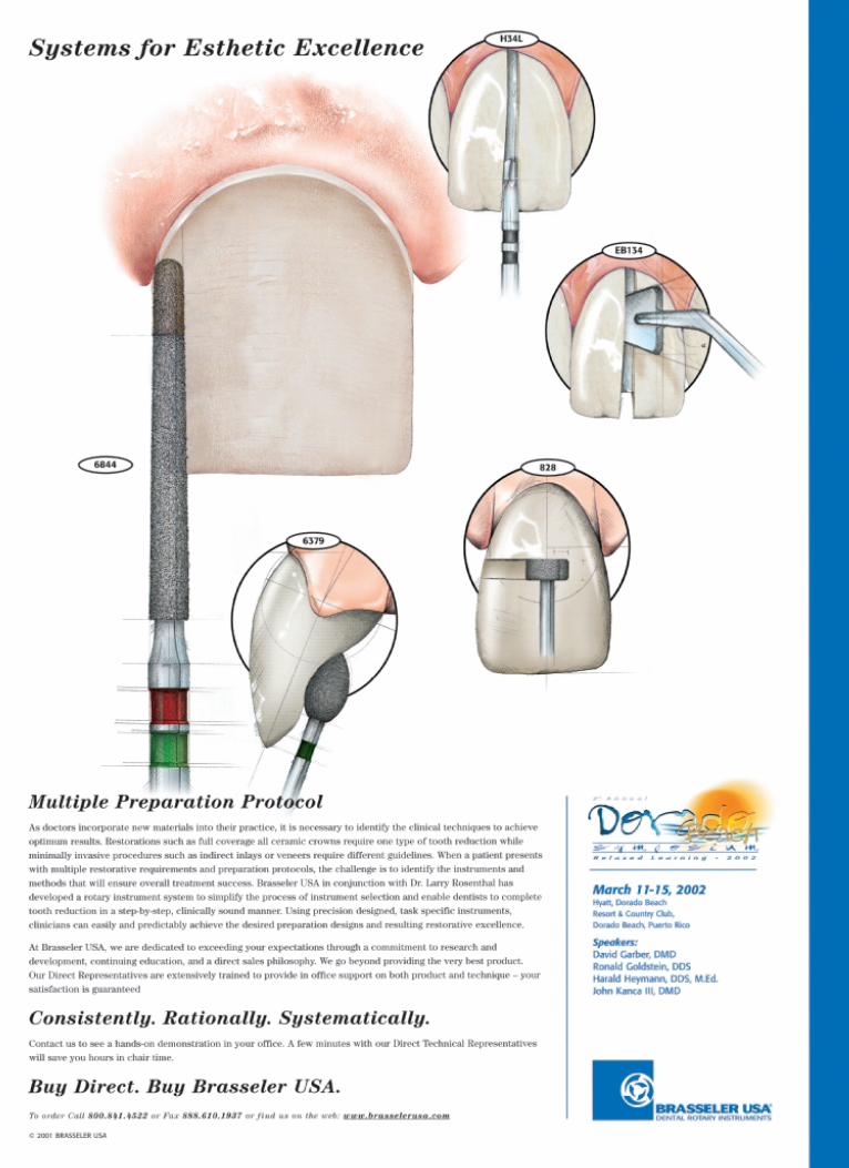
PREPARATIONSThe correct reduction for the
room necessary for the estheticfabrication of a Captek™ or, for thatmatter, any PFM restoration isparamount. Evaluation of morethan 700 Captek™ restorations inwhich the primary author per-formed all clinical and ceramic pro-cedures has led to the determina-tion that 1.2 mm of overall labialcrown thickness was the minimumideal dimension for predictableesthetics. All of those 700-plusrestorations were documented andmeasured for final crown dimen-sions before cementation, and sub-jective analysis was made as to theesthetic success of the cementedrestorations. It was determined
that a 1.2-mm facial crown dimen-sion allowed predictable shapereproduction and subjective esthet-ic success. Thus, it is recommend-ed to reduce facially to allow for afinal restoration with a facialdimension of 1.2 mm. This may notmean that 1.2 mm of tooth struc-ture needs to be removed facially.Esthetic and functional require-ments will dictate whether more orless tooth should be removed,because labializing or lingualizingthe facial surface may need to beaccomplished. The correct reduc-tion for a single crown is fairlyeasy, but the correct reduction formultiple-crown situations is muchmore complex.
SINGLE-CROWN SITUATIONSReduction for single crowns is
generally dictated by the adjacentteeth, which is easy to visualize andcompare. It is sometimes necessaryto build up the tooth to the desiredfinal shape before initiating thepreparation process to better visu-alize the correct amount of reduc-tion necessary for the final restora-tion. Before crown preparation,caries and old restoration removalwith concomitant foundationrestoration placement should beaccomplished.
Small carious lesions or oldrestorations can be removed duringthe gross reduction steps of thecrown preparation. Figure 4 dem-
It is possible to fab-
ricate PFM restora-
tions that rival any all-
ceramic restoration
esthetically with the
proper use of new-
generation porcelains
and alloy systems.
Figure 2—Preoperative condition
where the structural integrity of the
remaining tooth was ideal for bond-
ed porcelain.
Figure 3—Postoperative view of bond-
ed porcelain veneers on teeth Nos. 8 and
9. Note the esthetic integration of these
very conservative, bonded porcelain
restorations.
Figure 1—Porcelain-fused-to-gold
crowns on teeth Nos. 8 and 9 fabricat-
ed with a Captek™ substrate and Vita®
Omega 900, a new-generation, lower-
fusing porcelain.
5ESTHETIC TECHNIQUE VOL. 1, NO. 4, 2001
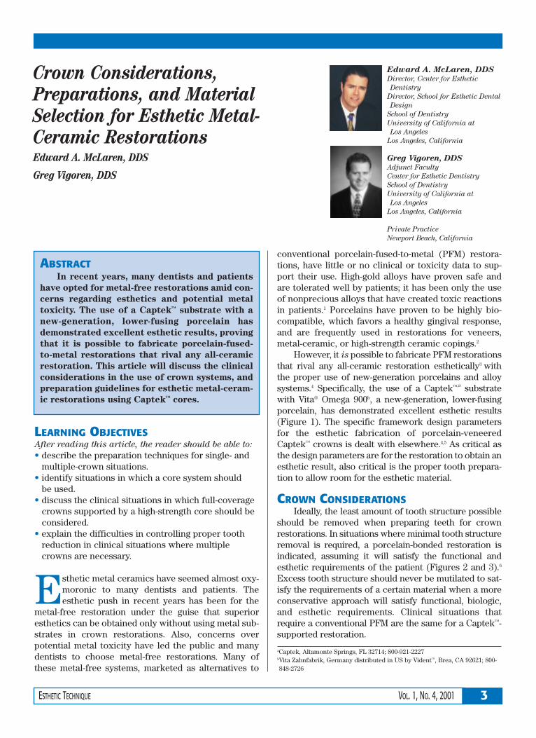
onstrates the UCLA Center forEsthetic Dentistry Metal-Ceramicand All-Ceramic Preparation Kit(338.31.620) by Dr. McLarenc. Thefirst step in the process is to breakcontact with the adjacent teeth(Figure 5) using the coarse diamond(#5858-014c). The marginal area isprepared next with either a KS1c orKS2c diamond (Figures 6A and 6B).The marginal preparation is doneright to the level of the gingiva(Figure 7). The marginal area is themost critical area when preparingfor a PFM with a porcelain margin oran all-ceramic crown, and experi-ence has shown that a 360-degree, 1-mm shoulder preparation with a 90-degree exit angle and rounded inter-nal line angles is ideal for theserestorations (Figure 8).
All other areas of the prepara-tion can be altered on the workeddie by the ceramist if necessary tocreate more room, and can subse-quently be adjusted intraorally bythe dentist. An underprepared
marginal area is impossible tocompensate for in the laboratoryand would require repreparationand reimpressioning, hence therationale for preparing the marginearly in the preparation process.
Axial reduction is next, and
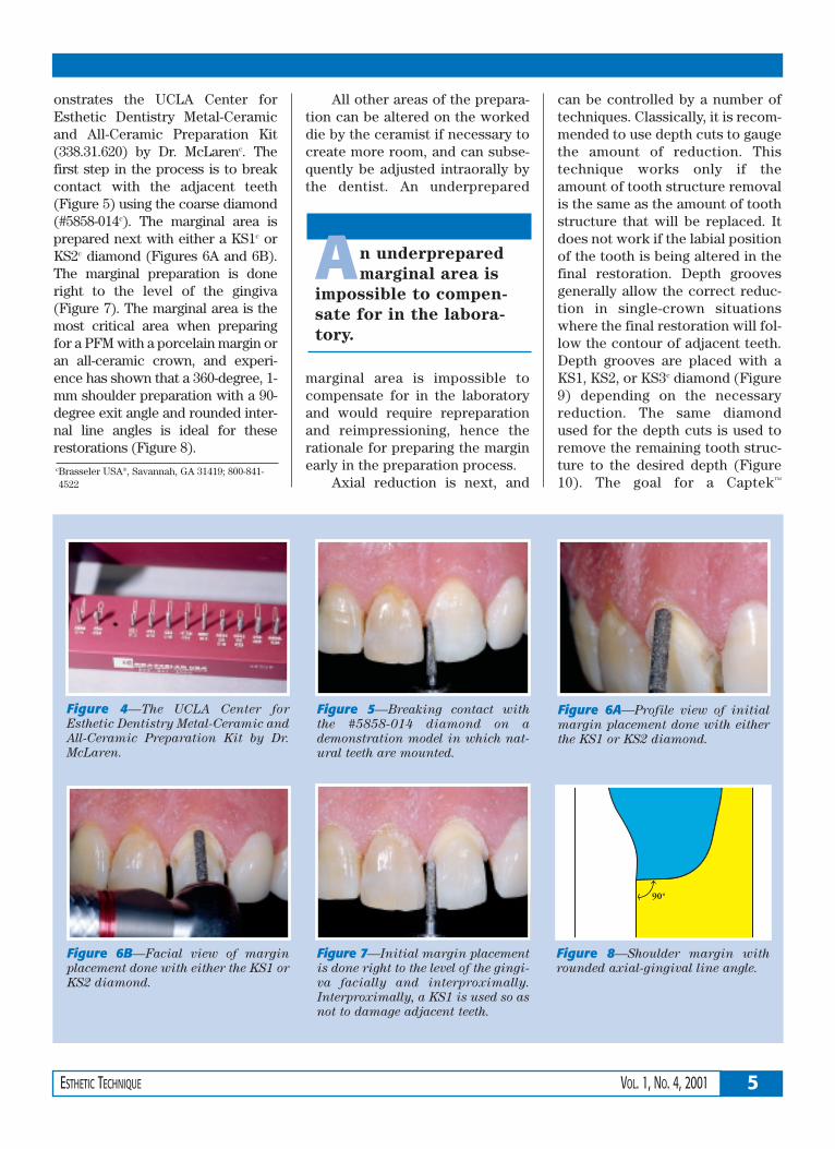
can be controlled by a number oftechniques. Classically, it is recom-mended to use depth cuts to gaugethe amount of reduction. Thistechnique works only if theamount of tooth structure removalis the same as the amount of toothstructure that will be replaced. Itdoes not work if the labial positionof the tooth is being altered in thefinal restoration. Depth groovesgenerally allow the correct reduc-tion in single-crown situationswhere the final restoration will fol-low the contour of adjacent teeth.Depth grooves are placed with aKS1, KS2, or KS3c diamond (Figure9) depending on the necessaryreduction. The same diamondused for the depth cuts is used toremove the remaining tooth struc-ture to the desired depth (Figure10). The goal for a Captek™
Figure 5—Breaking contact with
the #5858-014 diamond on a
demonstration model in which nat-
ural teeth are mounted.
Figure 6A—Profile view of initial
margin placement done with either
the KS1 or KS2 diamond.
Figure 6B—Facial view of margin
placement done with either the KS1 or
KS2 diamond.
Figure 7—Initial margin placement
is done right to the level of the gingi-
va facially and interproximally.
Interproximally, a KS1 is used so as
not to damage adjacent teeth.
An underprepared
marginal area is
impossible to compen-
sate for in the labora-
tory.
Figure 4—The UCLA Center for
Esthetic Dentistry Metal-Ceramic and
All-Ceramic Preparation Kit by Dr.
McLaren.
Figure 8—Shoulder margin with
rounded axial-gingival line angle.
cBrasseler USA®, Savannah, GA 31419; 800-841-4522
restoration should be to allow for1.2 mm of space labially. Incisal orocclusal reduction is initiated witha KS3 diamond. Incisal edge reduc-tion of 2 mm is adequate for goodesthetics. The diameter of the KS3is 1.6 mm, so going slightly deepergives the necessary 2-mm reduc-tion (Figure 11). The adjacentincisal edge can also be gauged as
a reduction guide. Posteriorly, it isnecessary to have 2.5 mm ofocclusal reduction for both esthet-ic metal-ceramic and all-ceramicrestorations, especially if natural,unworn occlusal anatomy isdesired in the final restoration.The best aid the authors havefound to accomplish this reduc-
tion is the 2-mm Reduction Guided.If the 2-mm guide passes with onlyslight binding through the occlud-ed opposing arches, then there isclose to 2.5 mm of interocclusalspace (Figure 12). Lingual reduc-tion is done with the KS4-024c dia-mond for anterior teeth (Figure13) and a KS2 or KS3 for posteriorteeth (Figure 14) to allow for atleast 0.7 mm of crown thicknessfor anterior teeth and 1.0 mmthickness for posterior teeth.
Before finishing the prepara-tion, one layer of Ultrapack® #000e
is placed in the sulcus (Figure 15).This generally gives 0.5 mm of gin-gival displacement. The margin isapically positioned 0.5 mm witheither a KS1 or KS2 (Figure 16)diamond or, in cases with exces-sively scalloped gingival margins,the KS6c. The depth to which themargin should be placed in the sul-cus is a complex issue and affect-
6 VOL. 1, NO. 4, 2001 ESTHETIC TECHNIQUE
Figure 11—Incisal edge reduction
using the KS3 diamond.
Figure 12—Using the 2-mm
Reduction Guide to check the occlusal
reduction on tooth No. 30.
Figure 13—Lingual reduction is
accomplished on anterior teeth with
the egg-shaped KS4-024 diamond.
Figure 15—A clinical case with
Ultrapak® #000 cord placed to
obtain initial gingival retraction so
that the margins can be finished.
The margin was prepared to the
level of the retraction cord.
Figure 16—The apical position of
the margin is placed with either
a KS1, KS2, or, as shown, a KS6
diamond.
Figure 14—Lingual reduction for
posterior teeth is done with a KS2 or
KS3 diamond.
Figure 9—Depth grooves are placed
with either a KS1 or KS2 diamond.
Figure 10—Axial reduction is com-
pleted using the same KS diamond
used for the depth cuts.
eUltradent Products, Inc, South Jordan, UT 84095;800-552-5512
dKerrLab, Orange, CA 92867; 800-537-7123
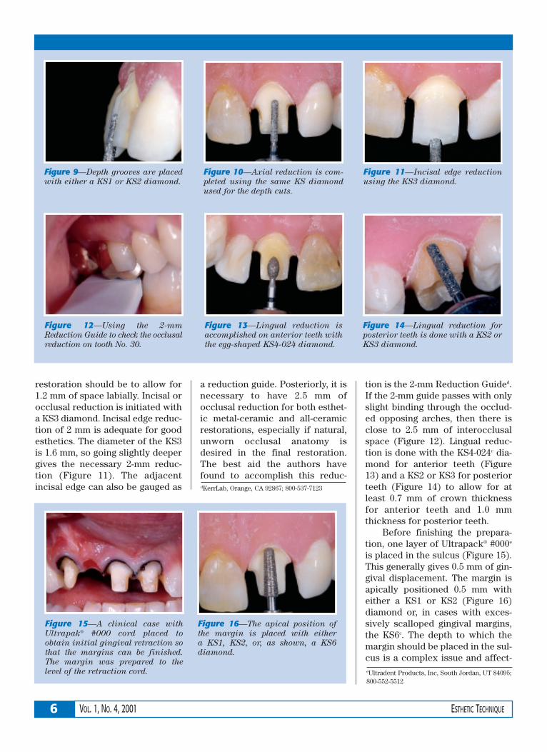
ed by many variables.9,10 The ulti-mate goal of margin placement isto have an esthetic restoration/gin-gival interface without biologiccomplications (ie, violation of bio-logic width). The marginal area isthen finished with either the#8845KR-018c or #8845KR-025c fin-ishing diamonds, or the H158-014c
carbide finishing bur (Figures 17and 18). Axial contours are fin-ished with the #8881-014c or#8856L-020c fine diamonds (Figure19). For all-ceramic crowns, it iscritical to round all internal lineangles with one of the fine dia-monds. This minimizes stress con-centrations in the ceramic crownby eliminating sharp angles.
PREPARATIONS FORMULTIPLE CROWNS
Clinical situations in whichmultiple crowns are necessary
present extreme difficulties incontrolling proper tooth reduc-tion. Often, old crowns are beingremoved or the three-dimensionalshape of the tooth is going to bealtered significantly. In these situa-
tions, axial depth grooves are oflimited value. It has generally beenrecommended to make a poly-propylene vacuum-formed matrixto be used intraorally to controltooth reduction (Figure 20). Whilethis is a useful adjunct, it is fraught
with potential problems. Whenplaced over the teeth, it is difficultto judge if the changes in toothform that are incorporated into thematrix are in fact correct estheti-cally and functionally. Also, it iseasy to displace the matrix in onedirection or another up to almost 1mm without knowing it. All of theabove conditions could easily leadto overprepared or underpreparedteeth. McLean described a tech-nique where the prototype (tempo-rary) is completed on the preparedteeth and then measured with acaliper to gauge proper toothreduction.11 Although this is thebest method, two or three relinesmay be necessary to finalize thereduction amount, which is notvery practical. One problem withthis technique is that acrylicmonomers left on the preparedtooth will inhibit the set of
7ESTHETIC TECHNIQUE VOL. 1, NO. 4, 2001
Figure 17—Final margin finishing
is completed with either the
#8845KR-018 or #8845KR-025 fine
diamond.
Figure 18—The finishing carbide
bur H158-014 can also be used to
finish the margin.
Figure 19—Axial contours are fin-
ished with either the #8881-014 or
#8856L-020 fine diamond.
Figure 22A—Rough preparations
immediately after old restoration
removal.
Figure 21—A clinical case in which
all maxillary teeth are to receive all-
ceramic crowns. In these situations
where adjacent teeth are prepared,
the contact is broken using either
the KS1 or KS2 diamond.
Figure 20—Using a polypropylene
matrix to gauge gross reduction.
The depth to which
the margin should
be placed in the sulcus
is a complex issue and
affected by many vari-
ables.
polyvinyl siloxane (PVS) impres-sion materials. Therefore, an alter-native technique will be discussedto control axial reduction.
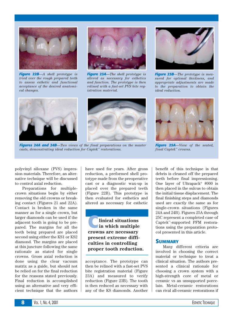
Preparations for multiple-crown situations begin by eitherremoving the old crowns or break-ing contact (Figures 21 and 22A).Contact is broken in the samemanner as for a single crown, butlarger diamonds can be used if theadjacent tooth is going to be pre-pared. The margins for all theteeth being prepared are placedsecond using either the KS1 or KS2diamond. The margins are placedat this juncture following the samerationale as stated for singlecrowns. Gross axial reduction isdone using the clear vacuummatrix as a guide, but should notbe relied on for the final reductionfor the reasons stated previously.Final reduction is accomplishedusing an alternative and very effi-cient technique that the authors
have used for years. After grossreduction, a preformed shell pro-totype made from the preoperativecast or a diagnostic wax-up isplaced over the prepared teeth(Figure 22B). This prototype isthen evaluated for esthetics andaltered as necessary for esthetic
acceptance. The prototype canthen be relined with a fast-set PVSbite registration material (Figure23A) and measured to verifyreduction (Figure 23B). The toothis then reduced as necessary withany of the KS diamonds. Another
benefit of this technique is thatdebris is cleaned off the preparedteeth before final impressioning.One layer of Ultrapack® #000 isthen placed in the sulcus to obtainthe initial tissue displacement. Thefinal finishing steps and diamondsused are exactly the same as forsingle-crown situations (Figures24A and 24B). Figures 25A through25C represent a completed case ofCaptek™-supported PFM restora-tions using the preparation proto-col presented in this article.
SUMMARYMany different criteria are
involved in choosing the correctmaterial or technique to treat aclinical situation. The authors pre-sented a clinical rationale forchoosing a crown system with ahigh-strength core of metal orceramic vs an unsupported porce-lain. Metal-ceramic restorationscan rival all-ceramic restorations if
8 VOL. 1, NO. 4, 2001 ESTHETIC TECHNIQUE
C linical situations
in which multiple
crowns are necessary
present extreme diffi-
culties in controlling
proper tooth reduction.
Figure 23B—The prototype is mea-
sured for optimal thickness, and
appropriate adjustments are made
to the preparation to obtain the
ideal reduction.
Figure 23A—The shell prototype is
altered as necessary for esthetics
and function. The prototype is then
relined with a fast-set PVS bite reg-
istration material.
Figure 22B—A shell prototype is
tried over the rough prepared teeth
to assess esthetic and functional
acceptance of the desired anatomi-
cal changes.
Figures 24A and 24B—Two views of the final preparations on the master
casts, demonstrating ideal reduction for Captek™ restorations.
Figure 25A—View of the seated,
final Captek™ crowns.
the proper materials and design cri-teria are followed. Captek™ restora-tions require 1.2 mm of facialreduction for a predictable estheticoutcome, which is less than what isgenerally required for conventionalmetal-ceramics. Proper prepara-tion techniques are of primaryimportance for esthetic successwith metal-ceramic restorations.
Authors’ Note: All clinical and
laboratory procedures were per-
formed by Dr. McLaren.
REFERENCES1. Schuster GS, LeFebvre CA, Watana JC, et al:
Biocompatibility of posterior restorativematerials. J Calif Dent Assoc 24(9):17-31,1996.
2. 1990 Survey of Dental Restorations. In:Survey of Dental Services 1990. Chicago,
ADA Survey Center, 1990.
3. Winter RR: Achieving esthetic ceramicrestorations. J Calif Dent Assoc 18(9):21-24,1990.
4. McLaren EA: Utilization of advanced metal-ceramic technology: clinical and laboratoryprocedures for a lower-fusing porcelain.Pract Periodontics Aesthet Dent 10(7):835-842, 1998.
5. McLaren EA: Forward to the past: a renais-sance in ceramometal technology. Contemp
Esthet Rest Pract 2(suppl 6):6-13, 1998.
6. Goldstein RE: Diagnostic dilemma: to bond,laminate or crown? Int J Periodontics
Restorative Dent 7(5):8-29, 1987.
7. McLaren EA: All-ceramic alternatives to conventional metal-ceramic restorations.Compend Contin Educ Dent 19(3):307-326,1998.
8. Scherrer SS, de Rijk WG: The fracture resis-tance of all-ceramic crowns on supportingstructures with different elastic moduli. Int J
Prosthodont 6(5):462-467, 1993.
9. Kois JC: Altering gingival levels: the restora-tive connection, part I: biologic variables. JEsthet Dent 6(1):3-9, 1994.
10. Kois JC: New paradigms for anterior toothpreparation: rationale and technique.Contemp Esthet Dent 2(1):1-8, 1996.
11. McLean JW: The Science and Art of Dental
Ceramics. Chicago, Quintessence PublishingCo, pp 263-268, 1979.
9ESTHETIC TECHNIQUE VOL. 1, NO. 4, 2001
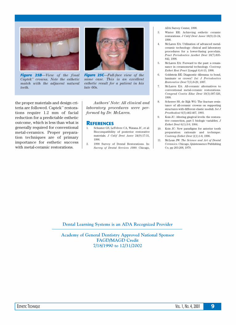
Figure 25C—Full-face view of the
same case. This is an excellent
esthetic result for a patient in his
late 60s.
Figure 25B—View of the final
Captek™ crowns. Note the esthetic
match with the adjacent natural
teeth.
Dental Learning Systems is an ADA Recognized Provider
Academy of General Dentistry Approved National Sponsor FAGD/MAGD Credit
7/18/1990 to 12/31/2002
10 VOL. 1, NO. 4, 2001 ESTHETIC TECHNIQUE
In developing the Goldstein Crown Design Kit™,a,the goal was to provide a selection of diamonds andstones that simplify crown preparation procedures in avariety of situations. Containing only nine instruments,the kit is designed to permit the dentist to address theesthetic requirements of anterior or posterior crowncases for patients with high or low lip lines. The kit’sdiamond selection was designed to accommodate allporcelain crowns, aluminous-core porcelain crowns,porcelain-fused-to-metal (PFM) crowns, or those fabri-
cated from gold. Smooth-finished, precise margins freeof jagged enamel edges can be produced supragingival-ly or subgingivally, according to specific case require-ments, enhancing the adaptation of the restoration tothe tooth. Impressions made after preparation with thekit components reveal exceptional detail and facilitateaccurate laboratory communications.
This article describes the multiple approaches toanterior preparations offered by the Goldstein CrownDesign Kit.
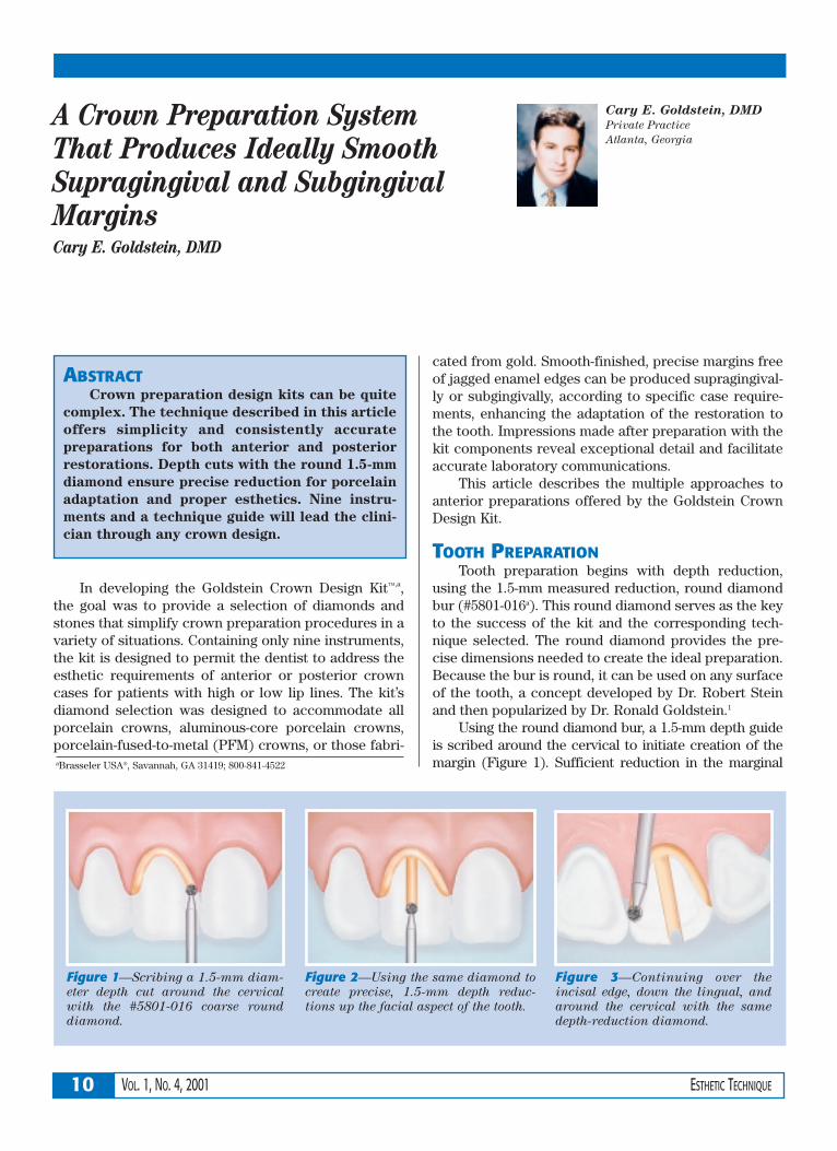
TOOTH PREPARATIONTooth preparation begins with depth reduction,
using the 1.5-mm measured reduction, round diamondbur (#5801-016a). This round diamond serves as the keyto the success of the kit and the corresponding tech-nique selected. The round diamond provides the pre-cise dimensions needed to create the ideal preparation.Because the bur is round, it can be used on any surfaceof the tooth, a concept developed by Dr. Robert Steinand then popularized by Dr. Ronald Goldstein.1
Using the round diamond bur, a 1.5-mm depth guideis scribed around the cervical to initiate creation of themargin (Figure 1). Sufficient reduction in the marginal
ABSTRACTCrown preparation design kits can be quite
complex. The technique described in this article
offers simplicity and consistently accurate
preparations for both anterior and posterior
restorations. Depth cuts with the round 1.5-mm
diamond ensure precise reduction for porcelain
adaptation and proper esthetics. Nine instru-
ments and a technique guide will lead the clini-
cian through any crown design.
A Crown Preparation System That Produces Ideally SmoothSupragingival and SubgingivalMarginsCary E. Goldstein, DMD
Cary E. Goldstein, DMD
Private Practice
Atlanta, Georgia
Figure 1—Scribing a 1.5-mm diam-
eter depth cut around the cervical
with the #5801-016 coarse round
diamond.
Figure 2—Using the same diamond to
create precise, 1.5-mm depth reduc-
tions up the facial aspect of the tooth.
Figure 3—Continuing over the
incisal edge, down the lingual, and
around the cervical with the same
depth-reduction diamond.
aBrasseler USA®, Savannah, GA 31419; 800-841-4522
11ESTHETIC TECHNIQUE VOL. 1, NO. 4, 2001
area permits ideal adaptation ofeither an all-ceramic crown or aPFM crown with a large enoughporcelain margin to obscure themetal. Although some clinicians mayprefer a depth reduction measuringless than 1.5 mm, the maximumdepth produced by the bur yields amargin of ideal thickness, particular-ly from the laboratory technician’sperspective, because space is suffi-cient to develop an esthetic margin.
After the initial depth-reduc-tion procedure, the same diamondis applied up the facial aspect of thetooth, producing an exact 1.5-mmfacial trough (Figure 2). The sameround depth-reduction diamondcan next be used over the incisaledge, swept down onto the lingualaspect of the tooth, and then usedto scribe a 1.5-mm line around thecervical of the lingual (Figure 3).
The next diamond employed in
this technique is either the small(#5856-016a) or large (#5856-021a)barrel-shaped, tapered, coarsebulk-reduction diamond. For asmall tooth, such as a lateralincisor, the small barrel-shaped dia-mond should be selected. For alarger tooth, such as a molar orcentral incisor, the larger diamondis an ideal choice.
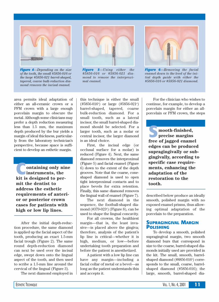
First, the incisal edge (orocclusal surface for a molar) isreduced (Figure 4). Next, the samediamond removes the interproximal(Figure 5) and facial enamel (Figure6) down to the extent of the depthgrooves. Note that the coarse, cone-shaped diamond is used to opentight interproximal contacts and toplace bevels for extra retention.Finally, this same diamond removesthe lingual wall of enamel (Figure 7).
The next diamond in thesequence, the football-shaped dia-mond (#379-023a) (Figure 8), can beused to shape the lingual concavity.
For all crowns, the healthiestmargin—that is, the least inva-sive—is placed above the gingiva;therefore, analysis of the patient’slip line is critical—whether it ishigh, medium, or low—beforeundertaking tooth preparation andbefore the patient is anesthetized.
A patient with a low lip line canhave any margin—including afacial collar of metal or a bevel—aslong as the patient understands thisand accepts it.
For the clinician who wishes tocontinue, for example, to develop aporcelain margin for either an all-porcelain or PFM crown, the steps
described below produce an ideallysmooth, polished margin with noexposed enamel prisms, thus allow-ing optimal adaptation of theporcelain to the preparation.
SUPRAGINGIVAL MARGINPOLISHING
To develop a smooth, polishedsupragingival margin, two smoothdiamond burs that correspond insize to the coarse, barrel-shaped dia-monds initially used are provided inthe kit. The small, smooth, barrel-shaped diamond (#8856-016a) corre-sponds to the small, coarse, barrel-shaped diamond (#5856-016); thelarge, smooth, barrel-shaped dia-
Figure 4—Depending on the size
of the tooth, the small #5856-016 or
the large #5856-021 barrel-shaped,
tapered, coarse bulk-reduction dia-
mond removes the incisal enamel.
Figure 5—Using either the
#5856-016 or #5856-021 dia-
mond to remove the interproxi-
mal enamel.
Figure 6—Removing the facial
enamel down to the level of the ini-
tial depth guide with either the
#5856-016 or #5856-021 diamond.
Containing only nine
instruments, the
kit is designed to per-
mit the dentist to
address the esthetic
requirements of anteri-
or or posterior crown
cases for patients with
high or low lip lines.
Smooth-finished,
precise margins
free of jagged enamel
edges can be produced
supragingivally or sub-
gingivally, according to
specific case require-
ments, enhancing the
adaptation of the
restoration to the
tooth.
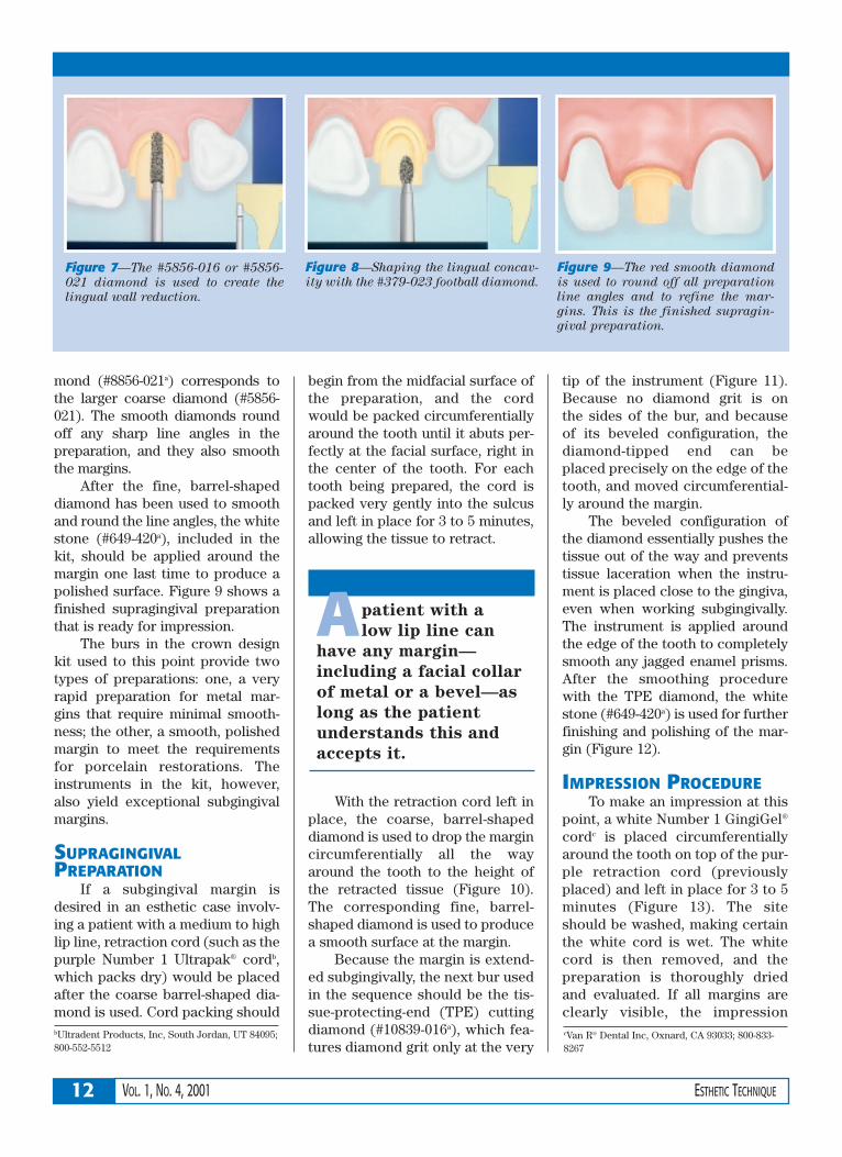
mond (#8856-021a) corresponds tothe larger coarse diamond (#5856-021). The smooth diamonds roundoff any sharp line angles in thepreparation, and they also smooththe margins.
After the fine, barrel-shapeddiamond has been used to smoothand round the line angles, the whitestone (#649-420a), included in thekit, should be applied around themargin one last time to produce apolished surface. Figure 9 shows afinished supragingival preparationthat is ready for impression.
The burs in the crown designkit used to this point provide twotypes of preparations: one, a veryrapid preparation for metal mar-gins that require minimal smooth-ness; the other, a smooth, polishedmargin to meet the requirementsfor porcelain restorations. Theinstruments in the kit, however,also yield exceptional subgingivalmargins.
SUPRAGINGIVALPREPARATION
If a subgingival margin isdesired in an esthetic case involv-ing a patient with a medium to highlip line, retraction cord (such as thepurple Number 1 Ultrapak® cordb,which packs dry) would be placedafter the coarse barrel-shaped dia-mond is used. Cord packing should
begin from the midfacial surface ofthe preparation, and the cordwould be packed circumferentiallyaround the tooth until it abuts per-fectly at the facial surface, right inthe center of the tooth. For eachtooth being prepared, the cord ispacked very gently into the sulcusand left in place for 3 to 5 minutes,allowing the tissue to retract.
With the retraction cord left inplace, the coarse, barrel-shapeddiamond is used to drop the margincircumferentially all the wayaround the tooth to the height ofthe retracted tissue (Figure 10).The corresponding fine, barrel-shaped diamond is used to producea smooth surface at the margin.
Because the margin is extend-ed subgingivally, the next bur usedin the sequence should be the tis-sue-protecting-end (TPE) cuttingdiamond (#10839-016a), which fea-tures diamond grit only at the very
tip of the instrument (Figure 11).Because no diamond grit is on the sides of the bur, and becauseof its beveled configuration, the diamond-tipped end can be placed precisely on the edge of thetooth, and moved circumferential-ly around the margin.
The beveled configuration ofthe diamond essentially pushes thetissue out of the way and preventstissue laceration when the instru-ment is placed close to the gingiva,even when working subgingivally.The instrument is applied aroundthe edge of the tooth to completelysmooth any jagged enamel prisms.After the smoothing procedurewith the TPE diamond, the whitestone (#649-420a) is used for furtherfinishing and polishing of the mar-gin (Figure 12).
IMPRESSION PROCEDURETo make an impression at this
point, a white Number 1 GingiGel®
cordc is placed circumferentiallyaround the tooth on top of the pur-ple retraction cord (previouslyplaced) and left in place for 3 to 5minutes (Figure 13). The siteshould be washed, making certainthe white cord is wet. The whitecord is then removed, and thepreparation is thoroughly driedand evaluated. If all margins areclearly visible, the impression
12 VOL. 1, NO. 4, 2001 ESTHETIC TECHNIQUE
Figure 7—The #5856-016 or #5856-
021 diamond is used to create the
lingual wall reduction.
Figure 8—Shaping the lingual concav-
ity with the #379-023 football diamond.
Figure 9—The red smooth diamond
is used to round off all preparation
line angles and to refine the mar-
gins. This is the finished supragin-
gival preparation.
cVan R® Dental Inc, Oxnard, CA 93033; 800-833-8267
Apatient with a
low lip line can
have any margin—
including a facial collar
of metal or a bevel—as
long as the patient
understands this and
accepts it.
bUltradent Products, Inc, South Jordan, UT 84095;800-552-5512
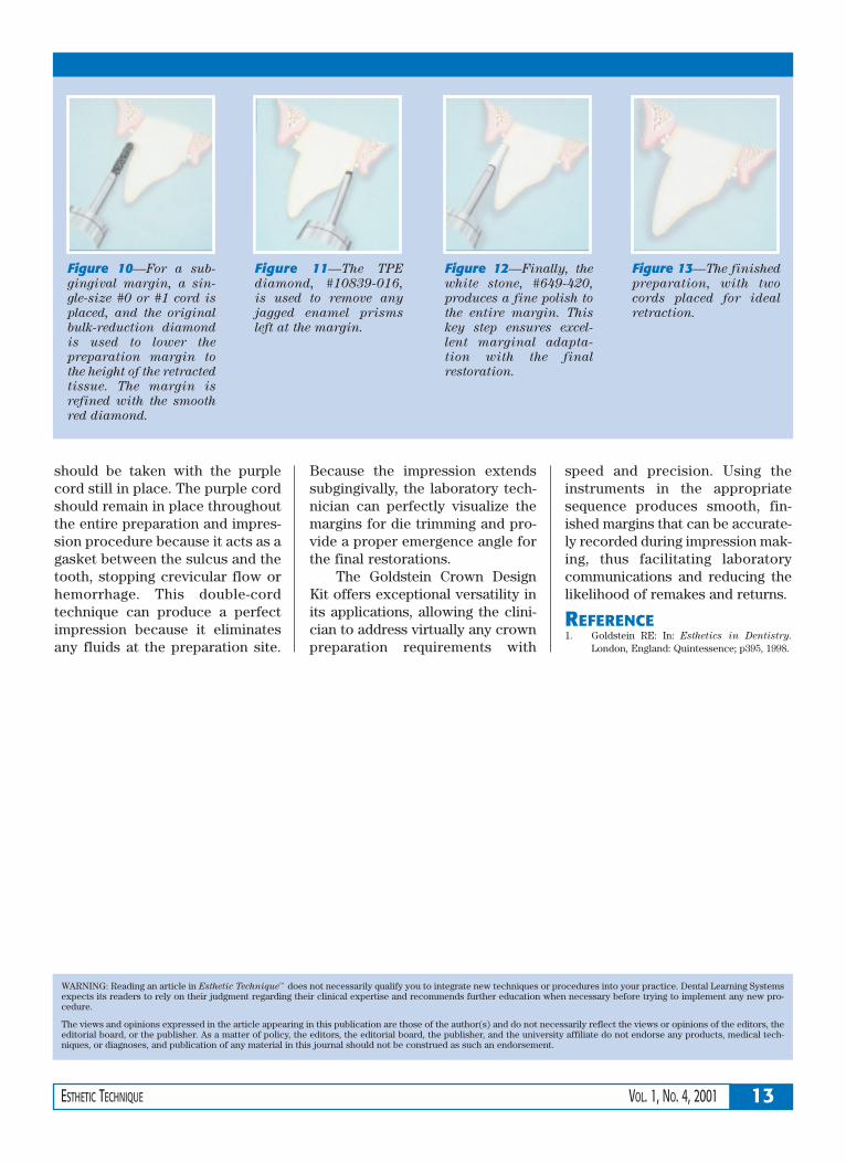
should be taken with the purplecord still in place. The purple cordshould remain in place throughoutthe entire preparation and impres-sion procedure because it acts as agasket between the sulcus and thetooth, stopping crevicular flow orhemorrhage. This double-cordtechnique can produce a perfectimpression because it eliminatesany fluids at the preparation site.
Because the impression extendssubgingivally, the laboratory tech-nician can perfectly visualize themargins for die trimming and pro-vide a proper emergence angle forthe final restorations.
The Goldstein Crown DesignKit offers exceptional versatility inits applications, allowing the clini-cian to address virtually any crownpreparation requirements with
speed and precision. Using theinstruments in the appropriatesequence produces smooth, fin-ished margins that can be accurate-ly recorded during impression mak-ing, thus facilitating laboratorycommunications and reducing thelikelihood of remakes and returns.
REFERENCE1. Goldstein RE: In: Esthetics in Dentistry.
London, England: Quintessence; p395, 1998.
13ESTHETIC TECHNIQUE VOL. 1, NO. 4, 2001
WARNING: Reading an article in Esthetic Technique™ does not necessarily qualify you to integrate new techniques or procedures into your practice. Dental Learning Systemsexpects its readers to rely on their judgment regarding their clinical expertise and recommends further education when necessary before trying to implement any new pro-cedure.
The views and opinions expressed in the article appearing in this publication are those of the author(s) and do not necessarily reflect the views or opinions of the editors, theeditorial board, or the publisher. As a matter of policy, the editors, the editorial board, the publisher, and the university affiliate do not endorse any products, medical tech-niques, or diagnoses, and publication of any material in this journal should not be construed as such an endorsement.
Figure 12—Finally, the
white stone, #649-420,
produces a fine polish to
the entire margin. This
key step ensures excel-
lent marginal adapta-
tion with the final
restoration.
Figure 10—For a sub-
gingival margin, a sin-
gle-size #0 or #1 cord is
placed, and the original
bulk-reduction diamond
is used to lower the
preparation margin to
the height of the retracted
tissue. The margin is
refined with the smooth
red diamond.
Figure 11—The TPE
diamond, #10839-016,
is used to remove any
jagged enamel prisms
left at the margin.
Figure 13—The finished
preparation, with two
cords placed for ideal
retraction.

14 VOL. 1, NO. 4, 2001 ESTHETIC TECHNIQUE
6. What was subsequently employed for the fabri-cation of the provisional restorations?a. matrixb. copper tubec. no provisional was usedd. alginate impression
7. It is sometimes necessary to build up the tooth tothe desired final shape before initiating thepreparation process in order to:a. fabricate the temporary.b. check the occlusion.c. ensure pulpal clearance.d. visualize the correct amount of reduction.
8. For the marginal area of a PFM, experience hasshown that what type of shoulder with roundedinternal line angles is ideal for these restorations?a. 180 degrees, 1 mmb. 270 degrees, 1.5 mmc. 270 degrees, 0.5 mmd. 360 degrees, 1 mm
9. How much incisal edge reduction is adequate forgood esthetics?a. 1 mmb. 1.5 mmc. 2 mmd. 2.5 mm
10. Posteriorly, it is necessary to have how muchocclusal reduction?a. 2 mmb. 2.5 mmc. 3 mmd. 3.5 mm
1. In situations where minimal tooth structureremoval is required, what restoration is indicated?a. full crownb. three-quarters crownc. porcelain-bondedd. amalgam
2. Full-coverage crowns supported by a high-strength core should be considered in whichclinical situation?a. bondingb. brittlec. feldspard. sintering
3. In one study,7 the failure load of bonded, pressedceramics to materials of different elastic moduliwas:a. proportional to the flexibility of the substrate.b. inversely proportional to the flexibility of the
substrate.c. logarithmically proportional to the flexibility
of the substrate.d. not proportional to the flexibility of the sub-
strate.
4. A core system should be used in situationswhere porcelain would be subjected to:a. high shear and high tensile stresses.b. low shear and low tensile stresses.c. high shear and low tensile stresses.d. low shear and high tensile stresses.
5. How many millimeters of overall labial crownthickness was the minimum ideal dimension forpredictable esthetics?a. 0.6 mmb. 1.2 mmc. 1.8 mmd. 2.4 mm
CE QUIZDental Learning Systems provides 2 hours of Continuing Education credit for those who wish to document
their continuing education endeavors. Participants are urged to contact their state registry boards for specialCE requirements.
To receive credit, complete the enclosed answer form, and mail with a check for $20, payable to DentalLearning Systems, to Dental Learning Systems CE Dept., 405 Glenn Drive, Suite 4, Sterling, VA 20164-4432.
Program #: D452
15ESTHETIC TECHNIQUE VOL. 1, NO. 4, 2001
Adjusting and polishing are inevitable for any pro-visional restoration, whether it is composite oracrylic, laboratory-fabricated or office-made.
These new Brasseler USA® instruments can makeadjusting temporaries faster, easier, and more precise.
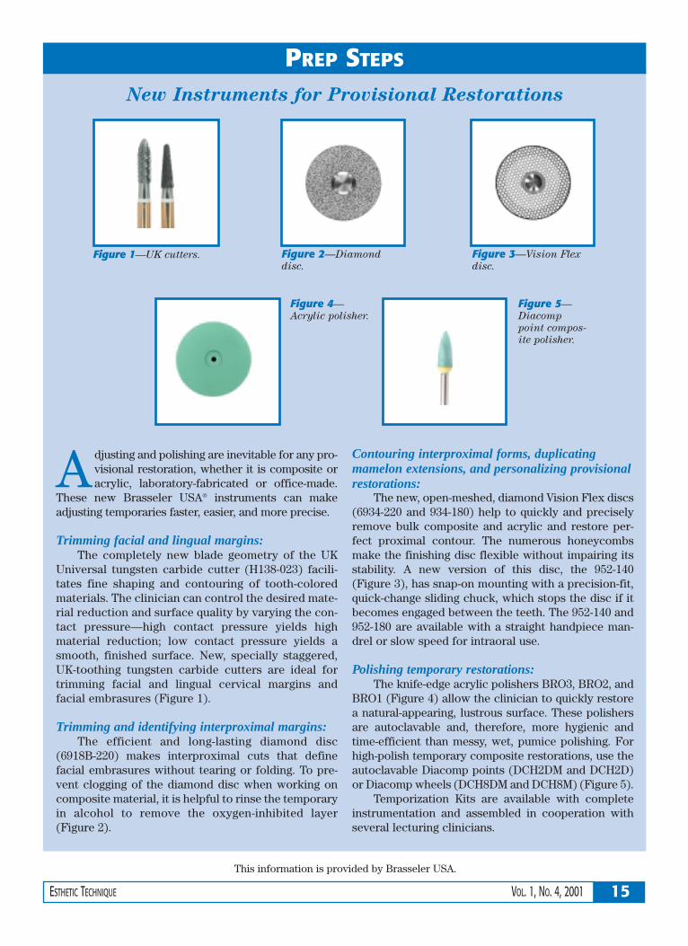
Trimming facial and lingual margins:The completely new blade geometry of the UK
Universal tungsten carbide cutter (H138-023) facili-tates fine shaping and contouring of tooth-coloredmaterials. The clinician can control the desired mate-rial reduction and surface quality by varying the con-tact pressure—high contact pressure yields highmaterial reduction; low contact pressure yields asmooth, finished surface. New, specially staggered,UK-toothing tungsten carbide cutters are ideal fortrimming facial and lingual cervical margins andfacial embrasures (Figure 1).
Trimming and identifying interproximal margins:The efficient and long-lasting diamond disc
(6918B-220) makes interproximal cuts that definefacial embrasures without tearing or folding. To pre-vent clogging of the diamond disc when working oncomposite material, it is helpful to rinse the temporaryin alcohol to remove the oxygen-inhibited layer(Figure 2).
Contouring interproximal forms, duplicatingmamelon extensions, and personalizing provisionalrestorations:
The new, open-meshed, diamond Vision Flex discs(6934-220 and 934-180) help to quickly and preciselyremove bulk composite and acrylic and restore per-fect proximal contour. The numerous honeycombsmake the finishing disc flexible without impairing itsstability. A new version of this disc, the 952-140(Figure 3), has snap-on mounting with a precision-fit,quick-change sliding chuck, which stops the disc if itbecomes engaged between the teeth. The 952-140 and952-180 are available with a straight handpiece man-drel or slow speed for intraoral use.
Polishing temporary restorations:The knife-edge acrylic polishers BRO3, BRO2, and
BRO1 (Figure 4) allow the clinician to quickly restorea natural-appearing, lustrous surface. These polishersare autoclavable and, therefore, more hygienic andtime-efficient than messy, wet, pumice polishing. Forhigh-polish temporary composite restorations, use theautoclavable Diacomp points (DCH2DM and DCH2D)or Diacomp wheels (DCH8DM and DCH8M) (Figure 5).
Temporization Kits are available with completeinstrumentation and assembled in cooperation withseveral lecturing clinicians.
This information is provided by Brasseler USA.
New Instruments for Provisional Restorations
PREP STEPS
Figure 5—
Diacomp
point compos-
ite polisher.
Figure 1—UK cutters. Figure 3—Vision Flex
disc.
Figure 2—Diamond
disc.
Figure 4—
Acrylic polisher.