Embed Size (px)
Citation preview
INTERNATIONAL JOURNAL OF HEALTH RESEARCH IN MODERN INTEGRATED MEDICAL SCIENCES, ISSN 2394-8612 (P), ISSN 2394-8620 (O), Vol-2, Issue-1, Jan-Mar 2015 17
Original Article
Study of Ventral Horn Cells of Spinal Cord in Different Gestations of
Fetuses
N. Bhimai Devi, V. Sunitha
Abstract: Study of the development of the nervous system is not of mere theoretical interest but it also enables to trace
homologies through various groups of vertebrates. A metameric arrangement of spinal cord has been described long ago
but no satisfactory explanation has been derived for the causes of the position, distribution of cells of origin, and their
myotomic distribution. Various interesting changes are incorporated in development of spinal cord. The differentiation
of alar and basal lamina in to ependymal, mantle, and marginal zones has been observed at 12 weeks of gestation.
Typical nuclear columns were observed at the same time.
Key Words : Greymatter, Multipolar cells, Spinal cord, Ventral horn cells.
Introduction
Corresponding author
Dr. N. Bhimai Devi,
Professor, Dept. of Anatomy
Maharajah’s Institute of Medical Sciences, Nellimarla,
Vizianagaram - 535 217, Andhra Pradesh, India.
It is of greatest utility to understand the logical concept of
complex nature of the nervous system. Consequent to the
development of technology and methodology the detailed
outline of cells that descend in the wall of neural tube
have been a great boon to all. Somewhat common pattern
has been described for all mammals. But differences in
the degree of development of the ventral horn cells in
various gestation of fetuses has been observed in the
present study. According to Anguloy Gonzalez1 there is
no segmental arrangement of motor neurons of the spinal
cord in albinorat. The observations of Kapper and Crosby2
showed that morphological differentiation of musculature
is perhaps dependent upon anatomical differentiation of
nuclear columns of anterior horn cells. No satisfactory
explanation has been derived for the causes of position
and distribution of cells of origin and their myotomic
distribution of neuraxis. Even though full details are
available in as far as adult spinal cord is concerned
literature on fetal spinal cord is still inadequate.
Materials and Methods- 50 dead fetuses were collected
from different private nursing homes of Vizianagaram and
fixed by injecting 10% formalin. Spinal cords were
dissected out. Various segments of spinal cords ranging
from 10 weeks gestation to full term were dissected. The
dissected sections of spinal cord were washed thoroughly
with water and then passed through graded alcohols. Next
they are cleared in Xylene till the tissue became
transparent. After clearing the tissue it was embedded in
the paraffin. The paraffin blocks were kept in refrigerator
for two to three hours before sectioning. Sections of six
to eight microns were taken. They were stained with
Haemotoxyline and Eosin. The ventral horn cells were
studied with regard to their appearance, shape, size and
grouping.
Observations- The grey matter of spinal cord of fetuses
of different gestational ages is been described as follows,
10 weeks- The grey matter does not show typical formation
of ventral and dorsal horns. Closely packed
indistinguishable neuroblasts are seen. The central canal
appeared but the lining epithelium was not clearly
discernable. (Fig-1)
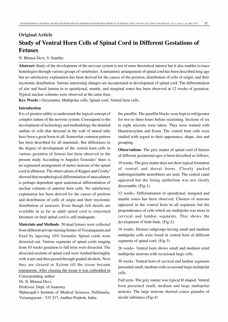
12 weeks- Differentiation of ependymal, marginal and
mantle zones has been observed. Clusters of neurons
appeared in the ventral horn in all segments but the
preponderance of cells which are multipolar was more in
cervical and lumbar segments. This shows the
development of limb buds. (Fig-2)
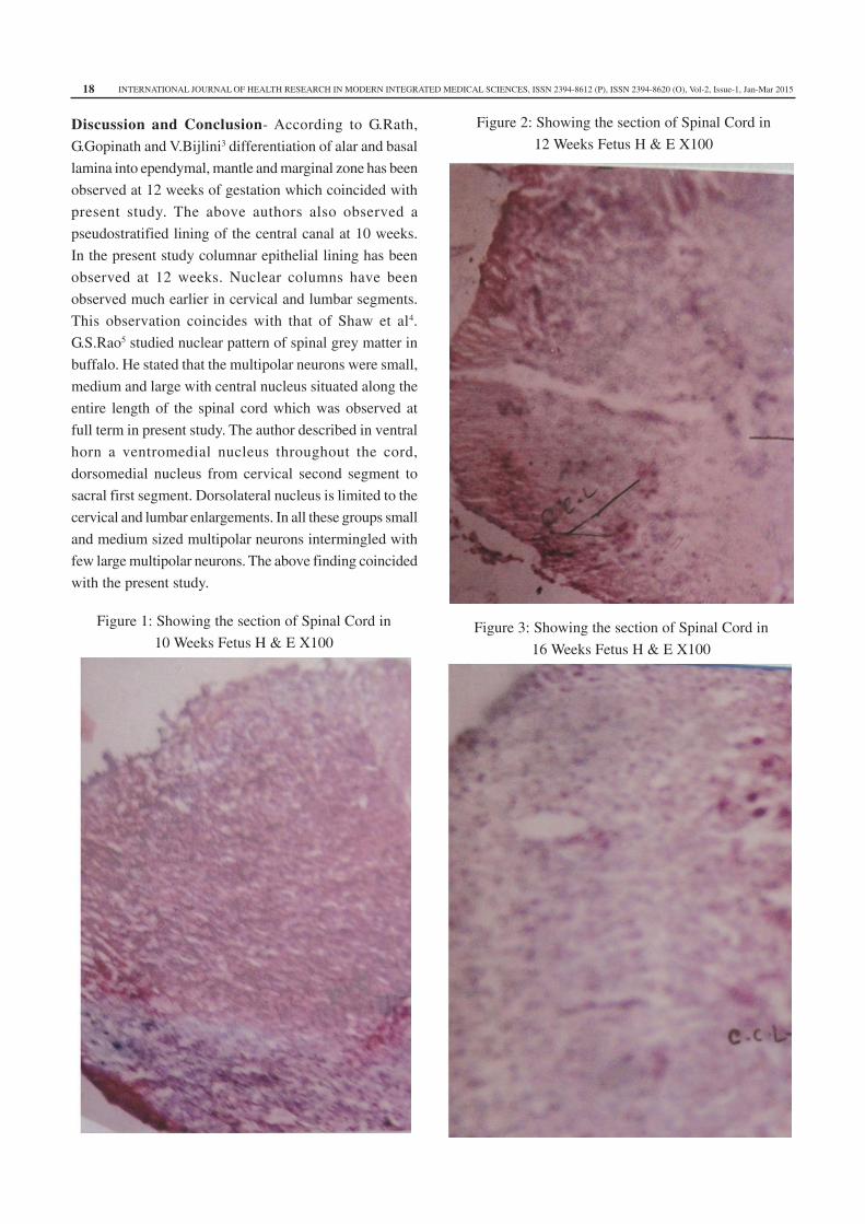
16 weeks- Distinct subgroups having small and medium
multipolar cells were found in ventral horn of different
segments of spinal cord. (Fig-3)
26 weeks- Ventral horn shows small and medium sized
multipolar neurons with occasional large cells.
30 weeks- Ventral horn of cervical end lumbar segments
presented small, medium with occasional large multipolar
cells.
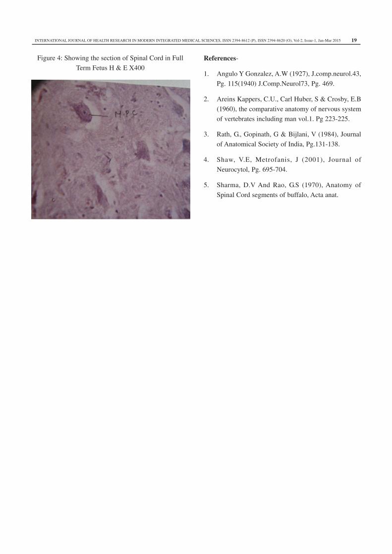
Full term- The grey matter was typical H shaped. Ventral
horn presented small, medium and large multipolar
neurons. The large neurons showed coarse granules of
nissile substance.(Fig-4)
INTERNATIONAL JOURNAL OF HEALTH RESEARCH IN MODERN INTEGRATED MEDICAL SCIENCES, ISSN 2394-8612 (P), ISSN 2394-8620 (O), Vol-2, Issue-1, Jan-Mar 201518
Discussion and Conclusion- According to G.Rath,
G.Gopinath and V.Bijlini3 differentiation of alar and basal
lamina into ependymal, mantle and marginal zone has been
observed at 12 weeks of gestation which coincided with
present study. The above authors also observed a
pseudostratified lining of the central canal at 10 weeks.
In the present study columnar epithelial lining has been
observed at 12 weeks. Nuclear columns have been
observed much earlier in cervical and lumbar segments.
This observation coincides with that of Shaw et al4.
G.S.Rao5 studied nuclear pattern of spinal grey matter in
buffalo. He stated that the multipolar neurons were small,
medium and large with central nucleus situated along the
entire length of the spinal cord which was observed at
full term in present study. The author described in ventral
horn a ventromedial nucleus throughout the cord,
dorsomedial nucleus from cervical second segment to
sacral first segment. Dorsolateral nucleus is limited to the
cervical and lumbar enlargements. In all these groups small
and medium sized multipolar neurons intermingled with
few large multipolar neurons. The above finding coincided
with the present study.
Figure 1: Showing the section of Spinal Cord in
10 Weeks Fetus H & E X100
Figure 2: Showing the section of Spinal Cord in
12 Weeks Fetus H & E X100
Figure 3: Showing the section of Spinal Cord in
16 Weeks Fetus H & E X100
INTERNATIONAL JOURNAL OF HEALTH RESEARCH IN MODERN INTEGRATED MEDICAL SCIENCES, ISSN 2394-8612 (P), ISSN 2394-8620 (O), Vol-2, Issue-1, Jan-Mar 2015 19
Figure 4: Showing the section of Spinal Cord in Full
Term Fetus H & E X400
References-
1. Angulo Y Gonzalez, A.W (1927), J.comp.neurol.43,
Pg. 115(1940) J.Comp.Neurol73, Pg. 469.
2. Areins Kappers, C.U., Carl Huber, S & Crosby, E.B
(1960), the comparative anatomy of nervous system
of vertebrates including man vol.1. Pg 223-225.
3. Rath, G., Gopinath, G & Bijlani, V (1984), Journal
of Anatomical Society of India, Pg.131-138.
4. Shaw, V.E, Metrofanis, J (2001), Journal of
Neurocytol, Pg. 695-704.
5. Sharma, D.V And Rao, G.S (1970), Anatomy of
Spinal Cord segments of buffalo, Acta anat.