Embed Size (px)
Citation preview

Vol. 6: 105-11 l , 1989 DISEASES OF AQUATIC ORGANISMS Dis. aquat. Org. I Published May 11
Rare renal neoplasms in Salmo gairdneri exposed to MNNG (N-methyl-Nr-nitro-N-
nitrosoguanidine)
Brian C. Lee*, Jerry D. Hendricks*', George S. Bailey
Department of Food Science and Technology, Oregon State University, Cowallis, Oregon 97331-6602, USA
ABSTRACT: N-methyl-N'-nitro-N-nitrosoguanidine (MNNG) is a potent liver, stomach, kidney, and swimbladder carcinogen in rainbow trout Salmo gairdnen. Histopathological examination of tissues from trout exposed as fry to 50 ppm MNNG for 30 min revealed 2 types of neoplastic renal lesions not previously reported for this species. Renal cell carcinoma in the corpuscle of Stannius and renal cell, cystic, papillary adenoma of the mesonephric duct were diagnosed. Nephroblastoma and nephrocal- cinotic lesions adjacent to the mesonephric duct adenoma suggested that occlusion of the duct could have been a contributing factor
INTRODUCTION
The rainbow trout has been a versatile and sensitive animal model in the study of chemical carcinogenesis (Hendricks et al. 1980, 1984, Hendricks 1982, Bailey et al. 1987). Inhibition, promotion, and enhancement of liver carcinogenicity by substances which may be pre- sent in the human diet, such as indoles, flavones, or chlorinated hydrocarbon pesticide residues, have been demonstrated in trout (Bailey et al. 1987, Dashwood et al. 1988, Fong et al. 1988, Nunez et al. 1988). Tissues in those studies are routinely subjected to histological examination to confirm gross observations and to determine the types of neoplastic responses.
Two renal tumor types, nephroblastoma and cys- tadenoma, have been reported for rainbow trout after exposure to N-methyl-NI-nitro-N-nitrosoguanidine (MNNG). Nephroblastoma (2.5 and 7.5 % Incidence after 12 mo) was produced after embryonic exposures to stahc solutions of 30 or 100 ppm, respectively, for l h (Hendricks et al. 1980a). Intragastric administration of 120 mg kg-' body weight followed 2 wk later by a 240 mg kg-' dose (Kimura et al. 1976) also produced an unspecified incidence of nephroblastoma. Exposure of
Present address: Health Sciences Directorate, U.S. Con- sumer Product Safety Commission, Washington, D.C. 20207, USA
' ' Adressee for reprint requests
trout fry, 8 wk post swimup, to a static aqueous solution of 50 ppm MNNG for 30 min produced a 35 O/O inci- dence of nephroblastoma after 1 yr (unpubl, data, this laboratory). Cystadenoma (40 O/O incidence after 32 mo) was found in addition to nephroblastoma after embry- onic exposure to 10 ppm for 24 h (Kimura et al. 1981). Other carcinogens, such as dimethylnitrosamine (Ash- ley 1970), methylazoxymethanol acetate (unpubl. data, this laboratory), and 7,12-dimethylbenz(a)anthracene (unpubl. data, this laboratory), can also produce ne- phroblaston~a in the trout.
Experimentally-induced renal cell adenomas and carcinomas have never been reported in rainbow trout, and no neoplasm of any origin has ever been reported in the corpuscle of Stannius of any fish. The 2 rare renal neoplasms reported here will serve to alert experimen- tal pathologists to these possibilities during examina- tion of carcinogen-exposed fish.
MATERIALS AND METHODS
Groups of ca 120 Shasta strain rainbow trout Salmo gairdneri were fed the semipurified Oregon Test Diet (Sinnhuber et al. 1977), containing known or suspected modulators of carcinogenicity, for 6 wk from swimup as part of an investigation into anti-carcinogenic chemi- cals. The group including Fish A received 2000 ppm indole-3-carbinol in its diet (Sigma Chemical Co., St.
O Inter-Research/Printed in F. R. Germany
106 Dis aquat. Org 6 105-11 1, 1989
Louis. IMO, USA) (Nlxon et al. 1984) Fish B, C, and D' were In a group fed 10 % soy-based textured vegetable proteln (Ralston Purina. St. LoulsJ substituted for the equivalent amount of casein m the diet
The fry were exposed to 50ppm MNNG (Sigma) for 30 mm, and raised on control Oregon Test Diet in tanks supphed wlth 14 ' C well water at the Food Tox~cology and Nutnbon Laboratory of Oregon State University Control fish were fed control diet and were sham- exposed.
At age 12 mo, the flsh were necropsied. Tissues were sampled, fixed in Bouin's soluhon, conventionally pro-
F ~ s h identity nos. A = EI109#38. B = EIlll#42, C =
EI112#12. D = EI112#38
cessed to paraffin sections, and stained with hematoxy- hn and eosin (H&E), or when necessary, McManus' ppriodlc acld-Schiff and hematoxylin (PAS & H) (Humason 1972). Distances between histological features were measured using a microscope ocular optical micrometer. Cases described in this report were the only ones of their types among 461 MNNG-treated and 120 control flsh.
RESULTS
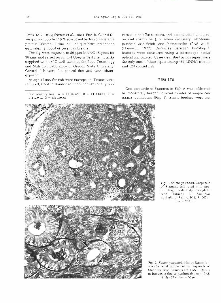
One corpuscle of Stannius in Fish A was infiltrated by moderately basophilic renal tubules of simple col- umnar epithehum (Fig. 1). Brush borders were not
Fig. 1. Salmo gairdneri. Corpuscle of Stannius infiltrated with pro- liferating moderately basophilic renal tubules of columnar epithelium. Fish A. H & E; 107x
Bar = 200 pm
i h g 2. S*a galrdnen Mitotic figure lar- 1 row) in renal tubule cell in corpuscle of
Stannius. Basal laminas are PAS+. Debris ' In lumens is due to nephrocalcinosis. PAS d & H; 432x. Bar = 50 pm

Lee et al.: Rare renal neoplasms ~n trout 107
present and tubules had either distal tubule or collect- ing duct features. No point of penetration of the tubu- les thro.ugh the capsule of the corpuscle was located in the 6 semi-serial sections examined. Debris occasion- ally present within the lumens of the tubules was associated with the nephrocalcinosis that was occa- sionally observed in MNNG-treated fish throughout the study. Few mitotic figures (Fig. 2) were found in the tubule epithelial cells, indicating the proliferation rate was not unusually rapid. Granular and agranular cells of the corpuscle (Krishnamurthy & Bern 1969) were normal and stained respectively PAS+ and PAS- (Figs. 2 and 3). A PAS+ reaction was ellcited along the luminal border of a few of the renal epithe- lial cells within the corpuscle (Fig. 3), a characteristic of fully differentiated collecting and mesonephric duct cells (Bulger & Trump 1968). Other corpuscles of Stan- nius in the same and adjacent kidney had normal cellular morphology and arrangement as described by Yasutake & Wales (1983).
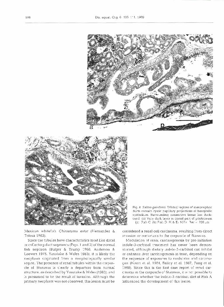
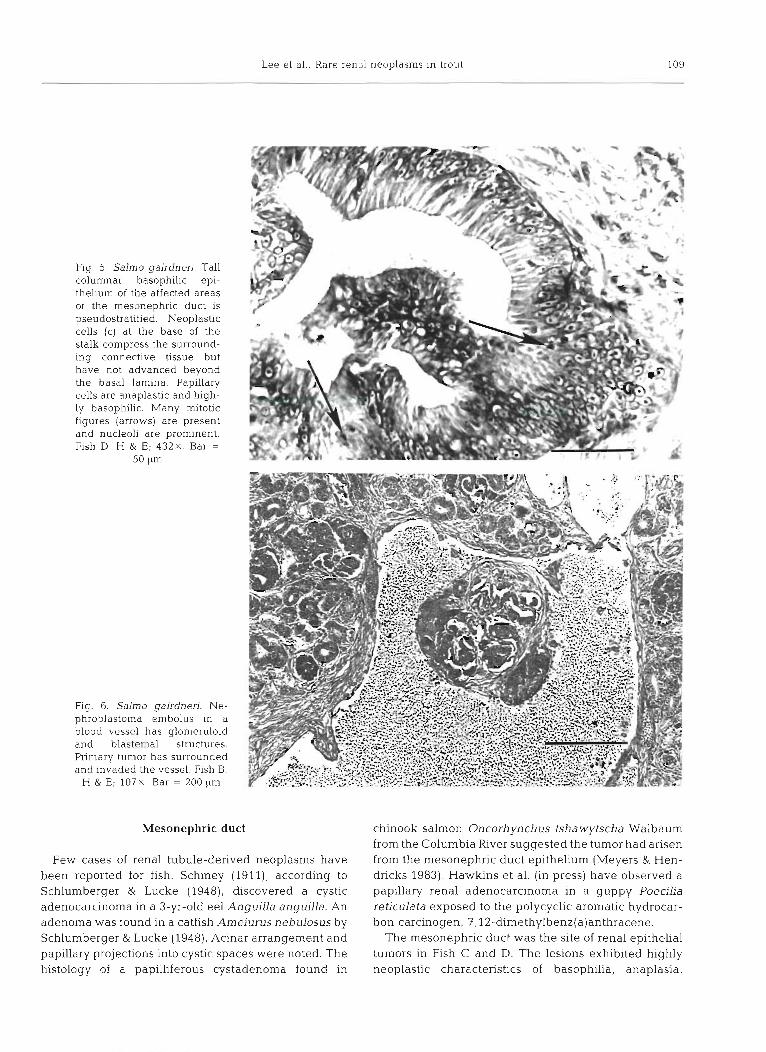
One region of a mesonephric duct in each of Fish C and D became widely dilated (Fig. 4a ,b ) . In these regions, the simple tall columnar epithelium became basophilic and occasionally stratified. The basophllic layer of rapidly proliferating epithelial cells projected into the lumen, forming highly basophilic cystic papil- lary structures of anaplastic cells. Nucleoli were darkly prominent and many mitotic figures were found (Fig. 5). Basal laminas of the mesonephric ducts remained intact although the neoplastic cells at the base of one stalk con~pressed the underlying connective tissue (Fig. 5) .
The shortest distance from the nearest affected por-
tion of the mesonephric duct to a cystadenoma in Fish C was 1.38 mm. However, a nephrocalcinotic lesion was in apposition to the d.uct. In Fish D, a nephroblas- toma was 0.14 mm from the duct. Increased nephrocal- cinosis was seen in the soy fed groups, but the lesions were not counted since the study was focused on asses- sing neoplastic responses.
Nephroblastoma has not been known to success- fully metastasize in fish, but the first case of a ne- phroblastoma embolus a t this lab was seen in Fish B (Fig. 6). The darkly basophilic embolus contained blastemal and glomeruloid features and was deficient in hemopoietic tissue as is typical of nephroblastoma in fish. The tumor of origin had surrounded and invaded the vessel wall where the embolus was found (Fig. 6).
Kidneys in the control fish were normal.
DISCUSSION
Corpuscle of Stannius
Corpuscles of Stannius are found only in teleosts but are functionally, histochemically, and immunologically similar to the parathyroid glands of higher vertebrates (Lopez et al. 1984). Neoplastic events within the cor- puscle have never been previously reported, and the 2 parenchyma1 cell types (granular and agranular) were normal in Fish A. However, the proxin~ity of other renal components may place the corpuscles at risk of neo- plastic invasion, except in fish with remotely located corpuscles, such as charal Chirostoma jordani and
Fig. 3. Salrno yairdneri. A few cells of the tubules in the corpuscle of Stannius were sufficiently differentiated to be- come PAS+ along the luminal border (arrow). Granular and agranular cells of the corpuscle were respectively PAS+ and PAS-. PAS & H; 432x Bar =
50 pm
108 Dis. aquat. Org. 6: 105-1 11, 1989
Mexican whitefish Chirostoma estor (Hernandez & Tolosa 1983).
Since the tubules have characteristics most Like distal or collecting duct segments (Figs. 1 and 2) of the normal fish nephron (Bulger & Trump 1968, Anderson & Loewen 1975. Yasutake & Wales 1983), it is likely the neoplasm originated from a morphologically similar region. The presence of renal tubules within the corpus- cle of Stannius is clearly a departure from normal structure, as described by Yasutake & Wales (19831, and is presumed to be the result of invasion. Although the primary neoplasm was not observed, this lesion must be
Fig. 4 . Salmo gairdneri. Dilated regions of mesonephric ducts contain cystic papillary projections of basophilic epithelium. Surrounding connective tissue has thick- ened. (a) Very dark layer is dorsal part of peritoneum
(p). Fish C. (b) Fish D. H & E; 1 0 7 ~ Bar = 200 pm
considered a renal cell carcinoma, resulting from direct invasion or metastasis to the corpuscle of Stannius.
Modulation of renal carcinogenesis by pre-initiation indole-3-carbinol treatment has never been demon- strated, although dietary indole-3-carbinol can inhibit or enhance liver carcinogenesis in trout, depending on the sequence of exposures to modulator and carcino- gen (Nixon et al. 1984, Bailey et al. 1987, Fong et al. 1988). Since this is the first case report of renal car- cinoma in the corpuscle of Stannius, it is not possible to determine whether the indole-3-carbinol diet of Fish A influenced the development of t.hls lesion.
Lee et al.. Rare renal neoplasms in trout 109
Fig. 5. Salmo gairdneri. Tall columnar basophilic epi- thelium of the affected areas of the mesonephric duct 1s pseudostratified. Neoplastic cells (c) a t the base of the stalk compress the surround- ing connective tissue but have not advanced beyond the basal lamina. Papillary cells are anaplastic and high- ly basophillc. Many mitotic figures (arrows) are present and nucleoli are prominent. Fish D. H & E; 4 3 2 ~ . Bar =
50 Itrn
Fig. 6. Salmo gairdneri. Ne- phroblastoina embolus in a blood vessel has glomeruloid and blasternal structures. Primary tumor has surrounded and invaded the vessel. Fish B.
H & E; 107x Bar = 2001tm
Mesonephric duct
Few cases of renal tubule-derived neoplasms have been reported for fish. Schmey (1911), according to Schlumberger & Lucke (1948), discovered a cystic adenocarcinoma in a 3-yr-old eel Anguilla anguilla. An adenoma was found in a catfish Ameiurus nebulosus by Schlumberger & Lucke (1948). Acinar arrangement and papillary projections into cystic spaces were noted. The histology of a papilliferous cystadenoma found in
chinook salmon Oncorhynchus tshawytscha Walbaum from the Columbia River suggested the tumor had arisen from the mesonephric duct epithelium (Meyers & Hen- dricks 1983). Hawkins et al. (in press) have observed a papillary renal adenocarcinoma in a guppy PoeciLia reticulata exposed to the polycyclic aromatic hydrocar- bon carcinogen, ?,12-dimethylbenz(a)anthracene.
The mesonephric duct was the site of renal epithelia1 tumors in Fish C and D. The lesions exhibited highly neoplastic characteristics of basophilia, anaplasia,
110 Dis. aquat. Org. 6: 105-111, 1989
rapid proliferation, and papillary growth (Figs. 4a, b Acknowledgements. This report is R8569 of the Oregeon and 5 ) . The cells were contained by the basal lamina, so Ag~cul tural Experiment Station, Oregon State University. w e
the lesions can not be as adenocarcinoma. thank Drs David E. Hinton and Mark Oluhiro of the University of California at Davis for their review of the lesions. We also
A diagnosis of 'ystic papillary adenOma of the express our gratitude to Dr Toshiko Morita for tissue proces- mesonephric duct is warranted. Considering the sing services, and to John Casteel and Ted Will for diet 'aggressiveness' of the cells, carcinoma in situ would preparation and animal care. This research was supported by be an appropriate alternative description to adenoma, U.S. Public Health Service grants ES 03850 and ES 00210 from
the National Institute of Environmental Health Sciences, and CA 34732 and CA 44317 from the National Cancer Institute.
Coincident neoplasms
It is highly unlikely that the coincident neoplasms, i.e. cystadenoma and nephroblastoma, were the origins of the rare renal neoplasms described in this report. Cystadenoma consists of widely distended tubule- derived structures of normally staining simple epithelium and lacks invasiveness (Kimura 1976). Metastasis of nephroblastoma in fish has never been reported, although the analogous tumor is transplant- able in rats (Hard & Noble 1981) and can metastasize in the cow, dog, hamster, macaque, and rabbit (reviewed by Hard 1984) as well as in humans (Borg et al. 1987). The finding of a nephroblastoma embolus in a rainbow trout (Fig. 6) indicates that at least the potential for metastasis exists. However, the carcinoma of the cor- puscle of Stannius and the renal cell papillary adenomas of the mesonephric ducts possessed none of the features typical of nephroblastoma, such as glomeruloid structures, blastema, or deficiency of hemopoietic tissue (Ashley 1970, Hendricks et al. 1980a, I(lmura et al. 1981).
The papillary adenomas occurred only in the group fed soy protein. Soy has not been previously reported to modulate renal neoplasia, although raw full-fat soy protein isolate has been shown to enhance the percent of pancreatic volume containing azasenne-induced acidophilic foci in rats (Roebuck et al. 1987). A regenerative stimulation of the mesonephric ductal epithelium in response to the tissue destruction due to more frequent nephrocalcinotic lesions in the soy-fed fish was not supported. Those processes were localized at the level of the tubules so that destruction and regeneration were not observed in the ducts.
Physical impingement of expansive or occlusive lesions on the mesonephric duct could cause a pro- liferative/metaplastic stimulus, analogous to the response of obstructed ducts of the pancreas or salivary gland (Robbins 1974, Shalimov et al. 1981). In both cases of papillary adenoma, a nephroblastoma or ne- phrocalcinotic lesion was sufficiently close to compress the duct. Although no blockages or occlusions were found, it was not possible to observe the entire ducts as they passed through the affected regions. Therefore, the role of coincident lesions in the promotion of the mesonephric duct adenoma remains speculative.
LITERATURE CITED
Anderson, B. G., Loewen, R. D. (1975). Renal n~orphology of freshwater trout. Am. J. Anat. 143: 93-1 14
Ashley, L. M. (1970). Pathology of fish fed aflatoxins and other andmetaboiites. 111: Sr~ieszko, S. F. (ed.) A symposium or, diseases of fishes aind shellfisches. American Fish. Soc. Special Publ. No. 5 , Washington, D.C , p. 366-379
B d e y , G. S., Selivonchick, D. P., Hendricks, J . D. (1987). tnitiation, promotion, and inhibition of carcjnogenesis in rainbow trout. Envir. Hlth Perspectives 71: 145-153
Borg, S. A., Rubin, P., DeWys, W D. (1987). Metastasis and disseminated disease. In: Rubin, P. (ed.) Clinical oncology, a multidisciplinary approach, 6th edn. American Cancer Soc., New York, Chap. 31, p. 498-515
Bulger, R. E., Trump, B. F. (1968). Renal morphology of the English sole (Parophrys vetulus). Am. J. Anat. 123: 195-226
Dashwood, R. H., Arbogast, D. N., Fong. A. T., Hendncks, J. D., Bailey, G. S. (1988). Mechanisms of anti-carcinogenesis by indole-3-carbinol. detailed In vivo DNA binding dose- response studies after dietary administration with aflatoxjn B,. Carclnogenesis 9. 427432
Fong, A. T.. Hendricks, J. D., Dashwood. R. H., Van Winkle, S., Lee, B. C., Bailey, G. S (1988) Modulation of diethylnit- rosamine-induced hepatocarc~nogenesis and O6-ethyl- guanine formation in rainbow trout by indole-3-carbinol, B-naphthoflavone, and Aroclor 1254. TOXIC. appl. Phar- mac. 96: 93-100
Hard, G. C (1984). Comparative oncology I. Nephroblastoma in laboratory mammals. 11. Nephroblastoma in domesti- cated and wild animals. In- Pochedly, C., Baum, E. S. (eds.) Wllms' tumor clinical and bioloy~cal manifestations. Elsevier, New York, p. 147-167, 169-214
Hard. G . C , Noble. R. L. (1981). Occurrence, transplantation, and histologic characteristics of nephroblastoma in the Nb hooded rat. Investig. Urol. 18: 371-376
Hawkins, W. E.. Walker, W. W.. Lytle, J. S., Lytle, T. F., Overstreet, R. M. (in press). Carcinogenic effects of 7,12- dimethylbenz[ajanthracene on the guppy (Poecilla ret~culata). Aquat. Toxic.
Hendricks, J. D. (1982) Chemical carcinogenesis in fish. In: Weber, L. J. (ed.) Aquatic toxicology. Vol. 1. Raven Press, New York, p. 149-211
Hendricks. J. D., Meyers, T R., Casteel, J. L., Nixon, J. E., Loveland, P. M., Bailey, G. S. (1984). Rainbow trout embryos. advantages and limitations for carcinogenesls research. Natl Cancer Inst., Monogr 65: 129-137
Hendricks, J. D., Scanlan, R. A., Williams. J. L . , Sinnhuber, R . 0.. Grieco. M. P. (1980a). Carcinogenicity of N-methy1-N'- nitro-N-nitrosoguanidine to the liver and kidney of rain- bow trout (Salmo gairdneri) exposed as embryos. J. natl Cancer Inst. 64: 1511-1519
Hendricks. J. D., Wales, J. H.. Sinnhuber, R . O., Nixon, J. E., Loveland, P. M,, Scanlan, R. A. (1980b). Rainbow trout
Lee et al.: Rare renal n ~ o p l a s m s in trout 111
(Salmo gairdner-i) embryos: a sensitrve animal for experi- mental carclnogenesis. Fed. Proc. 39: 2333-2339
Hernandcz, R. G., Tolosa, J . S. (1983). Caracteristicas his- tologicas e histoqujmicas de 10s corpusculos de Stannlus e n Chirostoma jordan y Chirostonid estor. Veterinaria (Mexico City) 14: 86-92
Humason, C;. L. (1972). Animal tlssue techniques, 3rd crln. W. 1I. Frrcrnan & CO, San Francisco, p. 325--328
Kimura, I . , Krtatori, H. , Yoshizaki, K , Tayama, K , Ito, M , Yarnada, S. (1981). Development of tumor in rainbow trout follo\ving embryonic esposure to N-nitroso compounds. In: Dawe, C. J., Harshbarger, J C., Kondo, S.. Suginlura, T., Takayama, S. (eds.) Phyletic approaches to cancer Japan Scientific Soc. Press, Tokyo, p. 241-252
I(lmura, I . , Mlyake, T., Yoshizah, K (1976). Induction of tumors of the stomach, of the liver and of the kidney in rainbow trout by intrastomach administration of N-methyl- N-nitroso-N'-nitrosoguanidine (MNNG). Proc. Jpn. Cdncer Assoc. 35: 16, abstract # 15
Krishnamurthy, V G., Bern, H. A. (1969). Correlative his- tologic study of the curpuscles of Stannius and the juxta- glomerular cells of teleost fishes. Gen. Comp. Endocrinol. 13: 313-335
Lopez, E . , Tisserand-Jochern, E . - M , V~da l , B., Milet, C . , Lal- lier, F. , MacIntyre, I. (1984). Les corpuscules de Stannius sont-ils les glandes parathyroides des Poissons teleos- teens? Arguments ultrastructuraux, cytologiques et irnmunocytochimiques. C. r Seanc. Acad, natn. Sci., Paris Serie I11 298: 359-364
l l e y ~ r s , T. R., Hendricks. J . D. (1983). Histopathology of four spontaneous neoplasms in thrre species of salmonid fishes. J. Fish Dis G . 4 8 1 4 9 9
Nixon, J. E , Hendricks, J D. , Pawlowski, N. E , Pereira, C. B., Sinnhuber, R. O., Bailey, G. S. (1984). Inhibition of afla- toxin B, carcinogenesis in rainbow trout by flavone and indole compounds. Carcinogenesis 5: 615-619
Nunez, O., IIendricks, J . D., Bailey, G. S. (1988). Enhrtncement of aflatoxin Bi and MNNG hepatocar- cinogenesis in rainholu trout (Salmo garrdnerl) by 178- eslradrol and other organlc chemicals. Dis. aquat. Org. 5: (3) 185-196
Robbins. S . L. (1974). hletaplasia. In: Robbins, S. L. (ed.) Pathokogic basis of disease. W B. Saunders Co., Philadel- phia. p. 17-18
Roebuck, B. D., Kaplita, P. V., Edwards, B. R.. Praissman, M. (1987). Effects of dietary fats and soybean protein on azasei-ine-lnduced pancreat~c carcinogenesls and plasma cholccystokinin in the rat. Cancer Res. 47: 1333-1338
Schlumberger, H. G . , Lucke, B. (1948). Tumors of fishes, amphibians, and reptiles. Cancer Res. 8: 657-754
Schmey. M. (1911). iJber Neubildungen Fischen. Frankf. 2. Pathol. 6: 230-253
Shalimov, A. A., Medvetskii, E. B., Keisevich, L. V (1981). hlechanisrns of metaplasia of the duct epithelium of organs of the pancreaticobiliary system. BuU. exp. Biol. Med. (USSR) 92: 991-993
Sinnhuber, R O., Hendricks, J D., Wales, J . H , Putnam, G. B. (1977). Neoplasms in rainbow trout, a sensitive animal model for environmental carcinogenesis. Ann. N.Y Acad. Sci. 298: 389-408
Yasutake, W T., Wales, J. H. (1983). Endocrine system. In: Microscopic anatomy of salmonids: an atlas U S. Dept. Interior, Fish and Wildlife Service Resource Publication 150, Washington, D.C., Chap. 13, p. 152-179
Responsiblc Subject Editor. Professor N. Peters; accepted for printing on February 22, 1989