Embed Size (px)
Citation preview
Journal of Clinical InvestigationVol. 41, No. 3, 1962
RHEUMATOIDFACTOR, COMPLEMENT,AND CONGLUTININ ABERRATIONSIN PATIENTS WITH SUBACUTEBACTERIAL ENDOCARDITIS*
By RALPH C. WILLIAMS, JR. AND HENRYG. KUNKEL
(From The Rockefeller Institute, New York, N. Y.)
(Submitted for publication September 28, 1961; accepted October 30, 1961)
Subacute bacterial endocarditis produces inman a complex situation of interaction of hostand invading organism. Little is known of anti-body response or of the specific mechanisms ofimmunity involved during the inception, course,and recovery from subacute bacterial endocarditis,although observations on streptococcal agglutinat-ing and complement-fixing antibodies have beenmade by numerous workers (1-7). Recent ob-servations (8, 9) have indicated that animalsimmunized with certain, streptococcal and coliformorganisms develop factors capable of agglutinatingy-globulin-coated red cells and other particles.These properties indicate a similarity to rheuma-toid factors, and the possibility is raised thatorganisms or their products play a role in theproduction of rheumatoid factors in human dis-ease. A natural parallel to production of suchrheumatoid factors in animals would be theiroccurrence in human disease states in which bac-teria are constantly fed into the circulation, asin miliary tuberculosis or subacute bacterial endo-carditis.
In the present study the effect of subacute bac-terial endocarditis on the serological behavior ofsystems designed to react with the rheumatoidfactor was examined. Preliminary observationsindicated a high incidence of positive tests forrheumatoid factor in these patients. It seemed ofspecial interest to see whether these reversed aftereradication of the offending organism undertherapy. The constancy and prolonged durationof these factors in patients with rheumatoidarthritis is well recognized (10, 11) althoughsome observers have reported decrease in titerwith remission (12). In addition, observationswere also made on conglutinin and serum com-plement in these patients because of certain gen-eral similarities of these various factors thatappear to be indirectly involved in immune phe-nomena.
* Aided by a grant from The National Foundation.
METHODSAND MATERIALS
Sera were collected from 51 patients with subacutebacterial endocarditis; 41 had the diagnosis of bacterialendocarditis supported by at least two positive bloodcultures. Autopsy confirmed the diagnosis in 3 patients.The remaining 10 had negative blood cultures but showedvarying degrees of persistent fever, anemia, splenomegaly,microscopic hematuria, and embolic phenomena sufficientfor a presumed diagnosis of bacterial endocarditis.
Latex fixation tests and sensitized sheep cell tests wereperformed on serially drawn sera using methods previ-ously described (13, 14).-' In addition, capillary tubeprecipitation of bacterial endocarditis sera with aggre-gated -y-globulin was performed (15). Precipitin curveswith both plain serum and serum heated to 56° C for 30minutes were determined by using aggregated -y-globulinas antigen (16). The difference between precipitin curvesof plain and heated serum was taken to indicate the rela-tive contributions of complement components to the pre-cipitate material. The height of the curves with serumpreviously heated to 56° C for 30 minutes was indicativeof the reaction between pure aggregates and rheumatoidfactors.
Many of the sera from bacterial endocarditis patientswere tested for direct agglutination by use of group 0Rh positive red cells coated with incomplete anti-Rh-antibody.
Serum complement determinations were per-formed by the modified method of Mayer, Eatonand Heidelberger as described by Fischel, Pauliand Lesh (17, 18). Immunoconglutinin was de-termined by the method of Coombs and Coombs(19).
Starch block electrophoresis and sucrose densitygradient ultracentrifugation were performed withmethods previously described from this labora-tory (20, 21).
RESULTS
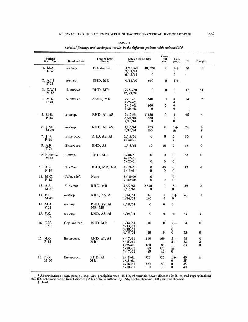
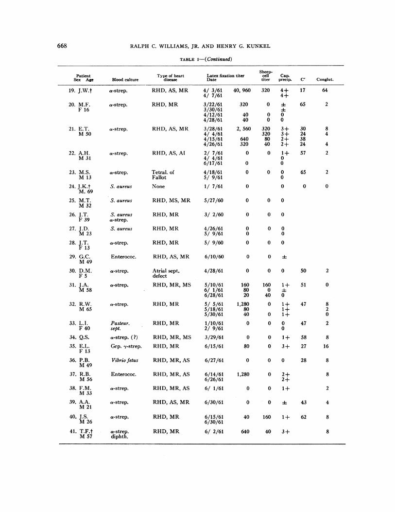
Measurement of rheumatoid factors. Positivelatex fixation tests were present in 26 of the 51sera (50 per cent). Ten, or 19 per cent, ofthese sera from patients with bacterial endo-carditis exhibited varying degrees of reactivitywith sensitized sheep cells (Table I). There
666
ABERRATIONSIN PATIENTS WITH SUBACUTEBACTERIAL ENDOCARDITIS
TABLE I
Clinical findings and serological results in the different patients with endocarditis*
ShPatient Type of heart Latex fixation titer c
Sex Age Blood culture disease Date ti
1. M.A. a-strep. Pat. ductusF 52
4/12/602/ 9/616/ 3/61
40, 96000
leep-cell Cap.iter precip.
0 4+00
C' Conglut.
51 0
2. A.J.t a-strep. RHD, MR 4/18/60 640 0 2+F 23
3. D.W.t S. aureus RHD, MR 12/23/60 0 0 0 13 64M85 12/29/60 0
4. M.D. S. aureus ASHD, MR 2/11/61 640 0 0 54 2F 70 2/24/61 0
3/ 2/61 160 0 03/24/61 0 0
5. G.K. a-strep. RHD, Al, AS 2/17/61 5,120 0 2+ 45 4F 28 3/24/61 320 4
7/13/61 0 0
6. J.Mc. a-strep. RHD, AI, AS 1/ 6/61 320 0 1+ 24 4M60 1/19/61 160 41 8
7. J.B. Enterococ. RHD, AS, AI, 1/ 5/61 0 0 0 36 8F 44 1/18/61 0
8. A.F. Enterococ. RHD, AS 1/ 8/61 40 40 0 46 0F 74
9. F.Mc.G. a-strep. RHD, MR 3/30/61 0 0 0 53 0M47 4/12/61 0
5/22/61 0 0 0
10. A.S. S. albus RHD, MR, MS 3/13/61 0 40 0 37 4F 19 4/3/61 0 0 0
11. M.C. Salm. chol. None 8/ 8/60 0 0F 45 9/20/60 0 0 0
12. A.S. S. aureus RHD, MR 3/29/61 2,560 0 2+ 89 2M57 6/ 6/61 0 0
13. P.U. a-strep. RHD, AS, Al 1/14/61 160 0 1+ 43 0M45 1/24/61 160 0 0
14. M.A. a-strep. RHD, AS, AI 4/ 9/61 0 0 0F 21 MR, MS
15. F.C. a-strep. RHD, AS, AI 4/19/61 0 0 ± 47 2F 76
16. E.N. Grp. a-strep. RHD, MR 1/14/61 40 0 2+ 34 0F 50 2/13/61 0
3/18/61 04/ 9/61 40 0 0 55 0
17. H.O. Enterococ. RHD, Al, AS 4/ 7/61 160 160 2+ 78 4F 53 MR 4/15/61 2+ 53 2
4/26/61 160 80 41 63 05/20/61 80 320 :17/ 7/61 80 40 0
18. P.O. Enterococ. RHD, Al 4/ 7/61 320 320 1+ 40 4M60 MR 4/15/61 0 35
4/26/61 320 80 0 355/20/61 0 0 0 40
667
*Abbreviations: cap. precip., capillary precipitin test; RHD, rheumatic heart disease; MR, mitral regurgitation;ASHD, arteriosclerotic heart disease; Al, aortic insufficiency; AS, aortic stenosis; MS, mitral stenosis.
t Dead.
RALPH C. WILLIAMS, JR. AND HENRYG. KUNKEL
TABLE i-( Continued)
Sheep-Patient Type of heart Latex fixation titer cell Cap.
Sex Age Blood culture disease Date titer precip. C' Conglut.
RHD, AS, MR
RHD, MR
RHD, AS, MR
RHD, AS, Al
Tetral. ofFallot
None
RHD, MS, MR
RHD, MR
RHD, MR
RHD, MR
RHD, AS, MR
Atrial sept.defect
RHD, MR, MS
RHD, MR
RHD, MR
RHD, MR, MS
RHD, MR
RHD, MR, AS
RHD, MR, AS
RHD, MR, AS
RHD, AS, MR
RHD, MR
RHD, MR
4/ 3/614/ 7/613/22/613/30/614/12/614/28/61
3/28/614/ 4/614/15/614/26/61
2/ 7/614/ 4/616/17/614/18/615/ 9/611/ 7/61
5/27/60
3/ 2/60
4/26/615/ 9/615/ 9/60
6/10/60
4/28/61
5/10/616/ 1/616/28/615/ 5/615/18/615/30/611/10/612/ 9/61
3/29/61
6/15/61
6/27/61
6/14/616/26/616/ 1/61
6/30/61
6/15/616/30/61
6/ 2/61
40, 960 320 4+4+
320 0 A
40 0 040 0 0
2, 560 320 3+320 3+
640 80 2+320 40 2+
0 0 1+0
0 0
0 0 00
0 0
0 0 0
0 0 0
0 0 00 0
0 0 0
0 0O
0 0 0
160 160 1+80 0 -
20 40 0
1,280 0 1+80 1+40 0 1+
0 0 00
0 0 1+
80 0 3+
0 0 0
1,280 0 2+2+
0 0 1+
0 0O
40 160 1+
640 40 3+
17 6419. J.W.t
20. M.F.F 16
21. E.T.M50
22. A.H.M31
23. M.S.M 13
24. J.K-tM. 69
25. M.T.M32
26. J.T.F 39
27. J.D.M23
28. J.T.F 13
29. G.C.M49
30. D.M.F 5
31. J.A.M58
32. R.W.M65
33. L.I.F 40
34. Q.S.
35. E.L.F 13
36. P.B.M49
37. R.B.M56
38. F.M.M33
39. A.A.M21
40. J.S.M26
41. T.F.tM57
a-strep.
a-strep.
a-strep.
a-strep.
a-strep.
S. aureus
S. aureus
S. aureusa-strep.
S. aureus
a-strep.
Enterococ.
a-strep.
a-strep.
a-strep.
Pasteur.sept.
a-strep. (?)
Grp. Sy-strep.
Vibrio fetus
Enterococ.
a-strep.
a-strep.
a-strep.
a-strep.diphth.
65 2
30 824 43824 4
57 2
65 2
0 0
50 2
51 0
47 820
47 2
58 8
27 16
28 8
8
2
43 4
62 8
8
668
ABERRATIONSIN PATIENTS WITH SUBACUTEBACTERIAL ENDOCARDITIS
TABLE i-( Continued)
Sheep-Patient Type of heart Latex fixation titer cell Cap.
Sex Age Blood culture disease Date titer precip. C' Conglut.
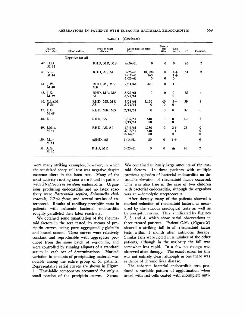
Negative for all
42. H.D. RHD, MR, MS 4/26/61 0 0 0 45 2M25
43. V.C. RHD, AS, Al 3/25/61 10, 240 0 4+ 54 2M44 4/ 7/61 160 1+
5/20/61 0 0 0
44. J.W. RHD, AS, MS 2/14/61 320 0 1+M48 MR
45. J.K. RHD, MR, MS 3/22/61 0 0 0 75 4M29 Al 3/27/61 0
46. C.La.M. RHD, MS, MR 2/24/61 5,120 40 2+ 39 8F 56 AS 3/24/61 0 0 0
47. L.O. RHD, MR, MS 2/18/61 0 0 0 32 0M48
48. D.L. RHD, AS 1/ 5/61 640 0 0 49 21/19/61 80 0
49. J.Mik. RHD, AS, Al 1/ 6/61 1,280 0 2+ 23 0M44 2/7/61 640 1+ 0
2/14/61 80 0 0
50. J.L.t ASHD, AS 1/14/61 80 0 1+ 0M54
51. A.G. RHD, MR 3/25/61 0 0 ± 70 2M46
were many striking examples, however, in whichthe sensitized sheep cell test was negative despiteextreme titers in the latex test. Many of themost actively reacting sera were found in patientswith Streptococcus viridans endocarditis. Organ-isms producing endocarditis and no latex reac-
tivity were Pasteurella septica, Salmonella chol-eraesuis, Vibrio fetus, and several strains of en-
terococci. Results of capillary precipitin tests inpatients with subacute bacterial endocarditisroughly paralleled their latex reactivity.
Weobtained some quantitation of the rheuma-toid factors in the sera tested, by means of pre-
cipitin curves, using pure aggregated y-globulinand heated serum. These curves were relativelyconstant and reproducible with aggregates pro-
duced from the same batch of y-globulin, andwere controlled by running aliquots of a standardserum in each set of determinations. Markedvariation in amounts of precipitating material was
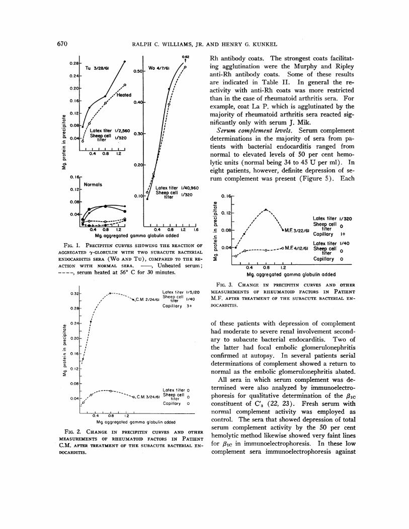
notable among the entire group of 51 patients.Representative serial curves are shown in Figure1. Heat-labile components accounted for only a
small portion of the precipitin curves. Serum
Wocontained uniquely large amounts of rheuma-toid factors. In three patients with multipleprevious episodes of bacterial endocarditis no de-tectable elevation of rheumatoid factor occurred.This was also true in the case of two childrenwith bacterial endocarditis, although the organismwas an a-hemolytic streptococcus.
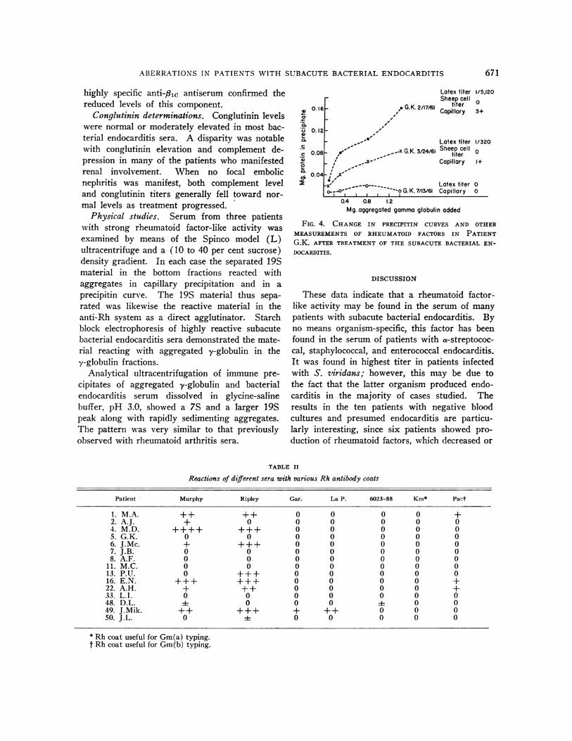
After therapy many of the patients showed amarked reduction of rheumatoid factors, as meas-ured by the various serological tests as well asby precipitin curves. This is indicated by Figures2, 3, and 4, which show serial observations inthree treated patients. Patient C.M. (Figure 2)showed a striking fall in all rheumatoid factortests within 1 month after antibiotic therapy.Similar falls were noted in a number of the otherpatients, although in the majority the fall wassomewhat less rapid. In a few no change wasobserved after therapy. The exact reason for thiswas not entirely clear, although in one there wasevidence of chronic liver disease.
The subacute bacterial endocarditis sera pro-duced a variable pattern of agglutination whentested with red cells coated with incomplete anti-
669
RALPH C. WILLIAMS, JR. AND HENRYG. KUNKEL
0.62 Rh antibody coats. The strongest coats facilitat-/,o ing agglutination were the Murphy and Ripley
anti-Rh antibody coats. Some of these resultsare indicated in Table II. In general the re-activity with anti-Rh coats was more restrictedthan in the case of rheumatoid arthritis sera. Forexample, coat La P. which is agglutinated by themajority of rheumatoid arthritis sera reacted sig-nificantly only with serum J. Mik.
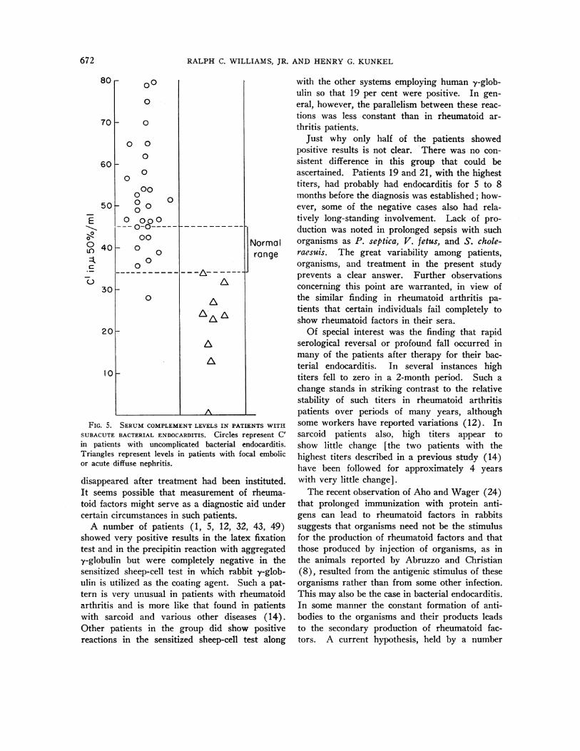
Serum complement levels. Serum complementdeterminations in the majority of sera from pa-tients with bacterial endocarditis ranged fromnormal to elevated levels of 50 per cent hemo-lytic units (normal being 34 to 45 U per ml). Ineight patients, however, definite depression of se-rum complement was present (Figure 5). Each
Latex titer 1/40,960Sheep cell 1/320
titer
4J- Lw w-----I0.4 0.8 1.2 0.4 0.8
Mg. aggregated gamma globulin added.2 1.6
FIG. 1. PRECIPITIN CURVESSHOWINGTHE REACTION OFAGGREGATEDly-GLOBULIN WITH TWOSUBACUTEBACTERIALENDOCARDITIS SERA (WO AND Tu), COMPAREDTO THE RE-ACTION WITH NORMAL SERA. -, Unheated serum;---- , serum heated at 56° C for 30 minutes.
.0---_ Latex titer 1/5,120C 2/24/61 Sheep cell 1/40
Capillary 3+
_-- ---.
'IO, -a. C. M. 3/24/61,P
0.4 0.8 1.2
Mg. aggregated gamma globulin added
FIG. 2. CHANGE IN PRECIPITIN CURVES AND OTHER
MEASUREMENTSOF RHEUMATOID FACTORS IN PATIENTC.M. AFTER TREATMENTOF THE SUBACUTEBACTERIAL EN-
DOCARDITIS.
0.16F
0. 12.
0.08
0.04
- A/ -,1 %
.1 \- , ; M.F 3/22/61
- - - --.o M. F4/l2/6I-o
Latex titer 1/320Sheep cell0
tilerCapillary 1+
Latex titer 1/40Sheep cell V
titerCapillary 0
-
0
._0.
T
C
C
.0.0.
0.4 0.8 1.2Mg. aggregated gammaglobulin added
FIG. 3. CHANGE IN PRECIPITIN CURVES AND OTHERMEASUREMENTSOF RHEUMATOID FACTORS IN PATIENTM.F. AFTER TREATMENTOF THE SUBACUTEBACTERIAL EN-DOCARDITIS.
of these patients with depression of complementhad moderate to severe renal involvement second-ary to subacute bacterial endocarditis. Two ofthe latter had focal embolic glomerulonephritisconfirmed at autopsy. In several patients serialdeterminations of complement showed a return tonormal as the embolic glomerulonephritis abated.
All sera in which serum complement was de-termined were also analyzed by immunoelectro-phoresis for qualitative determination of the /31cconstituent of C,3 (22, 23). Fresh serum withnormal complement activity was employed ascontrol. The sera that showed depression of totalserum complement activity by the 50 per centhemolytic method likewise showed very faint linesfor 8ic in immunoelectrophoresis. In these lowcomplement sera immunoelectrophoresis against
670
0.50k Wo4/7/61
0.40F
0.30-
0
0
._U
c
._c
._
0c2
0.4 0.8 1.2
0.16rNormals
0.121
0.08F0.10IO
0.04
0.32
0.28
w0 24
0.- 0.20
012c 0.16
0_1
0 2 F-~
0.081-
0 04
Latex titer oSheep cell
titerCapillary a
ABERRATIONSIN PATIENTS WITH SUBACUTEBACTERIAL ENDOCARDITIS
highly specific anti-/lic antiserum confirmed thereduced levels of this component.
Conglutinin determinations. Conglutinin levelswere normal or moderately elevated in most bac-terial endocarditis sera. A disparity was notablewith conglutinin elevation and complement de-pression in many of the patients who manifestedrenal involvement. When no focal embolicnephritis was manifest, both complement leveland conglutinin titers generally fell toward nor-
mal levels as treatment progressed.Physical studies. Serum from three patients
with strong rheumatoid factor-like activity was
examined by means of the Spinco model (L)ultracentrifuge and a (10 to 40 per cent sucrose)density gradient. In each case the separated 19Smaterial in the bottom fractions reacted withaggregates in capillary precipitation and in a
precipitin curve. The 19S material thus sepa-
rated was likewise the reactive material in theanti-Rh system as a direct agglutinator. Starchblock electrophoresis of highly reactive subacutebacterial endocarditis sera demonstrated the mate-rial reacting with aggregated y-globulin in they-globulin fractions.
Analytical ultracentrifugation of immune pre-
cipitates of aggregated y-globulin and bacterialendocarditis serum dissolved in glycine-salinebuffer, pH 3.0, showed a 7S and a larger 19Speak along with rapidly sedimenting aggregates.The pattern was very similar to that previouslyobserved with rheumatoid arthritis sera.
4)
4._a.
0
a)
c
._
Latex titer 1/5,120Sheep cell 0
titerCapillary 3+
Latex titer 1/320..x G.K. 3/24/61 Sheep cell 0
titer
Capillary 1+
,x"
__---o----..0Latex titer 0
0.4,~ , , ,~-o G. K. 7/13/61 Capillary 0
0.4 0.8 1.2Mg. aggregated gammaglobulin added
FIG. 4. CHANGE IN PRECIPITIN CURVES AND OTHER
MEASUREMENTSOF RHEUMATOID FACTORS IN PATIENTG.K. AFTER TREATMENTOF THE SUBACUTEBACTERIAL EN-
DOCARDITIS.
DISCUSSION
These data indicate that a rheumatoid factor-like activity may be found in the serum of many
patients with subacute bacterial endocarditis. Byno means organism-specific, this factor has beenfound in the serum of patients with a-streptococ-cal, staphylococcal, and enterococcal endocarditis.It was found in highest titer in patients infectedwith S. viridans; however, this may be due tothe fact that the latter organism produced endo-carditis in the majority of cases studied. Theresults in the ten patients with negative bloodcultures and presumed endocarditis are particu-larly interesting, since six patients showed pro-
duction of rheumatoid factors, which decreased or
TABLE II
Reactions of different sera with various Rh antibody coats
Patient Murphy Ripley Gar. La P. 6023-88 Km* Pact
l. M.A. ++ ++ 0 0 0 0 +2. A.J. + 0 0 0 0 0 04. M.D. ++++ +++ 0 0 0 0 05.G.K. 0 0 0 0 0 0 06. J.Mc. + +++ 0 0 0 0 07. J.B. 0 0 0 0 0 0 08. A.F. 0 0 0 0 0 0 0
II. M.C. 0 0 0 0 0 0 013. P.U. 0 +++ 0 0 0 0 016. E.N. +++ +++ 0 0 0 0 +22. A.H. + ++ 0 0 0 0 +33. L.I. 0 0 0 0 0 0 048. D.L. i 0 0 0 i 0 049. J.Mik. ++ +++ + ++ 0 0 050. J.L. 0 i 0 0 0 0 0
* Rh coat useful for Gm(a) typing.I Rh coat useful for Gm(b) typing.
671
RALPH C. WILLIAMS, JR. AND HENRYG. KUNKEL
80
0~~
n 0 ~~~~~~~rangeCJ
30-
0
20
0-A
A
FIG. 5. SERUMCOMPLEMENTLEVELS IN PATIENTS WITH
SUBACUTEBACTERIAL ENDOCARDITIS. Circles represent C'in patients with uncomplicated bacterial endocarditis.Triangles represent levels in patients with focal embolicor acute diffuse nephritis.
disappeared after treatment had been instituted.It seems possible that measurement of rheuma-toid factors might serve as a diagnostic aid undercertain circumstances in such patients.
A number of patients (1, 5, 12, 32, 43, 49)showed very positive results in the latex fixationtest and in the precipitin reaction with aggregatedy-globulin but were completely negative in thesensitized sheep-cell test in which rabbit y-glob-ulin is utilized as the coating agent. Such a pat-tern is very unusual in patients with rheumatoidarthritis and is more like that found in patientswith sarcoid and various other diseases (14).Other patients in the group did show positivereactions in the sensitized sheep-cell test along
with the other systems employing human y-glob-ulin so that 19 per cent were positive. In gen-eral, however, the parallelism between these reac-tions was less constant than in rheumatoid ar-thritis patients.
Just why only half of the patients showedpositive results is not clear. There was no con-sistent difference in this group that could beascertained. Patients 19 and 21, with the highesttiters, had probably had endocarditis for 5 to 8months before the diagnosis was established; how-ever, some of the negative cases also had rela-tively long-standing involvement. Lack of pro-duction was noted in prolonged sepsis with suchorganisms as P. septica, V. fetus, and S. chole-raesuis. The great variability among patients,organisms, and treatment in the present studyprevents a clear answer. Further observationsconcerning this point are warranted, in view ofthe similar finding in rheumatoid arthritis pa-tients that certain individuals fail completely toshow rheumatoid factors in their sera.
Of special interest was the finding that rapidserological reversal or profound fall occurred inmany of the patients after therapy for their bac-terial endocarditis. In several instances hightiters fell to zero in a 2-month period. Such achange stands in striking contrast to the relativestability of such titers in rheumatoid arthritispatients over periods of many years, althoughsome workers have reported variations (12). Insarcoid patients also, high titers appear toshow little change [the two patients with thehighest titers described in a previous study (14)have been followed for approximately 4 yearswith very little change].
The recent observation of Aho and Wager (24)that prolonged immunization with protein anti-gens can lead to rheumatoid factors in rabbitssuggests that organisms need not be the stimulusfor the production of rheumatoid factors and thatthose produced by injection of organisms, as inthe animals reported by Abruzzo and Christian(8), resulted from the antigenic stimulus of theseorganisms rather than from some other infection.This may also be the case in bacterial endocarditis.In some manner the constant formation of anti-bodies to the organisms and their products leadsto the secondary production of rheumatoid fac-tors. A current hypothesis, held by a number
672
ABERRATIONSIN PATIENTS WITH SUBACUTEBACTERIAL ENDOCARDITIS
of investigators, is that these factors representanti-antibodies (25, 26) developing against newgroups of the antibody molecule perhaps exposedby combination with antigen.
The present results with bacterial endocarditissuggest that, when the antigenic stimulus of or-ganisms is cut off by therapy, the level of rheu-matoid factors promptly falls. The constancy ofthe levels in rheumatoid arthritis patients, how-ever, suggests a constant and long continuedstimulus. This need not be of bacterial origin,although the latter deserves continued attention.But, simply because the rheumatoid factors donot directly cause the joint lesions of rheumatoidarthritis, there is no reason to think that theydo not represent a clue to the disease. Furtherobservations on these factors in experimental ani-mals and in other diseases such as bacterial endo-carditis may well prove valuable.
The eight patients with manifest focal embolicglomerulitis, or degrees of diffuse subacute glo-merulonephritis, are particularly interesting, sincethey exhibit uniform depression of serum comple-ment during the acute stages of their lesion. Thedepression of serum complement in the acutephase of the usual types of glomerulonephritis iswell established (27, 28). It has long beenknown that subacute bacterial endocarditis, withorganisms ranging from S. viridans to Haemophi-lus influenza or Neisseria gonorrhea, could becomplicated by an acute glomerulonephritis (29-31). The renal lesions of subacute glomerulo-nephritis are generally divided into three groups:1) focal embolic glomerulonephritis, 2) renal in-farctions, and 3) acute diffuse glomerulonephritis.It is most intriguing that right-sided bacterialendocarditis, with organisms ranging from N.gonorrhea to the pneumococcus, has been re-ported, accompanied by acute diffuse glomerulo-nephritis (30) or focal embolic glomerulonephritis(32). The possibility is raised that such renallesions are produced through an immune mecha-nism and not simply by minute bacterial emboli.In vivo fixation of complement might well explainconcurrent depression of serum complement asnoted in the patients studied.
Bacteria and living organisms may, underproper conditions, usurp the role of inciting anti-gen in the presumed immune pathogenesis of acuteglomerulonephritis. The recent case report by
Marmion, Higgins, Bridges and Edwards (31)of subacute glomerulonephritis complicating rick-ettsial endocarditis and the previous reports fol-lowing smallpox vaccination (33, 34) and trichi-nosis (35), accompanied by acute glomerulone-phritis, suggest that widely differing antigens maybe responsible in the pathogenesis of such lesions.In the present study various antibodies to theorganisms cultured from the patients with endo-carditis were measured. A wide variety of anti-bodies was found, including high titer antidextranantibodies with certain organisms that produceddextran. These could give rise to antigen-anti-body complexes. Dixon, Feldman and Vazquez(36) have been able to produce lesions closely sim-ulating acute and chronic glomerulonephritis byinjecting simple antigen-antibody complexes. Itseems that acute glomerulonephritis may well beassociated with circulating antigen-antibody com-plexes and that such complexes, although derivedfrom sources as divergent as a-hemolytic strepto-coccus and Rickettsia burneti, may be capable ofinducing the immune lesion of acute glomerulo-inephritis. Marks and Coombs (37) noted adefinite elevation of serum immunoconglutinintiters among a group of patients with acuteglomerulonephritis which is in line with the pres-ent findings. Coombs and Ingram and co-work-ers have postulated that immunoconglutinin is anantibody to complement fixed on antigen-antibodycomplexes (19, 38).
SUMMARY
Forty-four patients with subacute bacterial en-docarditis were studied. Sera from approxi-mately 50 per cent of these patients containedrheumatoid factor activity as confirmed by thelatex fixation test, sensitized sheep-cell test, pre-cipitin curves with aggregated y-globulin, andreactions with incomplete anti-Rh antibody-coatedred cells. The reactive factor was a 19S y-globulin as determined by density gradient ultra-centrifugation and analytical ultracentrifugationof dissolved immune precipitates. Presence of thereactive rheumatoid factor was not specificallyrelated to any single type of infecting organism.The titers were highest, however, in patients withStreptococcus viridans endocarditis. Absence ofability to produce such a rheumatoid factor wasnoticed in several patients who had previously
673
RALPH C. WILLIAMS, JR. AND HENRYG. KUNKEL
had multiple episodes of bacterial endocarditis, as
well as in two children.Serum complement levels were depressed in
eight patients with subacute bacterial endocarditiswith associated focal embolic or acute glomerulo-nephritis. Concurrent serial immunoconglutinintiters in most of these patients showed elevationwhich fell to normal as renal disease abated.
Striking falls in rheumatoid factors were ob-served in a number of these patients after anti-bacterial therapy. These changes were discussedin the light of the prolonged relative constancyof the levels in patients with rheumatoid arthritis.It is suggested that in the latter patients the rheu-matoid factors result from a continued stimulusof an unknown type.
REFERENCES
1. Billings, F. Chronic infectious endocarditis. Arch.intern. Med. 1909, 4, 409.
2. Major, R. H. Clinical and bacteriological studies on
endocarditis lenta. Bull. Johns Hopk. Hosp. 1912,23, 326.
3. Kinsella, R. A. Bacteriologic studies in subacutestreptococcus endocarditis. Arch. intern. Med.1917, 19, 367.
4. Wright, H. D. The bacteriology of subacute infec-tive endocarditis. J. Path. Bact. 1925, 28, 541.
5. Stone, G. K. Complement-fixation in streptococcalinfections. Brit. J. exp. Path. 1923, 4, 318.
6. Kreidler, W. A. Bacteriologic studies in endocardi-tis. J. infect. Dis. 1926, 39, 186.
7. Kurtz, C. M., and White, P. D. The treatment ofsubacute bacterial endocarditis by transfusion fromimmunized donors. Report of a case. New Engl.J. Med. 1929, 200, 479.
8. Abruzzo, J. L., and Christian, C. L. Induction of a
rheumatoid factor-like substance in rabbits (ab-stract). Arth. Rheum. 1961, 4, 103.
9. Eyquem, A., Guyot-Jeannin, N., and Podliachouk, L.Presence dans les immunserums anti-bacteriens defacteurs anti-globuliniques analogues a ceux de lapolyarthrite chronique evolutive. Ann. Inst. Pas-teur 1959, 96, 295.
10. Jacobson, A. S., Kammerer, W. H., Wolf, J., Ep-stein, W. V., and Heller, G. The hemagglutinationtest for rheumatoid arthritis. III. Clinical evalua-tion of the sheep erythrocyte agglutination (S.E.A.)test- and gamma globulin (FII) tests. Amer. J.
Med. 1956, 20, 490.11. Aho, K., Kirpila, J., and Wager, 0. The persistence
of the agglutination activating factor (AAF) inthe circulation. A nine-year study of twenty-sevenpatients. Ann. Med. exp. Fenn. 1959, 37, 377.
12. De Forest, G. K., Mucci, M. B., and Boisvert, P. L.The clinical behavior of the hemagglutination testfor rheumatoid arthritis. Amer. J. Med. 1956, 21,897.
13. Franklin, E. C., Holman, H. R., Mfiller-Eberhard,H. J., and Kunkel, H. G. An unusual proteincomponent of high molecular weight in the se-rum of certain patients with rheumatoid arthritis.J. exp. Med. 1957, 105, 425.
14. Kunkel, H. G., Simon, H. J., and Fudenberg, H.Observations concerning positive serologic reac-tions for rheumatoid factor in certain patients withsarcoidosis and other hyperglobulinemic states.Arth. Rheum. 1958, 1, 289.
15. Franklin, E. C., Kunkel, H. G., and Ward, J. R.Clinical studies of seven patients with rheuma-toid arthritis and uniquely large amounts of rheu-matoid factor. Arth. Rheum. 1958, 1, 400.
16. Mfiller-Eberhard, H. J., and Kunkel, H. G. Isola-tion of a thermolabile serum protein which precipi-tates 'y-globulin aggregates and participates inimmune hemolysis. Proc. Soc. exp. Biol. (N. Y.)1961, 106, 291.
17. Fischel, E. E., Pauli, R. H., and Lesh, J. Serologi-cal studies in rheumatic fever. II. Serum com-plement in the rheumatic state. J. clin. Invest.1949, 28, 1172.
18. Mayer, M. M., Eaton, B. B., and Heidelberger, M.Spectrophotometric standardization of comple-ment for fixation tests. J. Immunol. 1946, 53, 31.
19. Coombs, A. M., and Coombs, R. R. A. The con-glutination phenomenon-IX. The production ofimmuno-conglutinin in rabbits. J. Hyg. (Lond.)1953, 51, 509.
20. Kunkel, H. G., and Slater, R. J. Zone electrophore-sis in a starch supporting medium. Proc. Soc.exp. Biol. (N. Y.) 1952, 80, 42.
21. Edelman, G. M., Kunkel, H. G., and Franklin, E. C.Interaction of the rheumatoid factor with antigen-antibody complexes and aggregated gamma globu-lin. J. exp. Med. 1958, 108, 105.
22. Muller-Eberhard, H. J., Nilsson, U., and Aronsson,T. J. Isolation and characterization of two p,-glycoproteins of human serum. J. exp. Med. 1960,111, 201.
23. Muller-Eberhard, H. J., and Nilsson, U. Relation ofa #,-glycoprotein of human serum to the comple-ment system. J. exp. Med. 1960, 111, 217.
24. Aho, K., and Wager, 0. Production of "anti-anti-bodies" in rabbits. Appearance in rabbit serumof "anti-antibodies" reacting with autogenous andisogenous antibody, following autostimulation withprotein antigens. Ann. Med. exp. Fenn. 1961, 39,79.
25. Milgrom, F., and Witebsky, E. Studies on the rheu-matoid and related serum factors. I. Autoim-munization of rabbits with 'y-globulin. J. Amer.med. Ass. 1960, 174, 56.
26. Kunkel, H. G. The rheumatoid factors. A.M.A.Arch. intern. Med. 1959, 104, 832.
674
ABERRATIONSIN PATIENTS WITH SUBACUTEBACTERIAL ENDOCARDITIS
27. Veil, W. H., and Buchholz, B. Der Komplement-schwund im Blute. Eine bedeutsame immunobio-logische Reaktion beim rheumatischen Infekt undihre Beziehung zur allgemeinen Pathologie desRheumatismus. Klin. Wschr. 1932, 11, 2019.
28. Lange, K., Graig, F., Oberman, J., Slobody, L., Ogur,G., and Lo Castro, F. Changes in serum comple-ment during the course and treatment of glomeru-lonephritis. Arch. intern. Med. 1951, 88, 433.
29. Baehr, G. Renal complications of endocarditis.Trans. Ass. Amer. Phycns 1931, 46, 87.
30. Helpern, M., and Trubek, M. Necrotizing arteritisand subacute glomerulonephritis in gonococcicendocarditis. Toxic origins of periarteritis nodosa.Arch. Path. (Chicago) 1933, 15, 35.
31. Marmion, B. P., Higgins, F. E., Bridges, J. B., andEdwards, A. T. A case of subacute Rickettsialendocarditis; with a survey of cardiac patients forthis infection. Brit. med. J. 1960, 2, 1264.
32. Bain, R. C., Edwards, J. E., Scheifley, C. H., andGeraci, J. E. Right-sided bacterial endocarditisand endarteritis. A clinical and pathologic study.Amer. J. Med. 1958, 24, 98.
33. Herbut, P. A. Diffuse glomerulonephritis followingrevaccination for smallpox. Amer. J. Path. 1944,20, 1011.
34. Melnotte, P., and Duret, M. Nephrite aigue apresvaccination triple associee. Soc. Mid. milit. franc.Bull. 1938, 32, 105.
35. Reimann, H. A., Price, A. H., and Herbut, P. A.Trichinosis and periarteritis nodosa. Differentialdiagnosis; possible relationship. J. Amer. med.Ass. 1943, 122, 274.
36. Dixon, F. J., Feldman, J. D., and Vazquez, J. J.Experimental glomerulonephritis. The patho-genesis of a laboratory model resembling the spec-trum of human glomerulonephritis. J. exp. Med.1961, 113, 899.
37. Marks, J., and Coombs, R. R. A. The conglutinationphenomenon. XI. Immuno-conglutinin in humansera. J. Hyg. (Lond.) 1957, 55, 81.
38. Ingram, D. G., Barber, H., McLean, D. M., Soltys,M. A., and Coombs, R. R. A. The conglutinationphenomenon. XII. Immuno-conglutinin in experi-mental infections of laboratory animals. Immu-nology 1959, 2, 268.
675