Embed Size (px)
Citation preview
Vol.44 No.1January
2001
A Journal of Medical Sciences ofJapan and Other Asian Countries
CONTENTS
Feature:Risk Management in Medical Practice
Approach to risk managementin medical practice:Standpoint of a hospital Shozo MIYAKE . . . . . . . . . . . . . . 1
Approach to risk managementin medical practice:Standpoint of the blood transfusion Hisami IKEDA . . . . . . . . . . . . . . . 11
Approach to risk managementin medical practice:Standpoint of hospital-acquiredinfections Mitsuo KITAHARA . . . . . . . . . . . 22
Progress in Clinical Medicine
Music therapy and internal medicine Hiroshi BANDO . . . . . . . . . . . . . . 30
Insights into the pathophysiology ofheart failure based on a new concept Akira MATSUMORI . . . . . . . . . . 36
Control of triglyceride Nobuhiro YAMADA . . . . . . . . . . 42
ASIAN MEDICAL JOURNAL
Published by the Japan Medical Association2-28-16, Honkomagome, Bunkyo-ku, Tokyo 113-8621, JapanPresident: TSUBOI, Eitaka, M.D.Secretary General: KUMAGAI, Fujio, M.D.
Editorial correspondence and contribution to:AMJ Editorial OfficeInternational Affairs DivisionJapan Medical Association2-28-16, Honkomagome, Bunkyo-ku, Tokyo 113-8621, JapanTel: +81-(0)3-3946-2121Fax: +81-(0)3-3946-6295E-mail: [email protected]: http://www.med.or.jp/english/
Printed by Japan Printing Co., Ltd.Subscription Rate: Single Issue: ¥600
One Year: ¥7,200
* This article is a revised English version of a paper originally published in the Journal of theJapan Medical Association (Vol. 123 No. 5, 2000, pages 622–628).
** Vice President, Musashino Red Cross Hospital
1
APPROACH TO RISK MANAGEMENTIN MEDICAL PRACTICE:
STANDPOINT OF A HOSPITAL*
Shozo MIYAKE**
Asian Med. J. 44(1): 1–10, 2001Abstract: There had been a number of incidences of medical malpractice atMusashino Red Cross Hospital, and since 1995 the hospital has been engaged in“activities to prevent medical malpractice”. In the early days, efforts were focusedon quality control (QC) by introducing an incident reporting system that was pat-terned after risk management techniques developed in the field of aviation. Toevaluate these incident reports, a “Committee to Assess Medical Services” (laterrenamed the Medical Risk Management Committee) was established. Every monththis committee reviews each incident from the standpoint of medical technology,medical judgment, human factors, labor conditions, hospital systems, and the super-visory functions of the hospital. The results of these evaluations are used to improvedefects in the above system. The committee also conducts medical audits in thehospital. Our approach to the prevention of malpractice is described in this paper.
Key words: Risk management in health care; Medical risk management;Policies to prevent medical malpractice
Introduction
Generally speaking, when an accident occurs in an industry, every effort pos-sible is made to prevent a recurrence, thereby minimizing the risk of recurrence ofthe same type of accident within that industry. In health care, however, accidentsof the same type repeatedly occur in the same hospital. One might even suspectthat it is impossible to learn from mistakes in medicine. Confronted with thissituation, there is apprehension that doctors in a team practice may lose the trustthat the other team members have always placed in them. To improve this situa-tion, hospitals must make efforts systematically to change health care risk manage-ment so that medical malpractice can be prevented.
On the other hand, human beings always make mistakes, therefore, makingevery effort to prevent errors and provide safe and high-quality health care is themost important mission of health care organizations, and the practices employedat the Musashino Red Cross Hospital are described below from this standpoint.
Feature: Risk Management in Medical Practice
S. MIYAKE Asian Med. J. 44(1), 20012
The first goal we set in our attempt to prevent medical malpractice was toraise the awareness of those in the frontlines of medical practice. We believed thatquality control (QC) activity was the most appropriate means for this purpose andorganized a QC group in each work unit in a top-down format. The head of eachsection was to lead the group, and a total of 11 groups were formed. First, they setthe major goal of “prevention of malpractice” and selected topics accordingly.Each group held meetings once a month, from which we learned a great deal.However, the majority of the topics of the QC activities concerned problemsrelated to nurses alone, and because the results of the discussions overlappedaccident prevention activities in the nursing section and measures to improvenursing works, the burden on the nurses became even more onerous. After about2 years, all QC group activities ceased.
These initial efforts, however, established the basis for future activities toprevent medical malpractice in our institution. We came to realize that the QCactivities must be reorganized in the original bottom-up format, and we are cur-rently engaged in reorganizing our improvement activities, with the support of theUnion of Japanese Scientists and Engineers.
Construction of a Medical Risk Management Systemwithin an Organization
1. The nursing sectionThe nursing section has traditionally been involved in efforts to prevent medi-
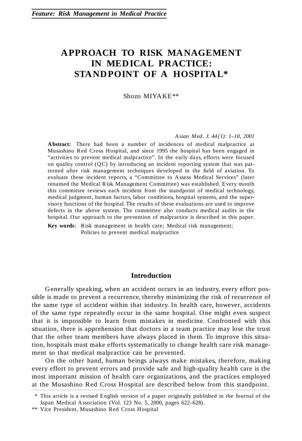
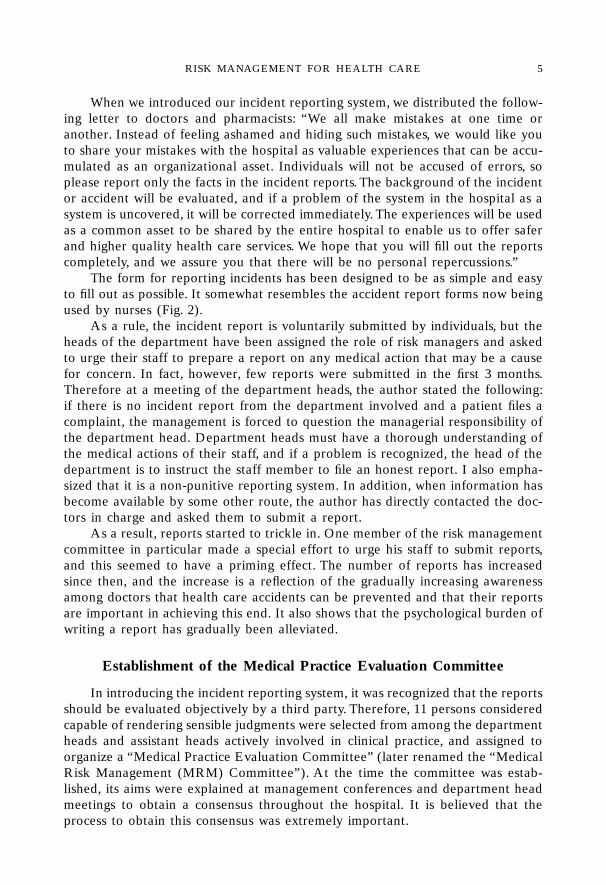
cal malpractice. In association with this new venture, they revised their accidentreport forms, organized a “committee to prevent accidents” within the nursingsection, reviewed accident reports forwarded from the wards, and fed the resultsback to the meetings of the chief nurses. The nursing section drew up a manualcalled “Accident Prevention” within a period of 18 months. Since early 1999, a riskmanagement nurse has been assigned to each ward to gather and analyze informa-tion on each incident and send back the details of the analysis in the form offeedback (Fig. 1).
2. DoctorsThere are numerous problems concerning doctors, and policies affecting doc-
tors will be mainly presented in this section.Traditionally, doctors have seemed to regard themselves as privileged and
expected everyone else to serve them, this attitude may have helped doctors tobecome self-righteous. Doctors have tended to avoid disclosing the details of theirpractice in investigations of medical malpractice, always leaving behind a so-called“gray zone”. However, the modern societies offer a wealth of medical information,and the public is better informed than ever. If doctors do not shake themselvesfree of their arrogant attitude, it may be impossible to prevent recurrences ofmedical malpractice, and doctors, as leaders of the health care groups, may lose thetrust of the other team members. Faced with the situation described above, ourprograms were undertaken.
Another motivation for starting these programs was doubts about the appro-
RISK MANAGEMENT FOR HEALTH CARE 3
priateness of our former method of hospital management. There are a number ofmedical departments within a hospital, and each operates within its own specialty.If each operates independently without regard for the other departments, thecohesion desired in a hospital is lost. Organized health care becomes possible onlywhen the goals and quality of medical service of the hospital as a whole are main-tained and managed. We believe that there is a definite need for a system tomonitor the health care actions of the hospital as a whole.
Introduction of an Incident Reporting System
Following the advice of Dr. Isao Kuroda, then a professor in the School ofHuman Science of Waseda University, who suggested “the introduction of riskmanagement technology that had been developed in the field of aviation becausemalpractice in health care resembles the accidents that occur in association withaviation” (1995), we decided to adopt the risk management incident reportingsystem for doctors.
There is a well known saying that there is a “chain of events” in aviationaccidents, because “3 or more minor incidents always occur in a row before alarger, more serious accident.” Every pilot is instructed to faithfully report everyincident that occurs during a flight regardless of its seriousness (including near-miss
Fig. 1 Organizational chart of medical risk management (Musashino Red Cross Hospital, June 1, 1999)
Head ofnursingsection
Director
Risk ManagementMedical Disputes
President, Vice president, Head of the nursing department, Head of the business department, Head of the general affairs section
Medical Affairs Conference General Risk Manager (Vice President)
Physician, Pharmacist, Medical technologist, Business section staff, Risk management nurse
Medical Risk Management Committee
Nursing sectionBusinessdepartment
Medicalsocial
servicedepartment
Laboratorydepartment
Pharmacydepartment
Medicaldepartment
Head of the nursing section
Risk Management Nurse
Nurse Risk ManagementCommittee
Risk Manager
Sectionhead
Sectionhead
Sectionhead
Departmenthead
Departmenthead
Departmenthead
Departmenthead
Risk Manager
Head ofnursingsection
Head ofnursingsection
Head ofnursingsection
Head ofnursingsection
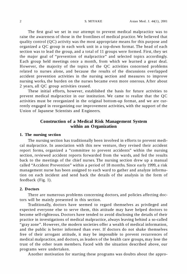
Incident/Accident Report(Circle one) Date: , 200
Work site Position Years of work experience Name Years (Personal seal)
Patient’s name Age Diagnosis(male or female)
Site of the accident Ward Department on an outpatient basisoccurred
Date and time of Date: Time:the accident
Date the accident was Date: Time:discovered
Time treatment was Date: Time:started
Time a report was made Date: Time:to the department head
Type of accident
[Classification] 1. oversight or misunderstanding, 2. misidentification,[Classification] 3. error in dosage, 4. complication, 5. iatrogenic disease, 6. others
Process during whichthe accident happened
Response and stepstaken after the accident
Explanation given afterthe accident andthe subsequentresponse of the patient
Evaluation of the gravity Llife-threating: □ very grave; □ grave; □ possible; □ little; □ noneof the risk involved Patient’s trust: □ greatly damaged; □ slightly damaged; □ not much affectedin the accident
Health status of □ good; physically fatigued [□ by work; □ for personal reasons]the medical personnel □ good; psychologically fatigued [□ by work; □ for personal reasons]involved □ others (remarks: )
Views on the causeof the accident
[Classification] 1. lack of observation, 2. delay in testing, 3. delayed diagnosis,[Classification] 4. inadequate technology, 5. surgical mistake,[Classification] 6. inadequate communication, 7. inadequate explanation, 8. others
Thoughts on the stepsto be taken in future
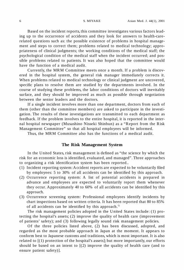
Fig. 2 Incident Report Form
incidents) by filing an incident report. They are assured that filing such reports willin no way affect their chances for promotion or future pay increases. Accidentprevention policies have been established and major accidents have been avertedby gathering and analyzing these reports describing minor incidents.
S. MIYAKE Asian Med. J. 44(1), 20014
When we introduced our incident reporting system, we distributed the follow-ing letter to doctors and pharmacists: “We all make mistakes at one time oranother. Instead of feeling ashamed and hiding such mistakes, we would like youto share your mistakes with the hospital as valuable experiences that can be accu-mulated as an organizational asset. Individuals will not be accused of errors, soplease report only the facts in the incident reports. The background of the incidentor accident will be evaluated, and if a problem of the system in the hospital as asystem is uncovered, it will be corrected immediately. The experiences will be usedas a common asset to be shared by the entire hospital to enable us to offer saferand higher quality health care services. We hope that you will fill out the reportscompletely, and we assure you that there will be no personal repercussions.”
The form for reporting incidents has been designed to be as simple and easyto fill out as possible. It somewhat resembles the accident report forms now beingused by nurses (Fig. 2).
As a rule, the incident report is voluntarily submitted by individuals, but theheads of the department have been assigned the role of risk managers and askedto urge their staff to prepare a report on any medical action that may be a causefor concern. In fact, however, few reports were submitted in the first 3 months.Therefore at a meeting of the department heads, the author stated the following:if there is no incident report from the department involved and a patient files acomplaint, the management is forced to question the managerial responsibility ofthe department head. Department heads must have a thorough understanding ofthe medical actions of their staff, and if a problem is recognized, the head of thedepartment is to instruct the staff member to file an honest report. I also empha-sized that it is a non-punitive reporting system. In addition, when information hasbecome available by some other route, the author has directly contacted the doc-tors in charge and asked them to submit a report.
As a result, reports started to trickle in. One member of the risk managementcommittee in particular made a special effort to urge his staff to submit reports,and this seemed to have a priming effect. The number of reports has increasedsince then, and the increase is a reflection of the gradually increasing awarenessamong doctors that health care accidents can be prevented and that their reportsare important in achieving this end. It also shows that the psychological burden ofwriting a report has gradually been alleviated.
Establishment of the Medical Practice Evaluation Committee
In introducing the incident reporting system, it was recognized that the reportsshould be evaluated objectively by a third party. Therefore, 11 persons consideredcapable of rendering sensible judgments were selected from among the departmentheads and assistant heads actively involved in clinical practice, and assigned toorganize a “Medical Practice Evaluation Committee” (later renamed the “MedicalRisk Management (MRM) Committee”). At the time the committee was estab-lished, its aims were explained at management conferences and department headmeetings to obtain a consensus throughout the hospital. It is believed that theprocess to obtain this consensus was extremely important.
RISK MANAGEMENT FOR HEALTH CARE 5
Based on the incident reports, this committee investigates various factors lead-ing up to the occurrence of accidents and they look for answers to health-care-related questions such as: the possible existence of problems in hospital manage-ment and steps to correct them; problems related to medical technology; appro-priateness of clinical judgments; the working conditions of the medical staff; thepsychological condition of the medical staff when the incident occurred; and pos-sible problems related to patients. It was also hoped that the committee wouldhave the function of a medical audit.
Currently, the MRM Committee meets once a month. If a problem is discov-ered in the hospital system, the general risk manager immediately corrects it.When problems related to medical technology or clinical judgment are uncovered,specific plans to resolve them are studied by the departments involved. In thecourse of studying these problems, the labor conditions of doctors will inevitablysurface, and they should be improved as much as possible through negotiationbetween the senior leaders and the doctors.
If a single incident involves more than one department, doctors from each ofthem (other than the committee members) are asked to participate in the investi-gation. The results of these investigations are transmitted to each department asfeedback. If the problem involves to the entire hospital, it is reported in the inter-nal hospital newspaper (Musashino Nisseki Shinbun) as a “Report from the RiskManagement Committee” so that all hospital employees will be informed.
Thus, the MRM Committee also has the functions of a medical audit.
The Risk Management System
In the United States, risk management is defined as “the science by which therisk for an economic loss is identified, evaluated, and managed”. Three approachesto organizing a risk identification system has been reported..(1) Incident reporting system: Accident reports are expected to be voluntarily filed
by employees: 5 to 30% of all accidents can be identified by this approach.(2) Occurrence reporting system: A list of potential accidents is prepared in
advance and employees are expected to voluntarily report them wheneverthey occur. Approximately 40 to 60% of all accidents can be identified by thisapproach.
(3) Occurrence screening system: Professional employees identify incidents bychart inspections based on written criteria. It has been reported that 80 to 85%of all accidents can be identified by this approach.1)
The risk management policies adopted in the United States include: (1) pro-tecting the hospital’s assets; (2) improve the quality of health care (improvementof patients’ safety); and (3) following legally sound risk management policies.
Of the three policies listed above, (2) has been discussed, adopted, andregarded as the most probable approach in Japan at the moment. It appears toconform best to Japanese customs and traditions, which is most important. It is alsorelated to [(1) protection of the hospital’s assets]; but more importantly, our effortsshould be based on an intent to [(2) improve the quality of health care (and toensure patient safety)].
S. MIYAKE Asian Med. J. 44(1), 20016
Introduction of the Risk Predicting System
To introduce (2) an occurrence reporting system for the risk managementsystem described above, the following steps were taken.
All of the department heads were assigned to the position of risk manager andasked to prepare a list of accidents that are most likely to occur in relation tomedical care in their department and to formulate and submit measures to preventthem. These reports have been incorporated into appropriate chapters of the“Manual to Prevent Medical Accidents”. As reference material, informed consentto various procedures that are frequently conducted in each department areincluded (when the consent is written, it is often accompanied by statistics on risk).A list of drug names that are easily confused and photographs and names ofampules containing drugs for parenteral use are included in the manual so errorsshould not go unnoticed.
It is said that about half of the disputes concerning medical care involvefinancial settlement, whereas the other half concern the personality of the doctor(appeals are made just to punish doctors for their actions). According to the sta-tistics in the United States, 70% of the medical disputes arise in the absence oferrors on the part of the medical staff. These disputes stem from a lack of commu-nication between the patients and doctors or other medical staff members. Appar-ently, what appears to have been a careless manner of speaking, attitude, or facialexpression of the doctor generates distrust on the part of the patient, which even-tually leads to medical complaint. With this in mind, the overview section of this“Manual to Prevent Medical Accidents” describes doctors’ methods and manner ofdealing with patients under the heading of “Basic Rules to Prevent Medical Acci-dents”. In this section, attention is called to basic manners required by doctors,including the need for a patient-oriented medical process, confirmation of eachprocedure, assuming a humble attitude, with undivided attention given to what thepatient wants to talk about, building a good patient-doctor relationship, and pro-viding methods for fill out medical records.
This “Manual to Prevent Medical Accidents” was distributed to all doctorsworking at the hospital. Doctors are expected to peruse even the sections that donot actually involve them. Their critiques are useful in preparing the next edition,and three revisions were made. It required 8 months to prepare the first edition,which was published in August 1997. The second edition was completed in October1998. In each of the revised editions we hope to include the experiences of doctorswho have learned from the accident prevention steps taken in other departmentsand re-evaluated and reinforced the accident preventive measures in their owndepartment. We also hope that the manual will be used by all doctors in thehospital, but the process of preparing it is even more important. The authorbelieves that by experiencing the process by which policies to prevent accidents aredrawn up, they will become more sensitive to the possibility of accidents. In otherwords, the manual to prevent medical accidents is most meaningful when eachhospital prepares its own unique version.
RISK MANAGEMENT FOR HEALTH CARE 7
Introduction of the Occurrence Screening System
The nursing section of our hospital assigned a risk management nurse late in1999 and prepared its own screening system. We expect good results from thismovement.
In the areas in which doctors are involved, the medical care in each depart-ment is extremely diverse and the magnitude of the risk involved is several timesthat of nursing section. Since the area involved is believed to be too large to bemanaged by a single risk manager, the department heads may have to be asked toact as risk managers; and doctors will be expected to improve their awareness ofthe need to prevent medical accidents.
Response to the Development of Medical Disputes
When a medical malpractice that might develop into a medical dispute occurs,the basic rule is that the doctor in charge immediately reports the accident to thehead of the department, and that the department head in turn reports it to thehead of the administrative department, the vice president (general risk manager),or the president of the hospital. The doctor then waits for their directions beforeresponding further.
What is important in these procedures is that those involved express theirsincerity and consideration toward the patients and their families by their attitudesand speech. Next, there is a need to establish a single channel to handle theprocedures for dealing with patients or their families. At our hospital, the generalaffairs section is in charge. It is essential that the doctors in charge or the head ofthe department involved not apologize to patients or their families on their ownnor tell them about the possible future response of the hospital based on their owninterpretation. Doctors or department heads should always discuss the matter withthe management of the hospital before they respond to outsiders. They shouldexplain to them that the matter will be handled by the general affairs section, andthen quickly report and discuss any future steps with hospital management throughthe procedures explained above. The initial response by the doctor in charge ordepartment head often determines future developments.
After taking the steps described above, those who are directly involved in theaccident should promptly fill out an “accident report” (a form that has been pre-pared by an insurance company) and submit it to the general affairs section. Basedon this accident report, future steps to be taken will be discussed at the MedicalAffairs Conference (composed of the president, vice president, head of the nursingsection, business manager, and head of the General Affairs Section). At Red CrossHospitals, such reports are sent to Red Cross Headquarters and the insurancecompany.
When the evidence is seized or a patient files a claim leading to a legal dispute,a conference is held between the attorney representing the patient and the insur-ance company, and the response by the attorney is discussed at a Medical AffairsConference.
S. MIYAKE Asian Med. J. 44(1), 20018
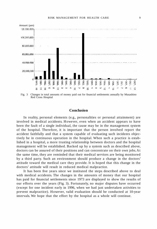
Fig. 3 Changes in total amounts of money paid out for financial settlements annually by MusashinoRed Cross Hospital
Amount (yen)
Conclusion
In reality, personal elements (e.g., personalities or personal attainment) areinvolved in medical accidents. However, even when an accident appears to havebeen the fault of a single individual, the cause may be in the management systemof the hospital. Therefore, it is important that the person involved report theaccident faithfully and that a system capable of evaluating such incidents objec-tively be in continuous operation in the hospital. When such a practice is estab-lished in a hospital, a more trusting relationship between doctors and the hospitalmanagement will be established. Backed up by a system such as described above,doctors can be assured of their positions and can concentrate on their own jobs. Atthe same time, they are reminded that their medical services are being monitoredby a third party. Such an environment should produce a change in the doctors’attitude toward the medical care they provide. It is hoped that this change in thedoctors’ attitude will result in reduced medical malpractice.
It has been five years since we instituted the steps described above to dealwith medical accidents. The changes in the amounts of money that our hospitalhas paid for financial settlements since 1973 are displayed to show the results ofour efforts over the years (Fig. 3). Fortunately, no major disputes have occurred(except for one incident early in 1996, when we had just undertaken activities toprevent malpractice). However, valid evaluation should be conducted at 10-yearintervals. We hope that the effort by the hospital as a whole will continue.
RISK MANAGEMENT FOR HEALTH CARE 9
REFERENCES
1) United States General Accounting Office: Health Care-Initiatives in Hospital RiskManagement. Washington DC, 1989.
2) Miyake, S.: Organizational approaches to medical accidents. Nippon Byoinkai Zasshi46: 775–787, 1999. (in Japanese)
3) Miyake, S.: Risk management in medicine. Nippon Byoinkai Zasshi 46: 1534–1568,1999. (in Japanese)
4) Miyake, S.: Medical risk management at Musashino Red Cross Hospital. Byoin 58:764–765, 860–861, 966–967, 1999. (in Japanese)
5) Miyake, S.: Practice of medical risk management. Chiryo 58: 2781–2881, 1999. (inJapanese)
S. MIYAKE Asian Med. J. 44(1), 200110
* This article is a revised English version of a paper originally published in the Journal of theJapan Medical Association (Vol. 123 No. 5, 2000, pages 638–644).
** Director, Hokkaido Red Cross Blood Center
11
APPROACH TO RISK MANAGEMENTIN MEDICAL PRACTICE:
STANDPOINT OF THE BLOOD TRASFUSION*
Hisami IKEDA**
Asian Med. J. 44(1): 11–21, 2000Abstract: In the field of blood transfusion, risk management is defined as assuringthe safety of blood transfusion. This includes prevention of errors in the process ofcollecting blood from donors, examination, and production of blood preparationsfrom the donated blood. Good Manufacturing Practice (GMP) and a system forensuring compliance with the GMP are thus necessary. Reliable retrospective exami-nation and provision of information, regarding adverse reactions, from medicalinstitutions are essential for prevention of adverse reactions to blood transfusion.On the basis of this information, such measures as highly sensitive screening, includ-ing viral nucleic acid amplification, and inactivation and elimination of leukocytescan be implemented. On the other hand, medical institutions need to clearly estab-lish the procedures to be followed when transfusing blood into patients in order toprevent human errors. There should be a system that allows confirmation of goodcompliance with the procedure. In numerous medical institutions in Japan, the pro-cess from placement of orders for blood to actual transfusion of the blood is com-plicated, thereby increasing the likelihood of errors. Unification of management ofthe blood transfusion process is necessary. Finally, a system that allows objectivemonitoring of appropriate production of blood preparations at blood centers, as wellas the blood transfusion process at medical institutions, must be established.
Key words: Window period; Error prevention system;Blood transfusion consent form;Blood Transfusion Therapy Committee
Introduction
Risk management can be interpreted as a procedure consisting of listing allpossible adverse reactions or errors, analysis of the probability of risk onset, evalu-ation of the seriousness of the risk, and prevention of adverse reactions or acci-dents by implementing preventive measures or making improvements. Amongvarious adverse reactions to blood transfusion, nonhemolytic reactions have beenreported in the largest number, but there have been only a few reports of seriouscases. In contrast, transfusion-transmitted infection, post-transfusion graft versus
H. IKEDA Asian Med. J. 44(1), 200112
host disease (GVHD), and hemolytic adverse reactions are infrequent, but can beserious. Therefore, priority should be placed on the more serious reactions in riskmanagement.1)
If we define risk management in blood transfusion as a process to assure itssafety, we should consider risk management from the perspective of the providersof blood preparations for transfusion, in other words, blood centers, and riskmanagement at medical institutions where blood transfusion is performed. Theformer aims at assuring the safety of donors and blood for transfusion, and thelatter aims at assuring the safety of blood transfusion recipients.
Risk Management at Blood Centers
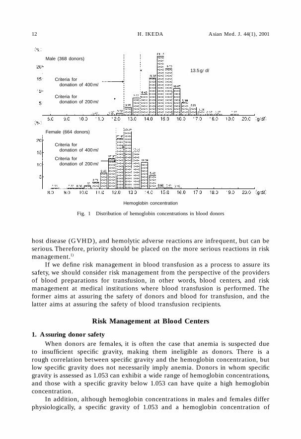
1. Assuring donor safetyWhen donors are females, it is often the case that anemia is suspected due
to insufficient specific gravity, making them ineligible as donors. There is arough correlation between specific gravity and the hemoglobin concentration, butlow specific gravity does not necessarily imply anemia. Donors in whom specificgravity is assessed as 1.053 can exhibit a wide range of hemoglobin concentrations,and those with a specific gravity below 1.053 can have quite a high hemoglobinconcentration.
In addition, although hemoglobin concentrations in males and females differphysiologically, a specific gravity of 1.053 and a hemoglobin concentration of
Fig. 1 Distribution of hemoglobin concentrations in blood donors
Hemoglobin concentration
Male (368 donors)
Criteria fordonation of 400 ml
Criteria fordonation of 200 ml
Female (664 donors)
Criteria fordonation of 400 ml
Criteria fordonation of 200 ml
13.5 g/dl
RISK MANAGEMENT EFFORTS 13
12.5 g/dl or more have been adopted as the criteria for collection of 400 ml ofblood in both males and females in Japan. The distribution of hemoglobin concen-trations in male donors is shifted toward the higher concentration in comparisonto that in female donors. Therefore, it may be necessary to change the criteria forcollection of 400 ml of blood to 13.5 g/dl or more for males and 12.5 g/dl or morefor females in order to assure donor safety (Fig. 1).
As to carriers of hepatitis viruses detected in screening for blood transfusion,not only notification but also more proactive management of their health is recom-mended. At our institution, we hold orientations for donors who have been provedto be carriers of hepatitis B virus (HBV) or hepatitis C virus (HCV), and carry outexaminations/health consultations two to four times a year under the auspices ofthe carrier clinic.2) In a sense, this can be regarded as an activity assuring the safetyof donors.
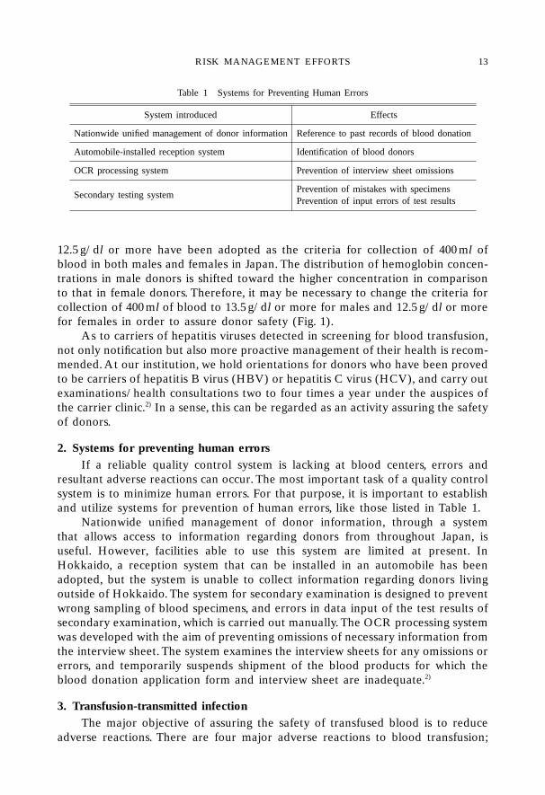
2. Systems for preventing human errorsIf a reliable quality control system is lacking at blood centers, errors and
resultant adverse reactions can occur. The most important task of a quality controlsystem is to minimize human errors. For that purpose, it is important to establishand utilize systems for prevention of human errors, like those listed in Table 1.
Nationwide unified management of donor information, through a systemthat allows access to information regarding donors from throughout Japan, isuseful. However, facilities able to use this system are limited at present. InHokkaido, a reception system that can be installed in an automobile has beenadopted, but the system is unable to collect information regarding donors livingoutside of Hokkaido. The system for secondary examination is designed to preventwrong sampling of blood specimens, and errors in data input of the test results ofsecondary examination, which is carried out manually. The OCR processing systemwas developed with the aim of preventing omissions of necessary information fromthe interview sheet. The system examines the interview sheets for any omissions orerrors, and temporarily suspends shipment of the blood products for which theblood donation application form and interview sheet are inadequate.2)
3. Transfusion-transmitted infectionThe major objective of assuring the safety of transfused blood is to reduce
adverse reactions. There are four major adverse reactions to blood transfusion;
Table 1 Systems for Preventing Human Errors
System introduced Effects
Nationwide unified management of donor information Reference to past records of blood donation
Automobile-installed reception system Identification of blood donors
OCR processing system Prevention of interview sheet omissions
Secondary testing systemPrevention of mistakes with specimensPrevention of input errors of test results
14
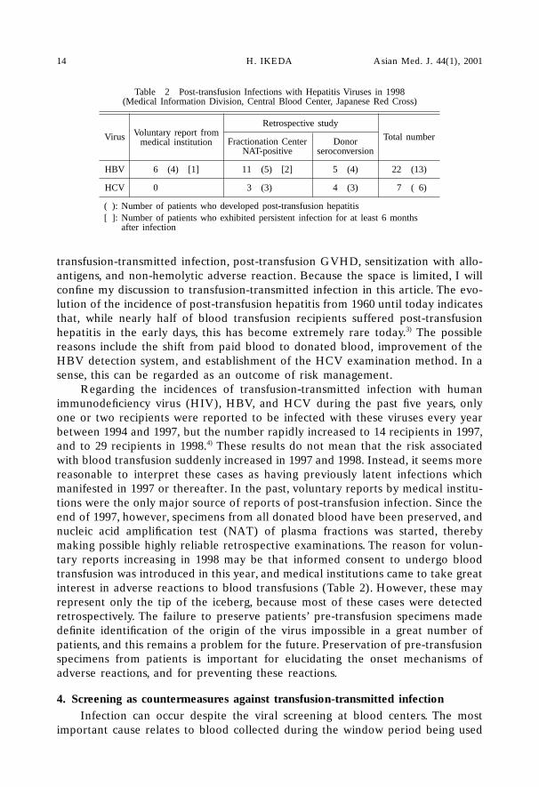
transfusion-transmitted infection, post-transfusion GVHD, sensitization with allo-antigens, and non-hemolytic adverse reaction. Because the space is limited, I willconfine my discussion to transfusion-transmitted infection in this article. The evo-lution of the incidence of post-transfusion hepatitis from 1960 until today indicatesthat, while nearly half of blood transfusion recipients suffered post-transfusionhepatitis in the early days, this has become extremely rare today.3) The possiblereasons include the shift from paid blood to donated blood, improvement of theHBV detection system, and establishment of the HCV examination method. In asense, this can be regarded as an outcome of risk management.
Regarding the incidences of transfusion-transmitted infection with humanimmunodeficiency virus (HIV), HBV, and HCV during the past five years, onlyone or two recipients were reported to be infected with these viruses every yearbetween 1994 and 1997, but the number rapidly increased to 14 recipients in 1997,and to 29 recipients in 1998.4) These results do not mean that the risk associatedwith blood transfusion suddenly increased in 1997 and 1998. Instead, it seems morereasonable to interpret these cases as having previously latent infections whichmanifested in 1997 or thereafter. In the past, voluntary reports by medical institu-tions were the only major source of reports of post-transfusion infection. Since theend of 1997, however, specimens from all donated blood have been preserved, andnucleic acid amplification test (NAT) of plasma fractions was started, therebymaking possible highly reliable retrospective examinations. The reason for volun-tary reports increasing in 1998 may be that informed consent to undergo bloodtransfusion was introduced in this year, and medical institutions came to take greatinterest in adverse reactions to blood transfusions (Table 2). However, these mayrepresent only the tip of the iceberg, because most of these cases were detectedretrospectively. The failure to preserve patients’ pre-transfusion specimens madedefinite identification of the origin of the virus impossible in a great number ofpatients, and this remains a problem for the future. Preservation of pre-transfusionspecimens from patients is important for elucidating the onset mechanisms ofadverse reactions, and for preventing these reactions.
4. Screening as countermeasures against transfusion-transmitted infectionInfection can occur despite the viral screening at blood centers. The most
important cause relates to blood collected during the window period being used
Table 2 Post-transfusion Infections with Hepatitis Viruses in 1998(Medical Information Division, Central Blood Center, Japanese Red Cross)
Retrospective study
Virus Voluntary report fromFractionation Center Donor Total numbermedical institution
NAT-positive seroconversion
HBV 6 (4) [1] 11 (5) [2] 5 (4) 22 (13)
HCV 0 (4) [1] 13 (3) [2] 4 (3) 7 ( 6)
( ): Number of patients who developed post-transfusion hepatitis[ ]: Number of patients who exhibited persistent infection for at least 6 months[ ]: after infection
H. IKEDA Asian Med. J. 44(1), 2001
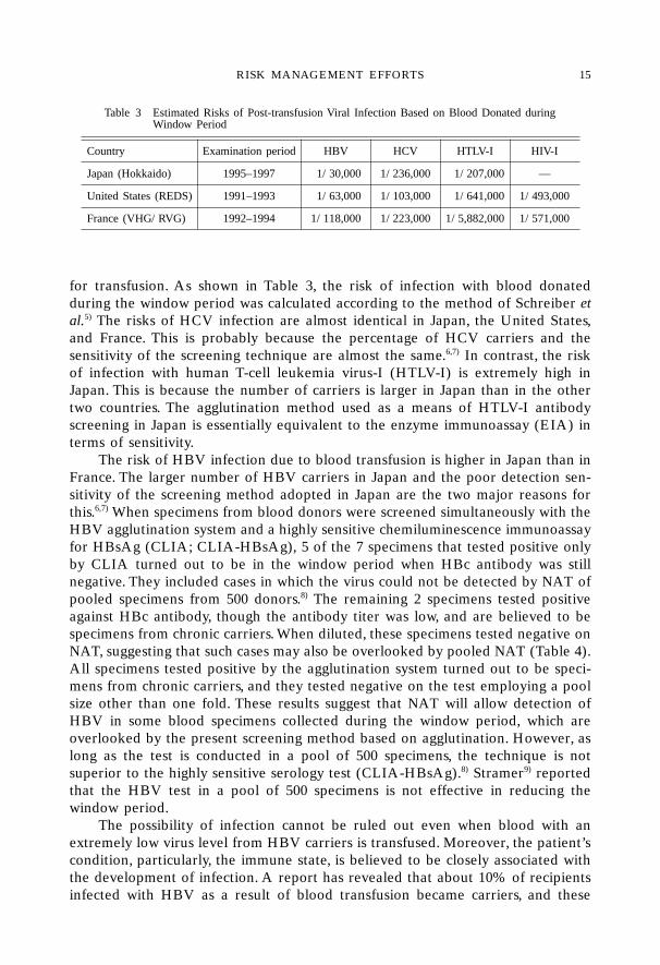
for transfusion. As shown in Table 3, the risk of infection with blood donatedduring the window period was calculated according to the method of Schreiber etal.5) The risks of HCV infection are almost identical in Japan, the United States,and France. This is probably because the percentage of HCV carriers and thesensitivity of the screening technique are almost the same.6,7) In contrast, the riskof infection with human T-cell leukemia virus-I (HTLV-I) is extremely high inJapan. This is because the number of carriers is larger in Japan than in the othertwo countries. The agglutination method used as a means of HTLV-I antibodyscreening in Japan is essentially equivalent to the enzyme immunoassay (EIA) interms of sensitivity.
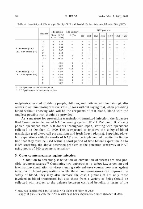
The risk of HBV infection due to blood transfusion is higher in Japan than inFrance. The larger number of HBV carriers in Japan and the poor detection sen-sitivity of the screening method adopted in Japan are the two major reasons forthis.6,7) When specimens from blood donors were screened simultaneously with theHBV agglutination system and a highly sensitive chemiluminescence immunoassayfor HBsAg (CLIA; CLIA-HBsAg), 5 of the 7 specimens that tested positive onlyby CLIA turned out to be in the window period when HBc antibody was stillnegative. They included cases in which the virus could not be detected by NAT ofpooled specimens from 500 donors.8) The remaining 2 specimens tested positiveagainst HBc antibody, though the antibody titer was low, and are believed to bespecimens from chronic carriers. When diluted, these specimens tested negative onNAT, suggesting that such cases may also be overlooked by pooled NAT (Table 4).All specimens tested positive by the agglutination system turned out to be speci-mens from chronic carriers, and they tested negative on the test employing a poolsize other than one fold. These results suggest that NAT will allow detection ofHBV in some blood specimens collected during the window period, which areoverlooked by the present screening method based on agglutination. However, aslong as the test is conducted in a pool of 500 specimens, the technique is notsuperior to the highly sensitive serology test (CLIA-HBsAg).8) Stramer9) reportedthat the HBV test in a pool of 500 specimens is not effective in reducing thewindow period.
The possibility of infection cannot be ruled out even when blood with anextremely low virus level from HBV carriers is transfused. Moreover, the patient’scondition, particularly, the immune state, is believed to be closely associated withthe development of infection. A report has revealed that about 10% of recipientsinfected with HBV as a result of blood transfusion became carriers, and these
Table 3 Estimated Risks of Post-transfusion Viral Infection Based on Blood Donated duringTable 3 Window Period
Country Examination period HBV HCV HTLV-I HIV-I
Japan (Hokkaido) 1995–1997 11/30,000 1/236,000 5,1/207,000 —
United States (REDS) 1991–1993 11/63,000 1/103,000 5,1/641,000 1/493,000
France (VHG/RVG) 1992–1994 1/118,000 1/223,000 1/5,882,000 1/571,000
RISK MANAGEMENT EFFORTS 15
recipients consisted of elderly people, children, and patients with hematologic dis-orders in an immunosuppressive state. It goes without saying that, when providingblood without knowing who will be the recipients of that blood, blood with thesmallest possible risk should be provided.
As a measure for preventing transfusion-transmitted infection, the JapaneseRed Cross has implemented NAT screening against HBV, HIV-1, and HCV usingpooled specimens from 500 donors throughout Japan, starting with specimenscollected on October 10, 1999. This is expected to improve the safety of bloodtransfusion (red blood cell preparations and fresh frozen plasma). Supplying plate-let preparations with the results of NAT must be implemented despite the limita-tion that they must be used within a short period of time before expiration. As toHBV screening, the above-described problem of the detection sensitivity of NATusing pools of 500 specimens remains.*
5. Other countermeasures against infectionIn addition to screening, inactivation or elimination of viruses are also pos-
sible countermeasures.10) Combining two approaches to safety, i.e., screening andinactivation/elimination of viruses, may greatly enhance countermeasures againstinfection of blood preparations. While these countermeasures can improve thesafety of blood, they may also increase the cost. Opinions of not only thoseinvolved in blood transfusion but also those from a variety of fields should becollected with respect to the balance between cost and benefits, in terms of the
Table 4 Sensitivity of HBs Antigen Test by CLIA and Pooled Nucleic Acid Amplification Test (NAT)
HBs antigen HBc antibodyNAT pool size
SpecimenCLIA (S/CO HI (2n) �1 �10 �25 �50 �100 �250 �500�/� (value)
1** � 11.10 � � � � � � � �
2** � 23.59 � � � � � � � �
CLIA-HBsAg (�)/ 3** � 21.34 � � � � � � � �
4** � 28.47 � � � � � � � �JRC HBV system (�)
5** � 29.18 � � � � � � � �
6** � 27.44 4 � � � � � � �
7** � 28.43 4 � � � � � � �
1** � �1.0 9 � � � � � � �
2** � �1.0 8 � � � � � � �
3** � �1.0 �100.0 � � � � � � �
CLIA-HBsAg (�)/ 4** � �1.0 6 � � � � � � �
JRC HBV system (�) 5** � �1.0 6 � � � � � � �
6** � �1.0 6 � � � � � � �
7** � �1.0 6 � � � � � � �
8** � �1.0 7 � � � � � � �
* 1–5: Specimens in the Window Period** 6,7: Specimens from low-viremic carriers
H. IKEDA Asian Med. J. 44(1), 200116
* JRC has implemented the 50 pool NAT since February of 2000.Supply of platelets with the NAT results have been implemented since October of 2000.
safety provided by these measures.Bacterial contamination of blood preparations should also be taken into con-
sideration. It has been indicated that, in sampling tests, bacterial contamination canbe detected in about 0.1% of specimens examined. However, the actual status ofadverse reactions to transfused blood preparations contaminated with bacteriaremains unknown, and systematic studies have not been conducted in Japan.
6. Retrospective studies of transfusion-transmitted infectionAt blood centers, retrospective studies are conducted when patients are
suspected of having developed transfusion-transmitted infection, or when NAT-positive blood preparations are suspected to have been given to patients. At bloodcenters, information regarding the patient is collected from his or her attendingphysician, and the physician is asked to submit specimens. Then, the patient’sspecimens as well as specimens from the donor are subjected to detailed examina-tion, and the results are reported to the attending physician. As described earlier,specimens from all donors have been preserved since 1997. When a transfusion-transmitted infection is suspected, the attending physician is requested to followup the patient’s condition, and the blood center cooperates with the physician incarrying out a detailed examination. Persons in charge of medical information atthe blood center (Medical Representative) play a central role in these series ofexaminations.
Risk management at Medical Institutions
Numerous hospital personnel (physicians, nurses, laboratory technicians, andpharmacists, etc.) are involved in blood transfusion therapy, and blood transfusionmay be performed at various places such as the general ward, operating room, andoutpatient clinic. Therefore, the commitment and cooperation of the entire hospi-tal staff are essential for prevention of errors related to blood transfusion. More-over, since highly specialized knowledge is required for blood transfusion therapy,accredited specialists and laboratory technicians specializing in transfusion medi-cine are believed to play important roles in transfusion therapy.
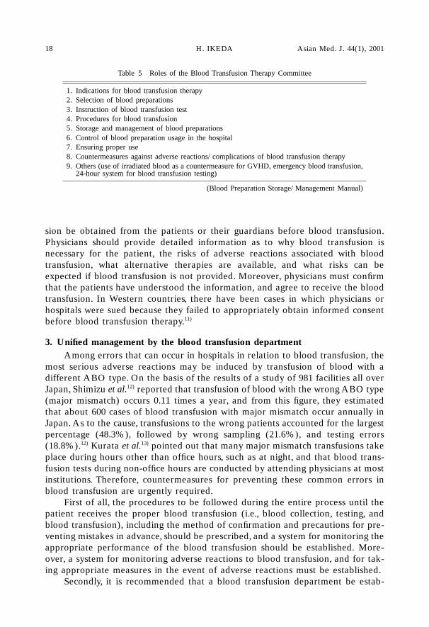
1. Foundation of Blood Transfusion Therapy Committee and its rolesAll medical institutions performing blood transfusions should establish a
Blood Transfusion Therapy Committee that monitors proper use of blood prepa-rations, examination of blood transfusions, storage and management of blood,actual procedures of blood transfusions, and adverse reactions. The role of such aBlood Transfusion Therapy Committee should be more than nominal. It shouldconstantly monitor whether blood transfusion is carried out in a safe and effectivemanner for patients, and when any problems are detected, it should demonstrateleadership and responsibility in resolving those problems. Roles of the BloodTransfusion Therapy Committee are summarized in Table 5.
2. Informed consentIn April 1997, it became mandatory that informed consent for blood transfu-
RISK MANAGEMENT EFFORTS 17
sion be obtained from the patients or their guardians before blood transfusion.Physicians should provide detailed information as to why blood transfusion isnecessary for the patient, the risks of adverse reactions associated with bloodtransfusion, what alternative therapies are available, and what risks can beexpected if blood transfusion is not provided. Moreover, physicians must confirmthat the patients have understood the information, and agree to receive the bloodtransfusion. In Western countries, there have been cases in which physicians orhospitals were sued because they failed to appropriately obtain informed consentbefore blood transfusion therapy.11)
3. Unified management by the blood transfusion departmentAmong errors that can occur in hospitals in relation to blood transfusion, the
most serious adverse reactions may be induced by transfusion of blood with adifferent ABO type. On the basis of the results of a study of 981 facilities all overJapan, Shimizu et al.12) reported that transfusion of blood with the wrong ABO type(major mismatch) occurs 0.11 times a year, and from this figure, they estimatedthat about 600 cases of blood transfusion with major mismatch occur annually inJapan. As to the cause, transfusions to the wrong patients accounted for the largestpercentage (48.3%), followed by wrong sampling (21.6%), and testing errors(18.8%).12) Kurata et al.13) pointed out that many major mismatch transfusions takeplace during hours other than office hours, such as at night, and that blood trans-fusion tests during non-office hours are conducted by attending physicians at mostinstitutions. Therefore, countermeasures for preventing these common errors inblood transfusion are urgently required.
First of all, the procedures to be followed during the entire process until thepatient receives the proper blood transfusion (i.e., blood collection, testing, andblood transfusion), including the method of confirmation and precautions for pre-venting mistakes in advance, should be prescribed, and a system for monitoring theappropriate performance of the blood transfusion should be established. More-over, a system for monitoring adverse reactions to blood transfusion, and for tak-ing appropriate measures in the event of adverse reactions must be established.
Secondly, it is recommended that a blood transfusion department be estab-
Table 5 Roles of the Blood Transfusion Therapy Committee
1. Indications for blood transfusion therapy2. Selection of blood preparations3. Instruction of blood transfusion test4. Procedures for blood transfusion5. Storage and management of blood preparations6. Control of blood preparation usage in the hospital7. Ensuring proper use8. Countermeasures against adverse reactions/complications of blood transfusion therapy9. Others (use of irradiated blood as a countermeasure for GVHD, emergency blood transfusion,
24-hour system for blood transfusion testing)
(Blood Preparation Storage/Management Manual)
H. IKEDA Asian Med. J. 44(1), 200118
lished to allow unified management of blood transfusion operations. At mostmedical institutions in Japan, blood preparations are managed by the pharmacy,and blood transfusion tests are conducted by the laboratory. Therefore, handling ofvarious slips, specimens and blood preparations is complicated during the processfollowing placement of the order for blood transfusion until the actual blood trans-fusion. Under such circumstances, errors are likely to occur. Unified managementis expected to facilitate rapid provision of blood preparations, promote proper use,allow monitoring of recipients of blood transfusion and effective use of bloodpreparations, as well as control adverse reactions. Unified management by theblood transfusion department is an important aspect of risk management in bloodtransfusion.
4. Inspection and accreditation (I&A) of blood transfusion proceduresThere is a system for objective evaluation by a third party regarding the
proper performance of blood transfusion procedures at an institution, and for useof the results to improve procedures at the institution. The Kanto KoshinetsuBlock of the Japanese Society of Blood Transfusion has established an I&A Sub-committee to offer this service to institutions that have requested evaluation.14)
This may be another approach to risk management. In the future, the JapaneseSociety of Blood Transfusion is expected to play a central role in establishing anationwide I&A system, and in demonstrating its effects.
Conclusion
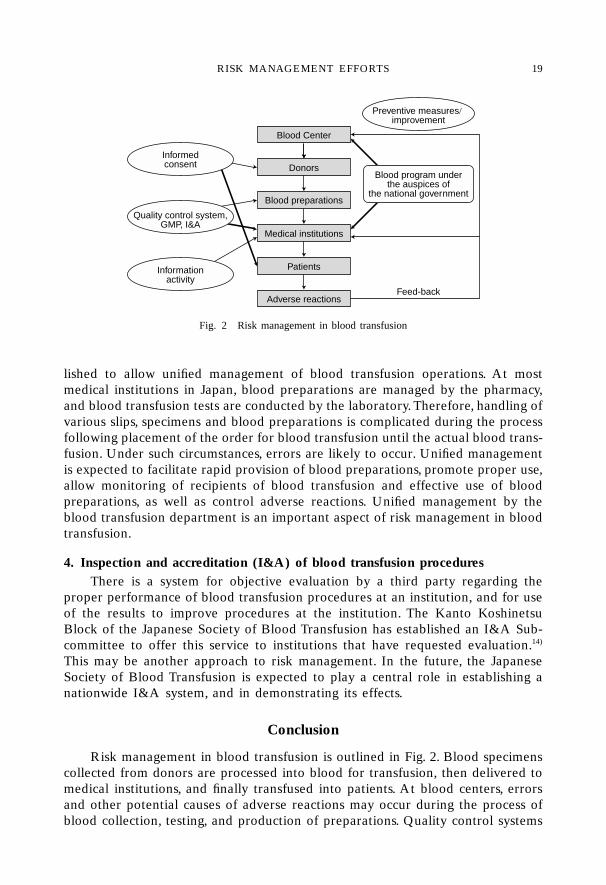
Risk management in blood transfusion is outlined in Fig. 2. Blood specimenscollected from donors are processed into blood for transfusion, then delivered tomedical institutions, and finally transfused into patients. At blood centers, errorsand other potential causes of adverse reactions may occur during the process ofblood collection, testing, and production of preparations. Quality control systems
Fig. 2 Risk management in blood transfusion
Feed-back
Preventive measures/improvement
Blood program underthe auspices of
the national government
Blood Center
Donors
Blood preparations
Medical institutions
Patients
Adverse reactions
Informedconsent
Quality control system,GMP, I&A
Informationactivity
RISK MANAGEMENT EFFORTS 19
and Good Manufacturing Practice (GMP) are important in their prevention. Majorfactors in risk management at medical institutions include establishment of aBlood Transfusion Therapy Committee, informed consent from patients, unifiedmanagement by the blood transfusion department, and I&A. If the causes areexplored and problems identified in the case of adverse reactions to blood trans-fusion, the information can be given to blood centers and medical institutions asfeedback to facilitate the establishment or improvement of preventive measuresfor the future.
Physicians, nurses, laboratory technicians, and pharmacists of a medical insti-tution will make it possible to provide safe and effective blood transfusions forpatients by carrying out their responsibilities and roles, under a cooperative systemwith the blood center. No matter how far blood transfusion therapy advances,however, potential risks of allogenic blood transfusion can never be completelyavoided. Therefore, strategies such as autologous blood transfusion and limitingblood transfusion to the minimum necessary cases should be continued.
Since blood transfusion is a medical practice that is intimately associated withthe safety of the general public, establishment of a system under which projectsrelated to blood preparations are carried out under the auspices of the nationalgovernment is awaited.
REFERENCES
1) Isbister, J.P.: Risk management in transfusion medicine. Transfus Med Rev 10: 183–202, 1996.
2) Sekiguchi, S.: Blood donation and informed consent. Nichijo Shinryo to Ketsueki 8:555–563, 1998. (in Japanese)
3) Sekiguchi, S. and Sato, S.: Current status of post-transfusion hepatitis and its preven-tion. Rinsho Kensa 43: 255–264, 1999. (in Japanese)
4) Takahashi, M. et al.: Transfusion-transmitted infection and criteria for its evaluation—Based on analysis of adverse reactions. Ketsueki Jigyo 21: 17–30, 1998. (in Japanese)
5) Schreiber, G.B. et al.: The risk of transfusion-transmitted viral infection. N Engl J Med334: 1685–1690, 1996.
6) Ikeda, H.: Serological tests and nucleic acid amplification in viral screening of blooddonors. Journal of the Japanese Society of Blood Transfusion (in press). (in Japanese)
7) Sato, S. et al.: Estimated risks of transfusion-transmitted infection. The 7th Red CrossBlood Symposium, Japanese Red Cross, 1999, pp.3–12. (in Japanese)
8) Ikeda, H. et al.: Sensitivity of nucleic acid amplification testing (NAT) using pooleddonor samples for HBV compared to a serological HBs antigen assay. Transfusion39(Suppl): 4S, 1999.
9) Strainer, S.L.: Practical concepts in molecular diagnostics testing donor screeningusing nucleic acid amplification assays. Evaluations of Genome Amplification Tech-niques (GAT) for testing of pooled donor samples. AABB 51st Annual Meeting, 1998,pp.470–474.
10) Sekiguchi, S.: Limitation to prevention of infection of blood preparations for transfu-sion. Ketsueki Jigyo 21: 173–184, 1998. (in Japanese)
11) Translated by Sekiguchi, S.: Informed consent for blood transfusion. AABB, 1998. (inJapanese)
12) Shimizu, M. et al.: A study by the Ministry of Health and Welfare on proper bloodtransfusion therapy based on the graft-versus-host disease—Report of the 1995 ques-
H. IKEDA Asian Med. J. 44(1), 200120
tionnaire survey, 1997. (in Japanese)13) Kurata, Y. et al.: Current status of transfusion of blood with different ABO type
during the past 5 years at 12 university hospitals in Kinki District and the preventivemeasures—Report of questionnaire survey. Abstract for the Journal of Japan Societyof Blood Transfusion 45: 200, 1998. (in Japanese)
14) Edited by Curriculum Committee of the Council of Accredited Blood TransfusionTechnician System: Text of the Standard Blood Transfusion Tests. Ishiyaku Shuppan,1999. (in Japanese)
RISK MANAGEMENT EFFORTS 21
22
* This article is a revised English version of a paper originally published in the Journal of theJapan Medical Association (Vol. 123 No. 5, 2000, pages 645–650).
** Vice Director, Tokyo Saiseikai Central Hospital
APPROACH TO RISK MANAGEMENTIN MEDICAL PRACTICE:
STANDPOINT OF THE CONTROLOF HOSPITAL-ACQUIRED INFECTIONS*
Mitsuo KITAHARA**
Asian Med. J. 44(1): 22–29, 2001Abstract: As an integral part of risk management in the hospital, the control ofnosocomial infections occupies a very important position in the prevention of a pooroutcome among patients and hospital employees. In this article, the need for surveil-lance, reasonable use of antimicrobial agents, and prevention of transmission infec-tions to hospital employees are discussed. Surveillance of hospital-acquired infec-tions begins with collecting data in certain designated areas, such as the ICU or incertain patients with foreign materials. Surveillance should include reporting the rateof the infections rather than just numbers of cases, and the results of surveillanceshould be returned to each section as a tool for further improvement. The use ofantimicrobial agents has an important role in discouraging the emergence of resis-tant strains. Each hospital should have guidelines for correct usage of antimicrobialagents, including antibiotic prophylaxis to prevent surgical wound infection. Stan-dard precautions and vaccinations are mentioned as a means of protecting hospitalemployees from infections.
Key words: Surveillance; Antimicrobial agents; Isolation strategies;Occupational infections
Introduction
Control of the incidence of hospital-acquired infections to the minimum isthe essence of infection control, representing an approach to risk management.Although the manifestation of hospital-acquired infection is already beyond thepreventive boundary of risk management, treatment of these infectious diseases atonset is of great importance. Preventive measures, however, should be undertakenin each hospital against the occurrence of such nosocomial infections.
This paper describes the necessity of surveillance, standards for the use ofantimicrobial agents, isolation strategies, and prevention of nosocomial infectionsin hospital personnels.
Necessity of Surveillance
1. Purpose of surveillanceSurveillance of hospital-acquired infections begins with the collection of data.
The purpose of surveillance is to reduce the mortality and morbidity of nosocomialinfections by minimizing their incidence. This leads to improvement of the qualityof medical care and minimizes the risks associated with nosocomial infections.
2. Target area for surveillanceWhole-hospital surveillance to understand the current status of nosocomial
infections may be impractical and confusing. It is therefore recommended (1) thatsurveillance be conducted in areas where nosocomial infections are frequentlyencountered, such as ICU and surgical wards, or (2) that attention be focused onpatients who are susceptible to infections owing to the use of foreign materialssuch as a central vascular lines, urinary catheters, and mechanical ventilators.Naturally, attention to both of the aforementioned areas would be acceptable.
3. Common terminology for surveillanceTo carry out surveillance of hospital-acquired infections and determine their
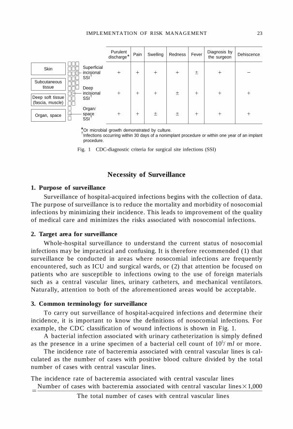
incidence, it is important to know the definitions of nosocomial infections. Forexample, the CDC classification of wound infections is shown in Fig. 1.
A bacterial infection associated with urinary catheterization is simply definedas the presence in a urine specimen of a bacterial cell count of 105/ml or more.
The incidence rate of bacteremia associated with central vascular lines is cal-culated as the number of cases with positive blood culture divided by the totalnumber of cases with central vascular lines.
The incidence rate of bacteremia associated with central vascular lines
�Number of cases with bacteremia associated with central vascular lines�1,000
The total number of cases with central vascular lines
Purulent Pain Swelling Redness Fever Diagnosis by Dehiscencedischarge* the surgeon
Superficialincisional � � � � � � �SSI†
Deepincisional � � � � � � �SSI†
Organ/space � � � � � � �SSI†
*Or microbial growth demonstrated by culture.†Infections occurring within 30 days of a nonimplant procedure or within one year of an implant
†procedure.
Skin
Subcutaneoustissue
Deep soft tissue(fascia, muscle)
Organ, space
Fig. 1 CDC-diagnostic criteria for surgical site infections (SSI)
IMPLEMENTATION OF RISK MANAGEMENT 23
4. Use of surveillance resultsThe results of surveillance should be returned to each section of the hospital
to enable the staff in the section to draw up measures for reducing the incidenceof nosocomial infections. Follow-up surveillance should then be continued. Thisfeedback mechanism helps greatly in reducing the incidence of nosocomial infec-tions and improving their outcome.
Hospital-acquired Infections and the Use of Antimicrobial Agents
Hospital inpatients often have diminished immunological competence, andforeign materials such as central vascular lines and urinary catheters are often usedin these patients, making them highly susceptible to infections. These patientsoften develop fever and frequently receive antibiotic therapy. The relationshipbetween the frequent use of antimicrobial agents and the emergence of drug-resistant strains must be emphasized here.
Before the occurrence of nosocomial infections attracted medical attention,the use of erythromycin and development of erythromycin-resistant hemolyticstreptococci was an important issue, as was the use of penicillin and the emergenceof penicillinase (�-lactamase)-producing staphylococci.
It has been shown that infection with MRSA (methicillin-resistant Staphylo-coccus aureus) is more closely related to the duration and dose of antibiotictherapy than is infection with MSSA (methicillin-sensitive Staphylococcus aureus).
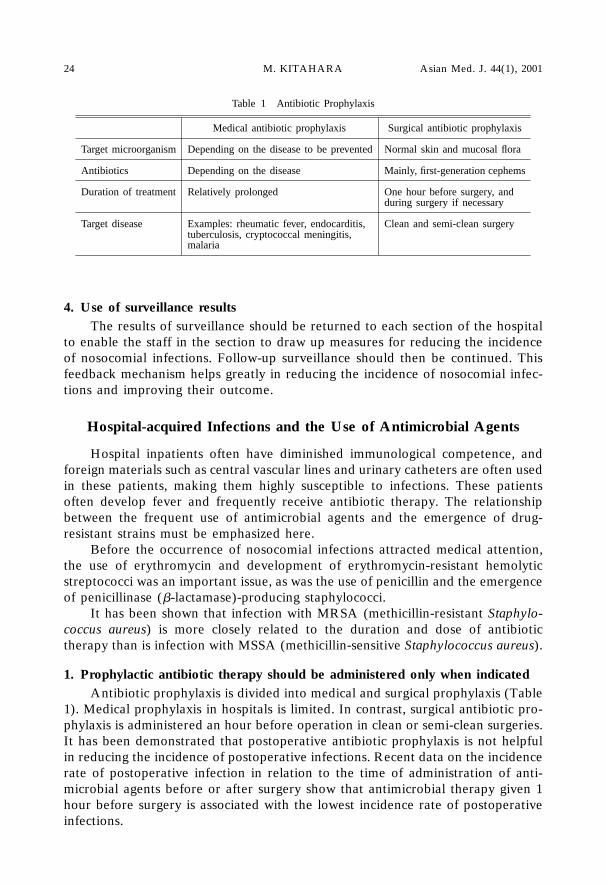
1. Prophylactic antibiotic therapy should be administered only when indicatedAntibiotic prophylaxis is divided into medical and surgical prophylaxis (Table
1). Medical prophylaxis in hospitals is limited. In contrast, surgical antibiotic pro-phylaxis is administered an hour before operation in clean or semi-clean surgeries.It has been demonstrated that postoperative antibiotic prophylaxis is not helpfulin reducing the incidence of postoperative infections. Recent data on the incidencerate of postoperative infection in relation to the time of administration of anti-microbial agents before or after surgery show that antimicrobial therapy given 1hour before surgery is associated with the lowest incidence rate of postoperativeinfections.
Table 1 Antibiotic Prophylaxis
Medical antibiotic prophylaxis Surgical antibiotic prophylaxis
Target microorganism Depending on the disease to be prevented Normal skin and mucosal flora
Antibiotics Depending on the disease Mainly, first-generation cephems
Duration of treatment Relatively prolonged One hour before surgery, andduring surgery if necessary
Target disease Examples: rheumatic fever, endocarditis, Clean and semi-clean surgerytuberculosis, cryptococcal meningitis,malaria
M. KITAHARA Asian Med. J. 44(1), 200124
Prophylactic vancomycin therapy against MRSA colonization facilitates thegrowth of vancomycin-resistant MRSA, rather than being of any benefit. Con-tinued vancomycin therapy in patients with urinary catheters may result in theappearance of vancomycin-resistant enterococci (VRE).
2. Simple antimicrobial therapyIn general, a single etiological agent is responsible for an infection, and there-
fore there is no need for multiple antimicrobial therapy. However, multiple anti-microbial therapy is recommended in some situations, e.g., in patients with acuteleukemia who have neutropenia and develop fever.
Diarrhea related to antimicrobial therapy, particularly that caused byClostridium difficile, is a common nosocomial infection reported in European andAmerican countries. It has become apparent that C. difficile-associated diarrhea isclosely related to the administration of certain types of antimicrobial drugs, suchas that, in particular, of second- or third-generation cephems, clindamycin,ampicillin, or amoxicillin. Impudent administration of broad-spectrum antimicro-bial agents should be particularly avoided.
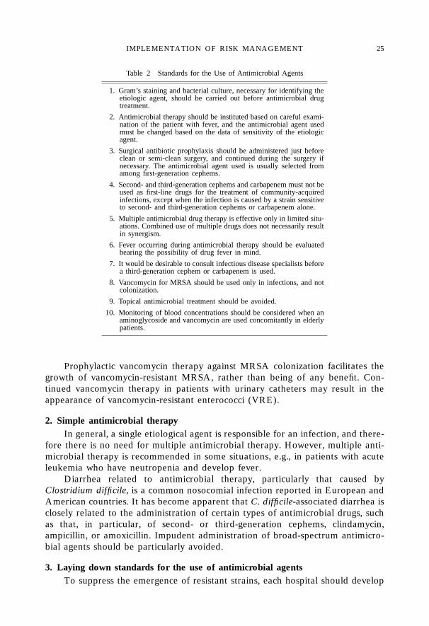
3. Laying down standards for the use of antimicrobial agentsTo suppress the emergence of resistant strains, each hospital should develop
Table 2 Standards for the Use of Antimicrobial Agents
1. Gram’s staining and bacterial culture, necessary for identifying theetiologic agent, should be carried out before antimicrobial drugtreatment.
2. Antimicrobial therapy should be instituted based on careful exami-nation of the patient with fever, and the antimicrobial agent usedmust be changed based on the data of sensitivity of the etiologicagent.
3. Surgical antibiotic prophylaxis should be administered just beforeclean or semi-clean surgery, and continued during the surgery ifnecessary. The antimicrobial agent used is usually selected fromamong first-generation cephems.
4. Second- and third-generation cephems and carbapenem must not beused as first-line drugs for the treatment of community-acquiredinfections, except when the infection is caused by a strain sensitiveto second- and third-generation cephems or carbapenem alone.
5. Multiple antimicrobial drug therapy is effective only in limited situ-ations. Combined use of multiple drugs does not necessarily resultin synergism.
6. Fever occurring during antimicrobial therapy should be evaluatedbearing the possibility of drug fever in mind.
7. It would be desirable to consult infectious disease specialists beforea third-generation cephem or carbapenem is used.
8. Vancomycin for MRSA should be used only in infections, and notcolonization.
9. Topical antimicrobial treatment should be avoided.
10. Monitoring of blood concentrations should be considered when anaminoglycoside and vancomycin are used concomitantly in elderlypatients.
IMPLEMENTATION OF RISK MANAGEMENT 25
institutional standards for the use of antimicrobial agents (Table 2). The standardsshould emphasize the need for Gram’s staining and restrictions on the use ofsecond- and third-generation cephems and carbapenem in patients with commu-nity-acquired infections. Topical antimicrobial treatment should be avoided.
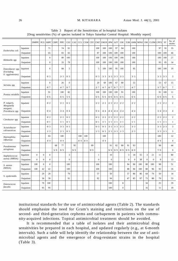
It is recommended that a table of isolates and their antimicrobial drugsensitivities be prepared in each hospital, and updated regularly (e.g., at 6-monthintervals). Such a table will help identify the relationship between the use of anti-microbial agents and the emergence of drug-resistant strains in the hospital(Table 3).
Table 3 Report of the Sensitivities of In-hospital Isolates[Drug sensitivities (%) of species isolated in Tokyo Saiseikai Central Hospital: Monthly report]
DMPPC PCG ABPC CBPC PIPC CEZ CAZ CCL CXM CPZ CMZ LMOX IPM AZT GM TOB AMK MINO EM CLDM CPFX ST No. ofstrains
Escherichia coliInpatient 71 74 91 100 100 100 97 94 100 97 91 35
Outpatient 65 83 65 87 100 100 100 100 100 100 100 46
Klebsiella sppInpatient 0 89 100 100 100 100 100 100 100 100 100 27
Outpatient 0 33 78 100 100 100 100 100 100 93 85 14
Enterobacter spp Inpatient 0 84 5 5 100 100 95 100 95 100 100 19(ExcludingE. agglomerans)
Outpatient 0/3 3/3 0/3 0/3 3/3 3/3 3/3 3/3 3/3 3/3 3/3 3
Serratia sppInpatient 0 26 0 20 60 100 67 80 53 53 67 15
Outpatient 0/7 4/7 0/7 2/7 4/7 6/7 6/7 7/7 4/7 3/7 6/7 7
Proteus mirabilisInpatient 91 100 82 100 100 100 100 91 100 91 100 11
Outpatient 6/6 6/6 5/6 6/6 6/6 6/6 6/6 6/6 6/6 6/6 6/6 6
P. vulgaris, Inpatient 0/2 2/2 0/2 2/2 2/2 2/2 2/2 2/2 2/2 2/2 2/2 2Morganellamorganii,
Outpatient 0/4 3/4 0/4 0/4 4/4 4/4 4/4 3/4 4/4 3/4 3/4 4Providencia spp
Citrobacter sppInpatient 0/2 2/2 0/2 0/2 2/2 2/2 2/2 2/2 2/2 2/2 2/2 2
Outpatient 0/1 1/1 0/1 0/1 1/1 1/1 1/1 1/1 1/1 1/1 1/1 1
Acinetobacter Inpatient 0/3 3/3 0/3 0/3 0/3 3/3 1/3 3/3 3/3 3/3 2/3 3
calcoaceticus Outpatient 1/3 2/3 0/3 1/3 0/3 2/3 3/3 1/3 2/3 3/3 3/3 3
Haemophilus Inpatient 83 100 100 100 100 100 12
influenzae Outpatient 4/4 4/4 4/4 4/4 4/4 4/4 4
Pseudomonas Inpatient 68 77 95 89 91 82 80 91 93 98 44
aeruginosa Outpatient 5/9 9/9 9/9 9/9 9/9 9/9 8/9 9/9 8/9 7/9 9
Staphylococcus Inpatient 0 0 0 3 1 10 3 18 55 3 21 11 80
aureus (MRSA) Outpatient 0 0 0 0 0 0 0 0 38 0 0 8 13
S. aureus Inpatient 100 0 0 100 100 100 94 96 100 88 100 98 51
(MSSA) Outpatient 100 42 42 100 100 100 88 96 100 88 96 92 26
S. epidermidisInpatient 29 29 79 57 50 57 86 86 64 79 50 14
Outpatient 36 39 91 82 94 67 85 97 73 88 79 33
Enterococcus Inpatient 79 100 100 8 34 25 29
faecalis Outpatient 95 95 100 0 16 11 19
M. KITAHARA Asian Med. J. 44(1), 200126
Prevention of occupational infections
The hospital is responsible for not only preventing infections among patients,but also for protecting hospital personnels, including physicians, nurses, and medi-cal technicians, who are in direct contact with patients, against infections.
1. Isolation of patientsStandard isolation precautions have been proposed by the Centers for Disease
Control and Prevention (CDC), USA, and must be followed in every inpatient(Table 4). Standard precautions must be followed in the handling of blood, bodyfluids, secretions (other than perspiration), and non-intact skin and mucosa.
Besides the standard precautions, precautions based on the routes of transmis-sion are also necessary (Table 4). These precautions pertain to three categories oftransmission, i.e., airborne, droplet, and contact transmissions. Airborne transmis-sion is the route of transmission of infections such as measles and tuberculosis, anddroplet transmission refers to the transmission of diseases via droplets measuringover 5�m in diameter generated from the patient. The droplets cause infectionwhen deposited on the hosts’ conjunctivae, nasal mucosa, or oral mucosa. Contacttransmission refers to the transmission of diseases via direct contact, or contactwith a contaminated intermediate object. Precautions against infections by theseroutes of transmission of infection must also be taken in addition to the standardprecautions.
However, to what extent such precautionary measures can be adhered to inhospitals in Japan is an issue of importance.
2. Prevention of tuberculosisIt appears that the number of health care professionals developing tuberculo-
sis as a result of exposure to patients with tuberculosis has recently been increas-ing. Insufficient knowledge regarding the transmission of tuberculosis underliessuch onset of the disease in health care professionals.
Health care professionals should always bear in mind the possibility of tuber-culosis when treating patients. When infection is suspected, the patient should beisolated in a private room until the possibility of tuberculosis is eliminated. Thehospital manager should keep records of tuberculin skin tests performed in hospi-tal personnel who come in contact with tuberculosis patients. When hospital per-sonnel are exposed to patients with tuberculosis, those with previously negativetuberculin tests should undergo repeat tuberculin testing and chest roentgeno-graphy to detect the occurrence of primary infection.
3. Prophylactic vaccination or drug treatmentThe hospital manager should set up a system that ensures immediate access
to prophylactic vaccination against infections transmitted by accidental needlestick injury, 24 hours of a day. If the accidental needle stick injury involves the riskof contracting type-B hepatitis virus infection, a globulin preparation should bemade available immediately. In case of needle stick injury involving the risk ofcontracting human immunodeficiency virus (HIV) infection, the system should
IMPLEMENTATION OF RISK MANAGEMENT 27
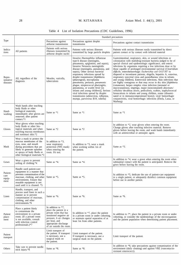
Table 4 List of Isolation Precautions (CDC Guidelines, 1996)
All patients
All, regardless of thediagnosis
Wash hands after touchingbody fluids or otherbiological materials;immediately after gloves areremoved; after patientcontact.*1
Wear gloves when touchingbody fluids or other bio-logical materials and whentouching mucous membranesand nonintact skin.*2
Wear a mask to protect themucous membranes of theeyes, nose, and mouthduring procedures that arelikely to generate splashesor sprays of body fluids orother biological materials.*3
Wear a gown to preventsoiling of clothing.*4
Handle used patient-careequipment in a manner thatprevents contamination of themucosae, clothing, and otherenvironments. Ensure thatreusable equipment is notused until it is cleaned. *5
Handle, transport, andprocess used linen in such amanner as to preventcontamination of mucosae,clothing, and otherenvironments.*6
Place a patient likelyto contaminate theenvironment in a privateroom. (If a private roomis not available, consultwith infection controlprofessionals.)*7
Take care to prevent needlestick injury.*8
Precautions againstairborne transmission
Patients with seriousillnesses transmitted byairborne droplet nuclei
Measles, varicella,tuberculosis
Same as *1
Same as *2
In addition to *3,wear respiratoryprotection (N95 mask)when entering theroom. Fit test.
Same as *4.
Same as *5.
Same as *6.
In addition to *7,place the patient in aprivate room that hasmonitored negative airpressure, 6 air changesper hour, andappropriate dischargeof air outside the room.
Limit transport ofthe patient. If transportis necessary, use asurgical mask onthe patient.
Same as *8.
Precautions against droplettransmission
Patients with serious illnessestransmitted by large particle droplets
Invasive Haemophilus influenzaetype-b disease (meningitis,pneumonia, epiglottitis, and sepsis),Invasive Neisseria meningitidisdisease (meningitis, pneumonia, andsepsis), Other serious bacterialrespiratory infections spread bydroplet transmission [diphtheria(pharyngeal), mycoplasmapneumonia, pertussis, pneumonicplague, streptococcal pharyngitis,pneumonia, or scarlet fever (ininfants and young children)], Seriousviral infections spread by droplettransmission (adenovirus, influenza,mumps, parvovirus B19, rubella)
Same as *1
Same as *2
In addition to *3, wear a maskwhen working within 1m ofthe patient.
Same as *4.
Same as *5.
Same as *6.
In addition to *7, place the patientin a private room or under cohorting,or maintain spatial separation of atleast 1m from other patients.
Limit transport of the patient.If transport is necessary, use asurgical mask on the patient.
Same as *8.
Precautions against contact transmission
Patients with serious illnesses easily transmitted by directpatient contact or by contact with infected material
Gastrointestinal, respiratory, skin, or wound infections orcolonization with multidrug-resistant bacteria judged to be ofspecial clinical and epidemiologic significance, and entericinfections by organisms requiring a low infectious dose or bythose capable of prolonged environmental survival (Clostridiumdifficile, enterohemorrhagic Escherichia coil O157:H7 indiapered or incontinent patients, shigella, hepatitis A, rotavirus,respiratory syncytial virus and parainfluenza virus in infantsand young children), Enteroviral infections, Skin infections thatare highly contagious or that may occur in dry skin [diphtheria(cutaneous), herpes simplex virus infection (neonatal ormucocutaneous), impetigo, major (noncontained) abscesses/cellulitis/decubitus ulcers, pediculosis, scabies, staphylococcalfurunculosis in infants and young children, zoster (dissemi-nated or in immunocompromised hosts)], viral/hemorrhagicconjunctivitis, viral hemorrhagic infections (Ebola, Lassa, orMarburg)
Same as *1
In addition to *2, wear gloves when entering the room.Change gloves after touching infective material. Removegloves before leaving the room, and wash hands immediatelywith an antimicrobial or antiseptic agent.
Same as *3.
In addition to *4, wear a gown when entering the room whensubstantial contact with the patient is anticipated. Remove thegown before leaving the room.
In addition to *5, dedicate the use of patient-care equipmentto a single patient, or adequately disinfect common equipmentbefore use in another patient.
Same as *6.
In addition to 7*, place the patient in a private room or undercohorting, or consider the epidemiology of the microorganismand the patient population when determining patient placement.
Limit transport of the patient.
In addition to *8, take precautions against contamination of theenvironment (daily cleaning) and against VRE (vancomycin-resistant enterococci).
Type
Indica-tions
Repre-sentativediseases
Hand-washing
Gloves
Mask
Gown
Patient-careequip-ment
Linen
Patientplace-ment
Patienttransport
Others
Standard precautions
M. KITAHARA Asian Med. J. 44(1), 200128
allow prompt initiation of anti-HIV, three-drug treatment.Health care professionals should receive vaccination against influenza virus
infection so as to prevent them from becoming sources of infection. Currently, itremains controversial as to whether BCG vaccination is a preventive measureagainst tuberculosis, particularly in adults. In the author’s opinion, BCG vaccina-tion should not be recommended routinely, and follow-up should be conducted bytuberculin testing.
Conclusion
Risk management pertaining to nosocomial infections is difficult to implementproperly in Japan, which has poor human resources and hospital economics. How-ever, failure to control nosocomial infections will undermine various aspects ofmedical care. Beginning with doing what can be done and proceeding steadily tohigher levels of infection control is recommended as a way of practicing riskmanagement.
REFERENCES
1) Satake, S.: To implement a fair surveillance: the concept of the denominator in surveil-lance (2). Infection Control 7: 536–541, 1998. (in Japanese)
2) Horan, T.C. et al.: CDC definitions of nosocomial surgical site infections, 1992:Amodification of CDC definitions of surgical wound infections. Infect Control HospEpidemiol 13: 606–608, 1992.
3) Classen, D.C. et al.: The timing of prophylactic administration of antibiotics and therisk of surgical infections. N Engl J Med 326: 281–286, 1992.
4) Hospital Infection Control Practices Advisory Committee: Guidelines for isolationprecautions in hospitals. Infect Control Hosp Epidemiol 17: 53–80, 1996.
IMPLEMENTATION OF RISK MANAGEMENT 29
30
* This article is a revised English version of a paper originally published in the Journal of theJapan Medical Association (Vol. 122 No. 7, 1999, pages 1173–1176).
** Assistant Professor, First Department of Internal Medicine, School of Medicine, University ofTokushima, Japan
MUSIC THERAPY AND INTERNAL MEDICINE*
Hiroshi BANDO**
Asian Med. J. 44(1): 30–35, 2001Abstract: Music therapy is an alternative and supplemental form of treatment usedto treat a variety of diseases in the field of internal medicine, including psycho-somatic and lifestyle-related diseases, specifically digestive ulcer, NUD (non-ulcerdyspepsia), chronic pancreatitis, hypertension, bronchial asthma, diabetes mellitus,simple obesity, Basedow’s disease, terminal cancer, and senile dementia. It has beenused in treating patients undergoing chronic dialysis, blood donation activities, inrehabilitation activities for cerebrovascular accidents (CVA), endoscopic examina-tions, in intensive care units (ICU), and to stabilize blood pressure, promote sleep,and reduce pain. Music therapy has also been applied in diet therapy using back-ground music (BGM) that is played during meals and karaoke is also a form ofmusic therapy that can be effectively utilized in social situations. It is also utilizedin physical and exercise therapy, in rehabilitating patients recovering from CVA andin treating the aged, and in promoting the interpersonal relationship between thepatient and the therapist. Music therapy is also effective in promoting the health ofprofessional athletes; and the Awa folk dance in Japan is also used as dance therapy.Rhythmical background music has also been effectively applied using the Walkman.
Key words: Music therapy; Lifestyle-related disease; Diet therapy;Exercise therapy
Introduction
Music therapy has been used to treat numerous diseases in the field of internalmedicine, including psychosomatic diseases. In this paper, the correlation betweenlifestyle-related diseases and diet and exercise is presented.
In this age of advanced technology, the science of medicine alone is unable tocure patients. The art of medicine, as advocated by Hippocrates, effectively uti-lized music in curing the mind and body of patients. Presently, music therapy isutilized in the fields of medicine, health care, music, education, and social welfare;and its cultural and artistic merits are equally important.
Music therapy allows patients to become directly involved and the benefits ofutilizing this method in patience guidance activities is presented in this paper.
Progress in Clinical Medicine
Human Beings and Rhythm
Human beings have an inborn love of music and a sense of rhythm. The fetusdevelops listening to the heartbeat and voice of the mother and infants will changefrom crying, smiling, to speaking in conjunction with the voice of the mother andother family members and the sounds emanating from the environment. Amother’s voice, that is filled with love and affection, is in the alto region and infantsare comforted when they are cradled and talked to for about one second in thearms of a mother whose voice is in the alto region.
Human beings maintain their health and live in harmony with melody andrhythm throughout their childhood, adulthood, and the twilight years of their lives.Health is lost when this harmonious balance is destroyed. The human body main-tains a pulse, body temperature, respiration, and other vital signs as well as ametabolism and blood flow that functions according to an inherent rhythm. Ourbehavioral patterns such as eating and sleeping are also based on a consistentrhythm and work activities are synchronized accordingly. The bodily organs arethe instruments and the heart is the conductor. The integrated role or function ofthese instruments are integrated to produce an orchestral performance.
Application and Treatment
Music therapy is widely applied in many areas of health care. However, it iscommonly utilized in internal medicine to treat digestive ulcer, NUD (non-ulcerdyspepsia), chronic pancreatitis, hypertension,1) bronchial asthma, diabetes mel-litus, simple obesity, Basedow’s disease, terminal cancer,2) and senile dementia.3) Ithas been used in treating patients undergoing chronic dialysis, blood donationactivities, in rehabilitation activities for CVA, during endoscopic examinations,4)
in intensive care units (ICU), cardial care unit (CCU), to stabilise blood pressure,promote sleep,5) and reduce pain. There are two categories of music therapy—theactive form of singing and playing an instrument and the passive form of listeningto music. Body sonics, pillows with stereo music, or BGM are the most commonforms of the latter category of music therapy.
Music therapy also has supplementary benefits, in addition to being an effec-tive means of treating diseases. Its effectiveness as a form of treatment must beinvestigated objectively.5) It helps rehabilitation activities to be performed moreeasily and lessens the apprehension of patients undergoing dialysis and other formsof treatment.
In the clinical setting, many hospitals and nursing homes utilize music therapyin their rehabilitation activities for patients recovering from CVA. Blood pressureis lowered and stabilized in patients suffering from hypertension; and orthostatichypotension is also stabilized with the use of music therapy. In addition, patiencecompliance has been favorable when music therapy is used in long-term treatmentand rehabilitation activities in the aftermath of acute myocardial infarction.6)
MUSIC THERAPY AND INTERNAL MEDICINE 31
Life-style Related Diseases
The term, lifestyle-related diseases, was initially used and discussed in Japanin 1978 by Dr. Shigeaki Hinohara, President of the Japan Biomusic Association.Recently, the Ministry of Health and Welfare has begun to utilize this term in lieuof the commonly known term, adult diseases.
The fundamental factor to maintaining good health is a balanced diet, mod-erate exercise, and mental rest. The key to sustaining good health is to maintainan established pattern of activity in our lifestyles. Maintaining a 24-hour dailycyclical rhythm produces a 1/f fluctuation, which induces the feeling of beingcomfortable. This is because some irregularity exists within lifestyles with an estab-lished pattern of activity.
Diet and Music
The most important meal of the day is breakfast, but an increasing number ofthe younger generation has begun to skip breakfast in recent years and this hasbecome a health issue in Japan. The general intake proportion of meals in Japanis 20 percent for breakfast, 30 percent for lunch, and 50 percent for dinner, incontrast to the ideal proportion of 40 percent for breakfast, 35 percent for lunch,and 25 percent for dinner. Simple obesity and lifestyle related diseases can beprevented if the ideal proportion of food intake is followed. Case examples thatmay prove useful in explanations to patients regarding the importance of incorpo-rating music in their daily lives have been given below.
1. Rising early in the morning is recommended and late risers should use analarm clock with a loud buzzer or digital alarm. For those who can get up easily,it may be a good idea to set the time switch on the radio to allow baroque or otherkinds of music to play in the morning. This will help such people to wake upquietly and comfortably.
2. After rising, drinking milk or other liquids is recommended, followed by ashort walk or light exercise. The added time spent after rising and before breakfasthelps to increase the appetite. In addition, fructose derived from fruits that areconsumed at breakfast are converted quickly into energy without insulin secretionfrom the pancreas. Hence, it is said in Japan that fruits which are consumed atbreakfast are comparable to gold, fruits eaten at lunch are comparable to silver,and fruits eaten at dinner are comparable to copper.
3. Many people rush through their breakfast, urged on by the time that ap-pears on their television screens. Listening to rhythmical BGM is recommended inthe morning. On holidays, it is recommended that the gurgling of small streams,singing birds, and other environmental sounds or refreshing BGM are playedduring breakfast.
4. Light exercise that induces perspiration, followed by a bath and dinner, isrecommended in the evenings. Bathing after an evening meal is not beneficial interms of blood circulation; and meals consumed late at night increase the onset oflifestyle-related disease, such as hyperlipemia. Therefore, they should be eatenearly in the evening. If BGM is played in the evenings, easy listening music that
H. BANDO Asian Med. J. 44(1), 200132
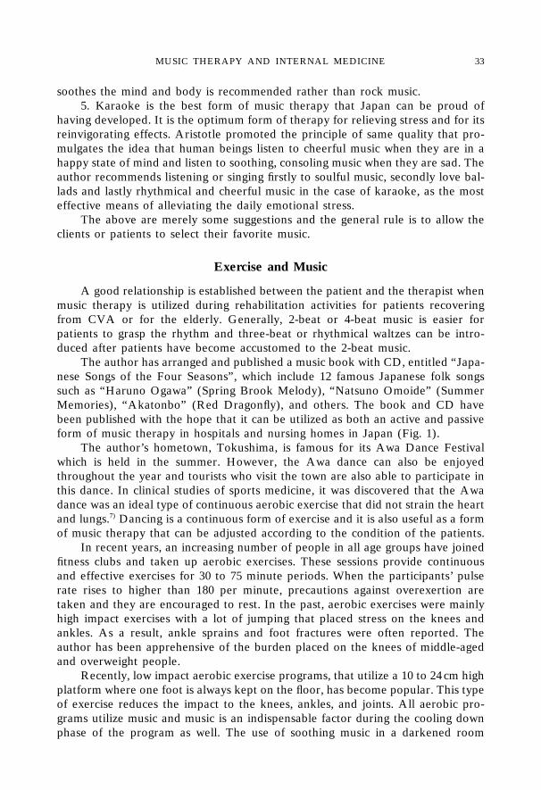
soothes the mind and body is recommended rather than rock music.5. Karaoke is the best form of music therapy that Japan can be proud of
having developed. It is the optimum form of therapy for relieving stress and for itsreinvigorating effects. Aristotle promoted the principle of same quality that pro-mulgates the idea that human beings listen to cheerful music when they are in ahappy state of mind and listen to soothing, consoling music when they are sad. Theauthor recommends listening or singing firstly to soulful music, secondly love bal-lads and lastly rhythmical and cheerful music in the case of karaoke, as the mosteffective means of alleviating the daily emotional stress.
The above are merely some suggestions and the general rule is to allow theclients or patients to select their favorite music.
Exercise and Music
A good relationship is established between the patient and the therapist whenmusic therapy is utilized during rehabilitation activities for patients recoveringfrom CVA or for the elderly. Generally, 2-beat or 4-beat music is easier forpatients to grasp the rhythm and three-beat or rhythmical waltzes can be intro-duced after patients have become accustomed to the 2-beat music.
The author has arranged and published a music book with CD, entitled “Japa-nese Songs of the Four Seasons”, which include 12 famous Japanese folk songssuch as “Haruno Ogawa” (Spring Brook Melody), “Natsuno Omoide” (SummerMemories), “Akatonbo” (Red Dragonfly), and others. The book and CD havebeen published with the hope that it can be utilized as both an active and passiveform of music therapy in hospitals and nursing homes in Japan (Fig. 1).
The author’s hometown, Tokushima, is famous for its Awa Dance Festivalwhich is held in the summer. However, the Awa dance can also be enjoyedthroughout the year and tourists who visit the town are also able to participate inthis dance. In clinical studies of sports medicine, it was discovered that the Awadance was an ideal type of continuous aerobic exercise that did not strain the heartand lungs.7) Dancing is a continuous form of exercise and it is also useful as a formof music therapy that can be adjusted according to the condition of the patients.
In recent years, an increasing number of people in all age groups have joinedfitness clubs and taken up aerobic exercises. These sessions provide continuousand effective exercises for 30 to 75 minute periods. When the participants’ pulserate rises to higher than 180 per minute, precautions against overexertion aretaken and they are encouraged to rest. In the past, aerobic exercises were mainlyhigh impact exercises with a lot of jumping that placed stress on the knees andankles. As a result, ankle sprains and foot fractures were often reported. Theauthor has been apprehensive of the burden placed on the knees of middle-agedand overweight people.
Recently, low impact aerobic exercise programs, that utilize a 10 to 24 cm highplatform where one foot is always kept on the floor, has become popular. This typeof exercise reduces the impact to the knees, ankles, and joints. All aerobic pro-grams utilize music and music is an indispensable factor during the cooling downphase of the program as well. The use of soothing music in a darkened room
MUSIC THERAPY AND INTERNAL MEDICINE 33
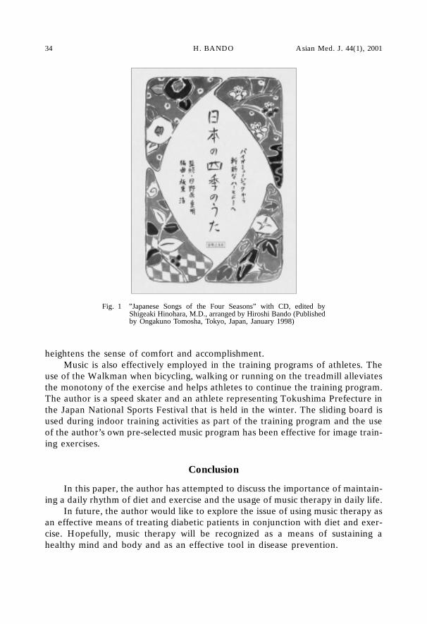
heightens the sense of comfort and accomplishment.Music is also effectively employed in the training programs of athletes. The
use of the Walkman when bicycling, walking or running on the treadmill alleviatesthe monotony of the exercise and helps athletes to continue the training program.The author is a speed skater and an athlete representing Tokushima Prefecture inthe Japan National Sports Festival that is held in the winter. The sliding board isused during indoor training activities as part of the training program and the useof the author’s own pre-selected music program has been effective for image train-ing exercises.
Conclusion
In this paper, the author has attempted to discuss the importance of maintain-ing a daily rhythm of diet and exercise and the usage of music therapy in daily life.
In future, the author would like to explore the issue of using music therapy asan effective means of treating diabetic patients in conjunction with diet and exer-cise. Hopefully, music therapy will be recognized as a means of sustaining ahealthy mind and body and as an effective tool in disease prevention.
Fig. 1 ”Japanese Songs of the Four Seasons” with CD, edited byShigeaki Hinohara, M.D., arranged by Hiroshi Bando (Publishedby Ongakuno Tomosha, Tokyo, Japan, January 1998)
H. BANDO Asian Med. J. 44(1), 200134
REFERENCES
1) Shinoda, T.: Stress management of patients with hypertension. Kongetsu no Chiryou7: 106–110, 1999. (in Japanese)
2) O’Callaghan, C.C.: Pain, music creativity and music therapy in palliative care. Am JHosp Palliat Care 13: 43–49, 1996.
3) Clark, M.E., Lipe, A.W. and Bilbrey, M.: Use of music to decrease aggressive behav-iors in people with dementia. J Gerontol Nurs 24: 10–17, 1998.
4) Bampton, P. and Draper, B.: Effect of relaxation music on patient tolerance of gas-trointestinal endoscopic procedures. J Clin Gastroenterol 25: 343–345, 1997.
5) Levin, Y.: “Brain music” in the treatment of patients with insomnia. Neurosci BehavPhysiol 28: 330–335, 1998.
6) White, J.M.: Effects of relaxing music on cardiac autonomic balance and anxiety afteracute myocardial infarction. Am J Crit Care 8: 220–230, 1999.
7) Suzuki, Y.: Exercise level of the Awa Dance. Shikoku Koshueisei 37: 69–72, 1992. (inJapanese)
MUSIC THERAPY AND INTERNAL MEDICINE 35
36
* This article is a revised English version of a paper originally published in the Journal of theJapan Medical Association (Vol. 121 No. 11, 1999, pages 1745–1748).
** Associate Professor, Department of Cardiovascular Medicine, Kyoto University GraduateSchool of Medicine
INSIGHTS INTO THE PATHOPHYSIOLOGYOF HEART FAILURE
BASED ON A NEW CONCEPT*
Akira MATSUMORI**
Asian Med. J. 44(1): 36–41, 2001Abstract: We have reported the increased myocardial expression of cytokinesin animal models of heart failure secondary to myocarditis, myocardial infarction,and hypertension and we have shown that the lesions in impaired hearts resembleinflammatory or proliferative changes in some respects. In addition, the occurrenceof crosstalk between cytokines and the adrenal axis, as well as between cytokinesand the renin-angiotensin or endothelin system, has been suggested. Crosstalkbetween the sympathetic nervous system, endocrine systems, and the immune sys-tem may be important in adaptation and failure of the myocardium caused by vari-ous factors, including mechanical overload, ischemia, and viral infection. Elucidationof the mechanisms of these processes may lead to the development of new therapyfor heart failure.
Key words: Heart failure; Cardiomyopathy; Cytokines; Immunity
Introduction
Over the years, the concept of heart failure has undergone dramatic changes.At first, heart failure was thought as a disease associated with renal failure due toalteration of cardiac function. Consequently, diuretics were once the mainstay oftreatment. Subsequently, cardiotonics and vasodilators were used for this condi-tion, because it was considered to arise from abnormal cardiac metabolism andabnormal circulatory responses, including the peripheral vessels. Recently, beta-adrenergic blockers and ACE inhibitors have become central to treatment becauseheart failure has considered as a disorder of the neurohumoral system.
Considering heart failure to be based on immune abnormalities, we hope thatimmunomodulation and anti-cytokine therapy will bring about major progress inits treatment. This article describes our attempts to gain new insights into thepathophysiology of heart failure based on such a viewpoint.
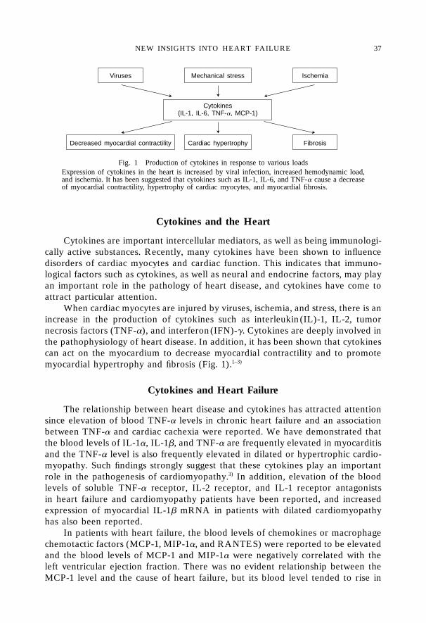
NEW INSIGHTS INTO HEART FAILURE 37
Cytokines and the Heart
Cytokines are important intercellular mediators, as well as being immunologi-cally active substances. Recently, many cytokines have been shown to influencedisorders of cardiac myocytes and cardiac function. This indicates that immuno-logical factors such as cytokines, as well as neural and endocrine factors, may playan important role in the pathology of heart disease, and cytokines have come toattract particular attention.
When cardiac myocytes are injured by viruses, ischemia, and stress, there is anincrease in the production of cytokines such as interleukin(IL)-1, IL-2, tumornecrosis factors (TNF-�), and interferon(IFN)-�. Cytokines are deeply involved inthe pathophysiology of heart disease. In addition, it has been shown that cytokinescan act on the myocardium to decrease myocardial contractility and to promotemyocardial hypertrophy and fibrosis (Fig. 1).1–3)
Cytokines and Heart Failure
The relationship between heart disease and cytokines has attracted attentionsince elevation of blood TNF-� levels in chronic heart failure and an associationbetween TNF-� and cardiac cachexia were reported. We have demonstrated thatthe blood levels of IL-1�, IL-1�, and TNF-� are frequently elevated in myocarditisand the TNF-� level is also frequently elevated in dilated or hypertrophic cardio-myopathy. Such findings strongly suggest that these cytokines play an importantrole in the pathogenesis of cardiomyopathy.3) In addition, elevation of the bloodlevels of soluble TNF-� receptor, IL-2 receptor, and IL-1 receptor antagonistsin heart failure and cardiomyopathy patients have been reported, and increasedexpression of myocardial IL-1� mRNA in patients with dilated cardiomyopathyhas also been reported.
In patients with heart failure, the blood levels of chemokines or macrophagechemotactic factors (MCP-1, MIP-1�, and RANTES) were reported to be elevatedand the blood levels of MCP-1 and MIP-1� were negatively correlated with theleft ventricular ejection fraction. There was no evident relationship between theMCP-1 level and the cause of heart failure, but its blood level tended to rise in
Fig. 1 Production of cytokines in response to various loadsExpression of cytokines in the heart is increased by viral infection, increased hemodynamic load,and ischemia. It has been suggested that cytokines such as IL-1, IL-6, and TNF-� cause a decreaseof myocardial contractility, hypertrophy of cardiac myocytes, and myocardial fibrosis.
Viruses Mechanical stress Ischemia
Cytokines(IL-1, IL-6, TNF-�, MCP-1)
Decreased myocardial contractility Cardiac hypertrophy Fibrosis
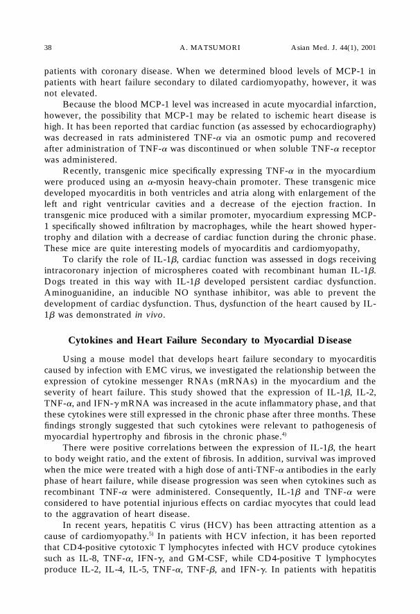
A. MATSUMORI Asian Med. J. 44(1), 200138
patients with coronary disease. When we determined blood levels of MCP-1 inpatients with heart failure secondary to dilated cardiomyopathy, however, it wasnot elevated.
Because the blood MCP-1 level was increased in acute myocardial infarction,however, the possibility that MCP-1 may be related to ischemic heart disease ishigh. It has been reported that cardiac function (as assessed by echocardiography)was decreased in rats administered TNF-� via an osmotic pump and recoveredafter administration of TNF-� was discontinued or when soluble TNF-� receptorwas administered.
Recently, transgenic mice specifically expressing TNF-� in the myocardiumwere produced using an �-myosin heavy-chain promoter. These transgenic micedeveloped myocarditis in both ventricles and atria along with enlargement of theleft and right ventricular cavities and a decrease of the ejection fraction. Intransgenic mice produced with a similar promoter, myocardium expressing MCP-1 specifically showed infiltration by macrophages, while the heart showed hyper-trophy and dilation with a decrease of cardiac function during the chronic phase.These mice are quite interesting models of myocarditis and cardiomyopathy,
To clarify the role of IL-1�, cardiac function was assessed in dogs receivingintracoronary injection of microspheres coated with recombinant human IL-1�.Dogs treated in this way with IL-1� developed persistent cardiac dysfunction.Aminoguanidine, an inducible NO synthase inhibitor, was able to prevent thedevelopment of cardiac dysfunction. Thus, dysfunction of the heart caused by IL-1� was demonstrated in vivo.
Cytokines and Heart Failure Secondary to Myocardial Disease
Using a mouse model that develops heart failure secondary to myocarditiscaused by infection with EMC virus, we investigated the relationship between theexpression of cytokine messenger RNAs (mRNAs) in the myocardium and theseverity of heart failure. This study showed that the expression of IL-1�, IL-2,TNF-�, and IFN-� mRNA was increased in the acute inflammatory phase, and thatthese cytokines were still expressed in the chronic phase after three months. Thesefindings strongly suggested that such cytokines were relevant to pathogenesis ofmyocardial hypertrophy and fibrosis in the chronic phase.4)
There were positive correlations between the expression of IL-1�, the heartto body weight ratio, and the extent of fibrosis. In addition, survival was improvedwhen the mice were treated with a high dose of anti-TNF-� antibodies in the earlyphase of heart failure, while disease progression was seen when cytokines such asrecombinant TNF-� were administered. Consequently, IL-1� and TNF-� wereconsidered to have potential injurious effects on cardiac myocytes that could leadto the aggravation of heart disease.
In recent years, hepatitis C virus (HCV) has been attracting attention as acause of cardiomyopathy.5) In patients with HCV infection, it has been reportedthat CD4-positive cytotoxic T lymphocytes infected with HCV produce cytokinessuch as IL-8, TNF-�, IFN-�, and GM-CSF, while CD4-positive T lymphocytesproduce IL-2, IL-4, IL-5, TNF-�, TNF-�, and IFN-�. In patients with hepatitis
caused by HCV, elevation of TNF-� and soluble TNF-� receptor in the blood hasbeen reported.
Cytokines and Heart Failure Secondary to Hypertension
We studied the expression of cytokines in Dahl salt-sensitive rats, whichdevelop heart failure secondary to cardiac hypertrophy caused by hypertension.Specimens of the myocardium were obtained from rats in the stages of hyper-trophy and heart failure, and the expression of IL-1� and TNF-� mRNA was inves-tigated. Although expression of TNF-� mRNA was not so remarkable, expressionof IL-1� and MCP-1 mRNA was increased in both stages. Their expression in thestage of heart failure was stronger than in the stage of hypertrophy.6) The expres-sion of IL-1� was positively correlated with the weight and diastolic diameter ofthe left ventricle, and was negatively correlated with left ventricular fractionalshortening. Consequently, IL-1� appears to play a role in cardiac hypertrophy andheart failure. The infiltration of numerous macrophages in the left ventricularmyocardium of Dahl rats suggested that IL-1� might have been produced by thesemacrophages and by endothelial cells.
The results of this study on Dahl rats led us to investigate whether mechanicalstress could induce the production of cytokines. We studied cytokine productionby human umbilical vein endothelial cells (HUVEC) that were subjected tostretching.7) This mechanical stretch induced a marked increase in the productionof IL-8 and MCP-1 by these cells. This increase showed that vascular endothelialcells can produce MCP-1, a macrophage chemotactic factor, when stimulatedmechanically by stretching. The enhanced ventricular expression of MCP-1 in Dahlrats suggested the possibility that infiltration of macrophages into the myocardiumwould be mediated by MCP-1 (Fig. 2).
Mechanical stress
Endothelial cells
Monocyte chemoattractant protein-1(MCP-1)
Macrophages
Cytokines(IL-1�, IL-6, TNF-�)
Fig. 2 Hemodynamic loading and the immune responseThe hemodynamic load induces synthesis of MCP-1 by vascular endothelialcells, leading to migration and activation of macrophages in the myocardium.The macrophages thus activated may produce cytokines such as IL-1�.
NEW INSIGHTS INTO HEART FAILURE 39
Cytokines in Heart Failure after Myocardial Infarction
As described above, the cytokine production can be induced by stresses suchas viral infection and hypertension. Whether cytokines can be induced by ischemiawas also studied.8) In rats, marked expression of the mRNAs for TNF-�, IL-1�,and IL-6 was noted in the infarcted area at one week after the development ofmyocardial infarction, and then in the non-infarcted region at 20 weeks after itsdevelopment. The expression of cytokine mRNAs in the non-infarcted region wascorrelated with the end-diastolic diameter of the left ventricle. Twenty weeks afterinfarction, the infarcted region was replaced with scar tissue, while vascular endo-thelial cells, smooth muscle cells, and macrophages were still IL-1� positive in thenon-infarcted region. In addition, the myocardial collagen content was related tothe expression of IL-1� in the non-infarcted region, suggesting an associationbetween remodeling after myocardial infarction and cytokine expression.8) Fur-thermore, it has been shown that expression of MCP-1 is increased during theacute phase of myocardial infarction, and that administration of anti-MCP-1 anti-bodies reduces the infarct size.9)
From these findings, it appears that the expression of cytokines may reflect thedevelopment and pathology of heart disease, so cytokines may be of value asdiagnostic and therapeutic markers.
Treatment of Heart Failure and Cytokines
Recently, it has been demonstrated that phosphodiesterase inhibitors, whichwere developed to treat heart failure, can inhibit the production of cytokines. Ithas been recognized that different inhibitors produced different effects. It has beenalso recognized that digitalis can increase the production of cytokines.10) Anantiarrhythmic agent amiodarone, which has been shown to improve the long-termprognosis of heart failure, was recently shown to inhibit the production of TNF-�and IL-6.11)
Because drugs that control the production of cytokines and signal transduc-tion seem to be effective for the treatment of heart failure, it is expected thatimmunological response modifiers will be developed as a new form of therapy inthe future.
Cytokines and Crosstalk between the Sympathetic Nervous System,Renin-angiotensin System, and Endothelin
The results of recent studies have highlighted the association between theimmune system and the central and autonomic nervous systems. It has becomeclear that increased production of adrenocortical hormones secondary to activa-tion of the hypothalamic-pituitary-adrenal axis reduces the production of cyto-kines. In heart failure, the sympathetic nervous system is activated, leading toincreased secretion of catecholamines, which act on many immune cells such asmacrophages and lymphocytes.
Many immune cells, including lymphocytes, macrophages, eosinophils, andneutrophils, possess �-receptors. It has become clear that when �-receptors are
A. MATSUMORI Asian Med. J. 44(1), 200140
stimulated, the intracellular cAMP level increases and various immune responsesare suppressed. Psychological stress is known to increase norepinephrine, whichsuppresses cellular immunity through modification of helper T-cell functions andalso increases humoral immunity.
The renin-angiotensin system and endothelin both play an important role incardiovascular diseases and both show crosstalk with cytokines. For example,TNF-� increases the production of angiotensinogen by liver cells, while angio-tensin II increases the production of IL-6, TNF-�, and MCP-1 by kidney cells. IL-1� and TNF-� increase the production of endothelin-1, which in turn increases theproduction of IL-6 by endothelial cells or IL-8 and GM-CSF by bronchial cells.
Thus, it is likely that cytokines form positive feedback circuits with the renin-angiotensin system and endothelin in cardiovascular disease.
Conclusion
In addition to the neural and endocrine factors that have been investigatedextensively to date, immunological factors such as cytokines seem to play animportant role in cardiovascular disease. If all these systems are taken into consid-eration, a new strategy for the treatment of heart failure may be designed.
REFERENCES
1) Matsumori, A.: Cytokines in myocarditis and cardiomyopathies. Curr Opin Cardiol11: 302–309, 1996.
2) Matsumori, A.: Molecular and immune mechanisms in the pathogenesis of cardio-myopathy. Jpn Circ J 61: 275–291, 1997.
3) Matsumori, A., Yamada, T., Suzuki, H. et al.: Increased circulating cytokines inpatients with myocarditis and cardiomyopathy. Br Heart J 72: 561–566, 1994.
4) Shioi, T., Matsumori, A. and Sasayama, S.: Persistent expression of cytokines in thechronic stage of viral myocarditis in mice. Circulation 94: 2930–2937, 1996.
5) Matsumori, A., Matoba, Y. and Sasayama, S.: Dilated cardiomyopathy associated withhepatitis C virus infection. Circulation 92: 2519–2525, 1995.
6) Shioi, T., Matsumori, A., Kihara, Y. et al.: Increased expression of interleukin-1� andmonocyte chemotactic and activating factor (MCAF)/monocyte chemoattractant pro-tein-1 (MCP-1) in the hypertrophied and failing heart with pressure overload. Circ Res81: 664–671, 1997.
7) Okada, M., Matsumori, A., Ono, K. et al.: Cyclical stretch upregulates the productionof interleukin-8 and monocyte chemotactic and activating factor/monocytechemoattractant protein-1 in human endothelial cells. Arterioscler Thromb Vasc Biol18: 894–901, 1998.
8) Ono, K., Matsumori, A., Shioi, T. et al.: Cytokine gene expression after myocardialinfarction in rat hearts. Circulation 98: 149–156, 1998.
9) Ono, K., Matsumori, A., Furukawa, Y. et al.: Prevention of myocardial reperfusioninjury in rats by an antibody against monocyte chemotactic and activating factor/monocyte chemoattractant protein-1. Lab Invest 79: 195–203, 1999.
10) Matsumori, A., Ono, K., Nishio, R. et al.: Modulation of cytokine production andprotection against lethal endotoxemia by the cardiac glycoside ouabain. Circulation 96:1501–1506, 1997.
11) Matsumori, A., Ono, K., Nishio, R. et al.: Amiodarone inhibits production of tumornecrosis factor-� by human mononuclear cells. A possible mechanism for its effect inheart failure. Circulation 96: 1386–1389, 1997.
NEW INSIGHTS INTO HEART FAILURE 41
42
* This article is a revised English version of a paper originally published in the Journal of theJapan Medical Association (Vol. 123 No. 4, 2000, pages 503–506). The Japanese text is atranscript of a lecture originally aired on June 7, 1999, by the Nihon Shortwave BroadcastingCo., Ltd., in its regular program “Special Course in Medicine”.
** Professor of Medicine, Institute of Clinical Medicine, Metabolism, Endocrinology and Athero-sclerosis, The University of Tsukuba
CONTROL OF TRIGLYCERIDE*
Nobuhiro YAMADA**
Asian Med. J. 44(1): 42–47, 2001Abstract: Hypertriglyceridemia has been considered to be a risk factor in progress-ing atherosclerosis because it increases the production of remnant and small denseLDL and frequently reduces HDL. It is also suggested that hypertrigriceridemia isinvolved in the development of atherosclerosis because it accelerates thrombophilia.Remnant is an atherogenic lipoprotein reported to increase in patients with diseasesassociated with multiple coronary risk factors, such as insulin resistance syndrome,and such diseases have recently become a matter for concern among medical pro-fessionals. Patients with increased remnant often develop hypercholesterolemia,hypertriglyceridemia, and reduced HDL. Remnant, which is abundant in cholesteroland apo-E, is incorporated into cells in a physiological form without being modifiedoxidatively. All long-term large-scale studies on the preventive effect of fibrates onhypertriglyceridemia have reported that improving the disease inhibits the develop-ment of cardiovascular events.
Key words: Triglyceride; Remnant; Syndrome X; HDL
Introduction
Hyperlipidemia has recently attracted widespread attention and it is consid-ered that care should be taken in selecting daily foods to prevent the disease. Thistrend has occurred because earlier studies revealed that the disease is the mostapparent risk factor in developing arteriosclerotic diseases, such as ischemic heartdisease. Therefore, determining the upper limit of lipid level that is considered notto cause complications such as atherosclerosis based on the result of epidemio-logical investigations for the definition of hyperlipidemia would seem to be anappropriate measure. Currently, the consensus of opinion among Japanese medicalprofessionals is that the upper limit of fasting total cholesterol is 220 mg/dL, andthat of triglyceride 150 mg/dL. These values are also applied to the new guidelines.
The lifestyles of Japanese people have changed dramatically in the last 50years: In particular, the changes in eating habits and reduction in the amount ofexercise are remarkable. Among the changes in eating habits, the increased intake
CONTROL OF TRIGLYCERIDE 43
of animal fat and the reduced intake of dietary fibers have greatly contributed tothe increase in the number of hyperlipidemia patients. In addition to these changesin eating habits, the reduction in the amount of exercise has accelerated the ten-dency toward obesity among Japanese people and had effects on the values oftriglyceride and HDL cholesterol, which are important parameters of dyslipidemia.These changes in the parameters are associated with increased caloric intake andreduced energy consumption. Various types of dyslipidemia including hyper-triglyceridemia develop as a result of such excessive calorie accumulation.
Patients with mild diabetes mellitus associated with obesity, hyperinsulinemiaor glucose intolerance disorder often have underlying insulin resistance as well asglucose intolerance, dyslipidemia, and hypertension simultaneously, although itdepends on the predisposition of individual patients. The combined risk factors areregarded to be serious metabolic disorders underlying various modern circulatorydiseases. It is also considered that when the severity of combined risk factorsaggravated, atheromatous plaque lesions may become unstable, resulting in theearlier development of ischemic heart disease. Hypertriglyceridemia has attractedattention as a hyperglycaemia that plays a central role in the process. The quali-tative abnormality of the disease is the increase in remnant.
Hypertriglyceridemia as a Risk Factor of Ischemic Heart Disease
Hypercholesterolemia has been established as an independent risk factor ofischemic heart disease, and aggressive preventive and treatment measures havebeen taken against it because the causal relationship with ischemic heart diseasehas been clearly demonstrated in the Framingham Study and investigative studieson primary hyperlipidemia conducted in Japan. Existing epidemiological studieshave indicated that increased triglyceride is a risk factor of ischemic heart disease,however, it is considered to be less significant than increased total cholesterol andreduced HDL. Triglyceride often increases in climacteric women.
However, hypertriglyceridemia has recently been attracting the attention ofphysicians after recent Studies that scrutinized patients with ischemic heart diseasehave shown that hypertriglyceridemia also developed at a high rate in suchpatients. The Framingham study also specified increased triglyceride as an inde-pendent risk factor of ischemic heart disease in women aged 50 years or older.Moreover, the Helsinki, BIP, and Becait Studies have also shown that the treat-ment of hypertriglyceridemia is as important as that of hypercholesterolemia inpreventing ischemic heart disease. Care should be taken when treating acutepancreatitis in patients with triglyceride of 1,000 mg/dL or higher.
Hyperlipidemia as a Cause of Atherosclerosis
Atherosclerosis is mainly considered to occur as a result of 2 mechanisms.One is the deposit of ester cholesterol on the vascular wall and the other iscytobiological pathologic response on the vascular wall. Many studies have demon-strated that hypercholesterolemia causes the deposit of cholesterol on the vascularwall, thereby inducing pathologic responses of vascular cells. It is accordingly quite
easy to understand that hypercholesterolemia is a risk factor of ischemic heartdisease. It has also been accepted that reduced HDL is a risk factor that accel-erates atherosclerosis because HDL removes cholesterol on the vascular wall.Triglyceride is used as a source of energy in the body and therefore cannot be amatrix for the lipid plaque of atherosclerosis because it is not deposited on thevascular wall.
Patients with hypertriglyceridemia frequently show reduced HDL becauseHDL is produced as a decomposition product of very low-density lipoprotein(VLDL) including triglyceride. Therefore, increased VLDL reduces HDL, makingit difficult to deliver cholesterol deposited on the vascular wall to outside the bloodvessel, and as a result atherosclerosis occurs. This indicates that, in contrast tocholesterol directly associated with atherosclerosis, triglyceride indirectly inducesatherosclerosis.
Remnant, which is a lipoprotein generated when VLDL is hydrolyzed bylipoprotein lipase, is easily incorporated into the vascular wall, and cholesterolin the remnant is deposited on the vascular wall. Therefore, those patients withincreased remnant present both hypercholesterolemia and hypertriglyceridemia.
Previously, LDL cholesterol was considered to be the risk factor of ischemicheart disease. Since LDL cholesterol contains remnant, investigators have startedto analyze the effect of remnant alone. Some consider that remnant is more impor-tant than LDL cholesterol as a risk factor of ischemic heart disease because itcontains a larger amount of triglyceride. Increased triglyceride reduces HDL pro-duction, thereby accelerating atherosclerosis. Another finding that has recentlyattracted attention is that small dense LDL, in addition to remnant is also increasedin patients with hypertriglyceridemia, thereby accelerating atherosclerosis.
Recent studies have reported that hypertriglyceridemia may be involved inthe development of atherosclerosis by inducing thrombophilia. Therefore,hypertriglyceridemia reduces HDL and increases remnant and small dense LDL inblood, thereby inducing the deposit of cholesterol on the vascular wall. At the sametime, the disease causes thrombotic formation and induces pathological reactionsof the vascular wall to advance atherosclerosis. Thus, hypertriglyceridemia is avery serious risk factor of atherosclerosis when it has occurred in patients withhypercholesterolemia or those with reduced HDL (Table 1).
Remnant
The treatment of patients with diabetes mellitus, obesity, excessive eating, or
Table 1 Hypertriglyceridemia and Atherosclerosis
• Increase in remnant
• Increase in small dense LDL
• Reduction of HDL
• Increase in thrombophilia
• Insulin-resistant pathology
N. YAMADA Asian Med. J. 44(1), 200144
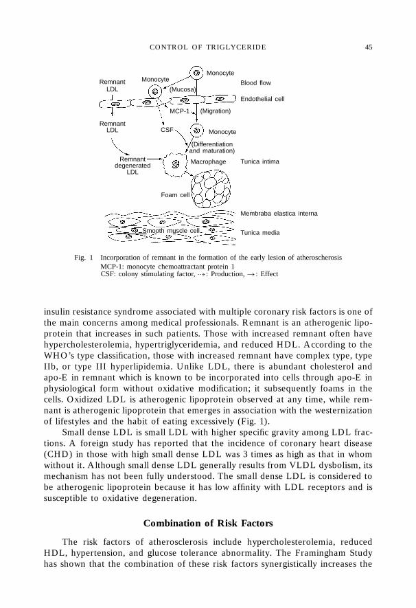
insulin resistance syndrome associated with multiple coronary risk factors is one ofthe main concerns among medical professionals. Remnant is an atherogenic lipo-protein that increases in such patients. Those with increased remnant often havehypercholesterolemia, hypertriglyceridemia, and reduced HDL. According to theWHO’s type classification, those with increased remnant have complex type, typeIIb, or type III hyperlipidemia. Unlike LDL, there is abundant cholesterol andapo-E in remnant which is known to be incorporated into cells through apo-E inphysiological form without oxidative modification; it subsequently foams in thecells. Oxidized LDL is atherogenic lipoprotein observed at any time, while rem-nant is atherogenic lipoprotein that emerges in association with the westernizationof lifestyles and the habit of eating excessively (Fig. 1).
Small dense LDL is small LDL with higher specific gravity among LDL frac-tions. A foreign study has reported that the incidence of coronary heart disease(CHD) in those with high small dense LDL was 3 times as high as that in whomwithout it. Although small dense LDL generally results from VLDL dysbolism, itsmechanism has not been fully understood. The small dense LDL is considered tobe atherogenic lipoprotein because it has low affinity with LDL receptors and issusceptible to oxidative degeneration.
Combination of Risk Factors
The risk factors of atherosclerosis include hypercholesterolemia, reducedHDL, hypertension, and glucose tolerance abnormality. The Framingham Studyhas shown that the combination of these risk factors synergistically increases the
(Migration)
Monocyte
Smooth muscle cell Tunica media
Membraba elastica interna
Tunica intimaMacrophage
Foam cell
Endothelial cell
Blood flow
MCP-1
CSFRemnant
LDL
Remnantdegenerated
LDL
(Differentiationand maturation)
Fig. 1 Incorporation of remnant in the formation of the early lesion of atheroscherosisMCP-1: monocyte chemoattractant protein 1CSF: colony stimulating factor, � : Production, � : Effect
MonocyteMonocyteRemnant
LDL (Mucosa)
CONTROL OF TRIGLYCERIDE 45
risk of ischemic heart disease. It is important to pay close attention to the combi-nation of such factors in addition to the severity of individual risk factors.
The concept of “syndrome X” or “deadly quartet” has been proposed for apatient group in whom the incidence of ischemic heart disease is high in spite ofthe mildness of individual risk factors. Both of them indicate a pathological statein which insulin sensitivity has been reduced by obesity, hyperalimentation, lack ofexercise, or genetic factors. Those with syndrome X or deadly quartet secrete anexcessive amount of insulin to compensate for insufficient insulin effect (insulinresistance), which results in hyperinsulinemia.
The insufficient insulin effect reduces the activity of lipoprotein lipase, increasesVLDL, and finally causes hypertriglyceridemia. As described above, HDL is nega-tively correlated with triglyceride in blood. Therefore, the reduced activity of lipo-protein lipase causes hypertriglyceridemia and reduces the production of HDL,thereby reducing HDL. Moreover, insulin resistance often causes glucose toler-ance disorder.
Diagnosis and Treatment of Hypertriglyceridemia
Triglyceride in blood is divided into that derived from the intake of food andthat synthesized in the liver. The blood triglyceride value is generally measuredafter fasting for at least 14 hours (often early in the morning after fasting) becauseit is easily influenced by that created from the intake of food.
Hyperlipidemia is considered to be primary when it has developed as a resultof the predisposition of the patient without the effect of other underlying diseasesor drugs. In contrast, the disease is considered secondary when it has developeddue to other diseases, such as diabetes mellitus, thyroid diseases, hepatic and bileduct disease, and renal insufficiency, intake of drugs, such as oral contraceptivesand hypotensive drugs, drinking, and stress. Secondary hypertriglyceridemia isgenerally improved by the treatment of such primary diseases and the discontinu-ance of causatory drugs.
The diseases that frequently develop with hypertriglyceridemia and are impor-tant to note in the prevention of ischemic heart diseases include type III hyper-lipidemia caused by the increased remnant in association with apo-E abnormality,complex type hyperlipidemia with increased cholesterol, and hyperlipidemia accom-panying diabetes mellitus, nephrotic syndrome, and hypothyroidism. It is alsonecessary to note that excessive drinking causes hypertriglyceridemia.
Hypertriglyceridemia is basically treated with diet therapy. The combinationof appropriate diet and exercise therapies is often effective in treating the disease.Care should be taken in starting pharmacotherapy in those with obesity or whohave a drinking problem. It is necessary to fully consider the pathological state andrisk factors of individual patients before commencing pharmacotherapy. That is,the future progress of atherosclerosis has to be considered in determining thera-peutic policy.
Patients with hypertriglyceridemia are divided into high risk and low riskgroups. High risk patients are those who have risk factors of ischemic heart disease(hyperlipidemia, hypertension, smoking, diabetes mellitus, men, arteriosclerotic
N. YAMADA Asian Med. J. 44(1), 200146
diseases other than ischemic heart disease, obesity, reduced HDL, and hyper-lipoproteinemia(a)) or those with a medical history of ischemic heart disease.
For those who have been affected by hypertriglyceridemia for a long periodof time and are classified into the high risk group, it should be considered thatatherosclerosis has already progressed, and the main therapeutic target is to reduceatherosclerosis. In such patients, pharmacotherapy should be actively performed.The target value should be set to be as low as possible. The target triglyceridevalue should be 150 mg/dL or lower.
When hypertriglyceridemia is of low risk with a short affected period and mildrisk factors, the treatment should be directed to the prevention of atherosclerosis.In such patients, it is not necessary to hurry the initiation of pharmacotherapy.Rather, it is important to check their compliance with diet therapy and provideinstruction for activities of daily living, particularly stopping drinking and adoptinga long-term perspective to continued exercise therapy.
Hypertriglyceridemia is mostly a result of excessive eating, lack of exercise,obesity, and excessive drinking. Therefore, it is natural that the disease is compli-cated by hypertension and diabetes mellitus in a large number of patients. Sincethe disease is closely related to daily lifestyle, it should be emphasized that patientsthemselves are responsible for controlling their health on a daily basis, includingmaintaining ideal body weight, doing moderate exercise, and refraining from drink-ing, and that favorable control of the above should be most effective in preventinghypertriglyceridemia and other adult diseases. Drugs used for treating the diseaseinclude fibrates, niacin preparations, and HMG-CoA reductase inhibitors.
REFERENCE
1) Yamada, N., Yoshinaga, H., Sakura, N. et al.: Increased risk factors for coronary arterydisease in Japanese subjects with hyperinsulinemia or glucose intolerance. DiabetesCare 17: 107, 1994.
CONTROL OF TRIGLYCERIDE 47