Embed Size (px)
Citation preview
ESTHETIC CROWN LENGTHENING - A HIGHLY GRATIFYING BUT OFTEN UNDIAGNOSED ESTHETIC ISSUE
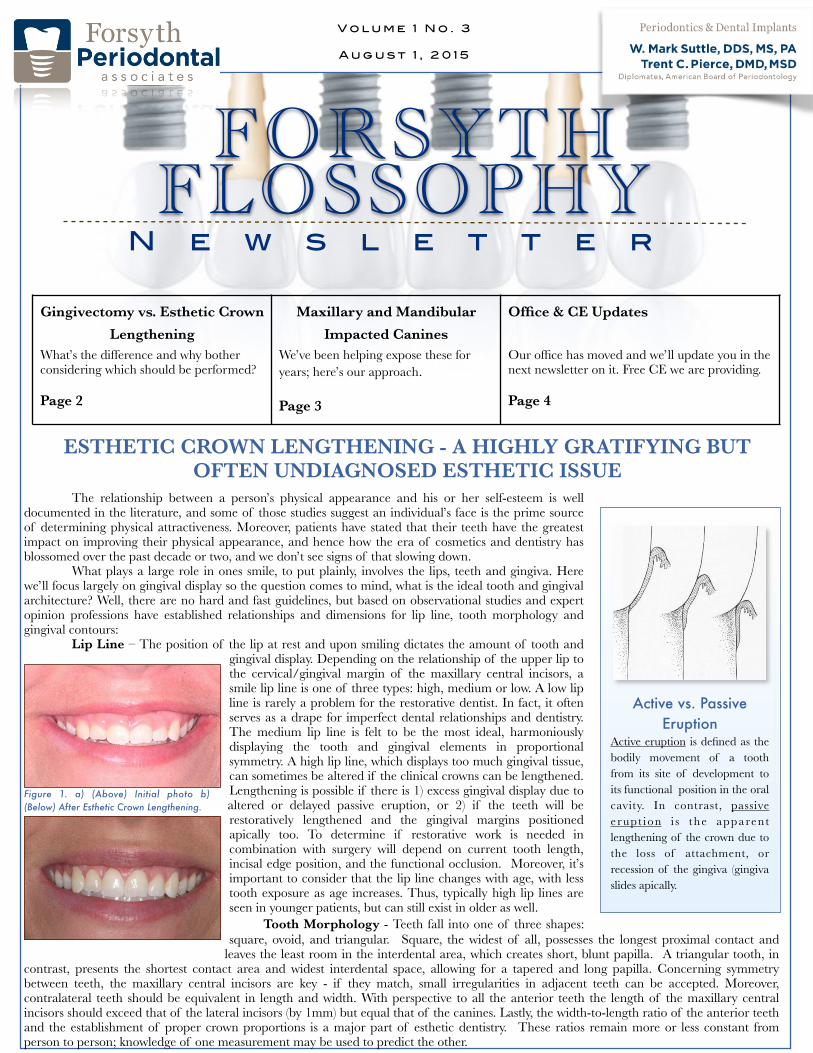
The relationship between a person’s physical appearance and his or her self-esteem is well documented in the literature, and some of those studies suggest an individual’s face is the prime source of determining physical attractiveness. Moreover, patients have stated that their teeth have the greatest impact on improving their physical appearance, and hence how the era of cosmetics and dentistry has blossomed over the past decade or two, and we don’t see signs of that slowing down. What plays a large role in ones smile, to put plainly, involves the lips, teeth and gingiva. Here we’ll focus largely on gingival display so the question comes to mind, what is the ideal tooth and gingival architecture? Well, there are no hard and fast guidelines, but based on observational studies and expert opinion professions have established relationships and dimensions for lip line, tooth morphology and gingival contours: Lip Line – The position of the lip at rest and upon smiling dictates the amount of tooth and
gingival display. Depending on the relationship of the upper lip to the cervical/gingival margin of the maxillary central incisors, a smile lip line is one of three types: high, medium or low. A low lip line is rarely a problem for the restorative dentist. In fact, it often serves as a drape for imperfect dental relationships and dentistry. The medium lip line is felt to be the most ideal, harmoniously displaying the tooth and gingival elements in proportional symmetry. A high lip line, which displays too much gingival tissue, can sometimes be altered if the clinical crowns can be lengthened. Lengthening is possible if there is 1) excess gingival display due to altered or delayed passive eruption, or 2) if the teeth will be restoratively lengthened and the gingival margins positioned apically too. To determine if restorative work is needed in combination with surgery will depend on current tooth length, incisal edge position, and the functional occlusion. Moreover, it’s important to consider that the lip line changes with age, with less tooth exposure as age increases. Thus, typically high lip lines are seen in younger patients, but can still exist in older as well. Tooth Morphology - Teeth fall into one of three shapes: square, ovoid, and triangular. Square, the widest of all, possesses the longest proximal contact and
leaves the least room in the interdental area, which creates short, blunt papilla. A triangular tooth, in contrast, presents the shortest contact area and widest interdental space, allowing for a tapered and long papilla. Concerning symmetry between teeth, the maxillary central incisors are key - if they match, small irregularities in adjacent teeth can be accepted. Moreover, contralateral teeth should be equivalent in length and width. With perspective to all the anterior teeth the length of the maxillary central incisors should exceed that of the lateral incisors (by 1mm) but equal that of the canines. Lastly, the width-to-length ratio of the anterior teeth and the establishment of proper crown proportions is a major part of esthetic dentistry. These ratios remain more or less constant from person to person; knowledge of one measurement may be used to predict the other.
Active vs. Passive Eruption
Active eruption is defined as the bodily movement of a tooth from its site of development to its functional position in the oral cavity. In contrast, passive e rupt ion i s the apparent lengthening of the crown due to the loss of attachment, or recession of the gingiva (gingiva slides apically.
FLOSSOPHYN e w s l e t t e r
Gingivectomy vs. Esthetic Crown Lengthening
What’s the difference and why bother considering which should be performed?
Page 2
Maxillary and Mandibular Impacted Canines
We’ve been helping expose these for years; here’s our approach.
Page 3
Office & CE Updates
Our office has moved and we’ll update you in the next newsletter on it. Free CE we are providing.
Page 4
FORSYTH
V o l u m e 1 N o . 3
A u g u s t 1 , 2 0 1 5
Figure 1. a) (Above) Initial photo b) (Below) After Esthetic Crown Lengthening.
a u g u s t 1 , 2 0 1 5
F o r s y t h F l o s s o p h y • F o r s y t h P e r i o d o n t a l A s s o c i a t e s • 3 3 6 . 7 6 5 . 9 2 2 4 - P a g e ! -2
FORSYTH FLOSSOPHYEsthetic Crown Lengthening Continued
Gingival Margin and Contour – The gingival margins of the maxillary central incisors and canines should typically exist at the same level with the margin of the lateral incisors falling about 1 mm coronal to its adjacent teeth. Additionally, the gingival zenith (height of gingival contour on the facial) of the maxillary central incisors and canines should match and peak at or near the distal line angle as they follow the curvature of the CEJ. The lateral incisor’s gingival zenith should exist at the center of the tooth mesiodistally. The degree of gingival scallop often depends on tooth morphology and tissue thickness. The etiology of excess gingival display, or a “gummy smile,” can vary, and can also be a combination of several factors. These include: Short or Hyper-mobile Upper Lip, Vertical Maxillary Excess, Dentoalveolar Extrusion, Gingival Enlargement, and Altered Passive Eruption. Treatment for each of these can also vary, but for this article we will focus on treating the “gummy smile” due to Altered Passive Eruption with esthetic crown lengthening for the adolescent patients. This procedure is one of the most rewarding treatments we provide since it can immediately and drastically improve a patient’s smile. Once the teeth reach their final position with respect to the alveolar bone (Active Eruption) then the gingival margin typically migrates to a level about 1 mm coronal to the CEJ (Passive Eruption). Early in passive eruption the epithelial attachment rests on the enamel surface, then progresses to just on the cemental surface. In about12% of patients passive eruption fails to completely occur, hence the term altered passive eruption, and results in excessive gingival display. Twelve percent seems small but 12% is actually a large number. For every 100 adolescents seen, 12 will present with this condition. This condition is extremely under-diagnosed, but for those diagnosed, fortunately, this can be corrected with a minor surgical procedure. The initial consultation helps determine if restorations will are necessary in combination with surgery due to expected root exposure post-surgery. In a later issue we will address cases that require combo restorative-
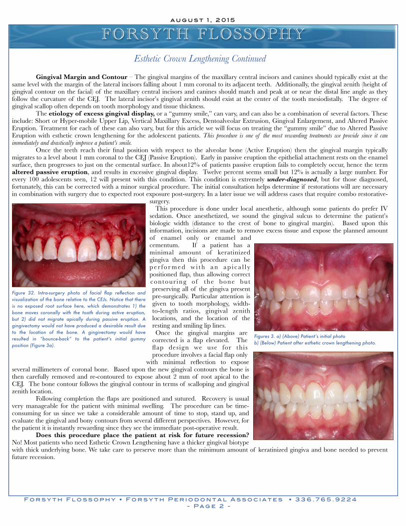
surgery. This procedure is done under local anesthetic, although some patients do prefer IV sedation. Once anesthetized, we sound the gingival sulcus to determine the patient’s biologic width (distance to the crest of bone to gingival margin). Based upon this information, incisions are made to remove excess tissue and expose the planned amount of enamel only or enamel and cementum. If a patient has a minimal amount of keratinized gingiva then this procedure can be p e r f o r m e d w i t h a n a p i c a l l y positioned flap, thus allowing correct contour ing o f the bone but preserving all of the gingiva present pre-surgically. Particular attention is given to tooth morphology, width-to-length ratios, gingival zenith locations, and the location of the resting and smiling lip lines. Once the gingival margins are corrected is a flap elevated. The flap design we use for this procedure involves a facial flap only
with minimal reflection to expose several millimeters of coronal bone. Based upon the new gingival contours the bone is then carefully removed and re-contoured to expose about 2 mm of root apical to the CEJ. The bone contour follows the gingival contour in terms of scalloping and gingival zenith location. Following completion the flaps are positioned and sutured. Recovery is usual very manageable for the patient with minimal swelling. The procedure can be time-consuming for us since we take a considerable amount of time to stop, stand up, and evaluate the gingival and bony contours from several different perspectives. However, for the patient it is instantly rewarding since they see the immediate post-operative result. Does this procedure place the patient at risk for future recession? No! Most patients who need Esthetic Crown Lengthening have a thicker gingival biotype with thick underlying bone. We take care to preserve more than the minimum amount of keratinized gingiva and bone needed to prevent future recession.
Figure 32. Intra-surgery photo of facial flap reflection and visualization of the bone relative to the CEJs. Notice that there is no exposed root surface here, which demonstrates 1) the bone moves coronally with the tooth during active eruption, but 2) did not migrate apically during passive eruption. A gingivectomy would not have produced a desirable result due to the location of the bone. A gingivectomy would have resulted in “bounce-back” to the patient’s initial gummy position (Figure 3a).
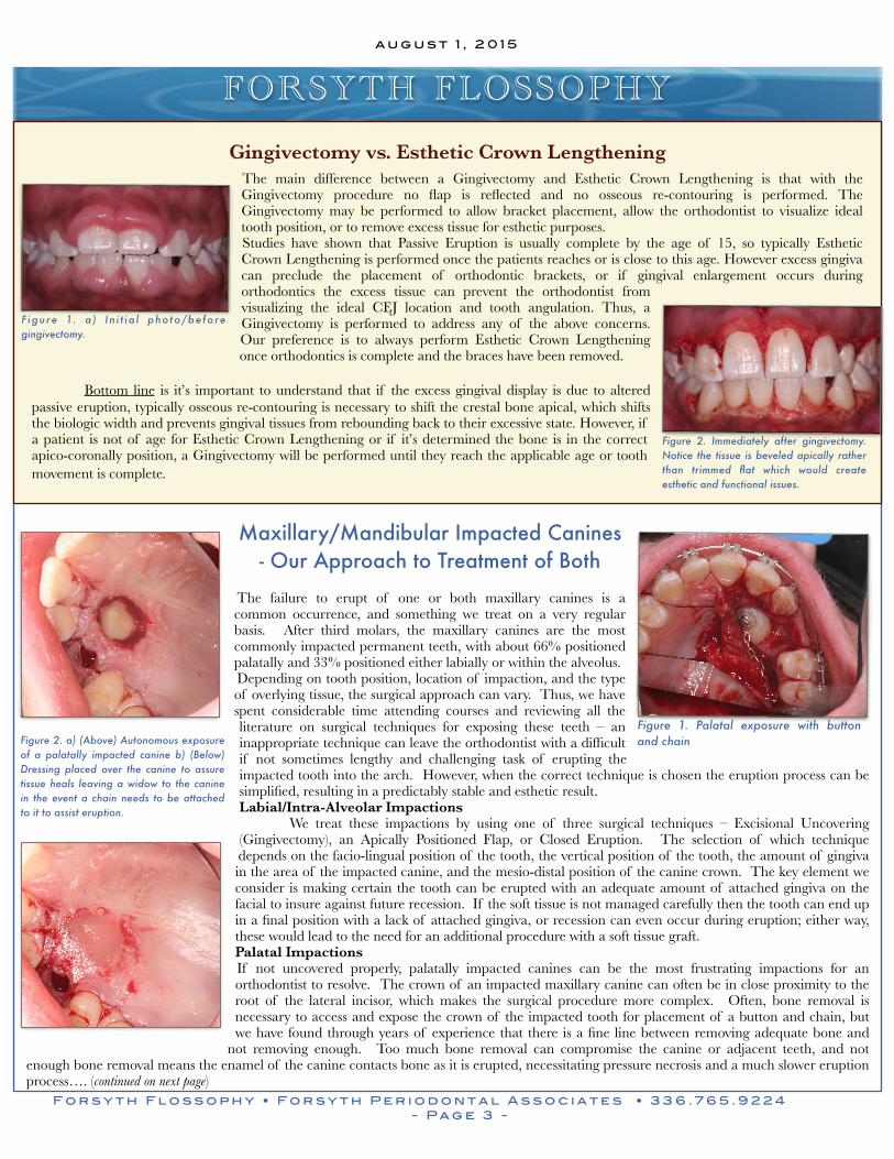
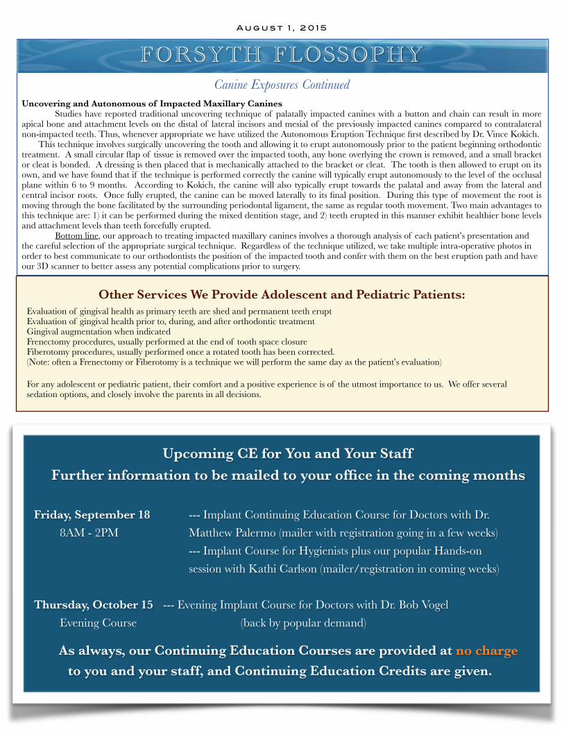
Figures 3. a) (Above) Patient’s initial photo b) (Below) Patient after esthetic crown lengthening photo.
a u g u s t 1 , 2 0 1 5
F o r s y t h F l o s s o p h y • F o r s y t h P e r i o d o n t a l A s s o c i a t e s • 3 3 6 . 7 6 5 . 9 2 2 4 - P a g e ! -3
Gingivectomy vs. Esthetic Crown Lengthening The main difference between a Gingivectomy and Esthetic Crown Lengthening is that with the Gingivectomy procedure no flap is reflected and no osseous re-contouring is performed. The Gingivectomy may be performed to allow bracket placement, allow the orthodontist to visualize ideal tooth position, or to remove excess tissue for esthetic purposes. Studies have shown that Passive Eruption is usually complete by the age of 15, so typically Esthetic Crown Lengthening is performed once the patients reaches or is close to this age. However excess gingiva can preclude the placement of orthodontic brackets, or if gingival enlargement occurs during orthodontics the excess tissue can prevent the orthodontist from visualizing the ideal CEJ location and tooth angulation. Thus, a Gingivectomy is performed to address any of the above concerns. Our preference is to always perform Esthetic Crown Lengthening once orthodontics is complete and the braces have been removed.
Bottom line is it’s important to understand that if the excess gingival display is due to altered passive eruption, typically osseous re-contouring is necessary to shift the crestal bone apical, which shifts the biologic width and prevents gingival tissues from rebounding back to their excessive state. However, if a patient is not of age for Esthetic Crown Lengthening or if it’s determined the bone is in the correct apico-coronally position, a Gingivectomy will be performed until they reach the applicable age or tooth movement is complete.
FORSYTH FLOSSOPHY
Maxillary/Mandibular Impacted Canines - Our Approach to Treatment of Both
The failure to erupt of one or both maxillary canines is a common occurrence, and something we treat on a very regular basis. After third molars, the maxillary canines are the most commonly impacted permanent teeth, with about 66% positioned palatally and 33% positioned either labially or within the alveolus. Depending on tooth position, location of impaction, and the type of overlying tissue, the surgical approach can vary. Thus, we have spent considerable time attending courses and reviewing all the literature on surgical techniques for exposing these teeth – an inappropriate technique can leave the orthodontist with a difficult if not sometimes lengthy and challenging task of erupting the impacted tooth into the arch. However, when the correct technique is chosen the eruption process can be simplified, resulting in a predictably stable and esthetic result. Labial/Intra-Alveolar Impactions We treat these impactions by using one of three surgical techniques – Excisional Uncovering (Gingivectomy), an Apically Positioned Flap, or Closed Eruption. The selection of which technique depends on the facio-lingual position of the tooth, the vertical position of the tooth, the amount of gingiva in the area of the impacted canine, and the mesio-distal position of the canine crown. The key element we consider is making certain the tooth can be erupted with an adequate amount of attached gingiva on the facial to insure against future recession. If the soft tissue is not managed carefully then the tooth can end up in a final position with a lack of attached gingiva, or recession can even occur during eruption; either way, these would lead to the need for an additional procedure with a soft tissue graft. Palatal Impactions If not uncovered properly, palatally impacted canines can be the most frustrating impactions for an orthodontist to resolve. The crown of an impacted maxillary canine can often be in close proximity to the root of the lateral incisor, which makes the surgical procedure more complex. Often, bone removal is necessary to access and expose the crown of the impacted tooth for placement of a button and chain, but we have found through years of experience that there is a fine line between removing adequate bone and
not removing enough. Too much bone removal can compromise the canine or adjacent teeth, and not enough bone removal means the enamel of the canine contacts bone as it is erupted, necessitating pressure necrosis and a much slower eruption process…. (continued on next page)
Figure 2. a) (Above) Autonomous exposure of a palatally impacted canine b) (Below) Dressing placed over the canine to assure tissue heals leaving a widow to the canine in the event a chain needs to be attached to it to assist eruption.
Figure 1. Palatal exposure with button and chain
F igure 1. a ) In i t ia l pho to/before gingivectomy.
Figure 2. Immediately after gingivectomy. Notice the tissue is beveled apically rather than trimmed flat which would create esthetic and functional issues.
A u g u s t 1 , 2 0 1 5
Canine Exposures Continued Uncovering and Autonomous of Impacted Maxillary Canines Studies have reported traditional uncovering technique of palatally impacted canines with a button and chain can result in more apical bone and attachment levels on the distal of lateral incisors and mesial of the previously impacted canines compared to contralateral non-impacted teeth. Thus, whenever appropriate we have utilized the Autonomous Eruption Technique first described by Dr. Vince Kokich.
This technique involves surgically uncovering the tooth and allowing it to erupt autonomously prior to the patient beginning orthodontic treatment. A small circular flap of tissue is removed over the impacted tooth, any bone overlying the crown is removed, and a small bracket or cleat is bonded. A dressing is then placed that is mechanically attached to the bracket or cleat. The tooth is then allowed to erupt on its own, and we have found that if the technique is performed correctly the canine will typically erupt autonomously to the level of the occlusal plane within 6 to 9 months. According to Kokich, the canine will also typically erupt towards the palatal and away from the lateral and central incisor roots. Once fully erupted, the canine can be moved laterally to its final position. During this type of movement the root is moving through the bone facilitated by the surrounding periodontal ligament, the same as regular tooth movement. Two main advantages to this technique are: 1) it can be performed during the mixed dentition stage, and 2) teeth erupted in this manner exhibit healthier bone levels and attachment levels than teeth forcefully erupted. Bottom line, our approach to treating impacted maxillary canines involves a thorough analysis of each patient’s presentation and the careful selection of the appropriate surgical technique. Regardless of the technique utilized, we take multiple intra-operative photos in order to best communicate to our orthodontists the position of the impacted tooth and confer with them on the best eruption path and have our 3D scanner to better assess any potential complications prior to surgery.
FORSYTH FLOSSOPHY
Upcoming CE for You and Your Staff Further information to be mailed to your office in the coming months
Friday, September 18 --- Implant Continuing Education Course for Doctors with Dr. 8AM - 2PM Matthew Palermo (mailer with registration going in a few weeks) --- Implant Course for Hygienists plus our popular Hands-on session with Kathi Carlson (mailer/registration in coming weeks)
Thursday, October 15 --- Evening Implant Course for Doctors with Dr. Bob Vogel Evening Course (back by popular demand)
As always, our Continuing Education Courses are provided at no charge to you and your staff, and Continuing Education Credits are given.
Other Services We Provide Adolescent and Pediatric Patients: Evaluation of gingival health as primary teeth are shed and permanent teeth erupt Evaluation of gingival health prior to, during, and after orthodontic treatment Gingival augmentation when indicated Frenectomy procedures, usually performed at the end of tooth space closure Fiberotomy procedures, usually performed once a rotated tooth has been corrected. (Note: often a Frenectomy or Fiberotomy is a technique we will perform the same day as the patient’s evaluation)
For any adolescent or pediatric patient, their comfort and a positive experience is of the utmost importance to us. We offer several sedation options, and closely involve the parents in all decisions.