Embed Size (px)
Citation preview

CLINICALTHYROIDOLOGY
VOLUME 16 ● ISSUE 1 MARCH 2004
A publication of the American Thyroid Association
THYROID DISEASE
Patients with Subclinical Thyroid Disease Have Few Clin-ical Abnormalities and Benefit Little from Treatment . . 1
HYPERTHYROIDISM
Cardiac Failure in Pregnant Women with Hyperthy-roidism Is Usually Precipitated by a Complication ofPregnancy . . . . . . . . . . . . . . . . . . . . . . . . . . . . . . . . . . . . 2
Children Who Accidentally Ingest Thyroid HormoneHave Few Symptoms and Signs of HyperthyroidismDespite High Serum Thyroid Hormone Concentrations . . . . . . . . . . . . . . . . . . . . . . . . . . . . . . . . 3
The Efficacy of Radioiodine Therapy for Hyperthy-roidism Is Not Reduced by Post-RadioiodineMethimazole Therapy. . . . . . . . . . . . . . . . . . . . . . . . . . . 4
HYPOTHYROIDISM
Thyroxine and Triiodothyronine Are Not More Effectivethan Thyroxine Alone in Patients with HypothyroidismWho Have Depressive Symptoms. . . . . . . . . . . . . . . . . . 5
Thyroxine and Triiodothyronine Are Not More Effectivethan Thyroxine Alone in Unselected Patients withHypothyroidism . . . . . . . . . . . . . . . . . . . . . . . . . . . . . . . 6
Triiodothyronine and Thyroxine Are Not More Effectivethan Thyroxine Alone in Ameliorating HypothyroidSymptoms or Improving Cognitive Function . . . . . . . . . 7
CONGENITAL HYPOTHYROIDISM
Mutations in a Cell–Membrane Thyroid Hormone–Transport Molecule Cause Neurologic and ThyroidAbnormalities . . . . . . . . . . . . . . . . . . . . . . . . . . . . . . . . . 8
Thyroid Radioiodine Imaging Is Superior to Ultrasonog-raphy in Evaluating Infants with Congenital Hypothy-roidism . . . . . . . . . . . . . . . . . . . . . . . . . . . . . . . . . . . . . . 9
Central Hypothyroidism Occurs in Infants of Motherswith Hyperthyroidism Due to Graves’ Disease duringTheir Pregnancies . . . . . . . . . . . . . . . . . . . . . . . . . . . . . 10
NODULAR GOITER
Thyroid Incidentalomas May Be Carcinomas . . . . . . . . 11
THYROID CANCER
Serum Calcitonin Should Be Measured in All Patientswith Thyroid Nodules. . . . . . . . . . . . . . . . . . . . . . . . . . 12
Preoperative Ultrasonography Is Valuable for DetectingLymph-Node Metastases and Local Recurrence inPatients with Thyroid Carcinoma . . . . . . . . . . . . . . . . . 13
THYROID FUNCTION IN PREGNANCY
The Rise in Serum Thyroxine Binding during Pregnancyin Women with Hypothyroidism Only Partly ExplainsTheir Increased Need for Thyroxine . . . . . . . . . . . . . . 14
THYROIDITIS
Painless Thyroiditis May Be Followed by Recurrence ofHyperthyroidism in Patients with Graves’ Disease . . . . 15
AUTOIMMUNE THYROID DISEASE
Seasonal Allergic Rhinitis May Be Followed by RecurrentHyperthyroidism in Women with Graves’ Disease . . . . 16
Cerebrospinal Fluid Concentrations of Antithyroid Anti-bodies Are High in Hashimoto’s Encephalopathy . . . . 17
IODINE DEFICIENCY
Iodine Deficiency Continues to Be a Major Health Prob-lem Worldwide . . . . . . . . . . . . . . . . . . . . . . . . . . . . . . . 18
DRUG EFFECTS ON THYROID FUNCTION
Short-Term Ingestion of Kelp Has a Weak AntithyroidEffect in Normal Subjects . . . . . . . . . . . . . . . . . . . . . . 19
THYROID HORMONE SECRETION
Megalin-Mediated Removal of Thyroid Hormone–PoorThyroglobulin Facilitates Thyroid HormoneSecretion . . . . . . . . . . . . . . . . . . . . . . . . . . . . . . . . . . . . 20

Editor-in-ChiefRobert D. Utiger, M.D.Thyroid DivisionDepartment of MedicineBrigham & Women’s Hospital 77 Avenue Louis Pasteur Boston, MA 02115(617) 525-5171 Telephone (617) 731-4718 Fax [email protected]
PresidentClark T. Sawin, M.D.
President-Elect Paul W. Ladenson, M.D.
SecretaryGregory A. Brent, M.D.
Treasurer Charles H. Emerson, M.D.
Executive DirectorBarbara R. Smith, C.A.E.American Thyroid Association6066 Leesburg Pike, Suite 650Falls Church, VA 22041Telephone: 703-998-8890Fax: 703-998-8893Email: [email protected]
Designed BySaratoga Graphics7 Kaatskill WayBallston Spa, NY 12020Kandra L. Files, Art Director
Clinical ThyroidologyCopyright © 2004American Thyroid Association, Inc.Printed in the USA. All rights reserved.
CLINICALTHYROIDOLOGYVOLUME 16 ● ISSUE 1 MARCH 2004
Clinical Thyroidology and condensed summaries of all articlesin Clinical Thyroidology are available on the ATA web site
(www.thyroid.org)
ATA News
Future Meetings*
76th Annual MeetingSeptember 29 to October 3, 2004Westin Bayshore Resort and MarinaVancouver, British Columbia, Canada
ATA Horizons in ThyroidologyApril 15 to 17, 2005Baltimore Marriott Waterfront HotelBaltimore, MD
13th International Thyroid CongressOctober 30 to November 4, 2005Buenos Aires, Argentina
77th Annual MeetingOctober 11 to 15, 2006Sheraton Wild Horse Pass Resort and SpaPhoenix, AZ
*For further information about these meetings contact the ATA at (703) 998-8890or www.thytoid.org

COMMENTARY
In a similar analysis, Helfand (1)reached the same conclusions as to thelack of benefit of treatment of subclini-cal hypothyroidism and subclinical hyper-thyroidism, and the U.S. PreventiveServices Task Force has recommendedagainst screening for thyroid dysfunctionin adults.
Patients who seek medical care oftenhave symptoms suggestive of thyroiddysfunction, and some of them have sub-clinical thyroid dysfunction. Given that
the changes in thyroid secretion in thesepatients are very small, even in the sub-groups with the more abnormal serumTSH concentrations (Tables 1 and 2), andthat the symptoms and other manifesta-tions of thyroid hormone deficiency orexcess are very nonspecific, it is not sur-prising that there is so little evidence ofeither disability or benefit of therapy. Ifnothing else, treatment should preventprogression to overt thyroid dysfunction.However, the rates of progression arelow (<5 percent/year for both subclinicalhypothyroidism and subclinical hyperthy-
roidism), and the rates of spontaneousnormalization of serum TSH values areabout the same.
Robert D. Utiger, M.D.
Reference
1. Helfand M. Screening for subclinicalthyroid dysfunction in nonpregnantadults: a summary of the evidence forthe U.S. Preventive Services Task Force.Ann Intern Med 2004;140:128-41.
SUMMARY
Background Subclinical hypothyroidism and subclinicalhyperthyroidism are defined, respectively, by the presence ofhigh or low serum thyrotropin (TSH) concentrations andnormal serum free thyroxine (T4) concentrations, inde-pendent of any symptoms or signs of thyroid dysfunction.Hypothyroidism and hyperthyroidism are considered overtonly when the serum free T4 concentration is low or high,respectively. The clinical effects of subclinical hypothy-roidism and subclinical hyperthyroidism are uncertain, asare the benefits of treatment. This article describes theresults of an analysis of these topics.
Methods The analysis was conducted by a panel of 13experts in endocrinology, epidemiology, and preventivehealth services appointed by the American ThyroidAssociation, the American Association of ClinicalEndocrinologists, and the Endocrine Society. Subclinicalhypothyroidism and subclinical hyperthyroidism weredefined as described above; for the purpose of this analysis,the normal range for serum TSH was considered to be 0.46to 4.4 mU/L.
The panel reviewed and then rated the strength of the avail-able information about the health effects and benefits oftreatment of subclinical hypothyroidism and subclinicalhyperthyroidism as good, fair, or insufficient. The panelthen made recommendations regarding evaluation andtreatment, with qualifications based on the perceivedstrength of the evidence.
Results The prevalence of subclinical hypothyroidism wasestimated to range from 4 to 8.5 percent among adults inthe United States, of whom about 75 percent have serumTSH concentrations of 4.5 to 10 mU/L. Table 1 showsthe panel’s summary of the health effects and benefits oftreatment of subclinical hypothyroidism.
The prevalence of subclinical hyperthyroidism was estimat-ed to be 3.2 percent, of whom about 75 percent have serumTSH concentrations of 0.1 to 0.45 mU/L. Table 2 showsthe panel’s summary of the findings regarding subclinicalhyperthyroidism.
The panel recommended against population-based screen-ing for thyroid dysfunction.
Conclusion There is little evidence that patients withsubclinical hypothyroidism or subclinical hyperthyroidismhave clinical manifestations of their thyroid dysfunction orbenefit from therapy.
THYROID DISEASE
Patients with subclinical thyroid disease have few clinical abnormalities andbenefit little from treatment
Surks MI, Ortiz E, Daniels GH, Sawin CT, Col NF, Cobin RH, Franklyn JA, Hershman JM, Burman KD, Denke MA,Gorman C, Cooper RS, Weissman NJ. Subclinical thyroid disease. Scientific review and guidelines for diagnosis andmanagement. JAMA 2004:291:228-38.
CLINICAL THYROIDOLOGY ● VOLUME 16 ● ISSUE 1 ● 1
Table 1. Assessment of Evidence Regarding Effects of Subclinical Hypo-thyroidism and Benefits of Treatment.
Strength of Evidence Treatment BenefitSerum TSH Serum TSH Serum TSH Serum TSH
4.5-10 mU/L >10 mU/L 4.5-10 mU/L >10 mU/LProgression to overt Good Good Yes Yes
hypothyroidismSymptoms of None Insufficient Insufficient Insufficient
hypothyroidismCardiac dysfunction Insufficient Insufficient Insufficient InsufficientHyper- Insufficient Fair Insufficient Insufficient
cholesterolemia
Table 2. Assessment of Evidence Regarding Effects of Subclinical Hyper-thyroidism and Benefits of Treatment.
Strength of Evidence Treatment BenefitSerum TSH Serum TSH Serum TSH Serum TSH
0.1-0.45 mU/L <0.1 mU/L 0.1-0.45 mU/L <0.1 mU/LProgression to Insufficient Good Yes Yes
overt hyperthyroidismSymptoms of None None None Insufficient
hyperthyroidismCardiac Insufficient Fair Insufficient Insufficient
dysfunctionAtrial fibrillation Insufficient Good None NoneOsteopenia None Fair* None Fair*In postmenopausal women.

2 ● CLINICAL THYROIDOLOGY ● VOLUME 16 ● ISSUE 1
SUMMARY
Background There are similar changes in cardiovascularfunction in normal pregnant women and patients withhyperthyroidism. They include a decrease in peripheral vas-cular resistance, and an increase in blood volume, heart rate,cardiac contractility, and cardiac output. Compensation forthe combined effects of the two conditions is usually ade-quate, but decompensation can occur. This study was doneto determine the factors associated with cardiac failure inpregnant women with hyperthyroidism.
Methods and Results From 1974 through 2001, slightlymore than 300,000 women delivered babies at the ParklandHospital in Dallas, Texas. Among them 150 (<0.05 percent)had overt hyperthyroidism at some time during their preg-nancy. Thirteen (9 percent) of the 150 women had cardiacfailure, based on standard clinical criteria and confirmed byradiographic findings of cardiomegaly and pulmonaryedema (and echocardiographic or other imaging evidence ofdepressed ventricular function in all five women tested).The women ranged in age from 17 to 34 years, and the dura-tion of pregnancy ranged from 18 to 37 weeks. All thewomen had symptoms of hyperthyroidism, a diffuse goiter,and high serum total or free thyroxine concentrations at thetime of hospitalization for cardiac failure.
Six women had cardiac failure before the time of fetal via-bility (23 weeks). Three also had an incomplete abortion,uterine hemorrhage, and sepsis, and two had pyelonephritisand septicemia; four of these women were anemic (hemat-
ocrit, <25 percent). Only one of the six women had nocomplicating problems. The extent of prenatal care, if any,in these women is not described.
Seven women had been pregnant for 29 or more weeks.Four were known to have hyperthyroidism, but had notbeen taking prescribed therapy, and three had received noprenatal care. Four of these women had severe preeclamp-sia or eclampsia, three were anemic, two had chorioam-nionitis and sepsis, and one had no complicating problems.
Treatment for heart failure consisted of intravenousfurosemide, and in some women digoxin or a β-adrenergicantagonist drug. Women with preeclampsia or eclampsiawere treated with magnesium sulfate and hydralazine. Allwere treated with propylthiouracil, potassium iodide, and aglucocorticoid. Other treatments included antibiotics, bloodtransfusion, and uterine curettage. All the women improvedrapidly, and cardiac evaluation was normal 5 weeks to 5months later. The outcome of the pregnancies is not stated.Eleven of the women were last evaluated 2 to 25 years later,and all had a normal cardiac evaluation at that time. Threehad been treated with iodine-131, one was taking propyl-thiouracil (3 years later), and seven were in remission (2 to25 years later).
Conclusion Most pregnant women with hyperthyroidismwho have cardiac failure also have sepsis, preeclampsia, orother complications of pregnancy.
COMMENTARY
Given the similarity of the changesin cardiovascular function in pregnantwomen and patients with hyperthy-roidism, and the presumption that thecardiovascular effects of the two condi-tions are additive, it is perhaps surprisingthat cardiac failure is not more commonin pregnant women with hyperthy-roidism. Thyroid hormone excess proba-bly has more direct chronotropic andinotropic effects on cardiac function thanpregnancy, and less effect on blood vol-ume, so perhaps the effects are less addi-tive than might be expected.
These women were encounteredover a period of many years, and relative-ly few had contemporary studies of car-diovascular function. In a study of sixpregnant women with hyperthyroidismbut not cardiac failure, cardiac output,
stroke volume, heart rate, and mean arte-rial pressure were higher, and peripheralresistance was lower, as compared withnormal pregnant women at the samestage of gestation, and the values for allmeasurements were more normal duringantithyroid drug therapy (1).
Cardiac failure may occur in patientswith hyperthyroidism as a result ofmarked tachycardia, atrial fibrillation, or,rarely, cardiomyopathy. However, it isuncommon, certainly in women of child-bearing age. Hence, the presence of car-diac failure in 9 percent of pregnantwomen with hyperthyroidism is excep-tional. This high frequency is probablyexplained by the high frequency of sepsisand disorders of pregnancy in thesewomen. Preeclampsia, which seems tooccur with increased frequency in womenwith hyperthyroidism, may increase after-load, and sepsis may increase preload,
beyond the capability of the heart tomaintain adequate output; the conse-quence is cardiac failure.
Some of the complications of preg-nancy may not be preventable, but severehyperthyroidism is. Women known tohave hyperthyroidism when they becomepregnant should be followed with specialcare, and those caring for pregnantwomen should remember that hyperthy-roidism can begin during pregnancy.
Robert D. Utiger, M.D.
Reference
1. Easterling TR, Schmucker BC, Carl-son KL, et al. Maternal hemodynamics inpregnancies complicated by hyperthy-roidism. Obstet Gynecol 1991;78:348-52.
HYPERTHYROIDISM
Cardiac failure in pregnant women with hyperthyroidism is usuallyprecipitated by a complication of pregnancy
Sheffield JS, Cunningham FG. Thyrotoxicosis and heart failure that complicate pregnancy. Am J Obst Gynecol2004;190:211-7.

SUMMARY
Background Thyroxine (T4) and other thyroid hormoneproducts are widely prescribed, and therefore are likely to beaccidentally ingested by young children. This studydescribes the findings in a group of young children whoingested large amounts of thyroid hormone.
Methods The study subjects were 30 children seen in onehospital between 1983 and 2000. The inclusion criteria wereage ≤5 years, a history of accidental ingestion of thyroidhormone, and the availability of results of measurements ofserum T4 and triiodothyronine (T3). Information concern-ing numbers of tablets ingested and timing of the ingestionwas obtained from the parents. The children were treated bygastric lavage, followed by administration of activated char-coal and sodium sulfate (0.25 g/kg). Until 1988, all childrenwere hospitalized, but thereafter they were hospitalized onlyif they were ill or were thought to have ingested >500 µg ofT4. Blood samples for hormone assay were usually obtainedtwo to six hours after ingestion, and multiple samples wereobtained from a few children.
Results The median age of the 30 children was 2.4 years(range, 1.6 to 4.8 years). Seven had ingested T4, in amountsranging from 300 to 9500 µg, one had ingested T3 (600 µg),and 16 had ingested both, in amounts ranging from 80 µg of
T4 and 20 µg of T3 to 2500 µg of T4 and 630 µg of T3; themedian doses were 760 µg of T4 and 150 µg of T3; no infor-mation on amount ingested was available for six children.Seven children were followed as outpatients and 23 werehospitalized. Only eight children (27 percent) had any signsof hyperthyroidism (usually tachycardia), and none hadseizures or other central nervous system abnormalities.
The initial serum T4 concentrations ranged from 7.1 to 94.5µg/dl (91 to 1219 nmol/L; median, 13.1 µg/dl [169nmol/L]), and the initial serum T3 concentrations rangedfrom 169 to 3195 ng/dl (2.6 to 49.2 nmol/L); median, 532ng/dl [8.2 nmol/L]). In four children, serum T4 and T3were measured repeatedly for up to 120 hours after theingestion. Their serum T4 concentrations were highest at 4to 8 hours; in two the value had fallen by 50 percent at 48hours and in one by 72 hours. In three of these four chil-dren the peak serum T3 concentration was at 6 hours and inthe fourth it was at 24 hours. There was no correlationbetween the initial serum T4 and T3 concentrations andclinical manifestations of hyperthyroidism.
Conclusion Children who accidentally ingest largeamounts of T4 and T3 have few or no clinical mani-festations of hyperthyroidism despite high serum T4 and T3concentrations.
HYPERTHYROIDISM
Children who accidentally ingest thyroid hormone have few symptomsand signs of hyperthyroidism despite high serum thyroid hormoneconcentrations
Willgerodt H, Keller E, Sorger D, Hoepffner W. Acute ingestion of thyroxine and triiodothyronine in young children.Eur J Pediatr 2003;162:639-41.
CLINICAL THYROIDOLOGY ● VOLUME 16 ● ISSUE 1 ● 3
COMMENTARY
Clinical manifestations of hyperthy-roidism were rare not only in these 30children, but also in larger studies of thisproblem (1,2). In the largest study, of 92children <6 years of age, only 8 (9 per-cent) had any clinical manifestations ofhyperthyroidism (2). Standard therapyconsists of gastric lavage and administra-tion of charcoal and a cathartic. The effi-cacy of these treatments is not known.
In these studies virtually all childrenwho had any symptoms or signs ofhyperthyroidism had ingested >5 mg ofT4, although other children toleratedthese doses well (1,2). These findingshave led to the recommendation thatgastrointestinal decontamination proce-dures need not be undertaken if the T4dose is ≤5 mg (1). There are multiple rea-sons why children tolerate single highdoses of T4 well. Fractional absorption isprobably lower than for therapeutic
doses of T4. The rate of T4 clearance ishigher in children than adults, and clear-ance of T4 when its serum concentrationis very high is more rapid than when itsconcentration is normal. Also, conver-sion to T3 is relatively slow, so thatserum T3 concentrations tend to rise andfall slowly.
In 2002, 4730 children <6 years ofage ingested a “thyroid preparation,”according to data accumulated by the 64centers that constitute the ToxicExposure Surveillance System of theAmerican Association of Poison ControlCenters (3). For all ages the total was9438, of which 8418 (89 percent) wereunintentional, 1899 (20 percent) weretreated in a health care facility, and 5 died(age not given). Given these statistics, itseems likely that most of the childrenwho ingested some thyroid preparationwere evaluated only by telephone andwere not treated, presumably because thedose was low. T4 and other thyroid
preparations are present in many homes,and care should be taken to keep themout of the hands of children.
Robert D. Utiger, M.D.
References
1. Litovitz TL, White JD. Levothyroxineingestions in children: an analysis of 78cases. Am J Emerg Med 1985;3:297-300.
2. Tunget CL, Clark RF, Turchen SG,Manoguerra, AS. Raising the decontami-nation level for thyroid hormone inges-tions. Am J Emerg Med 1995;13:9-13.
3. Watson WA, Litovitz TL, Rodgers GCJr, et al. 2002 Annual Report of theAmerican Association of Poison ControlCenters Toxic Exposure SurveillanceSystem. Am J Emerg Med 2003;21:353-421.

SUMMARY
Background Some patients with hyperthyroidism who aretreated with iodine-131 (I-131) are given an antithyroid drugbefore or after I-131, or at both times, but there has beenconcern that the antithyroid drug may reduce the efficacy ofI-131. In this study the effects of methimazole or no drugtherapy given after I-131 on thyroid function and size werecompared in patients with hyperthyroidism.
Methods The study subjects were 149 patients (124women, 25 men; median age, 59 years) with hyperthy-roidism caused by a toxic multinodular goiter in 81 (54 per-cent), Graves’ disease in 46 (31 percent), and a thyroid ade-noma in 22 (15 percent). All the patients had been treatedwith methimazole, and were euthyroid, as determined bynormal serum free thyroxine (T4) and free triiodothyronine(T3) index values, for at least three months. Then, methi-mazole was stopped, and four days later I-131 was given.The I-131 dose was based on thyroid volume, as determinedby ultrasonography; the mean (±SD) dose was 10.8±4.2mCi (400±154 MBq). Seven days later, the patients wererandomly assigned to resume the same dose of methima-zole or to receive no drug therapy.
Thyroid function was assessed periodically for one yearafter I-131 therapy. The methimazole was discontinued afterone or more months if the patient was euthyroid. Patientsin the no-drug-therapy group were given methimazole ifthey had hyperthyroidism six or more weeks after I-131 wasgiven. Patients in either group in whom methimazole couldnot be stopped were given a second dose of I-131 nine ormore months after the first dose.
Results At the time of I-131 therapy, the patients in thetwo groups were similar in age, causes of hyperthyroidism,thyroid volume, serum free T4 and free T3 index values, and24-hour thyroid I-131 uptake values.
At the end of the one year follow-up period, 84 patients (56percent) were euthyroid, 29 (20 percent) had hypothy-roidism, and 36 (24 percent) had persistent hyperthy-roidism. The patients with a toxic multinodular goiter weremore likely to be euthyroid (74 percent), than those withGraves’ disease (33 percent). There were no differences inoutcome in the methimazole and no-drug-therapy groups.For example, among the patients with Graves’ disease in themethimazole group, 24 percent were euthyroid, 35 percenthad hypothyroidism, and 41 percent had persistent hyper-thyroidism, as compared with 38 percent, 31 percent, and 31percent, respectively, in the no-drug-therapy group.
The mean serum free T4 and free T3 index values increasedby approximately 30 percent three and six weeks after I-131therapy in the no-drug-therapy group, whereas both valuesdeclined slightly in the methimazole group. Among thepatients who became euthyroid, thyroid volume decreasedby 43 percent (39 percent in the methimazole group and 48percent in the no-drug-therapy group; P<0.05).
Conclusion Among patients with hyperthyroidism previ-ously treated with methimazole, resumption of methima-zole after I-131 therapy does not alter the actions of I-131,except to reduce slightly its effect on thyroid volume.
HYPERTHYROIDISM
The efficacy of radioiodine therapy for hyperthyroidism is not reduced bypost-radioiodine methimazole therapy
Bonnema SJ, Bennedbaek FN, Gram J, Veje A, Marving J, Hegedus L. Resumption of methimazole after 131I therapy ofhyperthyroid diseases: effect on thyroid function and volume evaluated by a randomized clinical trial. Eur J Endocrinol2003;149:485-92.
4 ● CLINICAL THYROIDOLOGY ● VOLUME 16 ● ISSUE 1
COMMENTARY
The rationales for administration ofan antithyroid drug before or after I-131therapy, or at both times, are that thedrug may ameliorate hyperthyroidismsooner and reduce the likelihood of anexacerbation of hyperthyroidism as aresult of radiation-induced thyroiditis, ascompared with I-131 given alone. On theother hand, antithyroid drug therapy mayreduce the efficacy of I-131 therapy.
In any assessment of the availabledata, the differences in the timing ofantithyroid drug therapy in relation to I-131 therapybefore, after, orbothmust be kept in mind. Among
patients pretreated with methimazole,there is little evidence that the overallduration of hyperthyroidism is shorterthan in those given I-131 alone, and itdoes not diminish the long-term effect ofI-131 (1). In this new study, in which allpatients received methimazole before andmethimazole or no drug after I-131 ther-apy, there was no difference in the effica-cy of I-131 therapy. Comparable data arenot available for propylthiouracil.
It is good to know that treatmentwith methimazole does not alter thelong-term efficacy of I-131. It would beeven better to have stronger evidencethat it has overall benefit when givenbefore or after I-131, or at both times, as
compared with I-131 alone. Otherwise, itcomplicates therapy and exposes patientsto drug side effects unnecessarily.
Robert D. Utiger, M.D.
Reference
1. Andrade VA, Gross JL, Maia AL. Theeffect of methimazole pretreatment onthe efficacy of radioactive iodine therapyin Graves’ hyperthyroidism: one-year fol-low-up of a prospective, randomizedstudy. J Clin Endocrinol Metab 2001;86:3488-93.

SUMMARY
Background Most patients with hypothyroidism are treat-ed with thyroxine (T4), but despite restoration of normalserum T4 and thyrotropin (TSH) concentrations many con-tinue to have some symptoms, particularly psychologicalsymptoms, suggestive of hypothyroidism. Treatment withT4 plus triiodothyronine (T3) may ameliorate these symp-toms. In this study the effects of T4 alone and T4 plus T3were compared in patients with hypothyroidism receivingchronic T4 therapy who had symptoms of depression.
Methods Forty patients (36 women and 4 men; mean age,47 years) with primary hypothyroidism (mean duration, 9years) were studied. Specific inclusion criteria were treat-ment with a constant dose of T4 for at least six months,symptoms of depression (score >5 on the General HealthQuestionnaire, which is designed to identify psychiatric dys-function), and a normal serum TSH concentration. Patientswith a history of hyperthyroidism, thyroid carcinoma, orany thyroid surgery were excluded. The mean daily prestudydose of T4 was 120 µg in the T4 group and 132 µg in theT4-plus-T3 group.
The study patients were randomly assigned to receive theirusual dose of T4 once daily plus a placebo twice daily, orhalf their usual dose of T4 plus T3, 12.5 µg twice daily, for15 weeks. The dose of T3 was adjusted by an independentinvestigator to maintain normal serum TSH concentrations.The patients were evaluated using the Symptom Check-List-90 (SCL-90) and the Comprehensive EpidemiologicalScreens for Depression (CES-D), and serum TSH, free T4,and free T3 were measured, at base line and at 2, 4, 6, 9, 12,and 15 weeks. The patients were evaluated using the ShortForm 36 at base line and at 6 and 15 weeks. The first twoquestionnaires focus on psychological symptoms. The ShortForm 36 focuses on general physical and mental health andwell-being; it has eight subscales and two summary scores.Neither the patients nor the examiners were aware of treat-ment-group assignment.
Results Thirty-three of the 40 patients (82 percent) com-pleted the study. At the end of the study, the mean daily T4dose was 118 µg in the T4 group and 67 µg in the T4-plus-T3 group, and the mean T3 dose in the latter group was 19µg. The mean base-line serum TSH, free T4, and free T3concentrations were similar in the two groups. During andat the end of the study the mean serum TSH concentrationsin the two groups were similar, whereas the mean serumfree T4 concentration was lower and the mean serum freeT3 concentration was higher in the T4-plus-T3 group atall times (Table).
In both groups, the scores for the subtests (depression,anxiety, global severity index, positive symptom total, andpositive symptom distress index) of the SCL-90 test and thescore for the CES-D test were similar at base line. Thescores improved during the study, but there were no differ-ences between the groups at any time. The scores for all thesubscales and the summary scores of the Short Form 36also were similar at base line, and changed little in eithergroup (Table).
Conclusion In patients with hypothyroidism who havedepressive symptoms despite adequate T4 therapy, the com-bination of T4 plus T3 is no more effective than T4 alone inameliorating these symptoms and improving overall healthand well-being.
HYPOTHYROIDISM
Thyroxine and triiodothyronine are not more effective than thyroxine alonein patients with hypothyroidism who have depressive symptoms
Sawka AM, Gerstein HC, Marriott MJ, MacQueen GM, Joffe RT. Does a combination regimen of thyroxine (T4) and3,5,3′-triiodothyronine improve depressive symptoms better than T4 alone in patients with hypothyroidism? Results of adouble-blind, randomized, controlled trial. J Clin Endocrinol Metab 2003;88:4551-5.
CLINICAL THYROIDOLOGY ● VOLUME 16 ● ISSUE 1 ● 5
Table. Mean Serum TSH and Free T4 Concentrations and Results of Scores forSelected Subscales of the Short Form 36 in the T4 and T4-plus-T3 Groups atBase Line and after Treatment for 15 Weeks.
T4 Group T4-plus-T3 GroupBase Line 15 Weeks Base Line 15 Weeks
Serum TSH (mU/L) 2.2 1.7 1.8 1.8Serum free T4 (ng/dl)* 1.3 1.4 1.2 0.8Short Form 36**
Physical functioning 77 77 75 79General health 63 69 58 59Vitality 35 51 32 51Social functioning 70 73 55 76Mental health 63 70 56 63
*To convert to pmol/L, multiply by 12.9.**Scored on a scale of 0 (poorest health) to 100 (optimal health).
COMMENTARY
These three studies provide strongevidence against the suggestion, firstmade many years ago and resurrectedseveral years ago (1), that patients withhypothyroidism feel better when treatedwith T3 and T4 than when treated withT4 alone.
This suggestion was based on sever-al facts. One is that the thyroid producesand secretes both T4 and T3, and there-fore in patients with hypothyroidism T4alone is inadequate replacement. This istrue, even if the thyroidal component ofdaily T3 production is relatively small(approximately 20 to 25 percent). Torestore serum T3 concentrations fully to
normal, one then must give enough T4to raise serum T4 concentrations to theupper part of the normal range. Sincelittle is known about tissue penetrationof T4 and T3 and the intracellular con-version of T4 to T3 in different tissues,notably the brain, it is not hard to envi-sion that T4 alone might not restore nor-mality. In other words, the small amount
continued on page 6
of T3 contributed by the thyroid mightbe important.
The second fact is that manypatients with hypothyroidism thought tobe adequately treated with T4, mostly asdetermined by a normal serum TSH con-centration, do not feel well. The infor-mation on this point is largely but not
completely anecdotal (2); on the otherhand it is clear that the frequency ofsymptoms suggestive of hypothyroidismdiffers little between people with highserum TSH concentrations and normalsubjects (3). Then there are the questionsof what is a patient’s normal serum TSHconcentration, and is there a difference
in well-being when a patient’s serumTSH concentration is 0.8 or 2.8 mU/L?And is there too much reliance on serumTSH values anyhow, if only because theyare easy to measure?
Whatever the merits of the abovecomments, Sawka, Walsh, and Clyde andtheir colleagues found that replacing
SUMMARY
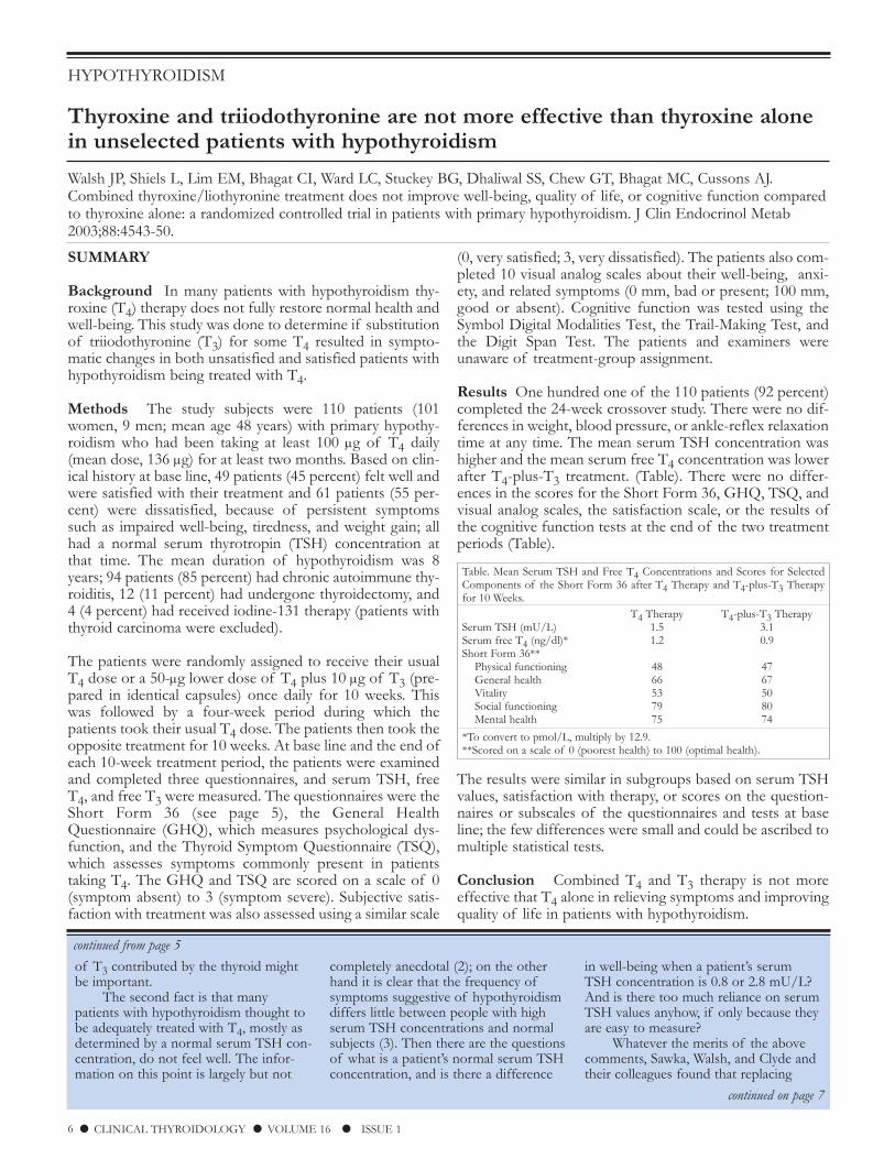
Background In many patients with hypothyroidism thy-roxine (T4) therapy does not fully restore normal health andwell-being. This study was done to determine if substitutionof triiodothyronine (T3) for some T4 resulted in sympto-matic changes in both unsatisfied and satisfied patients withhypothyroidism being treated with T4.
Methods The study subjects were 110 patients (101women, 9 men; mean age 48 years) with primary hypothy-roidism who had been taking at least 100 µg of T4 daily(mean dose, 136 µg) for at least two months. Based on clin-ical history at base line, 49 patients (45 percent) felt well andwere satisfied with their treatment and 61 patients (55 per-cent) were dissatisfied, because of persistent symptomssuch as impaired well-being, tiredness, and weight gain; allhad a normal serum thyrotropin (TSH) concentration atthat time. The mean duration of hypothyroidism was 8years; 94 patients (85 percent) had chronic autoimmune thy-roiditis, 12 (11 percent) had undergone thyroidectomy, and4 (4 percent) had received iodine-131 therapy (patients withthyroid carcinoma were excluded).
The patients were randomly assigned to receive their usualT4 dose or a 50-µg lower dose of T4 plus 10 µg of T3 (pre-pared in identical capsules) once daily for 10 weeks. Thiswas followed by a four-week period during which thepatients took their usual T4 dose. The patients then took theopposite treatment for 10 weeks. At base line and the end ofeach 10-week treatment period, the patients were examinedand completed three questionnaires, and serum TSH, freeT4, and free T3 were measured. The questionnaires were theShort Form 36 (see page 5), the General HealthQuestionnaire (GHQ), which measures psychological dys-function, and the Thyroid Symptom Questionnaire (TSQ),which assesses symptoms commonly present in patientstaking T4. The GHQ and TSQ are scored on a scale of 0(symptom absent) to 3 (symptom severe). Subjective satis-faction with treatment was also assessed using a similar scale
(0, very satisfied; 3, very dissatisfied). The patients also com-pleted 10 visual analog scales about their well-being, anxi-ety, and related symptoms (0 mm, bad or present; 100 mm,good or absent). Cognitive function was tested using theSymbol Digital Modalities Test, the Trail-Making Test, andthe Digit Span Test. The patients and examiners wereunaware of treatment-group assignment.
Results One hundred one of the 110 patients (92 percent)completed the 24-week crossover study. There were no dif-ferences in weight, blood pressure, or ankle-reflex relaxationtime at any time. The mean serum TSH concentration washigher and the mean serum free T4 concentration was lowerafter T4-plus-T3 treatment. (Table). There were no differ-ences in the scores for the Short Form 36, GHQ, TSQ, andvisual analog scales, the satisfaction scale, or the results ofthe cognitive function tests at the end of the two treatmentperiods (Table).
The results were similar in subgroups based on serum TSHvalues, satisfaction with therapy, or scores on the question-naires or subscales of the questionnaires and tests at baseline; the few differences were small and could be ascribed tomultiple statistical tests.
Conclusion Combined T4 and T3 therapy is not moreeffective that T4 alone in relieving symptoms and improvingquality of life in patients with hypothyroidism.
HYPOTHYROIDISM
Thyroxine and triiodothyronine are not more effective than thyroxine alonein unselected patients with hypothyroidism
Walsh JP, Shiels L, Lim EM, Bhagat CI, Ward LC, Stuckey BG, Dhaliwal SS, Chew GT, Bhagat MC, Cussons AJ.Combined thyroxine/liothyronine treatment does not improve well-being, quality of life, or cognitive function comparedto thyroxine alone: a randomized controlled trial in patients with primary hypothyroidism. J Clin Endocrinol Metab2003;88:4543-50.
6 ● CLINICAL THYROIDOLOGY ● VOLUME 16 ● ISSUE 1
Table. Mean Serum TSH and Free T4 Concentrations and Scores for SelectedComponents of the Short Form 36 after T4 Therapy and T4-plus-T3 Therapyfor 10 Weeks.
T4 Therapy T4-plus-T3 TherapySerum TSH (mU/L) 1.5 3.1Serum free T4 (ng/dl)* 1.2 0.9Short Form 36**
Physical functioning 48 47General health 66 67Vitality 53 50Social functioning 79 80Mental health 75 74
*To convert to pmol/L, multiply by 12.9.**Scored on a scale of 0 (poorest health) to 100 (optimal health).
continued on page 7
continued from page 5
SUMMARY
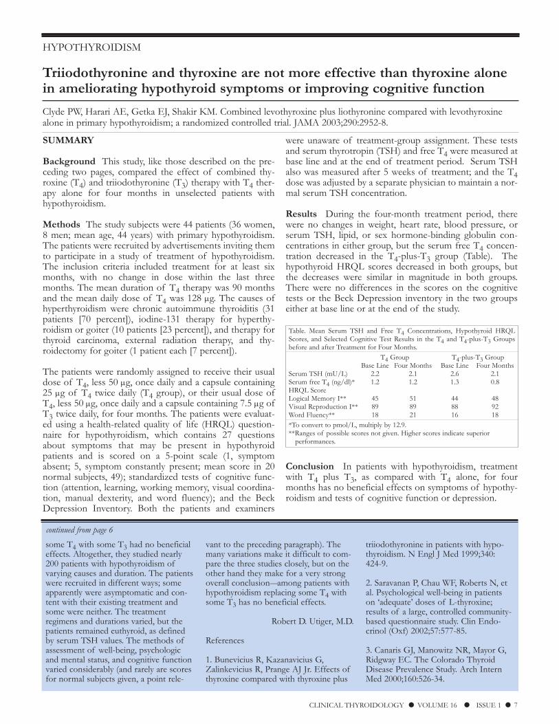
Background This study, like those described on the pre-ceding two pages, compared the effect of combined thy-roxine (T4) and triiodothyronine (T3) therapy with T4 ther-apy alone for four months in unselected patients withhypothyroidism.
Methods The study subjects were 44 patients (36 women,8 men; mean age, 44 years) with primary hypothyroidism.The patients were recruited by advertisements inviting themto participate in a study of treatment of hypothyroidism.The inclusion criteria included treatment for at least sixmonths, with no change in dose within the last threemonths. The mean duration of T4 therapy was 90 monthsand the mean daily dose of T4 was 128 µg. The causes ofhyperthyroidism were chronic autoimmune thyroiditis (31patients [70 percent]), iodine-131 therapy for hyperthy-roidism or goiter (10 patients [23 percent]), and therapy forthyroid carcinoma, external radiation therapy, and thy-roidectomy for goiter (1 patient each [7 percent]).
The patients were randomly assigned to receive their usualdose of T4, less 50 µg, once daily and a capsule containing25 µg of T4 twice daily (T4 group), or their usual dose ofT4, less 50 µg, once daily and a capsule containing 7.5 µg ofT3 twice daily, for four months. The patients were evaluat-ed using a health-related quality of life (HRQL) question-naire for hypothyroidism, which contains 27 questionsabout symptoms that may be present in hypothyroidpatients and is scored on a 5-point scale (1, symptomabsent; 5, symptom constantly present; mean score in 20normal subjects, 49); standardized tests of cognitive func-tion (attention, learning, working memory, visual coordina-tion, manual dexterity, and word fluency); and the BeckDepression Inventory. Both the patients and examiners
were unaware of treatment-group assignment. These testsand serum thyrotropin (TSH) and free T4 were measured atbase line and at the end of treatment period. Serum TSHalso was measured after 5 weeks of treatment; and the T4dose was adjusted by a separate physician to maintain a nor-mal serum TSH concentration.
Results During the four-month treatment period, therewere no changes in weight, heart rate, blood pressure, orserum TSH, lipid, or sex hormone-binding globulin con-centrations in either group, but the serum free T4 concen-tration decreased in the T4-plus-T3 group (Table). Thehypothyroid HRQL scores decreased in both groups, butthe decreases were similar in magnitude in both groups.There were no differences in the scores on the cognitivetests or the Beck Depression inventory in the two groupseither at base line or at the end of the study.
Conclusion In patients with hypothyroidism, treatmentwith T4 plus T3, as compared with T4 alone, for fourmonths has no beneficial effects on symptoms of hypothy-roidism and tests of cognitive function or depression.
some T4 with some T3 had no beneficialeffects. Altogether, they studied nearly200 patients with hypothyroidism ofvarying causes and duration. The patientswere recruited in different ways; someapparently were asymptomatic and con-tent with their existing treatment andsome were neither. The treatmentregimens and durations varied, but thepatients remained euthyroid, as definedby serum TSH values. The methods ofassessment of well-being, psychologicand mental status, and cognitive functionvaried considerably (and rarely are scoresfor normal subjects given, a point rele-
vant to the preceding paragraph). Themany variations make it difficult to com-pare the three studies closely, but on theother hand they make for a very strongoverall conclusionamong patients withhypothyroidism replacing some T4 withsome T3 has no beneficial effects.
Robert D. Utiger, M.D.
References
1. Bunevicius R, Kazanavicius G,Zalinkevicius R, Prange AJ Jr. Effects ofthyroxine compared with thyroxine plus
triiodothyronine in patients with hypo-thyroidism. N Engl J Med 1999;340:424-9.
2. Saravanan P, Chau WF, Roberts N, etal. Psychological well-being in patientson ‘adequate’ doses of L-thyroxine;results of a large, controlled community-based questionnaire study. Clin Endo-crinol (Oxf) 2002;57:577-85.
3. Canaris GJ, Manowitz NR, Mayor G,Ridgway EC. The Colorado ThyroidDisease Prevalence Study. Arch InternMed 2000;160:526-34.
HYPOTHYROIDISM
Triiodothyronine and thyroxine are not more effective than thyroxine alonein ameliorating hypothyroid symptoms or improving cognitive function
Clyde PW, Harari AE, Getka EJ, Shakir KM. Combined levothyroxine plus liothyronine compared with levothyroxinealone in primary hypothyroidism; a randomized controlled trial. JAMA 2003;290:2952-8.
CLINICAL THYROIDOLOGY ● VOLUME 16 ● ISSUE 1 ● 7
Table. Mean Serum TSH and Free T4 Concentrations, Hypothyroid HRQLScores, and Selected Cognitive Test Results in the T4 and T4-plus-T3 Groupsbefore and after Treatment for Four Months.
T4 Group T4-plus-T3 GroupBase Line Four Months Base Line Four Months
Serum TSH (mU/L) 2.2 2.1 2.6 2.1Serum free T4 (ng/dl)* 1.2 1.2 1.3 0.8HRQL ScoreLogical Memory I** 45 51 44 48Visual Reproduction I** 89 89 88 92Word Fluency** 18 21 16 18*To convert to pmol/L, multiply by 12.9.**Ranges of possible scores not given. Higher scores indicate superior
performances.
continued from page 6

COMMENTARY
Most, if not all, cells have cell−mem-brane molecules that can transport car-boxylic acids and amino acids, includingT4 and T3, into the cells. There are sever-al groups of these transporters; their dis-tribution among tissues varies, as doestheir affinity for different carboxylic andamino acids. MCT8 is one of a group of14 monocarboxylate transporters; amongthem it is probably the most specifictransporter of T4 and T3. It must beexpressed in neural tissue, given theclinical abnormalities in the two boys.However, these abnormalities were notthose of hypothyroidism (and there was
little systemic evidence of it), suggestingthat the primary role of MCT8 is totransport some other carboxylic acid(s).
What is the explanation for theabnormalities in serum thyroid hormonevalues in the two boys? TSH secretionmight be increased as a result of de-creased entry of T4 into the thyrotrophcells of the pituitary, not compensated bythe increase in type 2 deiodinase activitythat results from T4 deficiency in thesecells. The excess serum T3 could be com-ing from larger tissues rich in deiodinase,such as muscle, into which T4 is carriedby transporters other than MCT8, andthe thyroid, as a result of TSH stimula-tion of deiodinase activity there. Some
other carboxylic acid normally transport-ed by MCT8 may preferentially block T3transport by another transporter. Possiblecauses of the slightly low serum T4 con-centrations include decreased thyroidalsecretion, and preferential transport ofT4 by other transporters into tissues thatactively convert it to T3 and export theT3, which probably requires still othertransporters.
Robert D. Utiger, M.D.
SUMMARY
Background Thyroxine (T4) and triiodothyronine (T3)must enter cells to regulate gene expression, their major ifnot sole action. Their entry is facilitated by transporter mol-ecules located in the plasma membrane of the cells. Onesuch molecule is a member of a group of monocarboxylicacid transporters (MCT), MCT8. This study describes theresults of clinical and molecular studies of two families withmutations of the MCT8 gene.
Family 1 The proband was an 8-year-old boy whose par-ents were normal and who was born after a normal preg-nancy. He had onset of dystonia, irritability, and poor feed-ing within days after birth, and his subsequent developmentwas markedly delayed. At age 2 years he could not sit, crawl,or speak, and had dystonia. He later became quadriplegic.At age 17 months, his serum TSH concentration was 4.1mU/L, and at age 2 years it was 8.6 mU/L; he was treatedwith T4, with no benefit. When studied at age 8 years andthereafter, he repeatedly had low serum total T4 and free T4index, low reverse T3, high total and free T3, and high nor-mal or slightly high TSH values (Table). His mother hadsimilar but more normal values. His father, only sibling (aboy), and other maternal relatives were normal.
Family 2 The proband was a 3-year-old boy who was theproduct of a normal pregnancy. Neonatal screeningrevealed low blood-spot T4 and normal TSH values, con-firmed by serum assays on day 12. He was thought to havecentral hypothyroidism, and was treated with T4. At age 3months, he had feeding problems, disconjugate gaze, andhypertonia, and then quadriplegia. Later thyroid hormonevalues (while taking a low dose of T4) were similar to those
in the proband of family 1 (Table). His mother, a maternalaunt, the maternal grandmother had similar, but more nor-mal values; a maternal uncle had died at age 10 years withcerebral palsy and severe developmental delay.
Molecular Studies Analysis of the MCT8 gene, which islocated on the X chromosome, revealed different mutations,each likely to cause loss of function in the transporter, inthe probands and their mothers in both families. In family1, the mutant allele may have come from the maternalgrandfather (who had died), or occurred very early in devel-opment of the mother. In family 2, the mutant allele wasfound not only in the mother, but also in a maternal aunt,the maternal uncle who had died, and the maternal grand-mother.
Conclusion Mutations in the X-linked MCT8 gene, whichcan transport T4 and T3 into cells, results in severe devel-opmental delay and neurologic abnormalities and unusualabnormalities in thyroid function in hemizygous males andonly very mild abnormalities in thyroid function in het-erozygous females.
CONGENITAL HYPOTHYROIDISM
Mutations in a cell-membrane thyroid hormone-transport molecule causeneurologic and thyroid abnormalities
Dumitrescu AM, Liao XH, Best TB, Brockmann K, Refetoff S. A novel syndrome combining thyroid and neurologicalabnormalities is associated with mutations in a monocarboxylate transporter gene. Am J Hum Genet 2004;74:168-75.
8 ● CLINICAL THYROIDOLOGY ● VOLUME 16 ● ISSUE 1
Table. Serum Thyroid Hormone and TSH Concentrations in the Patients andTheir Parents.
Family 1 Family 2Serum Patient Mother Father Patient Mother FatherT4 (µg/dl) 4.7 5.9 6.5 4.6 6.5 7.1Free T4 index 4.8 5.7 7.5 5.7 5.5 8.0T3 (ng/dl) 251 174 116 263 169 136Reverse T3 (ng/dl) 7 12 20 8 12 25TSH (mU/L) 3.9 1.6 1.2 4.8 2.9 1.1Normal values: T4, 5-12; free T4 index, 6-10.5; T3, 90-180; reverse T3, 14-30;and TSH, 0.4-3.6. To convert serum T4 values to nmol/L, multiply by 12.9; toconvert serum T3 and reverse T3 values to nmol/L, multiply by 0.015.

SUMMARY
Background Most infants with congenital hypothy-roidism have developmental abnormalities of the thyroidgland. They include thyroid agenesis, hypoplasia, andectopic thyroid tissue, disorders collectively referred to asthyroid dysgenesis. The remaining infants have a normal orincreased mass of normally located thyroid tissue that haslittle thyroid biosynthetic activity; these disorders are collec-tively referred to as thyroid dyshormonogenesis. Thyroiddysgenesis and dyshormonogenesis are customarily distin-guished by radionuclide imaging. This study was done toevaluate the value of thyroid ultrasonography in distin-guishing among the causes of congenital hypothyroidism.
Methods The study subjects were 66 infants with con-genital hypothyroidism identified by newborn screening andconfirmed by clinical examination and measurements ofserum thyrotropin and free thyroxine (T4) at age 12±6(mean±SD) days after birth at a single hospital in France.Thyroid imaging 30 minutes after intravenous injection ofiodine-123 and thyroid ultrasonography were done at thattime or soon thereafter.
Results As determined by radioiodine imaging, 42 infants(64 percent) had ectopic thyroid tissue, 12 (18 percent) hadthyroid agenesis, and 12 (18 percent) had normally locatedthyroid tissue.
Among the 42 infants in the ectopic thyroid group, ultra-sonography revealed ectopic thyroid tissue high in the neckin 9 (21 percent) and no thyroid tissue in the other 33 (79percent). The mean serum free T4 concentration was high-er in these 9 infants than in the infants in whom no thyroid
tissue was detected by ultrasonography. Among the 12infants who had thyroid agenesis, as determined by radioio-dine imaging, ultrasonography revealed a normally locatedenlarged thyroid gland in one and a normally locatedhypoplastic thyroid gland in one.
Based on both tests, 42 infants (64 percent) had ectopic thy-roid tissue, 10 (15 percent) had thyroid agenesis, and 14 (21percent) had normally located thyroid tissue. Seven of thelatter had an enlarged thyroid gland, 2 had a normal-sizedthyroid gland, and 5 had an abnormal thyroid gland(hypoplasia, 1 infant; hemiagenesis of the left lobe, 2infants; hypoplasia of the left lobe, 1 infant; and markedasymmetry of the lobes, 1 infant). In these 5 infants, ultra-sonography was more informative than radioiodine imagingin defining the thyroid. All 14 infants in the group withsome normally located thyroid tissue had permanenthypothyroidism, as determined by recurrence when T4 ther-apy was stopped temporarily at some later time.
In all the infants with thyroid agenesis or ectopic thyroid tis-sue, ultrasonography revealed approximately 5 × 5 mmstructures in the thyroid area that were isoechoic withrespect to fat and hyperechoic with respect to muscle ornormal thyroid tissue. Six infants in the ectopic thyroidgroup had cysts in the thyroid area, and four infants had tis-sue with the appearance of thymus in the thyroid area.
Conclusion Radioiodine imaging is more informativethan thyroid ultrasonography in determining the cause ofcongenital hypothyroidism. However, ultrasonographyreveals additional abnormalities in infants with thyroid dys-genesis that may provide insight into the causes of congen-ital hypothyroidism.
COMMENTARY
There has been renewed interest inthe contribution of ultrasonography tothe etiologic diagnosis of congenitalhypothyroidism. There is general agree-ment that, even with the newest equip-ment and with the most skilled operators,ultrasonography is less sensitive thanradionuclide imaging for the detection ofectopic thyroid tissue, which results fun-damentally from arrested migration ofthe thyroid anlage into the lower neckand is the most common cause of con-genital hypothyroidism.
The contribution of this and similarrecent studies (1) using ultrasonographyis of a more theoretical nature. The pres-ence of structures resembling thymic
tissue in the usual location of thyroid tis-sue in four infants is consistent with thefinding that Hoxa-3-/- mice have abnor-malities in both thyroid and thymic devel-opment (2), although pathologic confir-mation of the presence of thymic tissuein this location in these infants is lacking.By contrast, the cystic structures detectedin six infants probably correspond to thecystic remnants of the ultimobranchialbodies seen on histologic studies of thethyroid bed in some patients with a lin-gual thyroid (3).
Guy Van Vliet, M.D.University of Montreal
Montreal, Quebec, Canada
References
1. Kreisner E, Camargo-Neto E, MaiaCR, Gross JL. Accuracy of ultrasonogra-phy to establish the diagnosis and aetiolo-gy of permanent primary congenitalhypothyroidism. Clin Endocrinol (Oxf)2003;59:361-5.
2. Manley NR, Capecchi MR. The role ofHoxa-3 in mouse thymus and thyroiddevelopment. Development 1995;121:1989-2003.
3. Williams ED, Toyn CE, Harach HR.The ultimobranchial gland and congenitalthyroid abnormalities in man. J Pathol1989;159:135-41.
CONGENITAL HYPOTHYROIDISM
Thyroid radioiodine imaging is superior to ultrasonography in evaluatinginfants with congenital hypothyroidism
Bubuteishvili L, Garel C, Czernichow P, Leger J. Thyroid abnormalities by ultrasonography in neonates with congenitalhypothyroidism. J Pediatr 2003;143:759-64.
CLINICAL THYROIDOLOGY ● VOLUME 16 ● ISSUE 1 ● 9

SUMMARY
Background Congenital hypothyroidism is usually causedby thyroid dysgenesis or dysfunction, but in a few infants itis caused by hypothalamic or pituitary dysfunction. Onecause of the latter is ongoing suppression of fetal hypo-thalamicpituitary function by hyperthyroidism in fetusesof mothers with hyperthyroidism caused by Graves’ disease.This study was done to define the clinical and biochemicalabnormalities and course of infants with this type of con-genital central hypothyroidism.
Methods The study subjects were 18 infants with centralcongenital hypothyroidism born to mothers with hyperthy-roidism caused by Graves’ disease. The mothers were eithernot treated or were inadequately treated during their preg-nancies. The infants were identified through the newbornscreening program in the Netherlands because they had lowor normal blood-spot thyroxine (T4) and low thyrotropin(TSH) values 4 to 14 days after birth. At recall, the diagno-sis of central hypothyroidism in these infants was based ona plasma free T4 concentration <0.9 ng/dl (12 pmol/L), aplasma TSH concentration <20 mU/L, and the presence ofGraves’ hyperthyroidism in the mother. Twelve of the 14mothers (86 percent) had high plasma TSH−receptor anti-body values (measured by receptor assay), as did 6 of the 11infants (54 percent).
Results There were 12 girls and 6 boys; their gestationalages ranged from 34.6 to 40.9 weeks, and their birth weightranged from 1840 to 3660 g. Their screening blood-spot T4and TSH concentrations ranged from 1.9 to 16.9 µg/dl
(24 to 218 nmol/L) and from <1 to 5 mU/L, respectively.
The mothers and infants were divided into three groups.Nine mothers were found to have hyperthyroidism duringthe first weeks after delivery, usually after the infants werediagnosed as having central hypothyroidism. Thyroid func-tion had not been assessed during pregnancy in any of thesemothers. At recall all of these infants had low plasma freeT4 and normal or low plasma TSH concentrations. Fourmothers were found to have hyperthyroidism during thesecond or third trimester of pregnancy All had been treatedwith an antithyroid drug and propranolol, and three hadnormal plasma free T4 concentrations at the time of deliv-ery. At recall these infants had low plasma free T4 and nor-mal or low plasma TSH concentrations. Four mothers (onehad a twin pregnancy) had hyperthyroidism and had beentreated with an antithyroid drug before pregnancy, but onlyone was treated throughout pregnancy, and all had plasmafree T4 concentrations >1.7 ng/dl (22 pmol/L) during theirpregnancies. At recall or later these infants had low plasmafree T4 and normal or low plasma TSH concentrations.
Seventeen infants were treated with T4, which was laterstopped without recurrence of hypothyroidism in oneinfant and with recurrence in another infant. One infantbecame euthyroid during evaluation and was not treated.
Conclusion Infants of mothers with hyperthyroidismcaused by Graves’ disease may have central hypothyroidismin the neonatal period, presumably as a result of hyperthy-roidism in utero.
COMMENTARY
The incidence of permanent con-genital hypothyroidism in TheNetherlands is approximately 1 in 20,000infants, and most have multiple pituitaryhormone deficiencies. Central congenitalhypothyroidism due to inadequately treat-ed gestational Graves’ disease is estimat-ed to occur in 1 in 35,000 newborninfants. Looked at from the perspectiveof Graves’ hyperthyroidism in the moth-er, the authors estimated that 1 in 70affected women (1.4 percent) will givebirth to a child with congenital centralhypothyroidism. This incidence is similarto the that of neonatal hyperthyroidismdue to maternal Graves’ hyperthyroidism.
Whatever the exact figures, pediatri-cians need to be aware of and test for
not only neonatal Graves’ hyperthy-roidism but also central hypothyroidismin infants of mothers with Graves’hyperthyroidism. The cause of the for-mer is fetal and neonatal stimulationcaused by transplacental passage of TSHreceptor-stimulating antibodies. Thecause of the latter is prolonged inhibitionof thyrotropin-releasing hormone andTSH secretion by preceding hyperthy-roidism, and there is no reason to doubtthat it could occur in an infant who hadhyperthyroidism at birth and during thefirst days or even weeks thereafter as wellas in utero.
Infants with congenital centralhypothyroidism caused by hyperthy-roidism in utero and soon after birth areprobably at risk for adverse neurologicaloutcomes, like infants with congenital
thyroidal hypothyroidism. The hypothy-roidism may not be permanent, but evenif it lasts only a few weeks it may have adeleterious effect on neurodevelopment.
Nine of the 17 mothers (53 percent)of these 18 infants had not been diag-nosed with hyperthyroidism during theirpregnancies, and in others the hyper-thyroidism had been poorly treated.Detecting and treating infants withcongenital central hypothyroidism inthe newborn period is laudable, butdetecting and treating hyperthyroidism inpregnant women will lead to the bestinfant outcome.
Cheryl E. Hanna, M.D.Oregon Health Sciences University
Portland, OR
CONGENITAL HYPOTHYROIDISM
Central hypothyroidism occurs in infants of mothers with hyperthyroidismdue to Graves’ disease during their pregnancies
Kempers MJ, van Tijn DA, van Trotsenburg AS, de Vijlder JJ, Wiedijk BM, Vulsma T. Central congenital hypothyroidismdue to gestational hyperthyroidism: detection where prevention failed. J Clin Endocrinol Metab 2003;88:5851-7.
10 ● CLINICAL THYROIDOLOGY ● VOLUME 16 ● ISSUE 1

NODULAR GOITER
Thyroid incidentalomas may be carcinomas
Nam-Goong IS, Kim HY, Gong G, Lee HK, Hong SJ, Kim WB, Shong YK. Ultrasonography-guided fine-needleaspiration of thyroid incidentaloma: correlation with pathological findings. Clin Endocrinol (Oxf) 2004;60:21-8.
CLINICAL THYROIDOLOGY ● VOLUME 16 ● ISSUE 1 ● 11
SUMMARY
Background Many thyroid nodules are detected inciden-tally, by ultrasonography or other imaging procedures doneto evaluate nonthyroid problems. There has been debate asto the importance of these incidentalomas and the extent towhich patients found to have them should be evaluated.This study was done to define the frequency of thyroid car-cinoma and its extent in patients with incidentalomas.
Methods The study subjects were 267 patients (209women and 58 men; mean age, 51 years [range, 26 to 75])referred for evaluation of thyroid nodules detected by ultra-sonography. None of the nodules was palpable. Nodules≥0.5 cm in diameter (and some smaller nodules) were biop-sied using a 21-gauge needle with ultrasound guidance. Thebiopsies were categorized as benign, indeterminate, suspi-cious for follicular tumor, suspicious for or consistent withpapillary carcinoma, or inadequate. Biopsies categorized asindeterminate were those in which the cellularity was too lit-tle and the amount of colloid too large to warrant the diag-nosis of suspicious for follicular tumor.
Results Three hundred seventeen nodules were biopsied inthe 267 patients. These nodules ranged from 0.2 to 1.5 cm(mean [±SD], 0.9±0.3) in diameter. Twenty-five noduleswere <0.5 cm, of which 13 were ≥0.5 cm at the initial ultra-sound study and 12 were newly detected at the time ofbiopsy in a patient who had a larger nodule. Overall, 130patients (49 percent) had a solitary nodule and 137 (51 per-cent) had multiple nodules. The cytologic findings areshown in the Table.
Among the 29 indeterminate nodules and the 101 nodulesfor which the biopsy was inadequate, 22 were biopsiedagain; 9 biopsies revealed benign cells, 6 were indeterminate,and 7 were inadequate.
The biopsy diagnosis was papillary carcinoma or othermalignant tumor in 8 percent of the <0.5-cm nodules, 15 percent of the 0.5- to 1.0-cm nodules, and 14 percent in the>1.0- to 1.5-cm nodules. Forty of the 48 patients who hada biopsy diagnosis of papillary carcinoma or other malig-nancy or follicular tumor underwent surgery. All of the 35patients with a biopsy diagnosis of papillary carcinoma hada histologic diagnosis of papillary carcinoma. Among the 3patients with a biopsy diagnosis of follicular tumor, 1 had afollicular carcinoma, 1 medullary carcinoma, and 1 follicularadenoma. One of the other 2 patients had an anaplastic car-cinoma and the other a lymphangioma.
At surgery, 16 of the 36 patients (44 percent) with papillaryor follicular carcinoma had extrathyroidal extension, 18 (50percent) had regional lymph-node metastases, and 14 (39percent) had multifocal tumors. The frequency of thesefindings was similar in the patients with nodules <1 cm andthose with nodules ≥1 cm.
Conclusion Among patients with thyroid incidentalomaswho had biopsy findings indicative of thyroid carcinomaand who underwent surgery, 12 percent had a papillary car-cinoma or other malignant tumor.
COMMENTARY
Thyroid incidentalomas are, by defi-nition, not palpable, but are identified byultrasonography or other imaging proce-dures. Most are <1 or 1.5 cm in diame-ter, but even larger nodules may not bepalpable if the nodule is embedded with-in the thyroid or behind or below it.
It is standard practice to biopsy onlyincidental thyroid nodules ≥1 cm, neces-sarily with ultrasound guidance. Thispractice is based on several premises.One is that it is harder to get adequatesamples from small nodules. The second
is that the proportion that are carcino-mas is lower than if the nodule is bigger.Last, even if the nodule is a carcinoma,there is no penalty for delayed diagnosis.In this and other biopsy studies of inci-dentalomas (1), size was not a determi-nant of the success of biopsy, and theproportion of nodules that were carcino-mas did not vary as a function of size.As for the possibility of penalty, thereprobably is none, notwithstanding thesubstantial frequency of extrathyroidalextension and regional node metastases.
Given the high frequency of thyroidincidentalomas in the population (30 to
60 percent), and the very low likelihoodthat those that are carcinomas will evercause trouble, it seems wise to continuethe above biopsy policy.
Robert D. Utiger, M.D.
Reference
1. Papini E, Guglielmi R, Bianchini A, etal. Risk of malignancy in nonpalpablethyroid nodules: predictive value of ultra-sound and color-Doppler features. J ClinEndocrinol Metab 2002;87:1941-6.
Table. Results of Ultrasound-Guided Fine-Needle Biopsies in 267 Patients with317 Thyroid Incidentalomas.
Nodule Diameter<0.5 0.5 to 0.9 cm ≥1.0 to 1.5 cm
(n=25) (n=153) (n=139)Benign 11 (44%) 62 (40%) 66 (48%)Indeterminate 3 (12%) 17 (11%) 9 (7%)Papillary carcinoma 2 (8%) 22 (15%) 18 (13%)Follicular tumor 2 (1%) 2 (1%)Other malignant tumor* 2 (1%)Inadequate 9 (36%) 50 (33%) 42 (30%)*Anaplastic carcinoma (1), lymphangioma (1).

SUMMARY
Background Medullary thyroid carcinoma is rare amongpatients with thyroid nodules, and therefore serum calci-tonin is not measured routinely in the evaluation of thesepatients. This study was done to determine the value of rou-tine measurements of serum calcitonin in patients with thy-roid nodules.
Methods Serum calcitonin was measured in 10,864patients (8692 women, 2172 men; mean age, 49 years) withthyroid nodular disease seen in a single center between 1991and 1998. The patients also underwent thyroid ultrasonog-raphy, radionuclide imaging, assessment of thyroid func-tion, and fine-needle aspiration biopsy. In patients with aserum calcitonin concentration ≥20 pg/ml (assay sensitivity,14 pg/ml; upper limit of the normal reference range, 20pg/ml), the measurement was repeated. If the value wasagain high, pentagastrin (0.5 µg/kg, intravenously) wasgiven, and serum calcitonin was measured 2, 5, 15, and 30minutes later; a serum calcitonin concentration >60 pg/mlwas considered abnormal. Thyroid surgery was recom-mended in patients with high basal serum calcitonin values,confirmed by a supranormal serum calcitonin response topentagastrin, and those in whom fine-needle aspirationbiopsy was suspicious for any type of thyroid carcinoma.
The results of follow-up in the patients in this group whohad medullary carcinoma were compared with the results in45 patients with medullary carcinoma diagnosed between1970 and 1990, before serum calcitonin screening.
Results Among the 10,864 patients, basal serum calcitoninconcentrations were high (20 to 6200 pg/ml) in 47(0.4 percent). Two patients with high values had chronic
renal failure; biopsies of their thyroid nodules revealedbenign thyroid cells. One patient (serum calcitonin, 30pg/ml) refused further testing. The peak post-pentagastrinserum calcitonin concentrations in the remaining 44patients ranged from 118 to 72,000 pg/ml.
The clinical diagnosis in 27 of these 44 patients was multi-nodular goiter. The biopsy diagnosis was medullary carcino-ma in 13, other thyroid carcinoma in 5, and benign thyroidcells in 7; the biopsy was inadequate in 2. The clinical diag-nosis in the other 17 patients was uninodular goiter. Amongthem, the biopsy diagnosis was medullary carcinoma in 7,other carcinoma in 4, and benign thyroid cells in 4; the biop-sy was inadequate in 2. These 44 patients underwent totalthyroidectomy and resection of lymph nodes in the centralcompartment of the neck. All but one proved to havemedullary carcinoma in the nodule that was biopsied (in thatpatient the medullary carcinoma was in a smaller adjacentnodule). Genetic screening in 40 of the 44 patients revealedret proto-oncogene mutations in 8 (20 percent).
In comparison with the 45 patients with medullary carcino-ma in the 1970 to 1990 group, in whom the diagnosis wasmade by biopsy (2 patients) or at surgery (43 patients), theTNM tumor stage was lower in the 1991–1998 group (stageI and II, 68 vs. 44 percent). The proportion of patients withundetectable serum calcitonin concentrations three monthsafter surgery in the 1991–1998 group was higher (66 vs. 3percent), and the mortality was lower (by 6 years after sur-gery the mortality rate was 4 percent in the 1991–1998group and 33 percent in the 1970–1990 group.
Conclusion Routine measurement of serum calcitoninshould be included in the routine evaluation of patientswith thyroid nodules.
COMMENTARY
It is clear from this and other stud-ies that routine measurement of serumcalcitonin in patients with thyroid nod-ules identifies some sporadic medullarycarcinomas that might otherwise bemissed, either because of biopsy misdiag-nosis or because the carcinoma is else-where in the thyroid gland. This doesn’thappen often, because these tumors arerare and most can be identified by biopsy.
According to surveys of membersof the European Thyroid Associationand the American Thyroid Association,43 percent of the former but only 5 per-cent of the latter order measurements ofserum calcitonin in patients with thyroid
nodules (1,2). Why the difference (assum-ing that physicians do what they say theydo)? Some possibilities are that physi-cians in the United States are more con-servative in ordering tests (true forpatients with thyroid nodules), unawareof this value of serum calcitonin meas-urements (maybe, because virtually all thesurveys have been done in Europe),unable to confirm a high basal serum cal-citonin value because pentagastrin is notavailable (unlikely), or satisfied with biop-sy (maybe). Whatever the explanation, itmay be time to reconsider.
Robert D. Utiger, M.D.
References
1. Bennedbaek FN, Perrild H, HegedusL. Diagnosis and treatment of the soli-tary thyroid nodule: results of aEuropean survey. Clin Endocrinol (Oxf)1999;50:357-63.
1. Bennedbaek FN, Hegedus L. Man-agement of the solitary thyroid nodule:results of a North American survey. JClin Endocrinol Metab 2000; 85:2493-8.
THYROID CANCER
Serum calcitonin should be measured in all patients with thyroid nodules
Elisei R, Bottici V, Luchetti F, Di Coscio G, Romei C, Grasso L, Miccoli P, Iacconi P, Basolo F, Pinchera A, Pacini F.Impact of routine measurement of serum calcitonin on the diagnosis and outcome of medullary thyroid carcinoma:experience in 10,864 patients with nodular thyroid disorders. J Clin Endocrinol Metab 2004;89:163-8.
12 ● CLINICAL THYROIDOLOGY ● VOLUME 16 ● ISSUE 1
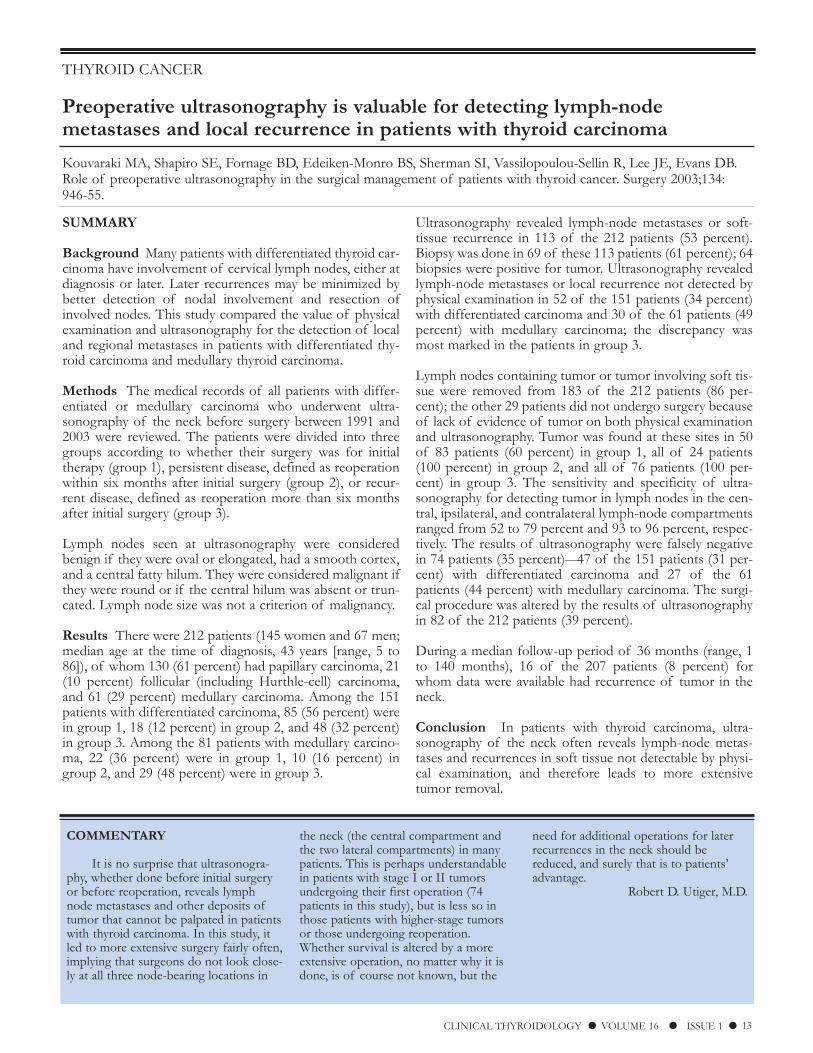
SUMMARY
Background Many patients with differentiated thyroid car-cinoma have involvement of cervical lymph nodes, either atdiagnosis or later. Later recurrences may be minimized bybetter detection of nodal involvement and resection ofinvolved nodes. This study compared the value of physicalexamination and ultrasonography for the detection of localand regional metastases in patients with differentiated thy-roid carcinoma and medullary thyroid carcinoma.
Methods The medical records of all patients with differ-entiated or medullary carcinoma who underwent ultra-sonography of the neck before surgery between 1991 and2003 were reviewed. The patients were divided into threegroups according to whether their surgery was for initialtherapy (group 1), persistent disease, defined as reoperationwithin six months after initial surgery (group 2), or recur-rent disease, defined as reoperation more than six monthsafter initial surgery (group 3).
Lymph nodes seen at ultrasonography were consideredbenign if they were oval or elongated, had a smooth cortex,and a central fatty hilum. They were considered malignant ifthey were round or if the central hilum was absent or trun-cated. Lymph node size was not a criterion of malignancy.
Results There were 212 patients (145 women and 67 men;median age at the time of diagnosis, 43 years [range, 5 to86]), of whom 130 (61 percent) had papillary carcinoma, 21(10 percent) follicular (including Hurthle-cell) carcinoma,and 61 (29 percent) medullary carcinoma. Among the 151patients with differentiated carcinoma, 85 (56 percent) werein group 1, 18 (12 percent) in group 2, and 48 (32 percent)in group 3. Among the 81 patients with medullary carcino-ma, 22 (36 percent) were in group 1, 10 (16 percent) ingroup 2, and 29 (48 percent) were in group 3.
Ultrasonography revealed lymph-node metastases or soft-tissue recurrence in 113 of the 212 patients (53 percent).Biopsy was done in 69 of these 113 patients (61 percent); 64biopsies were positive for tumor. Ultrasonography revealedlymph-node metastases or local recurrence not detected byphysical examination in 52 of the 151 patients (34 percent)with differentiated carcinoma and 30 of the 61 patients (49percent) with medullary carcinoma; the discrepancy wasmost marked in the patients in group 3.
Lymph nodes containing tumor or tumor involving soft tis-sue were removed from 183 of the 212 patients (86 per-cent); the other 29 patients did not undergo surgery becauseof lack of evidence of tumor on both physical examinationand ultrasonography. Tumor was found at these sites in 50of 83 patients (60 percent) in group 1, all of 24 patients(100 percent) in group 2, and all of 76 patients (100 per-cent) in group 3. The sensitivity and specificity of ultra-sonography for detecting tumor in lymph nodes in the cen-tral, ipsilateral, and contralateral lymph-node compartmentsranged from 52 to 79 percent and 93 to 96 percent, respec-tively. The results of ultrasonography were falsely negativein 74 patients (35 percent)47 of the 151 patients (31 per-cent) with differentiated carcinoma and 27 of the 61patients (44 percent) with medullary carcinoma. The surgi-cal procedure was altered by the results of ultrasonographyin 82 of the 212 patients (39 percent).
During a median follow-up period of 36 months (range, 1to 140 months), 16 of the 207 patients (8 percent) forwhom data were available had recurrence of tumor in theneck.
Conclusion In patients with thyroid carcinoma, ultra-sonography of the neck often reveals lymph-node metas-tases and recurrences in soft tissue not detectable by physi-cal examination, and therefore leads to more extensivetumor removal.
COMMENTARY
It is no surprise that ultrasonogra-phy, whether done before initial surgeryor before reoperation, reveals lymphnode metastases and other deposits oftumor that cannot be palpated in patientswith thyroid carcinoma. In this study, itled to more extensive surgery fairly often,implying that surgeons do not look close-ly at all three node-bearing locations in
the neck (the central compartment andthe two lateral compartments) in manypatients. This is perhaps understandablein patients with stage I or II tumorsundergoing their first operation (74patients in this study), but is less so inthose patients with higher-stage tumorsor those undergoing reoperation.Whether survival is altered by a moreextensive operation, no matter why it isdone, is of course not known, but the
need for additional operations for laterrecurrences in the neck should bereduced, and surely that is to patients’advantage.
Robert D. Utiger, M.D.
THYROID CANCER
Preoperative ultrasonography is valuable for detecting lymph-nodemetastases and local recurrence in patients with thyroid carcinoma
Kouvaraki MA, Shapiro SE, Fornage BD, Edeiken-Monro BS, Sherman SI, Vassilopoulou-Sellin R, Lee JE, Evans DB.Role of preoperative ultrasonography in the surgical management of patients with thyroid cancer. Surgery 2003;134:946-55.
CLINICAL THYROIDOLOGY ● VOLUME 16 ● ISSUE 1 ● 13

COMMENTARY
Many, but not all, women withhypothyroidism being treated with T4need higher doses during pregnancy (1).The likelihood of a need for more T4depends on multiple variables. One is theseverity of hypothyroidism. Othersinclude the prepregnancy level ofreplacement, how the women areassessed and what results lead to changesin therapy, and possible changes inabsorption of T4 (pregnant women areoften given iron and calcium supple-ments that can reduce T4 absorption).
Still others are the extent of the rise inserum TBG concentrations, loss of T4 tothe fetus, and extent of placental 5-deio-dination of T4 to reverse T3 and of T3to 3,3´-diiodothyronine.
This woman needed a substantialincrease in T4 dose during her pregnan-cies. The conclusion that she neededmore T4 than would be expected simplyfrom the increase in serum TBG concen-trations can be questioned, but is plausi-ble. It is supported by the infrequent andvery small changes indicative of hypothy-roidism that occur when women withhypothyroidism being treated with T4 are
given estrogen and have an increase intheir serum TBG concentrations (2).
Robert D. Utiger, M.D.
References
1. Kaplan MM. Management of thyrox-ine therapy during pregnancy. EndocrPract 1996;2:281-6.
2. Arafah BM. Increased need for thy-roxine in women with hypothyroidismduring estrogen therapy. N Engl J Med2001;344:1743-9.
SUMMARY
Background Thyroid secretion increases during pregnan-cy in normal women, and many women with hypothy-roidism who are being treated with thyroxine (T4) needhigher doses to maintain normal serum thyrotropin (TSH)concentrations when they are pregnant. One postulatedmechanism for the increased need for T4 during pregnancyis that serum thyroxine-binding globulin (TBG) concentra-tions increase, reducing serum free T4 concentrations, andtherefore more T4 is needed to restore serum free T4 con-centrations to normal. This case report describes a womanwith hypothyroidism and familial TBG deficiency whoneeded an increase in T4 dose during pregnancy.
Case Report The patient was a 42-year-old woman whowas found to have hypothyroidism (serum TSH concentra-tion, 86 mU/L; serum total T4 concentration, 2.2 µg/dl [28nmol/L]) in 1991. In 1993, while taking 0.125 mg of T4daily, her serum TSH concentration was low (0.03 mU/L)and her serum total T4 concentration was normal (7.1 µg/dl[92 nmol/L]). T4 therapy was stopped, with recurrence ofmild hypothyroidism; at the same time she had a triiodothy-ronine (T3)-resin uptake value of 44 percent (normal, 24 to37) and her serum TBG concentration was <1.0 mg/dl(normal, 1.2 to 3.0). T4 therapy was resumed. The patient’sparents and a sister were said to have hypothyroidism, and anephew to have congenital hypothyroidism that laterresolved; TBG deficiency was suspected but not document-ed in any of them.
In 1999, the patient had a normal term pregnancy. At two−
months’ gestation, while taking 0.112 mg of T4 daily, herserum TSH concentration was 4.1 mU/L. The dose wasraised to 0.112 mg nine times per week, after which herserum TSH concentrations were normal, her serum total T4
concentrations were normal (for nonpregnant women), andher serum free T4 index values were normal. Her serumTBG concentration near term was 1.3 mg/dl (normal, 1.7 to3.6). At term, she delivered a normal baby girl. Two monthspostpartum, her serum TBG concentration was 0.5 mg/dl.
She had another pregnancy in 2002. Her serum hormonalvalues and doses of T4 during and after this pregnancy areshown in the Table. This child was a boy, who had anundetectable serum TBG concentration and a normalserum TSH concentration, documenting the presence ofX-linked TBG deficiency in this family.
The patient’s serum TBG concentrations ranged from 0.3 to0.7 mg/dl when she was not pregnant and from 0.9 to 1.7mg/dl when she was pregnant, the mean increase was 1.0mg/dl (mean increase in normal women, 2.0 mg/dl). Incontrast, her need for 15 to 30 percent more T4 during herpregnancies approximated that of other women with spon-taneously occurring hypothyroidism.
Conclusion The need for higher doses of T4 during preg-nancy in women who have hypothyroidism is not causedsolely by the pregnancy-induced increase in serum TBGconcentrations.
THYROID FUNCTION IN PREGNANCY
The rise in serum thyroxine binding during pregnancy in women withhypothyroidism only partly explains their increased need for thyroxine
Zigman JM, Cohen SE, Garber JR. Impact of thyroxine-binding globulin on thyroid hormone economy during pregnan-cy. Thyroid 2003;12:1169-75.
14 ● CLINICAL THYROIDOLOGY ● VOLUME 16 ● ISSUE 1
Table. Serum TSH, Free T4 Index, and TBG Values and T4 Doses during andafter Pregnancy in a Woman with Partial TBG Deficiency.
Month of Pregnancy PostpartumMonth
1 3 5 7 9 4Serum TSH (mU/L) 3.2 1.5 4.7 2.2 2.8 0.08Serum free T4 index 2.8 2.2 2.4 1.8 2.1 2.3Serum TBG (mg/dl) 0.1 0.9 1.5 1.4 1.7 0.5Dose of T4 (mg/day) 0.1 0.1 0.114* 0.114* 0.1
Normal serum values: TSH, 0.35 to 5.5 mU/L; free T4 index, 1.8 to 3.8; andTBG, 1.7 to 3.6 mg/dl.*0.1 mg eight times/week.
SUMMARY
Background Thyroid inflammation may not only becaused by autoimmune thyroid disease, but may also induceor exacerbate it. In this study patients with painless thy-roiditis were followed to determine if the inflammatoryprocess led to the onset or recurrence of hyperthyroidismcaused by Graves’ disease.
Methods The study subjects were 92 patients with painlessthyroiditis, as defined by transient hyperthyroidism and apainless diffuse goiter; 24-hour radioiodine uptake, meas-ured in 88 patients, was low. Among them, 40 patients (39women and 1 man; mean [±SD] age, 33±11 years) had a his-tory of Graves’ hyperthyroidism. Nineteen were in remis-sion after prolonged antithyroid drug therapy; they hadonset of painless thyroiditis 33±22 months (range, 13 to122) after the therapy was stopped (in 10 the onset of thy-roiditis was postpartum). The other 21 patients with a his-tory of Graves’ hyperthyroidism had stopped antithyroiddrug therapy during a pregnancy; they had the onset ofpainless thyroiditis 8±2 months (range, 4 to 12) after thera-py was stopped. Fifty-two patients (43 women, 9 men; meanage, 40±14 years) with painless thyroiditis had no history ofGraves’ disease.
All the patients were followed for at least six months afterthe onset of painless thyroiditis. Serum thyrotropin (TSH),free thyroxine (T4), free triiodothyronine (T3), and TSHreceptor antibodies were measured on several occasions.The antibodies were measured as TSH binding-inhibitoryimmunoglobulins (TBII) using recombinant human TSHreceptors (normal, ≤10.4 percent inhibition of TSH bindingto the receptors). Recurrent hyperthyroidism after theepisode of painless thyroiditis was confirmed by a high
24-hour radioiodine uptake or a rise in serum thyroid hor-mone and TBII values.
Results At the time of diagnosis of painless thyroiditis, themean serum free T4 and free T3 concentrations were simi-lar in the patients with and those with no history of Graves’hyperthyroidism.
The serum TBII values in the patients with a history ofGraves’ hyperthyroidism, measured 2 to 12 months beforethe onset of painless thyroiditis, were normal. At the onsetof painless thyroiditis, their mean serum TBII value was7.7±9.8 percent, and the value in the patients with no his-tory of Graves’ hyperthyroidism was 1.4±5.4 percent(P=0.001). Twelve of the 40 patients (30 percent) with a his-tory of Graves’ hyperthyroidism had high serum TBII val-ues, as compared with 3 of the 52 patients (6 percent) withno history of Graves’ hyperthyroidism.
Seven of the 40 patients (18 percent) who had a history ofGraves’ hyperthyroidism had recurrent hyperthyroidism 8to 10 weeks after the onset of painless thyroiditis (two in thelong-term remission group and five in the group thatstopped therapy during pregnancy). At the onset of painlessthyroiditis, the mean serum TBII value in the 7 patients whohad recurrent hyperthyroidism was 17.0±11.8 percent, ascompared with 5.6±8.0 percent in the 33 patients who didnot have recurrent hyperthyroidism. No patient in thegroup with no history of Graves’ hyperthyroidism devel-oped hyperthyroidism during follow up.
Conclusion Patients with Graves’ hyperthyroidism whoare in remission may have a recurrence of Graves’ hyper-thyroidism after an episode of painless thyroiditis.
COMMENTARY
These results indicate that painlessthyroiditis, whether postpartum or other-wise, can reactivate production of TSHreceptor-stimulating antibodies, andtherefore be followed by recurrent hyper-thyroidism in patients with Graves’hyperthyroidism who are in remission,but it does not cause production of theseantibodies or Graves’ hyperthyroidism denovo. At the onset of painless thyroiditis,the patients with a history of Graves’hyperthyroidism—although euthyroid—presumably had less tolerance to thyroidantigens. Therefore, antigen release
induced by thyroid inflammation evokedan increase in production of TSH recep-tor-stimulating antibodies, and in some ofthem recurrence of Graves’ hyperthy-roidism. The relatively low frequency ofrecurrent hyperthyroidism in this groupmay have been due to variations in thecompleteness of remission at the onsetof thyroiditis, variations in the severity orduration of painless thyroiditis, continuedthyroid injury that limited thyroid respon-siveness to TSH receptor-stimulatingantibodies, or production of TSH recep-tor-blocking rather than TSH receptor-stimulating antibodies.
Thyroid injury can occasionallycause Graves’ hyperthyroidism, for exam-ple, in patients with Hodgkin’s diseasetreated with external radiation and inpatients with a nontoxic nodular goitertreated with radioiodine. Painless thy-roiditis alone does not seem to do so, asexemplified by this study and by the lim-ited long-term studies of women whohad postpartum thyroiditis. So it wouldseem that the susceptibility of the host ismore important than the nature of anythyroid injury in evoking Graves’ hyper-thyroidism.
Robert D. Utiger, M.D.
THYROIDITIS
Painless thyroiditis may be followed by recurrence of hyperthyroidism inpatients with Graves’ disease
Iitaka M, Morgenthaler NG, Momotani N, Nagata A, Ishikawa N, Ito K, Katayama S, Ito K. Stimulation of thyroid-stimulating hormone (TSH) receptor antibody production following painless thyroiditis. Clin Endocrinol (Oxf)2004;60:49-53.
CLINICAL THYROIDOLOGY ● VOLUME 16 ● ISSUE 1 ● 15
SUMMARY
Background Allergic rhinitis induced by Japanese cedarpollen is a seasonal disorder that has been associated withhigh serum immunoglobulin (Ig) E concentrations, as hasGraves’ disease. This study was undertaken to determine theeffect of seasonal allergic rhinitis on the clinical course andserum concentrations of antithyroid antibodies in patientswith hyperthyroidism caused by Graves’ disease. In Japan,cedar pollens are disseminated in February and March.
Methods The study subjects were 10 women aged 19 to 58years with Graves’ hyperthyroidism in the past; six were inremission and four were euthyroid while taking a low doseof methimazole. Five of the women had a history of sea-sonal allergic rhinitis caused by Japanese cedar pollen. Thewomen were evaluated clinically and biochemically at 2- to4-month intervals from August 1999 (or 2000) to December2000 (or 2001). The evaluations included measurements ofperipheral blood eosinophil counts and serum cedar-pollen-specific IgE (normal, ≤0.34 U/ml); thyrotropin (TSH)-receptor antibodies (measured by receptor assay; normal, ≤1U/L); and antithyroid peroxidase, antithyroglobulin, andantistreptolysin O antibodies.
Results At base line, all five women with allergic rhinitis,but not the other five women, had high serum cedar-pollen-specific IgE concentrations. All of the women in the formergroup, but none in the latter group, had an episode of aller-gic rhinitis the following March. This episode was followedby increases in eosinophil counts and serum concentrations
of cedar-pollen-specific IgE and, slightly later, anti TSH-receptor antibodies (Table), and, even later, antithyroid per-oxidase and antithyroglobulin antibodies. Serum antistrep-tolysin O concentrations did not change in either group.
Among the five women who had allergic rhinitis, three hadsubstantial increases in serum TSH-receptor antibody con-centrations (all values were undetectable in the other twowomen). The first woman developed subclinical hyperthy-roidism, and she ultimately resumed methimazole therapy.The second woman also had subclinical hyperthyroidism,which resolved coincident with a fall in the serum antibodyconcentration. The third woman had overt hyperthy-roidism, and she also resumed therapy. None of the womenwho did not have allergic rhinitis had as much as a twofoldrise in serum TSH-receptor antibody concentrations orrecurrent hyperthyroidism during the study period.
Conclusion Seasonal allergic rhinitis induced by cedarpollen may be followed by an increase in serum concentra-tions of TSH-receptor and other antithyroid antibodies andrecurrent hyperthyroidism in women with Graves’ disease.
AUTOIMMUNE THYROID DISEASE
Seasonal allergic rhinitis may be followed by recurrent hyperthyroidism inwomen with Graves’ disease
Takeoka K, Hidaka Y, Hanada H, Nomura T, Tanaka S, Takano T, Amino N. Increase in serum levels of autoantibodiesafter attack of seasonal allergic rhinitis in patients with Graves’ disease. Int Arch Allergy Immunol 2003;132:268-76.
16 ● CLINICAL THYROIDOLOGY ● VOLUME 16 ● ISSUE 1
COMMENTARY
It there a specific relationshipbetween seasonal allergic rhinitis, or morespecifically, seasonal allergic rhinitisinduced by cedar pollen, and Graves’hyperthyroidism, or was the increase inserum TSH-receptor and other antithy-roid antibody concentrations in thesewomen simply a nonspecific anamnesticresponse initiated by the allergic reaction?The answer is not known. Thyroid injurycan initiate and also reactivate productionof these antibodies and lead to hyperthy-roidism (see p. 15). There is no evidencefor thyroid injury during the course ofallergic rhinitis, but onset of Graves’hyperthyroidism after an episode of aller-gic rhinitis has been reported (1). Noother allergic disorders have been linkedto Graves’ hyperthyroidism, but has
anyone looked carefully? There are some other relevant data.
High serum IgE concentrations werefound in 41 of 107 Japanese patients(38 percent) with Graves’ hyperthy-roidism. Among those with high base-line serum IgE values, methimazole ther-apy was less effective in inducing remis-sion, and it resulted in long-term remis-sion less often, than in those with normalvalues (1). And in a study of Japanesepatients with Graves’ hyperthyroidism inremission after antithyroid drug therapy,recurrences were clustered from Marchthrough August (3).
It is time to look closely for a rela-tionship between allergic disorders, notjust allergic rhinitis, and Graves’ disease,and not only in Japan.
Robert D. Utiger, M.D.
References
1. Hidaka Y, Masai T, Sumizaki H, et al.Onset of Graves’ thyrotoxicosis after anattack of allergic rhinitis. Thyroid 1996;6:349-51.
2. Komiya I, Yamada T, Sato A, et al.Remission and recurrence of hyperthy-roid Graves’ disease during and aftermethimazole treatment when assessed byIgE and interleukin 13. J Clin EndocrinolMetab 2001;86:3540-4.
3. Misaki T, Iida Y, Kasagi K, Konishi J.Seasonal variation in relapse rate ofGraves’ disease after thionamide drugtreatment. Endocr J 2003;50:669-72.
Table. Peripheral Blood Eosinophil Counts and Serum Antibody Concentrationsduring Follow-up.
Eosinophil Cedar-Pollen IgE TSH-ReceptorCount (U/ml) Antibody
(cells/mm3) (U/L)Nov-Jan Apr-May Nov-Jan May-June Nov-Jan Jun-Oct
Allergic rhinitis 174 396 15.6 25.1 1.1 13.1No allergic rhinitis 166 151 <0.34 <0.34 1.5 1.7Values are means of two or three measurements in each patient during each timeinterval. Undetectable values assigned the lowest detectable value for each assay.

AUTOIMMUNE THYROID DISEASE
Cerebrospinal fluid concentrations of antithyroid antibodies are high inHashimoto’s encephalopathy
Ferracci F, Bertiato G, Moretto G. Hashimoto’s encephalopathy: epidemiologic data and pathogenetic considerations. JNeurol Sci 2004;217:165-8.
SUMMARY
Background Hashimoto’s encephalopathy is usuallydefined as the presence of encephalopathy in a patient whohas a high serum concentration of antithyroid peroxidase orantithyroglobulin antibodies. This study was done to deter-mine the frequency of high cerebrospinal fluid (CSF) con-centrations of these antibodies in patients with neurologicdisorders.
Methods From January, 2000, to October, 2002, 1978patients with an acute confusional state, progressive cogni-tive impairment, stroke, or seizures were evaluated at a sin-gle hospital in Italy. Serum samples were obtained fromthose patients with unexplained encephalopathy ormyelopathy for measurements of antithyroid peroxidaseand antithyroglobulin antibodies. In those patients withhigh serum concentrations of one or both antibodies, theantibodies were measured in CSF. The antibodies were alsomeasured in the serum and CSF of 26 patients with otherneurologic disorders (8 patients with multiple sclerosis, andlesser numbers of patients with headache, suspected menin-goencephalitis, peripheral neuropathy, hemichorea,Hashimoto’s thyroiditis and headache (1 patient), andHashimoto’s thyroiditis and ptosis (1 patient).
The diagnosis of Hashimoto’s encephalopathy was basedon the presence of neurologic abnormalities and a high CSFconcentration of one or both antithyroid antibodies. Thediagnosis of Hashimoto’s thyroiditis was based on a highserum antithyroid antibody concentration and the results of
thyroid ultrasonography and biopsy.
Results Among the 1978 patients, 143 had the findingsthat warranted measurements of serum antithyroid anti-bodies. Twelve of these 143 patients (8 percent) had a highserum concentration of one or both antithyroid antibodies.Nine of the 12 patients had high CSF concentrations ofone or both antithyroid antibodies, and therefore were con-sidered to have Hashimoto’s encephalopathy. There were 8women and 1 man, ranging in age from 17 to 77 years. Sixhad Hashimoto’s thyroiditis, 1 a goiter, and 1 a history ofhypothyroidism; all had normal thyroid function.
The symptoms in the nine patients considered to haveHashimoto’s encephalopathy were: confusion, 5; impairedconsciousness, 3; paresthesias, 3; memory loss, 2; ataxia, 2;sensory deficit, 2; paraparesis, 1; and hemiparesis, 1. Theserum to CSF ratio of antithyroid peroxidase antibodiesranged from 1 to 109, and ratio for antithyroglobulin anti-bodies ranged from 1 to 44. Four patients were treated withglucocorticoids, two of whom improved, as did four of thefive patients who were not treated.
Three of the patients in the control group, including thetwo patients with Hashimoto’s thyroiditis, had high serumantithyroid antibody concentrations, but no antithyroid anti-bodies were detected in their CSF.
Conclusion Hashimoto’s encephalopathy should be diag-nosed only in patients with high CSF concentrations ofantithyroid antibodies.
COMMENTARY
Brain et al., in the conclusion oftheir 1966 paper describing the firstpatient with encephalopathy and highserum antithyroid antibody concentra-tions, concluded that “Antibody studiesin future cases of unexplainedencephalopathy should show whether wehave described a syndrome or a coinci-dence” (1). Since then, several hundredpatients with these two findings, andoften other neurologic abnormalities aswell (seizures, focal deficits, myoclonus,and so on), have been reported under therubric Hashimoto’s encephalopathy. A2003 review in which the case definitionwas rather rigidclouding of conscious-ness (with or without other symptoms),no evidence of CSF infection, and a high
serum concentration of any antithyroidantibodyyielded 83 cases (2).
Ferracci et al. propose a new, lessexclusive, definitionneurologic abnor-malities and a high CSF concentrationof an antithyroid antibody. Excludinga requirement for encephalopathy (fourof the patients did not have cloudingof consciousness or even confusion),no matter what other neurologic abnorm-alities are present, will not make it easierto clarify the disorder. Furthermore, noone has determined the specificity ofa high CSF antithyroid antibody concen-tration as an indicator of any neurologicdisorder.
High serum and perhaps CSFantithyroid antibody concentrations seemto be a marker for some type ofencephalopathy, but what the fundamen-
tal problem might be is not known.Whether the antibodies define a syn-drome or are a coincidence remainsunanswered.
Robert D. Utiger, M.D.
References
1. Brain L, Jellinek EH, Ball K.Hashimoto’s disease and encephalopathy.Lancet 1966;2:512-4.
2. Chong JY, Rowland LP, Utiger RD.Hashimoto encephalopathy: syndrome ormyth? Arch Neurol 2003;60:164-71.
CLINICAL THYROIDOLOGY ● VOLUME 16 ● ISSUE 1 ● 17

COMMENTARY
This useful summary agrees general-ly with other information on globaliodine nutrition, for example, that collect-ed by the International Council for theControl of Iodine Deficiency Disorders(1). All such information is fragmentary,because national surveys of iodine nutri-tion are not conducted in many coun-tries. For example, people living inremote rural areas are often not sur-veyed, or are underrepresented in thesurveys that are done, and therefore sub-stantial iodine deficiency in these areasmay be overlooked. Also, staying currentis a major challenge: governmental poli-cies regarding iodine supplementationand monitoring and agricultural and ani-mal husbandry practices that involvesome use of iodine may change.Therefore information that is only a fewyears old may be out of date.
Iodine intake is best supplementedby iodization of salt, and salt iodizationshould be mandated wherever adequateiodine nutrition is not being met fromother sources. And regular monitoring ofiodine nutrition is essential, because
change can occur quietly and unpredict-ably, like the approximately 50 percentdecrease in iodine intake in the UnitedStates between 1971-4 and 1988-94 (2).
The best available measure of iodinenutrition in a population is the medianurinary iodine concentration, measured inspot urine samples. These measurementsare often done in schoolchildren, certain-ly an important group, but they shouldalso be done on a more widespread basis,particularly in pregnant women. This iswhen iodine deficiency has its most dam-aging effects, and therefore when theneed for an adequate iodine intake is thegreatest. Iodine nutrition is also moni-tored by collection of data on productionof iodized salt and surveys of householduse of iodized salt. These measures helpto document implementation of iodinesupplementation, but do not addressiodine nutrition per se.
Recent progress to improve iodinenutrition in many countries has been sub-stantial, but nonetheless many people inmany countries are still iodine-deficient.This short article by de Benoist et al.helps by highlighting the remarkabledurability of a condition that has devas-
tating, but entirely preventable, effects onboth individual people and societies.
John T. Dunn, M.D.University of Virginia
School of MedicineCharlottesville, VA
References
1. www.iccidd.org (accessed March 15,2004).
2. Hollowell JG, Staehling NW, HannonWH, et al. Iodine nutrition in the UnitedStates: trends and public health implica-tions: iodine excretion data fromNational Health and NutritionExamination Surveys I and III (1971-1974 and 1988-1994). J Clin EndocrinolMetab 1998;83:3401-8.
SUMMARY
Background Iodine deficiency is the most common causeof preventable mental deficiency in the world, despiteefforts to increase iodine intake in many countries. Thisstudy summarizes the results of surveys of iodine nutritionconducted by the World Health Organization (WHO) in thepast decade in many countries.
Methods Data were collected on urinary iodine excretion,measured in spot urine samples, in many countries from1993 to 2003. The country-wide prevalence of iodine defi-ciency, defined as urinary iodine excretion <100 µg/L, wasthen estimated. These estimates were based on the extent ofsampling within the country (local, regional, or national)and the people who were studied (children, adults, pregnantwomen). The data and the details of this analysis can beseen at http://www3.who.int/whosis/micronutrient/(accessed March 15, 2004 [ed]).
Results Data on urinary iodine excretion were availablefrom 192 WHO member states, representing 92 percent ofthe world’s population. Worldwide, based on population
estimates for 2002, more than 1.9 billion people wereiodine-deficient (Table). They included 285 million school-age children (6 to 12 years), representing 36 percent of allschool-age children.
There was considerable regional variation in the prevalenceof iodine deficiency. The prevalence was lowest in theAmericas and highest in Europe. These prevalence ratescorrelate with estimates of the proportions of householdsin which iodized salt is consumed, which are 90 percent inthe Americas and 27 percent in Europe.
Conclusion Iodine deficiency continues to be common inmost regions of the world.
IODINE DEFICIENCY
Iodine deficiency continues to be a major health problem worldwide
De Benoist B, Andersson MA, Takkouche B, Egli I. Prevalence of iodine deficiency worldwide. Lancet 2003;362:1859-60.
18 ● CLINICAL THYROIDOLOGY ● VOLUME 16 ● ISSUE 1
Table. Numbers and Percentages of People with Iodine Deficiency in 2003.All Ages (×106) School-Age Children (×106)
Africa 260 (43%) 49 (42%)Americas 75 (10%) 10 (10%)Eastern Mediterranean 228 (54%) 40 (55%)Europe 435 (57%) 42 (60%)Southeast Asia 624 (40%) 96 (40%)Western Pacific 365 (24%) 47 (26%)Total 1,989 (35%) 284 (36%)

COMMENTARY
The very weak antithyroid effect ofkelp taken daily for four weeks by thesenormal subjects is similar to that of com-parable and higher doses of inorganiciodine given for several weeks or months(1-3). Therefore, it is unlikely that anyother constituents of kelp contributed tothe changes.
Might these few capsules of kelp,taken chronically as a nutritional supple-ment, cause clinically important thyroiddysfunction? Possibly. Patients with somethyroid disorders, notably patients withchronic autoimmune thyroiditis or radia-tion-damaged thyroid glands, are moresensitive to the antithyroid actions ofiodine than are normal subjects, and theymay develop overt hypothyroidism whengiven inorganic or organic iodine. Theseantithyroid actions are inhibition of T4
and T3 synthesis and inhibition of thy-roglobulin proteolysis, and hence releaseof T4 and T3 from the thyroid gland.Conversely, patients with poorly func-tioning thyroid adenomas and multi-nodular goiters are at risk for iodine-induced hyperthyroidism. This prothy-roid action of iodine is due to the diffu-sion of iodine, when extracellular iodineconcentrations are high, into thyroid cellsthat have little iodine transport capacity.
The iodine doses associated withboth iodine-induced hypothyroidism andiodine-induced hyperthyroidism haveusually been large, e.g. many milligramsper dose or per day, and the minimumdose that has either antithyroid or pro-thyroid actions in patients with thyroiddysfunction is not known (but is proba-bly low).
Robert D. Utiger, M.D.
References
1. Gardner DF, Centor RM, Utiger RD.Effects of low dose oral iodide supple-mentation on thyroid function in normalmen. Clin Endocrinol (Oxf) 1988;28:283-8.
2. Phillippou G, Koutras DA, PiperingosG, et al. The effect of iodide on serumthyroid hormone levels in normal per-sons, in hypothyroid patients, and inhypothyroid patients on thyroxinereplacement. Clin Endocrinol (Oxf)1992; 36:573-8.
3. Jubiz W, Carlile S, Lagerquist LD.Serum thyrotropin and thyroid hormonelevels in humans receiving chronic potas-sium iodide. J Clin Endocrinol Metab1977;44:379-82.
DRUG EFFECTS ON THYROID FUNCTION
Short-term ingestion of kelp has a weak antithyroid effect in normalsubjects
Clark CD, Bassett B, Burge MR. Effects of kelp supplementation on thyroid function in euthyroid subjects. EndocrPract 2003;9:363-9.
CLINICAL THYROIDOLOGY ● VOLUME 16 ● ISSUE 1 ● 19
SUMMARY
Background High doses of iodine have a weak antithyroidaction in normal subjects, and can cause either hypothy-roidism or hyperthyroidism in patients with thyroid disease.Kelp is a type of seaweed that contains large amounts ofboth inorganic and organic iodine. It is eaten as food orconsumed in encapsulated form as a nutritional supplement.Given its iodine content, it has the potential to cause thyroiddysfunction. In this study the effects of kelp on thyroidfunction were determined in normal subjects.
Methods Thirty-six normal subjects (18 women, 18 men;mean age, 33 years) were randomly assigned to take fourcapsules of kelp (high-dose kelp group), two capsules ofalfalfa and two capsules of kelp (low-dose kelp group), andfour capsules of alfalfa (control group) daily for four weeks.The iodide content of the kelp capsules was 330 µg. Serumthyrotropin (TSH), free thyroxine (T4), and triiodothyronine(T3) were measured at base line and four weeks, and twoweeks after cessation of kelp ingestion. At base line andfour weeks, serum TSH was measured 30 minutes after theintravenous injection of thyrotropin-releasing hormone(TRH); urinary iodide and creatinine excretion and basalmetabolic rate were also measured at these two times.
Results The characteristics of the subjects in the threegroups were similar. In the kelp groups, the mean serumTSH concentration increased and the mean serum free T4
concentrations did not change. The mean serum T3 con-centration decreased slightly in the high-dose kelp group(by 13 ng/dl [0.2 nmol/L]; P=0.04). The serum TSHresponse to TRH increased only in the high-dose kelpgroup. Urinary iodide excretion increased substantially inboth kelp groups. In the alfalfa group, the mean serum TSHand T3 concentrations did not change (Table), but the meanserum free T4 concentration decreased slightly, from1.2±0.1 to 1.0±0.2 ng/dl (15±1 to 13±2 pmol/L, P=0.01).There was no change in basal metabolic rate in any group.
Two weeks after kelp ingestion was stopped, serum TSH,free T4, and T3 concentrations were similar to those at baseline, except that the serum TSH concentration was lower inthe high-dose kelp group (1.3±0.8 vs. 1.8±1.1 mU/L atbase line; P<0.05).
Conclusion Ingestion of kelp for four weeks has a weakantithyroid action, manifested by a small increase in TSHsecretion, in normal subjects.
Table. Mean (±SD) Serum TSH Concentrations and Urinary Iodide Excretion inthe High- and Low-Dose Kelp and Alfalfa Groups.
High-Dose Low-Dose Alfalfa GroupKelp Group Kelp Group
Serum TSH (mU/L) Base line 1.8±1.1 1.5±0.5 1.4±0.64 weeks 2.5±1.3 2.1±0.4* 1.7±1.0*
Serum TSH response Base line 15.8±7.8 11.8±4.9 12.5±5.4to TRH (mU/L) 4 weeks 20.1±9.0 15.0±8.9 12.5±5.1*
Urinary iodide Base line 190±89 223±98 229±140(µg/g creatinine) 4 weeks 982±224 545±77* 233±170*
*P≤0.04, as compared with the high-dose kelp group.

SUMMARY
Background Thyroglobulin, located in the lumen of thy-roid follicles, is both the site of synthesis and the site ofstorage of thyroxine (T4) and triiodothyronine (T3). It sub-sequently re-enters thyroid follicular cells in one of twoways. One is by fluid-phase endocytosis, with formation ofcolloid droplets that then fuse with lysosomes, after whichit undergoes proteolysis and its constituent T4 and T3 arereleased into the extracellular space and ultimately reach thecirculation. The other is by receptor-mediated uptake. Thereceptor is megalin, a thyrotropin (TSH)-dependent trans-membrane protein located in the apical membrane of thy-roid follicular cells. Thyroglobulin binds to megalin, and themegalinthyroglobulin complex is then internalized andcarried across the cell to the basolateral membrane (transcy-tosis), where the thyroglobulin, bound to a fragment ofmegalin, is released into the extracellular space. This may bethe major pathway by which thyroglobulin reaches the cir-culation. This study was done to determine whether thehormonal content of thyroglobulin affects its megalin-mediated uptake and transcytosis.
Methods and Results Rat thyroglobulin (Tg) containingno T4 and T3 (hormone-poor Tg) was taken up by andpassed through confluent rat thyroid (FRTL-5) cells grownon a filter to a lower chamber to a greater extent than wasTg containing T4 and T3 (hormone-rich Tg), and theincrease was blocked when anti-megalin antibodies wereadded. Hormone-poor Tg reduced the transcytosis of hor-mone-rich Tg to a greater extent than hormone-rich Tgreduced the transcytosis of hormone-poor Tg.
Addition of hormone-rich Tg to the cells resulted in theappearance of some T3 in the lower chamber, indicative ofproteolysis of Tg. Co-addition of anti-megalin antibodies,but not control antibodies, increased the content of T3in the lower chamber, indicating more proteolysis ofhormone-rich Tg despite inhibition of megalin-mediateduptake and transcytosis of Tg. Co-addition of hormone-
poor Tg, however, did not increase the T3 content in thelower chamber, indicating that it was less effective than anti-megalin antibodies in diverting hormone-rich Tg intothe proteolytic pathway.
In mice lacking both alleles of the gene for megalin, serumTSH concentrations were higher and serum Tg concentra-tions were lower than in heterozygous mice (which arenormal), whereas their serum free T4 concentrations weresimilar (n=3 in each group).
In rats given aminotriazole to inhibit thyroid peroxidase andT4 to prevent TSH hypersecretion, the thyroidal contentand distribution of megalin, as determined by immunofluo-rescence, was similar to that in control rats. Serum TSH andT3 concentrations were similar in the two groups, but serumTg concentrations were higher in the treated rats. Serum Tgin both groups was immunoprecipitated by anti-Tg anti-bodies, but only in the serum of the treated rats was the Tgimmunoprecipitated by anti-megalin antibodies, presumablybecause more of the Tg had reached the circulation viatranscytosis.
In thyroid tissue from patients with Graves’ hyperthy-roidism, megalin immunostaining was more intense than innormal thyroid tissue. In 13 patients who had been treatedwith methimazole for three months and were euthyroid andhad normal serum TSH concentrations, serum TSH recep-tor antibody concentrations were lower (but still high) andserum Tg concentrations were the same as before treatment(approximately 200 ng/ml). The proportion of serum Tgthat was bound to megalin, as determined by the reductionin serum Tg concentrations after adsorption with anti-megalin antibodies, increased after treatment.
Conclusion The megalin pathway of thyroglobulin metab-olism in the thyroid gland may serve to eliminate hormone-poor thyroglobulin from the gland, and therefore increaseproteolysis of hormone-rich thyroglobulin.
COMMENTARY
The megalin pathway of Tg metabo-lism in thyroid follicular cells seems tofacilitate the transfer of Tg from the fol-licular lumen to the circulation. However,it seems unlikely that the pathway existssimply for this purpose. Circulating Tghas no biologic actions, and even if hor-mone-rich, the amounts of T4 and T3 init are so small that complete extrathy-
roidal proteolysis of the Tg would addnegligibly to the serum pools of T4 andT3. What seems more likely is that themegalin pathway serves to divert hor-mone-poor Tg and perhaps Tg that isabnormal in other ways away from the Tgproteolytic pathway that provides the freeT4 and T3 that are secreted. This assumesthat the capacity of the proteolytic path-way to break down Tg is limited, or thatthe amounts of abnormal Tg are large, so
that a pathway for shunting these formsof Tg away from the proteolytic pathwayis needed to keep the latter open for hor-mone-rich Tg.
The role of megalin is just begin-ning to unfold, but it already seems clearthat it is more than just a way to carrynormal, hormone-rich Tg out of thethyroid.
Robert D. Utiger, M.D.
THYROID HORMONE SECRETION
Megalin-mediated removal of thyroid hormone-poor thyroglobulinfacilitates thyroid hormone secretion
Lisi S, Pinchera A, McCluskey RT, Willnow TE, Refetoff S, Marcocci C, Vitti P, Menconi F, Grasso L,Luchetti F, Collins AB, Marino M. Preferential megalin-mediated transcytosis of low-hormonogenic thyroglob-ulin: a control mechanism for thyroid hormone release. Proc Natl Acad Sci USA 2003;100:14858-63.
20 ● CLINICAL THYROIDOLOGY ● VOLUME 16 ● ISSUE 1

Thyroid Review Articles
Prabhakar BS, Bahn RS, Smith TJ. Current perspective on the pathogenesis of Graves' disease andophthalmopathy. Endocr Rev 2003;24:802-35.
Refetoff S. Resistance to thyrotropin. J Endocrinol Invest 2003;26:770-9.
Roberts CG, Ladenson PW. Hypothyroidism. Lancet 2004;363:793-803.
Schneider R, Reiners C. The effect of levothyroxine therapy of bone mineral density: a systematicreview of the literature. Exp Clin Endocrinol Diabetes 2003;111:435-70.
Corrections
The carpal tunnel syndrome is a feature of hypothyroidism, but several other musculoskeletal disorders arenot associated with thyroid dysfunction (November 2003:49). The last line in the Table should have read“Overt hypothyroidism”, and not “Overt hyperthyroidism”.
Low maternal serum free thyroxine concentrations are a risk factor for delayed infant neurodevelopment(November 2003:58). Lines 2 and 3 of the Conclusion should read “….low serum free T4 concentrations at12 weeks of gestation, ….”, not “10 weeks”.

CL
INIC
AL
TH
YR
OID
OL
OG
YAmerican Thyroid Association6066 Leesburg Pike, Suite 650Falls Church, VA 22041
NONPROFIT ORG.US POSTAGE
PAIDALBANY NYPERMIT #164
CLINICAL THYROIDOLOGY
is supported by an unrestricted educational grant from MONARCH PHARMACEUTICALS,a wholly owned subsidiary of KING PHARMACEUTICALS, INC. Bristol, Tennessee