Embed Size (px)
Citation preview
Page | 604
Revisiting the Hodgkin-Huxley and Fitzhugh-Nagumo models of action potential
propagation
Abraham Tsitlakidis 1, 2
, Nicolas Foroglou 2, Elias C. Aifantis
1
1School of Engineering, Aristotle University of Thessaloniki, Thessaloniki 54124, Greece 2Medical School, Aristotle University of Thessaloniki, Thessaloniki 54636, Greece
*corresponding author e-mail address: [email protected] | Scopus ID: 34871245600
ABSTRACT
The influence of neuron mechanical deformation on the generation and propagation of the action potential is studied by revisiting the
Hodgkin-Huxley (H-H) and the Fitzhugh-Nagumo (F-N) models within a coupled electromechanical framework. More specifically,
effects of flexoelectricity and cellular membrane deformation on the kinetics of potassium channels are studied. Their activation and
inactivation rate, as well as the appearance of time delay, are considered to describe changes in the propagation velocity of the action
potential due to the axon deformation. The results obtained are supported by experimental evidence, although such phenomena are
extremely challenging to analyze with existing tools. The electromechanical consideration of the generation and propagation of the
action potential is a very promising field with important clinical implications and wide perspectives for further understanding the
pathophysiology of various neurological disorders.
Keywords: action potential, dynamical systems, delay differential equations, electro-mechanical models.
1. INTRODUCTION
The components of the nervous system undergo destructive
or non-destructive mechanical deformation in the course of
various diseases or manipulations during neurosurgical operations.
For instance, the fibers of peripheral nerves may undergo
compression. The result of this deformation, in addition to
ischaemia, hypoxia, and non-reversible lesion, might manifest as
neuroapraxia, which can be reversed by eliminating the cause of
the compression. Furthermore, an increase of the intracranial
pressure, as in cerebral oedema or hydrocephalus, causes changes
in brain tissue perfusion, resulting in hypoxia and secondary
apoptosis of the neurons that influence cerebral function. Despite
all this, a least studied factor in the pathophysiology of increased
intracranial pressure is the mechanical deformation of neurons and
the influence that it may have on their function, i.e. the generation
and propagation of the action potential. Finally, it should also be
pointed out that the study of the local deformation of neural tissue,
caused by tumors, surgical manipulations, or the presence of
artificial implants, is of relevance here, as it may have
consequences for the function of neurons.
In view of the above introductory remarks, we proceed first with
the basics of earlier proposed action potential propagation models:
the Hodgkin-Huxley (H-H) model and its successor commonly
known as the Fitzhugh-Nagumo (F-N) model. In particular, the H-
H model is reviewed in Section 2.1 and the F-N model is
discussed in more detail in Section 2.2. In Section 2.3, a brief
discussion of Lord Kelvin’s cable theory for the nerve stimulation
propagation is given. Next, in Section 2.4, electromechanical
models for nerve stimulation propagation are presented by also
considering the effects of flexoelectricity and time delay. Finally,
in Section 3, a discussion on experiments, clinical implications,
and future directions is provided.
2. EXPERIMENTAL SECTION
2.1. The Hodgkin-Huxley Model.
The action potential (the electric signal transferred by the neurons)
is essentially a reversal of the polarity of the resting potential of
the neurons. The generation of the action potential is usually
described by a dynamical system. A dynamical system is
composed of a set of state variables, corresponding to the vector
p(t), and a rule that describes the evolution of these variables in
time. The state may depend on certain parameters, which
correspond to the vector a [1, 2]. The most distinguished such
model is the one proposed in the 1950s by Hodgkin and Huxley
(the H-H Nobel prize winner model), based on experiments
conducted on giant squid axons [3-7]. It has established the
theoretical foundations for modern neuroscience, based on the
existence and function of voltage-sensitive channels specialized
for each type of ion.
In this model, all factors taking part in electrical
perturbations are elements of an electric circuit. The cellular
membrane is regarded as a capacitor of capacitance Cm through
which current ICm passes, as two conductors (intracellular and
extracellular aqueous environment) are separated by a dielectric
(lipid bilayer). The channels are regarded as resistors of
conductivity GK (voltage-sensitive K+ channels), GNa (voltage-
sensitive Na+ channels) and GL (leakage channels), that are
constantly open - mainly Cl- channels of very small conductance.
Through them, ion currents IK, INa and IL pass respectively (the
ions that pass through the membrane). The sources EK = -77mV,
Volume 8, Issue 3, 2019, 604 - 612 ISSN 2284-6808
Open Access Journal Received: 04.08.2019 / Revised: 25.08.2019 / Accepted: 27.08.2019 / Published on-line: 29.08.2019
Original Research Article
Letters in Applied NanoBioScience www.NanoBioLetters.com
https://doi.org/10.33263/LIANBS83.604612
Revisiting the Hodgkin-Huxley and Fitzhugh-Nagumo models of action potential propagation
Page | 605
ENa = 50mV and EL = -54.4mV that represent the respective
equilibrium potentials are connected in series with the channels.
The total current that passes through the membrane is Im = 0μA /
cm2 in vivo. It may be non-zero in vitro, when a stimulus is used
experimentally to provoke the generation of the action potential.
The resting potential is Vrest = -65mV.
The currents (in μA/cm2) are described by the equations
mCm m
dVI C
dt (1),
K K m K K K m KI G V E g P V E (2),
Na Na m Na Na Na m NaI G V E g P V E (3) ,
L L m LI G V E (4),
m Cm K Na LI I I I I . (5)
The parameters Cm = 1μF / cm2 is the membrane capacitance (as a
capacitor); GL =R-1= 0.3mS / cm2 is the leakage channel
conductivity; gK = 36mS / cm2 and gNa = 120mS / cm2 are the K+
and Na+ channel special conductivities; and PK and PNa denote the
open channel proportion of each type.
During action potential evolution, the opening of the
voltage-gated Na+ channels allows the influx of Na+ ions into the
cytoplasm, resulting in the reversal of the resting potential
(depolarization). In consequence, opening of the voltage-gated K+
channels, while Na+ channels close, gradually restores the
membrane potential to its resting value (repolarization). Each
voltage-gated K+ channel consists of 4 identical n gates and each
Na+ channel consists of 3 m (activation) gates and 1 h
(inactivation) gate. Each gate follows a two-state (open or closed)
model. All gates should be simultaneously open, in order to keep a
channel open. Therefore, the proportion parameters 0 ≤ PK, PNa ≤
1 of open K+ and Na+ channels are given by the expressions 4
KP nnnn n (6),
3
NaP mmmh m h (7),
where 0 ≤ n, m, h ≤ 1 denote the probabilities of each type of gate
to be open. Combining equations (1) - (7) we obtain
4 3mm m K m K Na m Na L m L
dVI C g n V E g m h V E G V E
dt (8),
which can be rewritten in the following ordinary differential
equation for
3 4mm Na Na m K K m L L m m
dVC g m h E V g n E V G E V I
dt . (9)
If p=(m, n, h) denotes the proportion of open gates of each type,
the rate of gate opening and closing should respectively be αp and
βp, which depend on the membrane potential Vm as follows
(10),
with the rate variables αp and βp for each p given by
,
expp p
A Bv
v DC
F
(11),
where v = Vm - Vrest and A, B, C, D, and F are the coefficients
listed in Table 1.
Table 1: The coefficients of αp and βp variables, chosen by the fitting of
experimental data [3].
A B C D F
αm 2.5 -0.1 -1 -25 -10
αn 0.1 -0.01 -1 -10 -10
αh 0.07 0 0 0 20
βm 4 0 0 0 18
βn 0.125 0 0 0 80
βh 1 0 1 -30 -10
The time derivative of the variable p is given by
1p m p m
dpV p V p
dt .(12)
On defining the time scale and the steady state (asymptotic) value
of the variable p by the relation
1p m
p m p m
VV V
(13),
p m
m
p m p m
Vp V
V V
(14),
Eq. (12) is simplified to
m
p m
p V pdp
dt V
(15),
with solution
/
0( ) p mt V
m mp t p V p V p e
(16),
where p0 is the resting constant of p.
In summary, according to this model, changes in the variables m, h
and n are described in relation to the membrane potential Vm and
time t after an electric stimulus has emerged. Eq. (15) for
p=(m,n,h) and the change of membrane potential Vm in relation to
time t given by Eq. (9), comprise a non-linear dynamical system of
4 dimensions [2-9].
2.2. The Fitzhugh-Nagumo Model.
The H-H model describes quite accurately the action potential. It
originates from the fitting of experimental data and reflects
notions of cellular physiology, like ion channel structure and
function. However, the study of its behavior in the four
dimensions of the time dependent variables (Vm, m, h, n) is quite
complicated. Therefore, various reduced models with similar
behavior but fewer dimensions have been developed to facilitate
the study of neuronal dynamics.
A characteristic of the H-H model is that the m variable
changes much faster than the h and n variables (which have
similar time constants) and the passive membrane voltage, when
voltage-gated channels are closed. Thus,
(17),
where τm(Vm), τn(Vm), and τh(Vm) the time constants for the m, n
and h variables respectively, while the time constant for the
passive membrane is 1
v m LC G (18).
In consequence, m can be treated as an instantaneous variable
approximated by its asymptotic value m(t)≃m∞(Vm(t)), under
quasi-steady state approximation conditions. Furthermore, n∞(Vm)
and 1 – h∞(Vm) change in a similar way. Therefore, the h and n
variables can be both linearly approximated by a single variable w
[10]
Abraham Tsitlakidis, Nicolas Foroglou, Elias C. Aifantis
Page | 606
(19),
where k1, k2 > 0 are constants. Then, the model can be adequately
approximated by the following two ordinary differential equations
43
1 2m
m Na Na m K K m L L m m
dVC g m k w E V g w k E V G E V I
dt
(20),
w
w w tdw
dt
(21).
A straight-forward generalization of Eqs. (20) and (21) lead to the
following (non-dimensional) form
1
,v
dvF v w RI
dt (22),
1
,w
dwG v w
dt (23),
where v is the non-dimensional form of membrane potential Vm, w
is the recovery variable that represents both k1-h and k2n, and τw >
0. The functions F(v, w) and G(v, w) are defined accordingly [2, 8,
11-14].
Fitzhugh [15] was among the first to study numerically the
dynamics of the H-H model and noticed that the variables follow
fast (Vm and m) or slow (h and n) kinetics. He suggested one of the
first two-dimensional models approximating the behavior of the
H-H model. His model was based on the van der Pol oscillator
equation [16] modified with Lienard transformation (with the
addition of new coefficients) to read
3 21 1dv
v t a v t av t w t I v t a v t v t w t Idt
(24),
dw
bv t w tdt
(25).
He himself initially called this form of the model the Bonhoeffer-
van der Pol model [17]. Almost simultaneously, Nagumo et al.
[18] suggested an equivalent circuit model now commonly known
as the Fitzhugh-Nagumo (F-N) model. This model is of the above
generalized form given by Eqs. (22) and (23), where
, 1vF v w v t a v t v t w t (26),
,G v w bv w (27),
1w (28).
The F-N model can be treated analytically. Although not based
exclusively on neurophysiological data, it approximates quite well
the H-H model and maintains many of its properties, like the
existence of a stable equilibrium, the excitability, the absence of
all-or-none spikes, and the absence of a clearly defined threshold
[8, 14, 19]. Therefore, it can be considered as a suitable
representative of the H-H model [9].
In view of the above reduction, we proceed with the
phase plane analysis of the F-N model. The variable v(t)
corresponds to the membrane potential Vm of the H-H model. It is
the excitation variable and it changes according to the non-linear
differential Eq. (24). On the other hand, w(t), the recovery
variable, corresponds to variables h and n and it changes according
to the linear differential Eq. (25). Both equations are non-
dimensional with the parameters a, b, γ and ε being constants. The
parameter a, with 0 < a < 1, defines the quasi-threshold of the
model; (b, γ) > 0; and 0 < ε ≪ 1 defines the difference between the
time scales of the two variables. The quantity Ι corresponds to the
current Im applied to the membrane during the neurophysiological
experiments. The model is thus described by a continuous two-
dimensional dynamical system.
Such a system can be conveniently studied through the
analysis of its phase plane, that is the geometrical depiction of
specific orbits, like the equilibria, the separatrices, and the limit
cycles, that determine the topology of all other orbits, in the v-w
plane [1, 2, 20]. In particular, for the w nullcline, we have
0dw dt w b v (29),
which on the v-w plane corresponds to a straight line that passes
through (0, 0). Similarly, for the v nullcline, we have
0 1dv dt w v a v v I (30),
which on the v-w plane corresponds to a cubic curve with three
arms, a left descending, a middle ascending and a right descending
one. The local extrema of the curve consist of a local minimum
2
min
11 1
3v a a a (31),
between the left and the middle arm and a local maximum
2
max
11 1
3v a a a (32),
between the middle and the right arm. Consideration of the initial
condition that I = 0 simplifies Eq. (30) to
1w v a v v (33),
and the v axis is intercepted at the points where v = 0, v = a and v
= 1, corresponding to the left, middle and right arm respectively
(Figure 1A).
In the general case, the two curves intercept each other at
the points that satisfy the condition
1b v v a v v (34);
thus,
1 2 0i iv v v v v (35) ,
where
2
1, 2
11 1 4 /
2i iv a a b
(36).
In consequence, if
2
1 4 /a b (37),
the system would have three equilibria, (0, 0), (vi1, b/γ vi1) and (vi2,
b/γ vi2). However, in the classical F-N model the parameters b and
γ are chosen in a way that there is only the (0, 0) equilibrium, that
is 2(1 ) 4 /a b (38),
and the system is monostable, as the neuron physiology suggests.
At equilibrium, the Jacobian matrix of the linearized system is
1
av w I av w I
av w
bbv w bv w
v w
L (39),
with
0tr a L (40),
det 0a b L (41).
Revisiting the Hodgkin-Huxley and Fitzhugh-Nagumo models of action potential propagation
Page | 607
Therefore, the equilibrium is asymptotically stable and its
attraction domain extends to the whole plane. By further noting
that 2 2 2 2 2( ) 4det 2 4 4 ( ) 4tr a a a b a b L L ,
(42)
we can conclude that the equilibrium should be a “node” if 2( - ) - 4 0a b (43),
and a “focus” if 2( - ) - 4 0a b (44).
On the phase plane, dw/dt < 0 on the left and dw/dt > 0 on the
right of the w nullcline. Respectively, dv/dt < 0 above (on the left
of the middle arm) and dv/dt > 0 below the v nullcline (on the right
of the middle arm).
The model is usually studied by considering w(0) = 0,
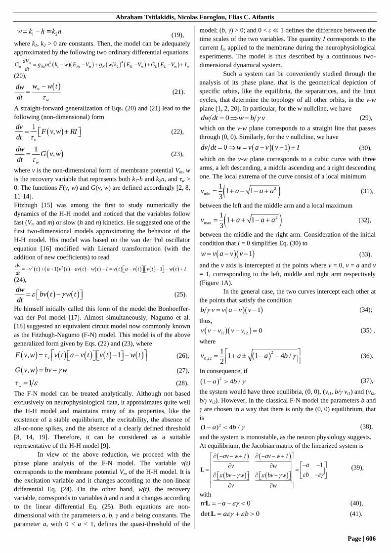
while the stimulus is v(0) = v0. If v0 < a (Figure 1A and 1B, point
1), then dv/dt < 0 and the orbit of the system returns to equilibrium
(Figure 1A and 1B, the orbit in red). If v0 > a (Figure 1A and 1B,
point 2), then dv/dt > 0 and v increases. The orbit of the system
ends at the sole attractor without crossing the middle arm, which
therefore has the character of a quasi-threshold. Instead, it travels
for a longer distance on the plane, which is perceived as an
excitation, before ending up at the equilibrium. Since ε ≪ 1, the
time scales between the two equations are separated, that is dv/dt
≫ dw/dt. Therefore, at all points except the v nullcline, dw/dt is
negligible in relation to dv/dt. In consequence, changes of w are
slower than changes of v and the system is able to generate
relaxation oscillations. The v nullcline is the unique set of points
in which dw/dt is not negligible, because dv/dt = 0.
Figure 1. (A) Phase plane analysis for the F-N model. In dotted-dashed
line is the w nullcline and in dashed line is the v nullcline. In red is an
orbit starting under the threshold. In blue is an orbit starting over the
threshold. (B) The evolution of v in time. The colors and the numbers
correspond to those of (A).
The various stages of an excitation stem from the traits of
the model, revealed by the phase plane analysis, as follows:
Initially, for a < v0 < 1, dv/dt ≫ 0 and dw/dt ≅ 0. The orbit
reaches rapidly the right arm (Figure 1A and 1B, point 3), without
significant deviations from the v axis.
At the right arm the relations dv/dt = 0 and dw/dt > 0 hold.
The orbit moves relatively slowly and close to the right arm until
the local maximum (Figure 1A and 1B, point 4).
At the local maximum the relations dv/dt ≪ 0 and dw/dt ≅ 0
hold. The orbit moves rapidly towards the left and in parallel to
the v axis, as v increases and w remains almost stable.
At last, at the left arm (Figure 1A and 1B, point 5), the
relations dv/dt = 0 and dw/dt < 0 hold. The state returns slowly to
the equilibrium [2, 8].
2.3. Propagation of Nerve Stimulation.
The action potential does not remain stationary after its
generation. The polarity in neighbouring areas of the membrane is
reversed due to the electric perturbation. As a result, the Na+
voltage-sensitive channels in these areas open and the action
potential gradually propagates along the axon. Meanwhile, the
resting potential is restored at areas the stimulation has already
passed through. The H-H and F-N models describe the potential
and the currents locally at a single point of the membrane of the
axon. The cable theory of Lord Kelvin, modified for the
appendages of neurons, is used to study the propagation of the
potential along the axon [21].
The system, regarding a cylindrical axon with cross
section area A and diameter d, is characterized by:
the cytoplasmic longitudinal resistance per unit of length of
the axon
2
4dR r rr
dx A d (45),
where r is the special resistivity of the cytoplasm per unit of
volume;
the resistance of the membrane per unit of length
mm
rr
d (46),
where mr is the special resistivity of the membrane per unit of
surface;
the capacitance of the membrane per unit of length
mm m
dCc dc
dx (47),
where mc is the special capacitance per unit of surface of the
membrane;
the ion current that passes through the thickness of the
membrane per unit of length, considered as a function of the
membrane potential and time
( , )m mion
m
dI f V tj
dx r (48);
the longitudinal (in the x direction) current i along the axon;
and
the propagation velocity c of the electric perturbation.
The cable equation, a special case of the reaction-diffusion
equation, is expressed as 2
2
1m ion
V Vc j
t r x
(49).
The time and space constants of the system are defined
respectively by
c m mr c (50),
mc
r
r (51).
Another expression for the cable equation can be derived from Eq.
(49) using Eqs. (50) and (51). It reads
Abraham Tsitlakidis, Nicolas Foroglou, Elias C. Aifantis
Page | 608
22
2
m mc c m ion
V Vr j
t x
(52),
or, by using Eq. (48) 2
2
2( , )m m
c c m
V Vf V t
t x
(53).
With regard to an active electric flow, as it applies to the axon
according to the H-H model, the nonlinear differential equation
2
2
2, , ,m m
c c m
V Vf V m n h
t x
(54),
along with Eq. (12) hold. In the case of the F-N model, the electric
flow is described by the equation
2
2
2,c c
v vf v w
t x
(55)
together with Eq. (12). For a traveling wave type solution, the
propagation velocity c is constant and its non-dimensional form
c
c
cC
(56)
is independent of time and position. Therefore, C depends only on
the f function and the following equation
2
24
c
c m mm m mm
C C C dc C
c r rc r r r rdc
d d
(57),
holds. In other words, the propagation velocity is dependent on the
properties of the cytoplasm, the membrane and the function that
determines the membrane potential, as well as on the diameter of
the axon [8, 22-26].
2.4. Coupled Electro-Mechanical Models.
The study of the influence of the mechanical deformation on the
electrical behavior of the axon has led to (i) the development of
models on the subcellular nanoscale level, in which the
deformation of the membrane and the channels is observed; and
(ii) the development of models at the micro/macroscale, in which
the deformation of peripheral nerves and the central nervous
system is observed. However, modeling of this coupling on the
level of the neurophysiology and the function of the neuron is
necessary, in order to bridge the gap between the nanoscale and
the micro/macroscale. This task is undertaken here by focusing on
specific generalizations of the F-N model. Analytical techniques,
when feasible, are used for the study of the effects of the
mechanical deformation on the behavior of the model. Numerical
analysis was further performed with the XPPAUT 8.0 software
(Ermentrout GB, 1996-2016) and the R 3.5.2 environment (R Core
Team, 2018) with the deSolve 1.22 package (Sotaert K et al.,
2010). The chosen values for the model parameters a = 0.1, I = 0,
ε = 0.01, b = 1 and γ = 2 fulfill the conditions of the classical F-N
model and have been used elsewhere in the literature as well [2].
2.4.1. Potential caused by deformation. The cellular membrane
has the structure of a liquid crystal bilayer consisting of electrical
dipoles, the lipids. By applying flexural deformation on the
membrane, the orientation of the dipoles changes and electrical
polarization between the surfaces of the membrane is created. As a
result, a potential Vf = f/ε0 2Η is observed, where f is a constant
(flexocoefficient) depending on the composition of the membrane
in lipids and proteins, ε0 the dielectric constant in vacuum and H
the mean curvature of the membrane [27]. In order to understand
how this effect (i.e. flexoelectricity) can influence the generation
and propagation of the action potential, we assume that Vf causes
an equivalent current if and the F-N equations are modified to read
dv/dt = v(a - v)(v - 1) – w + I + if and dw/dt = ε(bv - γw). The new
quantity if can be studied as an independent parameter with a
linear dependence on dv/dt. Related analyses have been done in
[28] by considering I as a bifurcation parameter.
The v nullcline follows the equation w = v(a – v)(v – 1) +
I + if, while the w nullcline obeys the equation w = b/γ v.
Consequently, the phase plane is similar to the phase plane of the
F-N model, with the difference that the v nullcline is transposed
higher by if. The two lines intercept only at the equilibrium. As if
increases, for some value if1, the equilibrium becomes unstable and
a limit cycle appears. For this value of if there is a Hopf
bifurcation and the usual conditions
0tr L (58),
det 0L (59)
hold for the Jacobian matrix of the linearized system at
equilibrium
23 2(1 ) 1v a v a
b
L (60).
Therefore, 23 2(1 ) 0tr v a v a L (61) ,
with solutions
2
1, 2
1(1 1 3 )
3h hv v a a a (62).
It follows that, for the values
1, 2 1, 2 1, 2 1, 2 1, 2( )( 1)f f h h h h h h h h
bi v a v v v
(63),
a Hopf bifurcation appears at the point (v, w) = (vh1,h2, b/γ vh1,h2),
while between the two values the equilibrium is unstable. At the
first bifurcation point the inequality
2 2
1 min
1 1(1 1 3 ) (1 1 )
3 3hv a a a a a a v (64),
holds and the equilibrium lies on the middle arm and slightly on
the right of the local minimum. Similarly, at the second
bifurcation point the inequality
2 2
2 max
1 1(1 1 3 ) (1 1 )
3 3hv a a a a a a v (65)
holds and the equilibrium lies on the middle arm and slightly on
the left of the local maximum.
Numerical analysis of the model was carried out, by
viewing if as the bifurcation parameter. The bifurcation diagram
for the modified F-N model with if as the bifurcation parameter is
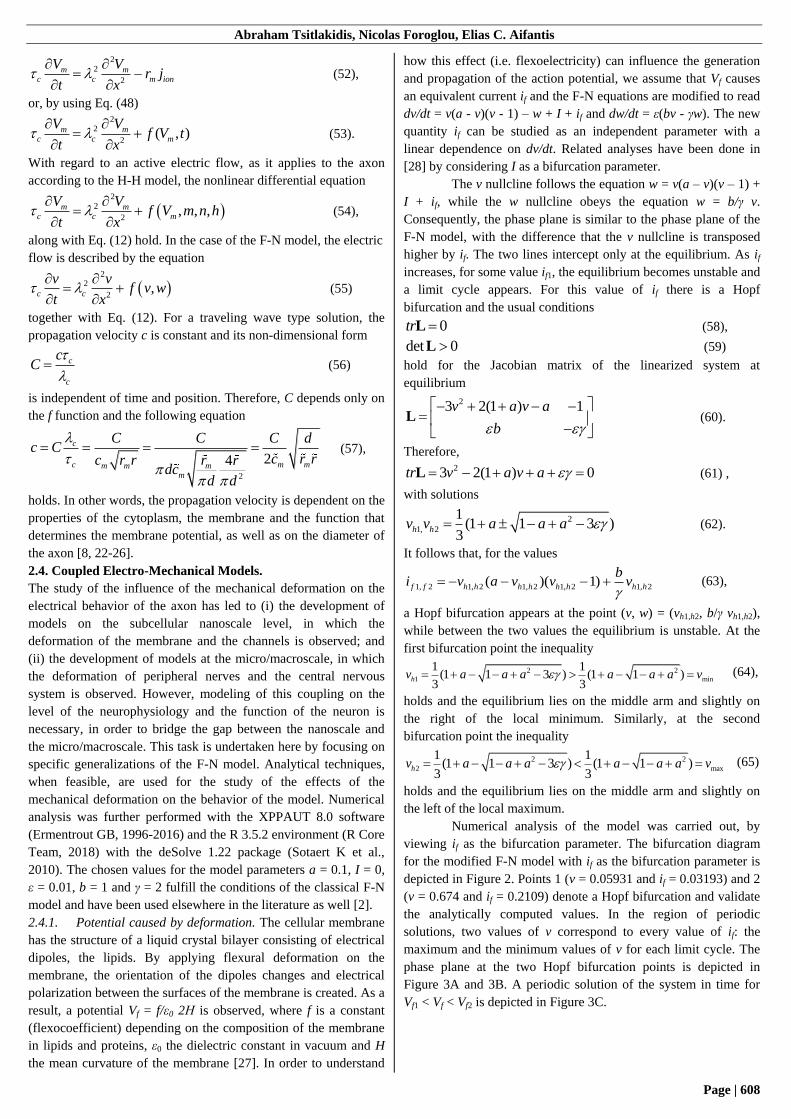
depicted in Figure 2. Points 1 (v = 0.05931 and if = 0.03193) and 2
(v = 0.674 and if = 0.2109) denote a Hopf bifurcation and validate
the analytically computed values. In the region of periodic
solutions, two values of v correspond to every value of if: the
maximum and the minimum values of v for each limit cycle. The
phase plane at the two Hopf bifurcation points is depicted in
Figure 3A and 3B. A periodic solution of the system in time for
Vf1 < Vf < Vf2 is depicted in Figure 3C.
Revisiting the Hodgkin-Huxley and Fitzhugh-Nagumo models of action potential propagation
Page | 609
Figure 2. Bifurcation diagram for the modified F-N model with if as the
bifurcation parameter. The equilibria (red line for asymptotically stable;
black line for unstable equilibria), the stable limit cycles (green lines)
generating periodic solutions, and the Hopf bifurcations (blue circles) are
shown.
Figure 3. (A) Phase plane at the Hopf bifurcation point for if = if1. (B)
Phase plane at the Hopf bifurcation point for if = if2. (C) The evolution of
v in time: periodic solutions of the modified F-N system.
2.4.2. Deformation Effects on the Kinetics of K+ Channels.
(i) K+ channels activated and inactived by tensile deformation:
The discovery of the mechanosensitivity of the ion channels led to
the study of the influence of the mechanical deformation of the
membrane on the kinetics of the electrosensitive K+ channels. In
some types of these channels the application of tensile
deformation (stretch) in the initial stages of their activation, when
they are closed, accelerates their activation. Conversely, when
tensile deformation is applied in the final stages of their activation,
it accelerates their inactivation [29]. Furthermore, these effects
have a dose-depended behavior. Therefore, the changes in the
activity of the channels are greater and faster with increased
membrane deformation, until the membrane ruptures.
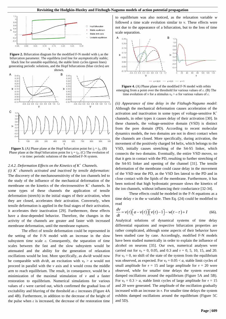
The effect of tensile deformation could be represented in
the setting of the F-N model with an increase in the slow
subsystem time scale ε. Consequently, the separation of time
scales between the fast and the slow subsystem would be
attenuated and the ability for the generation of relaxation
oscillations would be lost. More specifically, as dw/dt would now
be comparable with dv/dt, an excitation with v0 > a would not
proceed in parallel with the v axis and it would cross the middle
arm to reach equilibrium. The result, in consequence, would be a
minimization of the maximal stimulation of v and a faster
restoration to equilibrium. Numerical simulations for various
values of ε were carried out, which confirmed the gradual loss of
excitability and blurring of the threshold as ε increases (Figure 4A
and 4B). Furthermore, in addition to the decrease of the height of
the pulse when ε is increased, the decrease of the restoration time
to equilibrium was also noticed, as the relaxation variable w
followed a time scale evolution similar to v. These effects were
not due to the appearance of a bifurcation, but to the loss of time
scale separation.
Figure 4. (A) Phase plane of the modified F-N model with orbits
emerging from a point over the threshold for various values of ε. (B) The
time evolution of v for a stimulus v0 > α for various values of ε.
(ii) Appearance of time delay in the Fitzhugh-Nagumo model:
Although the mechanical deformation causes acceleration of the
activation and inactivation in some types of voltage-sensitive K+
channels, in other types it causes delay of their activation [30]. In
these channels, the voltage-sensitive domain (VSD) is distinct
from the pore domain (PD). According to recent molecular
dynamics models, the two domains are not in direct contact when
the channels are closed. More specifically, during activation, the
movement of the positively charged S4 helix, which belongs to the
VSD, initially causes stretching of the S4-S5 linker, which
connects the two domains. Eventually, the entire VSD moves, so
that it gets in contact with the PD, resulting to further stretching of
the S4-S5 linker and opening of the channel [31]. The tensile
deformation of the membrane could cause delay in the movement
of the VSD near the PD, as the VSD lies lateral to the PD and in
close contact with the lipids of the membrane. Furthermore, it has
been noticed that high hydrostatic pressure slows the kinetics of
the ion channels, without influencing their conductance [32-34].
These effects could be modeled in the F-N equations as a
time delay τ in the w variable. Then Eq. (24) could be modified to
read
1dv
v t a v t v t w t Idt
(66).
Analytical solutions of dynamical systems of time delay
differential equations and respective bifurcation properties are
rather complicated, although some aspects of their behavior have
been studied case by case. Accordingly, modified F-N models
have been studied numerically in order to explain the influence of
alcohol on neurons [35]. Our own, numerical analyses were
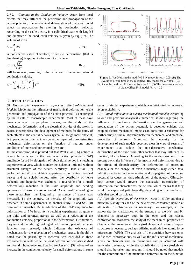
carried out for v0 = 0, 0.05, and 0.3 and τ = 0, 5, 10, 15, and 20.
For v0 = 0, no shift of the state of the system from the equilibrium
was observed, as expected. For v0 = 0.05 < a, stable limit cycles of
small amplitude for τ = 15 and large amplitude for τ = 20 were
observed, while for smaller time delays the system executed
damped oscillations around the equilibrium (Figure 5A and 5B).
For v0 = 0.3 > a, stable limit cycles of large amplitude for τ = 15
and 20 were generated. The amplitude of the oscillation gradually
increased with an increase in τ. For smaller time delays the system
exhibits damped oscillations around the equilibrium (Figure 5C
and 5D).
Abraham Tsitlakidis, Nicolas Foroglou, Elias C. Aifantis
Page | 610
2.4.2. Changes in the Conduction Velocity. Apart from local
effects that may influence the generation and propagation of the
action potential, the mechanical deformation of the axon could
affect its propagation by altering the conduction velocity.
According to the cable theory, in a cylindrical axon with length l
and diameter d the conduction velocity is given by Eq. (57). The
volume of axon
2
4V d l
(67),
is considered stable. Therefore, if tensile deformation (that is
lengthening) is applied to the axon, its diameter
2V
dl
(68),
will be reduced, resulting in the reduction of the action potential
conduction velocity
4
41
2m m
C Vc l
c r r
(69).
Figure 5. (A) Orbits in the modified F-N model for v0 = 0.05. (B) The
evolution of v in time in the modified FHN model for v0 = 0.05. (C)
Orbits in the modified F-N model for v0 = 0.3. (D) The time evolution of v
in the modified F-N model for v0 = 0.3.
3. RESULTS SECTION
(i) Macroscopic experiments supporting Electro-Mechanical
Models: Modeling the influence of mechanical deformation to the
generation and propagation of the action potential is encouraged
by the results of macroscopic experiments. Most of these have
been conducted on peripheral nerves, as the study of the
mechanical deformation and the electrical activity of the axons is
easier. Nevertheless, the development of methods for the study of
such effects in the central nervous system, although more difficult,
is imperative, in order to investigate the impact of non-destructive
mechanical deformation on the function of neurons under
conditions of increased intracranial pressure.
In this connection, it is pointed out that Wall et al. [36] noticed a
reversible reduction in the compound action potential (CAP)
amplitude for a 6 % elongation of rabbit tibial nerves in stretching
experiments in vivo, which is under the ischemia limit and without
structural changes of the nerves. Similarly, Ochs et al. [37]
performed in vitro stretching experiments on canine peroneal
nerves and rat sciatic nerves. After the possibility of nerve
ischemia and hypoxia was excluded, a reversible (for a small
deformation) reduction in the CAP amplitude and beading
appearance of axons were observed. As a result, according to
those researchers, the longitudinal resistance of axons was
increased. To the contrary, an increase of the amplitude was
observed in some experiments. In another study, Li and Shi [38]
observed a reversible 16 % reduction of the CAP amplitude for
deformation of 5 % in ex vivo stretching experiments on guinea-
pig tibial and peroneal nerves, as well as a reduction of the
conduction velocity, proportional to the deformation. Furthermore,
they noticed that, during prolonged deformation, part of the nerve
function was restored, which indicates the existence of
mechanisms for the relaxation of mechanical stress. It should be
noted that the possibility of ischemia was excluded in these
experiments as well, while the local deformation was also studied
and found inhomogeneous. Finally, Stecker et al. [39] observed an
increase in the conduction velocity for small deformations in some
cases of similar experiments, which was attributed to increased
axon excitability.
(ii) Clinical importance of electro-mechanical models: According
to our and previous analytical / numerical studies regarding the
influence of mechanical deformation on the generation and
propagation of the action potential, it becomes evident that
coupled electro-mechanical models can constitute a substrate for
further study of the relationship between mechanical and electrical
properties of neurons. Moreover, the necessity for the
development of such models becomes clear in view of results of
experiments that isolate the non-destructive mechanical
deformation of the axon from other factors that may influence its
function, like ischemia. According to the models studied in the
present work, the influence of the mechanical deformation, due to
the effects of flexoelectricity, the deformation of potassium
channels or the change of axon dimensions, may either have
inhibitory activity on the generation and propagation of the action
potential, or cause the tonic stimulation of the neuron. Clinically,
both effects would prevent the successful transmission of
information that characterizes the neuron, which means that they
would be expressed pathologically, depending on the number of
cells that would participate.
(iii) Possible extensions of the present work: It is obvious that a
meticulous study for each of the new effects considered heirein at
all scales of observation is necessary. For example, at the
nanoscale, the clarification of the structure of the Na+ and K+
channels is necessary both in the open and the closed
conformation. Moreover, the study of the mechanical properties of
channels, the membrane, the cytoskeleton and more complex
structures is necessary, perhaps utilizing methods like atomic force
microscopy (AFM). The analysis of the transition between open
and closed conformations, as well as the influence of mechanical
stress on channels and the membrane can be achieved with
molecular dynamics, while the contribution of the cytoskeleton
should also be taken into account. It should be noted that models
for the contribution of the membrane deformation on the function
Revisiting the Hodgkin-Huxley and Fitzhugh-Nagumo models of action potential propagation
Page | 611
of the channels have already been proposed [40-42] and they
could be extended to take into account the evolution of the action
potential in time. The contribution of the flexoelectricity may be
studied with techniques like patch clamping and AFM. The
coupling of mechanical and electrical properties can initially be
achieved with modifications in the parameters of simpler models,
like the F-N model, which was used in the present work.
Subsequently, it can be studied for more complex models, like the
H-H model, for example with alterations in parameters concerning
the Na+ and K+ channels. The combination of such models with
cable theory would also be interesting, in order to study the impact
of mechanical deformation on the propagation of the action
potential. Moreover, the consideration of models that propose the
companion of the propagated action potential by a mechanical
wave [43] could provide further insight into the interaction
between the electrical and the mechanical behavior of the axon. It
should be underlined that, in the present work, only the
mechanical deformation and the electric properties of the
unmyelinated axon were taken into account. However, their study
in myelinated nerve fibers, as well as in other elements of the
neuron, would also be useful, in order to analyze the impact of the
mechanical deformation on arrays of cells and neural circuits.
Furthermore, the presence of glia and the influence of mechanical
deformation on its glial and neuronal function should be taken into
account. Finally, the study of such effects at the macroscopic scale
would also be important, by considering the change of the
macroscopic conductance for various types of mechanical
deformation.
4. CONCLUSIONS
The generation and propagation of the action potential along the
axon are described adequately by neurophysiological models like
the H-H and F-N models and the cable theory. Such models are
used by neuroscientists to approach the function of the neuron and,
by extension, neural circuits and the nervous system as a whole.
The mechanical properties of the nervous tissue have also been
studied, to a degree, mainly in the context of traumatic injury.
However, the influence of the mechanical deformation on the
electric behavior of the nervous system has not been studied
thoroughly, with the exception of experiments on subcellular
structures and some macroscopic experiments on peripheral
nerves. Herein, we presented electromechanical models for the
nerve stimulation propagation by taking into account the effects of
flexoelectricity, as well as axon, cellular membrane and potassium
channels deformation. It becomes evident that the study of the
interaction of the electric and the mechanical properties of the
neural tissue is a field with notably useful clinical applications,
particular difficulties in its study, but also wide perspectives for
further research.
5. REFERENCES
[1] S. L. Campbell, R. Haberman, Introduction to Differential
Equations: with Dynamical Systems. Princeton University Press,
2008.
[2] E. M. Izhikevich, Dynamical Systems in Neuroscience. The
Geometry of Excitability and Bursting. The MIT Press, 2007.
[3] A. L. Hodgkin, A. F. Huxley, A quantitative description of
membrane current and its application to conduction and
excitation in nerve. J. Physiol., 117, 4, 500-544, 1952,
https://doi.org/10.1113/jphysiol.1952.sp004764.
[4] A. L. Hodgkin, A. F. Huxley, Currents carried by sodium
and potassium ions through the membrane of the giant axon of
Loligo. J. Physiol., 116, 4, 449-472, 1952,
https://doi.org/10.1113/jphysiol.1952.sp004717.
[5] A. L. Hodgkin, A. F. Huxley, The components of
membrane conductance in the giant axon of Loligo. J. Physiol.,
116, 4, 473-496, 1952,
https://doi.org/10.1113/jphysiol.1952.sp004718.
[6] A. L. Hodgkin, A. F. Huxley, The dual effect of membrane
potential on sodium conductance in the giant axon of Loligo. J.
Physiol., 116, 4, 497-506, 1952,
https://doi.org/10.1113/jphysiol.1952.sp004719.
[7] A. L. Hodgkin, A. F. Huxley, Propagation of electrical
signals along giant nerve fibres. Proc. R. Soc. Lond. B. Biol. Sci.,
140, 177-183, 1952, https://doi.org/10.1098/rspb.1952.0054.
[8] J. Keener, J. Sneyd, Mathematical Physiology, 2nd ed.
Springer Science+Business Media, LLC, 2009,
https://doi.org/10.1007/978-0-387-75847-3.
[9] J. D. Murray, Mathematical Biology. I. An Introduction,
3rd ed. Springer Science+Business Media, LLC, 2002,
https://doi.org/10.1007/b98868.
[10] V. I. Krinskii, Y. M. Kokoz, Analysis of equations of
excitable membranes - I. Reduction of the Hodgkin-Huxley
equations to a second order system. Biofizika, 18, 506-511, 1973
[11] W. Gerstner, W. Kistler, Spiking Neuron Models: Single
Neurons, Populations, Plasticity. Cambridge University Press,
2002.
[12] L. F. Abbott, T. Kepler, B. Model Neurons: From
Hodgkin-Huxley to Hopfield. In Statistical Mechanics of Neural
Networks: Proceedings of the XIth Sitges Conference, Sitges,
Barcelona, Spain, 3-7 June 1990. Garrido L, Ed. Springer, pp. 5-
18, 1990, https://doi.org/10.1007/3-540-53267-6.
[13] B. Alberts, A. Johnson, J. Lewis, M. Raff, K. Roberts, P.
Walter, Molecular Biology of the Cell, 5th ed. Garland Science,
2008.
[14] W. Gerstner, W. M. Kistler, R. Naud, L. Paninski,
Neuronal Dynamics. From Single Neurons to Networks and
Models of Cognition. Cambridge University Press, 2014.
[15] R. Fitzhugh, Mathematical models of threshold phenomena
in the nerve membrane. Bull. Math. Biophys., 17, 257-278, 1955,
https://doi.org/10.1007/BF02477753.
[16] B. van der Pol, A theory of the amplitude of free and forced
triode vibrations. Radio Review, 1, 701-710, 754-762, 1920.
[17] R. Fitzhugh, Impulses and physiological states in
theoretical models of nerve membrane. Biophys. J., 1, 445-466,
1961, https://doi.org/10.1016/s0006-3495(61)86902-6.
[18] J. Nagumo, S. Arimoto, S. Yoshizawa, An active pulse
transmission line simulating nerve axon. Proc. IRE, 50, 10,
2061-2070, 1962,
https://doi.org/10.1109/JRPROC.1962.288235.
[19] J. Rinzel, Electrical excitability of cells, theory and
experiment: review of the Hodgkin-Huxley foundation and an
update. Bull. Math. Biol., 52, 1/2, 5-23, 1990,
https://doi.org/10.1016/S0092-8240(05)80003-5.
[20] S. H. Strogatz, Nonlinear Dynamics and Chaos. With
Applications to Physics, Biology, Chemistry, and Engineering.
Perseus Books Publishing, 1994.
Abraham Tsitlakidis, Nicolas Foroglou, Elias C. Aifantis
Page | 612
[21] W. Thomson, On the theory of the electric telegraph. Proc.
R. Soc., 7, 382, 1855.
[22] A. Scott, Neuroscience: A Mathematical Primer. Springer-
Verlag, 2002, https://doi.org/10.1007/b98897.
[23] K. S. Cole, A. L. Hodgkin, Membrane and protoplasm
resistance in the squid giant axon. J. Gen. Physiol., 22, 671-687,
1939, https://doi.org/10.1085/jgp.22.5.671.
[24] W. Rall, Branching dendritic trees and motoneuron
membrane resistivity. Exp. Neurol., 1, 5, 491-527, 1959,
https://doi.org/10.1016/0014-4886(59)90046-9.
[25] A. L. Hodgkin, W. A. Rushton, The electrical constants of
a crustacean nerve fibre. Proc. R. Soc. Lond. B. Biol. Sci., 133,
873, 444-479, 1946, https://doi.org/10.1098/rspb.1946.0024.
[26] J. D. Murray, Mathematical Biology. II. Spatial Models and
Biomedical Applications, 3rd ed. Springer Science+Business
Media, LLC, 2003, https://doi.org/10.1007/b98869.
[27] A. G. Petrov, F. Sachs, Flexoelectricity and elasticity of
asymmetric biomembranes. Phys. Rev. E Stat. Nonlin. Soft
Matter Phys., 65, 021905, 2002,
https://doi.org/10.1103/PhysRevE.65.021905.
[28] W. C. Troy, Bifurcation phenomena in Fitzhugh's nerve
conduction equations. J. Math. Anal. Appl., 54, 3, 678-690,
1976, https://doi.org/10.1016/0022-247X(76)90187-6.
[29] C. X. Gu, P. F. Juranka, C. E. Morris, Stretch-activation
and stretch-inactivation of Shaker-IR, a voltage-gated K+
channel. Biophys. J., 80, 2678-2693, 2001,
https://doi.org/10.1016/S0006-3495(01)76237-6.
[30] U. Laitko, P. F. Juranka, C. E. Morris, Membrane stretch
slows the concerted step prior to opening in a Kv channel. J.
Gen. Physiol., 127, 6, 687-701, 2006,
https://doi.org/10.1085/jgp.200509394.
[31] M. O. Jensen, V. Jogini, D. W. Borhani, A. E. Leffler, R.
O. Dror, D. E. Shaw, Mechanism of voltage gating in potassium
channels. Science, 336, 229-233, 2012,
https://doi.org/10.1126/science.1216533.
[32] A. G. MacDonald, Ion channels under high pressure.
Comp. Biochem. Physiol. A Mol. Integr. Physiol., 131, 3, 587-
593, 2002, https://doi.org/10.1016/S1095-6433(01)00510-4.
[33] F. Conti, R. Fioravanti, J. R. Segal, W. Stuhmer, Pressure
dependence of the potassium currents of squid giant axon. J.
Membr. Biol., 69, 35-40, 1982,
https://doi.org/10.1007/BF01871239.
[34] F. Conti, R. Fioravanti, J. R. Segal, W. Stuhmer, Pressure
dependence of the sodium currents of squid giant axon. J.
Membr. Biol., 69, 23-34, 1982,
https://doi.org/10.1007/BF01871238.
[35] R. S. Franca, I. E. Prendergast, E.-S. Sanchez, M. Sanchez,
F. Berezovsky,The Role of Time Delay in the Fitzhugh-Nagumo
Equations: The Impact of Alcohol on Neuron Firing. Technical
Report, Report No. BU-1577-M, 2001
[36] E. J. Wall, J. Massie, B., M. K. Kwan, B. L. Rydevik, R. R.
Myers, S. R. Garfin, Experimental stretch neuropathy. Changes
in nerve conduction under tension. J. Bone Joint Surg. Br., 74,
126-129, 1992
[37] S. Ochs, R. Pourmand, K. Si, R. N. Friedman, Stretch of
mammalian nerve in vitro: effect on compound action potentials.
J. Peripher. Nerv. Syst., 5, 4, 227-235, 2000,
https://doi.org/10.1111/j.1529-8027.2000.00025.x.
[38] J. Li, R. Shi, Stretch-induced nerve conduction deficits in
guinea pig ex vivo nerve. J. Biomech., 40, 3, 569-578, 2007,
https://doi.org/10.1016/j.jbiomech.2006.02.009.
[39] M. M. Stecker, K. Baylor, J. Wolfe, M. Stevenson, Acute
nerve stretch and the compound motor action potential. J.
Brachial Plex. Peripher. Nerve. Inj., 6, 1, 4, 2011,
https://doi.org/10.1186/1749-7221-6-4.
[40] D. Reeves, T. Ursell, P. Sens, J. Kondev, R. Phillips,
Membrane mechanics as a probe of ion-channel gating
mechanisms. Phys. Rev. E, 78, 041901, 2008,
https://doi.org/10.1103/PhysRevE.78.041901.
[41] R. Phillips, T. Ursell, P. Wiggins, P. Sens, Emerging roles
for lipids in shaping membrane-protein function. Nature, 459,
7245, 379-385, 2009, https://doi.org/10.1038/nature08147.
[42] T. Ursell, K. C. Huang, E. Peterson, R. Phillips,
Cooperative gating and spatial organization of membrane
proteins through elastic interactions. PLoS Comput. Biol., 3, 5,
e81, 2007, https://doi.org/10.1371/journal.pcbi.0030081.
[43] A. El Hady, B. B. Machta, Mechanical surface waves
accompany action potential propagation. Nat. Commun., 6, 6697,
2015, https://doi.org/10.1038/ncomms7697.
6. ACKNOWLEDGEMENTS
This work is, in part, a result of the first author’s Master Thesis at the Lab of Mechanics and Materials (LMM).
© 2019 by the authors. This article is an open access article distributed under the terms and conditions of the
Creative Commons Attribution (CC BY) license (http://creativecommons.org/licenses/by/4.0/).


![Learning Dynamical Systems Requires Rethinking Generalization · 2020. 11. 16. · FitzHugh–Nagumo (FHN) [FitzHugh, 1961] and, independently, [Nagumo et al., 1962] derived the equations](https://img.pdfslide.net/doc/110x75/60d38b7ec48d57609971fbdd/learning-dynamical-systems-requires-rethinking-generalization-2020-11-16-fitzhughanagumo.jpg)