Embed Size (px)
Citation preview

Cell size and shape are determined by developmental programmes that involve changes in gene transcrip-tion1. In proliferating cells, cell size regulation depends on growth rates and cell cycle progression and has been most thoroughly studied in unicellular organisms. How cell size feeds back to transcription remains largely unknown, but it may involve, among other factors, the titration of DNA-binding proteins1. By contrast, vertebrate cell volume regulation2–4, as discussed here, involves the rapid adjustment (within minutes to a few hours) of cellular volume in response to external chal-lenges or during the execution of cellular functions. Cell volume regulation is of fundamental importance for almost every vertebrate cell. Cell swelling, shrink-age and regulatory volume adjustments generally occur without significant exocytic insertion or endocytic retrieval of surface membrane. As cells are not ideal spheres and often have membrane wrinkles, they have enough plasma membrane reserve to undergo substan-tial swelling before bursting5,6. Acute volume regulation involves the net transport of potassium (K+), sodium (Na+), chloride (Cl–) and organic osmolytes across the plasma membrane (FIG. 1). This transport occurs through various ion channels and transporters (BOX 1) and is followed by osmotically driven water transport, often through specialized water channels (BOX 2). As channels and transporters recruited for cell volume regulation almost always have additional important roles, cell vol-ume regulation is inextricably linked to other cellular functions (BOX 3).
While also addressing sensors of cell volume and subsequent signal transduction cascades, this Review
focuses on effectors of cell volume regulation4 — that is, proteins responsible for the transport of osmolytes across the plasma membrane — in mammals and dis-cusses their involvement in short-term cell volume regu-lation and other cellular processes. Particular attention is paid to volume-regulated anion channels (VRACs), which are key players in vertebrate cell volume regula-tion that not only transport Cl−, but also various organic osmolytes. VRACs have been studied biophysically and physiologically for decades7,8, but their molecular com-position (of LRRC8 proteins) and the ability of particular LRRC8 heteromers to transport certain metabolites and drugs have only been established recently9–11.
Necessity of volume regulationBiochemical reactions and equilibria depend on the concentrations of the reaction partners involved. Consequently, cells must avoid short-term changes in volume that will globally affect the concentrations of cellular constituents and thereby interfere with cellular functions. For instance, molecular crowding and ionic imbalance caused by cell shrinkage slows diffusion and affects vesicle traffic12. Furthermore, changes to intra cellular ion concentrations resulting from volume changes can affect cellular excitability and signalling by altering ion gradients across the plasma membrane. Cell swelling is dangerous as it may — in extreme cases — lead to cell bursting.
When vertebrate cells are exposed to a hypo- or hyper-tonic external medium, they swell or shrink, respectively, because they lack rigid extracellular cell walls (as found, for example, in plants and bacteria), and osmotic
Leibniz-Institut für Molekulare Pharmakologie (FMP) and Max-Delbrück-Centrum für Molekulare Medizin (MDC), Robert-Rössle-Strasse 10, 13125 Berlin, [email protected]
doi:10.1038/nrm.2016.29Published online 1 April 2016
OsmolytesDissolved substances that contribute to osmolarity; in biology, the term mainly refers to substances playing a significant part in osmoregulation. Osmolytes can be electrically charged or neutral, as only the concentration of particles matters.
Hypo- or hypertonicCondition in which the concentration of osmolytes that are not freely membrane permeable is lower or higher, respectively, on the reference side of a membrane, compared with the other side. This concentration difference creates an osmotic pressure.
VRACs and other ion channels and transporters in the regulation of cell volume and beyondThomas J. Jentsch
Abstract | Cells need to regulate their volume to counteract osmotic swelling or shrinkage, as well as during cell division, growth, migration and cell death. Mammalian cells adjust their volume by transporting potassium, sodium, chloride and small organic osmolytes using plasma membrane channels and transporters. This generates osmotic gradients, which drive water in and out of cells. Key players in this process are volume-regulated anion channels (VRACs), the composition of which has recently been identified and shown to encompass LRRC8 heteromers. VRACs also transport metabolites and drugs and function in extracellular signal transduction, apoptosis and anticancer drug resistance.
R E V I E W S
NATURE REVIEWS | MOLECULAR CELL BIOLOGY VOLUME 17 | MAY 2016 | 293
© 2016
Macmillan
Publishers
Limited.
All
rights
reserved.

OsmolarityConcentration of solute particles per kg of solution, irrespective of their ability to freely cross a given membrane.
gradients drive water across their outer membrane. Even disregarding differences in volume-regulatory processes, the rate of osmotic cell swelling or shrinkage can vary widely between cell types because of differences in the water permeability of their plasma membranes. Biological lipid bilayers have finite water permeability, which can be markedly increased by dedica ted water channels termed aquaporins13,14 (BOX 2). Although most mammalian cells are protected from large exter-nal osmotic disturbances, because the extracellular space of these tissue-embedded cells is controlled by systemic osmoregulation15, some cells — such as those in the gastrointestinal tract or distal kidney tubules — may be exposed to substantial changes in extracellular
osmolarity. Moreover, with a pathological decrease in extracellular Na+ levels (known as hyponatraemia), extracellular osmolarity may be reduced to a degree that causes cerebral oedema. Osmotic challenges may also originate in the cytoplasm, for instance, during the breakdown of macromolecules into a large number of (hence osmotically more active) building blocks or during glucose stimulation of pancreatic β-cells16.
In almost all cells, osmotic swelling or shrinkage is followed by cell-intrinsic regulatory processes that are called regulatory volume decrease (RVD) or regulatory volume increase (RVI), respectively, which tend to restore the initial cell volume (FIG. 1). RVD and RVI are generally studied (see Supplementary information S1 (box)) using
Nature Reviews | Molecular Cell Biology
TAUTNKCC KCC
VRAC
K+ channels
NHE
AE
HICC
H2O H
2O H
2O
HCO3
–
AE
Na+
Na+
Na+
Na+, K+
K+, Na+
Taurine
Organic osmolytes
CI–
CI–
CI–
CI–
H+
Ca2+-activatedCI– channels
Na+
K+
H+
CI–
HCO3
–
1455
10–7.4
12025
10–35 to –80 mV
140
10–7.2
5–4020
0 mV
Typical extra- and intracellularion concentrations (mM, or M for H+)and voltages in mammals
Extracellular Intracellular
K+
K+
Figure 1 | Plasma membrane channels and transporters involved in cell volume regulation. Cells regulate their volume by adjusting the levels of intracellular osmolytes, including the inorganic ions Cl−, K+ and Na+, and small organic compounds such as taurine or amino acids. This generates osmotic gradients, which then drive water across the membrane by diffusion through the bilayer or, more efficiently, through aquaporin water channels (BOX 2). Shrunken cells (left) recover their volume by regulatory volume increase (RVI). Depending on the cell type, this mainly involves the parallel activation of Na+/H+ exchangers (NHEs; mainly NHE1) and Cl−/HCO3
− anion exchangers (AEs; mainly AE2), or the stimulation of Na+/K+/2Cl− cotransporters (NKCCs; mainly NKCC1). Both processes increase cytoplasmic Na+ and Cl− concentrations. In some cells, the activation of hypertonicity-activated, non-selective cation channels (HICCs), for which molecular correlates have not been firmly established, can also be involved and mainly leads to Na+ influx, which requires a parallel conductance for electroneutrality of overall transport (not shown). In all three cases, the Na+/K+-ATPase (not shown) will gradually replace intracellular Na+ with K+. Na+-coupled uptake of taurine by Na+- and Cl−-dependent taurine transporter TAUT (also known as SLC6A6) rather contributes to long-term uptake of this organic osmolyte during RVI. In regulatory volume decrease (RVD; right), cells recover their volume by extruding K+, Cl− and organic osmolytes like taurine. Loss of KCl may occur through K+/Cl− cotransporters (KCCs; mainly KCC1, 3 or 4) or through parallel Cl− and K+ channels. Key players in RVD are the ubiquitously expressed volume-regulated anion channels (VRACs), which are LRRC8 heteromers. These, depending on subunit composition, conduct not only Cl−, but also organic osmolytes. In some cells, in which swelling induces a rise in intracellular Ca2+ concentration, Cl− may also exit through various Ca2+-activated Cl− channels. Channel-mediated Cl− exit must be electrically balanced by K+ efflux, which can occur through a range of constitutively open or swelling-activated K+ channels. Inset: typical extra- and intracellular concentrations of ions involved in mammalian volume regulation. Note that the direction of passive (channel-mediated) ion transport depends not only on the concentration difference, but also on the voltage across the plasma membrane (the electrochemical gradient as given by the Nernst equation; see glossary term ‘Electrochemical gradients’). The intracellular Cl− concentration shows substantial variation between cell types. In most cells, with the notable exception of most adult neurons, its electrochemical potential will favour Cl− exit (through VRACs or other Cl− channels).
R E V I E W S
294 | MAY 2016 | VOLUME 17 www.nature.com/nrm
© 2016
Macmillan
Publishers
Limited.
All
rights
reserved. ©
2016
Macmillan
Publishers
Limited.
All
rights
reserved.

DepolarizationA change in the resting voltage of cells (-40 to -80 mV inside versus outside) to more positive values. A change to more negative potentials is called hyperpolarization.
large non- physiological changes in osmolarity. Under most physiological conditions, however, cell volume regulation occurs in the absence of large osmotic gradi-ents or can be initiated without any osmotic imbalance, a process termed near-isotonic volume regulation. Cell volume changes, or ‘side-products’ of volume regulation such as changes in the voltage across the plasma mem-brane, may influence other cellular processes (BOX 3). For instance, hepatocyte metabolism can be influenced by cell swelling or shrinkage17, and glucose-induced swell-ing of β-cells may stimulate insulin secretion, because activation of VRACs may lead to Cl− efflux and thereby to depolarization of the cell18 (BOX 3). In these cases, changes in cell volume may be signals for separate cellular events.
Even in the absence of osmotic challenges, cells must regulate their volume, for instance, during cell division and growth2. Volume-regulatory mechanisms may also play a part in cell migration, with local intracellular
osmolarity and volume being increased at the leading edge and decreased at the trailing edge19,20. The passage of salt and water across apical and basolateral mem-branes must be closely balanced during transepithelial transport, and this may involve volume-sensitive ion transporters. Cells can also change their cell volume in response to external stimuli. For instance, hepato-cytes shrink or swell upon stimulation by glucagon and insulin, respectively21.
Finally, cell volume changes during cell death, with necrosis being generally associated with cell swelling and apoptosis with cell shrinkage. Cell shrinkage precedes other apoptotic changes22–24 in most mammalian cells, with only a few exceptions25. Inhibition of this apoptotic volume decrease (AVD) by extracellular hypertonicity26, various K+ channel blockers22,27, inhibitors of VRACs22,28 or genetic disruption of underlying LRRC8 proteins11 suppressed drug-induced apoptosis. These findings sug-gest a role for AVD in the progression of apoptosis and in drug resistance in cancer11,22–24.
Cellular osmolytesCells regulate their volume by adjusting the intra cellular concentrations of dissolved particles (osmolytes) that secondarily drive water flux across their membrane (FIG. 1). Osmolytes can be electrically charged (such as Cl−, K+ or glutamate−) or neutral (such as myo- inositol or taurine, a ‘zwitterion’ that carries both a positive and a negative charge at physiological pH). To exert signifi cant osmotic pressure, osmolytes must be poorly membrane- permeable and should be present at suffi-ciently high concentrations. Among inorganic ions, only K+ (with intracellular concentration ([K+]i)~140 mM), Na+ ([Na+]i~10 mM) and Cl– ([Cl−]i~5–40 mM, varying with cell type) are important osmolytes in vertebrate vol-ume regulation (see FIG. 1, inset). Although intra cellular cytosolic bicarbonate concentrations ([HCO3
−]i) are sufficiently high (~20 mM), the equilibrium between [HCO3
−]i and [H+]i at a given, constant concentration of CO2 (~5%) implies that large changes in [HCO3
−]i during volume regulation would lead to sizeable, pos-sibly harmful, changes in intracellular pH (pHi). Hence, HCO3
− is poorly suited as a volume-regulatory osmolyte. An important consideration with charged osmolytes is the requirement for the maintenance of overall electro-neutrality of transport. In RVD, for instance, the efflux of Cl− and K+ must be almost exactly matched, either by directly coupled, electroneutral transport through K+/Cl− cotransporters or by indirect electrical coupling of K+ and Cl− channels. Even very small imbalances between the transport of positive and negative charges lead to large changes in the plasma membrane voltage, which must be maintained within a maximal range of a few tens of mVs.
Besides inorganic ions, small organic compounds are key cellular osmolytes. These include taurine, sorbitol, betaine, myo-inositol and glycerophosphocholine. The concentrations of these compounds seem to be suffi cient for their function as osmolytes. For instance, tissue concentrations of taurine typically range from 3 to 40 mM29. Particularly high concentrations of organic
Box 1 | Ion transporting proteins
Ion transporting proteins are membrane-embedded proteins or protein complexes that span the lipid bilayer several times to create an ion translocation pathway. Some transporters function as monomers, whereas others are multimers of identical, homologous or non-homologous proteins. They may form a single translocation pathway, but some may have two or more translocation pathways (for instance, one per subunit in the dimeric CLC chloride channels or in tetrameric water channels (BOX 2)).
Functionally, ion transporting proteins can be classified into pumps, transporters and channels. Pumps can transport fixed numbers of ions against their electrochemical gradients through conformational changes driven by direct input of metabolic energy, mostly ATP hydrolysis. This type of transport is called ‘active’. Examples include H+-ATPases that transport protons, and the Na+/K+-ATPase (which extrudes 3 Na+ ions and imports 2 K+ ions in one transport cycle) that is a major determinant of all other ion plasma membrane ion gradients in mammalian cells. Ion transporters are generally defined as also requiring a conformational change per translocation of a fixed number of ions, but they operate without direct input of metabolic energy. The transporter subclasses are cotransporters (symports) that couple the movement of two or more ion species in the same direction (for instance, KCC K+/Cl+ cotransporters or NKCC Na+/K+/2Cl− cotransporters), or exchangers (antiports) that obligatorily exchange an ion from one side of the membrane for another one on the other side (such as Na+/H+ exchangers (NHEs) or Cl−/HCO3
− anion exchangers (AEs)). Strict coupling means that the gradient of one ion can drive the other ion against its gradients (termed ‘secondary active’ transport). Mammalian cells often use the large inwardly directed Na+ gradient to drive the transport of other ions or organic substrates. The charge of transported ions and the coupling ratio determines whether their transport is ‘electroneutral’ (meaning that it does not generate electrical currents; examples are KCC and NKCC cotransporters and AEs), or ‘electrogenic’ (such as CLC 2Cl−/H+ exchangers or NCX 3Na+/Ca2+ exchangers). Ion channels, by contrast, have a conduction pathway (that is, a ‘pore’) for the passive diffusion of ions along their electrochemical gradient. Their transport is intrinsically electrogenic. Channels are generally ‘gated’ (opened and closed) by a conformational change. Once a channel is open, a non-fixed, large number of ions can pass through. Ion channels generally have much higher transport rates than ion transporters or pumps.
Nature Reviews | Molecular Cell Biology
Cotransporter(symport)
Exchanger(antiport)
ChannelExtra-cellular
Cytosol
ATP ADP + Pi
Pump
Active transport Passive diffusionSecondary active transport
R E V I E W S
NATURE REVIEWS | MOLECULAR CELL BIOLOGY VOLUME 17 | MAY 2016 | 295
© 2016
Macmillan
Publishers
Limited.
All
rights
reserved. ©
2016
Macmillan
Publishers
Limited.
All
rights
reserved.

Electrochemical gradientsThe passive, diffusive transport of an ion across a membrane through a channel is driven not only by the concentration difference of that ion, but also by the voltage across that membrane. The driving force is given by the difference in electrochemical potential according to the Nernst equation, for example, for a singly charged anion A− by EA = (RT/F)*ln([A−]i / [A−]o), with R being the gas constant, T the absolute temperature, F Faraday’s constant, and [A−]i the intracellular and [A−]o the extracellular concentration of the anion.
Donnan effectsEffects on the voltage and concentration differences of charged particles across a semipermeable membrane, caused by the impermeability of the membrane to one ionic species.
osmolytes are found in kidney medulla30, which needs to be hyperosmolar to concentrate urine. After exposure to hypertonicity causing cell shrinkage and an initial, rapid RVI response based on ionic fluxes, these organic osmolytes accumulate in cells, mainly through Na+ gradient- driven transporters, which are regulated by the hypertonicity-activated transcription factor TONEBP (also known as NFAT5 or OREBP). The gradual substi-tution of the accumulated ions by biologically rather inert organic osmolytes allows intracellular ion concentrations to slowly return to near-normal levels, thereby avoiding the harmful effects of high intracellular ionic strength and altered plasma membrane ion gradients. Whereas organic osmolytes play a minor part in short-term RVI because of their low uptake rates, some organic osmolytes like taurine or glutamate play a part in short-term RVD, because they can be rapidly released by VRACs (FIG. 1).
Mechanisms of cell volume regulationShort-term regulatory volume changes (RVD and RVI) require a fast reversal of the osmotic gradients that led to cell swelling or shrinkage, respectively. This is accom-plished by the activation of plasma membrane channels and transporters (BOX 1) that transport Na+, K+, Cl− or (in RVD) organic osmolytes. The net transport by both channels and transporters is determined by the electro-chemical gradients of their substrates and does not involve direct expenditure of metabolic energy. However, ion pumps (mainly the Na+/K+-ATPase) are ultimately responsible for the generation of cellular ion gradients. Inhibition of the Na+/K+-ATPase — for example, during anoxia — eventually leads to a dissipation of ion gradi-ents and to cell swelling owing to Donnan effects. None of the channels or transporters that are involved in RVD or RVI is exclusively, and often not even mainly, dedicated to cell volume regulation. Given the molecular diversity of ion channels and transporters, there is a consider able redundancy of volume-regulatory effectors (TABLE 1).
Different cells may use different sets of ion transport proteins for regulating their volume. This section will describe the major mechanisms and principles governing volume regulation by these membrane effectors.
Regulatory volume increase. RVI mainly involves the cellular uptake of Na+, K+ and Cl−. Excess intra-cellular Na+ is secondarily exchanged for K+ by the Na+/K+-ATPase. Although large electrochemical gradi-ents favour Na+ entry, which could secondarily drive Cl− influx in response to cellular depolarization, Na+- and Cl−-selective channels are generally not used in RVI. However, hypertonicity-induced cation channels (HICCs) that conduct both Na+ and K+ can be involved in RVI in some cells31,32. The molecular identity of these channels has not yet been firmly established.
RVI probably mainly involves Na+/K+/2Cl− (NKCC) cotransporters or a parallel activation of Na+/H+ and Cl−/HCO3
− exchangers (FIG. 1; TABLE 1). NKCCs couple the movement of one Na+, one K+ and two Cl− ions in the same direction and hence do not generate cur-rents (that is, they are ‘electroneutral’). In this case, the inwardly directed Na+ and Cl− gradients of mammalian cells drive K+ uptake against its considerable outward gradient. Mammals have two genes encoding NKCC proteins. NKCC1 (also known as SLC12A2) is almost ubiquitously expressed, whereas NKCC2 (SLC12A1) is mainly found in the kidney, where it mediates salt reabsorption. NKCC1 is activated by cell shrinkage in a phosphorylation- dependent manner33 (see below). Apart from mediating RVI, it has basal transport activity that leads to the accumulation of intracellular Cl−. This role of NKCC1 is shared with Cl−/HCO3
− exchangers and is important for VRAC-mediated efflux of Cl− in RVD.
Other major players in RVI are Na+/H+ exchangers of the NHE (SLC9A) family34,35 (FIG. 1; TABLE 1). Mammals express nine different NHEs, which reside in the plasma membrane or in intracellular organelles. Consistent with its role in pHi regulation, the widely expressed NHE1 is activated by acidic pHi (REF. 36). Cell shrinkage activates NHE1 by shifting its activation curve towards more alka-line pHi and by relieving inhibition by extracellular Na+ (REF. 37). As the vast majority of H+ ions that are extruded by NHE1 are derived from intracellular buffers, proton efflux per se is osmotically irrelevant and NHE activation increases cellular osmolarity through Na+ influx. In the presence of CO2, the cytoplasmic alkalinization caused by NHE1 activation leads to slow HCO3
− accumulation, which is counteracted by Cl−/HCO3
− exchangers, mostly the widely expressed anion exchange 2 (AE2; also known as SLC4A2) isoform. AE2 is activated by intracellular alkalinization38 and, with physiological ion gradients, mediates Cl− influx and HCO3
− efflux (FIG. 1; TABLE 1). The indirect coupling of NHEs and anion exchangers by pHi results in electroneutral uptake of NaCl with only minor effects on pHi or [HCO3
−]i.
Regulatory volume decrease. RVD involves the exit of K+ and Cl− through electroneutral K+/Cl− cotransport-ers or through parallel K+ and Cl− channels (promi-nently including VRACs), as well as an efflux of organic
Box 2 | Water flux across membranes: the role of aquaporins
Although water can diffuse across lipid bilayers without the need for water-transporting proteins, dedicated water channels (aquaporins) can, depending on the cell type, drastically enhance (by 5–50-fold) the water permeability of biological membranes13,14. As cell volume regulation involves osmotically driven water transport across the plasma membrane, expression of aquaporins can significantly affect the speed of cell volume regulation (and, of course, the rate at which cells swell or shrink upon changes in extracellular osmolarity; see, for example, REF. 136). There are 13 aquaporin (AQP) genes in humans, of which many encode proteins (including AQP1, 2, 4, 5 and 8) that function as bidirectional water channels13. Some aquaporins can also transport glycerol and possibly other small molecules, owing to their wider channel pore. Aquaporin channels function as tetramers of ~30 kDa AQP proteins that have six membrane-spanning domains and two short helical segments that dip into the pore from either side, as revealed by crystal structures. Each of the four subunits has its own pore. Whereas aquaporins have a major, well-established role in osmotically driven water transport, it should be mentioned that some transporters, including Na+/K+/2Cl− cotransporter 1 (NKCC1) or K+/Cl− cotransporter 4 (KCC4) have been suggested to cotransport water together with ions137, and that one report suggested that volume-regulated anion channels (VRACs) may also be water-permeable to some degree138. Therefore, osmolyte transporters involved in volume regulation might also, to a minor degree, participate in facilitating water transport across membranes, and thereby further contribute to cell volume changes.
R E V I E W S
296 | MAY 2016 | VOLUME 17 www.nature.com/nrm
© 2016
Macmillan
Publishers
Limited.
All
rights
reserved. ©
2016
Macmillan
Publishers
Limited.
All
rights
reserved.

GABA (γ-aminobutyric acid) type A receptor(GABAA receptor). A pentameric anion channel that is opened by the binding of GABA. Channels of this type mediate fast synaptic inhibition in the nervous system by allowing a passive influx of Cl− which hyperpolarizes the membrane. This requires a low intracellular Cl− concentration, as found in most adult neurons.
Glycine receptorA pentameric anion channel that inhibits neuronal excitability by mediating a hyperpolarizing Cl− influx upon extracellular binding of the amino acid and neurotransmitter glycine.
ConductanceElectrical conductance is the inverse of electrical resistance. For instance, the K+ conductance of a biological membrane is a quantitative measure of the current mediated by K+ channels divided by the driving force (given by potential and concentration differences across the membrane). It depends on the number of open K+ channels and how readily they allow the passage of K+ ions.
osmolytes through VRACs. K+/Cl− cotransporters (KCCs) are encoded by four genes, KCC1–KCC4 (SLC12A4–SLC12A7). By coupling the large outward gradient of K+ (~140mM/5mM) to Cl− transport, KCCs lower [Cl−]i to less than 5 mM at thermodynamic equilib-rium. Low values of [Cl−]i (~5–10 mM) are maintained in adult neurons, in which KCC2 ensures synaptic inhib ition by generating the appropriate driving force for Cl− entry through GABA (γ-aminobutyric acid) type A receptor (GABAA receptor) and glycine receptor chan-nels39. In most other cells, [Cl−]i is about 20–40 mM, and activation of KCCs leads to net KCl efflux, thereby decreasing cell osmolarity and volume. All KCC iso-forms can be activated by cell swelling, with KCC3 being more sensitive than KCC1 (REF. 40). KCC2 additionally displays constitutive activity41. KCC1 is almost ubiqui-tously expressed, whereas KCC3 and KCC4 have more restricted expression patterns and KCC2 is neuron- specific. A role for K+/Cl− cotransport in erythrocyte RVD was recognized at an early stage42. Mammalian erythrocytes express KCC1 and KCC3 (REF. 43), with KCC3 mediating the bulk of swelling-induced K+/Cl− cotransport in mouse erythrocytes43. Increased eryth-rocyte volume and higher susceptibility to osmotic lysis suggested that RVD was severely compromised in Kcc1−/−
Kcc3−/− but not in either Kcc1−/− or Kcc3−/− erythrocytes43,
indicating that KCC1 and KCC3 can partially substitute for each other. Conversely, erythrocyte volume was reduced in mice carrying a Kcc1 mutation that constitu-tively activates KCC1 (REF. 44). These mouse models43,44 demonstrate that volume-regulatory processes can determine steady-state cell volume in vivo. RVD was also reduced in cultured hippocampal neurons from Kcc3−/− mice, and in renal proximal tubules, RVD was more severely affected in Kcc3−/− than in Kcc4−/− mice45.
Another, probably more widespread, mechanism in RVD involves the exit of K+ and Cl− through independ-ent K+ and Cl− channels that are coupled by the plasma membrane voltage2 (FIG. 1; TABLE 1). The opening of K+ channels hyperpolarizes the membrane and thereby increases the driving force for Cl− exit. Conversely, Cl− channel opening depolarizes the membrane of most cells and hence increases K+ efflux. K+ conductance exceeds that of Cl− under resting conditions in most cells. Swelling-induced opening of Cl− channels may then be sufficient to elicit RVD.
In many, but by no means all cells, swelling is associ-ated with an increase in [Ca2+]i (for a list of such cells, see REF. 2). In these cells, RVD may involve the opening of Ca2+-activated K+ and Cl− channels46–48. These channels are molecularly diverse. Ca2+-activated K+ channels belong to small, intermediate and big conductance classes
Box 3 | Additional processes associated with volume regulation
Cell division and migration, and necrotic and apoptotic cell death provide examples of processes that are directly linked to volume regulation. Obviously, overall cell volume increases during the generation of daughter cells, but so far it remains largely unclear whether and how this relates to the change in expression of various ion channels and transporters during the cell cycle. Cell migration may depend, in addition to cell–matrix interaction, on the cytoskeleton and motor proteins, as well as on cell volume regulatory processes19,20. It is thought that the leading edge of migrating cells recruits transporters involved in regulatory volume increase (RVI) — such as Na+/H+ exchanger 1 (NHE1) and anion exchange protein 2 (AE2) (for examples, see REFS 139,140), as well as aquaporins141,142 — thereby increasing local intracellular osmotic pressure and volume. Conversely, the trailing edge may decrease its volume by regulatory volume decrease (RVD), which may involve various K+ channels (for example, EAG2 (REF. 143)) and possibly volume-regulated anion channels (VRACs)144. Cells may indeed generate sufficient differences in hydrostatic pressure over distances of several micrometres and timescales of several seconds145, and recent experimental and theoretical work showed that under spatial confinement (as possibly found during cell migration within tissues), such ‘osmotic engines’ can support migration even if actin polymerization and myosin contractility are blocked20. Furthermore, activation of VRACs, in parallel to K+ channels, participates in apoptotic volume decrease (AVD) that might foster the progression of apoptosis11,22,146 (although probably not in all cell types25,146). For the role of VRACs in apoptosis, see the main text and FIG. 3a. It should be noted that in many processes, including cell migration and AVD, the primary trigger that activates the cell volume-changing channels and transporters is not a change in cell volume. This does not exclude, of course, the possibility that the activation of respective transporters uses part of the same, poorly understood signal transduction machineries that are used for RVI or RVD.
The ion transport associated with cell volume regulation not only changes intracellular osmolarity, but also affects the plasma membrane voltage (in the case of ion channels) or intracellular pH (pHi; in the case of Na+/H+ or Cl−/HCO3
− exchangers (NHEs or AEs)). In general, these potentially deleterious effects are avoided by neutralizing currents through other channels (for example, by parallel operation of K+ and Cl− channels) or by the activation of transporters with opposite effects on pHi (acid-extruding NHE1 is activated by acidic pHi, whereas base-extruding AE2 is activated by alkaline pHi, resulting in a pH-mediated coupling of these transporters). Nonetheless, relatively minor changes in pHi and plasma membrane voltage may be observed during volume regulation in some cells. In insulin-secreting pancreatic β-cells, for instance, an increase in glucose concentration might lead to cell swelling, activation of VRAC and Cl− efflux. The subsequent depolarization may open voltage-dependent Ca2+ channels and raise cytosolic Ca2+ levels, and thereby trigger exocytosis of insulin-containing granules16,147. However, this hypothesis, like many others, will require confirmation using the tools available after the identification of VRAC components9,10.
Many of the ion transporters and channels have crucial roles in other processes. For instance, NHE1 and AE2 are major regulators of pHi. Like NKCC1, they also have important roles in salt and water transport across several epithelia. An imbalance of ion transport across basolateral and epithelial membranes of epithelia may change cell volume, in particular, during acute stimulation of transepithelial transport. This might activate some of these volume-regulatory processes. In line with these considerations, recent work suggests a role for VRAC in β-adrenergic-induced fluid secretion in salivary glands148.
R E V I E W S
NATURE REVIEWS | MOLECULAR CELL BIOLOGY VOLUME 17 | MAY 2016 | 297
© 2016
Macmillan
Publishers
Limited.
All
rights
reserved. ©
2016
Macmillan
Publishers
Limited.
All
rights
reserved.

(SK, IK and BK channels). Both the bestrophin49 and TMEM16 (also known as anoctamin)50 gene fami-lies may encode Ca2+-activated Cl− channels. Whereas Drosophila melanogaster Bestrophin 1 is dually regulated by [Ca2+]i and cell swelling51,52, mammalian bestrophins are inhibited upon hypertonic shrinkage53 but, in the absence of increases in [Ca2+]i, fail to activate upon
hypotonic swelling51. Similarly, TMEM16 channels do not seem to be swelling-activated without an increase in [Ca2+]i (REFS 46,48). These channels must be distin-guished from canonical volume-regulated Cl− channels, which are nearly ubiquitously expressed in vertebrates, display a distinct, well-studied biophysical fingerprint7,54 and comprise LRRC8 heteromers9 (FIG. 2).
Table 1 | Main ion channels and transporters involved in cell volume regulation
Effector Protein(s) Transport Regulation Other roles
Regulatory volume increase (RVI)
Na+/H+ exchangers (NHEs)
Mainly NHE1 (possibly also NHE2 and NHE4), widely expressed across tissues
1:1 exchange of external Na+ for internal H+ (electroneutral)
• Activated by acidic intracellular pH (pHi)
• Several mechanisms discussed for shrinkage-activation
• Regulation of pHi (counteracts acidification)
• Transepithelial transport (of acid and Na+; also prominent for NHE3)
Cl−/HCO3− exchangers
Mainly anion exchanger 2 (AE2), widely expressed
1:1 exchange of external Na+ for internal H+ (electroneutral)
Activated by alkaline pHi • Regulation of pHi (counteracts alkalinization)
• Transepithelial transport (of acid and Cl−)
• Raising intracellular Cl− concentration ([Cl−]i)
Na+/K+/2Cl− (NKCC) cotransporters
Mainly NKCC1, widely expressed
Coupled uptake of 1Na+, 1K+ and 2Cl− into cells (electroneutral)
Activation through SPAK and OSR1 kinases that are activated by Cl−-sensing WNK kinases (inhibited by [Cl−]i), leading to inhibition of NKCC1 by [Cl−]i
• Transepithelial transport (of Cl− or K+)
• Raising [Cl−]i
Hyperosmolarity-induced non- selective cation (HICC) channels
Unclear (possibly several distinct channels, some candidates that require confirmation)
Passive influx of Na+ and efflux of K+ (electrogenic; may need parallel Cl− influx for electroneutrality)
? ?
Regulatory volume decrease (RVD)
K+/Cl− cotransporters (KCCs)
KCC3, KCC4 and KCC1 Coupled efflux of 1K+ and 1 Cl− (electroneutral)
Inhibited by SPAK and OSR1 kinases, which are in turn activated by WNK kinases. These are directly inhibited by [Cl−]i, leading to activation of KCCs by [Cl−]i
• Lowering of [Cl−]i• Transepithelial transport
Volume- regulated anion channels (VRACs)
Various heteromers of LRRC8A with LRRC8B, C, D or E (LRRC8D increases taurine permeation), nearly ubiquitously expressed
• Passive efflux of Cl− (in cells with appropriate Cl− gradient; electrogenic)
• Passive efflux of organic osmolytes (for example, taurine or glutamate; electrogenic or electroneutral (electrogenic operation needs parallel K+ channels))
Several mechanisms discussed (ionic strength, ROS, RHO signalling, cytoskeleton, tyrosine phosphorylation and others)
• Effector of apoptotic volume decrease (AVD)
• Release of signalling molecules (glutamate, aspartate, taurine)
• Drug uptake (for example, Pt-based anticancer drugs)
• Transepithelial transport (?), insulin secretion (?)
Calcium- activated Cl− channels
Bestrophins (BEST1, BEST2?), TMEM16/anoctamin proteins (ANO1, ANO6?), distinct tissue distributions
Passive efflux of Cl− (in cells with appropriate Cl− gradient; electrogenic (needs parallel K+ channels))
• Activated by rise in cytosolic Ca2+
• Can mediate RVD only in subset of cells that raise [Ca2+]i in response to swelling and express one of these proteins
Transepithelial Cl− transport and others
Calcium- activated K+ channels
Various SK, IK and BK channels, distinct tissue distributions
Passive efflux of K+ (electrogenic; needs parallel Cl− channels)
• Activated by rise in cytosolic Ca2+
• Can mediate RVD only in a subset of cells that raise [Ca2+]i in response to swelling and express one of these channels
Various
Other K+ channels
Two-pore domain K2P K+ channels (for example, TRAAK, TREK and TASK2) and various voltage-dependent Kv channels, distinct tissue distributions
Passive efflux of K+ (electrogenic; needs parallel Cl− channels)
Membrane stretch and curvature, lipid mediators (K2P channels)
Various
OSR1, oxidative stress-responsive 1 protein; Pt, platinum; ROS, reactive oxygen species; SK, IK and BK channels, small, intermediate and big conductance K+ channels; SPAK, SPS1-related proline/alanine-rich kinase; WNK, with no lysine.
R E V I E W S
298 | MAY 2016 | VOLUME 17 www.nature.com/nrm
© 2016
Macmillan
Publishers
Limited.
All
rights
reserved. ©
2016
Macmillan
Publishers
Limited.
All
rights
reserved.

Various Ca2+-independent K+ channels can be acti-vated by cell swelling. These include voltage- dependent Kv channels, such as several KCNQ (Kv7) channels55 and K2P channels56,57, some of which directly respond to membrane stretch58–60 (TABLE 1). Swelling- or stretch- activation of a channel, however, does not automatically imply its involvement in cell volume regulation. Even if the pharmacological inhibition of a channel impairs RVD, this does not prove that its swelling- activation is important for, or triggers, RVD. This stems from the fact that blocking any major constitutive K+ conduct-ance may impair RVD by preventing charge com-pensation for Cl− efflux through volume-regulated Cl− channels.
Two molecularly identified mammalian Cl− chan-nel types, VRACs7 and Cl– channel protein 2 (ClC-2)61, have been shown to be swelling-activated, and residues crucial for swelling-activation have been mapped in ClC-2 (REFS 61,62). Furthermore, a distinct swelling- activated, ATP-conducting ‘maxi-anion channel’ has been described63, but its molecular identity is unknown and its contribution to volume regulation is ill-defined. ClC-2 and VRACs are widely expressed across tissues but strongly differ in their biophysical properties7,63,64. Although glial cells appeared slightly swollen in Clcn2−/− mice65, convincing evidence for a role of ClC-2 in volume regulation is lacking66. This contrasts with VRACs, which play a major part in RVD in a wide range of cells7. This may be related, in part, to the fact that VRACs (unlike ClC-2) also conduct organic osmolytes and are more strongly regulated by cell swelling.
Sensors and transducers in volume regulation. While the list of effectors in cell volume regulation is becoming reasonably complete, our understanding of the mech-anisms involved in sensing cell volume and in the com-plex signal transduction to plasma membrane effectors is frustratingly incomplete. It is clear that cells cannot directly sense their volume, which is an extensive param-eter. Mechanical forces at the outer membrane, concen-trations of intracellular ions or molecules, or altered interactions of macromolecules following changes in cell shape are candidates for parameters that indirectly reflect volume changes.
In principle, ‘volume’ sensing might involve mechano sensitive plasma membrane proteins such as signalling receptors or ion channels. Mechanosensitive channels may directly be effectors (that is, significantly transport osmolytes), or they may change intracellular concentrations of ions such as Ca2+ that regulate other channels67 or transporters. Mechanosensitive channels may either directly sense the lateral tension in the lipid bilayer (known as the ‘force from lipid’ model) or be gated by mechanical push or pull from extra- or intra-cellular molecules (called the ‘tethered model’)68. The bacterial MscL channel is a prime example of a directly mechanosensitive channel69, and many vertebrate chan-nels may be similarly mechanosensitive70. For instance, TRAAK (also known as KCNK4) K2P K+ channels respond to changes in both pressure and membrane
Nature Reviews | Molecular Cell Biology
Hypotonic
0
–50
–100
–150
–2000 200 400 600
Cur
rent
den
sity
(pA
/pF)
Not transfectedLRRC8A siRNA
Time (s)
Extracellular
b
d
a
c
Cytosol CN
1
2
3
4
5
120 mV
–120 mV–80 mV
5 nA
0.5 sWild-type
LRRC8A–/–
LRRC8C–/–
LRRC8(B/C/D/E)–/–
Isotonic
Hypotonic
Figure 2 | Properties of volume-regulated anion channels (VRACs). a | VRAC currents (ICl,vol) can be measured using the patch-clamp technique. An isolated HEK cell attached to a cover slip is approached by a glass pipette filled with saline (shown on the right). After contact with the plasma membrane is established, negative pressure of the pipette solution (suction) seals the polished glass pipette with the membrane. This ensures mechanical stability and a high electrical resistance (GΩ range). Further suction ruptures the membrane patch and allows electrical access to the cell interior in the ‘whole-cell configuration’ of the patch-clamp technique. An electrical amplifier attached to the pipette allows measurements of cell voltage and current injection. The cell is then superfused with hypotonic solution (typically ~25%), leading to slow cell swelling as shown below. b | VRAC-mediated ICl,vol currents develop over minutes after exposure to hypotonicity during the course of cell swelling. Ionic conditions are chosen in such a way as to virtually exclude cation currents. Anion currents elicited by test pulses to negative voltages are shown. Short interfering RNA (siRNA)-mediated knockdown of the essential VRAC subunit LRRC8A nearly abolishes these currents. c | To obtain a biophysical fingerprint of currents, the cell interior is clamped by the amplifier through the patch pipette to several distinct voltages. The first panel (1) shows a superposition of these sequentially applied voltage steps. The current that the amplifier needs to inject into the cell to attain the desired voltage is measured. This equals the current flowing through all plasma membrane channels of the cell. Superimposed currents of hypotonically swollen wild-type HCT116 cells are shown in (2). These currents show outward rectification (larger currents at positive than at negative voltage) and inactivation at positive voltages (currents decline over time at constant voltage). Both features are typical of ICl,vol but are unlikely to be physiologically important. (3) LRRC8A–/– cells, which lack the essential VRAC subunit LRRC8A, lack ICl,vol currents, but (4) cells lacking LRRC8C (or B, D or E; not shown) display currents with changed inactivation kinetics. (5) Parallel disruption of LRRC(B–E) genes also abolishes ICl,vol. d | Topology model of a LRRC8 subunit (not to scale) with 4 transmembrane spans and a cytosolic carboxyl terminus containing 17 leucine-rich repeats that gave rise to its name (leucine-rich repeat-containing 8). VRACs are hypothesized to be hexamers105 of LRRC8A and at least one of the other LRRC8 proteins (B–E)9. Images in part a are courtesy of Florian Ullrich, Leibniz-Institut für Molekulare Pharmakologie (FMP) and Max-Delbrück-Centrum für Molekulare Medizin (MDC), Berlin, Germany. Images in parts b and c adapted with permission from REF. 9, AAAS.
R E V I E W S
NATURE REVIEWS | MOLECULAR CELL BIOLOGY VOLUME 17 | MAY 2016 | 299
© 2016
Macmillan
Publishers
Limited.
All
rights
reserved. ©
2016
Macmillan
Publishers
Limited.
All
rights
reserved.

Outward rectificationA channel is called outwardly rectifying if its currents are larger at inside-positive than at inside-negative voltages, which corresponds to an outward transport of positive charge. For anion channels this corresponds to an inward transport of anions.
Channel inactivationThe spontaneous closure of a channel at constant voltage.
curvature71. Recent biophysical and structural studies provide strong evidence for direct ‘force from lipid’ mechanosensitivity of TRAAK and TREK2 (also known as KCNK10) K+ channels58,59. However, the large plasma membrane reserve of most cells (in the form of wrinkles and infoldings)6 suggests that direct membrane stretch may require rather extreme swelling. Therefore, changes in membrane curvature or forces exerted by the cytoskeleton or the extracellular matrix are more likely to modulate ion transport during cell volume changes. Indeed, many channels and transporters involved in vol-ume sensing and/or regulation are thought to be gated or influenced by cytoskeletal elements72–74. However, only experiments involving procedures such as muta-tion73 or outcompeting72 of cytoskeletal binding sites on target proteins can prove the specificity of signal-ling; the mere disruption of cytoskeletal elements is not sufficient. ‘Force from lipid’ activation and ‘tether-ing’ may, in principle, converge on the same transport protein, as has been suggested for TRAAK channels71 and NHE1 (REF. 75). Alterations in cell volume can also affect the concentrations of plasma membrane lipids. These include phosphoinositide phosphates such as phosphatidylinositol- 4,5-bisphosphate that influence many targets, including various ion channels; however, the mechanisms by which cell volume affects these lipids remain unclear76,77.
In addition to processes at the plasma membrane, cell swelling or shrinkage may be sensed by associated changes in the intracellular concentrations of ions or other molecules. For instance, VRACs could be acti-vated by reducing the intracellular ionic strength78,79, even when purified VRACs were reconstituted into lipid bilayers80, suggesting that VRACs are both sen-sors and effectors of cell volume regulation. Fascinating insights were also obtained for the regulation of cation- Cl– cotransporters. Both NKCCs and KCCs are phosphoryl ated by SPAK (SPS1-related proline/alanine- rich kinase) and OSR1 (oxidative stress-responsive 1 protein), which are serine/threonine kinases that bind to a site in their carboxyl termini33,81,82. Phosphorylation of these transporters activates Cl−-accumulating Na+-driven transporters, but inhibits Cl− -extruding K+/Cl− cotransporters, resulting in a molecular switch for cytosolic Cl− homeostasis and volume regulation. SPAK and OSR1 themselves are activated by phosphoryl ation through WNK (with no lysine) kinases. WNKs are acti-vated by autophosphoryl ation in response to a reduc-tion in [Cl−]i or hypertonic shrinkage83,84. Excitingly, the crystal structure of the WNK1 kinase domain revealed a bound Cl− ion that inhibits autophosphoryl-ation85. Thus, WNK1 — like probably also VRACs78–80 — may represent a genuine (indirect) sensor of cell volume changes.
Various signalling pathways have been implicated in cell volume regulation2,7. As an example, VRACs were shown to be affected by cytoplasmic ionic strength78–80, NADPH oxidase and reactive oxygen species86,87, RHO-family GTPases and the cytoskeleton74,88, tyrosine phosphorylation89, leukotrienes90, plasma membrane cholesterol91, and integrins and epidermal growth factor
(EGF) receptor signalling92, among others. None of these regulatory processes seems to be prominent in all cell types. Generally, the promiscuity of the proposed sig-nalling pathways and their crosstalk hampers conclu-sions on their relevance in physiological settings. This problem is further complicated by the mostly drastic and often unspecific experimental interventions that have been used to investigate cell volume regulation and the underlying signalling pathways, and — until recently — by our ignorance concerning the molecular identity of VRACs.
Solving the puzzle of VRAC compositionSwelling-activated Cl– currents have been known since the 1980s93–95, and the biophysical properties, regulation and roles of the underlying VRACs have been studied extensively (reviewed in REFS 5,7,8,96). More than eight molecular VRAC ‘candidates’ (including the multidrug resistance P-glycoprotein pICln and ClC-3) have been proposed, but all were disproved5,8. Therefore, until the recent discovery of LRRC8 proteins as crucial VRAC components9,10, physiological roles ascribed to VRACs mostly hinged on experiments with nonspecific drugs, and the field stagnated.
VRACs are also known as VSORs (volume expansion- sensing outwardly rectifying anion chan-nels5) or VSOACs (volume-stimulated organic osmolyte and anion channels97), although the latter name refers specifically to channels also transporting organic osmolytes, which some authors thought to be distinct from VRACs48,98,99. We now know that the observed vari-ability in properties of VRACs, including their ability to transport organic substances, is due to differences in their LRRC8 subunit composition9,11.
VRAC-generated currents. VRAC currents (named ICl,vol or ICl,swell when activated by swelling) show moderate outward rectification, variable channel inactivation at inside- positive voltages (FIG. 2c) and I−>Cl−>Br− selectivity. They also require intracellular ATP97 and background levels of [Ca2+]i and can be inhibited by various nonspecific drugs7. Depending on their subunit composition11, VRACs also conduct organic substances such as taurine9–11,100. ICl,vol activates over minutes after reducing extracellular osmolarity by ~25% to induce cell swelling (FIG. 2a,b). However, VRACs can also be activated to a lesser degree without cell swelling by pro-apoptotic drugs11,87 and can be opened or potentiated by the stimulation of certain plasma membrane receptors86,101–103.
Typical ICl,vol currents have been observed in many vertebrate cell types7,104, suggesting an almost ubiquitous expression. However, these currents have not been found in non-chordates such as insects51. This is consistent with the fact that the expression of protein components of VRACs (discussed below) is observed in vertebrates and some other chordates, but not in non-chordates (with the surprising exception of sea anemones)105. In many studies2,7, pharmacological inhibition of VRACs severely impaired or blocked RVD. This pivotal role of VRACs in volume regulation may partially be owing to its ability to also mediate efflux of organic osmolytes.
R E V I E W S
300 | MAY 2016 | VOLUME 17 www.nature.com/nrm
© 2016
Macmillan
Publishers
Limited.
All
rights
reserved. ©
2016
Macmillan
Publishers
Limited.
All
rights
reserved.

LRRC8 proteins as core components of VRACs. Using a functional assay in genome-wide short interfering RNA (siRNA) screens, two groups independently identi fied LRRC8A as an essential VRAC component9,10. CRISPR–Cas9-mediated gene ablation9 and partial knockdown with siRNAs10 revealed that LRRC8A is required for ICl,vol. However, LRRC8A needs at least one other LRRC8 isoform (LRRC8B, C, D or E) to form functional chan-nels, as demonstrated by parallel disruption of multiple LRRC8 genes9 (FIG. 2c) or by re- expressing LRRC8 iso-forms in cells engineered to lack all five LRRC8 proteins9. LRRC8A physically interacts with the other LRRC8 isoforms9,106 and is required for the transport of these isoforms (in a heteromeric complex with LRRC8A) to the plasma membrane9. Hence, LRRC8 heteromers are crucial VRAC components9. However, hetero logous co-expression of LRRC8 isoforms has so far failed to signifi cantly increase ICl,vol above native levels9. It is therefore possible that yet another subunit is necessary to form functional VRACs in cells, although purified LRRC8 heteromers were sufficient to yield currents in artificial bilayers80. Alternatively, heterologous expres-sion may have yielded few correctly assembled hetero-mers, or strict feedback regulation might exist to limit VRAC currents despite an increased abundance of channels.
RVD was reduced in HeLa cells upon LRRC8A knockdown10 and was virtually abolished in LRRC8A−/− or LRRC8(B/C/D/E)−/− HEK cells9,11, demonstrating that VRACs are major players in cell volume regula-tion. Notably, swelling-induced taurine efflux was also reduced by LRRC8A knockdown in HeLa cells10, and by genomic disruption of either LRRC8A or all other LRRC8 subunits (B to E) in HCT116 and HEK cells9,11. Hence, LRRC8 heteromers are also essential for swell-ing-induced taurine release, and VRACs and ‘VSOACs’ are either identical or at least share subunits.
LRRC8 proteins have four predicted transmem-brane domains and up to 17 carboxy-terminal leucine- rich repeats (hence their name leucine-rich repeat containing 8) (FIG. 2d). They were originally specu-lated to be receptors with an extracellular leucine-rich repeat-containing interaction domain107, but it is now established that their amino and carboxyl termini are cytoplasmic9,10,105,106. The transmembrane part of LRRC8 proteins has weak but significant sequence homo-logy to pannexins108, which are probably hexameric plasma membrane channels that have structural but not sequence similarity to gap junction- forming con-nexins105. Similar to VRACs, pannexins conduct ions and organic compounds and are blocked by carbenox-olone108. On the basis of their similarity to pannex-ins105, it was suggested that VRACs function as LRRC8 heterohexamers9, but the exact number of LRRC8 proteins constituting VRACs remains unknown. As there are five different LRRC8 proteins, an assumed heterohexameric assembly suggests that many differ-ent VRACs, with different subunit composition and properties, may exist. These may coexist in the same cell, as most LRRC8 genes are widely expressed across tissues and cells.
LRRC8 composition and VRAC function. First insights into the roles of different VRAC subunits have been obtained. For instance, the combination of LRRC8 sub-units determines the inactivation of ICl,vol at positive volt-ages (FIG. 2c), and inactivation characteristics of native ICl,vol correlate with the expression of LRRC8 isoforms9. Importantly, the subunit composition of a VRAC deter-mines its substrate selectivity11 and single- channel con-ductance80. Small changes in halide ion selectivity with LRRC8 composition were observed in one study80 but not in another9. Much larger effects of subunit com-position were observed when comparing the transport of Cl– with that of organic substrates11. LRRC8D was crucial, although not absolutely required, for the trans-port of taurine and the anticancer drug cisplatin11, and of the antibiotic blasticidin106. The ratio of cisplatin or taurine to Cl– transport was much larger with LRRC8A/LRRC8D than with LRRC8A/LRRC8C heteromers11. These changes in selectivity, together with more recent reconstitution experiments80, strongly bolster previous suggestions9,10 that LRRC8 proteins themselves form the channel pore. The reported differences between Cl−-conducting ‘VRACs’ and organic osmolyte-conducting ‘VSOACs’, which were based on divergent regulation and drug sensitivities of ICl,vol and osmolyte fluxes46,98,99,109, probably reflect the distinct properties of differently composed LRRC8 heteromers11.
VRACs beyond volume regulationAs discussed above, volume-regulatory transport pro-teins have roles beyond volume regulation. These addi-tional roles are particularly evident in the case of VRACs, as these channels transport not only Cl−, but also organic osmolytes, metabolites that are involved in extracellular signalling and even clinically important drugs.
VRACs in apoptosis and cancer drug resistance. VRAC-like currents can be induced without cell swelling by pro-apoptotic compounds such as staurosporine or by platinum-based drugs (Pt-drugs) such as cispla-tin87,110. These currents probably play a major part in AVD, which is a hallmark of programmed cell death that generally precedes other features of apoptosis such as caspase induction22. Inhibition of drug-induced apopto-sis by several nonspecific VRAC inhibitors (and various K+ channel blockers27) suggested that AVD facilitates the progression of apoptosis22,23,28,111 by mechanisms that are poorly understood but that might involve a decrease in intracellular cations27. Several drug-resistant cancer cell lines display less ICl,vol, AVD, RVD and swelling-activated taurine efflux compared to their parent cells28,112,113. Of note, a histone deacetylase inhibitor (which can activate transcription of certain genes) restored cisplatin sensitiv-ity and ICl,vol of resistant cells113. It was therefore hypoth-esized that VRAC downregulation causes drug resistance by reducing AVD and, as a consequence, apoptosis.
Following the identification of LRRC8 proteins as VRAC components, this hypothesis was rigorously tested11. Disruption of the essential VRAC subunit LRRC8A indeed suppressed cisplatin- or staurosporine- induced activation of anion and taurine transport and of
R E V I E W S
NATURE REVIEWS | MOLECULAR CELL BIOLOGY VOLUME 17 | MAY 2016 | 301
© 2016
Macmillan
Publishers
Limited.
All
rights
reserved. ©
2016
Macmillan
Publishers
Limited.
All
rights
reserved.
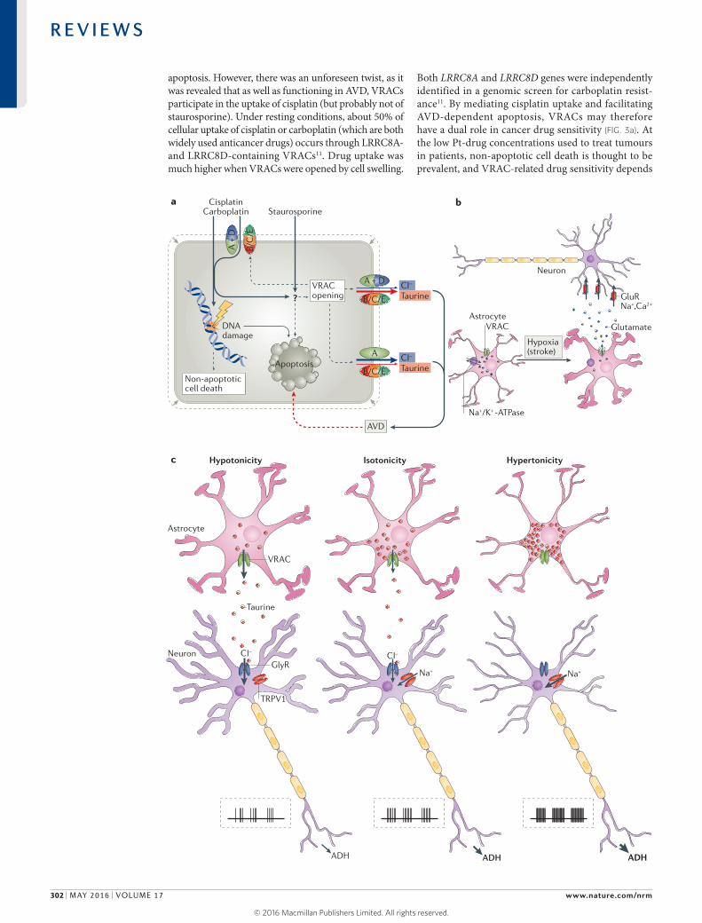
apoptosis. However, there was an unforeseen twist, as it was revealed that as well as functioning in AVD, VRACs participate in the uptake of cisplatin (but probably not of staurosporine). Under resting conditions, about 50% of cellular uptake of cisplatin or carboplatin (which are both widely used anticancer drugs) occurs through LRRC8A- and LRRC8D-containing VRACs11. Drug uptake was much higher when VRACs were opened by cell swelling.
Both LRRC8A and LRRC8D genes were independently identified in a genomic screen for carboplatin resist-ance11. By mediating cisplatin uptake and facilitating AVD-dependent apoptosis, VRACs may therefore have a dual role in cancer drug sensitivity (FIG. 3a). At the low Pt-drug concentrations used to treat tumours in patients, non-apoptotic cell death is thought to be prevalent, and VRAC-related drug sensitivity depends
Nature Reviews | Molecular Cell Biology
Na+/K+ -ATPase
Na+
GlutamateVRAC
Hypoxia(stroke)
GluRNa+,Ca2+
Astrocyte
Neuron
Neuron
Astrocyte
Na+
Taurine
ADH ADH ADH
VRAC
GlyR
TRPV1
Hypotonicity HypertonicityIsotonicity
CI–CI–
A +
D
A
CisplatinCarboplatin Staurosporine
ba
c
?
DNAdamage
Non-apoptoticcell death
Apoptosis
TaurineCI–
TaurineCI–
AVD
VRACopening
B/C
/E
B/C/E
B/C/E
A + D
R E V I E W S
302 | MAY 2016 | VOLUME 17 www.nature.com/nrm
© 2016
Macmillan
Publishers
Limited.
All
rights
reserved. ©
2016
Macmillan
Publishers
Limited.
All
rights
reserved.

AstrocytesA major class of glial (non-neuronal) cells in the central nervous system, which have important functions, for instance, in buffering extracellular ion concentrations, in removal of neurotransmitters and in neuronal metabolism.
predominantly on VRAC-dependent drug uptake rather than on VRAC-dependent AVD11. The role of VRACs in Pt-drug sensitivity may be clinically relevant, as LRRC8D downregulation correlated with poor survival of patients with cancer who were treated with Pt-drugs11. No down-regulation of LRRC8A was apparent in the patients’ tumours, suggesting that VRAC-dependent cell volume regulation, which is abolished with a loss of LRRC8A, but not LRRC8D, is important for the growth of tumour cells.
VRAC-mediated transport of metabolites. As VRACs are able to transport anticancer Pt-drugs11 and structurally distinct antibiotics106, these channels could potentially transport a broad range of small compounds. Such trans-port is surely not indiscriminate, as compounds such as the larger Pt-drug oxaliplatin apparently do not permeate VRACs11.
Metabolites such as taurine, glutamate, aspartate and inositol (and possibly even ATP114) are believed to pass through VRACs or have been confirmed as LRRC8 channel substrates9–11,115. As already mentioned, their release may be important for cell volume regulation. Indeed, quantitative considerations116 indicate that loss of inorganic ions can only decrease cell volume by 20–30%, less than is typically observed in AVD, suggesting an important role for organic osmolytes116. A key role for taurine in volume regulation is indicated by diminished, but not abolished, RVD in LRRC8D−/− HEK cells that dis-play severely reduced swelling-induced taurine efflux but normal ICl,vol (REF. 11). A VRAC-mediated release of
organic osmolytes might be particularly rele vant for RVD in neurons, which (as discussed above) maintain a low [Cl−]i that is set by constitutively active KCC2 (REF. 39) to enable inhibitory Cl− influx through GABAA receptors. Indeed, Cl− entry through VRACs, together with Na+ influx through glutamate receptors, may contribute to neuronal swelling in excitotoxicity117 (see below). The loss of valuable intracellular metabolites through VRACs probably has no deleterious consequences during AVD, as such cells will typically die. In RVD, the disadvantage of such a loss may be offset by the advantage of avoid-ing cell lysis with extreme swelling. Moreover, the loss of metabolites is less pronounced and more easily com-pensated for following small volume changes. Released osmolytes such as glutamate and taurine, however, can have significant effects on neighbouring cells. Two notable examples are discussed here.
Glutamate is an excitatory neurotransmitter that acti-vates cognate receptors on neurons. In excess, glutamate stimulation can lead to excitotoxicity, which prominently involves overstimulation of Ca2+-permeable glutamate receptors, contributing to neuronal cell death in stroke and other pathologies of the central nervous system. This condition is induced upon ischaemia, and several mechanisms for ischaemia-induced glutamate release have been proposed118, including reversal of Na+-driven glutamate transporters of astrocytes. Notably, one of the proposed mechanisms involves the release of glutamate from astrocytes through VRACs119–122 (FIG. 3b). Astrocytes are more susceptible to ischaemic swelling than neu-rons122 and may be the main source of VRAC-released amino acids in the brain. VRAC-dependent glutamate release probably occurs mainly in the area surrounding the hypoxic core (termed the penumbra)121, because VRACs need permissive intracellular concentrations of ATP and hence sufficient oxygen levels. Although cell swelling may be less pronounced in the penumbra, astrocytic VRAC opening may be potentiated by stroke- related increases of substances like ATP and react ive oxygen species102,115,123. Animal experiments indeed showed that VRAC inhibitors blunted hypoxia-induced release of excitatory amino acids in the brain124. In addi-tion to its role in astrocytic glutamate release, Cl− influx into neurons through VRACs might further contribute to pathology by promoting neuronal swelling initiated by Na+ entry through glutamate receptors117. Thus, VRAC inhibition might be beneficial in stroke. Several studies have indeed reported protective effects of anion transport inhibitors in stroke models125,126. However, all of the VRAC inhibitors used in those studies also affect other channels and cellular processes. Using LRRC8 mutant mouse models subject to stroke might provide more definitive results.
Besides glutamate, the VRAC substrate taurine has many cellular functions29. For instance, taurine acti-vates inhibitory neuronal glycine receptor and GABAA receptor channels127,128. Interestingly, VRAC may link cellular to systemic volume regulation by releasing tau-rine from glia in the osmosensing supraoptical nucleus (SON)15,127,129,130 (reviewed in REF. 131) (FIG. 3c). Neurons of the SON secrete antidiuretic hormone (ADH; also
Figure 3 | Some proposed roles of volume-regulated anion channels (VRACs) beyond volume regulation. a | Apoptosis-inducing compounds such as the anticancer drug cisplatin or the alkaloid staurosporine can enter cells by passive diffusion across the plasma membrane and promote cell death. Additionally, by an unknown mechanism (indicated here by ‘?’) that may involve reactive oxygen species, both drugs open VRACs, leading to efflux of Cl− and taurine (the latter preferentially through LRRC8D-containing VRACs11) and, consequently, to apoptotic volume decrease (AVD), which is believed to stimulate the progression of apoptosis. Opening of VRACs also further potentiates cisplatin uptake in a feedforward mechanism, as cisplatin (but not staurosporine) can also enter cells through LRRC8D-containing VRACs11. Therefore, whereas disruption of the indispensable VRAC subunit LRRC8A suppresses both cisplatin- and staurosporine-induced apoptosis through inhibition of AVD, LRRC8D disruption suppresses cisplatin- but not staurosporine-induced cell death through inhibition of enhanced cisplatin uptake11. A, B, C and D indicate the various LRRC8 subunits. b | A role for VRAC in stroke has been proposed. Occlusion of brain blood vessels leads to hypoxia, thereby decreasing cellular ATP levels and impairing ion pumping by the Na+/K+-ATPase. This entails swelling of astrocytes, opening of VRACs on their plasma membranes and the subsequent release of glutamate that binds to neuronal glutamate-gated cation channels (GluR). The massive opening of these receptors results in an influx of Na+ and Ca2+, which leads to neuronal damage (termed ‘excitotoxicity’), in addition to that directly caused by hypoxia. c | VRAC-mediated taurine release may link cellular to systemic osmoregulation15,129,130. Neurons of the supraoptical nucleus (SON) poorly regulate their volume132,133. In hypertonic solution, these neurons shrink and depolarize, owing to the opening of transient receptor potential cation channel subfamily V member 1 (TRPV1) cation channels and Na+ influx. This results in increased action potential firing of SON neurons (shown schematically in graphs), thereby stimulating the release of antidiuretic hormone (ADH) in the pituitary, which in turn increases renal water retention. Conversely, under hypotonic conditions, astrocytes neighbouring SON neurons undergo swelling and open their VRACs, thereby releasing taurine. Taurine then activates glycine receptors (GlyR) on SON neuronal cell bodies, leading to Cl− influx and hyperpolarization and inhibition of SON neurons, thereby decreasing ADH release. This collectively results in a push–pull mechanism controlling SON neuronal activity and body fluid homeostasis. Image in panel c adapted with permission from REF. 129, Elsevier.
▶
R E V I E W S
NATURE REVIEWS | MOLECULAR CELL BIOLOGY VOLUME 17 | MAY 2016 | 303
© 2016
Macmillan
Publishers
Limited.
All
rights
reserved. ©
2016
Macmillan
Publishers
Limited.
All
rights
reserved.

known as vasopressin) and oxytocin, which are hor-mones involved in body fluid homeostasis. These neurons function as osmosensors and were previously thought to lack RVD and RVI132; however, they do slowly develop ICl,vol and undergo RVD when swollen for more than 10 minutes133. In a hypertonic environment, SON neurons shrink and depolarize owing to Na+ influx through transient receptor potential cation channel subfamily V member 1 (TRPV1) cation channels that are opened by a shrinkage-induced pushing force from a channel-attached microtubule scaffold72. Depolarization, in turn, increases their firing rate, leading to increased secretion of ADH and resulting in enhanced renal water retention. Whereas extracellular hypertonicity directly activates SON neurons, hypotonicity is sensed by SON astrocytes, which release taurine in response to hypo-tonic swelling through VRAC-like channels127,129,130. By activating glycine receptor Cl− channels on neuronal cell bodies, taurine hyperpolarizes and thereby inhibits the activity of SON neurons and secretion of vasopressin. The interplay between direct hypertonicity-induced excitation of SON neurons and inhibition by VRAC-mediated astrocytic taurine release upon hypotonicity allows for efficient systemic osmoregulation in both directions (FIG. 3c).
Conclusions and outlookCell volume regulation is crucial for almost every verte-brate cell. It involves the regulated transport of osmolytes such as ions or small organic compounds across the plasma membrane. The resulting osmotic gradients drive water across the plasma membrane and restore cell volume. The transport proteins (effectors) involved in osmolyte transport almost always have additional cellu-lar functions. This is particularly true for VRACs, which transport not only inorganic anions, but also metabolites with functions in extracellular signalling. The recent dis-covery of LRRC8 proteins as essential components of VRACs9,10 has already led to the conclusion that the same type of channel is able to conduct both ions and organic osmolytes, and that it is involved both in RVD and
1. Ginzberg, M. B., Kafri, R. & Kirschner, M. On being the right (cell) size. Science 348, 1245075 (2015).
2. Hoffmann, E. K., Lambert, I. H. & Pedersen, S. F. Physiology of cell volume regulation in vertebrates. Physiol. Rev. 89, 193–277 (2009).This is an exhaustive review of the mechanisms involved in volume regulation and their physiological importance.
3. Lang, F. et al. Functional significance of cell volume regulatory mechanisms. Physiol. Rev. 78, 247–306 (1998).
4. Koivusalo, M., Kapus, A. & Grinstein, S. Sensors, transducers, and effectors that regulate cell size and shape. J. Biol. Chem. 284, 6595–6599 (2009).
5. Okada, Y. Volume expansion-sensing outward-rectifier Cl− channel: fresh start to the molecular identity and volume sensor. Am. J. Physiol. 273, C755–C789 (1997).
6. Groulx, N., Boudreault, F., Orlov, S. N. & Grygorczyk, R. Membrane reserves and hypotonic cell swelling. J. Membr. Biol. 214, 43–56 (2006).
7. Nilius, B. et al. Properties of volume-regulated anion channels in mammalian cells. Prog. Biophys. Mol. Biol. 68, 69–119 (1997).This is an excellent, detailed review on the properties of VRACs.
8. Pedersen, S. F., Klausen, T. K. & Nilius, B. The identification of VRAC (Volume Regulated Anion Channel): an amazing odyssey. Acta Physiol. 213, 868–881 (2015).
9. Voss, F. K. et al. Identification of LRRC8 heteromers as an essential component of the volume-regulated anion channel VRAC. Science 344, 634–638 (2014).This study shows that LRRC8 heteromers, composed of LRRC8A and at least one other LRRC8 isoform, are indispensable for swelling-activated VRAC currents and taurine efflux, and that subunit composition determines ICl,vol inactivation.
10. Qiu, Z. et al. SWELL1, a plasma membrane protein, is an essential component of volume-regulated anion channel. Cell 157, 447–458 (2014).This study identifies LRRC8A (renamed SWELL1) as a crucial VRAC component and shows, using knockdown experiments, that it is important for volume regulation and taurine fluxes.
11. Planells-Cases, R. et al. Subunit composition of VRAC channels determines substrate specificity and cellular resistance to Pt-based anti-cancer drugs. EMBO J. 34, 2993–3008 (2015).This paper reports, for the first time, that the selectivity of a VRAC depends on its LRRC8 subunit composition (thereby indicating that LRRC8
proteins form its pore) and demonstrates that VRACs have a dual role in cellular drug resistance by mediating cisplatin uptake and facilitating apoptosis.
12. Nunes, P. et al. Ionic imbalance, in addition to molecular crowding, abates cytoskeletal dynamics and vesicle motility during hypertonic stress. Proc. Natl Acad. Sci. USA 112, E3104–E3113 (2015).
13. Verkman, A. S. Aquaporins at a glance. J. Cell Sci. 124, 2107–2112 (2011).
14. Gomes, D. et al. Aquaporins are multifunctional water and solute transporters highly divergent in living organisms. Biochim. Biophys. Acta 1788, 1213–1228 (2009).
15. Bourque, C. W. Central mechanisms of osmosensation and systemic osmoregulation. Nat. Rev. Neurosci. 9, 519–531 (2008).
16. Miley, H. E., Sheader, E. A., Brown, P. D. & Best, L. Glucose-induced swelling in rat pancreatic β-cells. J. Physiol. 504, 191–198 (1997).
17. Schliess, F. & Häussinger, D. Osmosensing and signaling in the regulation of liver function. Contrib. Nephrol. 152, 198–209 (2006).
18. Best, L., Brown, P. D., Sener, A. & Malaisse, W. J. Electrical activity in pancreatic islet cells: the VRAC hypothesis. Islets 2, 59–64 (2010).
AVD. This discovery has also revealed that VRACs can mediate the uptake of anticancer drugs, an observation of potential importance for tumour drug resistance11. The biological importance of VRACs is highlighted by a recently reported study in Lrrc8a−/− mice134. The pheno types of these mice, which were erroneously inter-preted134 in terms of VRACs being plasma membrane receptors with extracellular leucine-rich repeats107,135, include high embryonic and postnatal lethality and pathological changes in many organs134. It has also been reported that a heterozygous truncation of the LRRC8A protein is associated with a congenital absence of serum γ-globulins, but this observation was based only on a single patient135. Although the severe phenotypes of Lrrc8a−/− mice underline the importance of VRACs, the survival of a few mice134 and the viability of Lrrc8−/− cells lacking all LRRC8 isoforms9,11 prove that VRACs are not strictly required for processes such as apoptosis, cell death, division, growth and migration. However, this does not exclude the important, but partially redundant, roles of VRACs in these processes. The diverse cellular and systemic roles of VRACs can now be addressed by cell- and tissue-specific disruption of individual sub-units. It may emerge that the transport of metabolites and signalling molecules by VRACs is as important as their role in volume regulation. LRRC8 proteins may also have roles independent of their channel function, for instance, by interacting with other proteins.
Identification of sensors and transducers involved in cell volume regulation remains another major challenge. The difficulty of identifying specific pathways is prob-ably due, at least in part, to redundancies at the levels of sensors and effectors. As it is likely that all effectors have additional cellular roles and sometimes only ‘moonlight’ in cell volume regulation, their regulation is expected to be complex. A good way to study the signalling pathways involved may be to work back from known effectors to identify signals directly impinging on them. Owing to the recent progress in our understanding of the com-position and functions of VRACs, such analysis should now also be possible for these channels.
R E V I E W S
304 | MAY 2016 | VOLUME 17 www.nature.com/nrm
© 2016
Macmillan
Publishers
Limited.
All
rights
reserved. ©
2016
Macmillan
Publishers
Limited.
All
rights
reserved.

19. Schwab, A., Fabian, A., Hanley, P. J. & Stock, C. Role of ion channels and transporters in cell migration. Physiol. Rev. 92, 1865–1913 (2012).
20. Stroka, K. M. et al. Water permeation drives tumor cell migration in confined microenvironments. Cell 157, 611–623 (2014).These experiments with cells migrating in narrow channels, together with mathematical modelling, show that osmolarity-driven asymmetric water transport can support cell migration in an actin- and myosin-independent manner.
21. Vom Dahl, S., Hallbrucker, C., Lang, F., Gerok, W. & Häussinger, D. Regulation of liver cell volume and proteolysis by glucagon and insulin. Biochem. J. 278, 771–777 (1991).
22. Maeno, E., Ishizaki, Y., Kanaseki, T., Hazama, A. & Okada, Y. Normotonic cell shrinkage because of disordered volume regulation is an early prerequisite to apoptosis. Proc. Natl Acad. Sci. USA 97, 9487–9492 (2000).This paper shows that AVD precedes caspase induction and other features of apoptosis, and that both AVD and apoptosis can be suppressed by pharmacological inhibitors of Cl− and K+ channels.
23. Okada, Y. et al. Volume-sensitive chloride channels involved in apoptotic volume decrease and cell death. J. Membr. Biol. 209, 21–29 (2006).
24. Lang, F. & Hoffmann, E. K. Role of ion transport in control of apoptotic cell death. Compr. Physiol. 2, 2037–2061 (2012).
25. Hortelano, S., Zeini, M., Castrillo, A., Alvarez, A. M. & Boscá, L. Induction of apoptosis by nitric oxide in macrophages is independent of apoptotic volume decrease. Cell Death Differ. 9, 643–650 (2002).
26. Wehner, F., Shimizu, T., Sabirov, R. & Okada, Y. Hypertonic activation of a non-selective cation conductance in HeLa cells and its contribution to cell volume regulation. FEBS Lett. 551, 20–24 (2003).
27. Bortner, C. D. & Cidlowski, J. A. Cell shrinkage and monovalent cation fluxes: role in apoptosis. Arch. Biochem. Biophys. 462, 176–188 (2007).
28. Poulsen, K. A. et al. Deregulation of apoptotic volume decrease and ionic movements in multidrug-resistant tumor cells: role of chloride channels. Am. J. Physiol. Cell Physiol. 298, C14–C25 (2010).
29. Lambert, I. H., Kristensen, D. M., Holm, J. B. & Mortensen, O. H. Physiological role of taurine — from organism to organelle. Acta Physiol. 213, 191–212 (2015).
30. Burg, M. B., Ferraris, J. D. & Dmitrieva, N. I. Cellular response to hyperosmotic stresses. Physiol. Rev. 87, 1441–1474 (2007).
31. Wehner, F. et al. Hypertonicity-induced cation channels. Acta Physiol. 187, 21–25 (2006).
32. Numata, T. et al. The ΔC splice-variant of TRPM2 is the hypertonicity-induced cation channel in HeLa cells, and the ecto-enzyme CD38 mediates its activation. J. Physiol. 590, 1121–1138 (2012).
33. Alessi, D. R. et al. The WNK-SPAK/OSR1 pathway: master regulator of cation-chloride cotransporters. Sci. Signal. 7, re3 (2014).
34. Alexander, R. T. & Grinstein, S. Na+/H+ exchangers and the regulation of volume. Acta Physiol. 187, 159–167 (2006).
35. Rotin, D. & Grinstein, S. Impaired cell volume regulation in Na+-H+ exchange-deficient mutants. Am. J. Physiol. 257, C1158–C1165 (1989).
36. Wakabayashi, S., Hisamitsu, T., Pang, T. & Shigekawa, M. Kinetic dissection of two distinct proton binding sites in Na+/H+ exchangers by measurement of reverse mode reaction. J. Biol. Chem. 278, 43580–43585 (2003).
37. Dunham, P. B., Kelley, S. J. & Logue, P. J. Extracellular Na+ inhibits Na+/H+ exchange: cell shrinkage reduces the inhibition. Am. J. Physiol. Cell Physiol. 287, C336–C344 (2004).
38. Stewart, A. K., Kurschat, C. E., Vaughan-Jones, R. D. & Alper, S. L. Putative re-entrant loop 1 of AE2 transmembrane domain has a major role in acute regulation of anion exchange by pH. J. Biol. Chem. 284, 6126–6139 (2009).
39. Hübner, C. A. et al. Disruption of KCC2 reveals an essential role of K-Cl cotransport already in early synaptic inhibition. Neuron 30, 515–524 (2001).
40. Mount, D. B. et al. Cloning and characterization of KCC3 and KCC4, new members of the cation-chloride cotransporter gene family. J. Biol. Chem. 274, 16355–16362 (1999).
41. Mercado, A., Broumand, V., Zandi-Nejad, K., Enck, A. H. & Mount, D. B. A C-terminal domain
in KCC2 confers constitutive K+-Cl− cotransport. J. Biol. Chem. 281, 1016–1026 (2006).
42. Dunham, P. B., Stewart, G. W. & Ellory, J. C. Chloride-activated passive potassium transport in human erythrocytes. Proc. Natl Acad. Sci. USA 77, 1711–1715 (1980).
43. Rust, M. B. et al. Disruption of erythroid K-Cl cotransporters alters erythrocyte volume and partially rescues erythrocyte dehydration in SAD mice. J. Clin. Invest. 117, 1708–1717 (2007).Using mice with disrupted Kcc1 and Kcc3 genes, this work shows that KCC3 is more important than KCC1 in erythrocyte volume regulation, and that erythrocytes have a larger volume when both isoforms are eliminated.
44. Brown, F. C. et al. Activation of the erythroid K-Cl cotransporter Kcc1 enhances sickle cell disease pathology in a humanized mouse model. Blood 126, 1683–1670 (2015).An activating mutation in the gene encoding the K+-Cl cotransporter KCC1 identified in a mouse random mutagenesis study is shown here to constitutively decrease erythrocyte volume in mice.
45. Boettger, T. et al. Loss of K-Cl co-transporter KCC3 causes deafness, neurodegeneration and reduced seizure threshold. EMBO J. 22, 5422–5434 (2003).
46. Hoffmann, E. K., Holm, N. B. & Lambert, I. H. Functions of volume-sensitive and calcium-activated chloride channels. IUBMB Life 66, 257–267 (2014).
47. Milenkovic, A. et al. Bestrophin 1 is indispensable for volume regulation in human retinal pigment epithelium cells. Proc. Natl Acad. Sci. USA 112, E2630–E2639 (2015).Using ATP-free intracellular solutions that suppress VRAC currents, these authors suggest that the Ca2+-activated Cl− channel bestrophin 1 is crucial for volume regulation in retinal pigment epithelial cells.
48. Juul, C. A. et al. Anoctamin 6 differs from VRAC and VSOAC but is involved in apoptosis and supports volume regulation in the presence of Ca2+. Pflügers Arch. 466, 1899–1910 (2014).
49. Hartzell, H. C., Qu, Z., Yu, K., Xiao, Q. & Chien, L. T. Molecular physiology of bestrophins: multifunctional membrane proteins linked to best disease and other retinopathies. Physiol. Rev. 88, 639–672 (2008).
50. Pedemonte, N. & Galietta, L. J. Structure and function of TMEM16 proteins (anoctamins). Physiol. Rev. 94, 419–459 (2014).
51. Stotz, S. C. & Clapham, D. E. Anion-sensitive fluorophore identifies the Drosophila swell-activated chloride channel in a genome-wide RNA interference screen. PLoS ONE 7, e46865 (2012).This paper reports a genome-wide siRNA screen in D. melanogaster cells, which are devoid of canonical VRAC (LRRC8 heteromers), and identifies Bestrophin 1 as a swelling-activated anion channel.
52. Chien, L. T. & Hartzell, H. C. Rescue of volume-regulated anion current by bestrophin mutants with altered charge selectivity. J. Gen. Physiol. 132, 537–546 (2008).Using ion selectivity-changing mutants to confirm the identity of the channel, this paper demonstrates that the D. melanogaster anion channel Bestrophin 1 can be activated by cell swelling.
53. Fischmeister, R. & Hartzell, H. C. Volume sensitivity of the bestrophin family of chloride channels. J. Physiol. 562, 477–491 (2005).
54. Okada, Y. Cell volume-sensitive chloride channels: phenotypic properties and molecular identity. Contrib. Nephrol. 152, 9–24 (2006).
55. Grunnet, M. et al. KCNQ1 channels sense small changes in cell volume. J. Physiol. 549, 419–427 (2003).
56. Niemeyer, M. I., Cid, L. P., Barros, L. F. & Sepúlveda, F. V. Modulation of the two-pore domain acid-sensitive K+ channel TASK-2 (KCNK5) by changes in cell volume. J. Biol. Chem. 276, 43166–43174 (2001).
57. Barrière, H. et al. Role of TASK2 potassium channels regarding volume regulation in primary cultures of mouse proximal tubules. J. Gen. Physiol. 122, 177–190 (2003).
58. Lolicato, M., Riegelhaupt, P. M., Arrigoni, C., Clark, K. A. & Minor, D. L. Jr. Transmembrane helix straightening and buckling underlies activation of mechanosensitive and thermosensitive K2P channels. Neuron 84, 1198–1212 (2014).
59. Brohawn, S. G., Su, Z. & MacKinnon, R. Mechanosensitivity is mediated directly by the lipid
membrane in TRAAK and TREK1 K+ channels. Proc. Natl Acad. Sci. USA 111, 3614–3619 (2014).
60. Patel, A. J. et al. A mammalian two pore domain mechano-gated S-like K+ channel. EMBO J. 17, 4283–4290 (1998).
61. Gründer, S., Thiemann, A., Pusch, M. & Jentsch, T. J. Regions involved in the opening of CIC-2 chloride channel by voltage and cell volume. Nature 360, 759–762 (1992).
62. Jordt, S. E. & Jentsch, T. J. Molecular dissection of gating in the ClC-2 chloride channel. EMBO J. 16, 1582–1592 (1997).
63. Sabirov, R. Z., Merzlyak, P. G., Islam, M. R., Okada, T. & Okada, Y. The properties, functions, and pathophysiology of maxi-anion channels. Pflügers Arch. 468, 405–420 (2016).
64. Thiemann, A., Gründer, S., Pusch, M. & Jentsch, T. J. A chloride channel widely expressed in epithelial and non-epithelial cells. Nature 356, 57–60 (1992).
65. Hoegg-Beiler, M. B. et al. Disrupting MLC1 and GlialCAM and ClC-2 interactions in leukodystrophy entails glial chloride channel dysfunction. Nat. Commun. 5, 3475 (2014).
66. Nehrke, K. et al. Loss of hyperpolarization-activated Cl− current in salivary acinar cells from Clcn2 knockout mice. J. Biol. Chem. 277, 23604–23611 (2002).
67. Cahalan, S. M. et al. Piezo1 links mechanical forces to red blood cell volume. eLife 4, e07370 (2015).This study shows that mechanical activation of the PIEZO1 channel in red blood cells leads to Ca2+ influx that activates the Ca2+-sensitive Gardos (KCa3.1) K+ channel and thereby leads to smaller erythrocyte volume.
68. Ranade, S. S., Syeda, R. & Patapoutian, A. Mechanically activated ion channels. Neuron 87, 1162–1179 (2015).
69. Sukharev, S. I., Blount, P., Martinac, B., Blattner, F. R. & Kung, C. A large-conductance mechanosensitive channel in E. coli encoded by mscL alone. Nature 368, 265–268 (1994).
70. Anishkin, A., Loukin, S. H., Teng, J. & Kung, C. Feeling the hidden mechanical forces in lipid bilayer is an original sense. Proc. Natl Acad. Sci. USA 111, 7898–7905 (2014).
71. Maingret, F., Fosset, M., Lesage, F., Lazdunski, M. & Honoré, E. TRAAK is a mammalian neuronal mechano-gated K+ channel. J. Biol. Chem. 274, 1381–1387 (1999).
72. Prager-Khoutorsky, M., Khoutorsky, A. & Bourque, C. W. Unique interweaved microtubule scaffold mediates osmosensory transduction via physical interaction with TRPV1. Neuron 83, 866–878 (2014).This paper shows that the TRPV1 cation channel is activated in osmosensory neurons by cell shrinkage through a pushing force of an attached microtubule scaffold.
73. Zhang, W. et al. Ankyrin repeats convey force to gate the NOMPC mechanotransduction channel. Cell 162, 1391–1403 (2015).
74. Tilly, B. C. et al. Activation of the osmo-sensitive chloride conductance involves P21rho and is accompanied by a transient reorganization of the F-actin cytoskeleton. Mol. Biol. Cell 7, 1419–1427 (1996).
75. Fuster, D., Moe, O. W. & Hilgemann, D. W. Lipid- and mechanosensitivities of sodium/hydrogen exchangers analyzed by electrical methods. Proc. Natl Acad. Sci. USA 101, 10482–10487 (2004).
76. Nasuhoglu, C. et al. Modulation of cardiac PIP2 by cardioactive hormones and other physiologically relevant interventions. Am. J. Physiol. Cell Physiol. 283, C223–C234 (2002).
77. Yamamoto, M. et al. Hypertonic stress increases phosphatidylinositol 4,5-bisphosphate levels by activating PIP5KIβ. J. Biol. Chem. 281, 32630–32638 (2006).
78. Voets, T., Droogmans, G., Raskin, G., Eggermont, J. & Nilius, B. Reduced intracellular ionic strength as the initial trigger for activation of endothelial volume-regulated anion channels. Proc. Natl Acad. Sci. USA 96, 5298–5303 (1999).This work convincingly shows that VRACs can be stimulated by reducing intracellular ionic strength, independent of cell volume changes.
79. Emma, F., McManus, M. & Strange, K. Intracellular electrolytes regulate the volume set point of the organic osmolyte/anion channel VSOAC. Am. J. Physiol. 272, C1766–C1775 (1997).
R E V I E W S
NATURE REVIEWS | MOLECULAR CELL BIOLOGY VOLUME 17 | MAY 2016 | 305
© 2016
Macmillan
Publishers
Limited.
All
rights
reserved. ©
2016
Macmillan
Publishers
Limited.
All
rights
reserved.

80. Syeda, R. et al. LRRC8 proteins form volume-regulated anion channels that sense ionic strength. Cell 164, 499–511 (2016).Using reconstitution of purified LRRC8 heteromers into lipid bilayers, this paper shows that LRRC8 proteins are sufficient for forming a VRAC channel, and that it can be activated under conditions of low ionic strength.
81. Rinehart, J. et al. Sites of regulated phosphorylation that control K-Cl cotransporter activity. Cell 138, 525–536 (2009).
82. de Los Heros, P. et al. The WNK-regulated SPAK/OSR1 kinases directly phosphorylate and inhibit the K+-Cl− co-transporters. Biochem. J. 458, 559–573 (2014).
83. Melo, Z. et al. N-terminal serine dephosphorylation is required for KCC3 cotransporter full activation by cell swelling. J. Biol. Chem. 288, 31468–31476 (2013).
84. Gagnon, K. B., England, R. & Delpire, E. Volume sensitivity of cation-Cl-cotransporters is modulated by the interaction of two kinases: Ste20-related proline-alanine-rich kinase and WNK4. Am. J. Physiol. Cell Physiol. 290, C134–C142 (2006).
85. Piala, A. T. et al. Chloride sensing by WNK1 involves inhibition of autophosphorylation. Sci. Signal. 7, ra41 (2014).This report of the crystal structure of the catalytic domain of the WNK1 kinase reveals that it directly binds to Cl–, and this binding is shown to prevent kinase activation.
86. Varela, D., Simon, F., Riveros, A., Jørgensen, F. & Stutzin, A. NAD(P)H oxidase-derived H2O2 signals chloride channel activation in cell volume regulation and cell proliferation. J. Biol. Chem. 279, 13301–13304 (2004).
87. Shimizu, T., Numata, T. & Okada, Y. A role of reactive oxygen species in apoptotic activation of volume-sensitive Cl− channel. Proc. Natl Acad. Sci. USA 101, 6770–6773 (2004).This paper shows that ICl,vol-like currents are activated by several pro-apoptotic stimuli and provides evidence for a role of reactive oxygen species in the activation of those currents and in apoptosis.
88. Carton, I. et al. RhoA exerts a permissive effect on volume-regulated anion channels in vascular endothelial cells. Am. J. Physiol. Cell Physiol. 283, C115–C125 (2002).
89. Lepple-Wienhues, A. et al. The tyrosine kinase p56lck mediates activation of swelling-induced chloride channels in lymphocytes. J. Cell Biol. 141, 281–286 (1998).
90. Holm, J. B., Grygorczyk, R. & Lambert, I. H. Volume-sensitive release of organic osmolytes in the human lung epithelial cell line A549: role of the 5-lipoxygenase. Am. J. Physiol. Cell Physiol. 305, C48–C60 (2013).
91. Levitan, I., Christian, A. E., Tulenko, T. N. & Rothblat, G. H. Membrane cholesterol content modulates activation of volume-regulated anion current in bovine endothelial cells. J. Gen. Physiol. 115, 405–416 (2000).
92. Browe, D. M. & Baumgarten, C. M. EGFR kinase regulates volume-sensitive chloride current elicited by integrin stretch via PI-3K and NADPH oxidase in ventricular myocytes. J. Gen. Physiol. 127, 237–251 (2006).
93. Grinstein, S., Clarke, C. A., Dupre, A. & Rothstein, A. Volume-induced increase of anion permeability in human lymphocytes. J. Gen. Physiol. 80, 801–823 (1982).
94. Hoffmann, E. K., Simonsen, L. O. & Lambert, I. H. Volume-induced increase of K+ and Cl− permeabilities in Ehrlich ascites tumor cells. Role of internal Ca2+. J. Membr. Biol. 78, 211–222 (1984).
95. Hazama, A. & Okada, Y. Ca2+ sensitivity of volume-regulatory K+ and Cl− channels in cultured human epithelial cells. J. Physiol. 402, 687–702 (1988).
96. Okada, Y., Sato, K. & Numata, T. Pathophysiology and puzzles of the volume-sensitive outwardly rectifying anion channel. J. Physiol. 587, 2141–2149 (2009).
97. Jackson, P. S., Morrison, R. & Strange, K. The volume-sensitive organic osmolyte-anion channel VSOAC is regulated by nonhydrolytic ATP binding. Am. J. Physiol. 267, C1203–C1209 (1994).
98. Stutzin, A. et al. Separate taurine and chloride efflux pathways activated during regulatory volume decrease. Am. J. Physiol. 277, C392–C402 (1999).
99. Lambert, I. H. & Hoffmann, E. K. Cell swelling activates separate taurine and chloride channels in Ehrlich mouse ascites tumor cells. J. Membr. Biol. 142, 289–298 (1994).
100. Jackson, P. S. & Strange, K. Volume-sensitive anion channels mediate swelling-activated inositol and taurine efflux. Am. J. Physiol. 265, C1489–C1500 (1993).
101. Akita, T. & Okada, Y. Regulation of bradykinin-induced activation of volume-sensitive outwardly rectifying anion channels by Ca2+ nanodomains in mouse astrocytes. J. Physiol. 589, 3909–3927 (2011).
102. Mongin, A. A. & Kimelberg, H. K. ATP potently modulates anion channel-mediated excitatory amino acid release from cultured astrocytes. Am. J. Physiol. Cell Physiol. 283, C569–C578 (2002).
103. Deng, W., Baki, L. & Baumgarten, C. M. Endothelin signalling regulates volume-sensitive Cl− current via NADPH oxidase and mitochondrial reactive oxygen species. Cardiovasc. Res. 88, 93–100 (2010).
104. Nilius, B. et al. Volume-activated Cl− currents in different mammalian non-excitable cell types. Pflügers Arch. 428, 364–371 (1994).This paper suggests that VRACs are widely, and maybe ubiquitously, expressed in mammalian cells.
105. Abascal, F. & Zardoya, R. LRRC8 proteins share a common ancestor with pannexins, and may form hexameric channels involved in cell-cell communication. Bioessays 34, 551–560 (2012).This is a bioinformatic analysis showing that LRRC8 proteins are homologous to pannexins and suggesting that LRRC8 proteins form hexameric ion channels.
106. Lee, C. C., Freinkman, E., Sabatini, D. M. & Ploegh, H. L. The protein synthesis inhibitor blasticidin S enters mammalian cells via leucine-rich repeat-containing protein 8D. J. Biol. Chem. 289, 17124–17131 (2014).This work shows that the cellular uptake of blasticidin depends on LRRC8D, a gene that was previously identified in a genome-wide reporter screen for genes involved in NF-κB activation that used blasticidin as trigger.
107. Kubota, K. et al. LRRC8 involved in B cell development belongs to a novel family of leucine-rich repeat proteins. FEBS Lett. 564, 147–152 (2004).
108. Bond, S. R. & Naus, C. C. The pannexins: past and present. Front. Physiol. 5, 58 (2014).
109. Shennan, D. B. Swelling-induced taurine transport: relationship with chloride channels, anion-exchangers and other swelling-activated transport pathways. Cell Physiol. Biochem. 21, 15–28 (2008).
110. Min, X. J. et al. Dysfunction of volume-sensitive chloride channels contributes to cisplatin resistance in human lung adenocarcinoma cells. Exp. Biol. Med. 236, 483–491 (2011).
111. Ise, T. et al. Roles of volume-sensitive Cl− channel in cisplatin-induced apoptosis in human epidermoid cancer cells. J. Membr. Biol. 205, 139–145 (2005).
112. Sørensen, B. H., Thorsteinsdottir, U. A. & Lambert, I. H. Acquired cisplatin resistance in human ovarian A2780 cancer cells correlates with shift in taurine homeostasis and ability to volume regulate. Am. J. Physiol. Cell Physiol. 307, C1071–C1080 (2014).
113. Lee, E. L. et al. Impaired activity of volume-sensitive Cl− channel is involved in cisplatin resistance of cancer cells. J. Cell. Physiol. 211, 513–521 (2007).In this work, sensitivity to cisplatin and VRAC currents are restored in parallel in a drug-resistant cell line that displays small VRAC currents by nonspecifically changing gene expression with a histone deacetylate inhibitor.
114. Hisadome, K. et al. Volume-regulated anion channels serve as an auto/paracrine nucleotide release pathway in aortic endothelial cells. J. Gen. Physiol. 119, 511–520 (2002).
115. Hyzinski-García, M. C., Rudkouskaya, A. & Mongin, A. A. LRRC8A protein is indispensable for swelling-activated and ATP-induced release of excitatory amino acids in rat astrocytes. J. Physiol. 592, 4855–4862 (2014).
116. Model, M. A. Possible causes of apoptotic volume decrease: an attempt at quantitative review. Am. J. Physiol. Cell Physiol. 306, C417–C424 (2014).
117. Inoue, H. & Okada, Y. Roles of volume-sensitive chloride channel in excitotoxic neuronal injury. J. Neurosci. 27, 1445–1455 (2007).
118. Lai, T. W., Zhang, S. & Wang, Y. T. Excitotoxicity and stroke: identifying novel targets for neuroprotection. Prog. Neurobiol. 115, 157–188 (2014).
119. Kimelberg, H. K., Goderie, S. K., Higman, S., Pang, S. & Waniewski, R. A. Swelling-induced release of glutamate, aspartate, and taurine from astrocyte cultures. J. Neurosci. 10, 1583–1591 (1990).
120. Pasantes-Morales, H., Murray, R. A., Sánchez-Olea, R. & Morán, J. Regulatory volume decrease in cultured astrocytes. II. Permeability pathway to amino acids and polyols. Am. J. Physiol. 266, C172–C178 (1994).
121. Feustel, P. J., Jin, Y. & Kimelberg, H. K. Volume-regulated anion channels are the predominant contributors to release of excitatory amino acids in the ischemic cortical penumbra. Stroke 35, 1164–1168 (2004).
122. Mongin, A. A. Volume-regulated anion channel-a frenemy within the brain. Pflügers Arch. 468, 421–441 (2015).
123. Haskew-Layton, R. E., Mongin, A. A. & Kimelberg, H. K. Hydrogen peroxide potentiates volume-sensitive excitatory amino acid release via a mechanism involving Ca2+/calmodulin-dependent protein kinase II. J. Biol. Chem. 280, 3548–3554 (2005).
124. Phillis, J. W., Song, D. & O’Regan, M. H. Inhibition by anion channel blockers of ischemia-evoked release of excitotoxic and other amino acids from rat cerebral cortex. Brain Res. 758, 9–16 (1997).
125. Zhang, Y., Zhang, H., Feustel, P. J. & Kimelberg, H. K. DCPIB, a specific inhibitor of volume regulated anion channels (VRACs), reduces infarct size in MCAo and the release of glutamate in the ischemic cortical penumbra. Exp. Neurol. 210, 514–520 (2008).This is one of several studies showing that anion channel inhibitors reduce cerebral infarct size, now using a somewhat more specific VRAC inhibitor that is applied intracisternally.
126. Inoue, H., Ohtaki, H., Nakamachi, T., Shioda, S. & Okada, Y. Anion channel blockers attenuate delayed neuronal cell death induced by transient forebrain ischemia. J. Neurosci. Res. 85, 1427–1435 (2007).
127. Hussy, N., Deleuze, C., Pantaloni, A., Desarmenien, M. G. & Moos, F. Agonist action of taurine on glycine receptors in rat supraoptic magnocellular neurones: possible role in osmoregulation. J. Physiol. 502, 609–621 (1997).This seminal paper supports the notion that hypotonicity-induced release of taurine from astrocytes activates glycine receptors on nearby osmosensing neurons and suggests that this plays an important part in systemic volume regulation.
128. Le-Corronc, H., Rigo, J. M., Branchereau, P. & Legendre, P. GABAA receptor and glycine receptor activation by paracrine/autocrine release of endogenous agonists: more than a simple communication pathway. Mol. Neurobiol. 44, 28–52 (2011).
129. Hussy, N., Deleuze, C., Desarmenien, M. G. & Moos, F. C. Osmotic regulation of neuronal activity: a new role for taurine and glial cells in a hypothalamic neuroendocrine structure. Prog. Neurobiol. 62, 113–134 (2000).
130. Choe, K. Y., Olson, J. E. & Bourque, C. W. Taurine release by astrocytes modulates osmosensitive glycine receptor tone and excitability in the adult supraoptic nucleus. J. Neurosci. 32, 12518–12527 (2012).
131. Prager-Khoutorsky, M. & Bourque, C. W. Mechanical basis of osmosensory transduction in magnocellular neurosecretory neurones of the rat supraoptic nucleus. J. Neuroendocrinol. 27, 507–515 (2015).
132. Zhang, Z. & Bourque, C. W. Osmometry in osmosensory neurons. Nat. Neurosci. 6, 1021–1022 (2003).This work suggests that osmosensory neurons from the SON function as osmometers, because they apparently lack both RVD and RVI.
133. Sato, K., Numata, T., Saito, T., Ueta, Y. & Okada, Y. V2 receptor-mediated autocrine role of somatodendritic release of AVP in rat vasopressin neurons under hypo-osmotic conditions. Sci. Signal. 4, ra5 (2011).Investigating RVD and ICl,vol on longer time scales, this work contradicts reference 132 by showing that these neurons slowly activate VRACs and RVD.
134. Kumar, L. et al. Leucine-rich repeat containing 8A (LRRC8A) is essential for T lymphocyte development and function. J. Exp. Med. 211, 929–942 (2014).Although based on the wrong assumption that LRRC8A is a receptor, this first description of the severe phenotype of constitutive Lrrc8a-knockout mice highlights the importance of VRACs in many tissues.
R E V I E W S
306 | MAY 2016 | VOLUME 17 www.nature.com/nrm
© 2016
Macmillan
Publishers
Limited.
All
rights
reserved. ©
2016
Macmillan
Publishers
Limited.
All
rights
reserved.

135. Sawada, A. et al. A congenital mutation of the novel gene LRRC8 causes agammaglobulinemia in humans. J. Clin. Invest. 112, 1707–1713 (2003).
136. Krane, C. M. et al. Salivary acinar cells from aquaporin 5-deficient mice have decreased membrane water permeability and altered cell volume regulation. J. Biol. Chem. 276, 23413–23420 (2001).
137. Zeuthen, T. Water-transporting proteins. J. Membr. Biol. 234, 57–73 (2010).
138. Nilius, B. Is the volume-regulated anion channel VRAC a “water-permeable” channel? Neurochem. Res. 29, 3–8 (2004).
139. Klein, M., Seeger, P., Schuricht, B., Alper, S. L. & Schwab, A. Polarization of Na+/H+ and Cl−/HCO3
− exchangers in migrating renal epithelial cells. J. Gen. Physiol. 115, 599–608 (2000).
140. Denker, S. P. & Barber, D. L. Cell migration requires both ion translocation and cytoskeletal anchoring by the Na-H exchanger NHE1. J. Cell Biol. 159, 1087–1096 (2002).
141. Saadoun, S., Papadopoulos, M. C., Hara-Chikuma, M. & Verkman, A. S. Impairment of angiogenesis and cell migration by targeted
aquaporin-1 gene disruption. Nature 434, 786–792 (2005).
142. Papadopoulos, M. C., Saadoun, S. & Verkman, A. S. Aquaporins and cell migration. Pflügers Arch. 456, 693–700 (2008).
143. Huang, X. et al. EAG2 potassium channel with evolutionarily conserved function as a brain tumor target. Nat. Neurosci. 18, 1236–1246 (2015).
144. Schneider, L. et al. H-ras transformation sensitizes volume-activated anion channels and increases migratory activity of NIH3T3 fibroblasts. Pflügers Arch. 455, 1055–1062 (2008).
145. Charras, G. T., Yarrow, J. C., Horton, M. A., Mahadevan, L. & Mitchison, T. J. Non-equilibration of hydrostatic pressure in blebbing cells. Nature 435, 365–369 (2005).
146. Orlov, S. N., Platonova, A. A., Hamet, P. & Grygorczyk, R. Cell volume and monovalent ion transporters: their role in cell death machinery triggering and progression. Am. J. Physiol. Cell Physiol. 305, C361–C372 (2013).
147. McGlasson, L., Best, L. & Brown, P. D. The glucokinase activator GKA50 causes an increase in cell volume and activation of volume-regulated anion channels in rat
pancreatic β-cells. Mol. Cell. Endocrinol. 342, 48–53 (2011).
148. Catalán, M. A. et al. A fluid secretion pathway unmasked by acinar-specific Tmem16A gene ablation in the adult mouse salivary gland. Proc. Natl Acad. Sci. USA 112, 2263–2268 (2015).
AcknowledgementsThe author thanks Rosa Planells-Cases for help with figures, and members of his laboratory for critical reading of the manuscript. Work on VRACs is supported in the author’s laboratory by the European Research Council Advanced Grant (FP/2007-2013) 294435 ‘Cytovolion’ and the Deutsche Forschungsgemeinschaft (Exc 257 ‘Neurocure’ and JE164/12).
Competing interests statementThe author declares no competing interests.
SUPPLEMENTARY INFORMATIONSee online article: S1 (box)
ALL LINKS ARE ACTIVE IN THE ONLINE PDF
R E V I E W S
NATURE REVIEWS | MOLECULAR CELL BIOLOGY VOLUME 17 | MAY 2016 | 307
© 2016
Macmillan
Publishers
Limited.
All
rights
reserved. ©
2016
Macmillan
Publishers
Limited.
All
rights
reserved.

![Ions channels/transporters and chloroplast regulation · transporters/pumps and secondary transporters (according to the Transport Classification system [1]). Channels transport](https://img.pdfslide.net/doc/110x75/601623c1d6936b1074546c48/ions-channelstransporters-and-chloroplast-transporterspumps-and-secondary-transporters.jpg)


![Ions channels/transporters and chloroplast regulation · to the CorA super-family (transporters for both Mg and Co, for review see [22]), may transport Mg into the stroma (Fig. 1)](https://img.pdfslide.net/doc/110x75/601624b446f20b5f037e6743/ions-channelstransporters-and-chloroplast-to-the-cora-super-family-transporters.jpg)