Embed Size (px)
Citation preview

WADC TECHNICAL REPORT 52-286
A PLASTIC DRESSING (AEROPLAST) FOR BURNS AND SURGICAL WOUNDS
DANIEL S. J. CJIOY, CAPT, USAF, MC
AERO MEDICAL LABORATORY
bLLC'CEMBEI( 1952
Statement AApproved for Public Release
WRIGHT AIR DEVELOPMENT CENTER
2OOO/4O3 7/

NOTICES
When Government drawings, specifications, or other data are usedfor any purpose other than in connection with a definitely related Govern-ment procurement operation, the United States Government thereby in-curs no responsibility nor any obligation whatsoever; and the fact .thatthe Government may have formulated, furnished, or in any way suppliedthe said drawings, specifications, or other data, is not to be regardedby implication or otherwise as in any manner licensing the holder orany other person or corporation,or conveying any rights or permissionto manufacture, use, or sell any patented invention that may in anywaybe related thereto.
The information furnished herewith is made available for studyupon the understanding that the Government's proprietary interests inand relating thereto shall not be impaired. It is desired that the TudgeAdvocate (WCT), Wright Air Development Center, Wright-PattersonAir Force Base, Ohio, be promptly notified of any apparent conflict be-tween the Government's proprietary interests and those of others.
: • ia It II* S . I I I I !

WADC TECHNICAL REPORT 52-286
A PLASTIC DRESSING (AEROPLAST) FOR BURNS AND SURGICAL WOUNDS
Daniel S. J. Choy, Capt, USAF, MCAero Medical Laboratory
December 1952
RDO No. 696-69
Wright Air Development Center
Air Research and Development Command
United States Air ForceWright-Patterson Air Force Base, Ohio
McGregor & Werner, Inc., Dayton, 0.300 June, 1953

FOREWORD
This Report by Captain Daniel S. J. Choy, USAF, 'C, the projectengineer, was prepared under Research and Development Order Eo. 696-69,"IHuman Thermal Tolerance." The work was performed under the auspicesof the Aero Medical Laboratory, Wright Air Developnent Center, Wright-Patterson Air Force Base, Ohio, 'The experimental work described in3ection I was performed in collaboration with Captain Wallace E. Wondt,USAF, VC, at the Acro Medical Laboratory and 3ampson Air Force Dase.The clinical investigations described in Section III were conducted onthe III Surgical Division, Bellevue lospital, New York City, Yew York.The author is indebted for the invaluable assistance of TechnicalSergeant James Edwards.
,'ADC T" 52-236
ABSTRACT
In August 1951, the author initiated a project to develop a rapidlyapplied, sprayable, film-forming plastic to be dispensed from aerosolbombs or other spraying equipment, for the mass local therapy of burns.
This report of the achievements of this project is divided into threeSections:
Section I is concerned with the treatment of experimental burns onhogs with the plastic dressing and the evaluation of the plastic for possiblelocal and systemic toxicity.
Section II deals with studies on comparative wound healing rates in rats,using the plastic dressing and a control dressing of vaseline gauze.
Section III describes the clinical trials of the aeroplast dressing onbfirns and a wide variety of surgical wounds in humans.
PUBLICATION REVIEW
This report has been reviewed and is approved.
FCR THE COMMANDING GENERAL:
ROBERT H. BLOUNTColonel, USAF (Mi)Chief, Aero Mbdical LaboratoryDirectorate of Research
WADC TR 52-286 iii

TABLE OF CONTENTS
Section I A Local Treatment of Burns and Surgical Wounds......... 1
Results. .. *.... .... ... .. ... ........ .. ... .... *. ........... 4
14
Toxicity.. .......................... 12
Summary ............................................... 1/4
Section II Comparative Wound Healing Rates of Standard Burn Dress-ing and Aeroplast................................••... 15
Nethods... ............................................ 15
Discussion and Summary .............................. 15
Section III Clinical Trials ............................. 18
Skin-Graft Donor Sites .............................. 18
rns ....... . ......................................... 20
Other Surgical Wounds .............................. 25
Summary ..... .... ...................................... 30
Bibliography .................................. • .... • 35
WADC TR 52-286 iv
LIST OF ILLUSTRATIONS
FIGURE Page
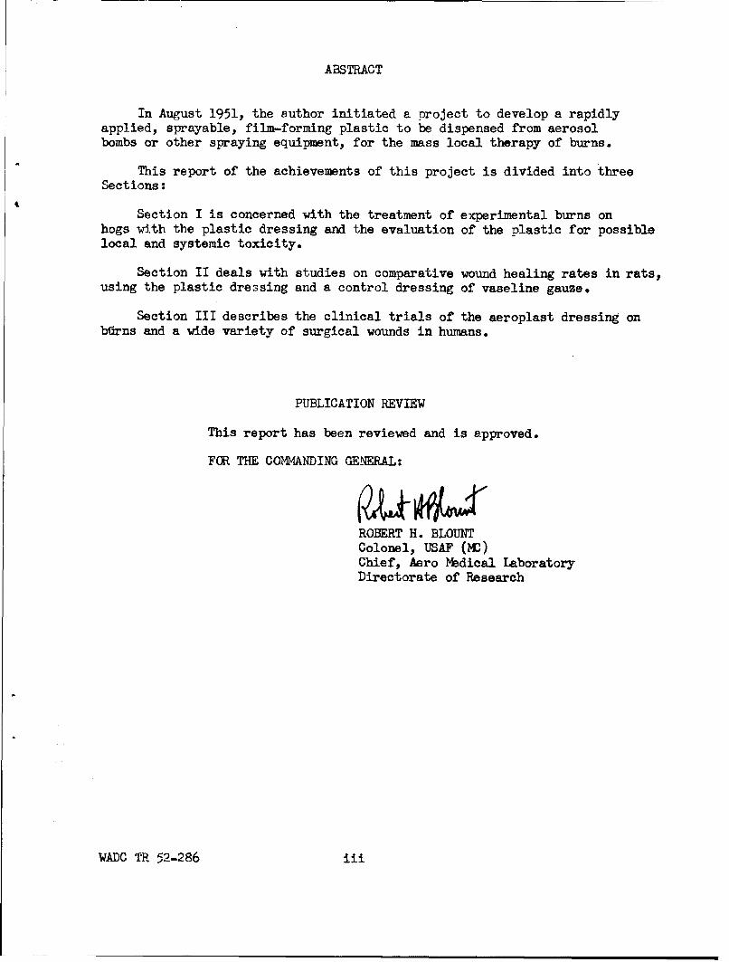
1. An agar plateA streaked with Staphylococcus aureusB seeded with Pseudomonas aeruginosaC seeded with an aerobic spore former and Proteus
vulgaris .................... 9 -....... ...... ............ 2
2. Pattern of burn on dorsal surface of hog .................. 3
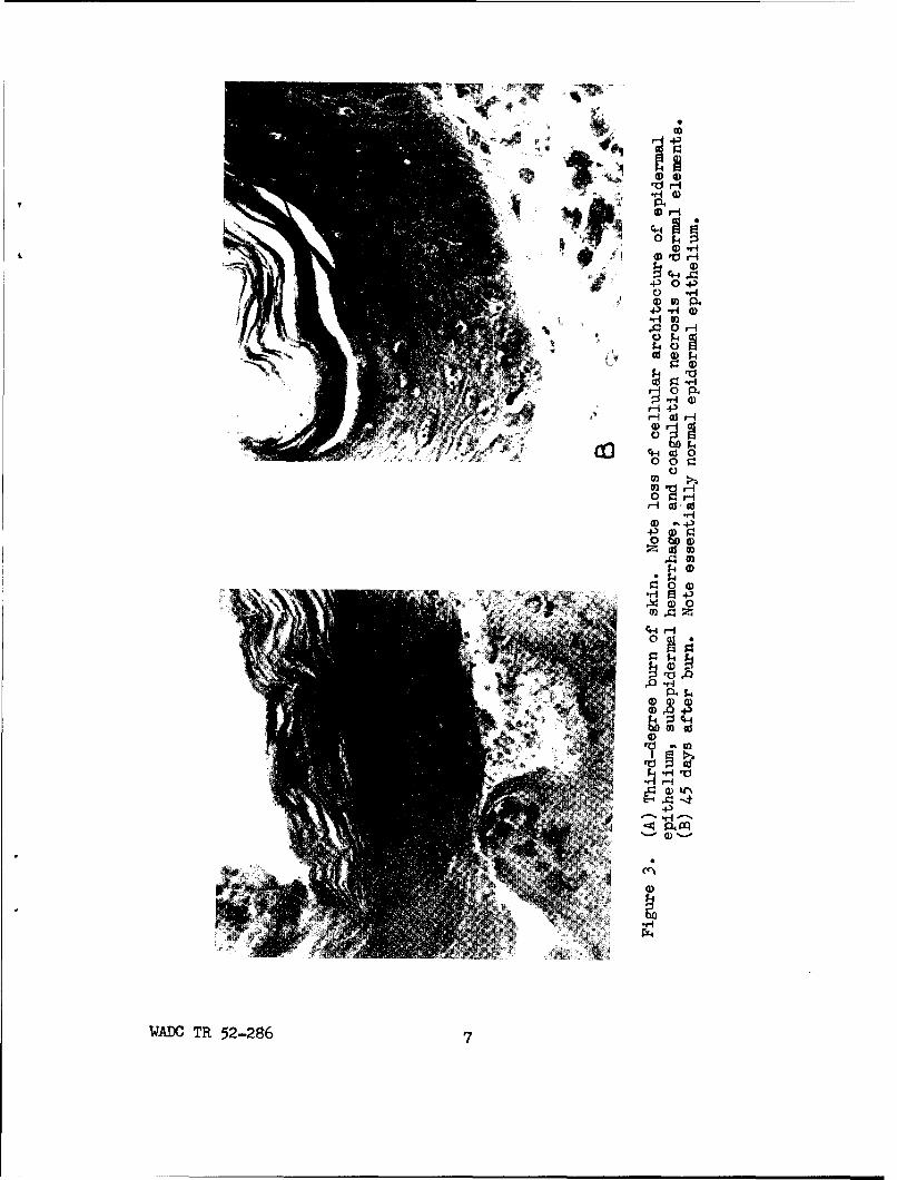
3. A Third-degree burn of skinB 45 days after burn .................................. 7
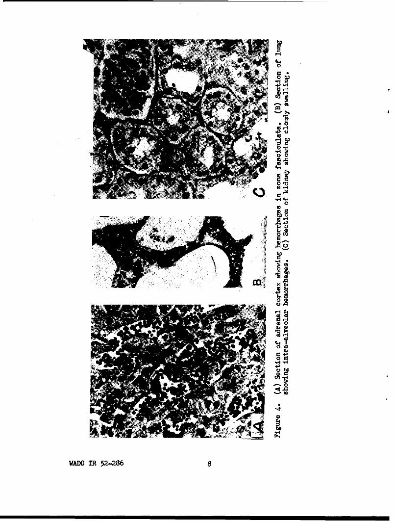
4ý. A Section of adrenal cortex showing hemorrhages in zonafasciculata
B Section of lung showing intra-alveolar hemorrhagesC Section of kidney showing cloudy swelling .......... 0 9.. 8
5% A Section of liver showing small focal abscessB Section of intestine showing submucous abscessC Section of lung showing acute interstitial pneumonitis 9
6, A Saction of liver showing focal necrosisB Section of lung showing acute lobar pneumonia0...... .. 10
7o Section of skin, 33 days after burn, showing complete epi-dermal regeneration with moderate dermal cicatrix formation 11
8. Section of kidney showing focal hydropic degeneration oftubular epithelium . 0 .. . . ........ .. 11
9. Comparison of treatment of 50% body burn byP Standard pressure bandageB Aeroplast dressing ................. ........ 14
10. Representative wound-healing rates in two rats, usingaeroplast and control dressing ............................. 17
11. A Case 1, E. J. Skin-graft donor site, seven days postoperative
B Case 1, E. J. Skin-graft donor site with aeroplastdressing, 14 days post operative
C Case 1, E. J. Skin-graft donor site, healed, 18 dayspost operative ................... ............. 19
12. A Case 2, C. B. Second-degree burn, dorsum of leftforearm
B Case 2, C. B. Second-degree burn with aeroplast ...... 21
WADC TR 52-286 v

FIGURE Page
13. A Case 7, G. D. Second-degree burn of left hand withaeroplast dressing
B Case 7, G. D. Same burn after excision of blistersand application of aeroplast........................ • 23
C Case 7, G. D. Same burn with complete re-epithelization 12 days post-burn
D Removal of aeroplast dressing as an entire "glove"... 24
14. Case 22, F. L. laceration of face with aeroplast dressing 26
15. A Case 27, L. S. Ileostomy with excoriated surround-ing skin
B Case 27, L. S. Ileostomy, 24 hours after applica-tion of aeroplast ...... 27
16. Case 24, A. W. Traumatic ulcer treated with aeroplast,completely epithelialized 16 days post-trauma............. 28
LIST OF TABLES
TABLE Page
1. Summary of findings following burns ................. •.... 5Summary of findings following burns--continued ........... 6
2. Results of patch-testing with aeroplast on 1052 subjects. 12
3. Comparative wound-healing rates of petrolatum gauze and
4. Skin-graft donor sites ................................. 31
5. Surgical wounds treated with aeroplast ................... 32
6. Results of surgical lesions treated with aeroplast....... 33Results of surgical lesions treated with aeroplast--cont. 34
WADN TR 52-286 vi

SECTION I
A LOCAL TREATMENT OF BURNS AND SURGICAL WOUNDS
Present-day warfare, especially bombardment with nuclear reactors,brings with it the threat of hundreds of t ousands of burn casualties pro-duced within a moment. Pearse and Pa, e-/ estimate that from 65 to 85%of all casualties resulting from an atomic explosion will have thermalinjuries. The need for revision of traditional methods of burn therapyis evident. Research into this problem has been directed toward the develop-ment of plasma expanders and the development of new technics of local therapy.
Nearly all of the technics of local burn therapy today are variationsof the classical ointment-gauze pressure dressing. The disadvantages ofthis method and its modifications are: (1) the dressings are bulky and re-quire much storage space; (2) they require periodic re-sterilization; (3)tieir application requires a fairly high level of training, and is time-consuming; (4) the dressings are poorly adapted to some parts of the body;(5) around limbs they may produce a tourniquet effect, leading to stasis,phlebothrombosis, and tissue anoxia; (6) around the thorax or abdomen theyrestrict respiration; (7) massive pressure dressings in hot weather havebeen observed to result in elevation of body temperature and discomfort; and(8) inspection of the lesion is not possible without removal of the dressing.It is onrly fair to state that the hydrolyzed casein gel dressing of Curtiset al.2/ has been used with success on 22 patients by Spangler 2 /.
The open-air method dispenses with the logistical problems posed by thepressure-bandage technic, but its inherent limitations are (1) its inappli-cability to circumferential burns; (2) the unrestricted local loss of tissuefluids; (3) swelling; (4) the great nursing load during the drying-out period;and (5) the difficulties of transporting the patient.
Against this background, we envisaged an occlusive type of dressingwhich could be sprayed on over a burn by relatively untrained personnel inminimal time. The dressing would seal contaminants out and vital fluids andelectrolytes in. We thought that a nontoxic, biologically and chemicallyinert, adhesive, elastic, transparent, rapidly drying, film-forming, andpeelable plastic, dispensed from an aerosol bomb or spray gun, would bestmeet this requirement and overcome most of the difficulties enumerated forstandard burn dressings. Of a number of plastics tested in the laboratory,
•/ Pearse, H. E., and Payne, J. T.: Medical Progress; Mechanical and ThermalInjury from Atomic Bomb. New England J. Med. 241: 647-653, 27 October 1949
2/ Curtis, R. M.; Brewer, J. H.; and Rose, Il W., Jr.: New Technique for LocalTreatment of Burns. J. A. M. A. 147: 741-743, 20 October 1951
2/ Spangler, P. E.: New Local Treatment for Burns. U. S. Armed Forces M. J.3: 105-114, January 1952
WADC TR 52-286 1

a modified polyvinyl chloride acetate copolymer in ethyl acetate solvent(XC7-9C) was selected for evaluation as a plastic dressing (aeroplast) to.be sprayed on experimental animals.
Methods:
Preliminary studies included tests for permeability to bacteria andbacteriostatic properties and gross qualitative tests for adhesiveness toskin, elasticity, water resistance, flexibility, and permeability to watervapor.
"Gloves" of aeroplast averaging from 2 to 3 mils in thickness wereapplied to the (unburned) digits and hands of several subjects by immersionor spraying. The subjects were then instructed to go about their dailyaffairs without favoring the gloved digit or hand. Inspection of the glovewas made 24 hours later. In all cases, the gloves were found to be adherent,except at the edges; sufficiently elastic and flexible after 24 hours topermit maximum flexion and extension of a digit without cracking; water-proof; and sufficiently permeable to the diffusion of water vapor so thatno "water blisters" formed from trapped perspiration. The plastic peeledoff easily, exposing skin that was not puckered or wrinkled. There wereno complaints of subjective discomfort.
Bacteriologic studies indicated that films from 2 to 3 mils thick wereimpermeable to motile coliform group organisms, and when sprayed on toseeded agar plates, possessed a moderate bacteriostatic effect (Fig. 1).
Figure 1. An agar plate (A) streaked withStaphylococcus aureus, (B) seeded withPseudomonas aeruginosa, (C) seeded with anaerobic spore former and Proteus vulgaris.Aeroplast was sprayed on top half of plateA within two hours and on plates B and Cafter 24 hours' incubation. Note inhibitionof growth on top half of all plates 24 hourslater.
WADC TR 52-286 2

Clinical. Chester-White, weaned, healthy male and female hogs, fromsix to eight weeks old, weighing from 15 to 20 kwere used because of thehistologic similarity between hog and human skin4/. Barbiturate anesthesiawas given through an ear vein. Complete blood counts were made before burn-ing and repeated at intervals of from two to seven days. Color photographswere taken immediately before and after burning, after aeroplast was applied,and repeated serially every five to seven days. A standard burn patternwas made by cutting arbitrarily shaped windows in a one-fourth inch thicksheet of asbestos. With this shield in place, a third-degree burn, involv-ing about 20 percent of the body surface, was applied with a blow torch tothe clipped dorsal surface of the animal (Fig. 2). Joints and orificeswere avoided. A biopsy specimen of burned skin was then taken.
Figure 2. Pattern of burn on dorsal surface of hog
The degree of burn was Judged (1) clinically, to be third-degree whenthe skin turned from a normal pink to a charred yellow and formed a rigid,depressed eschar which eventually sloughed off as re-epithelization occurred,and (2) histologically, to be third degree when there was full-thicknessdestruction of tn hepidermis and coagulation necrosis of more than the upperfourth of dermis2/.
/Moritz, A. R., and Henriques, F. C., Jr.: Studies on Thermal Injury;Relative Importance of Time and Surface Temperature in Causation ofCutaneous Burns. Am. J. Path. 23: 695-720, September 1947.
•/ Christopher, F.: Textbook of Surgery. 3d edition. W. B. Saunders Co.,Philadelphia, Pa., 1942. p. 60
WADC TR 52-286 3

Aeroplast was sprayed on the burned area with an ordinary paint sprayerwithin 10 minutes of the burning. The ethyl acetate solvent evaporatedwithin from 15 to 20 seconds, leaving a tough, elastic, and transparentplastic film 2 or 3 mils thick (The tensile strength of a 6 ail thick filmat 7QOF is 1332 psi.) adhering to the burned areas and the surrounding,healthy skin. From 150 to 300 cc of isotonic saline solution was adminis-tered intraperitoneally to each animal.
The open-air method of therapy was employed in the control animalsbecause it was found technically not feasible to apply and maintain theintegrity of conventional pressure bandages in the ambulatory hog.
On recovery from anesthesia, the animals were allowed protein-supplementhog feed and fluids ad lib. All animals were ambulatory from recovery ofconsciousness to autopsy. Nhen complete healing had occurred, the animalswere sacrificed by exsanguination. Sections were taken of skin, heart,lungs, kidneys, liver, adrenals, and spleen for histologic study.
Results:
Aeroplast was exposed to traumatic conditions which would not obtainin the case of a human being. Because of cramped quarters, the dressingwas repeatedly chewed on by the bogs and abraded against the wire fence.Despite these conditions, the dressings remained intact for an average ofthree days. After seven days, an average of 35% of the burned area was ex-posed. Healing of the burns occurred with eschar formation, sloughing, andre-epithelization with moderate cicatrix formation (Fig. 3). There was arelative absence of local infection.
Third-degree burns involving 20% of the body surface was administeredto 18 hogs, four of which were controls. This report includes only I hogson which complete data are available. Of the 11 being reported, three werecontrols (Table 1).
Of the control group, one exhibited about 68% skin regeneration atdeath from bronchopneumonia 28 days after the burn. One died eight daysafter burn, and had sloughing of the burn and no healing. Death was dueto so-called "burn toxemia" characterized by cloudy swelling of the kidneys,and diffuse punctate hemorrhages in the adrenal cortex, pulmonary parenchyma,epicardium, and gastrointestinal tract (Fig. 4). One exhibited complete re-generation of the skin after 43 days, and at autopsy no pathologic changeswere noted.
Of the treated group, one exhibited 85% skin regeneration at deathfrom pneumonitis, bacteremia, and metastatic abscesses in liver and gastro-intestinal tract (Fig. 5) 28 days after the burn. One died after eight daysand had sloughing on the burn and no healing. Death was due to acute lobarpneumonia and focal necrosis of the liver (Fig. 6).
The remaining six exhibited complete regeneration of the skin withmoderate cicatrix formation (Fig. 7), from 24 to 37 days after the burn.Of these, one exhibited cloudy swelling of the liver; one, focal hydropic
WADC TR 52-286 4

0~ 4)0 :00 0 X
C ~ 00 0
z zz vz :
08
0 z~ 00 z z
.- 0 co u ( 4
a0 0
(j UU
40 4) u u
-z z z : 4)"oU4
0O :: : U
0-4 0 0
.r 0A -0C
.44
II)
4) u '
80 0
0 U
W0fl TR 5228 5uu

(U.-
&4~z z -~C
00
u43 U, o004 0..04 04
C3
u u00
I-a Co V
040 040
-. 40
o 0
(U) 4-
(QU
- 0 Co a
1.10% Co
0) lo(C
4) U
0 (U
o8 q
C4 ::.44 03
ho ~ -~ U0 v a2 .. 4
Z- 0
WADC o TR 2-26
0 q
-P 0d
r--4O4)
0d 0
00U)
0 -
.02
C'
WAflO TR 52-2867
00
PPk
v 04
2
0eliWADO" TR5228

Al,
0
0 0to4-
¶0 Ints
WADC T 52-28

0
05
r-4
Cdo
0004
H
C)
0
"r-t
a
0
0-1.452
WADO TR 52-286 10

4,
"4-
to
"41
b.0
0
00 P
'0.0
*~H
j 4"MH0q
.s .-
as $w ýt
'd4-IWADC R 52-86 1

degeneration of the renal tubular epithelium (Fig. 8); and two, nematodeinfestation of the lungs with partial atelectasis. No other pathologicfindings were noted.
Toxicity:
Little or no toxicity was expected from the use of aeroplast becauseits base is a chemically inert vinyl resin. The properties of its solvent,ethyl acetate, are well known. In air concentrations lower than 400 p.p.m.,it is nontoxic; above this level, it can produce headache and mild narcosisif inhaled, and irritation to conjunctiva, gums, and respiratory passagesby contact. Chronic poisoning leads to secondary anemia, leukocytosis, andcloudy swelling and degeneration of visceraW. Because of its rapid evapo-rative loss, its contact time with the burn lesion is negligible; with ini-tial dry spraying (spraying with the spray nozzle at some distance from theskin so that the initial layer is dry vinyl plastic, which forms an insula-tion barrier) it is possible to reduce the contact time to zero. Because itis not envisaged to apply aeroplast more than once to each patient, the prob-lem of chronic poisoning does not arise. Aeroplast is, however, inflammable,and suitable precautions should accompany its use.
Patch tests were performed to determine possible local irritant ortoxic effects. The aeroplast was applied with an applicator to a 1 sq cmarea on the volar surface of tUs shaved forearm and was covered with a band-aid. The area was inspected at 24 and 48 hours for signs of irritation.Of 1052 subjects tested, five showed a ± to + reaction (doubtful to slighterythema in 24 hours, and three showed a i to + reaction in 48 hours (Table2). Those who exhibited positive reactions in 24 hours were negative in48 hours and those who exhibited positive reactions in 48 hours had beennegative at 24 hours. The total number of subjects reacting to aeroplastwas eight or 0.76%. By contrast, it was noted with interest that about 20%of the group exhibited mild to severe reactions to the band-aid.
Reactions after 24 hours 48 hours
0 1047 1049
+ 42
+ 1 1Percent of positivereactions* 0.48 0.29
*Including ±
Table 2. Results of patch-testing with aeroplast on 1052 subjects
W Sax, N. I.: Handbook of Dangerous Materials. Reinhold Publishing Corp.,New York, N. Y., 1951. p. 58
WAC TR 52-286 12

Inasmuch as vinyl polymer is insoluble in tissue fluids, the questionof systemic toxicity from absorption (as in the case of tannic acid2 ) wasconsidered to be more hypothetical than real. Observations were, however,made of the hematologic, renal, and hepatic systems. Serial blood countsrevealed no evidence of myeloid depression and no morphologic changes ofthe erythrocytes such as those commonly associated with various systempoisons.
Of the treated group of eight animals one had small focal abscesses,one had focal necrosis, and another (hog 3) had cloudy swelling of the liver(Table 1). The first, however, was associated with pneumonitis and bacter-emia with diffuse metastatic abscesses and the second with acute lobar pneu-monia, both bearing possible etiologic relationship to the hepatic changes.The third, sacrificed by exsanguination, had no other pathologic finding.
Focal hydropic degeneration of the tubular epithelium was observed inone of the treated hogs. The significance of this and the cloudy swellingof liver in hog 3 is difficult to evaluate in the light of well-known he-patorenal changes in "burn toxemia." The evidence suggests an absence ofsystemic toxic effect from polyvinyl chloride acetate copolymer.
Discussion:
This form of local burn therapy has these advantages: (1) a markedsaving of time over conventional pressure dressings in application (Fig. 9);(2) the feasibility of its use by relatively untrained personnel; (3) appli-cability to parts of the body poorly adapted to pressure dressings; (4)transparency allowing frequent inspection of the burned area without removalof the dressing; (5) flexibility allowing relatively unrestricted earlyexercise of burned hands and digits without loss of integrity of the dress-'ing; (6) impermeability to bacteria; (7) moderate bacteriostasis; (8) re-duction of local loss of electrolytes and fluids and sealing of proteolyticenzymes within the area of the burn, thus enhancing autodebridement andearly sloughing of eschar in third-degree burns; (9) elimination of necessi-ty for periodic resterilization; (10) minimal storage problems; (11) theabsence of tourniquet effect on limbs and restriction of respiration whenapplied to the abdomen or thorax; (12)-portability and feasibility of useunder adverse conditions in the field; and (13) adaptability to the masstherapy of burns.
These considerations are advanced with the inescapable premise in mindthat adequate systemic therapy with fluid and electrolyte replacement, anti-biotics, analgesics, and tetanus antitoxin, take precedence over any form oflocal therapy, however effective. No statistically valid conclusions can bederived from such a small series, especially with reference to relative heal-ing times of the control and treated groups. It can only be stated thatresults so far are encouraging. Further evaluation of aeroplast on animals,with emphasis on possible toxicity, is indicated.
2/ Jackson, A. V.: Liver Necrosis in Burns Treated with Tannic Acid.M. J. Australia 2: 352-354, 30 September 1944
WADC TR 52-286 13

Burn therapy technics need revision in view of the demands of atomicwarfare. A method of local treatment of burns, using aeroplast has beentried on 11 hogs with excellent results.
(~\~o I DOCTOR4A 1 NURSE
- ~ LI j 2Y'z LBS. OINTMENT
S120 YDS. BANDAGE
+
60 + MINUTESA
H 2 -MEDICAL CORPSMEN
-Iii AEROSOL BOMBS
5 MINUTESB
Figure 9. Comparison of treatment of 50% body burn by(A) Standard pressure bandage and (B) Aeroplast dressing
WADC TR 52-286 14
SECTION II
COMPARATIVE WOUND HEALING RATES OFSTANDARD BURN DRESSING AND AEROPIAST
Methods:
Fourteen adult male and female albino rats, Sprague-Dawley strain,weighing 250 to 350 grams, were used. Each animal was anesthetized withnembutal intraperitoneally, and shaved on both sides of the abdomen.Both areas were painted with merthiolate. A circle 1.0 cm in diameterwas traced on both sides with a template. The areas were then repaintedwith merthiolate. With surgically clean (but not aseptic) forceps andcurved scissors, the 1.0 cm diameter circular areas of skin were excised,exposing subcutaneous fascia. The skin defects were traced on cellophane.Aeroplast was applied with an applicator stick to the right wound, andallowed to dry, leaving a transparent film approximately 2 mils thick.Petrolatum was applied to the left wound. A sterile 2 x 2 gauze was ap-plied over both wounds and kept in place with adhesive tape wound circum-ferentially.
The wounds were inspected at three to four day intervals under etheranesthesia, and the defects recorded on cellophane by direct tracing (Fig.10). At this time, the dressings were changed. Only in three or fourinstances, when the plastic film was inadvertently removed due to adherenceto the overlying gauze, was it necessary to re-apply aeroplast. In theremainder, the original application of aeroplast sufficed until completehealing had occurred.
Results:
Healing times ranged from 13 to 14 days after the operation (Table 3).Of the wounds treated with petrolatum gauze, five healed in 13 days, andnine in 14. Of the wounds treated with aeroplast, six healed in 13 days,and eight in 14.
There was thus no significant difference in wound healing rates in
this series of 14 comparisons.
Discussion and Summary:
A study by Brush et al.8/ of a number of substances recommended forthe treatment of burns revealed that among those which delayed wound heal-ing were tannic acid in solution and in jellies, proflavine dihydrochloride,hydrosulphosol, Biodyne ointment, and two types of carbowax base. Woundsdressed with petrolatum gauze healed in the same time as control woundsdressed with dry gauze.
8/ Brush, B. E., aiam, C. R., and Ponka, J. L.: Wound Healing Studies onSeveral Substances Recommended for the Treatment of Burns. Surgery 21:662-667, 1947.
WADC TR 52-286 15

In the evaluation of aeroplast as a burn dressing, the questionnecessarily arose as to whether or not it delayed wound healing. Theresults of this experiment, with 14 sets of comparisons, indicate thatwounds dressed with aeroplast and those dressed with petrolatum gauzeheal at identical rates.
.HEALING TIME: N.... R OF WOUNDSDAYS
PETROLATUM'. •".... AUZE AEROPLASTGAUZE
13 5 6
14 9 8
TOTAL 14 14
Table 3. Comperative wound-healing rates of petrolatum gauzeand aeroplast
WADC TR 52-286 16

Number of days Rat No. 1 Rat No. 2
Post-operative Co A Co A
00
13 X X X
14
X = Complete healingCo = Control
A = Aeroplast
Figure 10. Representative wound-healing rates in tworats using aeroplast and control dressing
WADC TR 52-286 17

SECTION III
CLINICAL TRIALS
Methods:•
Clinical trials of the aeroplast were conducted on The Third SurgicalDivision, Bellevue Hospital, New York City.
All wounds were treated in accordance with usual surgical practice.The aeroplast was then applied either by (I) sprayiag from an aerosol bombor a spray gun operated by a portable air compressor, or (2) direct paintingonto the lesion with a 2 x 2 on straight forceps or an applicator stick.In all cases, a from 1 to 2 inch margin of normal skin was included in thedressing. Color photographs were taken before and after the dressing andserially during wound healing. Clinical observations were made daily andthe aeroplast dressing changed as necessary by simply peeling it off as asheet and reapplying.
Serial liver profile, renal function, and hematological studies werecontemplated to further confirm previous animal experiments demonstratingthe nontoxicity of the aeroplast dressing. These, however, were soonabandoned because of the prevalence in our series of cases of acute alco-holism, Laennec's cirrhosis, malnutrition, and pulmonary tuberculosis.
It was decided to include a wide variety of surgical wounds, otherthan burns, in order to determine the precise areas of efficacy and thelimitations of the aeroplast.
Results:
Of a total of 50 cases treated, Ii were burns, from first- to third-degree; 8 were skin-graft donor sites, which were regarded as second-degreeburn equivalents; and 31 were other types of surgical lesions, includingoperative wounds, lacerations of hands, neck, face and scalp, open reduc-tions of fractures, decubiti, granulating wounds and colostomies.
Skin-Graft Donor Sites:
Of the eight skin-graft donor sites treated, all healed without infec-tion in an average of 11 days. (Table 2)
Because of fluid collection underneath the dressing, Case No. 1, E. J.(Fig. 11), required two, and Case No. 2, F. F., and 3, W. H., each requiredone change of dressing. The remaining five re-epithelialized under thesingle, original dressing.
Case No. 3 (W. H.), was a chronic alcoholic with active pulmonary tuber-culosis and stasis ulcers of the right lower leg. A split-thickness skingraft was taken from the inner aspect of the left thigh. After 76 days,
WADC TR 52-286 18

4-
04 4 -4-3 04. 4.a)
0t4 4. 0) *d0 o
co04,4 0 -H
* 0 Sý4
t-~02CH) 0-
r44-
m~ 4-, o4)f~) 14. 14+
WADC TR 52-286 19

the donor site, treated first with vaseline gauze and subsequently withthe open-air exposure method, was approximately 70% non-epithelializedbecause of repeated infection with B. pyocyaneus and breakdown of the newepithelium. The aeroplast dressing was then tried, successfully, withcomplete epithelization 10 days later.
Burns:
Of the 11 burns treated, 5 were infected with B. pyocyaneus; of these,3 were infected prior to, and 2 were infected subsequent to the applicationof aeroplast. Two were lost to follow-up. The three outstanding observa-tions made were: (1) the aeroplast could be applied very quickly; (2) infirst- and second-degree burns there was pronounced subjective relief ofpain (after the initial, sharp "stinging" sensation wore off with evapora-tion of the ethyl acetate solvent; this usually lasted from 30 to 45 sec-onds) and (3) the transparency of the aeroplast permitted early detectionof infection.
Case-I (E. J.) The patient was a 22-year old, colored male admitted4 July 1952, for first-, second-, and third-degree burns of the right thighand leg, sustained when a fire-rocket entered his trouser leg. The first-and second-degree burns healed in from four to seven days with open-airexposure. The third-degree burn measuring 9 x 8 cm was grafted on 30 July1952. There was an incomplete take (approximately 75%), with a superimposedB. pyocyaneus infection under the pressure dressing. The lesion was exposedfor 24 hours before he was first seen by the author. The aeroplast dressingwas applied on 7 August 1952 and changed once on 8 August 1952 because ofmoderate fluid accumulation underneath. The pyocyaneus infection subsided,no other dressings were required and the burn epithelialized completely on18 August 1952.
Case 2 (C. B.) The patient was a 24-year old, colored male admitted21 August 1952, for a second-degree friction burn of the entire dorsal sur-face of the left forearm, sustained in an automobile accident (Fig. 12).He first received surgical attention 12 hours after the injury, at which timethe burn was debrided and dressed with vaseline gauze. This was removed on22 August and aeroplast applied. Between this time and 2 September 1952,the dressing was changed twice because of a low-grade B. pyocyaneus infec-tion. On 2 September, there was no further evidence of infection, and evi-dence of healing appeared with the return of pigmentation in scattered areas.The burn was completely epithelialized by 8 September 1952.
Case 3 (C. B.) The patient was a 31-year old, white male admitted 23August 1952, for second-degree circumferential burns of the digits and dor-sum of both hands, and second- and third-degree burns of the entire, left,lower leg, except for the plantar surface of the foot, sustained in a gaso-line fire. The aeroplast dressing was applied with marked, subjectiverelief from pain. The patient was encouraged to use his hands, and he didso, so vigorously that the dressing over the finger pads and web spaces hadto be "mended" daily by painting over with plastic as breaks developed. Onthe third hospital day, the blisters over the fingers, hands, left foot andankle were excised and the aeroplast reapplied. A second change of dressing
WADC TR 52-286 20

Figure 12. (A) Case 2, C. B. Second-degree burn, dorsum of left forearm.
Figure 12. (B) Case 2, C. B. Second-degree burn with aeroplast dressing.
WADO TR 52-286 21

was done on 29 August. On 2 September 1952, both hands were completely epi-thelialized with full function. However, the burn on the leg became infectedwith B. pyocyaneus, the aeroplast was removed, and the burn successivelytreated with acetic acid soaks, open exposure, antibiotics and physiotherapy.On 29 September 1952, the burn had healed sufficiently to warrant dischargeof the patient.
Case 4 (E. We) The patient was a 54-year old, white male admitted 15August, with a diagnosis of active pulmonary tuberculosis and a 3 x 4 cm,third-degree burn of the right ankle, sustained on I August 1952. On fura-cin and saline dressings, the burn became grossly infected with B. pyocy-aneus. Aeroplast dressings were started on 25 August. Until 4 September,the aeroplast was changed every two-three days with soap, peroxide and salinepreparation. The pyocyaneus infection remained stationary; new epitheliumfrom 0.5 to 1.0 cm wide grew in from the margins, and there were healthygranulations. On 4 September, the lesion was completely epithelialized.
Case 5 (J. H.) The patient was a 41-year old, colored male admitted12 August 1952 for first- and second-degree flash burns of the face, neck,upper trunk, volar surfaces of both arms, the right forearm, and the rightpalm, sustained while tending a furnace. Of the burned area, 90% was first-degree., All the burns, except those on the face, were sprayed with aeroplast.There was almost immediate subjective relief of pain in the sprayed areas;the burns on the face remained painful. The first-degree burns were com-pletely epithelialized by 18 August, there having been no difference in heal-ing time between those treated with exposure and those treated with aeroplast.
Case 6 (M. W.) The patient was a 29-year old, colored male admittedfrom another hospital 2 September 1952, to the prison ward for a deep lacera-tion involving joint capsule and a deep, second-degree friction burn of theleft shoulder, measuring 14 x 17 cm, and two second-degree friction burns ofthe left wrist and dorsal surface of the left forearm, measuring 10 x 4 cmand 15 x 6 cm respectively. These injuries were sustained in a stolen truckon 30 August 1952. The patient was first seen by us on 3 September, fourdays after the injury, at which time the shoulder lesion was clean and theforearm lesions were grossly infected with B. pyocyaneus. The vaseline-gauze dressings were replaced with aeroplast; this was changed every threedays. On 11 September, the patient was transferred to prison. At this time,the B. pyocyaneus infection of the forearm lesions had neither improved norprogressed; the shoulder lesion was 50% healed, with zones of re-epithelizationfrom 1.0 to 4.0 cm wide.
Case 7 (G. D.) The patient was a 38-year old, white waitress first seenin the Out Patient Department on 11 September 1952, with circular, second-degree burns of all fingers and the dorsum of the left hand, sustained throughimmersion in boiling water (Fig. 13). Aeroplast was applied within 30 minuteswith no debridement, the blisters were exicised on the third day and aeroplastreapplied. The patient was given a small supply of aeroplast and instructedto "touch-up" any cracks that developed. She was seen every other day and atno time was there evidence of infection. The aeroplast was changed again on19 September. On 23 September 1952, there was complete healing with fullfunction. Throughout this period she continued working as a waitress, usingher injured hand, although she was advised to keep the hand protected.
WADC TR 52-286 22

Figure 13. (A) Case 7, G. D. Second-degree burn of left hand with aero-plast dressing. Note blister formation.
Figure 13. (B) Case 7, G. D. Same burn after excision of blisters andapplication of aeroplast.
WADC TR 52-286 23

Figure 13. (C) Case 7, G. D. Same burn with complete re-epithelization12 days post-burn.
Figure 13. (D) Note removal of aeroplast dressing as an entire "glove".
WADO MR 52-286 24

Case 8 (B. S.) The patient was a 36-year old, white male admitted 12September 1952, with a first-, second- and third-degree burn involving theposterior surface of the distal half of the left arm, the elbow and the leftforearm. These were sustained in a freak accident in which the patient waswaiting for a traffic light with his arm on the window sill of his car, whena gas explosion occurred in an open man hole. Aeroplast was applied withdramatic relief of pain. On the second hospital day, however, the patientsigned out to seek private care.
Case 9 (M. B.) The patient was a 42-year old, white male seen in theOut Patient Department 17 September 1952, with first-degree flash burns ofthe posterior aspect of the neck and both shoulders (a vest-distribution),sustained in a rubber-cement fire. The patient complained bitterly of painuntil the aeroplast was applied. The patient was instructed to return thefollowing day, but failed to do so.
Case 10 (G. A.) The patient was an 11-year old, white girl first seenin the Out Patient Department 24 September 1952, with second-degree burnsof the dorsal aspects of the third, fourth and fifth fingers of the righthand, sustained four days previously in a frying-pan fire. When seen, theblisters had ruptured, and aeroplast was applied without debridement. On25 September, the blisters were excised and aeroplast reapplied. No otherdressings were required and healing was complete, without infection, by 1October 1952.
Case 11 (A. B.) The patient was a 22-year old, white nurse who sus-tained first- and second-degree burns of the dorsal aspects of the third andfourth fingers, and the distal half of the dorsum of the left hand. Aero-plast was applied, with relief of pain. No change of dressing was requiredand complete healing occurred five days later.
Other Surgical Wounds:
The aeroplast dressing was successfully used on a wide variety of sur-gical wounds (Table 4). The results of treatment are summarized in Table 5.In most cases, one dressing applied in the operating room was sufficientuntil the sutures were removed. The advantages of the window-effect oftransparency, and the elimination of dressing changes in most of these cases,proved not unacceptable to the hard-working house staff. The aeroplast wasexcellent for lacerations of the scalp, face (Fig. 14), neck and hands, areaswhich are poorly suited for gauze and adhesive tape dressings. Perhaps themost dramatic application of the aeroplast was its use on the abdominal wall,markedly excoriated by an ileostomy (Case No. 27, L. S.); there was almostimmediate subjective relief of discomfort and 24 hours later there was an80% clearing of the excoriations (Fig. 15). It was used with moderate suc-cess in one decubitus ulcer (Case 23, N. C.) and with marked success on atraumatic ulcer (Case 24, A. W., Fig. 16). Its use on open reductions offractures made feasible the application of skin-tight plaster casts, sincethe bulk of the usual gauze dressings was eliminated. The sites of emergenceof Kirschner wires from skin are often subject to low-grade infections. Theuse of aeroplast in two cases (Cases No. 30, H. M., and No. 31, J. W.) sealedoff these areas effectively for three and four weeks respectively. The
WADC TR 52-286 25

Figure 14. Case 22., F, L. Laceration of face with aeroplast dressing
WAflO TR 52-286 26

Figure .15. (A) Case 27, L. S. Ileostomy with excoriated surrounding skin
Figure 15. (B) Case 27, L. S. Ileostomy, 24 hours after application ofaeroplast
WADC TR 52-286 27

Figure 16. Case 24, A. W. Traumatic ulcer treatedwith aeroplast, completely epithelialized 16 dayspost-trauma
WADC TR 52-286 28
absence of infection in these two cases must be attributed at least inpart to the aeroplast. One wound infection (Case 11, L. B. ) was encount-ered. Four cases warrant comment:
Case 11 (L. B.) The patient was a 64-year old, white male with an
indirect, left, inguinal hernia. A herniorrhaphy, performed on 3 September
1952, was dressed with aeroplast on the first post-operative day. On the
fifth post-operative day, the wound was observed to be infected and from
five to seven cc of dark red blood was aspirated foom the inferior end ofthe wound. The aeroplast was replaced with a gauze dressing and antiobiotics
begun. The wound healed completely on the 17th post-operative day after theinfection gradually subsided.
Case 25 (W. H.) The patient was a 42-year old, white male with a large
stasis ulcer of the right heel, measuring 20 x 15 cm. Concurrent diagnoses:y ,Imonary tuberculosis, active; chronic alcoholism; Laennec's cirrhosis; and
malnutrition. After a skin graft failed to take, the aeroplast dressing wasapplied on 26 September 1952. After 10 days, it was obvious there was no
progression of healing. The aeroplast was therefore discontinued.
Case 26 (A. C.) The patient was a 65-year old, white female with dia-
betes and a severe weeping stasis dermatitis of both lower legs. It wasdecided to attempt to prevent a breakdown of the skin by protecting the legs
with aeroplast. The aeroplast was discontinued after two days because ofexcessive accumulation of fluid underneath.
Case 29 (M. G.) The patient was a 45-year old, white female with the
diagnoses: psychosis, manic-depressive type; obesity, severe; and varicose
veins. A bilateral saphenous vein ligation was performed on 4 September
1952. Post-operatively the wounds were macerated by (1) a pendulous abdomenand pubic hairs abrading the upper thighs, and (2) urinary incontinence. On
the second post-operative day, the macerated wounds were treated with aero-
plast to exclude the urine. Gauze and adhesive were applied over the aero-plast to protect the plastic dressing against mechanical trauma from thependulous abdomen. Under this regimen, the wounds healed in five days, andthe macerated areas gradually returned to normal after 12 days.
Discussion
The aeroplast, originally designed as an emergency, initial local dress-ing for the mass treatment of thermal burns pending (1) the mobilizationof existing medical resources, and (2) the evacuation of these burn casual-,ties to centers for definitive therapy, must, in the light of clinical trialsdescribed in the foregoing, be regarded as a general surgical dressing withcertain advantages and certain limitations. The evidence indicates that theessentials of a good surgical dressing are met: (1) no retardation of woundhealing, (2) ability to maintain the sterility of a clean wound, (3) ease ofapplication and removal, (4) transparency.
The chief drawback of the aeroplast proved to be the 30-45 second periodof sharp "stinging" felt when the dressing was applied to a raw area. The"stinging" sensation was of the order of magnitude of the burning caused by
WADO TR 52-286 29

merthiolate or alcohol. It was felt that the following considerations tendto minimize the importance of this undesirable characteristic s
(1) The aeroplast does not cause "stinging" in first- and third-degreeburns, and in granulating wounds.
(2) The burning sensation lasts only 30-45 seconds, and is no worsethan that caused by standard antiseptics such as merthiolate and alcohol.
(3) All surgical wounds can be dressed while the patient is stillunder anesthesia.
(4) The problem of burning is trivial in relation to the time andpersonnel saving advantages of the aeroplast In the treatment of burncasualties of disaster proportions.
Use of the aeroplast as an initial, temporary dressing to preventfurther wound contamination, opens channels of use in front-line battalionaid stations, in bomber aircraft on extended missions over hostile territory,in airfield crash ambulances, and in military and civilian rescue operationsfollowing atomic attack.
Use of the aeroplast as a definitive dressing for surgical wounds
affords the advantages previously described.
Summary
A general surgical dressing (aeroplast), utilizing a modified poly-vinyl plastic dispensed from an aerosol bomb, is presented.
'The results of two months of clinical trials with the aeroplast arepresented.
The advantages and limitations, and the possible uses of the aeroplastdressing are discussed.
WADC TR 52-286 30

Table 4
SKIN-GRAFT DONCR SITES
Case
No. Name Sex &e Primary Dx Infection Healing Ti2e
1 E.J. M 27 Third-degree burn No 18 days
2 F.F. M 65 Stasis ulcers, left leg No 13 days
3 W.H. M 42 Stasis ulcers, right leg; No 10 days afterPulmonary tuberculosis, applicationactive; Chronic alcoholism of aeroplast.
Incompletehealing for76 days priorto aeroplasto
4 G.W. M 57 Gangrene, right buttock No 9 daysand scrotum; Malnutrition;Severe chronic alcoholism
5 M.W. M 50 Paraplegia; Skin defect, No 12 daysleft foot, secondary toextravasated infusion
6 T.W. M 49 Third-degree burn; No 8 daysKorsakoff' s syndrome
7 P.M. M 65 Chronic alcoholism; Trau- No 7 daysmatic ulceration, left leg
8 J.S. M 59 Malnutrition; Stasis ulcers, No 9 daysleft leg
WADC TR 52-286 31

Table 5
SURGICAL WOUNDS TREATED WITH AEROPLAST
Won Number
Appendectomies 4
Laparotomies 2
Cholecystectomies 2
Herniorrhaphies 5
Open Reductions of Fractures 3
Kirschner Wire Fixations 2
Incision and Drainage of Abscesses 2
Lacerations 4
Ulcers, Skin 3
Gastrostomy and Ileostomr 2
Saphenous Vein Ligation 1
Stasis Dermatitis 1
TOTAL 31
WADC TR 52-286 32

Table 6
RESULTS OF SURGICAL LESIONS TREATED WITH AEROPLAST
Case Healing Time
No. Name Sex Age Primary Wound (Days) Infection
1 T.C. M 20 Appendectomy 7 No
2 M.C. M 20 Appendectomy 6 No
3 A.W. M 34 Appendectomy 9 No
4 E.G. F 20 Appendectomy; Pregnancy 5 mos. 6 No
5 H.K. M 63 Exploratory Laparotomy 10 No
6 D.L. M 61 Exploratory Laparotomy 8 No
7 L.M. M 43 Cholecystectomy 7 No
8 E.A. F 31 Cholecystectomy 13 No
9 J.E. M 69 Herniorrhaphy, Bilateral 8 No
10 F.I. M 48 Herniorrhaphy, Bilateral Right - 7 NoLeft wounddehisced onfirst post op-erative day
11 L.B. M 64 Herniorrhaphy 17 Yes
12 0.0. M 38 Herniorrhaphy 6 No
13 D.E. M 80 Herniorrhaphy 13 No
14 R.S. F 77 Smith-Peterson Intramedullary 9 Nonailing of hip
15 M.S. F 32 Open Reduction, left femur 5 No
16 E.M. F 35 Open Reduction, left radius 11 Noand ulna
17 M.S. F 34 I & D of large abscesses, both 1 Nobuttocks
18 G.T. M 65 I & D of carbuncle, left tern- 11 Noporal scalp, required stamp graft
WADC TR 52-286 33

Table 6 (continued)
Case Healing TimeNo. Nam Sex A"e Primary Wound (days) Infection
19 H.B. M 32 Laceration, left hand 9 No
20 A.M. F 34 Laceration, left hand 6 No
21 T.B. M 43 Laceration, neck, 15 cm long 4 No
22 F.L. M 67 Laceration, face, 10 cm long 4 No
23 N.C. F 70 Decubitus ulcer, 7.0 x 5.0 cm In 30 days Noulcer was2.0 x 1.0 cm
24 A.W. M 31 Traumatic ulcer, left foot, 16 No7 x 3 cm
25 W.H. M 42 Stasis ulcer, right heel, No healing20 x 15 cm
26 A.C. F 65 Stasis Dermatitis, both No healinglower legs
27 L.S. M 32 Excoriated abdomen from 1 Noileostony
28 J.S. M 25 Excoriated abdomen from I Nogastrostomy
29 M.G. F 45 Bilateral saphenous vein 5 Noligation
30 H.M. M 65 Kirschner wire fixation, - Nosupracondylar fracture, rightfemur
31 J.W. M 34 Kirschner wire fixation, trans- Noverse fracture, right tibiaand fibula
WADC TR 52-286 34

BIBLIOGRAPHY
Pearse, H. E., and Payne, J. T. Medical Progress; Mechanical and Thermal
Injury from Atomic Bomb. New England Journal of Medicine. 241:647-653,27 October 1949.
Curtis, R. M., Brewer, J. H., and Rose, I. W., Jr. New Technique for
Local Treatment of Burns. Journal of the American Medical Association.147: 741-743, 20 October 1951.
Spangler, P. E. New Local Treatment for Burns. United States Armed ForcesMedical Journal. 3: 105-114, January 1952.
Nbritz, A. R., and Henriques, F. C.,Jr. Studies on Thermal Injury; Rela-
tive Importance of Time and Surface Temperature in Causation of CutaneousBurns. American Journal of Pathology. 23: 695-720, September 1947.
Christopher, F. Textbook of Surgery. 3d Edition. W. B. Saunders Company,
Philadelphia, Pennsylvania, 1942. p. 60.
Sax, N. I. Handbook of Dangerous Materials. Reinhold Publishing Corp.,New York, New York, 1951. p. 58.
Jackson, A. V. Liver Necrosis in Burns Treated with Tannic Acid. MedicalJournal of Australia. 2: 352-354, 30 September 1944.
Brush, B. E., Lam, C. R., and Ponka, J. L. Wound Healing Studies onSeveral Substances Recommended for the Treatment of Burns. Surgery. 214
662-667, 1947.
WADC TR 52-286 35


![Eight Positive Principles of Thinking Finally, brethren, whatsoever things are true, whatsoever things [are] honest, whatsoever things [are] just, whatsoever](https://img.pdfslide.net/doc/110x75/56649c9f5503460f9495f3b4/eight-positive-principles-of-thinking-finally-brethren-whatsoever-things.jpg)