Embed Size (px)
Citation preview

Ways Cells Divide
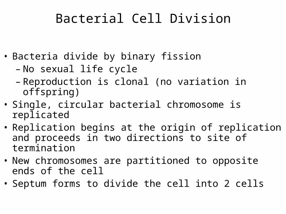
Bacterial Cell Division
• Bacteria divide by binary fission– No sexual life cycle– Reproduction is clonal (no variation in offspring)
• Single, circular bacterial chromosome is replicated• Replication begins at the origin of replication and
proceeds in two directions to site of termination• New chromosomes are partitioned to opposite ends of
the cell• Septum forms to divide the cell into 2 cells


Eukaryotic Chromosomes
• Every species has a different number of chromosomes• Humans have 46 chromosomes in 23 nearly identical
pairs– Additional/missing chromosomes usually fatal

Eukaryotic Chromosomes
Chromosomes
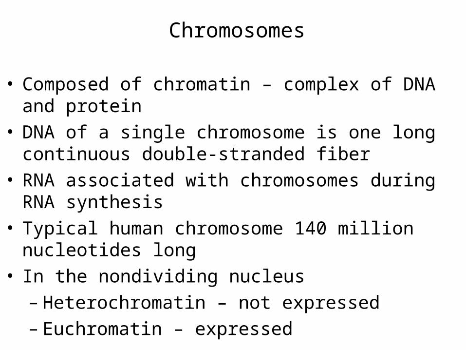
• Composed of chromatin – complex of DNA and protein
• DNA of a single chromosome is one long continuous double-stranded fiber
• RNA associated with chromosomes during RNA synthesis
• Typical human chromosome 140 million nucleotides long
• In the nondividing nucleus– Heterochromatin – not expressed– Euchromatin – expressed

Chromosome Structure
• Nucleosome– Complex of DNA and histone proteins– Promote and guide coiling of DNA– DNA duplex coiled around 8 histone proteins every
200 nucleotides– Histones are positively charged and strongly
attracted to negatively charged phosphate groups of DNA


• Nucleosomes wrapped into higher order coils called solenoids– Leads to a fiber 30 nm in diameter– Usual state of nondividing (interphase) chromatin
• During mitosis, chromatin in solenoid arranged around scaffold of protein to achieve maximum compaction– Radial looping aided by condensin proteins

Karyotype
• Particular array of chromosomes in an individual organism– Arranged according to size, staining properties,
location of centromere, etc.• Humans are diploid (2n)
– 2 complete sets of chromosomes– 46 total chromosomes
• Haploid (n) – 1 set of chromosomes– 23 in humans
• Pair of chromosomes are homologous– Each one is a homologue


Replication
• Prior to replication, each chromosome composed of a single DNA molecule
• After replication, each chromosome composed of 2 identical DNA molecules– Held together by cohesin proteins
• Visible as 2 strands held together as chromosome becomes more condensed– One chromosome composed of 2 sister chromatids

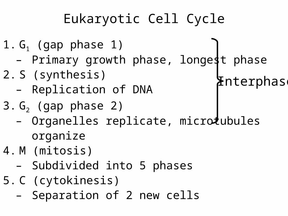
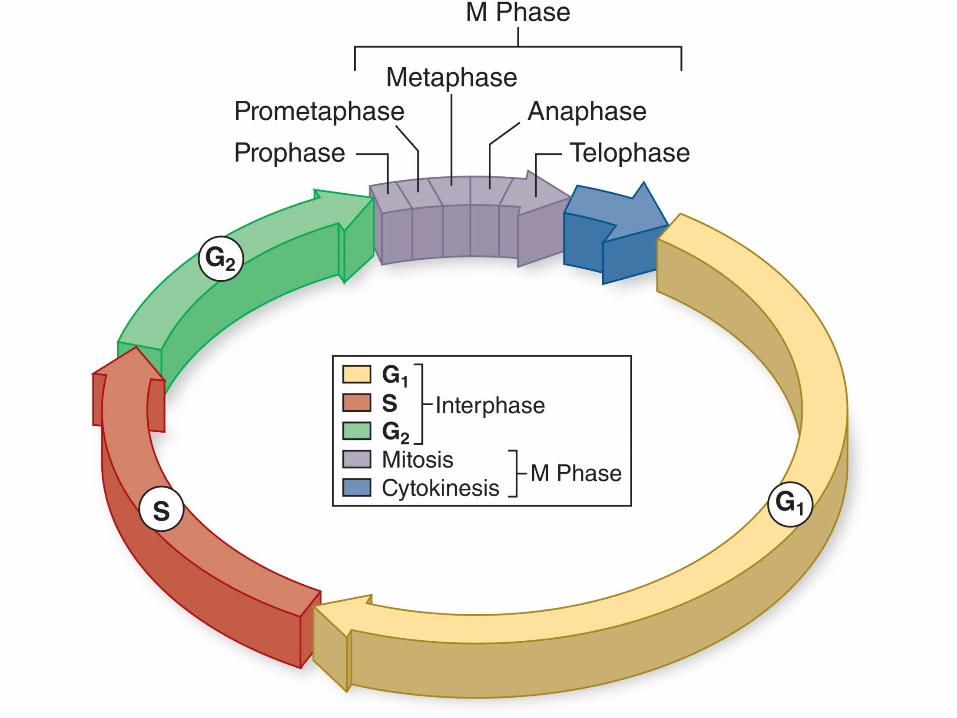
Eukaryotic Cell Cycle
1. G1 (gap phase 1)– Primary growth phase, longest phase
2. S (synthesis)– Replication of DNA
3. G2 (gap phase 2)– Organelles replicate, microtubules
organize4. M (mitosis)
– Subdivided into 5 phases5. C (cytokinesis)
– Separation of 2 new cells
Interphase

Duration
• Time it takes to complete a cell cycle varies greatly• Fruit fly embryos = 8 minutes• Mature cells take longer to grow
– Typical mammalian cell takes 24 hours– Liver cell takes more than a year
• Growth occurs during G1, G2, and S phases– M phase takes only about an hour
• Most variation in length of G1

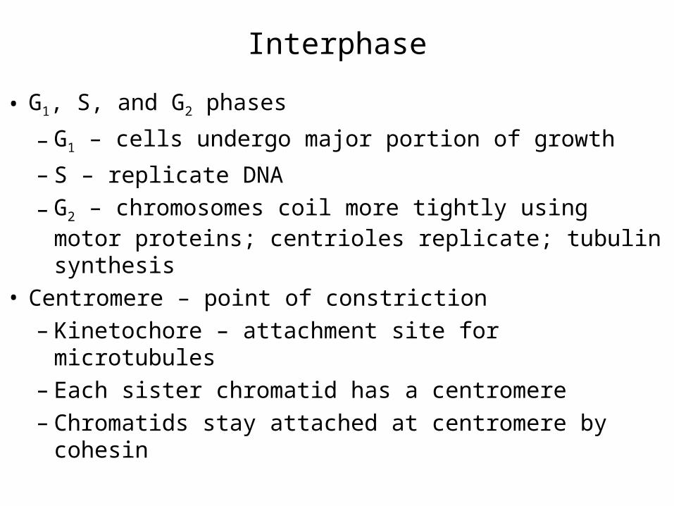
Interphase
• G1, S, and G2 phases
– G1 – cells undergo major portion of growth
– S – replicate DNA
– G2 – chromosomes coil more tightly using motor proteins; centrioles replicate; tubulin synthesis
• Centromere – point of constriction– Kinetochore – attachment site for microtubules– Each sister chromatid has a centromere– Chromatids stay attached at centromere by cohesin


M phase
Mitosis is divided into 5 phases:
1. Prophase
2. Prometaphase
3. Metaphase
4. Anaphase
5. Telophase
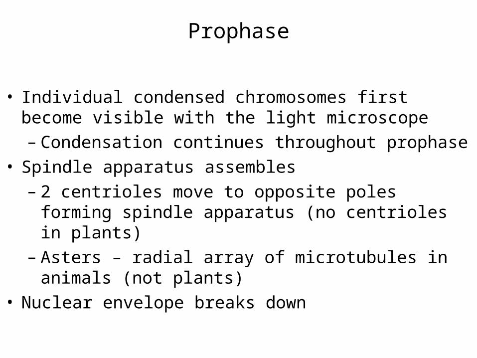
Prophase
• Individual condensed chromosomes first become visible with the light microscope– Condensation continues throughout prophase
• Spindle apparatus assembles– 2 centrioles move to opposite poles forming spindle
apparatus (no centrioles in plants)– Asters – radial array of microtubules in animals (not
plants)• Nuclear envelope breaks down


Prometaphase
• Transition occurs after disassembly of nuclear envelope
• Microtubule attachment– 2nd group grows from poles and attaches to
kinetochores– Each sister chromatid connected to opposite poles
• Chromosomes begin to move to center of cell – congression – Assembly and disassembly of microtubules– Motor proteins at kinetochores


Metaphase
• Alignment of chromosomes along metaphase plate– Not an actual structure– Future axis of cell division


Anaphase
• Begins when centromeres split• Key event is removal of cohesin proteins from all
chromosomes• Sister chromatids pulled to opposite poles• 2 forms of movements
– Anaphase A – kinetochores pulled toward poles– Anaphase B – poles move apart


Telophase
• Spindle apparatus disassembles• Nuclear envelope forms around each set of sister
chromatids• Chromosomes begin to uncoil• Nucleolus reappears in each new nucleus


Cytokinesis
• Cleavage of the cell into equal halves• Animal cells – constriction of actin filaments produces
a cleavage furrow• Plant cells – cell plate forms between the nuclei• Fungi and some protists – nuclear membrane does
not dissolve; mitosis occurs within the nucleus; division of the nucleus occurs with cytokinesis

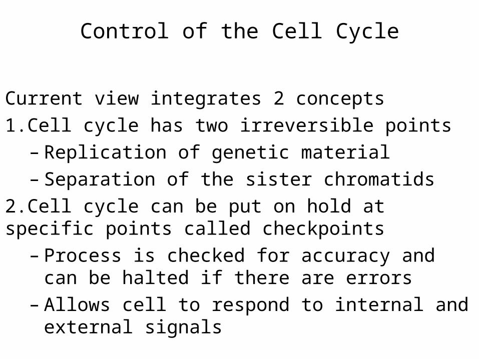
Control of the Cell Cycle
Current view integrates 2 concepts
1.Cell cycle has two irreversible points– Replication of genetic material– Separation of the sister chromatids
2.Cell cycle can be put on hold at specific points called checkpoints
– Process is checked for accuracy and can be halted if there are errors
– Allows cell to respond to internal and external signals
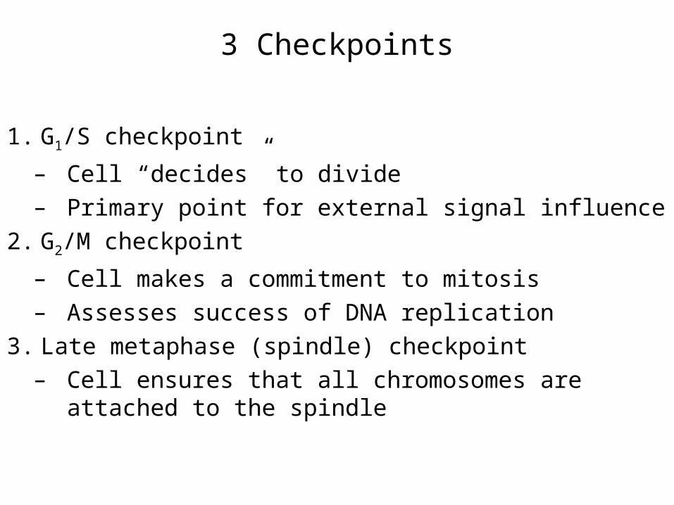
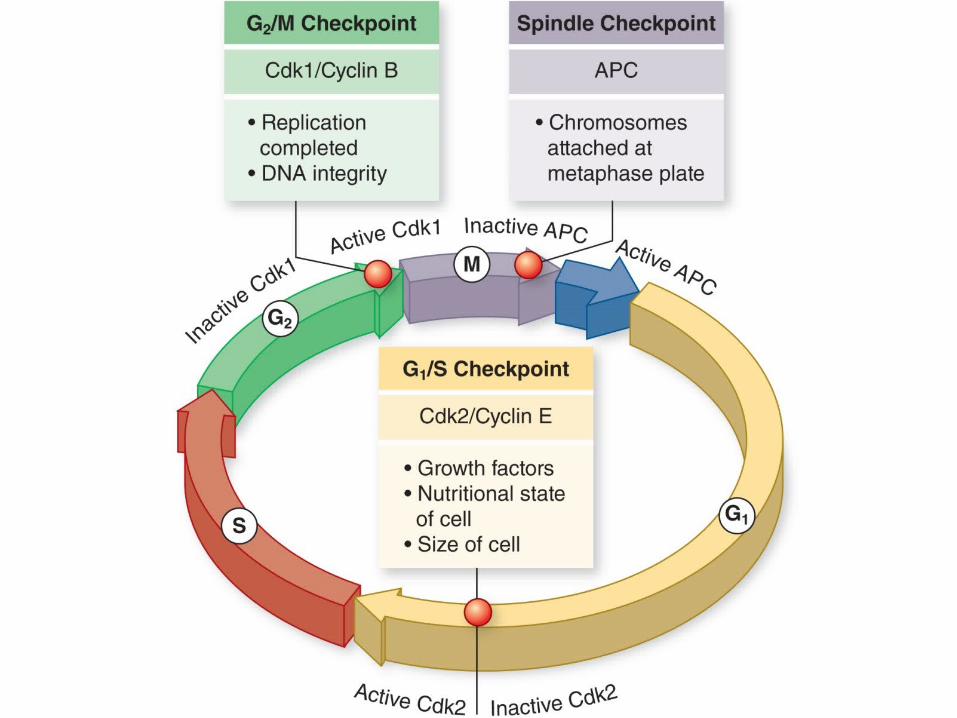
3 Checkpoints
1. G1/S checkpoint
– Cell “decides” to divide– Primary point for external signal influence
2. G2/M checkpoint
– Cell makes a commitment to mitosis– Assesses success of DNA replication
3. Late metaphase (spindle) checkpoint– Cell ensures that all chromosomes are attached to
the spindle


Cyclin-dependent kinases (Cdks)
• Enzymes that phosphorylate proteins• Primary mechanism of cell cycle control• Cdks partner with different cyclins at different points in
the cell cycle• For many years, a common view was that cyclins
drove the cell cycle – that is, the periodic synthesis and destruction of cyclins acted as a clock
• Now clear that Cdk itself is also controlled by phosphorylation

• Cdk – cyclin complex– Also called mitosis-promoting factor (MPF)
• Activity of Cdk is also controlled by the pattern of phosphorylation– Phosphorylation at one site (red) inactivates Cdk– Phosphorylation at another site (green) activates
Cdk