Embed Size (px)
Citation preview

CASE I: V13-1511 (JPC 4028574 ).
Signalment: 11-year-old, male castrated Shih Tzu, canine (Canis familiaris).
History: Presented with glaucoma OS. Increased IOP OS, significantly deceased tear production OU and moderate to severe pigmentary keratitis OU. Unable to view intraocular structures OU.
Gross Pathology: Entire globe, anterior lens luxation with fusion of lens to cornea and loss of lens structure.
Histopathologic Description: None provided.
Contributor’s Morphologic Diagnosis: Eye: End stage globe with: corneal vascularization and pigmentation; lens luxation, rupture and fusion to cornea; anterior chamber collapse; synechia of iris
1
J o i n t P a t h o l o g y C e n t e rVe t e r i n a r y P a t h o l o g y S e r v i c e s
WEDNESDAY SLIDE CONFERENCE 2012-2013
C o n f e r e n c e 2 3 1 May 2013
1-1. Eye, dog: The globe is mildly enlarged with anterior displacement of the lens and detachment of the retina. (HE 0.63X)
1-2. Eye, dog: The lens is flattened in an anterior-posterior direction, and present completely within the anterior chamber, where it is attached to the posterior aspect of the cornea (anterior synechia). (HE 15X)

to caudal lens; chronic retinal atrophy and detachment and optic nerve cupping.
Contributor’s Comment: Lens displacement refers to a change in position of the lens from its normal anatomical location and can be partial (subluxation) or complete (luxation). This condition affects several species and is a well-documented clinical disease in dogs. Primary lens displacement (PLD) occurs spontaneously,
and it is likely due to an inherited defect in the lens zonules. This defect leads to spontaneous rupture of the lens zonules inducing initial lens instability that may eventually result in luxation of the lens. Secondary lens displacement occurs when the zonules are disrupted by a prior or concomitant disease such as glaucoma, cataracts, uveitis, or trauma. In these cases, the zonules are not defective, but rather damaged by the underlying disease, and they break down causing lens instability.1 A recent study found ADAMTS17 mutation associated with primary lens luxation is widespread among breeds.3,7
If the zonular disinsertion is partial and the lens still rests in the patella fossa of the anterior vitreous, it is described as a posterior subluxation. If the zonular disinsertion is total, with migration of the lens posteriorly onto the floor of
the vitreous, or anteriorly through the pupil, into the anterior chamber, the lens is described as luxated. Pathogenesis of sequelae involve the distortion of the visual optics, physical irritation by the displaced lens to retina, anterior uvea, and/or cornea, and pupillary block by the lens itself of the adherent vitreous with resultant secondary glaucoma. Luxated lenses become cataractous if they are not already, likely due to altered nutrition.
WSC 2012-2013
2
1-3. Eye, dog: Internal changes within the lens include liquefaction and mineralization of lens fibers (green arrows), and fibrous metaplasia of lens fibers at the posterior aspect. (HE 60X)
1-4. Eye, dog: There is diffuse marked atrophy of the detached retina, with loss of the ganglion cell layer, inner nuclear and plexiform layers (consistent with glaucoma), and photoreceptor layer. (HE 85X)
1-5. Eye, dog: There is cupping of the optic nerve, also consistent with chronic increased intraocular pressure. (HE 25X)

Primary luxation occurs in the terrier breeds; dogs are usually middle-aged and the condition is always bilateral although not necessarily concurrently so. The affected eye may present with iridodonesis, an aphakic crescent, and irritation manifested by redness, blepharospasm, and tearing if the lens is subluxated; IOP may be variable. Anterior luxation is characterized by visualization of the lens equator and obscuration of the pupil; IOP will usually be elevated and if it is not, it soon will be. Posterior luxations are diagnosed by observing a deep anterior chamber with a concavity to the iris surface, and the lens on the floor of the posterior segment; IOP is usually not elevated. Examine fellow eyes closely; iridodonesis may be present and dilation may reveal an aphakic crescent. Dilation may turn an innocuous posterior subluxation into an emergent anterior luxation. In cats, the majority of luxations occur secondary to chronic uveitis; secondary glaucoma is uncommon compared to the dog, related to the deep feline anterior chamber and the liquefaction of the vitreous that accompanies chronic inflammation.
Primary lens displacement has been documented to be heritable in many terrier breeds as well as Tibetan Terriers, Border Collies, and Shar-Peis. PLD is invariably a bilateral condition, albeit usually not simultaneous. When a patient presents with one eye affected, the fellow eye may vary in presentation from no apparent instability to early signs of instability such as iridodensis, phacodonesis, and vitreal herniation, to clear evidence of subluxation or complete luxation. The reported time to displacement in the fellow eye varies from days to years. Lens displacement often leads to vision- threatening complications, of which glaucoma is the most common and can occur regardless of lens position (anterior, posterior, or subluxated). Other reported complications include retinal detachment and uveitis.4
JPC Diagnosis: Eye: Anterior lens luxation, with corneal adhesions, anterior and posterior synechiae, drainage angle occlusion, retinal detachment and atrophy, and optic nerve degeneration and excavation.
Conference Comment: There is significant slide variation in this case, with not all slides exhibiting all the described changes.
Conference participants discussed the difficulty in determining primary versus secondary lens luxation from histology alone, concluding that similar lesions can be seen in both. As the contributor notes, primary luxation of the lens is due to rupture of the lens zonules, which function to keep the lens in its normal position between the ciliary processes. The zonular fibers are made of microfibrils (primarily the glycoproteins fibrillin-1 and fibrillin-2). Genetic abnormalities in genes coding for fibrillin-1 have been shown to cause lens displacement in humans and possibly in cattle.6 A truncating gene mutation in ADAMTS17 on CFA03 has been found to be associated with the development of primary lens displacement in 17 dog breeds; however, its mode of inheritance and penetrance have yet to be fully elucidated.2 Primary lens displacement in dogs appears to be affected by both dose and age (i.e. heterozygotes are affected later in life than homozygotes, and penetrance increases with age), which is consistent with an additive allelic model.2 ADAMTS17 is a member of the secreted metalloproteinase family of proteins which bind extracellular matrix; it is believed to play a role in the formation of crystalline lens zonules and connective tissue. Mutations in ADAMTS17 cause abnormalities similar to Weill-Marchesani syndrome (WMS) in humans. WMS, which is associated with mutations in a related gene (ADAMTS10), as well as FBN1, is a rare connective tissue disorder in which patients develop eye and skeletal abnormalities.5
Contributing Institution: Tri Service Research Lab4141 Petroleum RoadFort Sam Houston, TX 78234
References : 1 . A la r io AF, P i zz i r an i S , P i r i e CG. Histopathologic evaluation of the anterior segment of eyes enucleated due to glaucoma secondary to primary lens displacement in 13 canine globes. Vet Ophthalmol. 2012;(4):doi 10.1111.2. Gharahkhani P, O’Leary C, Duffy D, Bernays M, Kyaw-Tanner M. Primary lens luxation in Australian Tenterfield and Miniature Bull Terriers is due to an old ADAMTS17 mutation and is an additive trait. The Open Genomics Journal. 2012;5:7-13.
WSC 2012-2013
3

3. Gould D, Pettitt L, McLaughlin B, Holmes N, Forman O, Thomas A, Ahonen S, Lohi H, O’Leary C, Sargan D, Mellersh C. ADAMTS17 mutation associated with primary lens luxation is w idesp read among b reeds . Ve te r inary Ophthalmology. 2011;14, 6, 378–384.4. Gelatt K, MacKay E. Secondary glaucomas in the dog in North America . Veter inary Ophthalmology. 2004;7:245–259. 5. Morales J, Al-Sharif L, Khalil DS, Shinwari JMA, Bavi P, Al-Mahrouqi RA, et al . Homozygous mutations in ADAMTS10 and ADAMTS17 cause lenticular myopia, ectopia lentis, glaucoma, spherophakia, and short stature. Am J Hum Genet. 2009;85(5):558–568.6. Morris RA, Dubielzig RR. Light-microscopy evaluation of zonular fiber morphology in dogs with glaucoma: secondary to lens displacement. Vet Ophthalmol. 2005;8(2)81-44. 7. Sargan DR, Withers D, Pettiite I, Squire M, Gould DJ, Mellersh CS. Mapping the mutation causing lens luxation in several terrier breeds. Journal of Heredity. 2007:98(5):534–538.Blackwell Publishing, Ltd.
WSC 2012-2013
4

CASE II: JPC012 (JPC 4020974).
Signalment: One month old, intact male Dorper lamb (Ovis aries).
History: This lamb was smaller than normal at birth and showed poor weight gain while nursing. The owner noticed that it became weak with labored breathing and increased lung sounds. Antibiotic treatment (oxytetracycline) was initiated, but the animal was found dead five days later.
Gross Pathology: Approximately 70-80% of the lungs contained moderately well-demarcated, often confluent, firm, white-tan nodular areas, with only a small portion of the dorsal-caudal regions unaffected. On section, many bronchi were filled with a white-tan material. No thymic tissue was identified. No other abnormalities were noted.
Laboratory Results: Nocardia sp. was isolated from the cut surface of affected lung.
Histopathologic Description: Lung: Filling or effacing up to 75% of alveoli and bronchioles are multifocal to coalescing, variably sized and shaped pyogranulomas characterized by central areas of necrotic cellular debris, high numbers of degenerate and necrotic neutrophils and macrophages, and mineralization surrounded by variable mixtures of neutrophils , large m a c r o p h a g e s , f e w e r l y m p h o c y t e s a n d multinucleated giant cells. Adjacent, less affected
airways and alveoli are often compressed and contain congestion and hemorrhage, fewer numbers of inflammatory cells and multifocal areas of edema and/or basophilic-staining material (mucus). On H&E stained sections, faintly-staining, fine filamentous organisms can be seen sporadically at the margins of the necrotic areas.
Special stains revealed numerous, approximately 1 micron thick, beaded to filamentous and occasionally branching, gram-positive (Brown & Brenn) and variably acid-fast (Fite-Faraco) bacteria within areas of inflammation.
Contributor’s Morphologic Diagnosis: Lung: Pyogranulomas, multifocal and coalescing, marked, with necrosis, mineralization and numerous branching, filamentous gram-positive and acid-fast bacteria, lamb (Ovis aries).
Contributor’s Comment: Nocardia spp. are ubiquitous, saprophytic, aerobic bacteria that are commonly associated with opportunistic infections in animals and people characterized by pyogranulomatous inflammation. The organisms are observed as long, thin, beaded filaments with frequent right-angle branching that are said to have a “Chinese character” pattern. They can be easily overlooked on standard H&E stained sections and are better demonstrated with special stains including Gomori methenamine silver (GMS), Gram stains and modified acid-fast stains such as Fite-Faraco.9
WSC 2012-2013
5
2-1. Lung, sheep: Randomly scattered throughout the lung, there are numerous foci of inflammation which compress the adjacent hypercellular pulmonary parenchyma. (HE 0.63X)
2-2. Lung, sheep: Within inflammatory foci, alveoli are filled with large numbers of degenerate neutrophils and cellular debris; there is central septal necrosis. (HE 168X)

The primary differential diagnosis for pulmonary nocardiosis includes infections with Actinomyces and some atypical mycobacteria.9 Actinomycosis is more commonly associated with sulfur granules and Splendore-Hoeppli material, and the organisms are not acid fast.10 Atypical
mycobacteria are much shorter beaded bacilli and do not exhibit true branching.
Nocardia are commonly found in the environment, particularly soil enriched with decayed organic matter. Routes of infection include inhalation, inoculation, and ingestion. In people, Nocardia are generally cons ide red pa thogen ic bacteria with relatively low v i ru lence . Wi th the exception of cutaneous disease, human infection is typically opportunistic, occurring more commonly in the elderly and people with compromised T cell-mediated immunity, as with prolonged steroid treatment or HIV infection.6 In the present case, it is not clear if the absence of a normal a p p e a r i n g t h y m u s a t necropsy was due to a
primary condition predisposing the animal to infection (thymic hypoplasia) or a secondary response to the severe infection (thymic atrophy).
WSC 2012-2013
6
2-3. Lung, sheep: Occasional multinucleated giant cell macrophages are scattered throughout the parenchyma adjacent to inflammatory foci. (HE 180X)
2-4. Lung, sheep: A tissue Gram stain reveals moderate numbers of 1um diameter, filamentous, occasionally beaded gram-positive bacteria within inflammatory foci. (Photo courtesy of: Covance Laboratories, Inc, Madison, Wisconsin, USA. http://www.covance.com/products/nonclinical/toxicology/risk-assessment/index.php) (Brown-Brenn, 400X)
2-5. Lung, sheep: An acid-fast stain reveals moderate numbers of 1µm diameter, filamentous, occasionally beaded, variably acid-fast bacteria within inflammatory foci. (Photo courtesy of: Covance Laboratories, Inc, Madison, Wisconsin, USA. http://www.covance.com/products/nonclinical/toxicology/risk-assessment/index.php) (Brown-Brenn, 400X)

Nocardiosis is an important cause of pneumonia, pleuritis and empyema in dogs, particularly hunting/sporting dogs in which, following inhalation or ingestion of plant material, migrating grass awns can contaminate the pleural spaces with bacteria resulting in a putrid “tomato soup-like” hemorrhagic pyothorax. In most other species, pulmonary nocardiosis more closely resembles the nodular pyogranulomatous and necrotizing inflammation observed in this lamb. Nocardia infections have been infrequently reported in primates, cats, horses, cattle and marine mammals.1,2,5,8 In cattle, infections have been associated with mastitis, pneumonia, dermatitis, placentitis with abortion, and disseminated disease.1 Nocardia infections in other ruminants are apparently very uncommon, but have been reported in sheep, goats, llama, bison and reindeer.3,7,10 Nocardiosis is reportedly a significant and often fatal disease in marine mammals.8 Pinnipeds, particularly hooded seals, and cetaceans appear to be prone to a systemic form of infection which typically involves the lung and thoracic lymph nodes and, to lesser extent, brain and skin.
J P C D i a g n o s i s : L u n g : P n e u m o n i a , pyogranulomatous, multifocal to coalescing, severe, with numerous filamentous gram-positive, acid-fast bacteria.
Conference Comment: In addition to discussing nocardiosis in the various species so well described by the contributor, conference participants also discussed the so-called “nocardioform” placentitis associated with equine abortion. Nocardioform placentitis can be caused by several genera of gram positive, branching actinomycetes that share similarities with Nocardia species, including the following: Crossiella equi, Amycolatopsis kentuckyensis, Amycolatopsis lexingtonensis, Amycolatopsis pretoriensis, Streptomyces atriruber, and Streptomyces silaceus.4 Clinically, nocardioform placentitis presents as late gestation abortion, stillbirth, prematurity or full term but weak foals. Grossly, a thick, light brown exudate is often observed in the chorion at the bifurcation of the horns of the affected placenta. The organisms do not reach the fetus; thus fetal lesions are those of placental insufficiency. Recently, nocardioform placentitis resulted in a record number of equine abortions in the 2011 foal crop in Kentucky. The most prominent actinomycetes found in this series
of third trimester abortions were Amycolatopsis spp. and Crosiella equi; fewer Streptomyces, Microbacterium, Nocardia and Allokutzneria species were identified.4
Contributing Institution: Covance Laboratories, IncMadison, Wisconsin, USA http://www.covance.com/products/nonclinical/toxicology/risk-assessment/index.php
References:1. Bawa B, Bai J, Whitehair M, Purvis T, et al. Bovine abortion associated with Nocardia farcinica. J Vet Diagn Invest. 2010;22(1):108-11.2. Bolon B, Buergelt CD, Cooley AJ. Abortion in two foals associated with Nocardia infection. Vet Pathol.1989;26(3):277–278.3. Chang CD, Boosinger TR, Dowling PM, et al. Nocardiosis in a llama. J Vet Diagn Invest. 1993;5(4):631-4.4. Erol E, Sells SF, Williams NM, Kennedy L, Locke SJ, Labeda DP, et al. An investigation of a recent outbreak of nocardioform placentitis c a u s e d a b o r t i o n s i n h o r s e s . V e t Microbiol. 2012:17;158(3-4):425-30.5. Klumpp SA, McClure HM. Nocardiosis, lung. In: Jones TC, Mohr U, Hunt RD, eds. Monographs on Pathology of Laboratory Animals: Nonhuman Primates II. Berlin and New York: Springer-Verlag; 1993:99–103. 6. McAdam AJ, Sharpe AH. Infectious diseases. In: Kumar V. Abbas A. Fausto N, eds. Robbins and Cotran: Pathologic Basis of Disease. 8th edition. Philadelphia, PA: Elsevier Saunders; 2010:362-363. 7. Pal M. Nocardia asteroides as a cause of pneumonia in a buffalo calf. Review of Scientific Technical Office International Des Epizootics. 1997;16:881-884. 8. St. Leger JA, Begeman L, Fleetwood M, et al. Comparative pathology of nocardiosis in marine mammals. Vet. Pathol. 2009;46:299–308.9. Travis WD, Colby TV, Koss MN, et al. Lung Infections. Atlas of Nontumor Pathology. Non-neoplastic Disorders of the Lower Respiratory Tract. Washington, DC: American Registry of Pathology; 2002:557-563.10. Vemireddi V, Sharma A, Wu CC, et al. Systemic nocardiosis in a reindeer (Rangifer tarandus tarandus), J. Vet. Diagn. Invest. 2007;19(3):326-329.
WSC 2012-2013
7

CASE III: AR12-0051 (JPC 4017933).
Signalment: Adult male Sprague-Dawley rat, Rattus norvegicus.
History: This transgenic rat for the mouse Ren2 gene was found dead.
Gross Pathology: The small intestinal mesenteric arteries were diffusely dark red, nodular, tortuous and measured up to 5mm in diameter. Both kidneys had many 0.5mm round indentations on their surfaces.
Histopathologic Description: The renal changes are consistent with an end stage kidney with polyarteritis nodosa and hypertensive arterial disease. The renal capsule is irregularly indented throughout. The underlying interstitium has tubular loss with replacement by fibrous connective tissue and infiltration by lymphocytes and plasma cells. The glomerular mesangium is regularly thickened by pale eosinophilic amorphous material, the tufts are large and prominently segmented, and periglomerular fibrosis is prominent. Occasional glomeruli are shrunken, have synechia or are sclerotic. The cortical renal tubules are dilated, up to 1mm across, by eosinophilic amorphous material, and are lined by attenuated epithelium. Tubular epithelial cells are occasionally swollen with vacuola ted cy toplasm, and some have hypereosinophilic cytoplasm and pyknotic nuclei.
The tunica media of medium-sized arteries is thickened by concentric rings of excessive smooth muscle and sometimes fibrous connective tissue, and the walls are infiltrated by lymphocytes and fewer macrophages. Hypereosinophilic amorphous material obscures the tunica media in some slides (fibrinoid change).
Contributor’s Morphologic Diagnosis: 1. Glomerulonephropathy, diffuse, chronic, severe with tubular proteinosis and interstitial lymphocytic nephritis and fibrosis (chronic progressive nephropathy).2. Arteritis, segmental, chronic, moderate to severe, lymphohistiocytic with fibrinoid change, kidney (polyarteritis nodosa).3. Smooth muscle hyperplasia, segmental, chronic, moderate, arterial tunica media, kidney (hypertensive change).
Contributor ’s Comment: This case demonstrates three histological renal lesions in a hypertensive rat: chronic progressive nephropathy (CPN), polyarteritis nodosa and hypertensive arterial disease. CPN is a common age-related lesion most common in males, with increased incidences in Sprague-Dawley and Fischer 344 rats. Grossly, the renal cortices are irregular and pitted, as in this case. The kidneys may be enlarged, pale and have linear streaks in the cortex and medulla. Histologically, glomerular changes include basement membrane and mesangial thickening, segmental sclerosis, and
WSC 2012-2013
8
3-1. Mesentery, Ren2 transgenic rat: Mesenteric arteries in the region of the small intestine are diffusely dark red, nodular, tortuous and measure up to 5mm in diameter. (Photo courtesy of: Wake Forest University Health Sciences, Animal Resources Program, Medical Center Boulevard, Winston-Salem, NC 27157 http://www.wfubmc.edu/schoolofMedicine/schoolofMedicine_default.aspx?id=26651
3-2. Kidney, Ren2 transgenic rat: At the subgross level, the marked ectasia of tubules and pitted capsule are evident, suggesting chronic progressive nephropathy. (HE 0.63X)

synechia. Tubules are regularly dilated, protein-filled, and lined by flattened epithelial cells. Interstitial fibrosis and inflammation are common. Advanced cases may also have secondary hyperparathyroidism with metastatic calcification in the kidneys, gastric mucosa, lungs and/or tunica media of larger arteries. Late-stage CPN has been associated with hypertension and polyarteritis nodosa.7
Similar to CPN, polyarteritis nodosa is a chronic progressive degenerative disease occurring most frequently in aged Sprague-Dawley rats, spontaneous hypertensive rats, and rats with late-stage chronic nephropathy. At necropsy the mesenteric vessels are often enlarged, tortuous and segmentally thickened, as in this case. Polyarteritis nodosa is characterized histologically by fibrinoid degeneration and thickening of the
tunica media of medium-sized arteries with infiltrating mononuclear cells and fewer neutrophils. The lumina may vary in size and have thromboses, some of which may recanalize. Polyarteritis nodosa has also been reported in mice,7 cats,1 dogs,11 pigs,2,4 cynomolgus macaques9 and foxes.6 Although the cause is uncertain, the lymphohistiocytic inflammation suggests it may have an immune-mediated basis.4,7,9
The rat is a common animal model of hypertension. Spontaneously hypertensive Wistar rats, Dahl-salt sensitive rats, transgenic mREN2 rats, and rats administered deoxycorticosterone acetate (DOCA) in combination with a high salt diet are the most common models.3,8 In all of these models, the hypertensive animals have impaired endothelium-dependent relaxation and vascular remodeling which lead to increased
WSC 2012-2013
9
3-3. Kidney, Ren2 transgenic rat: Multifocally, the intima of renal arterioles is expanded by bright eosinophilic protein, and the intima and media are infiltrated by neutrophils admixed with cellular debris (fiibrinoid necrosis). Additionally, there is lamellar fibrosis of the tunica adventitia, suggesting prolonged hypertension. (HE 144X)

vascular resistance and renal damage.3,8 Severe end-organ damage involving the heart, brain and kidney is only seen in a subset of animals within some models. This rat was transgenic (TGR[mREN2]27), with over-expression of the mouse Ren-2 gene which increases renin activity. Renin catalyzes the conversion of angiotensinogen to angiotensin I, which is then converted to angiotensin II by angiotensin-converting enzyme. Angiotensin II causes vasoconstriction and increased blood pressure. Hypertensive changes are further exacerbated by angiotensin II-induced a ldos te rone sec re t ion which inc reases mineralocorticoid levels and sodium reabsorption, thus increasing blood volume osmotically and further increasing blood pressure.10
JPC Diagnosis: 1. Kidney, arteries: Arteritis, proliferative and necrotizing, multifocal, severe, with fibrinoid necrosis.2. Kidney, arteries: Arteriosclerosis, multifocal, moderate.3. Kidney: Glomerulonephritis, diffuse, with tubular degeneration, necrosis and regeneration, proteinosis, and chronic interstitial nephritis.
Conference Comment: The contributor provides an excellent summary of the various changes observed in this animal model of hypertension. Veterinary species are not as commonly affected with idiopathic (essential) hypertension as humans; however, several species, including cats and dogs, develop systemic hypertension which is often secondary to other conditions. In the cat, hyper tens ion i s o f ten assoc ia ted wi th
hyperthyroidism and chronic renal failure; whereas in the dog, it is more likely to be assoc ia ted wi th chronic rena l fa i lu re , hyperadrenocorticism, or pheochromocytomas.5
Conference participants discussed the vessel wall changes observed in this case, noting that hypertension results in several pathologic changes in the walls of small arteries and arterioles, including two types of arteriolosclerosis: hyaline and hyperplastic. Small blood vessels in hyaline arteriolosclerosis exhibit a homogeneous, eosinophilic thickening of the wall and narrowing of the lumen due to endothelial cell damage and the subsequent increased vascular permeability and plasma protein leakage into the vessel wall. Hyperplastic arteriolosclerosis, on the other hand, is characterized by a concentric, laminated thickening of the wall (“onion skinning”) and narrowing of the lumen which is due to smooth muscle cell hyperplasia and reduplication of b a s e m e n t m e m b r a n e s . H y p e r p l a s t i c arteriolosclerosis can also be associated with fibrinoid necrosis in the vessel walls.10
Contributing Institution: Wake Forest University Health SciencesAnimal Resources ProgramMedical Center BoulevardWinston-Salem, NC 27157http://www.wfubmc.edu/schoolofMedicine/schoolofMedicine_default.aspx?id=26651
WSC 2012-2013
10
3-4. Kidney, Ren2 transgenic rat: Less affected arterioles contain degenerate neutrophils admixed within the inner tunica media. (HE 144X)
3-5. Kidney, Ren2 transgenic rat: Lamellar fibrosis of the tunica adventitia is a common finding in hypertension. (HE 168X).

References: 1. Altera KP and Bonasch H. Periarteritis nodosa in a cat. J Am Vet Med Assoc. 1966;149(10):1307-11. 2. Elling F. Nutritionally induced necrotizing glomerulonephritis and polyarteritis nodosa in pigs. Acta Pathol Microbiol Scand A. 1979;87A(1-6):387-392. 3. Dornas W, Silva ME. Animal models for the study of arterial hypertension. J Biosci. 2011;36(4): 731-737. 4. Hélie P, Drolet R, Germain MC, Bourgault A. “ S y s t e m i c n e c r o t i z i n g v a s c u l i t i s a n d glomerulonephritis in grower pigs in southwestern Quebec.” Can Vet J. 1995;36(3):150–154. 5 . Mi l l e r LM, Van V lee t JF, Ga l A . Cardiovascular system and lymphatic vessels. In: McGavin MD, Zachary JF, eds. Pathologic Basis of Veterinary Disease. 5th ed. St. Louis, MO: Elsevier Mosby; 2012:552. 6. Nordstoga K, Westbye K. Polyarteritis nodosa associated with nosematosis in blue foxes. Acta Pathol Microbiol Scand A. 1976;84(3):291-296. 7. Percy, DH, Barthold SW. Rat. In: Pathology of Laboratory Rodents and Rabbits. 3rd ed. Ames, Iowa: Blackwell Publishing; 2007:125-206. 8. Pinto YM, Paul M, Ganten D. Lessons from rat models of hypertension: from Goldblatt to genetic engineering. Cardiovasc Res. 1998;39:77-88. 9. Porter BF, Frost P, Hubbard GB. Polyarteritis Nodosa in a Cynomolgus Macaque (Macaca fascicularis). Vet Pathol. 2003;40(5):570-573. 10. Schoen FJ. Blood Vessels. In: Robbins and Cotran Pathologic Basis of Diseases. 8th ed. P h i l a d e l p h i a , PA : S a u n d e r s E l s e v i e r ; 2010:487-528. 11. Scott-Moncrieff JC, Snyder PW, Glickman LT, Davis EL, Felsburg PJ. Systemic necrotizing vasculitis in nine young beagles. J Am Vet Med Assoc. 1992;201(10):1553-1558.
WSC 2012-2013
11
CASE IV: V08-10649 (JPC 3141626).
Signalment: 10-year-old castrated male Belgian, equine (Equus caballus).
History: A 10-year-old Belgian gelding was euthanized following progressively worsening neurological signs, including severe ataxia, gait abnormalities, and sudden loss of balance with falling. The horse owner determined this was a severe safety risk for the handlers tending the horse. Originally, this horse was diagnosed positive for equine protozoal myeloencephalitis (EPM) eight months earlier. Despite appropriate medical treatment, clinical signs progressively deteriorated. This horse was insured and the insurance carrier requested a necropsy.
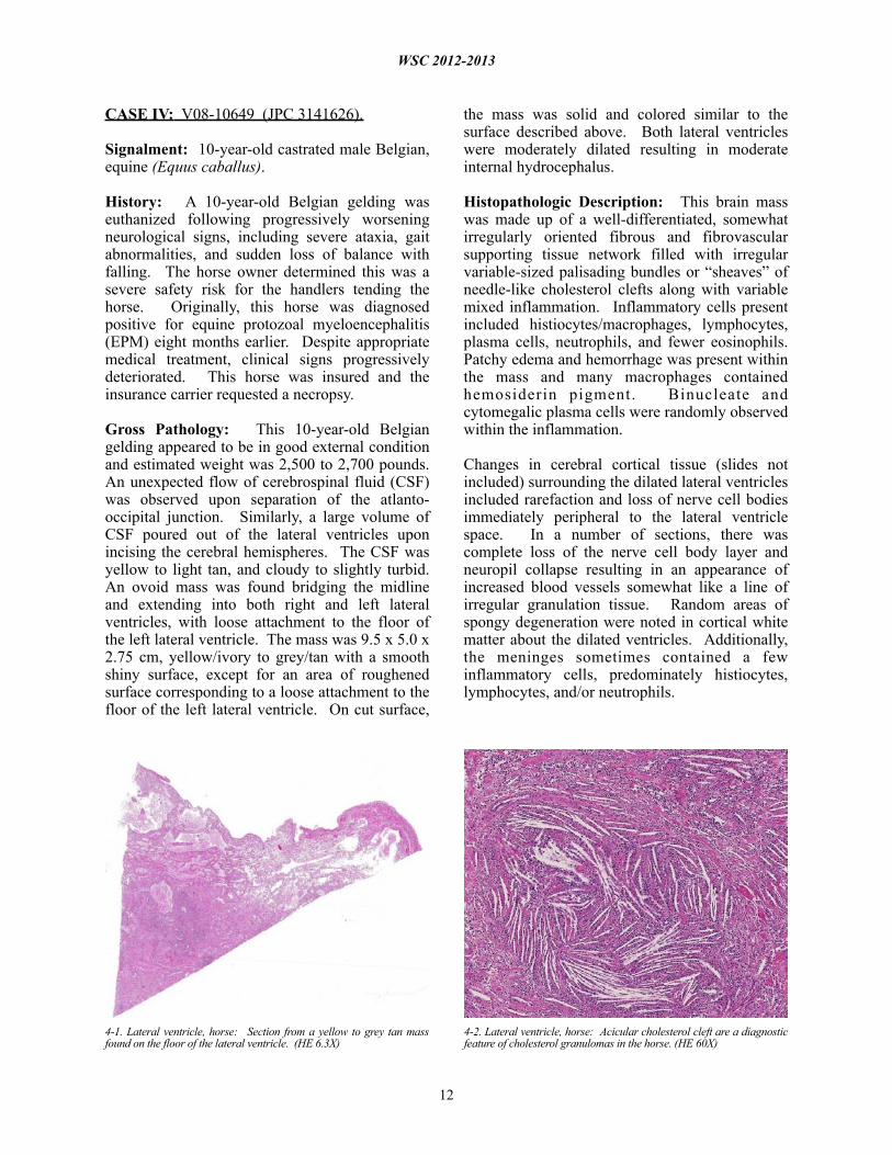
Gross Pathology: This 10-year-old Belgian gelding appeared to be in good external condition and estimated weight was 2,500 to 2,700 pounds. An unexpected flow of cerebrospinal fluid (CSF) was observed upon separation of the atlanto-occipital junction. Similarly, a large volume of CSF poured out of the lateral ventricles upon incising the cerebral hemispheres. The CSF was yellow to light tan, and cloudy to slightly turbid. An ovoid mass was found bridging the midline and extending into both right and left lateral ventricles, with loose attachment to the floor of the left lateral ventricle. The mass was 9.5 x 5.0 x 2.75 cm, yellow/ivory to grey/tan with a smooth shiny surface, except for an area of roughened surface corresponding to a loose attachment to the floor of the left lateral ventricle. On cut surface,
the mass was solid and colored similar to the surface described above. Both lateral ventricles were moderately dilated resulting in moderate internal hydrocephalus.
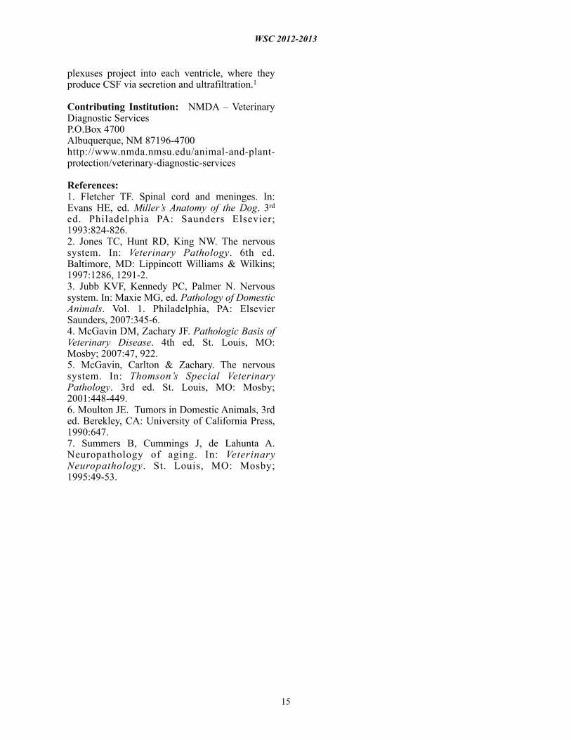
Histopathologic Description: This brain mass was made up of a well-differentiated, somewhat irregularly oriented fibrous and fibrovascular supporting tissue network filled with irregular variable-sized palisading bundles or “sheaves” of needle-like cholesterol clefts along with variable mixed inflammation. Inflammatory cells present included histiocytes/macrophages, lymphocytes, plasma cells, neutrophils, and fewer eosinophils. Patchy edema and hemorrhage was present within the mass and many macrophages contained hemosiderin pigment. Binucleate and cytomegalic plasma cells were randomly observed within the inflammation.
Changes in cerebral cortical tissue (slides not included) surrounding the dilated lateral ventricles included rarefaction and loss of nerve cell bodies immediately peripheral to the lateral ventricle space. In a number of sections, there was complete loss of the nerve cell body layer and neuropil collapse resulting in an appearance of increased blood vessels somewhat like a line of irregular granulation tissue. Random areas of spongy degeneration were noted in cortical white matter about the dilated ventricles. Additionally, the meninges sometimes contained a few inflammatory cells, predominately histiocytes, lymphocytes, and/or neutrophils.
WSC 2012-2013
12
4-1. Lateral ventricle, horse: Section from a yellow to grey tan mass found on the floor of the lateral ventricle. (HE 6.3X)
4-2. Lateral ventricle, horse: Acicular cholesterol cleft are a diagnostic feature of cholesterol granulomas in the horse. (HE 60X)

Contributor’s Morphologic Diagnosis: Brain mass: Cholesterol granuloma, right and left lateral ventricles, cerebrum, Belgian, equine.
Contributor ’s Comment: This case demonstrates the sometimes perplexing situation for clinical practitioners and veterinary pathologists alike, when a horse is presented for evaluation of what is often loosely referred to as an “equine neurological case.” Many of these cases may be relatively long-standing and may have been previously diagnosed, erroneously or not, with a specific neurological disease (in this case, equine protozoal encephalomyelitis [EPM]). Additionally, some cases may have been treated for a specific disease with or without improvement. At the time the veterinary pathologist receives the case, most likely some set of events or changes in the clinical state of the horse have taken place that warrant euthanasia of the horse.
Knowing the above chain of events is likely in many cases, then, the veterinary pathologist should preface the undertaking of an equine neurological case necropsy, with a set of all possible differential diagnoses, in addition to any previous clinical diagnosis. Many equine neurological cases, and particularly CNS cases, have overlap of signs. Even if a case has a diagnosis confirmed by reliable clinical testing, as in this case, that particular diagnosis may not be the cause of the recent chain of events that brought about the euthanasia of the horse. Finally, there are some equine neurological cases that may be a “rule out diagnosis.” Botulism is one disease that always should come to mind as an example of this. Because the levels of botulinum toxin required to affect and even kill a horse are so low that current modes of testing may be unable to detect, the pathologist should always have a conversation with the horse owner and clinical veterinarian, about any and all of the
WSC 2012-2013
13
4-3. Lateral ventricle, horse: The cholesterol granuloma is largely composed of mature collagen, large numbers of epithelioid macrophages and lymphocytes, and rare plasma cells and multinucleated giant cell macrophages. In this mass, vessels are surrounded by moderate amounts of fibrous connective tissue. (HE 220X)

details of environment and husbandry that could possibly lead to a final rule-out diagnosis of botulism.
Clearly then, cholesterol granulomas should be considered as a differential diagnosis in any equine neurological case, especially those in older horses with predominantly CNS signs. In fact, it has been reported that 15-20% of old horses have cholesterol granulomas, although many may be present in the absence of clinical signs.
Cholesterol granulomas and cholesteatoma have been used in veterinary literature to describe the lesion diagnosed in this case.2,3,4,6,7 There is some disagreement of sorts as to whether cholesteatoma should be used to describe this brain lesion, specifically found in old horses.2 Cholesteatoma is a term also used to describe a nodular mass in the middle ear, although this aural cholesteatoma has not been reported in horses.5 It may be a prudent move to restrict the use of cholesterol granulomas only to describe this specific entity of aged horses, rather than use cholesteatoma interchangeably with cholesterol granuloma.
Despite this sort of disagreement of terminology for equine cholesterol granulomas, the mechanism of development is basically agreed upon.2,3,4,6,7 The exact underlying pathogenesis, however, seems to be uncertain.7 Equine cholesterol granulomas are often described as an aging or degenerative phenomenon.2,3,5,7 Repeated episodes of hemorrhage and/or chronic intermittent congestion, edema and congestive hemorrhage in the choroid plexuses are believed to be the pathological processes responsible for development of cholesterol granulomas of older horses.2,3 Erythrocytes are likely the source of the cholesterol.6 It is when hemorrhage results in formation of a hematoma, subsequently becoming organized, followed by breakdown of red blood cells to release cholesterol that the granuloma develops.2 Cholesterol accumulation, both extracellular and within macrophages, is observed and the cholesterol itself stimulates a “foreign body response” or “giant cell reaction,”6 resulting in developing and progressive granulomatous i n f l a m m a t i o n a n d f i b r o v a s c u l a r “organization.”2,3,4,6,7 Whether this is or can result from a single event or requires multiple episodes is speculative, but likely the larger cholesterol granulomas develop because of multiple events of this type.
Small cholesterol granulomas may remain clinically silent and are discovered incidentally. The larger cholesterol granulomas produce clinical CNS signs and signs can be variable and progressive. Most, if not all, of the effects of large cholesterol granulomas are caused by blockage of interventricular foramen resulting in development of internal hydrocephalus and progressive degeneration and compression a t rophy o f t he ce r eb ra l hemi sphe re s . Inflammation in the brain itself is usually minimal and represented as small infi l trates of macrophage, lymphocytes, and neutrophils in the meninges. It may be fortunate that even though the occurrence of cholesterol granulomas in old horses is reported to be 15-20%, the majority are found in the fourth ventricle; and, development of hydrocephalus, the usual cause of clinical signs, is limited to those in the lateral ventricles that can by location block the interventricular formen causing hydrocephalus.3
JPC Diagnosis: Ventricle (left lateral, per c o n t r i b u t o r ) : C h o l e s t e r o l g r a n u l o m a (cholesteatoma).
Conference Comment: Conference participants discussed the typical gross and histopathologic findings as well the pathogenesis of cholesterol granulomas, all of which are discussed in the contributor’s excellent summary. Conference participants also used this case as an opportunity to review the anatomy of the ventricular system of the brain. There is one lateral ventricle within each cerebral hemisphere, each of which communicates with the third ventricle through an interventricular foramen. The third ventricle is a narrow chamber that lies along the midline, between the two thalami in the diencephalon; it communicates with the fourth ventricle in the hindbrain through the mesencephalic aquaduct. The fourth ventricle communicates with the central canal of the spinal cord and the subarachnoid space via lateral recesses and apertures. This system of cavities through which cerebrospinal fluid (CSF) flows is lined by ependymal epithelial cells. Tela choroidea, (an area in which nervous tissue is absent and pia mater contacts the ependymal cells directly) forms a portion of the floor of the lateral ventricles and roof of the third and fourth ventricles. It gives rise to the choroid plexuses, which are composed of ependymal cells and microvascular proliferations. The choroid
WSC 2012-2013
14
plexuses project into each ventricle, where they produce CSF via secretion and ultrafiltration.1
Contributing Institution: NMDA – Veterinary Diagnostic ServicesP.O.Box 4700Albuquerque, NM 87196-4700http://www.nmda.nmsu.edu/animal-and-plant-protection/veterinary-diagnostic-services
References:1. Fletcher TF. Spinal cord and meninges. In: Evans HE, ed. Miller’s Anatomy of the Dog. 3rd ed. Philadelphia PA: Saunders Elsevier; 1993:824-826.2. Jones TC, Hunt RD, King NW. The nervous system. In: Veterinary Pathology. 6th ed. Baltimore, MD: Lippincott Williams & Wilkins; 1997:1286, 1291-2.3. Jubb KVF, Kennedy PC, Palmer N. Nervous system. In: Maxie MG, ed. Pathology of Domestic Animals. Vol. 1. Philadelphia, PA: Elsevier Saunders, 2007:345-6.4. McGavin DM, Zachary JF. Pathologic Basis of Veterinary Disease. 4th ed. St. Louis, MO: Mosby; 2007:47, 922. 5. McGavin, Carlton & Zachary. The nervous system. In: Thomson’s Special Veterinary Pathology. 3rd ed. St. Louis, MO: Mosby; 2001:448-449.6. Moulton JE. Tumors in Domestic Animals, 3rd ed. Berekley, CA: University of California Press, 1990:647.7. Summers B, Cummings J, de Lahunta A. Neuropathology of aging. In: Veterinary Neuropathology. St. Louis, MO: Mosby; 1995:49-53.
WSC 2012-2013
15