Embed Size (px)
DESCRIPTION
wells
Citation preview

PULMONARY /OR IG INAL RESEARCH
Prospective Validation of Wells Criteria in the
Evaluation of Patients With Suspected Pulmonary
Embolism
Stephen J. Wolf, MDTracy R. McCubbin, MDKim M. Feldhaus, MDJeffrey P. Faragher, MDDorothy M. Adcock, MD
From the Department of Emer-
gency Medicine, Denver Health
Medical Center (Wolf, Feld-
haus, Faragher); the Depart-
ment of Emergency Medicine,
Kaiser Permanente/Exempla St.
Joseph Hospital (McCubbin);
and Esoterix, Inc. (Adcock),
Denver, CO.
0196-0644/$30.00
Copyright � 2003 by the American
College of Emergency Physicians.
doi:10.1016/
j.annemergmed.2004.04.002
Study objective: The literature suggests that the D-dimer is useful in patients
suspected of having pulmonary embolism and who have a low pretest probability of
disease. A previously defined clinical decision rule, the Wells Criteria, may provide
a reliable and reproducible means of determining this pretest probability. We evaluate
the interrater agreement and external validity of Wells Criteria in determining pretest
probability in patients suspected of having pulmonary embolism.
Methods: This was a prospective observational study. Trained research assistants
enrolled patients during 120 random 8-hour shifts. Patients who underwent imaging for
pulmonary embolism after a medical history, physical examination, and chest
radiograph were enrolled. Treating providers and research assistants determined
pretest probability according to Wells Criteria in a blinded fashion. Two D-dimer assays
were run. Three-month follow-up for the diagnosis of pulmonary embolism was
performed. Interrater agreement tables were created. k Values, sensitivities, and
specificities were determined.
Results: Of the 153 eligible patients, 3 patients were missed, 16 patients declined, and
134 (88%) patients were enrolled. Sixteen (12%) patients were diagnosed with
pulmonary embolism. The k values for Wells Criteria were 0.54 and 0.72 for the
trichotomized and dichotomized scorings, respectively. When Wells Criteria were
trichotomized into low pretest probability (n=59, 44%), moderate pretest probability
(n=61, 46%), or high pretest probability (n=14, 10%), the pulmonary embolism prevalence
was 2%, 15%, and 43%, respectively. When Wells Criteria were dichotomized into
pulmonary embolism–unlikely (n=88, 66%) or pulmonary embolism–likely (n=46, 34%),
the prevalence was 3% and 28%, respectively. The immunoturbidimetric and rapid
enzyme-linked immunosorbent assay D-dimer assays had similar sensitivities (94%) and
specificities (45% versus 46%).
Conclusion: Wells Criteria have a moderate to substantial interrater agreement and
reliably risk stratify pretest probability in patients with suspected pulmonary embolism.
[Ann Emerg Med. 2004;44:503-510.]
NOVEMBER 200 4 44 : 5 ANNALS OF EMERGENCY MEDIC INE 5 0 3

WEL LS CR I T E R I A AND PULMONARY EMBOL I SM Wolf et al
Editor’s Capsule Summary
What is already known on this topic
The Wells Criteria have been proposed as a clinical predictionrule that can be used to determine the pretest probability ofpulmonary embolism in emergency department patients.
What question this study addressed
This study was designed to evaluate the interrater agreement andexternally validate the ability of theWells Criteria to prospectivelydetermine pretest probability of pulmonary embolism.
What this study adds to our knowledge
In 134 patients prospectively studied with 3-month follow-up,there was moderate to substantial interrater agreement in theevaluation of the criteria. Low prior probability patients by WellsCriteria had a 2% incidence of pulmonary embolism, moderate15%, and high 43%. D-Dimer screening performed similarly to theWells Criteria.
How this might change clinical practice
Clinicians can have increased confidence in the reproducibilityand reliability of the Wells Criteria in predicting the pretestprobability of pulmonary embolism in emergency patients.
I N T R O D U C T I O N
The role of the D-dimer assay in evaluating patients with
suspected pulmonary embolism remains unclear to many
physicians. Many clinicians are uncertain about the
differences between the various types of D-dimer assays
and the appropriate clinical setting in which to use them.
Recent reviews1-6 and published studies7-13 have shown
that the sensitivities and negative likelihood ratios of the
rapid enzyme-linked immunosorbent assay and the
immunoturbidimetric D-dimer assays are the most
promising for the emergency department (ED) setting.
However, use of these assays alone is insufficient to
exclude the diagnosis of pulmonary embolism in all
patients.2,4-7 Several authors suggest that the D-dimer
assay has the greatest utility in the patient with a low
pretest probability.2,3,5-8
A renewed interest in pretest probability has developed
in recent years, leading investigators to develop more
reliable means of determining pretest probability using
clinical decision rules. Several such rules have been
recently published.14-19 Unfortunately, many of these
rules are complicated and difficult to apply, especially in
a busy ED environment.14,15,17,18 In recent years, Wells et
al14,19,20 published, refined, and then internally validated
a clinical decision rule for pretest probability of pulmo-
nary embolism.
Wells Criteria for pretest probability of pulmonary
embolism consist of the 7 following weighted criteria: (1)
5 0 4
clinical signs and symptoms of deep venous thrombosis
(13.0); (2) an alternative diagnosis that is less likely than
pulmonary embolism (13.0); (3) pulse rate greater than
100 beats/min (11.5); (4) immobilization or surgery in the
previous 4 weeks (11.5); (5) previous deep venous
thrombosis/pulmonary embolism (11.5); (6) hemoptysis
(11.0); and (7) malignancy (on treatment, treated in the
past 6 months, or palliative;11.0). Summation of these
point values can be trichotomized into low (<2), moderate
(2 to 6), or high (>6) pretest probability with prevalences
for pulmonary embolism of 2% to 4%, 19% to 21%, and
50% to 67%, respectively. Alternatively, the total score can
be dichotomized into a pulmonary embolism–unlikely
(�4) or a pulmonary embolism–likely (>4) pretest proba-
bility with prevalences for pulmonary embolism of 5% to
8% and 39% to 41%, respectively.14,19
The studies by Wells et al14,19,20 suggest that for
a patient with a low pretest probability, as defined by
Wells Criteria, and a negative whole-blood D-dimer assay,
the diagnosis of pulmonary embolism can be reliably
excluded. External, retrospective validation of Wells
Criteria’s ability to reliably risk stratify patients according
to pulmonary embolism prevalence has also recently been
published.21 However, the interrater agreement and val-
idity of Wells Criteria have not been reported indepen-
dently and prospectively.
We therefore conducted a study with 2 objectives: (1)
to evaluate the interrater agreement of the Wells Criteria;
and (2) to externally validate the ability of the Wells
Criteria to determine pretest probability prospectively.
M A T E R I A L S A N D M E T H O D S
Study Design
This was a prospective, observational study of patients
presenting to the ED with suspected pulmonary embolism
and who underwent diagnostic imaging for pulmonary
embolism. The study hospital’s institutional review board
approved the study protocol, and written informed
consent was obtained from all study participants.
Setting
The study was conducted from August 2001 through
June 2002 at an emergency medicine residency–affiliated,
community-based ED, serving a predominantly managed-
care patient population (Kaiser Permanente). This ED has
an annual census of approximately 55,000 and performs
an estimated 1,500 radiographic evaluations for pulmo-
nary embolism annually, about 1 pulmonary embolism
workup per 8-hour shift.
ANNALS OF EMERGENCY MEDIC INE 44 : 5 NOVEMBER 20 04

WEL LS CR I T E R I A AND PULMONARY EMBOL I SM Wolf et al
Data Collection
Before the data collection phase of our study, all
departmental emergency care providers (emergency
physicians, emergency medicine residents, and physi-
cian’s assistants) participated in a training session about
the proper application of Wells Criteria. In addition, all
research assistants (emergency physicians, emergency
medicine residents, medical students, and nurses) were
trained in a 2-hour session in data collection and the
application of Wells Criteria. Data were collected during
120 randomly generated 8-hour periods, representing all
hours of the day and all days of the week, throughout a 6-
month period. During these shifts, the EDwas staffed with
a research assistant to perform data collection. Any shift
that could not be filled at the originally assigned time
block was moved to the same time block and day of the
week of the nearest following week.
All patients presenting during a study shift who were
referred for imaging for pulmonary embolism, after the
emergency care provider’s history taking, physical exam-
ination, chest radiograph, and ECG, were considered
eligible. Eligibility and subsequent enrollment were
completed before the D-dimer assays or the diagnostic
imaging was obtained. Patients were excluded if they were
non–English speaking, recently (<6 months before) or
currently pregnant, morbidly obese (>350 lb), diagnosed
with a previous genetic clotting disorder, younger than 18
years or older than 85 years, critically ill or unable to
consent, or known to have a recently elevated or normal
D-dimer assay result. Exclusion criteria 2 to 5 were
established because of potential limitations of the D-dimer
assays22-24 and because of the weight limitations of the
table of the computed tomographic (CT) scanner used.
The purpose of the last exclusion criterion was to exclude
patients referred from outlying clinics for evaluation of
a positive D-dimer. Eligible patients who consented for
participation underwent an interview by the research
assistant, who collected demographic data and historical
information and performed a physical examination.
All blood samples were collected by standard veni-
puncture into glass evacuated tubes containing 3.2%
sodium citrate and centrifuged within 1 hour of collec-
tion. Plasma aliquots were frozen within 2 hours of
collection at 2708C (–948F) in polypropylene tubes after
a second centrifugation at 1500g for 10 minutes. An
automated immunoturbidimetric D-dimer assay (Liatest
d-di; Diagnostica Stago, Parsippanny, NJ) was performed
on the fresh plasma within 1 hour of centrifugation,
according to the manufacturer’s instructions. Thawed
frozen samples were evaluated within 3 months using
NOVEMBER 200 4 44 : 5 ANNALS OF EMERGENCY MEDIC INE
a rapid enzyme-linked immunosorbent assay D-dimer
assay (VIDAS D-dimer; bioMerieux, Marcy L’etoile,
France), following the manufacturer’s instructions. The
cutoff values to determine ‘‘positive’’ or ‘‘negative’’ D-dimer
values were 400 ng/mL fibrinogen equivalent units for the
immunoturbidimetric assay and 500 ng/mL for the auto-
mated enzyme-linked immunosorbent assay.25-27
The research assistant and the treating provider eval-
uated patients for Wells Criteria. They were blinded to
each other’sWells Criteria results, the D-dimer values, and
the results of any diagnostic radiographic evaluation. The
patient then underwent imaging for pulmonary embolism
according to a clinical diagnostic algorithm used at the
study institution. Patients with signs of deep venous
thrombosis underwent lower-extremity duplex ultraso-
nography first. Otherwise, patients underwent either
a ventilation/perfusion lung scan (if their chest radiograph
was interpreted as normal) or a helical CT angiogram of
the chest (if the chest radiograph was interpreted as
abnormal). An abnormal chest radiograph was defined as
one demonstrating congestive heart failure, pleural effu-
sion, parenchymal infiltrate, or frank bullae suggesting
significant chronic obstructive pulmonary disease.
Because the immunoturbidimetric D-dimer assay was in
current use by the study hospital, the results were
disclosed to the treating provider after completion of
Wells Criteria. The rapid enzyme-linked immunosorbent
assay results were not available to the treating care
provider at the initial ED evaluation.
All enrolled patients not diagnosed with pulmonary
embolism on their initial evaluation were followed up
with a 3-month telephone interview. Contacted patients
were asked whether they had been diagnosed with a blood
clot since their initial ED visit and, if so, where and when.
A positive response was confirmed in the Kaiser Perma-
nente medical records. Patients who could not be reached
by telephone for the follow-up interview after 3 attempts
had a review of their Kaiser Permanente and hospital
medical record and the hospital’s anticoagulation clinic
enrollment for evidence of pulmonary embolism as
a proxy for the interview. Kaiser Permanente maintains an
electronic medical record that is comprehensive and
includes clinic, emergency, and hospital visits. Patient
demographics are continually updated. Any hospital, ED,
or clinic visit to any local area hospital is logged into the
electronic medical record system during authorization for
the visit. If there was no entry after the initial ED
encounter, the Kaiser Permanente database was queried
to confirm that Kaiser Permanente insurance was still
active.
5 0 5

WEL LS CR I T E R I A AND PULMONARY EMBOL I SM Wolf et al
A research assistant interviewed each enrolled patient,
obtaining historical and physical data. Wells Criteria were
determined and recorded by the research assistant and the
treating provider. Wells Criteria were then dichotomized
to yield a pulmonary embolism–likely or pulmonary
embolism–unlikely pretest probability, as well as tricho-
tomized to yield a low, moderate, or high pretest proba-
bility.
The diagnosis of pulmonary embolism was made when
any of the following criteria were met: (1) high-proba-
bility ventilation/perfusion scan using modified Prospec-
tive Investigation of Pulmonary Embolism Diagnosis
(PIOPED) study criteria; (2) contrast-enhanced CT scan
of the chest diagnostic for pulmonary embolism (General
Electric single detector helical scanner, General Electric
CTI, General Electric, Fairfield, CT); (3) intermediate
probability ventilation/perfusion scan with a high pretest
clinical suspicion as determined by the treating provider;
(4) pulmonary angiogram diagnostic for pulmonary em-
bolism; (5) follow-up telephone interview; or (6) medical
record review documenting diagnosis of pulmonary em-
bolism or deep venous thrombosis. Patients not diagnosed
with a pulmonary embolism were considered to be ruled
out for the diagnosis of pulmonary embolism.
Primary Data Analysis
Statistical analysis was performed by an independent
statistician using SPSS software (version 10.1, SPSS, Inc.,
Chicago, IL). Confidence intervals (CIs) were calculated
using Confidence Interval Analysis (version 1.0, CIA, BMJ
Publishing Group, London, England). Interrater agree-
ment tables were created. k Values were calculated for
Wells Criteria as a measure of agreement and were
interpreted according to a traditionally accepted scale,
with 0.41 to 0.60, 0.61 to 0.80, and 0.81 to 1.0 being
considered moderate, substantial, and excellent agree-
ment, respectively.28
R E S U L T S
Onehundred seventy-six patients whopresented to the ED
during the 120 study shifts met our enrollment criteria.
Demographic data are listed in Table 1. Of these patients,
23 (23/176, 13%) were excluded for the following reasons:
non–English speaking (2); pregnant at the timeof the study
(1); pregnant within the previous 6 months (3); morbidly
obese (3); age older than 85 years (5); age younger than 18
years (2); unable to consent or critically ill (3); and known
to have a recently elevated or normal D-dimer assay result
(6). Two excluded patients met more than 1 exclusion
5 0 6
criterion. Of the 153 (153/176, 87%) patients who met the
inclusion criteria, 16 (16/153, 10%) declined to participate
and 3 (3/153, 2%) were missed because of high volume in
the ED, leaving 134 of the eligible patients to be enrolled
(134/153, 88%). Despite being enrolled in the study, 17
patients (17/134, 13%) were discharged without an
imaging procedure for pulmonary embolism after the
treating provider received negative results of an
immunoturbidimetric D-dimer. The remaining 117 (117/
134, 87%) patients underwent imaging for pulmonary
embolism on their initial ED visits. One patient had
severely hemolyzed blood samples, andbecause of concern
that this might compromise results, neither D-dimer assay
was performed. Although this patient’s data were not used
for statistical calculations involving the D-dimer assays,
theywereused in thedeterminationof interrater agreement
and external validation of Wells Criteria. Three-month
follow-up data were obtained on 112 (112/120, 93%)
enrolled patients not diagnosedwith pulmonary embolism
during their initial visit.Of these,14 (14/112, 13%)patients
had the proxy follow-up because they could not be reached
on the telephone. Eight (8/120, 7%) patients were lost to
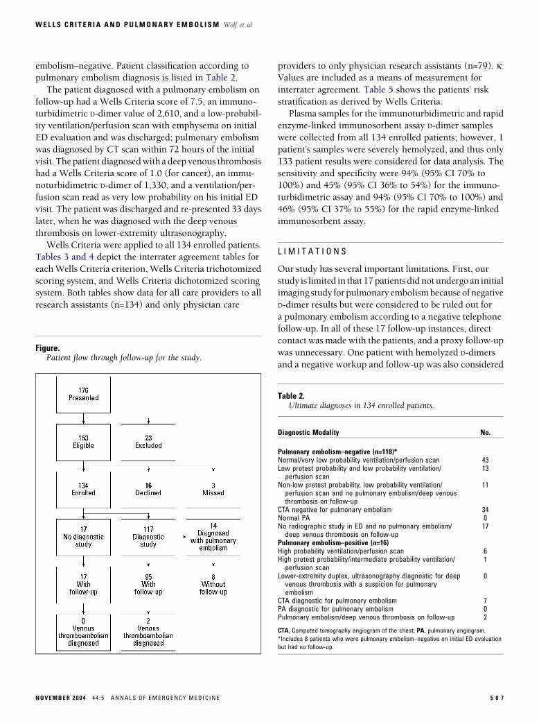
follow-up.TheFiguredepicts patient flowand follow-up in
the study.
Fourteen patients (14/134, 10%) were diagnosed with
pulmonary embolism (pulmonary embolism–positive) on
their initial ED evaluation. Two patients (2/112, 2%) were
found to have venous thromboembolism on 3-month
follow-up (1 deep venous thrombosis, 1 pulmonary
embolism). The overall venous thromboembolism preva-
lence for the study was 12% (16/134). All of the 17
patients who received no initial imaging study and had
a negative D-dimer result had no venous thromboembo-
lism on direct telephone follow-up. The 8 patients who
were lost to follow-up but had negative imaging on
initial presentation were considered pulmonary
Table 1.Demographic characteristics of enrolled patients.
Demographic Characteristic No. (n=134) %
Male sex 61 46Median age, y 58Interquartile range (43–72)Race/ethnicityWhite 99 74Hispanic 19 14Black 11 8Asian/Pacific Islander 2 2Other 3 2
ANNALS OF EMERGENCY MEDIC INE 44 : 5 NOVEMBER 20 04
WEL LS CR I T E R I A AND PULMONARY EMBOL I SM Wolf et al
embolism–negative. Patient classification according to
pulmonary embolism diagnosis is listed in Table 2.
The patient diagnosed with a pulmonary embolism on
follow-up had a Wells Criteria score of 7.5, an immuno-
turbidimetric D-dimer value of 2,610, and a low-probabil-
ity ventilation/perfusion scan with emphysema on initial
ED evaluation and was discharged; pulmonary embolism
was diagnosed by CT scan within 72 hours of the initial
visit. Thepatient diagnosedwith a deep venous thrombosis
had a Wells Criteria score of 1.0 (for cancer), an immu-
noturbidimetric D-dimer of 1,330, and a ventilation/per-
fusion scan read as very low probability on his initial ED
visit. The patient was discharged and re-presented 33 days
later, when he was diagnosed with the deep venous
thrombosis on lower-extremity ultrasonography.
Wells Criteria were applied to all 134 enrolled patients.
Tables 3 and 4 depict the interrater agreement tables for
eachWells Criteria criterion,Wells Criteria trichotomized
scoring system, and Wells Criteria dichotomized scoring
system. Both tables show data for all care providers to all
research assistants (n=134) and only physician care
Figure.Patient flow through follow-up for the study.
NOVEMBER 200 4 44 : 5 ANNALS OF EMERGENCY MEDIC INE
providers to only physician research assistants (n=79). kValues are included as a means of measurement for
interrater agreement. Table 5 shows the patients’ risk
stratification as derived by Wells Criteria.
Plasma samples for the immunoturbidimetric and rapid
enzyme-linked immunosorbent assay D-dimer samples
were collected from all 134 enrolled patients; however, 1
patient’s samples were severely hemolyzed, and thus only
133 patient results were considered for data analysis. The
sensitivity and specificity were 94% (95% CI 70% to
100%) and 45% (95% CI 36% to 54%) for the immuno-
turbidimetric assay and 94% (95% CI 70% to 100%) and
46% (95% CI 37% to 55%) for the rapid enzyme-linked
immunosorbent assay.
L I M I T A T I O N S
Our study has several important limitations. First, our
study is limited in that 17patientsdidnotundergo an initial
imagingstudy forpulmonaryembolismbecauseofnegative
D-dimer results but were considered to be ruled out for
a pulmonary embolism according to a negative telephone
follow-up. In all of these 17 follow-up instances, direct
contact was made with the patients, and a proxy follow-up
was unnecessary. One patient with hemolyzed D-dimers
and a negative workup and follow-up was also considered
Table 2.Ultimate diagnoses in 134 enrolled patients.
Diagnostic Modality No.
Pulmonary embolism–negative (n=118)*Normal/very low probability ventilation/perfusion scan 43Low pretest probability and low probability ventilation/perfusion scan
13
Non-low pretest probability, low probability ventilation/perfusion scan and no pulmonary embolism/deep venousthrombosis on follow-up
11
CTA negative for pulmonary embolism 34Normal PA 0No radiographic study in ED and no pulmonary embolism/deep venous thrombosis on follow-up
17
Pulmonary embolism–positive (n=16)High probability ventilation/perfusion scan 6High pretest probability/intermediate probability ventilation/perfusion scan
1
Lower-extremity duplex, ultrasonography diagnostic for deepvenous thrombosis with a suspicion for pulmonaryembolism
0
CTA diagnostic for pulmonary embolism 7PA diagnostic for pulmonary embolism 0Pulmonary embolism/deep venous thrombosis on follow-up 2
CTA, Computed tomography angiogram of the chest; PA, pulmonary angiogram.
*Includes 8 patients who were pulmonary embolism–negative on initial ED evaluation
but had no follow-up.
5 0 7

WEL LS CR I T E R I A AND PULMONARY EMBOL I SM Wolf et al
to be ruled out for pulmonary embolism.We believed that
excluding these patients, for whom there was a possibility
of venous thromboembolismon follow-up,wouldhavehad
a greater potential to distort our findings than including
them, with the possibility of a false negative follow-up.
In addition, there were 8 patients who did not receive
follow-up. These patients could not be contacted by
telephone, and they did not have reliable medical records
in the hospital and managed-care system. They were
categorized as pulmonary embolism–negative according
to their initial ED visit with a negative result for pulmo-
nary embolism. Finally, 3 patients were missed and no
data were available on them.
5 0 8
A second limitation is related to ourdecision to consider
pulmonary embolism excluded by a negative CT angio-
gram of the chest, which continues to be an area of debate
in the literature and in EDs across the country. Several
studies do suggest that CT angiography can be used as
a primary modality of diagnosis of pulmonary embolism
and that the false negative rate can be low.29-33 However,
our studywas not designed to evaluate the CT angiogram’s
performance. Of note, both of the pulmonary embolisms
diagnosed on follow-up in our study were in patients who
underwent an initial ventilation/perfusion scan.
A final limitation is the potential selection bias in-
troduced by including only patients who underwent an
Table 3A.Interrater agreement table comparing all care providers (n=134).
Rater 2
Criterion 1 Criterion 2 Criterion 3 Criterion 4 Criterion 5 Criterion 6 Criterion 7
Rater 1 Yes No Yes No Yes No Yes No Yes No Yes No Yes No
Yes 17 4 54 17 34 7 15 4 24 1 6 0 9 0No 5 108 11 52 6 87 1 112 0 109 0 128 3 122k (95% CI) 0.76 (0.60–0.92) 0.58 (0.44–0.72) 0.78 (0.66–0.90) 0.79 (0.63–0.95) 0.97 (0.92–1.00) 1.00 0.83 (0.64–1.00)
Table 3B.Interrater agreement table comparing physician care providers to physician research assistants (n=79).
MD 2
Criterion 1 Criterion 2 Criterion 3 Criterion 4 Criterion 5 Criterion 6 Criterion 7
MD 1 Yes No Yes No Yes No Yes No Yes No Yes No Yes No
Yes 13 2 35 6 19 2 11 3 14 0 4 0 4 0No 3 61 8 30 4 54 1 64 0 65 0 75 1 74k (95% CI) 0.78 (0.60–0.96) 0.64 (0.46–0.82) 0.81 (0.67–0.96) 0.82 (0.64–0.99) 1.00 1.00 0.85 (0.57–1.00)
MD, Physician.
Table 4A.Trichotomized scoring table comparing all care providers with allcare providers.*
Rater 2
Rater 1 Low Moderate High
Low 40 8 0Moderate 18 52 5High 0 2 9
*k=0.54 (95% CI 0.40–0.68)
Table 4B.Trichotomized scoring table comparing physician care providerswith physician research assistants.*
MD 2
MD 1 Low Mod High
Low 23 7 0Moderate 7 34 2High 0 0 66
*k=0.62 (95% CI 0.44–0.80)
ANNALS OF EMERGENCY MEDIC INE 44 : 5 NOVEMBER 20 04

WEL LS CR I T E R I A AND PULMONARY EMBOL I SM Wolf et al
imaging study for pulmonary embolism. Enrolling all
patients with chest pain, shortness of breath, or syncope
would have made this study prohibitively costly, and
would also have resulted in a lower apparent prevalence of
pulmonary embolism in our population, diluted by
patients for whom the suspicion or pulmonary embolism
was extremely low to begin with. This inclusion would
have severely limited the generalizability of this study. In
any case, our goal was to study the application of Wells
Criteria to patients who were going to undergo imaging
for pulmonary embolism.
D I S C U S S I O N
To our knowledge, this is the first study to examine the
interrater agreement and prospective external validity of
Wells Criteria. This clinical decision rule appears to
reproducibly and reliably risk stratify patients with
suspected pulmonary embolism into either a low,
moderate, and high classification or a pulmonary
embolism–unlikely and pulmonary embolism–likely
classification. Furthermore, the 2 D-dimer assays used in
our study appeared to perform with equal sensitivities and
Table 4D.Dichotomized scoring table comparing physician care providerswith physician research assistants.*
MD 2
MD 1 PE-UL PE-L
PE-UL 44 5PE-L 6 24
*k=0.70 (95% CI 0.53–0.87)
Table 4C.Dichotomized scoring table comparing all care providers with allcare providers.*
Rater 2
Rater 1 PE-UL PE-L
PE-UL 77 6PE-L 12 39
PE-L, Pulmonary embolism–likely; PE-UL, pulmonary embolism–unlikely.
*k=0.72 (95% CI 0.60–0.84)
NOVEMBER 200 4 44 : 5 ANNALS OF EMERGENCY MEDIC INE
specificities. Our results do not seem to support the use of
either assay alone to rule out the diagnosis of pulmonary
embolism, although the CIs are large.
As the first study looking at the interrater agreement of
the Wells Criteria, our findings are encouraging. We
found that the dichotomized scoring system had sub-
stantial interrater agreement, whereas the trichotomized
system had moderate interrater agreement. Furthermore,
our study found that the interrater agreement for both
scoring systems was essentially independent of the level of
training of the emergency care provider (0.72 versus 0.70
and 0.54 versus 0.62), which is interesting, given the
subjective nature of the second criterion, which asks
whether an alternative diagnosis is considered as likely or
more likely than pulmonary embolism. This answer un-
doubtedly would vary with education and experience, as
the traditional ‘‘gestalt’’ means of determining pretest
probability does.34 In fact, the second criterion generated
the lowest k value of all the Wells Criteria. We speculate
that when the clinical decision rule is considered as
a whole, the other 6 criteria may act to moderate the
results of the second, causing experience and education to
influence the final score less.
Wells et al14,19 previously published studies reporting
the incidence of pulmonary embolism to be 2% to 4% for
patients with a low pretest probability, 19% to 21% for
patients with a moderate pretest probability, and 50% to
67% for patients with a high pretest probability. Addi-
tionally, they found that the pulmonary embolism–
unlikely and pulmonary embolism–likely groups had
incidences of 5% to 8% and 39% to 41%, respectively.19
Our findings are consistent with these numbers, providing
independent external validity to Wells Criteria for both
the dichotomized and trichotomized scoring systems. It is
interesting to note that none of the patients with a negative
D-dimer result in the low pretest probability or pulmonary
embolism–unlikely groupings were diagnosed with a pul-
monary embolism.
Table 5.Results of Wells Criteria.
Wells CriteriaScore No. %
PulmonaryEmbolism, No.
PulmonaryEmbolism, % 95% CI
Low (<2) 59 44 1 2 0–9Moderate (2–6) 61 46 9 15 7–26High (>6) 14 10 6 43 18–71PE-UL(�4) 88 66 3 3 0–9PE-L (>4) 46 34 13 28 18–71
5 0 9

WEL LS CR I T E R I A AND PULMONARY EMBOL I SM Wolf et al
The sensitivities we found for both of the D-dimer
assays were not as promising as those that have previously
been reported. However, our sample size is relatively
small, and it may be that with a greater sample size our
numbers would converge with those previously pub-
lished. Still, keeping in mind the very large CIs, these
numbers are consistent with recent literature that states
that a negative D-dimer assay alone should not be used to
rule out pulmonary embolism.2,4-7
In summary, we conclude that Wells Criteria appears
to have a moderate to substantial interrater reliability,
affording emergency medicine care providers with a re-
producible means of determining pretest probability
among ED patients for whom the diagnosis of pulmonary
embolism is being considered.
Author contributions: TRM and DMA conceived the study andobtained funding. SJW, TRM, and KMF supervised the conduct of thestudy. All authorswere involved in data collection, and SJWmanagedthe data. SJW, TRM, and KMF were responsible for quality control.Statistical analysis was performed by an independent statistician andin part by SJW. SJW drafted the manuscript, and SJW, TRM, RMF,JPF, and DMA contributed substantially to its revision. SJW takesresponsibility for the paper as a whole.
Received for publicationMay 18, 2003. Revisions received October 13,2003; December 15, 2003; March 8, 2004; and April 5, 2004. Acceptedfor publication April 7, 2004. Available online August 25, 2004.
Presented at the Society for Academic Emergency Medicine annualmeeting, St. Louis, MO, May 2002.
Supported in part by a Medicare grant (Medicare 2001 KaiserPermanente/Exempla Saint Joseph Hospital Research Fund).
Reprints not available from the authors.
Address for correspondence: Stephen J. Wolf, MD, Department ofEmergencyMedicine, Denver HealthMedical Center,Mail Code 0108,777 Bannock Street, Denver, CO 80204; 303-436-8842, fax 303-436-7541; E-mail [email protected].
R E F E R E N C E S1. Goldhaber SZ. Pulmonary embolism. N Engl J Med. 1998;339:93-104.
2. Brown MD, Rowe BH, Reeves MJ, et al. The accuracy of the enzyme-linked
immunosorbent assay D-dimer test in the diagnosis of pulmonary embolism:
a meta-analysis. Ann Emerg Med. 2002;40:133-144.
3. Kline JA, Johns KL, Colucciello SA, et al. New diagnostic tests for pulmonary
embolism. Ann Emerg Med. 2000;35:168-172.
4. Wolfe TR, Hartsell SC. Pulmonary embolism: making sense of the diagnostic
evaluation. Ann Emerg Med. 2001;37:504-514.
5. Sadosty AT, Goyal DG, Boie ET, et al. Emergency department D-dimer testing. J
Emerg Med. 2001;21:423-429.
6. Kline JA, Wells PS. Methodology for a rapid protocol to rule out pulmonary
embolism in the emergency department. Ann Emerg Med. 2003;42:266-275.
7. Bounameaux H, de Moerloose P, Perrier A, et al. Plasma measurement of d-dimer
as diagnostic aid in suspected venous thromboembolism: an overview. Thromb
Haemost. 1994;71:1-6.
8. Goldhaber SZ, Simons GR, Elliott CG, et al. Quantitative plasma D-dimer levels
among patients undergoing pulmonary angiography for suspected pulmonary
embolism. JAMA. 1993;270:2819-2822.
5 1 0
9. Ginsberg JS, Brill-Edwards PA, Demers C, et al. D-Dimer in patients with clinically
suspected pulmonary embolism. Chest. 1993;104:1679-1684.
10. De Moerloose P, Desmarais S, Bounameaux H, et al. Contribution of a new, rapid,
individual and quantitative automated D-dimer ELISA to exclude pulmonary embolism.
Thromb Haemost. 1996;75:11-13.
11. Perrier A, Desmarais S, Miron MJ, et al. Non-invasive diagnosis of venous
thromboembolism in outpatients. Lancet. 1999;353:190-195.
12. Knecht MF, Heinrich F. Clinical evaluation of an immunoturbidimetric D-dimer assay
in the diagnostic procedure of deep vein thrombosis and pulmonary embolism. Thromb
Res. 1997;88:413-417.
13. Duet M, Benelhdj S, Kedra W, et al. A new quantitative D-dimer assay appropriate
in emergency: reliability of the assay for pulmonary embolism exclusion diagnosis.
Thromb Res. 1998;91:1-5.
14. Wells PS, Ginsberg JS, Anderson DR, et al. Use of a clinical model for safe
management of patients with suspected pulmonary embolism. Ann Intern Med. 1998;
129:997-1005.
15. Wicki J, Perneger TV, Junod AF, et al. Assessing clinical probability of pulmonary
embolism in the emergency ward: a simple score. Arch Intern Med. 2001;161:92-97.
16. Kline JA, Nelson RD, Jackson RE, et al. Criteria for the safe use of D-dimer testing
in emergency department patients with suspected pulmonary embolism: a multicenter
US study. Ann Emerg Med. 2002;39:144-152.
17. British Thoracic Society Standards of Care Committee Pulmonary Embolism
Guideline Development Group. British Thoracic Society guidelines for the management
of suspected acute pulmonary embolism. Thorax. 2003;58:470-484.
18. Miniati M, Monti S, Bottai M. A structured clinical model for predicting the
probability of pulmonary embolism. Am J Med. 2003;114:173-179.
19. Wells PS, Anderson DR, Rodger M, et al. Derivation of a simple clinical model to
categorize patients’ probability of pulmonary embolism: increasing the model’s utility
with the SimpliRED D-dimer. Thromb Haemost. 2000;83:416-420.
20. Wells PS, Anderson DR, Rodger M, et al. Excluding pulmonary embolism at the
bedside without diagnostic imaging: management of patients with suspected
pulmonary embolism presenting to the emergency department by using a simple
clinical model and D-dimer. Ann Intern Med. 2001;135:98-107.
21. Chagnon I, Bounameaux H, Aujesky D, et al. Comparison of two clinical prediction
rules and implicit assessment among patients with suspected pulmonary embolism. Am
J Med. 2002;113:269-275.
22. Chabloz P, Reber G, Boehlen F, et al. TAFI antigen and D-dimer levels during normal
pregnancy and at delivery. Br J Haematol. 2001;115:150-152.
23. Righini M, de Moerloose P, Reber G, et al. Should the D-dimer cut-off value be
increased in elderly patients suspected of pulmonary embolism? [letter] Thromb
Haemost. 2001;85:744.
24. De Lorenzo F, Mukherjee M, Kadziola Z, et al. Association of overall adiposity
rather than body mass index with lipids and procoagulant factors. Thromb Haemost.
1998;80:603-606.
25. Perrier A, Desmarais S, Miron MJ, et al. Non-invasive diagnosis of venous
thromboembolism in outpatients. Lancet. 1999;353:190-195.
26. Freyburger G, Trillaud H, Labrouche S, et al. D-Dimer strategy in thrombosis
exclusion. Thromb Haemost. 1998;79:32-37.
27. Heit JA, Meyers BJ, Plumhoff EA, et al. Operating characteristics of automated
latex immunoassay fibrin D-dimer in the diagnosis of angiographically-defined acute
pulmonary embolism. Thromb Haemost. 2000;83:970.
28. Landis JR, Koch GG. The measurement of observer agreement for categorical data.
Biometrics. 1977;33:159-174.
29. Teigen CL, Maus TP, Sheedy PF II, et al. Pulmonary embolism: diagnosis with
contrast-enhanced electron-beam CT and comparison with pulmonary angiography.
Radiology. 1995;194:313-319.
30. Mayo JR, Remy-Jardin M, Muller NL, et al. Pulmonary embolism: prospective
comparison of spiral CT with ventilation-perfusion scintigraphy. Radiology. 1997;205:
447-452.
31. Garg K, Welsh CH, Feyerabend AJ, et al. Pulmonary embolism: diagnosis with spiral
CT and ventilation-perfusion scanning: correlation with pulmonary angiographic results
or clinical outcome. Radiology. 1998;208:201-208.
32. Goodman LR, Lipchik RJ, Kuzo RS, et al. Subsequent pulmonary embolism: risk after
a negative helical CT pulmonary angiogram: prospective comparison with scintigraphy.
Radiology. 2000;215:535-542.
33. Swensen SJ, Sheedy PF II, Ryu JH, et al. Outcomes after withholding
anticoagulation from patients with suspected acute pulmonary embolism and negative
computed tomographic findings: a cohort study. Mayo Clin Proc. 2002;77:130-138.
34. Stein PD, Terrin ML, Hales CA, et al. Clinical, laboratory, roentgenographic, and
electrocardiographic findings in patients with acute pulmonary embolism and no
pre-existing cardiac or pulmonary disease. Chest. 1991;100:598-603.
ANNALS OF EMERGENCY MEDIC INE 44 : 5 NOVEMBER 20 04


![ENVIRONMENTAL PROTECTION COMMISSION[567] · 2014-06-25 · wells, recreational-use wells, monitoring wells, heat pump supply wells or GHEX loop boreholes, industrial wells, and dewatering](https://img.pdfslide.net/doc/110x75/5f3f728939b254613866ae00/environmental-protection-commission567-2014-06-25-wells-recreational-use-wells.jpg)