Embed Size (px)
Citation preview

What are the four tissues of the body?
Our body is made from these 4 tissues

Epithelial Tissue
Covers exposed surfaces
Lines internal passageways
Forms glands

Connective Tissue
Fills internal spaces
Supports other tissues
Transports material - cells
Stores energy

Muscle Tissue
Specialized for contraction
Skeletal muscle, heart muscle, and walls of hollow organs
Neural Tissue
Carries electrical signals from 1 part of the body to another

KEY CONCEPT
Tissues are collections of cells and cell products that perform specific, limited functions
4 tissue types form all the structures of the human body:
epithelial, connective, muscle, and neural
What are the special structures and functions of epithelial tissues?

Characteristics of Epithelia
1. Cellularity (cell junctions)
2. Polarity (apical and basal surfaces)
3. Attachment (basal lamina)
4. Avascularity
5. Regeneration
Functions of Epithelial Tissue
1. Provide physical protection
2. Control permeability
3. Provide sensation
4. Produce specialized secretions (glandular epithelium)

Specializations of Epithelial Cells
1. Move fluids over the epithelium (protection)
2. Move fluids through the epithelium (permeability)
3. Produce secretions (protection and messengers)

Figure 4–1
Free Surface and Attached Surface
Polarity: apical and basolateral surfaces

Increasing Surface Area
Microvilli increase absorption or secretion
Cilia (ciliated epithelium) move fluids

Effective Barriers
Physical integrity is maintained by:intercellular connections
attachment to basal lamina
maintenance and repair
Cell Junctions
Form bonds with other cells or extracellular material:
tight junctions
gap junctions
desmosomes

Tight Junctions
Between 2 cell membranes
Figure 4–2b

Tight Junctions
Adhesion belt attaches to terminal web
Prevents passage of water and solutes
Isolates wastes in the lumen

Gap Junctions
Allow rapid communications
Figure 4–2c

Gap Junctions
Held together by channel proteins (junctional proteins, connexons)
Allow ions to pass
Coordinated contractions in heart muscle

Desmosomes
CAMs, dense areas, and intercellular cement
Figure 4–2d

Desmososmes
Button desmosomes
Ties cells together
Allow bending and twisting
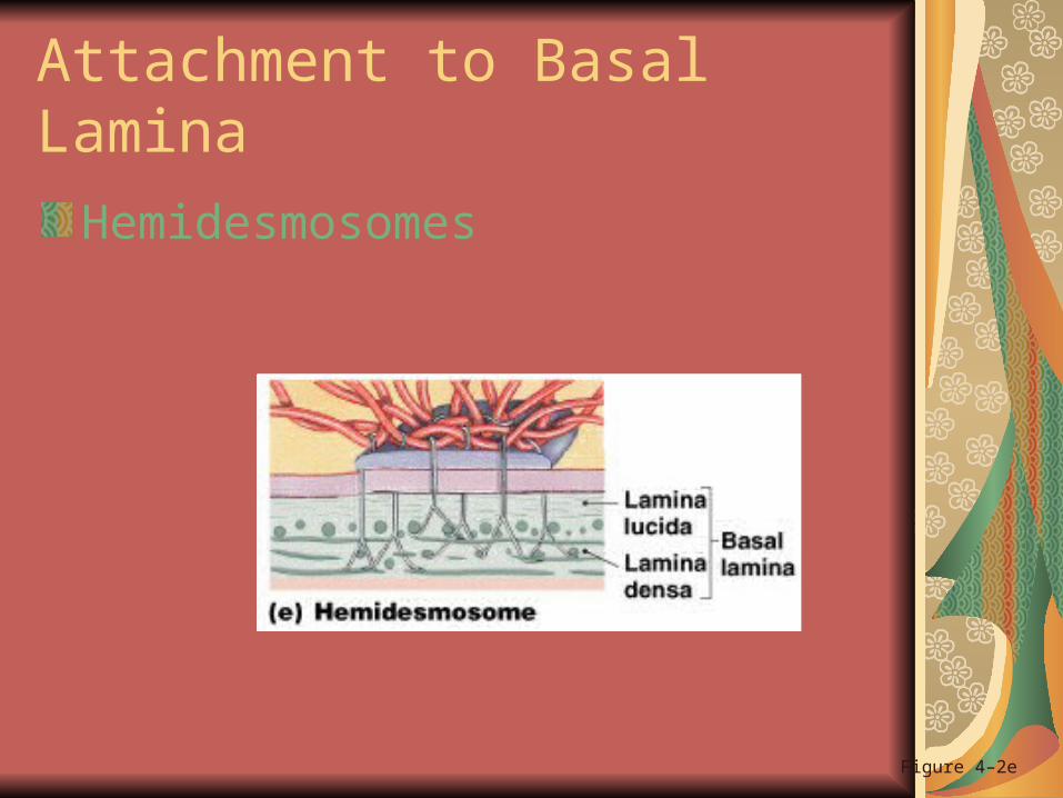
Attachment to Basal Lamina
Hemidesmosomes
Figure 4–2e
Basal Lamina
Lamina lucida: thin layer secreted by epitheliabarrier to proteins
Lamina densa: thick fibersproduced by connective tissuestrength and filtration
Repairing and Replacing Epithelia
Epithelia are replaced by division of germinative cells (stem cells)
Near basal lamina
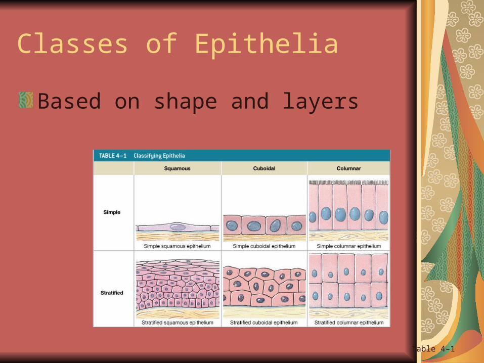
Classes of Epithelia
Based on shape and layers
Table 4–1

Layers
Simple epithelium: single layer of cells
Stratified epithelium:several layers of cells

Cell Shape
Squamous epithelia:flat shaped
Cuboidal epithelia:square shaped
Columnar epithelia:tall shaped

Squamous Epithelia
Simple squamous epithelium: absorption and diffusion
Mesothelium: lines body cavities
Endothelium: lines heart and blood vessels

Figure 4–3a
Simple Squamous Epithelium

Figure 4–3b
Stratified Squamous Epithelium
Stratified Squamous Epithelium
Protects against attacks
Keratin proteins add strength and water resistance

Cuboidal Epithelia
Simple cuboidal epithelium:secretion and absorption
Stratified cuboidal epithelia:sweat and mammary ducts

Simple Cuboidal Epithelium
Kidney tubules
Figure 4–4a
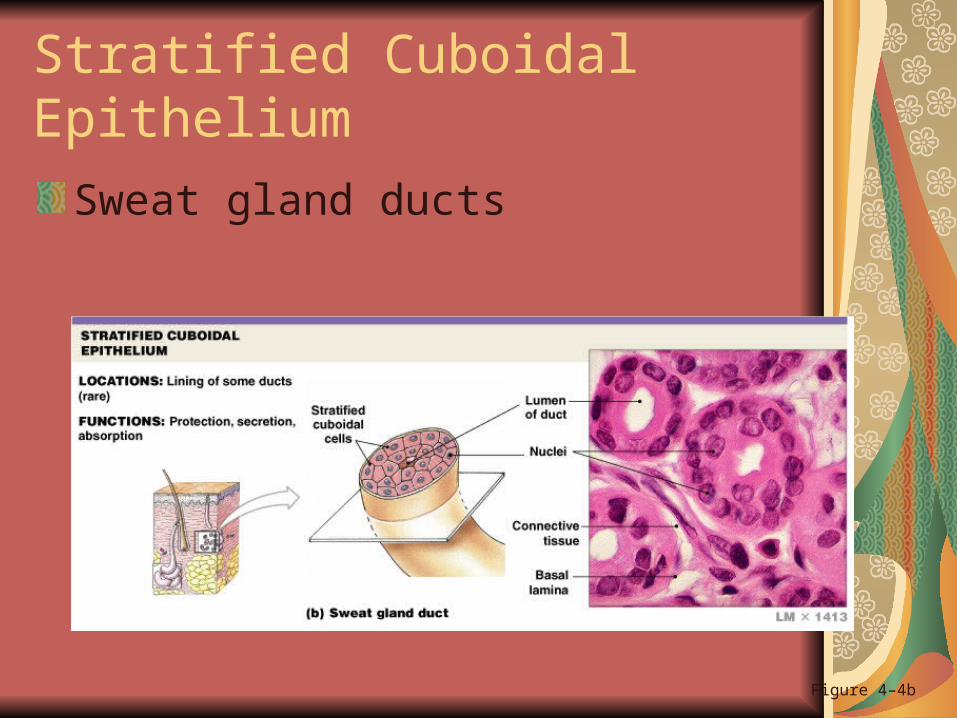
Stratified Cuboidal Epithelium
Sweat gland ducts
Figure 4–4b

Transitional Epithelium
Urinary bladder
Figure 4–4c
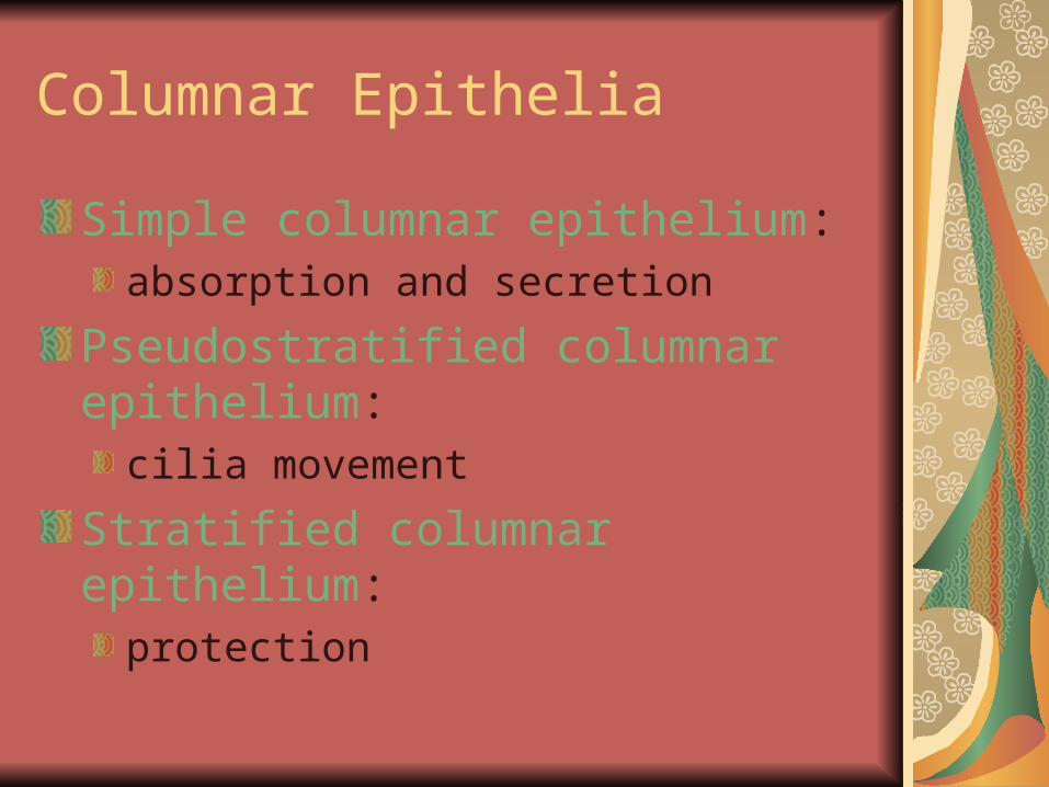
Columnar Epithelia
Simple columnar epithelium:absorption and secretion
Pseudostratified columnar epithelium:cilia movement
Stratified columnar epithelium:protection
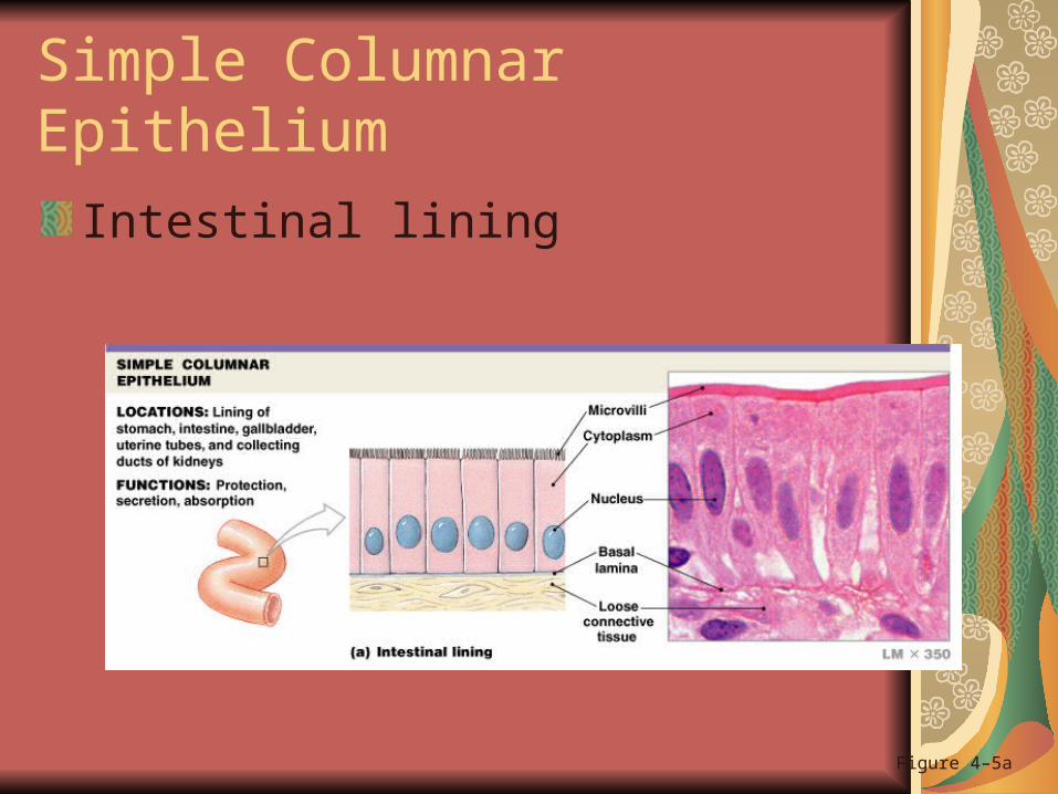
Simple Columnar Epithelium
Intestinal lining
Figure 4–5a

Pseudostratified Columnar Epithelium
Trachea
Figure 4–5b

Stratified Columnar Epithelium
Salivary gland duct
Figure 4–5c
Mechanisms of Glandular SecretionPLAYPLAY
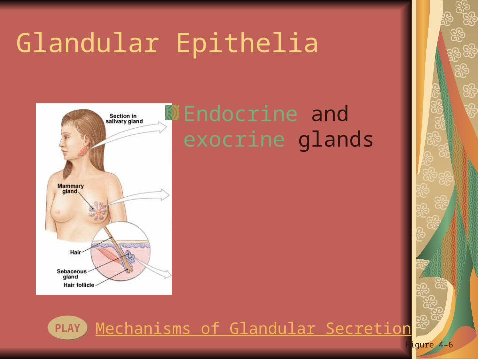
Glandular Epithelia
Endocrine and exocrine glands
Figure 4–6

Endocrine Glands
Release hormones:into interstitial fluid
no ducts
Exocrine Glands
Produce secretions:onto epithelial surfaces
through ducts

Modes of Secretion
Merocrine secretion (GA produces and secreted by vesicles)
Figure 4–6a
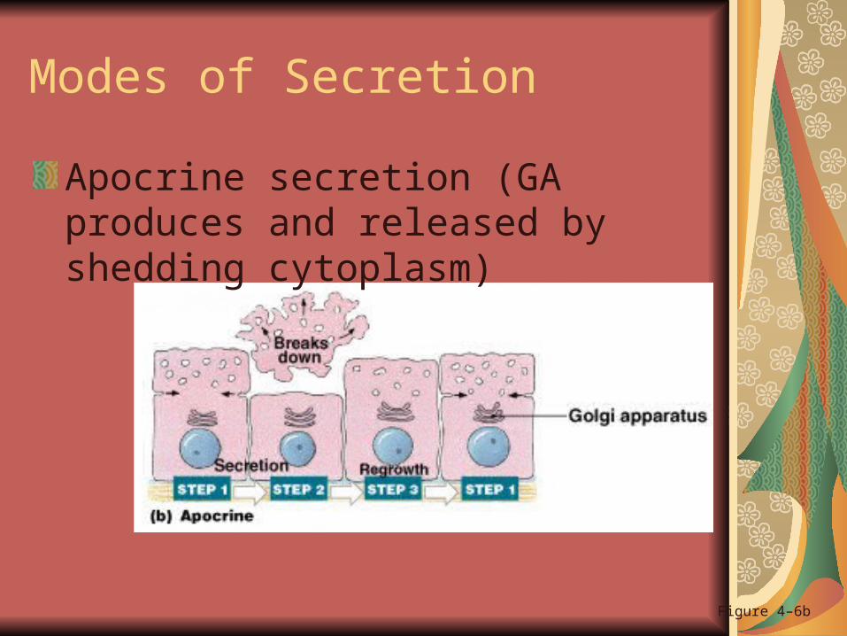
Modes of Secretion
Apocrine secretion (GA produces and released by shedding cytoplasm)
Figure 4–6b
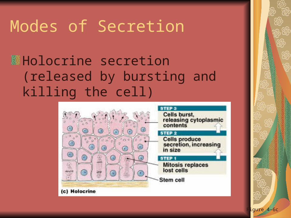
Modes of Secretion
Holocrine secretion (released by bursting and killing the cell)
Figure 4–6c
Types of Secretions
Serous glands:watery secretions
Mucous glands:secrete mucins
Mixed exocrine glands:both serous and mucous
Gland Structure
Exocrine glands can be classified as:unicellular glands
multicellular glands

Structure of Multicellular Exocrine Glands
Structural classes of exocrine glands
Figure 4–7 (1 of 2)

Structure of Multicellular Exocrine Glands
Figure 4–7 (2 of 2)

What are the structures and functions of different types of connective tissues?

Connective Tissues
Connect epithelium to the rest of the body (basal lamina)
Provide structure (bone)
Store energy (fat)
Transport materials (blood)
Have no contact with environment

The Matrix
The extracellular components of connective tissues (fibers and ground substance):
majority of cell volume
determines specialized function

Classification of Connective Tissues
Connective tissue proper:connect and protect
Fluid connective tissues:transport
Supportive connective tissues:structural strength

Connective Tissue Proper
Figure 4–8
Categories of Connective Tissue Proper
Loose connective tissue:more ground substance, less fibers
e.g., fat (adipose tissue)
Dense connective tissue:more fibers, less ground substance
e.g., tendons

8 Cell Types of Connective Tissue Proper
Fibroblasts
Macrophages
Adipocytes
Mesenchymal cells
Melanocytes
Mast cells
Lymphocytes
Microphages

Fibroblasts
The most abundant cell type:found in all connective tissue proper
secrete proteins and hyaluronan (cellular cement)
Makes ground substance and structural fibers

Macrophages
Large, amoeba-like cells of the immune system:
eat pathogens and damaged cells
fixed macrophages stay in tissue
free macrophages migrate

Adipocytes
Fat cells: each cell stores a single, large fat droplet

Mesenchymal Cells
Stem cells that respond to injury or infection:
differentiate into fibroblasts, macrophages, etc.
Melanocytes
Synthesize and store the brown pigment melanin

Mast Cells
Stimulate inflammation after injury or infection:
release histamine and heparin
Basophils are mast cells carried by blood

Lymphocytes
Specialized immune cells in lymphatic system:e.g., plasma cells which produce antibodies
Microphages
Phagocytic blood cells:respond to signals from macrophages and mast cells
e.g., neutrophils and eosinophils

Fibers in Connective Tissue Proper
Collagen fibers: most common fibers in CTP
long, straight, and unbranched
strong and flexible
resists force in 1 direction
e.g., tendons and ligaments

Fibers in Connective Tissue Proper
Reticular fibers: network of interwoven fibers (stroma)
strong and flexible
resists force in many directions
stabilizes functional cells (parenchyma) and structures
e.g., sheaths around organs

Fibers in Connective Tissue Proper
Elastic fibers: contain elastin
branched and wavy
return to original length after stretching
e.g., elastic ligaments of vertebrae

Loose Connective Tissues
The packing materials of the body
3 types in adults:areolar
adipose
reticular

Areolar Tissue
Least specialized
Open framework
Viscous ground substance
Elastic fibers
Holds blood vessels and capillary beds:e.g., under skin (subcutaneous layer)
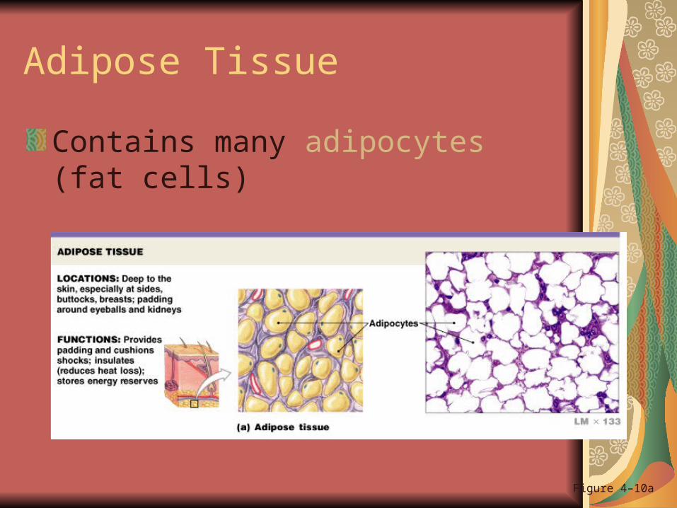
Adipose Tissue
Contains many adipocytes (fat cells)
Figure 4–10a
Adipose Cells
Adipocytes in adults do not divide:expand to store fat
shrink as fats are released
Mesenchymal cells divide and differentiate:
to produce more fat cells
when more storage is needed