Embed Size (px)
DESCRIPTION
hematuri untuk dokter praktek umum
Citation preview

What is significant hematuria for the primary care physician ?
Agus Darmanto
Erlin Irawati
Rakhmiana

Introduction Hematuria is a clinical finding that is
commonly encountered by primary care physicians and urologist
Roughly 4 % of patients with microscopic hematuria and up to 40 % of patients with gross hematuria could be harboring a malignancy
Therefore, it is important to effectively and efficiently manage patient who have hamturia

What is significant hematuria? When confronted with a patient with
hematuria, a clinician must establish whether the hematuria is clinically significant.
Gross hematuria should always be considered significannt, and it may also be regarded as sign of malignancy until proven otherwise
If the patient’s clinical history confirms the presence of hematuria, further questions may localize the source of bleeding

If gross hematuria appears throughout the urinary stream, then it probably orginates above the level of the bladder outlet, including the upper urinary tract and kidneys
If blood is detected only at the initiation of urination, then it is likely from the urethra
Gross hematuria at the end of urination may be from the prostate or bladder neck

Microscopic hematuria is a more common finding than gross hematuria
Microscopic hematuria is often detected from a positive urine dipstick test
Teh color change can be affected by factor affecting the dipstick reagent, urine, or their interaction
Therefore, microscopic urinalysis is recomended to confirm a positive dipstick test for hematuria


In the canadian guidlines, significant microscopic hematuria is defined as 2 or more red blood cells per high power field, confirmed in two microscopic urinalysis tests performed when there is no benign etiology such as menstruation, recent exercise, recent sexual activity, or recent intrumentation of the urinary tract
The american recommendations define significant microscopic hematuria as 3 or more RBCs per high power field in a properly collected specimen and in the absence of a benign etiology

Causes of significant hematuria Urinary tract infection Microscopic hematuria is usually associated
with UTIs and can even cause gross hematuria, although this is less common
Patients with a UTI generally also have other voiding symptoms, especially dysuria and acute urinary frequency

Urolithiasis Kidney stones are common and both men and
women may have them Smaller stones that reside in the kidney are
usually asymptomatic, and microscopic hematuria may be the only manifestation
Larger stones, including staghorn calculim, may present with gross hematuria
Hematuria associated with acute onset of lateralizing flank pain is the classic presentation of a stone thet moves into the ureter

Malignancies Gross hematuria is not as common, but it may
be seen if the patient has a larger mass located more centrally in the kidney
Urothelial carcinoma arises from the lining of the collecting system.
Gross hematuria tneds to be the common symptom with this type of tumor

Patients who have a tumor that arises from the renal pelvis or ureter may present with flank pain, and they may note the passage of “worm like clots” on micturition
Lesions arising from the bladder often cause no pain, but patient with these lesions may have large blood clots when they urinate
Although incidental microscopic hematuria may be present when prostate cancer is diagnosed, this hematuria is more likely due to benign prostatic hyperplasia (BPH) rather than the cancer

BPH While the classic patient presentation is
usually that of lower urinary tract obstructive or irritative symptoms, hematuria is also common
BPH may, in fact, be the most common cause of microscopic hematuria in men
Less commonly, BPH may be the sole cause of gross hematuria and clot retention
A diagnosis of BPH can be made only after full urological evaluation and exclusion of other causes of hematuria

Nephropathies and nephritis The medical cause of hematuria should not be
overlooked Patients with nefropathies and nephritis may
present with hypertension, edema, and renal insufficiency, and urine tests may reveal the presence of proteinuria and RBS casts

Patient Management Physicians should take a thorough clinical
history These include age older than 40 years, past of
current analgesic abuse, smoking, or exposure to chemicals or dyes or history of pelvic irradiation, gross hematuria, irritative voiding symptoms, or UTIs
The patient’s current medications or prior urinary tract interventions might be linked to the bleeding


Ancillary study Laboratory investigation Imaging

Renal function•BUN•Serum creatinine•Estimated glomerular filtration rate (eGFR)
Identifikasi koagulopati •Blood coagulation parameter (INR and PTT)
In pregnant women •Serum beta-HCG test
Laboratory investigations
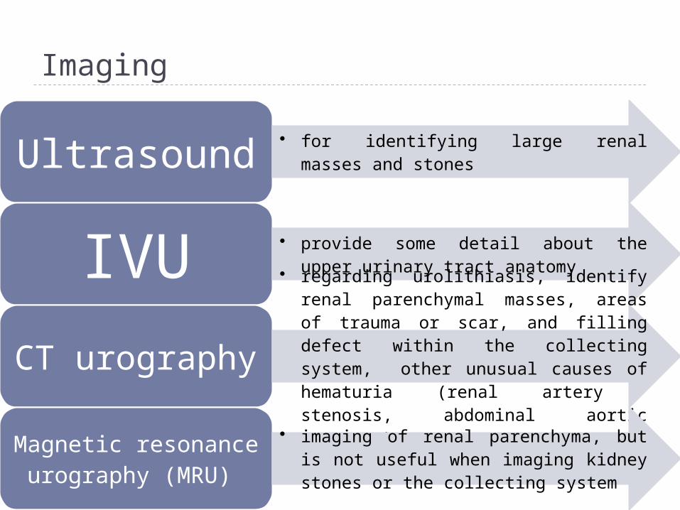
Imaging
Ultrasound • for identifying large renal masses and stones
IVU • provide some detail about the upper urinary tract anatomy
CT urography
• regarding urolithiasis, identify renal parenchymal masses, areas of trauma or scar, and filling defect within the collecting system, other unusual causes of hematuria (renal artery stenosis, abdominal aortic aneurysm)
Magnetic resonance
urography (MRU)
• imaging of renal parenchyma, but is not useful when imaging kidney stones or the collecting system

Terima Kasih