Embed Size (px)
Citation preview

BIFURCATIONCHALLENGING CASES
62 CARDIAC INTERVENTIONS TODAY SEPTEMBER/OCTOBER 2019 VOL. 13, NO. 5
CASE PRESENTATIONA 62-year-old man with non–insulin-dependent dia-
betes and no cardiac history was diagnosed with angina pectoris Canadian Cardiovascular Society grade II by means of a positive stress echocardiogram at the inferior and posterior left ventricular wall with normal systolic function. Diagnostic angiography revealed two-vessel dis-ease: a severely calcified subocclusive lesion in the left cir-cumflex artery (LCX) at the first obtuse marginal (OM1) branch bifurcation as the main culprit vessel (Figure 1) and a long chronic total occlusion (CTO) at the mid-right coronary artery (RCA) with excellent retrograde fill-ing flow from the left anterior descending (LAD) artery.
Two months later, the patient underwent percutane-ous coronary intervention (PCI) in a staged procedure targeting the LCX. After receiving a 600-mg loading dose of clopidogrel orally and intravenous heparin and nitrates, access was achieved through the right femoral artery with angiographic guidance using a 7-F XB 3.5 Vista Brite Tip catheter (Cordis, a Cardinal Health com-pany) to visualize the target lesion. A Runthrough NS Floppy guidewire (Terumo Interventional Systems) was used to progress and position a FineCross MG micro-catheter (Terumo Interventional Systems) at the proxi-mal tip of the LCX lesion (Figure 2). Two Fielder XT-R guidewires (Asahi Intecc USA, Inc.) were passed to the LCX and OM1, respectively. However, after crossing to the distal LCX, the microcatheter did not advance to the OM1 branch.
How would you approach this complex two-vessel disease?
Dr. Choi: The patient is a middle-aged diabetic man with complex two-vessel non-LAD disease and normal left ventricular systolic function. By my cal-
culations, his SYNTAX score is 13 due to the LCX calci-fied bifurcation and RCA CTO. Although the SYNTAX score was originally designed for left main and/or three-vessel coronary artery disease, the lower tertial score and absence of LAD disease (use of left internal mammary artery graft) makes successful PCI, with a goal of full revascularization, the preferred option over coronary artery bypass grafting (CABG). Both lesions are complex and challenging. I think a reasonable strategy would be to attempt PCI in the more difficult lesion first, which would most likely be the RCA CTO. If unsuccessful, given the large amount of ischemic ter-ritory at risk, I would consider CABG of the RCA, OM1, and OM2. If successful, PCI could be performed for the LCX/OM bifurcation lesion later as a staged procedure.
Prof. Gonçalves: This is really a complex interven-tion, not only because it is a true bifurcation lesion with significant stenosis in two important vessels (both
PCI in a Complex Bifurcation CTOMODERATOR: RUI CAMPANTE TELES, MD, FESC, FACC
PANELISTS: JAMES W. CHOI, MD, FACC, FSCAI; LAURENT DROGOUL, MD;
AND PEDRO de ARAÚJO GONÇALVES, MD, PhD
WHAT WOULD YOU DO?
Figure 1. Angiography of the left coronary artery showing a
calcified subocclusive lesion at the circumflex and RCA CTO.

VOL. 13, NO. 5 SEPTEMBER/OCTOBER 2019 CARDIAC INTERVENTIONS TODAY 63
CHALLENGING CASESBIFURCATION
branches are > 2 mm), but especially because of the severe calcification. In addition, the takeoff of the OM1 imposes further complexity. Some kind of debulking could be useful in the face of the calcification, but rotablation would not be possible with a guidewire protecting the side branch. Other debulking devices such as cutting and scoring balloons could be an alter-native option, but their crossing profile is less than ideal and therefore would probably be difficult to deliver to the lesions in the complex calcified bifurcation. My strategy would be to treat this with a two-stent strat-egy (due to the size of the side branch) with aggressive predilatation of both branches.
Crossing the lesion with the guidewire might also be difficult in this setting, and more aggressive CTO wires and microcatheter support could be useful. The two-stent strategy could be a culotte (because the two vessels have similar diameters) or a minicrush/balloon crush technique.
Dr. Drogoul: I would use radial access via a Glidesheath Slender introducer sheath (Terumo Interventional Systems) with a 6-F external size and the ability to use a 7-F guiding catheter to reduce radial thrombosis, as well as an extra-backup 3.75 Launcher guide catheter (Medtronic) for the largest internal lumen. To cross the lesion, I would proceed with a Fielder XT-R guidewire for narrow channel tracking because its low profile, flexible tip, and high-lubricity
polymer coating are useful in calcified lesions and are less “aggressive” than the Fielder XT. I would use this with the Finecross microcatheter (for example). In this case, and because of the tortuosity and massive calci-fications, I wouldn’t directly use the RotaWire (Boston Scientific Corporation) to cross the lesion. After cross-ing with the Fielder XT-R and microcatheter, you can exchange the Fielder with the RotaWire.
After the first intervention, I rotablate with a 1.25-mm burr (small to avoid captive burr). For heav-ily calcified lesions (particularly in this case), the risk of coronary rupture is very high if you use a balloon first.
How do you ameliorate your support in complex lesions and/or CTOs?
Prof. Gonçalves: First, I would use a 7- to 8-F guiding catheter, which might require changing to or planning a second procedure via femoral access, assum-ing there is no significant peripheral artery disease. If diagnostic coronary angiography is performed by radial access and the operators decide to convert to femoral access, it could be useful to check the quality of the iliac-femoral axis by making a pigtail injection in the distal abdominal aorta to exclude significant disease that could limit the use of 7- to 8-F femoral access. It could also be useful for looking at the height of the femoral bifur-cation for the ideal use of a femoral closure device to decrease the risk of femoral bleeding after the complex PCI (expected to be a long procedure with larger heparin dose and larger 7- to 8-F sheath). Second, catheter exten-sions (GuideLiner [Teleflex], Guidezilla [Boston Scientific Corporation], Telescope [Medtronic]) could also help to deliver stents/balloons in calcified LCX lesions. Third, stents with thinner struts and good deliverability should be used to increase the likelihood of successful tracking along these calcified vessels.
Dr. Choi: In challenging cases like this, guide catheter support is critical for procedural success. I would favor femoral access over radial, and I think Dr. Teles’ use of a 7-F system was prudent. In the current case, it was noted that the FineCross microcatheter could not initially cross the OM1 lesion. In my experience, I tend to favor the “spinning” braided-type microcatheters such as Turnpike (Teleflex), which may have been able to navigate and bluntly dilate the lesion. If further support is required for balloon or microcatheter advancement, guide catheter extension catheters, such as the one used in this case, should be considered. Given the severity and calcifica-tion of the stenosis, I don’t think a buddy wire deflection method would be of much help in balloon or microcath-eter advancement.
Figure 2. The Fielder XT-R guidewire was not able cross to the
OM1 lesion.

64 CARDIAC INTERVENTIONS TODAY SEPTEMBER/OCTOBER 2019 VOL. 13, NO. 5
BIFURCATIONCHALLENGING CASES
Dr. Drogoul: In the first intervention, I would use a guide catheter. You can use a 1.25-mm burr with a guide extension, which is not possible with an anchoring bal-loon. For CTOs, the anchoring balloon is very useful—particularly for the RCA. The choice of guiding catheter is important, and the support is the key to success. For the RCA, I would choose the Amplatz left (0.75 or 1.0); for the left circumflex, I would choose the extra-backup guiding catheters.
How would you resolve the specific problem described here of a severely calcified bifurcation lesion with equiv-alent branch sizes?
Dr. Choi: The lesion being treated in this case is as difficult and challenging as they come because it is (1) heavily calcified, (2) at a bifurcation involving all three segments (Medina classification 1,1,1), and (3) a functional CTO requiring use of microcatheters with spe-cialty CTO wires. Ideally, with this degree of calcification, I would consider rotational atherectomy with a 1.5-mm burr (Rotablater, Boston Scientific Corporation) into the OM branch, given that was the more difficult segment to cross with the microcatheter. However, this would require removal of the wire in the LCX branch and poten-tially shutting down and losing wire access in the ather-ectomy process. As such, I agree with lesion preparation with aggressive noncompliant or semicompliant balloon inflation sized 1:1 with high pressure. Given the equally important territories supplied by both the OM and LCX and the involvement of all three segments of the bifur-cation, I think a planned two-stent strategy is required. With the exception of kissing stents, I would encourage operators to use the two-stent strategy (T-stent, double-kissing crush, or culotte) they are most comfortable with, placing emphasis on good poststenting proximal segment balloon dilation and final kissing balloon inflation. If adept in all two-stent techniques, I think this case sets up ideally for a culotte strategy because the two side branches are similar in size. I would also encourage the use of Dr. Teles’ technique of poststenting proximal segment optimiza-tion prior to any side branch wiring through stent struts, which helps to eliminate the chance of the side branch wire getting behind the stent struts in the main body seg-ment of the bifurcation.
Prof. Gonçalves: In true bifurcation lesions with sig-nificant disease in the two branches that are of similar size, a culotte technique is a good option. This strat-egy is technically demanding and it is very important to perform a proximal optimization technique after implantation of the first stent, so that when a third wire
is advanced to the side branch, it decreases the chances that the wire crosses between the stent struts and vessel wall, which could lead to stent distortion and difficulties recrossing to the side branch. Another important issue with the culotte technique is to stent from the main branch to the side branch with higher angulation, so that the second stent can be more easily implanted. In addition, after culotte, the procedure should ideally be completed with a kissing balloon inflation. Last, and if possible, it would be desirable to avoid a long segment of stent overlap in the proximal main branch and go for the so-called mini-culotte.
Dr. Drogoul: When using rotablation, it is extremely rare to lose the side branch because you create a calcaneal tun-nel. If difficulty is encountered in crossing the side branch, a double-lumen microcatheter can be useful. In this case, it’s difficult to avoid the two-stent technique. A two-stent double-kissing crush technique or culotte would be my first attempt. The routine practice of the proximal optimization technique for the two-stent strategy in bifurcation lesions is important to minimize the risk of going outside the stent strut and facilitate access to the side branch.
What is your antiaggregation regimen and duration in this setting?
Prof. Gonçalves: In this complex lesion treat-ed with two stents with overlap in the proximal main branch (two layers of stent struts in the proximal calcified
Figure 3. Guidezilla support and lesion preparation with
balloon predilation.
VOL. 13, NO. 5 SEPTEMBER/OCTOBER 2019 CARDIAC INTERVENTIONS TODAY 65
CHALLENGING CASESBIFURCATION
LCX), I would recommend 12 months of dual antiplatelet therapy (DAPT) with aspirin and clopidogrel or ticagrelor because strut endothelialization might be slower in this setting. This recommendation should also take into con-sideration the overall bleeding risk of the patient.
Dr. Drogoul: Before angioplasty, I would prescribe clopidogrel (75 mg daily) and aspirin (75 mg daily). During the procedure, I would use heparin 1 mg/kg. In this complex case (and for CTOs), we use activated clot-ting time at 10 minutes and every 30 minutes to mini-mize the risk of thrombosis. Finally, I would recommend 1 year (vs 6 months) of aspirin (75 mg daily) plus clopi-dogrel (75 mg daily) because of the two-stent technique.
Dr. Choi: For cost and patient compliance reasons, I tend to primarily use low-dose aspirin and clopidogrel. Given the complex two-stent bifurcation stent technique used, I would favor longer DAPT, ideally for 1 year. However, with the use of second-generation stents and an excellent angiographic result in this case, I would not be opposed to a shorter DAPT duration should the clinical need arise.
APPROACH OF THE MODERATORThe procedure was continued by gaining support with
a 6-F Guidezilla II extension. Predilations were alternative-ly done, in both branches, with semicompliant balloons (a 1.25- X 15-mm Tazuna [Terumo Europe] at 20 atm and then a 2.5- X 15-mm Tazuna at 18 atm) (Figure 3). The culotte technique was chosen considering the similar size of both branches and the presence of severe calcium. A Coroflex ISAR NEO stent (2.75 X 19 mm; B. Braun Melsungen AG) was advanced and implanted at 14 atm at the LCX across the OM1 ostium. Proximal optimization therapy was performed with a 3.5- X 8-mm
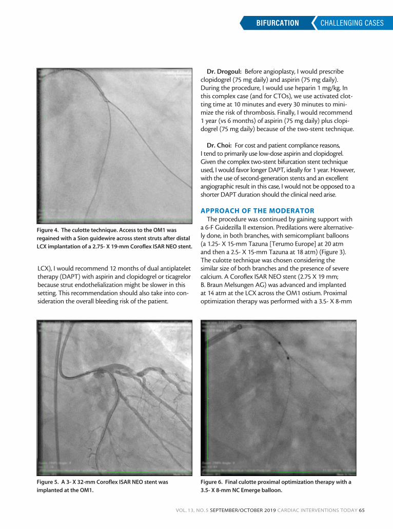
Figure 4. The culotte technique. Access to the OM1 was
regained with a Sion guidewire across stent struts after distal
LCX implantation of a 2.75- X 19-mm Coroflex ISAR NEO stent.
Figure 5. A 3- X 32-mm Coroflex ISAR NEO stent was
implanted at the OM1.
Figure 6. Final culotte proximal optimization therapy with a
3.5- X 8-mm NC Emerge balloon.

66 CARDIAC INTERVENTIONS TODAY SEPTEMBER/OCTOBER 2019 VOL. 13, NO. 5
BIFURCATIONCHALLENGING CASES
NC Emerge balloon (Boston Scientific Corporation) at 12 atm. After regaining the OM1 access with a Sion guidewire (Asahi Intecc USA, Inc.), the stent struts were opened to the OM1 with a 2.5- X 15-mm Tazuna balloon at 10 atm (Figure 4) and a 3- X 32-mm Coroflex ISAR NEO stent was implanted at the OM1 (Figure 5).
After recrossing with a Sion guidewire, a 2.5- X 15-mm Tazuna balloon was used to open the stent struts. A kissing balloon technique was attempted with a 2.5- X 15-mm Trek balloon (Abbott) and a 3- X 15-mm Ikazuchi Zero (Cordis, a Cardinal Health com-pany,) but rupture occurred after forcing both balloons across the 6-F Guidezilla. Finally, stent postdilation in both arms was performed with a 2.5- X 15-mm Trek balloon at 20 atm followed by proximal optimiza-tion therapy with a 3.5- X 8-mm NC Emerge balloon (Figure 6). The final angiographic result was excellent without any residual stenosis and excellent flow to both bifurcation branches (Figure 7).
The patient was discharged the next day on DAPT for at least 12 months, according to the grade 2 isch-emic risk evaluated by the DAPT score. The DAPT score should be requantified at 12-month follow-up, and if no hemorrhages have occurred, prolonged DAPT can be useful. The RCA CTO PCI will be discussed according to the clinical symptoms and ischemia. n
Acknowledgment: The moderator would like to acknowl-edge Dr. Henrique Mesquita Gabriel for sharing this case performed at our institution (Hospital de Santa Cruz CHLO, CEDOC, Nova Medical School in Lisbon, Portugal).
Rui Campante Teles, MD, FESC, FACC Hospital de Santa Cruz, CHLOHospital da Luz, Luz-SaudeNova Medical School, CEDOCLisbon, [email protected]: None.
James W. Choi, MD, FACC, FSCAICardiology Consultants of TexasBaylor Scott & White Heart and Vascular HospitalBaylor University Medical CenterMedical DirectorCoronary, Renal and Structural Heart TechnologiesBaylor Research InstituteAssociate Professor of MedicineTexas A&M College of MedicineDallas, [email protected]: Advisory board for Medtronic; speaker’s bureau for Boston Scientific Corporation.
Laurent Drogoul, MDClinique St GeorgeArnault Tzanck Institute Nice, [email protected]: CTO proctor for Medtronic, Abbott, Boston Scientific Corporation, Biotronik, and Biosensors International Group, Ltd.
Pedro de Araújo Gonçalves, MD, PhDHospital de Santa Cruz, CHLO Hospital da Luz, Luz-SaudeNova Medical School, CEDOCLisbon, [email protected]: None.
Figure 7. Final culotte result.

![Stability and bifurcation analysis of a Van der Pol–Duffing ... · study the post-bifurcation behavior of the system, which loses stability through a simple Hopf bifurcation [7]](https://img.pdfslide.net/doc/110x75/5f0434bd7e708231d40cd631/stability-and-bifurcation-analysis-of-a-van-der-poladufing-study-the-post-bifurcation.jpg)


![Nonlinear bifurcation analysis of stiffener profiles via ...especially for imperfection-sensitive shells where multiple bifurcation paths are possible [1], makes the bifurcation analysis](https://img.pdfslide.net/doc/110x75/60e0b8694695dc175a47d4ad/nonlinear-bifurcation-analysis-of-stiffener-profiles-via-especially-for-imperfection-sensitive.jpg)