Embed Size (px)
Citation preview
LECTURE PRESENTATIONSFor CAMPBELL BIOLOGY, NINTH EDITION
Jane B. Reece, Lisa A. Urry, Michael L. Cain, Steven A. Wasserman, Peter V. Minorsky, Robert B. Jackson
© 2011 Pearson Education, Inc.
Lectures byErin Barley
Kathleen Fitzpatrick
Cell Communication
Chapter 11

Overview: Cellular Messaging
• Cell-to-cell communication is essential for both multicellular and unicellular organisms
• Biologists have discovered some universal mechanisms of cellular regulation
• Cells most often communicate with each other via chemical signals
• For example, the fight-or-flight response is triggered by a signaling molecule called epinephrine
© 2011 Pearson Education, Inc.
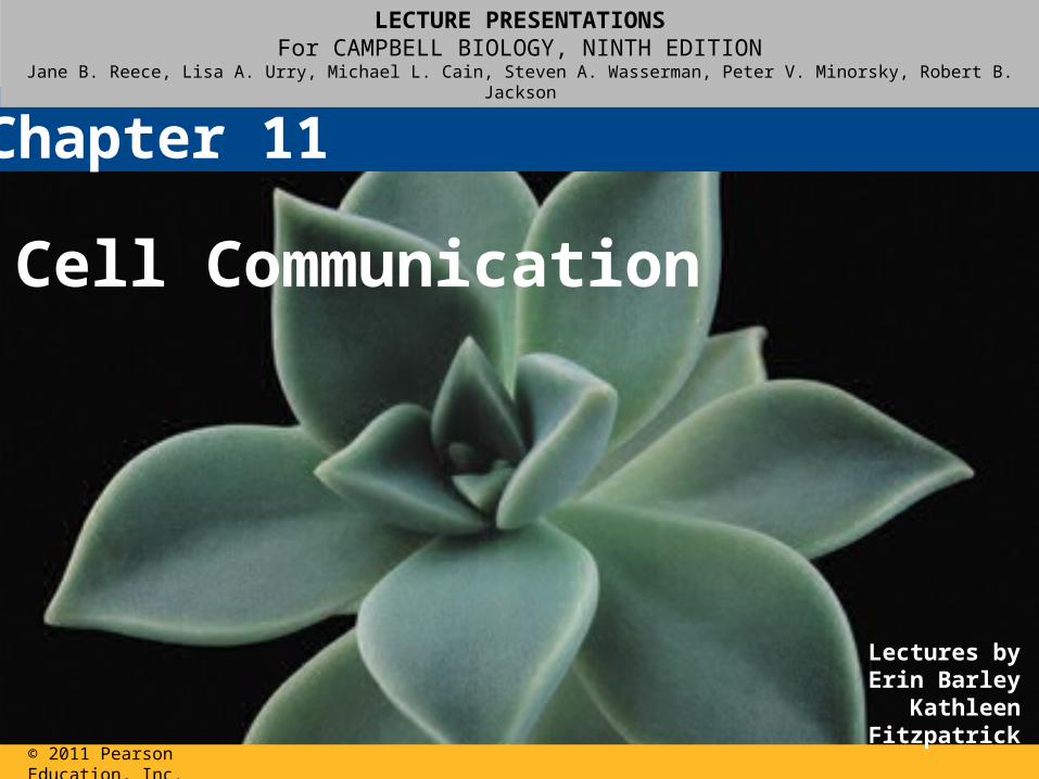
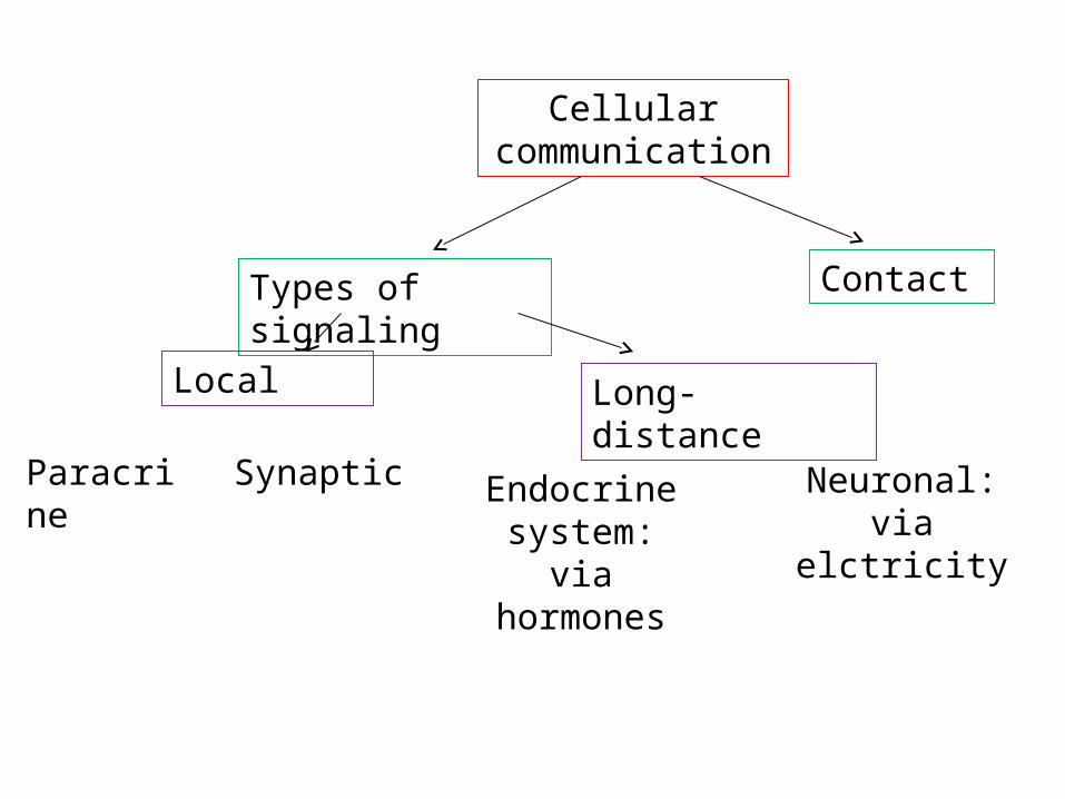
Cellular communication
Types of signaling Contact
Local
Paracrine Synaptic
Long-distance
Endocrine system: via hormones
Neuronal: via elctricity
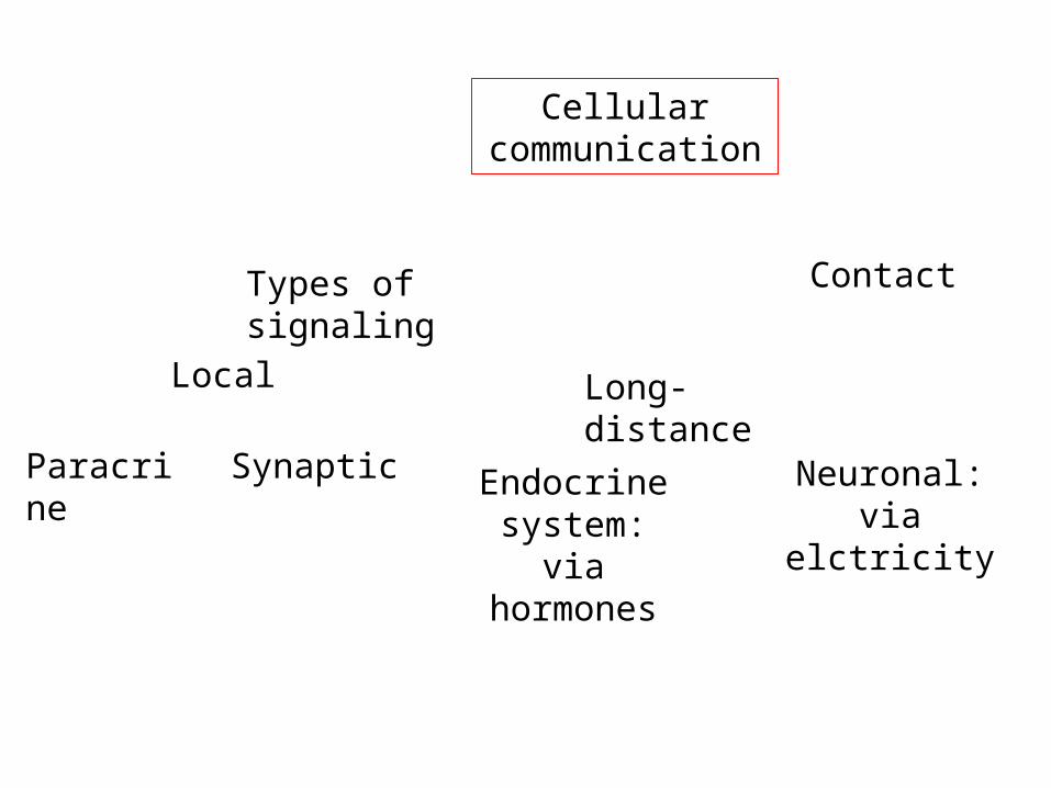
Cellular communication
Types of signaling Contact
Local
Paracrine Synaptic
Long-distance
Endocrine system: via hormones
Neuronal: via elctricity
Cellular communication
Types of signaling Contact
Local
Paracrine Synaptic
Long-distance
Endocrine system: via hormones
Neuronal: via elctricity
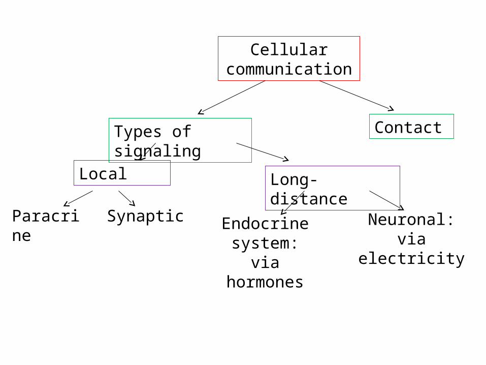
Cellular communication
Types of signaling Contact
Local
Paracrine Synaptic
Long-distance
Endocrine system: via hormones
Neuronal: via electricity

Figure 11.1
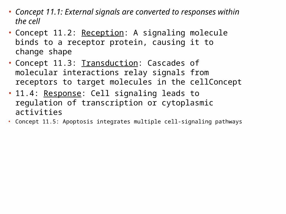
• Concept 11.1: External signals are converted to responses within the cell
• Concept 11.2: Reception: A signaling molecule binds to a receptor protein, causing it to change shape
• Concept 11.3: Transduction: Cascades of molecular interactions relay signals from receptors to target molecules in the cellConcept
• 11.4: Response: Cell signaling leads to regulation of transcription or cytoplasmic activities
• Concept 11.5: Apoptosis integrates multiple cell-signaling pathways
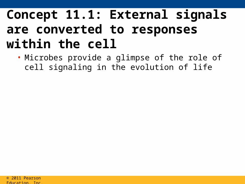
Concept 11.1: External signals are converted to responses within the cell
• Microbes provide a glimpse of the role of cell signaling in the evolution of life
© 2011 Pearson Education, Inc.
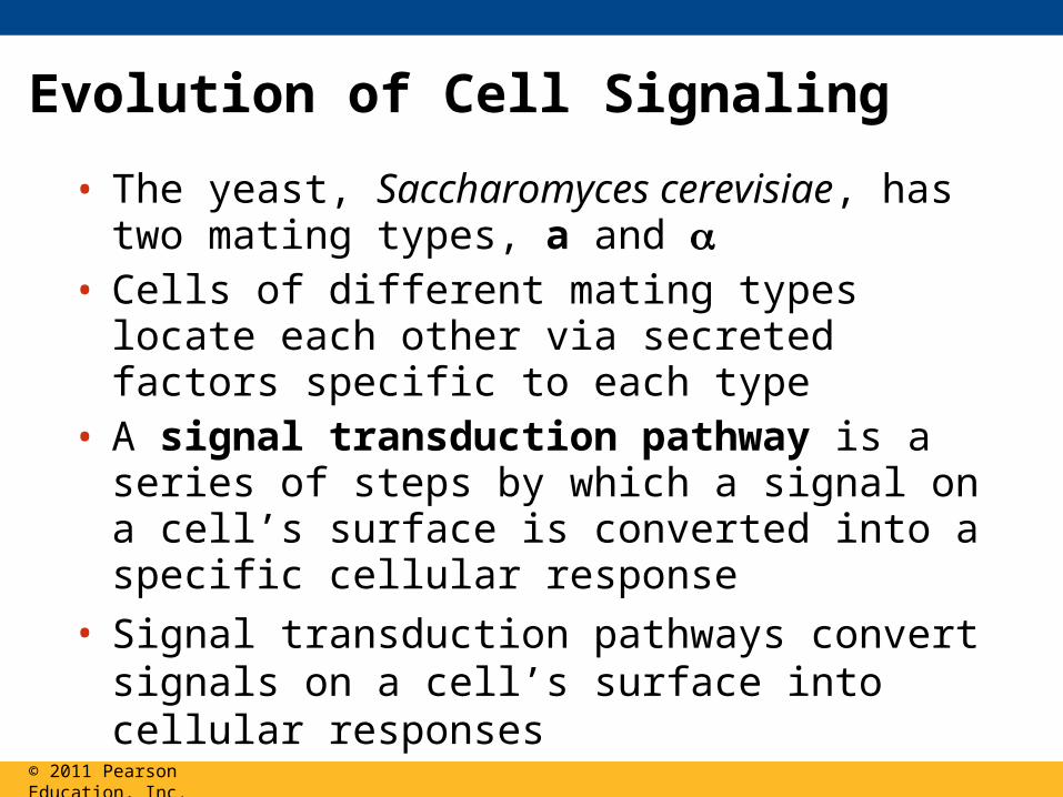
Evolution of Cell Signaling
• The yeast, Saccharomyces cerevisiae, has two mating types, a and
• Cells of different mating types locate each other via secreted factors specific to each type
• A signal transduction pathway is a series of steps by which a signal on a cell’s surface is converted into a specific cellular response
• Signal transduction pathways convert signals on a cell’s surface into cellular responses
© 2011 Pearson Education, Inc.
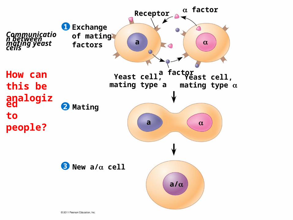
Communication between mating yeast cells
How canthis beanalogized
to people?
Exchange of mating factors
Receptor factor
a factorYeast cell,
mating type aYeast cell,
mating type
Mating
New a/ cell
1
2
3
a
a
a/
• How could we find out how long ago cell communication evolved?
© 2011 Pearson Education, Inc.
• How could we find out how long ago cell communication evolved?
• See if similar mechanisms are present in bacteria and recently developed organisms like people.
© 2011 Pearson Education, Inc.
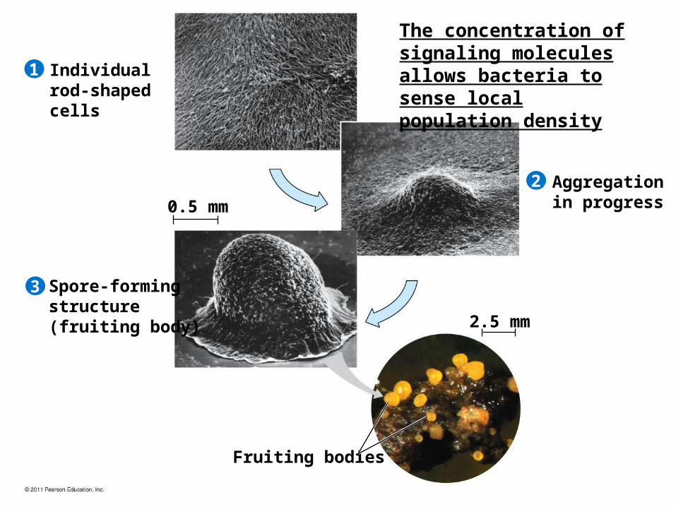
Individualrod-shapedcells
Spore-formingstructure(fruiting body)
Aggregation in progress
Fruiting bodies
1
2
3
0.5 mm
2.5 mm
The concentration of signaling molecules allows bacteria to sense local population density
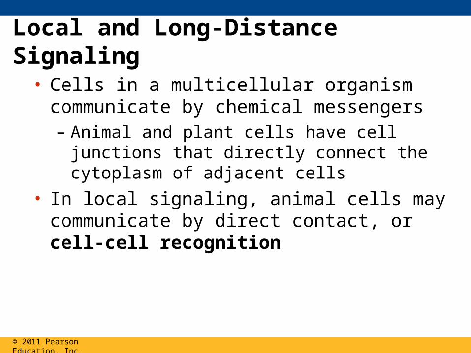
Local and Long-Distance Signaling
• Cells in a multicellular organism communicate by chemical messengers– Animal and plant cells have cell junctions that
directly connect the cytoplasm of adjacent cells
• In local signaling, animal cells may communicate by direct contact, or cell-cell recognition
© 2011 Pearson Education, Inc.
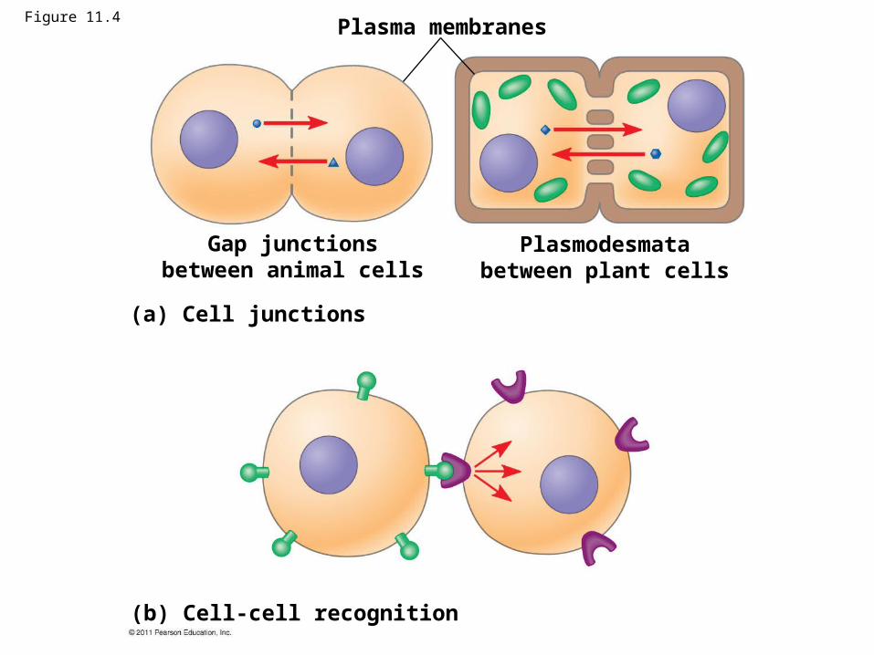
Figure 11.4Plasma membranes
Gap junctionsbetween animal cells
Plasmodesmatabetween plant cells
(a) Cell junctions
(b) Cell-cell recognition
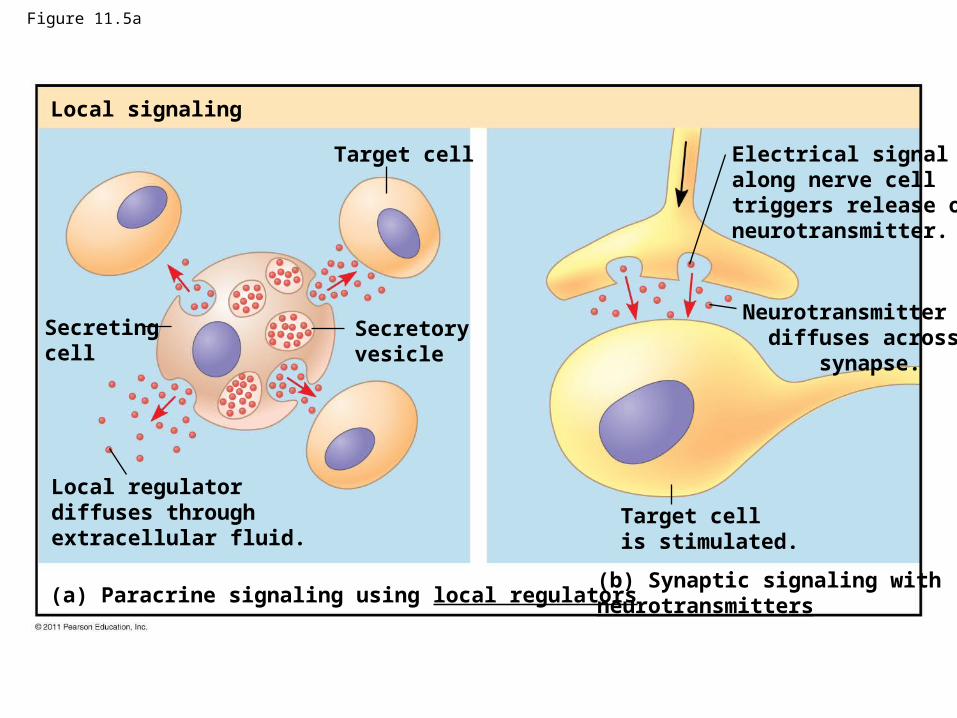
Figure 11.5a
Local signaling
Target cell
Secretingcell
Secretoryvesicle
Local regulatordiffuses throughextracellular fluid.
(a) Paracrine signaling using local regulators(b) Synaptic signaling with neurotransmitters
Electrical signalalong nerve celltriggers release ofneurotransmitter.
Neurotransmitter diffuses across synapse.
Target cellis stimulated.
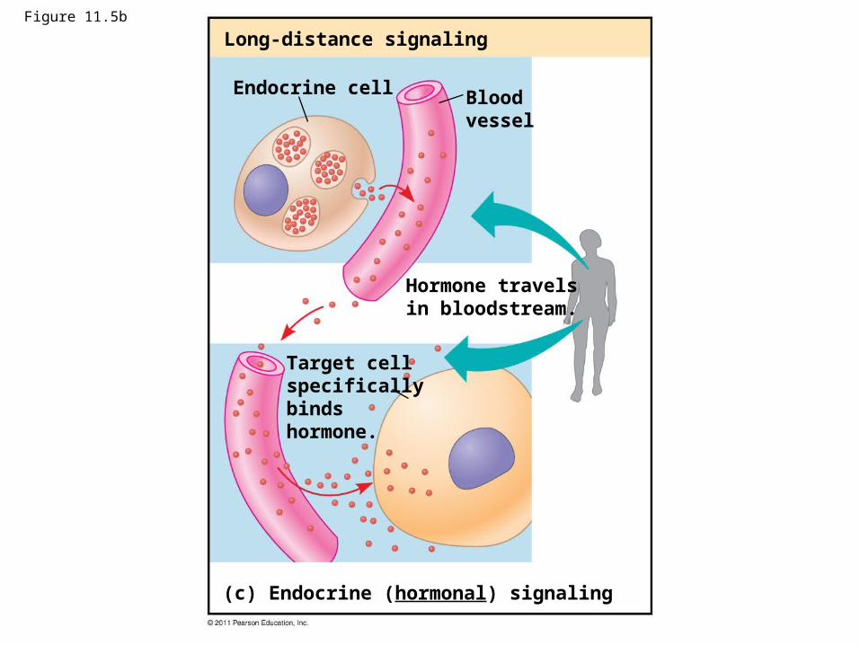
Figure 11.5b
Long-distance signaling
Endocrine cell Bloodvessel
Hormone travelsin bloodstream.
Target cellspecificallybinds hormone.
(c) Endocrine (hormonal) signaling
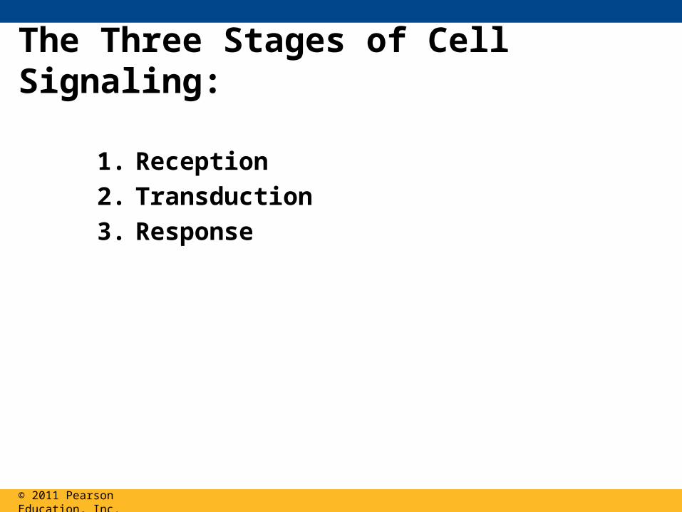
The Three Stages of Cell Signaling:
1. Reception
2. Transduction
3. Response
© 2011 Pearson Education, Inc.
© 2011 Pearson Education, Inc.
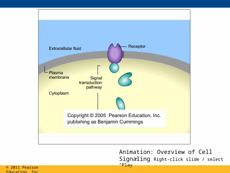
Animation: Overview of Cell Signaling Right-click slide / select “Play”
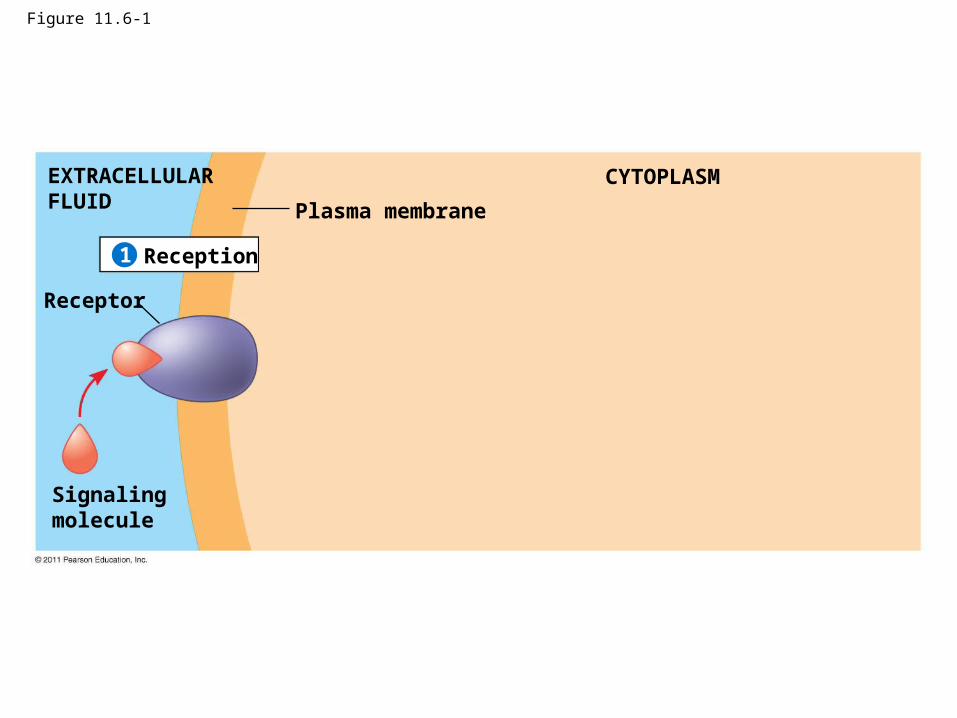
Figure 11.6-1
Plasma membrane
EXTRACELLULARFLUID
CYTOPLASM
Reception
Receptor
Signalingmolecule
1
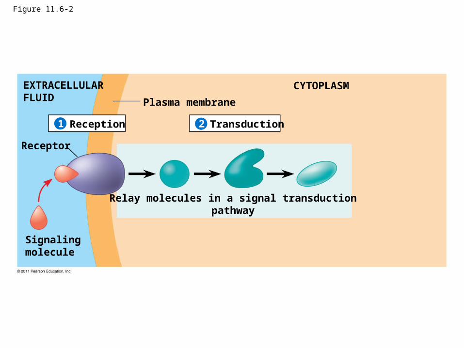
Figure 11.6-2
Plasma membrane
EXTRACELLULARFLUID
CYTOPLASM
Reception Transduction
Receptor
Signalingmolecule
Relay molecules in a signal transductionpathway
21
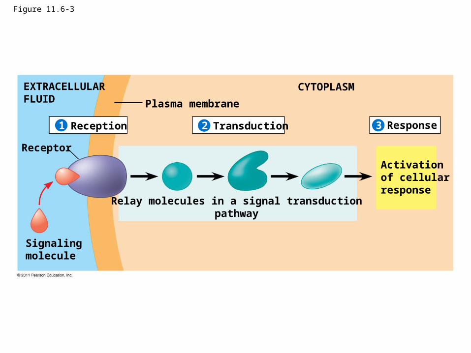
Figure 11.6-3
Plasma membrane
EXTRACELLULARFLUID
CYTOPLASM
Reception Transduction Response
Receptor
Signalingmolecule
Activationof cellularresponse
Relay molecules in a signal transductionpathway
321
Concept 11.2: Reception: A signaling molecule binds to a receptor protein, causing it to change shape
© 2011 Pearson Education, Inc.
Receptors in the Plasma Membrane
• There are three main types of membrane receptors
1. G protein-coupled receptors
2. Receptor tyrosine kinases
3. Ion channel receptors
© 2011 Pearson Education, Inc.
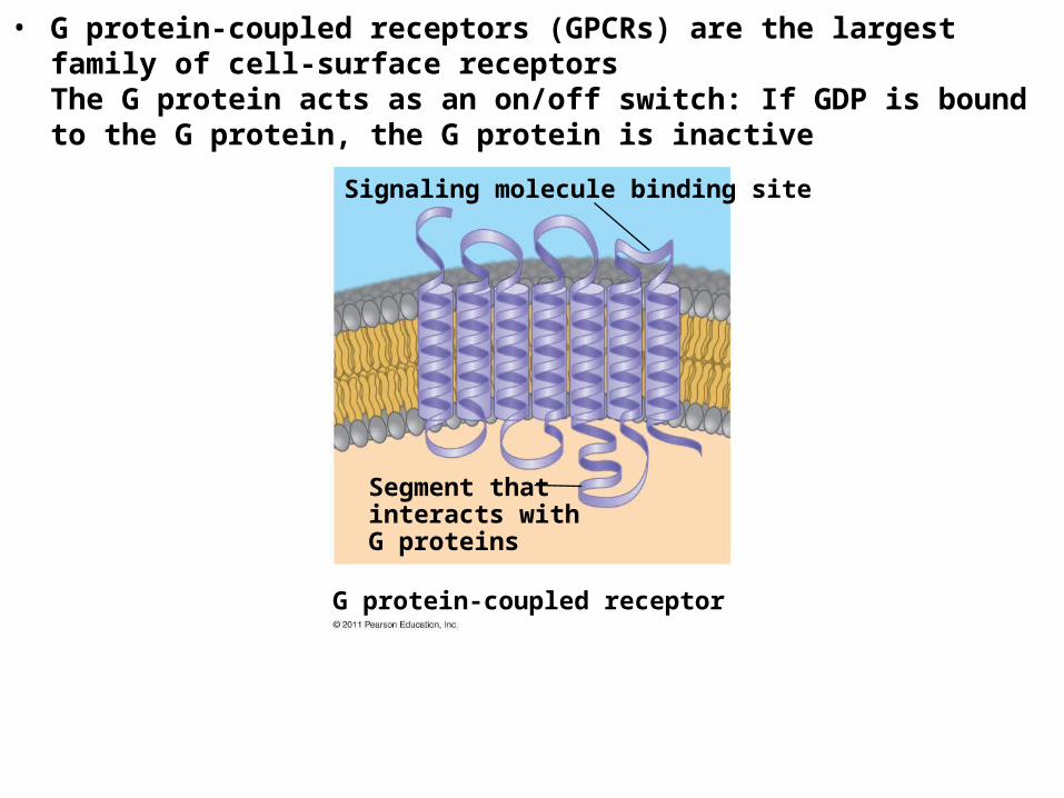
• G protein-coupled receptors (GPCRs) are the largest family of cell-surface receptorsThe G protein acts as an on/off switch: If GDP is bound to the G protein, the G protein is inactive
G protein-coupled receptor
Signaling molecule binding site
Segment thatinteracts with G proteins
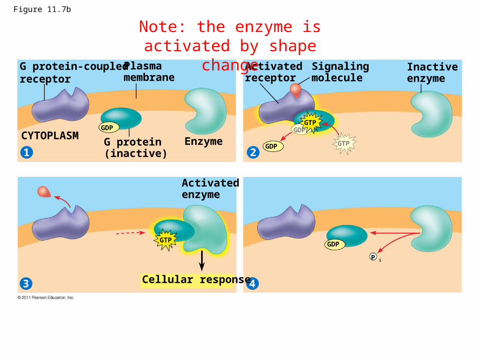
Figure 11.7b
G protein-coupledreceptor
21
3 4
Plasmamembrane
G protein(inactive)
CYTOPLASM Enzyme
Activatedreceptor
Signalingmolecule
Inactiveenzyme
Activatedenzyme
Cellular response
GDPGTP
GDPGTP
GTP
P i
GDP
GDP
Note: the enzyme is activated by shape change
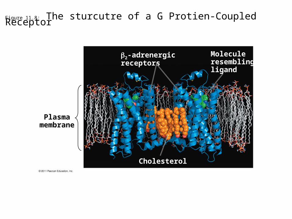
Figure 11.8: The sturcutre of a G Protien-Coupled Receptor
Plasmamembrane
Cholesterol
2-adrenergicreceptors
Moleculeresemblingligand
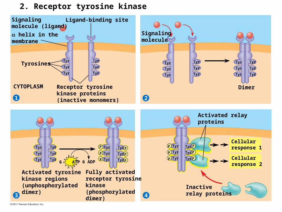
2. Receptor tyrosine kinase
Signalingmolecule (ligand)
21
3 4
Ligand-binding site
helix in themembrane
Tyrosines
CYTOPLASM Receptor tyrosinekinase proteins(inactive monomers)
Signalingmolecule
Dimer
Tyr
Tyr
Tyr
Tyr
Tyr
Tyr
Tyr
Tyr
Tyr
Tyr
Tyr
Tyr
Tyr
Tyr
Tyr
Tyr
Tyr
Tyr
Tyr
Tyr
Tyr
Tyr
Tyr
Tyr
Tyr
Tyr
Tyr
Tyr
Tyr
Tyr
Tyr
Tyr
Tyr
Tyr
Tyr
Tyr
P
P
P
P
P
P
P
P
P
P
P
P
Activated tyrosinekinase regions(unphosphorylateddimer)
Fully activatedreceptor tyrosinekinase(phosphorylateddimer)
Activated relayproteins
Cellularresponse 1
Cellularresponse 2
Inactiverelay proteins
6 ATP 6 ADP
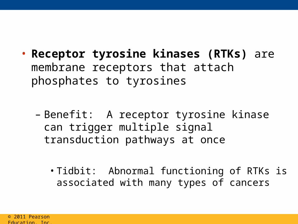
• Receptor tyrosine kinases (RTKs) are membrane receptors that attach phosphates to tyrosines
– Benefit: A receptor tyrosine kinase can trigger multiple signal transduction pathways at once
• Tidbit: Abnormal functioning of RTKs is associated with many types of cancers
© 2011 Pearson Education, Inc.
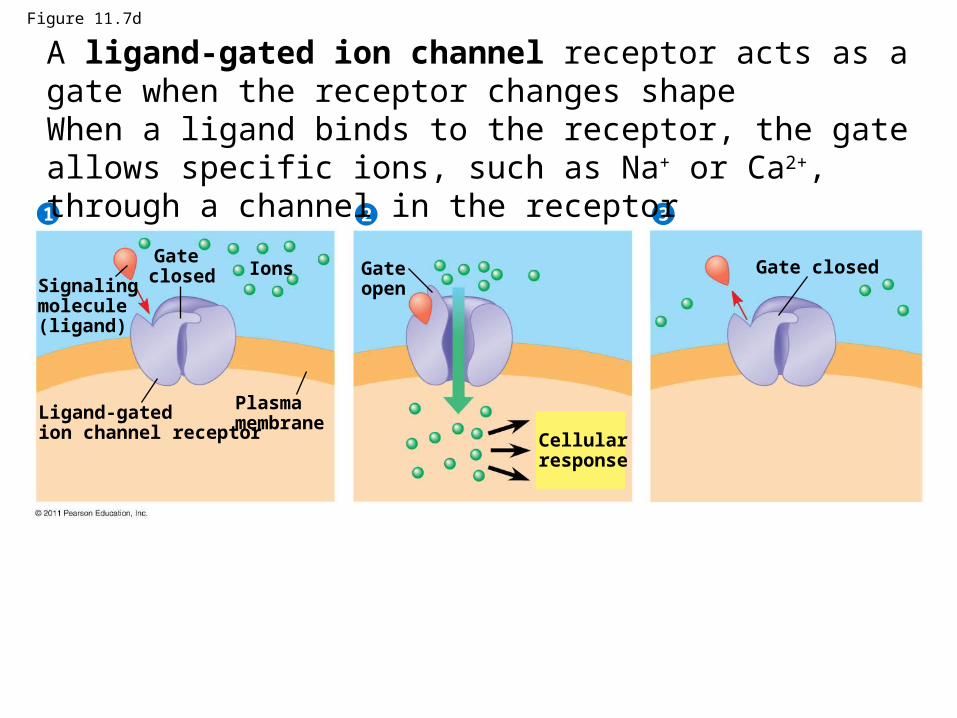
Figure 11.7d
Signalingmolecule (ligand)
21 3
Gate closed Ions
Ligand-gatedion channel receptor
Plasmamembrane
Gate open
Cellularresponse
Gate closed
A ligand-gated ion channel receptor acts as a gate when the receptor changes shapeWhen a ligand binds to the receptor, the gate allows specific ions, such as Na+ or Ca2+, through a channel in the receptor
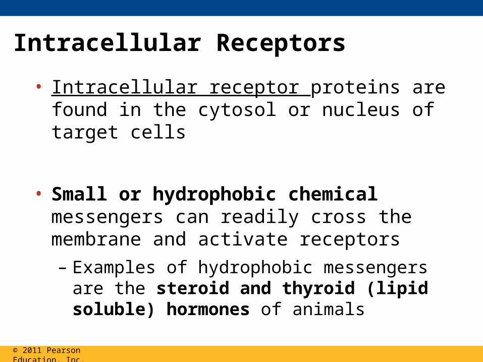
Intracellular Receptors
• Intracellular receptor proteins are found in the cytosol or nucleus of target cells
• Small or hydrophobic chemical messengers can readily cross the membrane and activate receptors
– Examples of hydrophobic messengers are the steroid and thyroid (lipid soluble) hormones of animals
© 2011 Pearson Education, Inc.
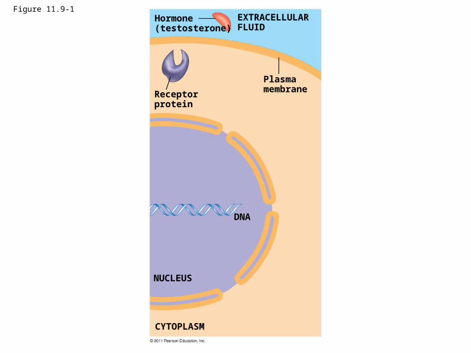
Figure 11.9-1Hormone(testosterone)
Receptorprotein
Plasmamembrane
DNA
NUCLEUS
CYTOPLASM
EXTRACELLULARFLUID
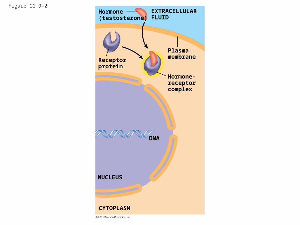
Figure 11.9-2Hormone(testosterone)
Receptorprotein
Plasmamembrane
Hormone-receptorcomplex
DNA
NUCLEUS
CYTOPLASM
EXTRACELLULARFLUID
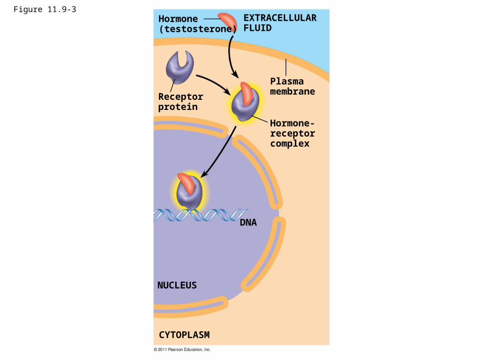
Figure 11.9-3Hormone(testosterone)
Receptorprotein
Plasmamembrane
Hormone-receptorcomplex
DNA
NUCLEUS
CYTOPLASM
EXTRACELLULARFLUID
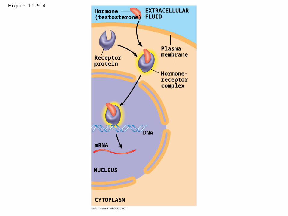
Figure 11.9-4Hormone(testosterone)
Receptorprotein
Plasmamembrane
Hormone-receptorcomplex
DNA
mRNA
NUCLEUS
CYTOPLASM
EXTRACELLULARFLUID
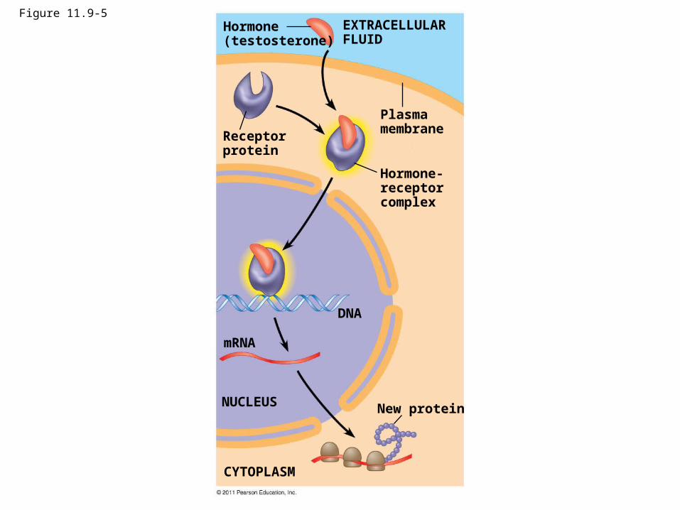
Figure 11.9-5Hormone(testosterone)
Receptorprotein
Plasmamembrane
EXTRACELLULARFLUID
Hormone-receptorcomplex
DNA
mRNA
NUCLEUS
CYTOPLASM
New protein
Concept 11.3: Transduction: Cascades of molecular interactions relay signals from receptors to target molecules in the cell
• Signal transduction usually involves multiple steps– What are some benefits of a multistep pathway
a.k.a. cascade?
© 2011 Pearson Education, Inc.
Concept 11.3: Transduction: Cascades of molecular interactions relay signals from receptors to target molecules in the cell
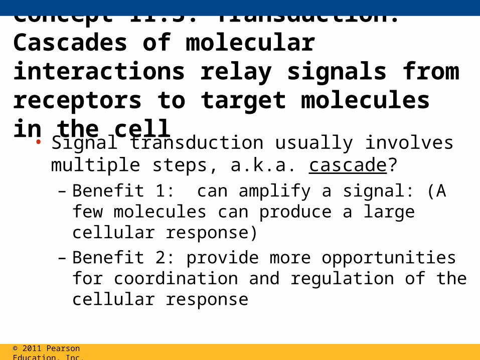
• Signal transduction usually involves multiple steps, a.k.a. cascade?– Benefit 1: can amplify a signal: (A few molecules
can produce a large cellular response)
– Benefit 2: provide more opportunities for coordination and regulation of the cellular response
© 2011 Pearson Education, Inc.
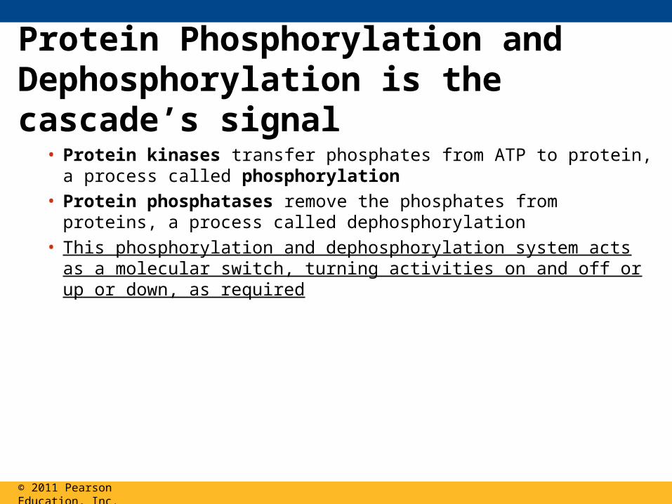
Protein Phosphorylation and Dephosphorylation is the cascade’s signal
• Protein kinases transfer phosphates from ATP to protein, a process called phosphorylation
• Protein phosphatases remove the phosphates from proteins, a process called dephosphorylation
• This phosphorylation and dephosphorylation system acts as a molecular switch, turning activities on and off or up or down, as required
© 2011 Pearson Education, Inc.
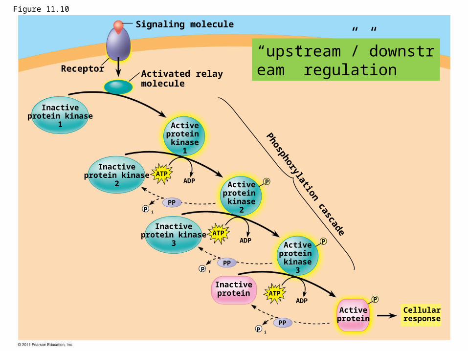
Receptor
Signaling molecule
Activated relaymolecule
Phosphorylation cascade
Inactiveprotein kinase
1 Activeprotein kinase
1
Activeprotein kinase
2
Activeprotein kinase
3
Inactiveprotein kinase
2
Inactiveprotein kinase
3
Inactiveprotein
Activeprotein
Cellularresponse
ATPADP
ATPADP
ATPADP
PP
PP
PP
P
P
P
P i
P i
P i
Figure 11.10
“upstream”/”downstream” regulation
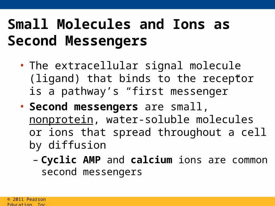
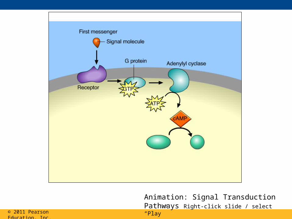
Small Molecules and Ions as Second Messengers
• The extracellular signal molecule (ligand) that binds to the receptor is a pathway’s “first messenger”
• Second messengers are small, nonprotein, water-soluble molecules or ions that spread throughout a cell by diffusion– Cyclic AMP and calcium ions are common second
messengers
© 2011 Pearson Education, Inc.
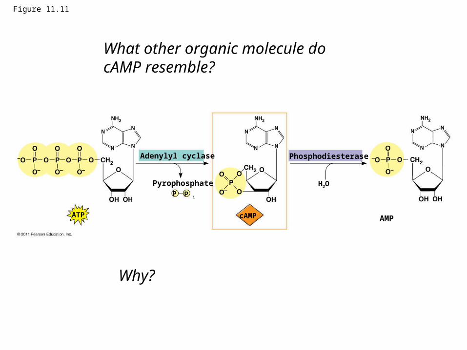
Figure 11.11
Adenylyl cyclase Phosphodiesterase
Pyrophosphate
AMP
H2O
ATP
P iP
cAMP
What other organic molecule do cAMP resemble?
Why?
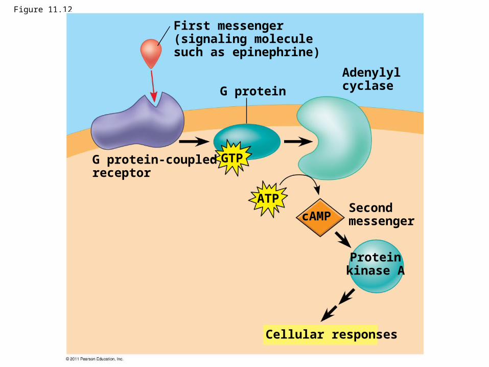
Figure 11.12
G protein
First messenger(signaling moleculesuch as epinephrine)
G protein-coupledreceptor
Adenylylcyclase
Second messenger
Cellular responses
Proteinkinase A
GTP
ATP
cAMP
Calcium Ions and Inositol Triphosphate (IP3)
• Calcium ions (Ca2+) act as a second messenger in many pathways
• Calcium is an important second messenger because cells can regulate its concentration
© 2011 Pearson Education, Inc.
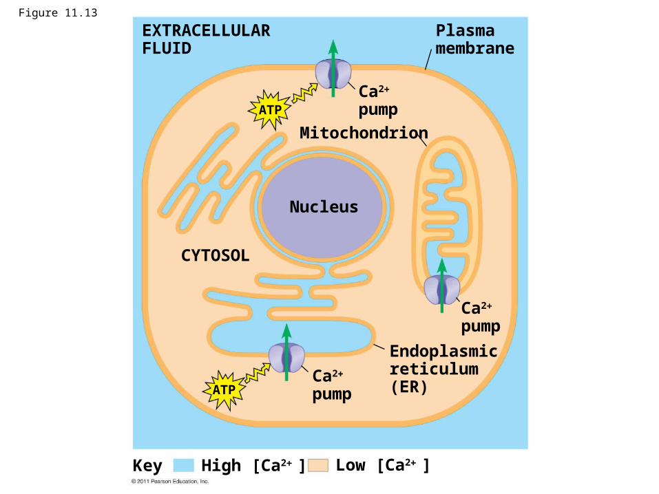
Figure 11.13
Mitochondrion
EXTRACELLULARFLUID
Plasmamembrane
Ca2
pump
Nucleus
CYTOSOL
Ca2
pump
Ca2
pump
Endoplasmicreticulum(ER)
ATP
ATP
Low [Ca2 ]High [Ca2 ]Key
© 2011 Pearson Education, Inc.
Animation: Signal Transduction Pathways Right-click slide / select “Play”
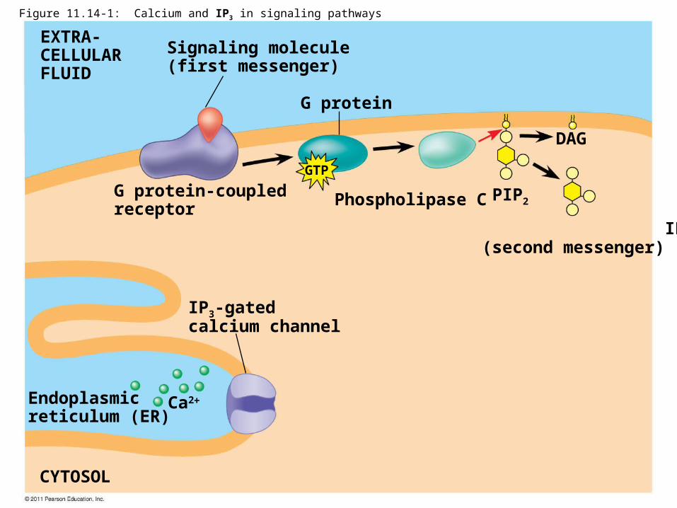
G protein
EXTRA-CELLULARFLUID
Signaling molecule(first messenger)
G protein-coupledreceptor
Phospholipase C
DAG
PIP2
IP3
(second messenger)
IP3-gatedcalcium channel
Endoplasmicreticulum (ER)
CYTOSOL
Ca2
GTP
Figure 11.14-1: Calcium and IP3 in signaling pathways
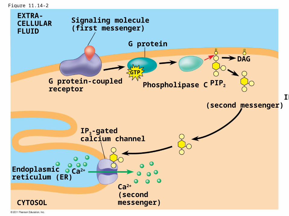
Figure 11.14-2
G protein
EXTRA-CELLULARFLUID
Signaling molecule(first messenger)
G protein-coupledreceptor
Phospholipase C
DAG
PIP2
IP3
(second messenger)
IP3-gatedcalcium channel
Endoplasmicreticulum (ER)
CYTOSOL
Ca2
(secondmessenger)
Ca2
GTP
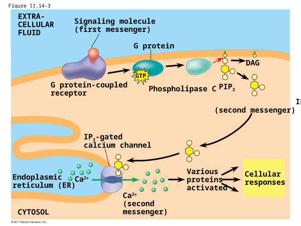
Figure 11.14-3
G protein
EXTRA-CELLULARFLUID
Signaling molecule(first messenger)
G protein-coupledreceptor
Phospholipase C
DAG
PIP2
IP3
(second messenger)
IP3-gatedcalcium channel
Endoplasmicreticulum (ER)
CYTOSOL
Variousproteinsactivated
Cellularresponses
Ca2
(secondmessenger)
Ca2
GTP
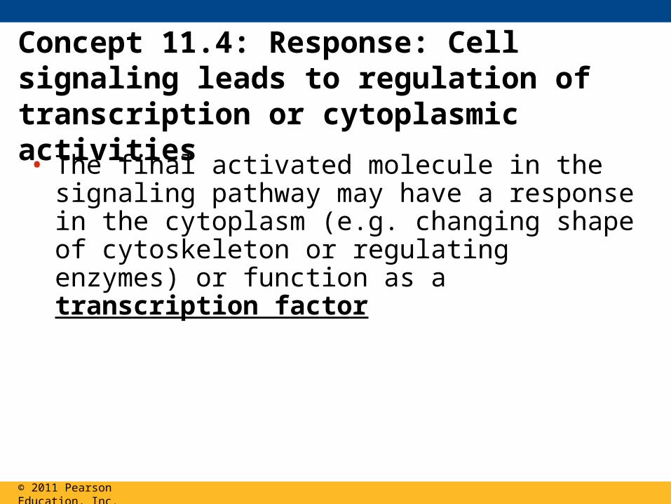
Concept 11.4: Response: Cell signaling leads to regulation of transcription or cytoplasmic activities• The final activated molecule in the signaling
pathway may have a response in the cytoplasm (e.g. changing shape of cytoskeleton or regulating enzymes) or function as a transcription factor
© 2011 Pearson Education, Inc.
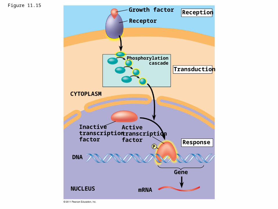
Figure 11.15Growth factor
Receptor
Reception
Transduction
CYTOPLASM
Response
Inactivetranscriptionfactor
Activetranscriptionfactor
DNA
NUCLEUS mRNA
Gene
Phosphorylationcascade
P
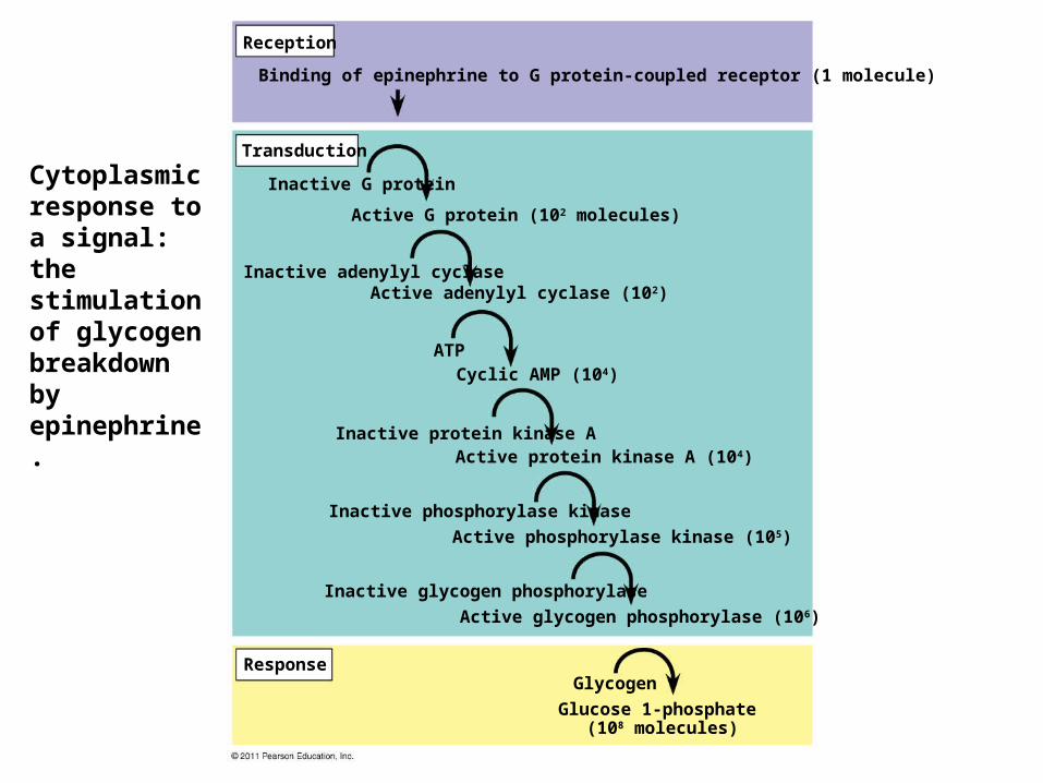
Cytoplasmic response to a signal: the stimulation of glycogen breakdown by epinephrine.
Reception
Transduction
Response
Binding of epinephrine to G protein-coupled receptor (1 molecule)
Inactive G protein
Active G protein (102 molecules)
Inactive adenylyl cyclaseActive adenylyl cyclase (102)
ATPCyclic AMP (104)
Inactive protein kinase AActive protein kinase A (104)
Inactive phosphorylase kinase
Active phosphorylase kinase (105)
Inactive glycogen phosphorylase
Active glycogen phosphorylase (106)
Glycogen
Glucose 1-phosphate (108 molecules)
• Signaling pathways can also affect the overall behavior of a cell, for example, changes in cell shape
© 2011 Pearson Education, Inc.
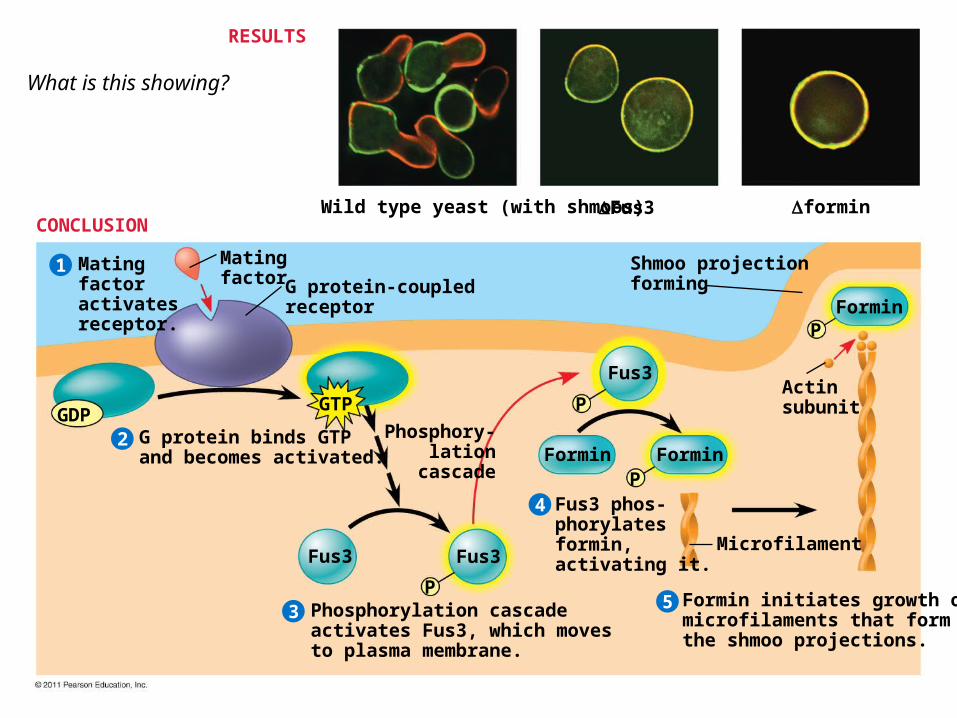
Wild type yeast (with shmoos) Fus3 formin
Matingfactoractivatesreceptor.
Matingfactor G protein-coupled
receptor
Shmoo projectionforming
Formin
G protein binds GTPand becomes activated.
2
1
3
4
5
P
P
P
PForminFormin
Fus3
Fus3Fus3
GDPGTP
Phosphory- lation cascade
Microfilament
Actinsubunit
Phosphorylation cascadeactivates Fus3, which movesto plasma membrane.
Fus3 phos-phorylatesformin,activating it.
Formin initiates growth ofmicrofilaments that formthe shmoo projections.
RESULTS
CONCLUSION
What is this showing?
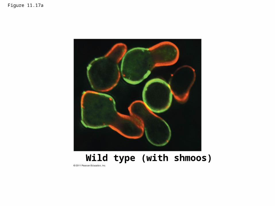
Figure 11.17a
Wild type (with shmoos)
Fine-Tuning of the Response
• There are four aspects of fine-tuning to consider:1. Amplification of the signal (and thus the
response)
2. Specificity of the response
3. Overall efficiency of response, enhanced by scaffolding proteins
4. Termination of the signal
© 2011 Pearson Education, Inc.
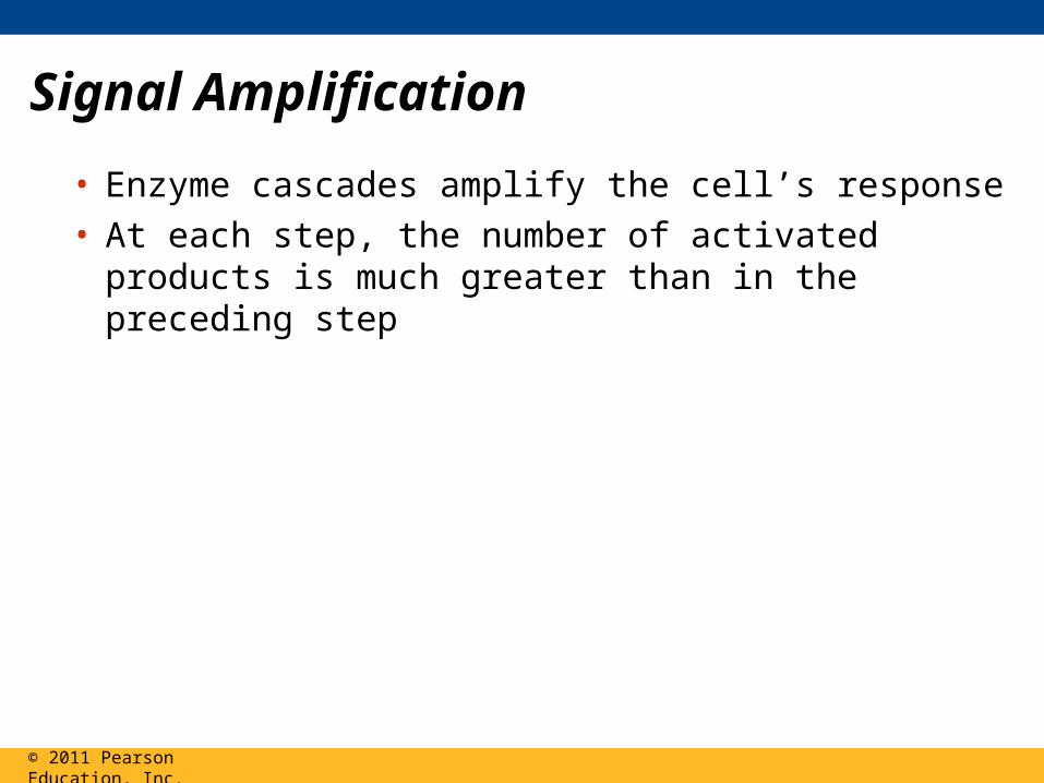
Signal Amplification
• Enzyme cascades amplify the cell’s response• At each step, the number of activated products is
much greater than in the preceding step
© 2011 Pearson Education, Inc.
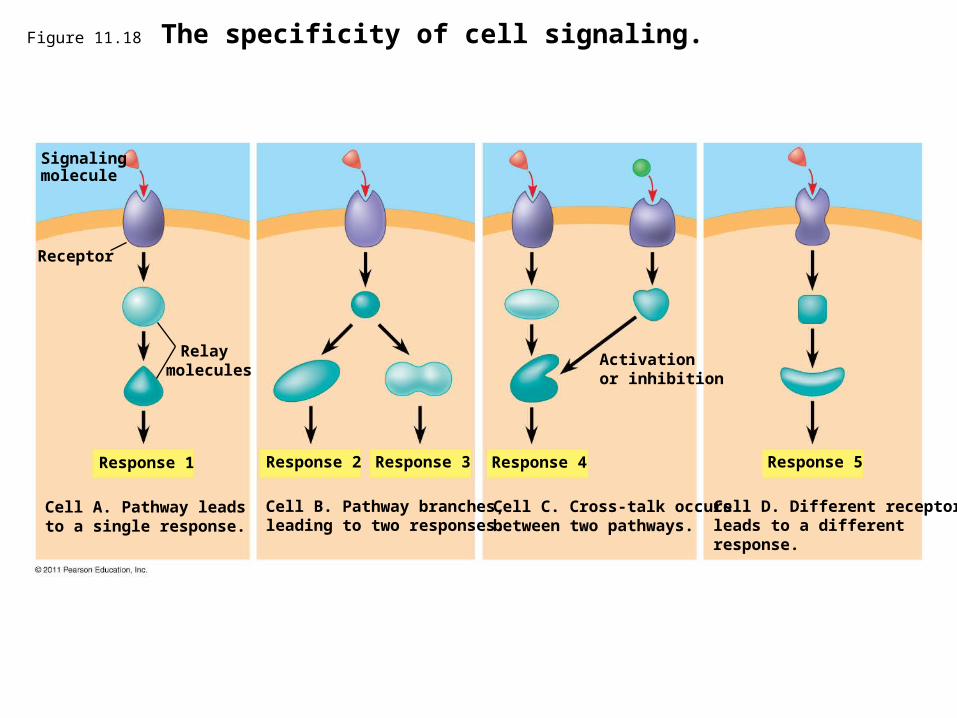
Figure 11.18 The specificity of cell signaling.
Signalingmolecule
Receptor
Relay molecules
Response 1
Cell A. Pathway leadsto a single response.
Response 2 Response 3 Response 4 Response 5
Activationor inhibition
Cell B. Pathway branches,leading to two responses.
Cell C. Cross-talk occursbetween two pathways.
Cell D. Different receptorleads to a differentresponse.
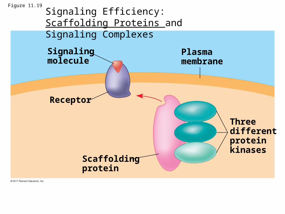
Figure 11.19
Signalingmolecule
Receptor
Plasmamembrane
Scaffoldingprotein
Threedifferentproteinkinases
Signaling Efficiency: Scaffolding Proteins and Signaling Complexes
Termination of the Signal
• Inactivation mechanisms are an essential aspect of cell signaling
• If ligand concentration falls, fewer receptors will be bound
• Unbound receptors revert to an inactive state
© 2011 Pearson Education, Inc.
Concept 11.5: Apoptosis integrates multiple cell-signaling pathways
• Apoptosis is programmed or controlled cell suicide
• WHY IS THIS FUNCTION CRITICAL?
© 2011 Pearson Education, Inc.
Concept 11.5: Apoptosis integrates multiple cell-signaling pathways
• Apoptosis is programmed or controlled cell suicide
• Components of the cell are chopped up and packaged into vesicles that are digested by scavenger cells
• Apoptosis prevents enzymes from leaking out of a dying cell and damaging neighboring cells
© 2011 Pearson Education, Inc.
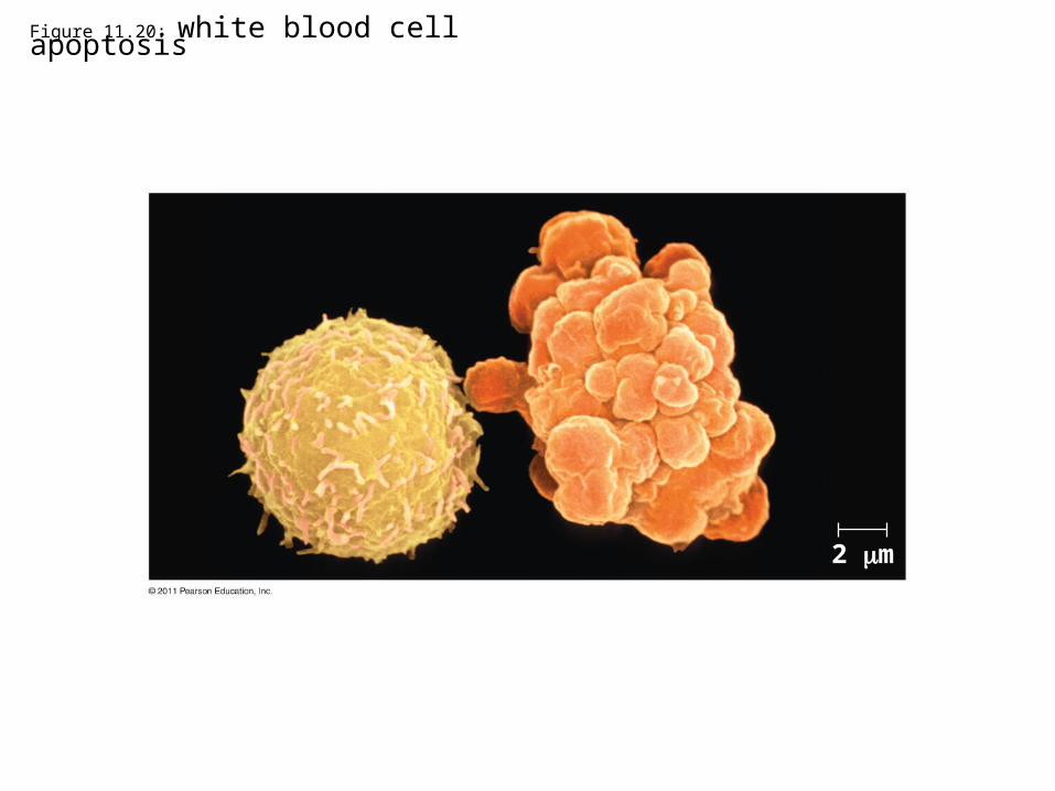
Figure 11.20: white blood cell apoptosis
2 m
Apoptosis in the Soil Worm Caenorhabditis elegans
• Apoptosis is important in shaping an organism during embryonic development
• The role of apoptosis in embryonic development was studied in Caenorhabditis elegans
• In C. elegans, apoptosis results when proteins that “accelerate” apoptosis override those that “put the brakes” on apoptosis
© 2011 Pearson Education, Inc.
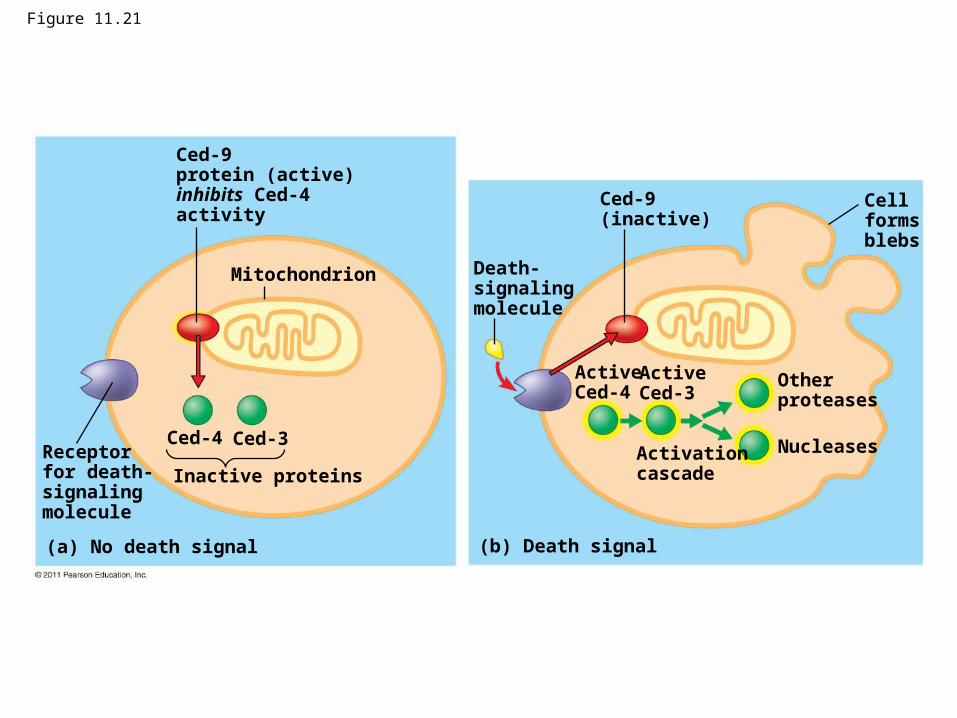
Figure 11.21
Mitochondrion
Ced-9protein (active)inhibits Ced-4activity
Receptorfor death-signalingmolecule
Ced-4 Ced-3
Inactive proteins
(a) No death signal
Death-signalingmolecule
Ced-9(inactive)
Cellformsblebs
ActiveCed-4
ActiveCed-3
Otherproteases
NucleasesActivationcascade
(b) Death signal
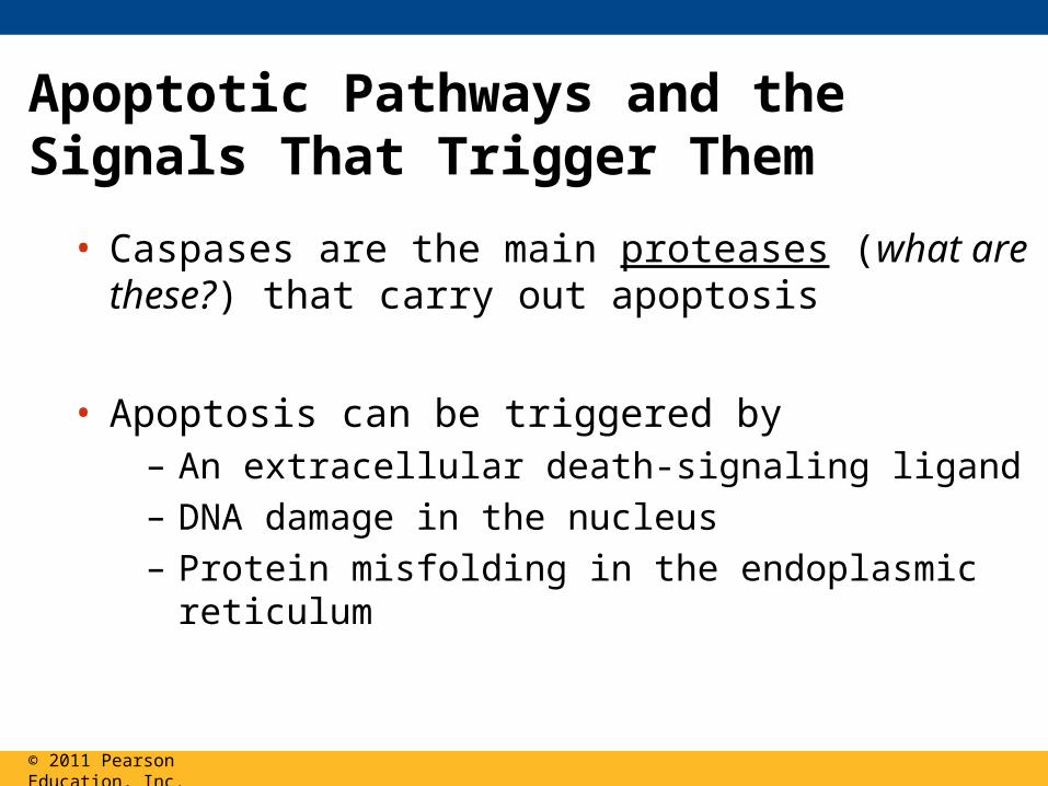
Apoptotic Pathways and the Signals That Trigger Them
• Caspases are the main proteases (what are these?) that carry out apoptosis
• Apoptosis can be triggered by– An extracellular death-signaling ligand – DNA damage in the nucleus– Protein misfolding in the endoplasmic reticulum
© 2011 Pearson Education, Inc.
• Apoptosis may be involved in some diseases (for example, Parkinson’s and Alzheimer’s); interference with apoptosis may contribute to some cancers
© 2011 Pearson Education, Inc.
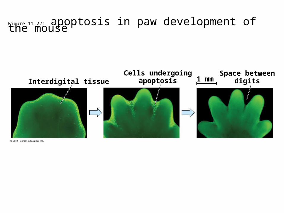
Figure 11.22: apoptosis in paw development of the mouse
Interdigital tissueCells undergoing
apoptosisSpace between
digits1 mm
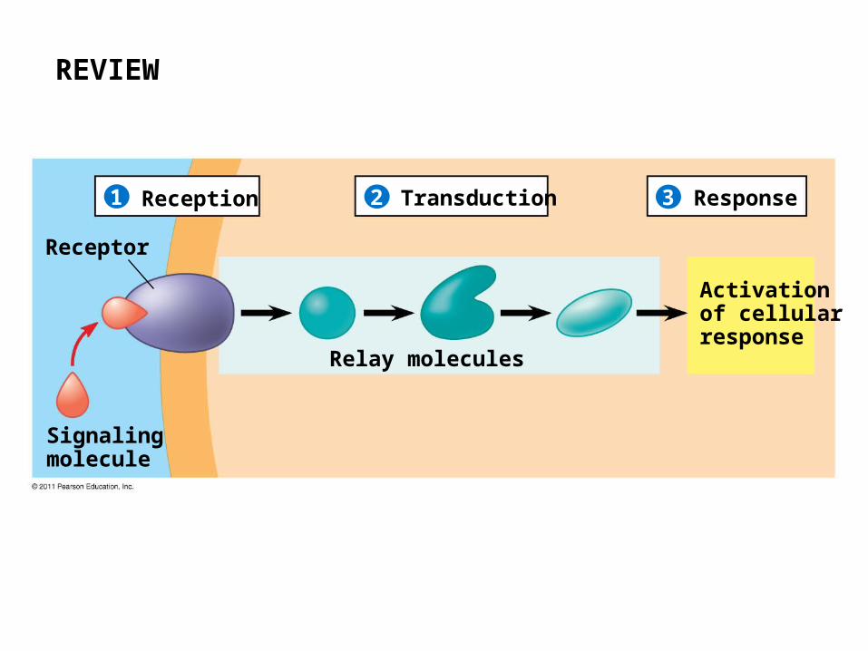
REVIEW
Reception1 2 3Transduction Response
Receptor
Signalingmolecule
Relay molecules
Activation of cellularresponse