Embed Size (px)
Citation preview
What’s New and Important in Pediatric Ophthalmology and Strabismus for Spring 2008
AAPOS Annual Meeting Washington, D.C. April 2008
American Association for Pediatric Ophthalmology and Strabismus – Professional Education Committee
A. Melinda Rainey, MD Dean Bonsall,MD Daniel Weaver, MD Earl R. Crouch, MD Douglas Fredrick, MD Deborah M. Alcorn, MD Daniel Karr, MD Oscar Cruz, MD Barry Wasserman, MD Jitka Zobal-Ratner, MD Michael Chiang, MD
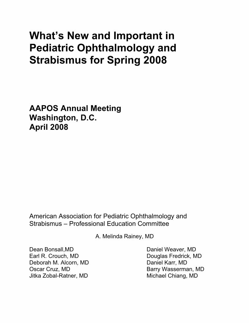
Sections
p. 2 ROP p. 7 Prematurity p. 9 Strabismus p. 17 Strabismus Surgery p. 23 Thyroid p. 24 Cataract p. 29 Cataract Surgery p. 37 Refractive Error p. 37 Refractive Surgery p. 41 Anterior Segment p. 42 Cornea p. 46 Vision Screening p. 48 Amblyopia p. 55 Genetics p. 69 Retinoblastoma p. 74 Neuro p. 76 Trauma p. 78 Uveitis p. 82 Retina p. 86 Nasolacrimal p. 87 Orbit p. 89 Plastics p. 92 Glaucoma p. 95 Congential Infections p. 96 Clinical Care p. 97 Pediatrics p. 100 Infantile Disease p. 101 Systemic p. 104 Visual Impairment p. 108 Nystagmus p. 109 Myopia
2
ROP
Retinopathy of Prematurity: the relationship between the Pediatric Ophthalmologist and the Hospital. JPOS 2007; 44: 145-149. Across the United States, the care of premature infants with retinopathy of prematurity (ROP) is becoming increasingly complex, often involving multiple specialists in multiple clinical settings. Adding to this complexity are the legal ramifications of poor outcomes for these tiny infants and the potential for high dollar litigation. The central role of the Pediatric Ophthalmologist in this maze is discussed by a panel of experts in a report generated at the 2006 AAO meeting. Effect of Diode Laser Retinal Ablative therapy for Threshold Retinopathy of Prematurity on the Visual field: Results of Goldman Perimetry at a Mean Age of 11 Years. JPOS 2007; 44:170-173. The peripheral visual field in premature children who received diode laser photocoagulation for threshold ROP was compared with a group of children with subthreshold ROP that had regressed spontaneously without laser treatment. The mean followup was eleven years. Goldman visual field testing using the II4e and V4e stimulus was utilized. Laser treated eyes showed a slight constriction of peripheral visual fields compared with untreated subthreshold eyes. This may have been due to either the laser therapy or the more severe ROP in the laser group. However, the limited reduction in visual field extent is comparable to that reported for cryotherapy and is unlikely to be of functional significance. Severe visual impairment in children with mild or moderate retinal residua following regressed threshold retinopathy of prematurity. Siatkowski RM, Dobson V., et al. J AAPOS 2007; 11: 148-152. CRYO-ROP 10-year follow-up exam and data was used to identify 16 of the 247 patients examined who had retinal outcomes of no ROP residua, no straightened temporal vessels, and no macular heterotropia in at least one eye, but with a visual acuity less than 20/200 in both eyes. The authors discuss the anterior, posterior, or combined visual pathway conditions that can occur in these rare patients.
3
Severe retinopathy of prematurity in infants <30 weeks gestation in New South. Wales and the Australian Capital Territory from 1992-2002. Todd DA, Wright A, Smith J, NICUS Group Arch Dis Child. 2007. Data was collected from Neonatal Intensive Care Units (NICUS) over an 11-yr period of infants <30 wks. Divided into three groups: < 24 wks, 25-26 wks and 27-29 wks gestation. In the 24 wk group the incidence of ROP and treatment increase from 41.5% to 53.9%. In infants 25-26 wks the incidence decreased, but the number of treated pts increase slightly. In infants 27-29 wks there was no increase in incidence or treatment. Visual and cerebral sequelae of very low birth weight in adolescents. Hellgren K, Hellstrom A, Jacobson L, Flodmark O, Wadsby M, Martine L. Arch Dis Child. 2007. 59 15-year old former VLBW infants were compared to 55 sex and age matched controls with normal birth weight. Va, stereo and cycloplegic refraction were measured as was the WISC intelligence scale. All previously VLBW also had MRIs of the brain. The VLBW adolescents had poorer vision, worse stereo and lower I.Q.s 30% had abnormal MRI findings and these children performed worse that those VLBWs with normal MRIs. The findings found a cerebral causative component for the visual dysfunction. Children born weighing less than 1701 g: visual and cognitive outcomes at 11-14 years. Stephenson T, Wright S, O’Conner A, Fielder A, Johnson A, Ratib S. Arch. Dis. Child. 2007. Prospective study of ROP infants, 1701 g born started in 1985. 7,254 consented to an ophthalmic examination at 10-13 years. 198 consented to a neuropsychological assessment at 11-14 years. 99/198 had an adverse ophthalmic outcome (AOO), 106/198 had ROP, 98 had mild ROP with no increase in poor vision later. All children with an AOO performed worse on the neuropsycological assessment. Interexpert agreement of plus disease diagnosis in retinopathy of prematurity. Michael Chiang MD MA, Lei Jiang BA, Rony Gelman MD, Yunling Du PhD, John Flynn MD Arch Ophthalmol 2007;125(7)857-880 A set of 34 wide-angle retinal photographs from infants with ROP was compiled on a secure Web site and was interpreted independently by 22 recognized ROP experts. Diagnostic agreement was analyzed using 3-level (plus, pre-plus, or neither) and 2-level (plus or not plus) categorizations. In the 3-level categorization, all experts agreed on the
4
same diagnosis in 4 of 34 images (12%), and the mean weighted ĸ statistic for each expert compared with all others was fair agreement for 7 experts (32%), and moderate agreement for 15 experts (68%). In the 2-level categorization, all experts who provided a diagnosis agreed in 7 of 34 images (21%), and the mean ĸ statistic for each expert compared with all others was slight agreement for 1 expert (5%), fair agreement for 3 experts (14%), moderate agreement for 12 experts (55), and substantial agreement for 6 experts (27%). Conclusions: Interexpert agreement of plus disease diagnosis is imperfect. This may have important implications for clinical ROP management, continued refinement of the international ROP classification system, development of computer-based diagnostic algorithms, and implementation of ROP telemedicine systems. The Editorial Comment by Dale L. Phelps, M.D., on page 963 of this issue of Archives, provides excellent remarks to this report. Plus disease in retinopathy of prematurity: Pilot study of computer-based and expert diagnosis. Gelman R, Jiang L, Du Y, Martinez-Perez E, Flynn J, Chiang M. J AAPOS 2007, Dec; 11(6); 533 Twenty-two ROP experts were asked to interpret a set of 34 wide-angle retinal images for the presence of plus disease. Images were also analyzed by a computer-based image analysis Retinal Image multiScale Analysis (RISA). The computer-based analysis included integrated curvature (IC), diameter, and tortusity index (TI). The RISA diagnosis of plus disease was compared with the reference standard, as were the expert panels’ diagnosis. When using the arteriolar IC and TI, the venular diameter, IC and TI, the RISA system was better at diagnosing plus disease than 18 of the 22 experts (81.8%) The dilemma of digital imaging in retinopathy of prematurity. Quinn G. J AAPOS 2007, Dec.11(6): 529 Nice editorial by Dr. Quinn on the difficulties of diagnosing ‘serious” ROP and absence of a quantitative standard for plus disease and the two prototype digital imaging analysis systems.
5
Incidence, progression, and duration of retinopathy in Hispanic and white non-hispanic infants. Eliason K, Osborn D, Amsel E, Richards S. J AAPOS 2007 Oct 11(5); 447 The charts of 671 white non-Hispanics and 128 Hispanic infants with BW < 1751g were retrospectively examined and multiple regression analysis was used to control for weight, gestational age and year of birth. The duration of untreated ROP was compared for the two ethnic groups. ROP was found to occur wth similar frequency in Hispanic and white non-Hispanic infants, as does subthreshold or worse ROP. Some Hispanic infants had an unusually short or long duration of ROP before regression. This may indicate the natural history of ROP is different in this group. Treatment for retinopathy of prematurity in Denmark in a ten-year period (1996-2005): Is the incidence increasing? Slidsborg C, Olesen H, Jensen P, Jensen H et al. Pediatrics 2008 Jan; 121(1):97-105. In a study conducted in Denmark the incidence of treated retinopathy of prematurity increased from 1.3% to 3.5% over a 10 year period time. The increase could not be fully explained by increased survivor rates for the infants or by changes in investigated neonatal risk factors. The increase in treated cases could not be related to changing indications for treatment as the same senior treating ophthalmologist used threshold retinopathy of prematurity criterion for the decision to treat rather than using early treatment recommendations. The authors posit that perhaps screening regimes were more efficacious in finding children at risk for the development of ROP compared to the previous time period.
6
PREMATURITY Perinatal care in the threshold of viability: An international comparison of practical guidelines for the treatment of extremely preterm births. Pignotti M, Donzelli G. Pediatrics 2008; 121(1):e193-e198. The authors conducted a review of published guidelines concerning the different approaches to the care of extremely preterm births in various countries. They found that intensive care was justified in age greater than 25 weeks, compassionate care should be delivered for less than 22 weeks and a variable individual approach to 23 – 24 weeks. As developing countries increased their ability to resuscitate and care for extremely low birth weight infants the prevalence of retinopathy of prematurity will surely increase in these countries. Neurodevelopmental outcome in survivors of periventricular hemorrhagic infarction. Bassan H, Limperopoulos C, Visconti K, Mayer D et al. Pediatrics 2007 Oct; 120(4):785-792. In the study conducted in Boston 30 premature infants were followed prospectively to evaluate developmental and cognitive deaths associated with periventricular hemorrhagic infarction. 26% of the children had significant involvement of the visual pathways leading to decreased visual acuity or significant visual field deficits. Children discharged from the hospital with a diagnosis of periventricular hemorrhagic required close ophthalmic follow-up and early intervention. Neonatologists’ practices and experiences in arranging retinopathy of prematurity screening services. Kemper A, Wallace D. Pediatrics 2007 Sep; 120(3):527-531. The authors conducted a mail survey of 300 neonatologists to determine their practices and experiences related to coordinating, screening and treatment for ROP. The response rate was 62%. The authors found that only 19% of the respondents used the recommended gestational age criterion of 30 weeks for initiating screening with 6% using lower, more restrictive criteria, and 70% using a higher, more inclusive criterion. 86% used 1,500 grams as the birth weight for criterion for screening. 46% reported that retina specialists provided the treatment, although 67% stated that screening was performed by pediatric ophthalmologists. 30% reported that they could not back
7

transfer a patient to a lower acuity hospital because of lack of specialists who were able to screen for retinopathy or prematurity.
8
STRABISMUS
Causes and outcomes for patients presenting with diplopia to an eye casualty department. Comer RM, Dawson E, Plant G, Acheson JF, Lee JP. Eye. 2007 Mar;21(3):413-8 Patients presenting with diplopia as a principal symptom, who were referred to the Orthoptic Department from Moorfields Eye Casualty over a 12-month period, were retrospectively investigated. One hundred and seventy-one patients were identified with complete records in 165 cases. There were 99 men and 66 women with an age range of 5-88 years. Monocular diplopia accounted for 19 cases (11.5%), whereas 146 patients (88.5%) had binocular diplopia. Cranial nerve palsies were the most common cause of binocular diplopia accounting for 98 (67%) of cases. Isolated sixth nerve palsy was the largest diagnostic group (n=45). Microvascular disease (hypertension or diabetes mellitus, or both) was present in 59% of patients with cranial nerve palsies, and of this group, 87% resolved spontaneously by 5 months rising to 95% by 12 months. Causes of binocular diplopia other than cranial nerve palsies included thyroid eye disease, myasthenia gravis, myositis, superior oblique myokymia and previous strabismus surgery. Patients with clinically isolated single cranial nerve palsies associated with diabetes or hypertension are likely to recover spontaneously within 5 months and initially require observation only. However, patients with unexplained binocular diplopia and those who progress or fail to recover should be investigated to establish the underlying aetiology and managed as appropriate. New approach for treating vertical strabismus: decentered intraocular lenses. Nishimoto H, Shimizu K, Ishikawa H, Uozato H. J Cataract Refract Surg. 2007 Jun;33(6):993-8. PURPOSE: To evaluate a new surgical procedure that uses a decentered intraocular lens (IOL) to correct vertical strabismus in cataract patients. METHODS: Six patients (11 eyes) with vertical strabismus had small-incision cataract surgery. The continuous curvilinear capsulorhexis was decentered, and the asymmetrical span of the IOL haptics located on the side to be bent was inserted after phacoemulsification and aspiration. Some relaxing incisions were made in the anterior capsule. Postoperatively, the alternate prism cover test was used to assess changes in ocular position. In addition, the EAS-1000 (Nidek) and KR-9000PW (Topcon) were used to evaluate IOL decentration, tilt, and aberrations. RESULTS: The mean age of the patients was 66 years (range 58 to 77 years). The mean preoperative vertical strabismus was 7.3 prism diopters (PD) (range 4 to 12 PD). Two years after surgery, the mean angle of vertical deviation was 1.3 PD (range 0 to 5
9
PD) without affectivity coma-like aberrations (S3). The mean amount of decentration was 0.52 mm +/- 0.29 (SD) and the mean tilt, 4.30 +/- 2.85 degrees (n = 10 eyes). CONCLUSION: Decentered IOL implantation was effective in cataract patients with vertical strabismus and can be performed during cataract surgery. High-resolution magnetic resonance imaging of the extraocular muscles and nerves demonstrates various etiologies of third nerve palsy. Kau HC, Tsai CC, Ortube MC, Demer JL. Am J Ophthalmol. 2007 Feb;143(2):280-287. Epub 2006 Nov 27. PURPOSE: The etiology of third nerve palsy is usually diagnosed by history, motility examination, and presence of lid and pupil involvement, as well as cranial and vascular imaging. We used high-resolution magnetic resonance imaging (hrMRI) of the oculomotor nerve and affected extraocular muscles (EOMs) to investigate oculomotor palsy. METHODS: Twelve patients with nonaneurysmal oculomotor palsy of 0.75 to 252 months' duration were studied. In the orbit and along the intracranial oculomotor nerve, hrMRI at 1- to 2-mm thickness was performed. Coronal plane images of each orbit were obtained in multiple, controlled gaze positions. Structural abnormalities of the oculomotor nerve and associated changes in EOM volume and contractility were evaluated. RESULTS: Cases were categorized as tumor related, congenital, diabetic, traumatic, and idiopathic according to clinical characteristics and hrMRI findings. Reduction of volume and contractility of affected EOMs were noted in six patients; however, there was no marked EOMs atrophy in two cases of diabetic oculomotor palsy, and there were four cases of aberrant regeneration. hrMRI demonstrated the oculomotor nerve at the midbrain and at EOMs in all cases, and in two cases with previous normal neuroimaging elsewhere that demonstrated contrast-enhancing tumors on the oculomotor nerve. One patient with apparently unilateral congenital inferior division oculomotor palsy had no detectable ipsilateral and a hypoplastic contralateral oculomotor nerve exiting the midbrain. CONCLUSIONS: hrMRI provides valuable information in patients with oculomotor palsy, such as structural abnormalities of the orbit and oculomotor nerve, and atrophy and diminished contractility of innervated EOMs. This information could be helpful in diagnosis and management of oculomotor palsy. Age at strabismus diagnosis in an incidence cohort of children. Mohney BG, Greenberg AE, Diehl NN. Am. J Ophthalmol. 2007 Sep;144(3):467-9 PURPOSE: To compare the age at diagnosis of children with esotropia, exotropia, and hypertropia.
10
METHODS: The medical records of all Olmsted County, Minnesota, residents < 19 years diagnosed with esotropia, exotropia, or hypertropia from January 1, 1985 through December 31, 1994 were reviewed. RESULTS: The median age at diagnosis of esotropia (n = 380), exotropia (n = 205), and hypertropia (n = 42) was 3.1 years, 7.2 years, and 6.1 years, respectively (P = .001). In the first six years of life, esotropia had the highest incidence and was more likely to occur than either exotropia or hypertropia; exotropia predominated between age seven and 12 years; and each form was similarly likely to occur between 13 and 18 years of age (P = .001). CONCLUSIONS: The age at diagnosis was significantly different for the various forms of strabismus in this population. Esotropia is the most common form in the first six years of life; beyond this age exotropia predominates until the teenage years when the three forms have a similar but decreased incidence. Common forms of childhood strabismus in an incidence cohort. Mohney BG. Am. J Ophthalmol. 2007 Sep;144(3):465-7. PURPOSE: To report the prevalent forms of childhood strabismus. METHODS: The medical records of all Olmsted County, Minnesota, residents younger than 19 years diagnosed with esotropia, exotropia, or hypertropia from January 1, 1985 through December 31, 1994, were reviewed. RESULTS: Six hundred twenty-seven new cases of childhood strabismus were identified during the 10-year study period, including 380 (60.1%) with esotropia, 205 (32.7%) with exotropia, and 42 (6.7%) with hypertropia. The five most common forms of strabismus included accommodative esotropia (27.9%), intermittent exotropia (16.9%), acquired nonaccommodative esotropia (10.2%), esotropia in children with an abnormal central nervous system (7.0%), and convergence insufficiency (6.4%). CONCLUSIONS: This study provides population-based data on the most prevalent forms of childhood strabismus. Accommodative esotropia, intermittent exotropia, and acquired nonaccommodative esotropia were the predominant forms of strabismus in this Western population. The Effects of Strabismus on Quality of Life in Adults. Hatt SR, Leske DA, Kirgis PA, Bradley EA, Holmes JM. Am J Ophthalmol. 2007 Aug 16; [Epub ahead of print] PURPOSE: As a first step in the development of a health-related quality of life (HRQOL) instrument, we conducted in-depth interviews to identify the specific concerns of adults with strabismus. METHODS: Thirty adults with strabismus, 17 with diplopia, and 13 without were recruited. Individual interviews, using 11 open-ended questions, were audiotaped, transcribed, and transcripts reviewed independently by three investigators. Phrases
11
regarding how strabismus affected everyday life were grouped into topic areas and the frequency of each topic analyzed for subjects with and without diplopia. RESULTS: A total of 1,508 phrases were extracted: 207 (14%) of 1,508 were excluded because they did not pertain to HRQOL. From the remaining 1,301 phrases, 48 topic areas were apparent. For patients with diplopia, the most frequently occurring topics were: nonspecific negative feeling (15/17; 88%) ("really hard"); general disability (15/17; 88%) ("affects everything"); and driving (14/17; 82%). In those without diplopia, the most frequently mentioned topics were appearance to others (12/13; 92%) ("people notice my eyes") followed by problems with eye contact (10/13; 77%) and interpersonal relationships (10/13; 77%). Of the topics that were common to both groups (n = 42), two of the most frequent were driving and nonspecific negative feeling. CONCLUSIONS: Multiple individual interviews revealed many topics that negatively affect quality of life in strabismic adults. The frequency and type of concerns confirm the importance of HRQOL assessment as an important aspect of strabismus management. Strabismus in unicoronal synostosis: ispilateral or contralateral? Macintosh C, Wall S, Leach C. J Cranofac Surg 2007 May; 18(3):465-9 This is a retrospective case series of 59 patients with confirmed unicoronal synostosis. Thirty-four (57.6%) were found to have manifest strabismus in primary position. The most common being esotropia with a vertical component (21/34). In 55.9% this occurred contralateral to the fused suture and in 26.5% ipsilateral to the fused suture. Thirty cases showed an apparent IO OA/SO UA and was bilateral in 14 cases, ipsilateral in 15 cases.
Twenty seven (46%) had a significant refractive error. Of those with anisometropia, the higher error was in the eye contralateral to the fused suture. Fifteen (62%) had more astigmatism on the contralateral, nonsynostotic side. All cases of unicoronal synostosis with a mutation of the FGFR2 or FGFR3 gene had manifest strabismus. Statistical analysis proved for this sample, that manifest strabismus was no more likely to be found on either the contralateral or ipsilateral side to the synostosis. Superior oblique myokymia: Efficacy of medical treatment. Williams PE, Purvin VA, Kawasaki A. J AAPOS 2007; 11: 254-57. This is a retrospective review of patients with SOM. They identified 27 patients of which a subset of 20 patients received medical therapy. 80% of these 20 patients had
12
initial positive response to medication with sustained benefits and 45% of the patients. Carbamazepine was effective in the greatest number of patients. Baclofen showed no treatment success. The authors suggested that a trial of one or more medications should be performed in patients with SOM. Changes in the functional binocular status of older children and adults with previously untreated infantile esotropia following late surgical realignment. Murray ADN, Orpen J., Calcutt C. J AAPOS 2007; 11: 125-129. The authors evaluated 17 patients aged eight years and older with a history of untreated esotropia present within the first six months of life. All had visual acuity of 20/30 or better in the worse eye, monocular optokinetic asymmetry, and binocular function assessments pre-and postoperatively. All were aligned within eight^ of orthotropia. 15 of 17 patients acquired postoperative binocular function with Bagolini lenses and 13 of 17 demonstrated an increase in the binocular visual field. The authors conclude that these patients derive functional benefits with late surgical realignment. Accommodative ability in exotropia: Predictive value of surgical success. Somer D, Demirice S, Cinar G, Duman S. J AAPOS 2007 Oct 11(5) 460 Dynamic retinoscopy was performed preoperatively on 47 pts with basic X(T) with the capacity for fusion who were undergoing a first surgery. The patients were divided into two groups: those with “equal effective accommodative response” and those with an “unequal accommodative response between” the two eyes. They then either had BLR or R&R. Sixty-eight percent of pts (32) had a difference in accommodative amplitudes between the two eyes. The data suggests that these patients did better with R&R than BLR. They conclude a decrease accommodative response of the nondominate eye may be a predicting factor on the surgical success.
Variability of Stereoacuity in Intermittent Exotropia.
Hatt SR, Mohney BG, Leske DA, Holmes JM.
Am J Ophthalmol. 2008 Jan 15 [Epub ahead of print]
PURPOSE: Distance stereoacuity is used to monitor deterioration of intermittent exotropia (IXT), but variability of stereoacuity has not been studied rigorously. The purpose of this study was to assess the variability of stereoacuity over one day in children with IXT. DESIGN: Prospective cohort study.
13
METHODS: Twelve children with IXT were recruited. Stereoacuity was assessed using the Frisby Davis Distance test and the Distance Randot test at distance, and the Frisby and Preschool Randot tests at near. Tests were repeated three or four times over the day, with at least two hours between assessments. The main outcome measure was variable stereoacuity defined as a change by two or more log levels between any two time points over the day.
RESULTS: Variable stereoacuity at distance was found in five (42%) of 12 patients. Four (33%) of 12 patients demonstrated variable results using the Distance Randot test, three of whom also showed variable results using the Frisby Davis Distance test. One patient had variable results using the Frisby Davis Distance test only. Nine (75%) of 12 patients completed near stereoacuity testing; two (22%) of nine showed variable near stereoacuity. Two (22%) of nine showed variable results using the Preschool Randot test, one (11%) of whom also had variable results using the Frisby test. In some cases, stereoacuity changed from measurable stereoacuity on one assessment to nil on another.
CONCLUSIONS: Nearly half of children with IXT show marked changes in stereoacuity over the course of a single day. When based on isolated measures, an apparent change in distance stereoacuity between visits should be interpreted with caution.
DISSOCIATED HORIZONTAL DEVIATION AFTER SURGERY FOR INFANTILE ESOTROPIA Clinical Characteristics and Proposed Pathophysiologic Mechanisms Michael D Brodsky MD; Katherine J Fray CO Arch Ophthalmol 2007;125(12):1683-1692 The objective of this study was to examine the results of reversed fixation testing in patients who develop consecutive exotropia after surgery for infantile esotropia. Twenty-eight patients were included. The reversed fixation test was performed prospectively; all patients were also assessed for adduction weakness, latent nystagmus, dissociated vertical divergence, and neurologic disease. A positive reversed fixation test, indicating a presence of dissociated horizontal deviation, was found in 14 of 28 patients (50%) with consecutive exotropia. In patients with dissociated horizontal deviation, the exodeviation was usually smaller with the nonpreferred eye fixating than with the preferred eye fixating, and smaller with the preferred eye fixating than during periods of visual inattention or under general anesthesia. Dissociated horizontal deviation correlated with the findings of dissociated vertical divergence, but not with asymmetric adduction weakness, latent nystagmus, or neurologic disease. The authors conclude that dissociated horizontal deviation is a clinical expression of dissociated esotonus. The common clinical presentation of dissociated horizontal
14
deviation as an intermittent exodeviation of 1 eye results from the superimposition of a dissociated esotonus on a baseline exodeviation. Variability of Control in Intermittent Exotropia Ophthalmology 2008;115:371-376. Purpose: To assess the presence and degree of any change in control occurring over the course of day using a previously described 6-point clinical control scale. Design: A prospective case series of 25 patients with intermittent exotropia. Variability over 1 day was assessed comparing 3 or 4 assessments at least 2 hours apart. Results: Interobserver agreement was high. Change in control was defined as ≥2 levels. Twenty-four percent of patients tested twice within 5 minutes showed change in control. Of the 13 patients assessed over 1 day, 6 (46%), showed change in control. Conclusions: Control can vary throughout the day including phoric to tropic and vice versa. The worst level of control was not always later in the day. Reviewer’s Comments: A single assessment of control can not be relied upon to represent severity in an individual patient. Apparent worsening may in fact represent normal variability of control in intermittent exotropia. Comparing Methods of Quantifying Diplopia Sarah R. Hatt, David A. Leske, Jonathan M. Holmes Ophthalmology 2007;114:2316-2322 Purpose: Quantification of diplopia is important for describing severity of strabismus, measuring change over time, and reporting surgical outcomes. The cervical range of motion (CROM) method has been proposed as a simple, inexpensive alternative to the Goldmann perimeter for quantifying diplopia. Purpose of this study was to compare these 2 techniques. Methods: Seventy-six consecutive patients underwent diplopia assessment with the CROM method and Goldmann perimeter. Where CROM and Goldmann results were disparate, the medical record was reviewed independently by 2 clinicians. Results: Overall agreement between the 2 tests was good. The most frequent reason for worse diplopia using the Goldmann technique was poor ability to fuse or suppress compared with the real-world targets used in free space for the CROM method. Worse diplopia using the CROM method most often was the results of the deviation being present for distance only. Conclusions: In most cases, both provide equivalent measures of diplopia severity. The Goldmann method seems to overestimate diplopia in patients with fragile fusion or tenuous suppression. The CROM method maybe more representative of diplopia severity as experienced in every day life.
15

Reviewer’s Comments: This study from the Mayo Clinic supports the use of the CROM method as the best method for quantifying diplopia in clinic practice and for clinical trials of strabismus. There are limitations to the practical administration of the CROM method. It requires a magnet to be worn and is also not suitable for those with severely restricted head or neck mobility.
16
STRABISMUS SURGERY
Unilateral recession-resection in children with exotropia of the convergence insufficiency type. Choi MY, Hyung SM, Hwang JM. Eye. 2007 Mar;21(3):344-7. The purpose of this study was to evaluate prospectively the long-term surgical results of unilateral lateral rectus muscle recession and medial rectus muscle resection in children with intermittent exotropia of the convergence insufficiency type. A total of 14 children with intermittent exotropia greater at near than at distance by 10 prism diopters (PD) or more were included in this prospective study. The amounts of resection and recession were based on near and distance deviation, respectively. Minimum follow-up was 1 year (mean 26.6 months; range, 12-68 months) after surgery. Significant postoperative reduction was achieved in terms of mean distance exodeviation, from 22.5 PD to 9.1 PD (P=0.000), and mean near exodeviation from 33.8 PD to 13.6 PD (P=0.000). Mean near-distance difference reduced from 11.3 PD preoperatively to 4.6 PD postoperatively (P=0.000). Fresnel prism was used temporarily to treat postoperative esotropia in only one patient for postoperative 6 months. Unilateral surgery biased to MR strengthening more than LR weakening in children with intermittent exotropia of the convergence insufficiency type, was found to successfully reduce both distance and near deviation and to collapse near-distance differences with a low risk of long-term postoperative esotropia.
Effect of prophylactic brimonidine instillation on bleeding during strabismus surgery in adults. Hong S, Kim CY, Seong GJ, Han SH. Am J Ophthalmol. 2007 Sep;144(3):469-70. PURPOSE: To investigate the effects of preoperative brimonidine-purite 0.15% instillation on intraoperative bleeding and postoperative subconjunctival hemorrhage during strabismus surgery in adult patients. METHODS: One hundred and eighteen eyes of 90 consecutive adult patients were instilled with either a single drop of brimonidine-purite 0.15% (42 eyes), phenylephrine 1% (38 eyes), or sodium hyaluronate 0.1% (38 eyes) 15 minutes prior to strabismus surgery. Intraoperative bleeding and postoperative subconjunctival hemorrhage were graded on a scale of one to three. The scores were compared among the study groups. RESULTS: Scores of the intraoperative bleeding and the postoperative subconjunctival hemorrhage of the treatment groups were significantly less than that of the control group (P < .001). The scores of the brimonidine group were similar to those of the phenylephrine group (intraoperative bleeding score, P = .405; subconjunctival hemorrhage score, P = .722).
17
CONCLUSIONS: Topical brimonidine administration before strabismus surgery may reduce intraoperative bleeding and postoperative subconjunctival hemorrhage in adult patients. Inferior oblique muscle fixation into the orbital wall: A profound weakening procedure. Ela-Dalman N, Velez FG, et al. J AAPOS 2007; 11: 17-22. 10 consecutive patients with V- pattern strabismus and/or inferior oblique overaction underwent inferior oblique orbital fixation by attaching its insertion into the periostium of the lateral orbital wall. Both the V-pattern ( pre 22^ to post 7^) and inferior oblique overaction (pre +2.5 to ost +0.1) improved. Advantages of this procedure compared to extirpation, myotomy, and myectomy include the permanent removal of the muscle from the globe which is reversible if needed. Anterior and nasal transposition of the inferior oblique muscles in patients with missing superior oblique tendons. Hussein MA, Stager Sr. DR, et al. . J AAPOS 2007; 11: 29-33. Nine children, 2 unilateral and 7 bilateral, with absent superior oblique tendons underwent anterior and nasal transposition of the inferior oblique muscles. Some cases were combined with horizontal practice muscle surgery. The transposition reduced overelevation in abduction and improved or eliminated divergence in upgaze. Both unilateral cases were orthotropic with no abnormal head position. The bilateral cases were improved but had vertical deviation inside gaze (three patients) and V-pattern esotropia in downgaze (two patients). Use of the combined recession and resection of a rectus muscle procedure in the management of the incomitant strabismus. Dawson E., Boyle N, et al. J AAPOS 2007; 11: 131-134. This is a retrospective review on 22 patients approximately evenly split between paralytic, mechanical/restrictive and residual childhood strabismus patients with gaze incomitance. 20 of the 22 patients had prior surgical procedures. 21 of 22 patients had measurable improvement in the incomitance and 11 of 12 showed improvement of field of binocular single vision.
18
The effect of topical tetracaine eyedrops on the emergence behavior and pain relief after strabismus surgery. Anninger W, Forbes B, et al. J AAPOS 2007; 11: 273-76. The authors performed a double masked randomized controlled trial of 88 patients from one to 12 years of age undergoing strabismus surgery. They had three groups randomized to receive either saline drops before and after surgery, saving drugs before and tetracaine 1% after surgery, and tetracaine drops before and after surgery. They masked observers then assessed each patient in the Post anesthesia care unit. They found that postoperative strabismus surgery pain was lessened by the use of preoperative and pre-and postoperative tetracaine drops. Management of nonresolving consecutive exotropia following botulinum toxin treatment of childhood esotropia. Jaime Tejedor MD PhD, José Rodriguez MD Arch Ophthalmol 2007;125(9):1210-1213 Retrospective medical records review; 2445 patients treated with botulinum toxin bimedial injection; patients operated on after 1 year of consecutive exotropia were selected. A total of 5 children with acquired esotropia and 2 with infantile esotropia were included. A high dose of toxin per injection might increase the risk of consecutive exotropia. Preoperative mean exotropic deviation was 15.42 prism diopters, and stereoacuity was not measurable before surgery. Postoperative mean deviation was 6 PD, and mean stereoacuity was 447.14 arc seconds. In 2 patients, suppression of the nondominant eye was detected. Three children had poor stereoacuity. Conclusion: Surgery for exotropia following botulinum toxin injection in children is effective from a motor and sensory point of view. A 10-year overview of double elevator muscle weakening procedures. Richard Saunders MD, Stacey Kruger MD, Joel Lall-Trail MD, Philip Rust PhD Arch Ophthalmol 2007;125:634-638 Observational case series; 10-year period; consecutive patients who underwent bilateral 5- to 11-mm superior rectus muscle recessions combined with an IO muscle recession, myectomy, or anterior transposition. Effects on ocular rotations and eyelid position were recorded for 37 patients (69 eyes) who were followed up for at least 6 months postoperatively. Supraduction deficiency was significantly associated with transposition of the IO muscle anterior to the inferior rectus muscle insertion compared with the standard IO muscle recession and IO muscle myectomy. Y-pattern exotropia occurred more frequently after transposition of the IO muscle anterior to the inferior rectus muscle insertion than other weakening procedures.
19
Surgical outcomes of intermittent exotropia associated with concomitant hypertropia including simulated superior oblique palsy after horizontal muscles surgery only. Cho YA, Kim SH. Eye. 2007 Dec;21(12):1489-92
The authors investigated the clinical features and obtained guidelines of treatment in intermittent exotropia associated with hypertropia including simulated superior oblique palsy. They retrospectively reviewed the charts of 93 patients of intermittent exotropia aligned with horizontal muscle surgery only, who showed hypertropia of more than 2 PD in primary gaze before surgery which disappeared after surgery. Pre-operatively, the average amount of distant horizontal deviation in primary gaze was 32.3+/-9.58 (25-53) PD, hypertropia was 3.50+/-2.52 (2-14) PD. After horizontal surgery, the amount of hypertropia was 1.2 PD at postoperative 1 day. This state was maintained up to 1 year postoperatively. The authors conclude that a small amount of hypertropia, up to 14 PD, in intermittent exotropia could be eliminated with horizontal muscle surgery only. However, careful examinations for head tilt history, fovea extorsion, oblique dysfunction, and Maddox rod test should be preceded to rule out true superior oblique palsy.
Ultrasound biomicroscopy (UBM) characteristics of scleral tunnels created with suture needles commonly used during strabismus surgery.
Hussein MA, Coats DK, Harris LD, Sanchez CR, Paysse EA.
Binocul Vis Strabismus Q. 2007;22(2):102-8.
In order to enhance the safety and efficacy of surgical treatment of strabismus,the authors sought to measure and determine the ultrasound biomicroscopy (UBM) profile of scleral tunnels created with needles commonly used during strabismus surgery and to determine which needles are less likely to create the complication of scleral perforation. Adult cadaver eyes were secured in a styrofoam head. Intraocular pressure was maintained between 15 and 21 mm Hg. Then S14, S24, S28 and TG100 needles were used to create scleral tunnels simulating those created during routine strabismus surgery. Ten scleral tunnels were created with each needle type at 3 different sites on the globe, for a total of 120 passes. The thickness of the sclera and the maximum depth and length of each scleral tunnel were measured using UBM. RESULTS: The mean tunnel depth below the scleral surface (+/- SD) was 0.43 +/-0.11 mm, 0.37 +/-0.09, 0.40
20
+/-0.08 and 0.34 +/-0.07 mm, for the S14, S24, S28 and TG100 needles, respectively (P=0.002, One way ANOVA). For both the S14 and S28 needles, there was a "statistically significant" P 0.05) linear trend of an increase in the depth of the pass as the length of the pass increased (P=0.01 for the S14 and P=0.02 for the S28). A similar trend was found with the S24 needle but the trend was not "statistically significant" (P=0.35). No such trend was found with the TG100 needle. CONCLUSIONS: Needle design had a definite impact on the characteristics of scleral tunnels created to simulate those made during strabismus surgery and may influence needle selection by the surgeon for different or various surgical circumstances, but the differences were not such as to predicate for or against the general use of any of these four needles for strabismus surgery.
Essential infantile esotropia in neurologically impaired pediatric patients: is botulinum toxin better primary treatment than surgery?
Hauviller V, Gamio S, Sors MV
Binocul Vis Strabismus Q. 2007;22(4):221-6
A prospective study was performed over a 10 year period on 25 children with infantile esotropia and neurological problems to answer this question. From November 1996 to March 2006 they were treated with injections of botulinum toxin (Botox) of both medial rectus muscles. Mean age was 26.4 months, (range 9-76 months) and mean initial angle was 35 prism diopters (PD)(range 20-60 PD). RESULTS: 18 patients (72%) remained orthotropic+/-10 PD at 29 months (range 6-59 months). Average number of injection treatments was 1.5 per patient. The authors compared their success rates with those obtained with primary conventional strabismus surgical procedures in 2 previously published series. Treatment with botulinum toxin seemed to produce better results than one surgical series and at least equally similar results to the other one. The authors advise that because there are other advantages to the injection procedure including superior safety and economy, they now use botulinum injections as the primary treatment in these patients.
A comparison of hang-back with conventional recession surgery for exotropia.
Orlin A, Mills M, Ying G-S, Liu C.
J AAPOS. 2007; Dec 11(6);597.
Retrospective review of 55 pts having surgery by a single surgeon between 200-2006 for comitant exotropia by either hang-back suspension loop or conventional surgery. Per the authors, the hang-back technique has better exposure, shorter preceduer duration
21
and lower risk of perforations. However, earlier reports had indicated poor surgical outcome in lateral rectus muscle recessions.
These authors found no statistical differences between the two techniques when adjusted fro age severity and other preoperative factors.
Myectomy versus anterior transposition for inferior oblique overaction.
Ghazawy S, Reddy A, Kipioti A, McShane P, Arora S, Bradbury J.
J AAPOS 2007;Dec 11(6); 601
Retrospective review of 120 eyes of 81 patients with IO overaction. Twenty had anterior transposition of the IO and 100 eyes had myectomy. There was no statistically significant difference between myectomy and anterior transposition in either primary overaction or secondary overaction. However, myectomy is more effective in improving superior oblique underaction in all cases.
Immediate Postoperative Strabismus Management. Del Monte, MA, O"Keefe, M, and Johnson, A with Nelson, LB (moderator). JPOS 2007; 44: 330-332. An informative and interactive panel discussion initially presented at the 2006 American Academy of Ophthalmology meeting involving postoperative strabismus management by three experienced surgeons (moderated by a fourth experienced surgeon). Practical tips are discussed, including activities, return to school and work, use of antibiotics, and frequency of subsequent follow-up visits.This is helpful information, especially for the recently-trained strabismus surgeon to consider in their care of patients following eye muscle surgery.
.
22
THYROID Cigarette smoking and thyroid eye disease: a systematic review. Thornton J, Kelly SP, Harrison RA, Edwards R. Eye. 2007 Sep;21(9):1135-45. The purpose of this study was to evaluate the epidemiological evidence for a causal association between tobacco smoking and thyroid eye disease (TED). Systematic review, including quality assessment, of published epidemiological studies and evaluation of the evidence was done using established causality criteria. Fourteen papers describing 15 studies were included. There was a positive association between smoking and TED in four case-control studies when compared with control patients with Graves' disease but no ophthalmopathy (odds ratio (OR) 1.94-10.1) and in seven case-control studies in which control subjects did not have thyroid disease (OR 1.22-20.2). Two cohort studies examined the occurrence of new cases of TED; one study found an increased incidence of TED with smoking. Four cohort studies investigated progression or outcome of treatment in patients with established TED, three finding an association between smoking and poorer outcome. The quality of the studies was variable, but the association with smoking was strong in the most methodologically rigorous studies. Other evidence supporting a causal link was a consistent association across studies, a dose-response effect, a reduced risk of TED in ex-smokers, and a temporal relationship. CONCLUSION: This systematic review provided strong evidence for a causal association between smoking and development of TED. Current-smokers were also more likely to experience disease progression or poorer outcome of treatment. The Characteristics and Surgical Outcomes of Medial Rectus Recessions in Grave’s Ophthalmopathy. JPOS 2007; 44: 93-100. The clinical records of 32 adult patients with Grave’s ophthalmopathy were retrospectively reviewed. All surgeries were performed by the same surgeon. Patient with Grave’s ophthalmopathy who undergo MR recession for restrictive esotropia are prone to undercorrection. A history of decompression is associated with a less favorable outcome. Augmented surgery and adjustable sutures are recommended for inproved clinical outcomes. .
23
CATARACT
Outcome of early surgery for bilateral congenital cataracts in eyes with microcornea. Nishina S, Noda E, Azuma N. Am J Ophthalmol. 2007 Aug;144(2):276-280. Epub 2007 May 29. PURPOSE: To report the outcome of early surgery for bilateral congenital cataracts in eyes with microcornea. METHODS: We retrospectively reviewed 22 eyes of 11 patients with microcorneas who underwent early surgery for bilateral congenital cataracts. All patients underwent lensectomy and anterior vitrectomy via the limbal approach by 12 weeks of age. The corneal diameters at the time of surgery ranged from 7.0 to 9.0 mm. The mean age at the time of surgery was 7.7 +/- 3.3 weeks (range, two to 12 weeks); the follow-up period was 115 +/- 58 months (range, 40 to 199 months). Aphakic eyes were corrected with spectacles or contact lenses. Visual acuities and the postoperative complications were evaluated periodically. RESULTS: The morphologic types of cataract were nuclear (12 eyes), complete (eight eyes), and membranous (two eyes). Other preoperative ocular abnormalities included iris hypoplasia in 10 eyes and persistent fetal vasculature in three eyes. Systemic abnormalities were found in four patients. Postoperative complications occurred in 11 eyes (50%), including glaucoma (nine eyes), exudative retinal detachment (two eyes), rhegmatogenous retinal detachment, and secondary membrane formation, in one eye each. The binocular visual acuity was 20/40 to 20/20 in six patients (55%), 20/200 to 20/100 in two patients (18%), and 2/100 or worse in three patients (27%) who developed postoperative glaucoma. CONCLUSION: Despite microcorneas, favorable visual outcomes were achieved after early surgery in this series. However, adequate management of postoperative complications, especially glaucoma, is required. Central corneal thickness: Congenital cataracts and aphakia. Muir KW, Duncan L, Enyedi LB, Wallace DK, Freedman SF. Am J Ophthalmol. 2007 Aug 13; [Epub ahead of print] PURPOSE: To evaluate central corneal thickness (CCT) in normal children (controls) and in those with cataracts, pseudophakia, and aphakia. METHODS: CCT was measured in 369 eyes of 223 children. Subjects with glaucoma, anterior segment abnormalities, or intraocular pressure of more than 30 mm Hg were excluded. Group means were compared for controls and for eyes with pediatric cataracts, pseudophakia, and aphakia. RESULTS: The mean CCT of eyes with cataracts was more than that of controls (574 +/- 54 mum [n = 46] and 552 +/- 38 mum [n = 230], respectively; P = .001). After
24
excluding from the cataract group those eyes with aniridia, Down syndrome, Marfan syndrome, or glaucoma surgery, the mean CCT (564 +/- 34 mum [n = 36]) was no longer greater than that of controls (P = .07). The mean CCT of pseudophakic eyes (598 +/- 56 mum [n = 29]) was greater than the mean CCT of controls (P < .001) and was similar to the mean CCT of eyes with cataracts (P = .06). The mean CCT of aphakic eyes (642 +/- 88 mum [n = 64]) was greater than the mean CCT of controls (P < .001), eyes with cataracts (P < .001), and eyes with pseudophakia (P = .003). CONCLUSIONS: In the absence of factors known to affect CCT (Down syndrome, Marfan syndrome, and aniridia), CCT is similar in eyes with pediatric cataracts and normal controls and increases after cataract surgery. Pediatric cataract extraction with intraocular lens implantation: Visual acuity outcome when measured at age 4 years and older. Ledoux DM, Trivedi RH, et al. J AAPOS 2007; 11: 218-24. The authors present 139 eyes gleamed from charts of 510 consecutive pediatric patients undergoing cataract extraction which meant the exclusion criteria for conditions such as tramatic cataract, secondary IOL implantation, ROP, severe developmental delay, and less than four years of age at last follow-up. The median acuity of all eyes was 20/30, with the bilateral cases being slightly better than the unilateral cases (20/25 to 20/40 respectively). They also found that older age at time of surgery and more normal interocular length measurements were associated with better visual acuity. Amblyopia was the major cause of residual visual deficit. 19 of 139 eyes have final visual acuity worse than 20/200. Changes in interocular axial length after pediatric cataract surgery. Trivedi RH, Wilson Jr ME. J AAPOS 2007; 11: 225-29. The authors retrospectively looked at the pre-and postoperative axial links of 47 eyes receiving pediatric cataract surgery and intraocular lens implantation. They found that eyes with a shorter axial length than the fellow eye showed an accelerated rate of postoperative gross compared to eyes with a longer interocular axial length. Eyes with a longer axial length than the fellow eye showed a slower rate of axial growth postoperatively. The growth rates showed a trend of postoperative intraocular axial length differences to move towards zero.
25
Recessive congenital total cataract with microcornea and heterozygote carrier signs caused by a novel missense CRYAA mutation (R54C). Khan AO, Aldahmesh MA, Meyer B. Am J Ophthalmol. 2007 Dec;144(6):949-952. Epub 2007 Oct 15. PURPOSE: To determine the genetic basis for congenital total white cataract with microcornea in three affected siblings. DESIGN: Prospective interventional case series. METHODS: Clinical ophthalmic examination, venous blood sampling for linkage analyses, and diagnostic testing of identified candidate gene(s). RESULTS: Three siblings had congenital total white cataract with microcornea; the parents and seven other siblings were asymptomatic. Linkage analysis mapped the phenotype to Hsa 21q22.3, the region of the gene for the alpha-A component of alpha-crystallin (CRYAA), with a logarithm of odds (LOD) score of 2.5. Diagnostic CRYAA sequencing revealed a novel homozygous nonsense mutation (R54C) in the three affected individuals only. One other sibling and the two parents were heterozygotes; these individuals had punctuate lenticular opacities evident by careful slit-lamp biomicroscopy which were not present in the noncarriers, all of whom had unremarkable ophthalmic examinations. CONCLUSION: R54C is the second reported recessive CRYAA mutation associated with congenital cataract and the first with described morphology: punctuate lenticular opacities in carriers and congenital total white cataract with microcornea in homozygotes. The microcornea may have been caused by an inductive effect on the developing cornea from the abnormal lens and/or reduced CRYAA molecular chaperoning of the cornea.
Megalocornea and bilateral developmental cataracts.
Berry-Brincat A, Chan TK.
J Cataract Refract Surg. 2008 Jan;34(1):168-70.
We present the case of a 9-year-old boy with megalocornea and juvenile cataracts. Bilateral lens aspiration and acrylic intraocular lens (IOL) implantation were performed under general anesthesia. After the surgery, both IOLs gradually decentered. Posterior capsule opacification was a further complication, necessitating bilateral neodymium:YAG (Nd:YAG) capsulotomy under local anesthesia. Five years after the Nd:YAG capsulotomy, the decentered IOLs remained in the same position and the vision remained stable with glasses. Cataract extraction in megalocornea is difficult, and complications are frequent. The type of IOL, IOL size, and need for surgery should be carefully considered.
26
Long-Term Results of Lensectomy in Children With Ectopia Lentis. So, YK, et al. JPOS 2008;45:13-19. A retrospective analysis was performed with the records of 78 eyes of 42 patients operated on for ectopia lentis and observed for at least three years. The clinical features and long-term visual results in children with ectopia lentis after lensectomy are examined. New microsurgical techniques have made surgical intervention in these patients safe and promising with regard to good visual out- comes after 7.1 years of follow-up. However, the possibility of amblyopia must be considered due to differences in the degree of dislocation between the two eyes.
Subtle signs of anterior vitreous face disturbance during posterior capsulorhexis in pediatric cataract surgery.
Praveen MR, Vasavada AR, Koul A, Trivedi RH, Vasavada VA, Vasavada VA.
J Cataract Refract Surg. 2008 Jan;34(1):163-7.
In 4 patients with congenital cataract, subtle signs of anterior vitreous face (AVF) disturbance were documented during posterior continuous curvilinear capsulorhexis. The signs were vitreous strands in the anterior chamber, vitreous strands attached to the capsule flap, and distortion of the anterior and posterior capsulorhexis margins; the latter is considered a pathognomonic sign of AVF disturbance. As a result of the early recognition, modifications to the cataract surgery technique were made.
KERATOMETRY IN PEDIATRIC EYES WITH CATARACT
Rupal Trivedi MD MSCR; M Edward Wilson MD
Arch Ophthalmol 2008;126(1):38-42
This was a retrospective review of preoperative data of 299 pediatric cataractous eyes (randomly selected single eye of bilateral cases; cataractous eye of unilateral cases). The objective was to report keratometry data and to compare keratometry data of the unilateral cataractous eye with the corresponding noncataractous fellow eye. All patients underwent cataract surgery prior to age 18 years. Eyes with traumatic cataract or lens subluxation were excluded.
27

Keratometry values of younger children (aged 0-6 months) were significantly steeper from those of older children (P<.001). Girls had steeper corneas when compared with boys (P=-.03). The values of eyes with cataract in monocular cases were steeper than that of bilateral cases (P=.07). For unilateral cataract, the eye with the cataract had a significantly steeper cornea than the fellow eye (P=.02).
28
CATARACT SURGERY
Pediatric anterior capsulotomy preferences of cataract surgeons worldwide: comparison of 1993, 2001, and 2003 surveys. Bartholomew LR, Wilson ME Jr, Trivedi RH. J Cataract Refract Surg. 2007 May;33(5):893-900. Comparison of the pediatric anterior capsulotomy preferences of members of the American Society of Cataract and Refractive Surgery (ASCRS) and the American Association of Pediatric Ophthalmology and Strabismus (AAPOS) reported in 3 surveys (1993, 2001, and 2003). In 1993 and 2001, more than 50% of ASCRS respondents preferred manual anterior capsulotomy techniques; in 2001 and 2003, AAPOS respondents preferred manual and vitrector techniques. The ASCRS preferences remained unchanged when subdivided into domestic and international, as did AAPOS domestic preferences; however, more than 50% of AAPOS international preferences changed from manual alone in 2001 to a manual-vitrector combination in 2003. In 2003, more than 50% of AAPOS respondents worldwide preferred this combination: the vitrector for the very young patient and the manual anterior capsulotomy for the older child. Intraocular lens power calculation in children. Eibschitz-Tsimhone M, Archer SM, Del Monte MA. Surv Ophthalmol 2007; 52: 474-82. The acceptable age for placing IOLs in infants and children undergoing cataract surgery is becoming younger. IOL implantation in children ≥2 years of age has become widely accepted, although implantation during infancy continues to be controversial. Most current tools for selecting IOL power were developed using theoretical or regression formulas based on data from adult eyes. These may not be optimal for children because of differences in axial lengths, anterior chamber depth, and keratometric values; continuing ocular growth during childhood; and potential inaccuracy in measurement of children. This paper reviews current tools and considerations for IOL power prediction in infants and children. In particular, the authors discuss normal ocular development, postoperative refractive goals in older children and infants, measurement of axial length, and methodologies for IOL power calculation in adults and children. This paper is worth a read by anybody who performs pediatric cataract surgery.
29
Bag-in-the-lens intraocular lens implantation in the pediatric eye. Tassignon MJ, De Veuster I, Godts D, Kosec D, Van den Dooren K, Gobin L. J Cataract Refract Surg. 2007 Apr;33(4):611-7. PURPOSE: To study the efficacy, safety, and feasibility of implantation of a bag-in-the-lens intraocular lens (IOL) in children and babies. METHODS: Thirty-four eyes of 22 children had implantation of a bag-in-the-lens IOL. The ages ranged from 2 months to 14 years. Congenital cataract was present in 26 eyes, and persistent fetal vasculature (PFV) was concomitantly present in 4 eyes. Fifteen patients had bilateral cataract, and 6 had unilateral cataract. RESULTS: In 3 eyes, the IOL could not be properly implanted. In these cases, secondary intervention was necessary because of early posterior capsule opacification. The mean postoperative follow-up was 17.45 months +/- 17.12 (SD) (range 4 to 68 months). None of the children except those presenting with PFV had anterior vitrectomy during surgery. The optical axis remained clear during the follow-up in all patients who had successful IOL implantation. CONCLUSIONS: The bag-in-the-lens implantation technique in children and babies was safe and kept the visual axis clear after cataract surgery. In the near future, 4.0 or 4.5 mm IOLs will be available that may improve the success rate of IOL implantation in the small eyes of babies. Prognostic factors for strabismus surgery after cataract surgery. Chung SE, Kyung SE, Oh SY. J Cataract Refract Surg. 2007 Feb;33(2):297-300. PURPOSE: To evaluate the clinical features of strabismus that present after cataract surgery and determine the motor and sensory results after surgical correction of the strabismus. METHODS: Thirty-one patients who had strabismus surgery after cataract surgery between January 1996 and June 2004 were included in the study. The clinical features of strabismus and the factors contributing to successful strabismus surgery results were retrospectively analyzed. Sensory functional tests were performed postoperatively. RESULTS: Fifteen patients (48.4%) had exotropia. The types of cataract included traumatic (35.5%), congenital (32.3%), and senile (25.8%). Prolonged deviation was the statistically significant factor contributing to final alignment (P = .023). Fourteen of 31 patients had stereoacuity measurement; all achieved a stereoacuity of 3000 seconds of arc. Five of the 14 patients (35.7%) had better than 200 seconds of arc. CONCLUSIONS: The anatomical results and sensory function of the patients were generally good. When appropriate, surgical intervention to treat strabismus after cataract surgery should be offered, and this is important for restoration of fusion.
30
Suture-related complications after congenital cataract surgery: Vicryl versus Mersilene sutures. Bar-Sela SM, Spierer O, Spierer A. J Cataract Refract Surg. 2007 Feb;33(2):301-4. PURPOSE: To evaluate 10-0 polyester sutures (Mersilene) and 10-0 absorbable polyglactin sutures (Vicryl) for small-incision congenital cataract surgery. METHODS: A retrospective review comprised 51 patients (70 eyes) who had small-incision congenital cataract extraction and intraocular lens implantation between 1999 and 2005. Surgery was done using Mersilene sutures or Vicryl sutures. Retinoscopy and a careful examination for suture-related complications were done 1 week after surgery and then every month for 6 months. The sutures were removed in cases of local tissue reaction but not for high postoperative astigmatism. The t test was used to evaluate postoperative astigmatism and the Fisher exact test, to evaluate the difference in the incidence of suture-related complications. RESULTS: The patients' age ranged from 2 months to 15 years. Ten cases (18%) of corneal vascularization occurred in the Mersilene group during the 6-month follow-up period. This necessitated suture removal, after which 1 incident of endophthalmitis occurred. In contrast, no suture-related complications were noted in the Vicryl group during that time. The difference in the incidence of complications between the 2 groups approached statistical significance (P = .07). Mean astigmatism 1 week postoperatively was 2.3 diopters (D) +/- 2.1 (SD) in the Mersilene group, which was significantly higher than in the Vicryl group (mean 1.4 +/- 1.1 D) (P = .038). However, the mean astigmatism decreased to less than 1.0 D in both groups during the 6-month follow-up period. CONCLUSION: Vicryl sutures are recommended for small-incision congenital cataract surgery. Pediatric intraoperative floppy-iris syndrome. Wilson ME Jr, Trivedi RH, Mistr S. J Cataract Refract Surg. 2007 Jul;33(7):1325-7. CASE REPORT: An unusual intraoperative finding in the case of a 4-month-old infant with bilateral congenital cataracts removed within a 1-week period. Surgery in the right eye was uneventful. During removal of the cataract in the left eye, signs of the intraoperative floppy-iris syndrome (IFIS) were observed; ie, iris floppiness, iris prolapse to the incisions, and progressive miosis. The surgical technique was identical in both eyes except that epinephrine was added to the irrigating solution in the right eye but inadvertently omitted in the left-eye surgery. Use of intracameral epinephrine has been documented to prevent IFIS in adult eyes at risk for developing the syndrome. Our case highlights the importance of epinephrine in the irrigating solution in pediatric cataract surgery.
31
Four-incision capsulorhexis in pediatric cataract surgery. Mohammadpour M. J Cataract Refract Surg. 2007 Jul;33(7):1155-7. TECHNIQUE: Pediatric cataract surgery is challenging, with multiple differences from cataract surgery in adults; however, an ideal capsulorhexis is the major prerequisite for both. Capsulorhexis in children is more difficult due to the more elastic nature of the anterior capsule. I describe a technique for anterior and posterior continuous curvilinear capsulorhexes in pediatric cataract surgery using 4 arcuate incisions. The results in 10 eyes of 10 children are presented. Preventing posterior capsular opacification with an endocapsular equator ring in a young human eye 2-year follow-up Tsutomu Hara MD, Takeshi Hara MD, Takako Hara MD Arch Ophthalmol 2007;125:483-486 One eye of a 22-year-old atopic patient underwent endocapsular equator ring implantation with a 1-piece polymethylmethacrylate intraocular lens immediately after phacoemulsification. The solid flexible silicone ring has an outer diameter of 9.0 mm, is 1.0 mm wide and 1.0 mm thick, and has a square edge. The loops of the IOL are fixed in the inner groove of the ring. The contralateral control eye underwent phacoemulsification and implantation with a conventional IOL implantation. The Hayashi method was used to determine the posterior capsule opacification score. Two year follow-up results: The ring retained the transparency of the entire posterior capsule. The posterior capsular opacification score in the central area was 3.75 in the ring eye and 15.25 in the control eye, which underwent Nd:YAG laser capsulotomy 2.5 years postoperatively.
Determinants of pediatric cataract program outcomes and follow-up in a large series in Mexico.
Congdon NG, Ruiz S, Suzuki M, Herrera V.
J Cataract Refract Surg. 2007 Oct;33(10):1775-80.
PURPOSE: To report determinants of outcomes and follow-up in a large Mexican pediatric cataract project. SETTING: Hospital Luis Sanchez Bulnes, Mexico City, Mexico.
METHODS: Data were collected prospectively from a pediatric cataract surgery program at the Hospital Luis Sanchez Bulnes, implemented by Helen Keller International. Preoperative data included age, sex, baseline visual acuity, type of cataract, laterality, and presence of conditions such as amblyopia. Surgical data
32
included vitrectomy, capsulotomy, complications, and use of intraocular lenses (IOLs). Postoperative data included final visual acuity, refraction, number of follow-up visits, and program support for follow-up.
RESULTS: Of 574 eyes of 415 children (mean age 7.1 years +/- 4.7 [SD]), IOLs were placed in 416 (87%). At least 1 follow-up was attended by 408 patients (98.3%) (mean total follow-up 3.5 +/- 1.8 months); 40% of eyes achieved a final visual acuity of 6/18 or better. Children living farther from the hospital had fewer postoperative visits (P = .04), while children receiving program support had more visits (P = .001). Factors predictive of better acuity included receiving an IOL during surgery (P = .04) and provision of postoperative spectacles (P = .001). Predictive of worse acuity were amblyopia (P = .003), postoperative complications (P = .0001), unilateral surgery (P = .0075), and female sex (P = .045).
CONCLUSIONS: The results underscore the importance of surgical training in reducing complications, early intervention before amblyopia (observed in 40% of patients) can develop, and vigorous treatment if amblyopia is present. The positive impact of program support on follow-up is encouraging, although direct financial support may pose a problem for sustainability. More work is needed to understand reasons for worse outcomes in girls.
ASCRS white paper. Hydrophobic acrylic intraocular lenses in children.
Wilson ME Jr, Trivedi RH, Buckley EG, Granet DB, Lambert SR, Plager DA, Sinskey RM, Vasavada AR.
J Cataract Refract Surg. 2007 Nov;33(11):1966-73.
Summary:
1. Hydrophobic acrylic IOLs have improved the intra-operative performance of pediatric cataract surgery. These hydrophobic acrylic IOLs not only allow easier and safer implantation in small (even microphthalmic) pediatric eyes, they also help the surgeon consistently achieve the desired in-the-bag fixation in these eyes.
2. We recommend hydrophobic acrylic IOL implantation in children. Implantation is usually combined with a posterior capsulectomy and an anterior vitrectomy from infancy until the age of 5 years. In children older than an infant, combined posterior capsulectomy, vitrectomy, and hydrophobic acrylic IOL implantation avoids the need for a secondary intervention in most eyes.
3. In the eyes of infants, VAO (visual axis opacification) is much more common when an IOL of any type is implanted than in cases of primary aphakia, even when a posterior
33
capsulectomy and an anterior vitrectomy are performed. Surgical removal of VAO is usually uncomplicated and rarely has to be repeated.
4. In pediatric eyes with an intact posterior capsule, PCO (Posterior capsular opacification) develops in most eyes, even those with hydrophobic acrylic IOLs. However, some studies document a delay in PCO development in eyes with hydrophobic acrylic IOLs compared with eyes with PMMA IOLs. This delay may allow the child to reach an age at which he or she can cooperate during an Nd:YAG laser capsulotomy in the office. Also, during the amblyopic ages, any delay in the onset or progression of PCO may be beneficial. In children, proliferative PCO is more common with hydrophobic acrylic IOLs than with PMMA IOLs, with which fibrous PCO is more common.
5. Patients having cataract surgery during early infancy are at high risk for the development of glaucoma with or without IOL implantation. Children who have surgery and IOL implantation later in childhood are at a much lower risk for glaucoma.
6. Finally, an IOL implanted in a child’s eye must remain there for several decades, perhaps 70 years or more, without biodegrading. To date, hydrophobic acrylic IOLs have been found to be efficacious in providing good short-term to intermediate-term results after implantation in pediatric cataract surgery. Longer-tgerm outcomes will continue to be evaluated.
Comparison of epilenticular IOL implantation vs technique of anterior and primary posterior capsulorhexis with anterior vitrectomy in paediatric cataract surgery.
Rastogi A, Monga S, Khurana C, Anand K. Eye. 2007 Nov;21(11):1367-74
This is a prospective comparison of the technique of epilenticular intraocular lens (IOL) implantation and the technique of anterior continuous curvilinear capsulorhexis (ACCC), posterior continuous curvilinear capsulorhexis (PCCC) with vitrectomy and in-the-bag IOL implantation in paediatric cataract surgery. Epilenticular IOL implantation is performed with the IOl placed over the cataractous lens into the ciliary sulcus. A pars plana lensectomy and vitrectomy is then performed. Forty eyes of 33 children with developmental or traumatic cataract, whose mean age was 2-12 years, were randomly divided into two groups. Group A patients underwent epilenticular IOL implantation while in group B patients, ACCC, PCCC with anterior vitrectomy with in-the-bag IOL implantation was performed. Equal number of eyes (10 each) with developmental cataracts (subgroups A1 and B1) and traumatic cataracts (subgroups A2 and B2) were allotted to both the groups. RESULTS:. One eye in subgroup B2 developed central posterior capsular opacification and hence required a secondary capsulotomy. All cases in group A maintained a clear visual axis at the last follow-up. Minimal postoperative inflammation was noticed in all groups, which subsided with anti-inflammatory
34
medication. At the last follow-up, all eyes in group A gained visual acuity >/=6/18. Visual acuity >/=6/18 was obtained in 85.7% cases with the epilenticular IOL implantation technique and in 83.3% cases with ACCC and PCCC with anterior vitrectomy technique.The authors conclude that epilenticular IOL implantation offers a safe and effective alternative for management of paediatric cataract. In selected cases of traumatic cataract, it is the preferred treatment modality.
Outcomes of Bilateral Cataract Surgery in Tanzanian Children
Richard J.C. Bowman, Joy Kabiru, Guy Negretti, Mark L. Wood
Ophthalmology 2007;114:2287-2292
Design: Retrospective interventional case series of 243 children who underwent bilateral cataract surgery in east Africa.
Results: Intraocular lenses were inserted in first eyes of 232 children. Fifty-eight (62%) with final visual acuity recorded in both eyes achieved 20/60 or better in their better eye and 13% were blind.
Conclusions: Preoperative blindness was stronger predictor of poor postoperative visual outcomes. The use of AcrySof lenses as opposed to PMMA lenses made “in-the-bag fixation” more likely and also reduced postoperative astigmatism.
Reviewer’s Comments: Cataract is now the leading cause of childhood blindness in many parts of sub-Saharan Africa. Existing data suggest that many cataract blind or visual impaired children may have undergone previous surgery but their vision remains poor because of suboptimal surgical results or visual rehabilitation. Tanzania has adopted the WHO strategy of encouraging restriction of surgery of this condition to recognized tertiary referral centers.
Long-term Results of Scleral Fixation of Posterior Chamber Intraocular Lenses in Children
Reza Asadi, Ahmad Kheirkhah
Ophthalmology 2008;115:67-72.
35
Design: Noncomparative interventional case series. Twenty-five eyes of 23 children who underwent primary (6 eyes) or secondary (19 eyes) implantation of SF-PCIOLs. All eyes lacked adequate capsular support.
Results: The mean age of patients at the time of SF-PCIOLs was 6.7 years. The mean duration of follow-up was 81 months. Best-corrected visual acuity improved postoperatively in 12 eyes (48%)
by >1 Snellen line. Complications included transient intraocular hemorrhage in 13 eyes (52%), transient choroidal effusion, late endophthalmitis, retinal detachment, and late IOL dislocation due to breakage of polypropylene sutures after 7 to 10 years in 6 eyes (24%).
Conclusion: Can be visually awarding in selected cases but there is a high rate of complications.
Reviewer’s Comments: The treatment of children with aphakia is a significant challenge to pediatric ophthalmologists. This study (from Iran) adds further information about Sulcus fixation.
36
REFRACTIVE ERROR / REFRACTIVE SURGERY Corrective Lens Wear Among Adolescents: Findings from the National Health and Nutrition Examination Survey. Kemper, AR et al. JPOS 2007; 44:356-362. Cross-sectional analysis of 3,916 adolescents between 12 and 18 years of age who participated in the National Health and Nutrition Survey demonstrated that 32.2% reported wearing corrective lenses. The data suggest that both undertreatment and over-treatment contribute to the variations observed in the use of corrective lenses across demographic characteristics. It is equally important to understand the social and behavioral pressures that discourage adolescents from complying with corrective lenses. Longitudinal changes in the cylinder power children with accommodative esotropia. Lambert SR, Lynn M., et al. J AAPOS 2007; 11: 55-59. The authors evaluated the longitudinal changes in the astigmatic refractive error of 120 children with accommodative esotropia. 120 children were followed for a mean of 4.4 +/- 2.5 years. They were analyzed based on the age at the time spectacles were prescribed (<2, 2 to <4 and 4 to 8 years of age). The average astigmatism for the group was approximately 1 D. if all children were grouped together the mean astigmatism remained relatively stable for the 4.4 years of follow-up. There was an increase of cylinder and children prescribed glasses under four years of age compared to a slight decrease in the cylinder of children prescribed glasses for years of age or older. The authors discuss potential reasons for this discrepancy Association between amblyopia and higher-order aberrations. Prakash G, Sharma N, Chowdhary V, Titiyal JS. J Cataract Refract Surg. 2007 May;33(5):901-4. CASE REPORT: A case with asymmetric higher-order aberrations and wavefront profiles possibly leading to unilateral amblyopia. Stimulus deprivation, strabismus, substantially unequal refractive error, microtropia, and organic causes were ruled out.
37
Keratographic assessment was similar bilaterally, but there was a between-eye difference by wavefront analysis. The predominant aberration in the left eye was defocus and in the right eye, x-axis trefoil. This resulted in different wavefront profiles and point-spread functions, which could have caused amblyopia during the critical age period. The case shows a previously unreported but logically probable cause of amblyopia. Since a single case can suggest the biological plausibility of a hypothesis but cannot prove the strength of the association, further research is required in patients with no cause or an insufficient explanation for amblyopia, especially patients with a mild refractive error difference.
Refractive surgery in children.
Kohnen T.
J Cataract Refract Surg. 2007 Dec;33(12):2001.
Comment on: J Cataract Refract Surg. 2002 Jun;28(6):932-41. Photorefractive keratectomy in children. Astle WF, Huang PT, Ells AL, Cox RG, Deschenes MC, Vibert HM.
PURPOSE: To evaluate photorefractive keratectomy (PRK) in pediatric patients who fail traditional methods of treatment for myopic anisometropic amblyopia and high myopia. SETTING: Nonhospital surgical facility with follow-up in a hospital clinic setting.
METHODS: Photorefractive keratectomy was performed in 40 eyes of 27 patients. The patients were divided into 4 groups based on the type of myopia: myopic anisometropic amblyopia (15 eyes/13 patients), bilateral high myopia (20 eyes/10 patients), high myopia post-penetrating keratoplasty (3 eyes/2 patients), and combined corneal scarring and anisometropic amblyopia (2 eyes/2 patients). All procedures were performed under general anesthesia using the VISX 20/20 B laser and a multizone, multipass ablation technique. Appropriate corneal fixation was achieved with appropriate head positioning (turn and tilt) and an Arrowsmith fixation ring. Myopia was as high as -25.00 diopter (D) spherical equivalent (SE), but no treatment was for more than -17.50 D SE.
RESULTS: The mean SE decreased from -10.68 D to -1.37 D at 1 year, a mean change of -9.31 D. At 1 year, the mean best corrected visual acuity improved from 20/70 to 20/40 in the entire group. Forty percent of eyes were within +/-1.0 D of the targeted refraction. There was no haze in 59.5% of eyes. Three eyes initially had 3+ haze; 1 improved to 2+ and 2 required repeat PRK with significant haze reduction. Five eyes (3 patients) with greater than -17.00 D SE myopia before PRK (range -17.50 to -25.00 D) had 3.42 D more effect than predicted (range 0.50 to 5.50 D). A functional vision survey
38
demonstrated a positive effect on the children's ability to function in their environments after the laser treatment.
CONCLUSION: Photorefractive keratectomy in children represents another method of providing long-term resolution of bilateral high myopia and myopic anisometropic amblyopia.
Laser-assisted subepithelial keratectomy for anisometropic amblyopia in children: outcomes at 1 year.
Astle WF, Rahmat J, Ingram AD, Huang PT.
J Cataract Refract Surg. 2007 Dec;33(12):2028-34.
PURPOSE: To assess the refractive, visual acuity, and binocular results of laser-assisted subepithelial keratectomy (LASEK) for anisomyopia, anisohyperopia, and anisoastigmatia in children with various levels of amblyopia secondary to the anisometropic causes. SETTING: Nonhospital surgical facility with follow-up in a hospital clinic setting.
METHODS: This retrospective review was of 53 children with anisometropia who had LASEK to correct the refractive difference between eyes. All LASEK procedures were performed using general anesthesia. Patients were divided into 3 groups according to their anisometropia as follows: myopic difference greater than 3.00 diopters (D), astigmatic difference greater than 1.50 D, and hyperopic difference greater than 3.50 D. The children were followed for at least 1 year, and their refractive status, visual acuity, and binocular vision were assessed and recorded at 2 and 6 months as well as 1 year.
RESULTS: The mean age at treatment was 8.4 years (range 10 months to 16 years). The mean preoperative anisometropic difference was 6.98 D in the entire group, 9.48 D in the anisomyopic group, 3.13 D in the anisoastigmatic group, and 5.50 D in the anisohyperopic group. One year after LASEK, the mean anisometropic difference decreased to 1.81 D, 2.43 D, 0.74 D, and 2.33 D, respectively, and 54% of all eyes were within +/-1.00 D of the fellow eye, 68% were within +/-2.00 D, and 80% were within +/-3.00 D. Preoperative visual acuity and binocular vision could be measured in 33 children. Postoperatively, 63.6% of children had an improvement in best corrected visual acuity (BCVA) and the remainder had no noted change. No patient had a reduction in BCVA or a loss in fusional ability after LASEK. Of the 33 children, 39.4% had positive stereopsis preoperatively and 87.9% had positive stereopsis 1 year after LASEK.
39

CONCLUSION: Laser-assisted subepithelial keratectomy is an effective surgical alternative to improve visual acuity in anisometropic children unable to tolerate conventional methods of treatment or in whom these methods fail.
40
ANTERIOR SEGMENT
Conjunctival Melanoma 3 Years after Radiation and Chemotherapy for Retinoblastoma. JPOS 2007; 44:300-302. An 8-month old infant with advanced bilateral retinoblastoma was managed with enucleation of the right eye and chemotherapy and radiotherapy of the left eye. Three years after treatment, excisional biopsy of a non-pigmented mass in the anopthalmic right socket demonstrated amelanotic melanoma of the conjunctiva with superficial orbital invasion. No tumor recurrence has been noted in 20 months of follow-up. Potential causes of this unusual occurrence are discussed including germline mutation and the importance of systemic monitoring in patients with retinoblastoma. Malignant Melanoma of the Conjunctiva in Children: An Review of the International Literature 1965-2006. JPOS 2007; 44:277-282. Malignant melanoma of the conjunctiva is an uncommon but potentially life-threatening ocular tumor which primarily affects white adults between the fourth and seventh decades of life. Conjuctival melanoma is rare in children. A review of the literature resulted in 28 reported cases in children younger than 15 years and only 8 had adequate details noted. Conjunctival melanoma is approx-imately 18-40 times less frequent than uveal melanoma. Conjunctival melanomas can evolve from melanocytic nevi or primary aquired melanosis or they can arise de novo. Due to the rarity of conjunctival melanoma in children, other more common diagnoses should be considered in the work-up. . The small numbers provided in this review prevent drawing conclusions with any confidence.
41
CORNEA Long-term visual prognosis in children after corneal transplant surgery for Peters anomaly type I. Zaidman GW, Flanagan JK, Furey CC. Am J Ophthalmol. 2007 Jul;144(1):104-108. PURPOSE: To evaluate the long-term visual prognosis in children with corneal transplant surgery for Peters anomaly type I. METHODS: Twenty-four children treated in a university-based practice were divided into two groups for analysis: a younger preverbal group and an older group of children three years of age or older. Children underwent corneal transplantation surgery (penetrating keratoplasty [PKP]) for Peters anomaly type I as infants (age range, two to 18 months). Visual acuity using Snellen or Allen charts and glaucoma and other complications were tabulated. RESULTS: Twenty-four patients had Peters anomaly; 16 had unilateral disease, eight had bilateral disease. Thirty eyes underwent PKP. Average age at PKP was five months. The mean follow-up from PKP to the most recent visit was 78.9 months. Fifteen eyes (50%) were treated for glaucoma. Five transplants (17%) had graft rejection episodes; two of these failed and were regrafted. Six eyes (20%) required cataract surgery. One eye had a retinal detachment. Currently, 27 eyes (90%) have clear grafts. In the younger group of children, five of six grafts are clear (83%). In the older group of 24 eyes of verbal children, seven eyes (29%) have visual acuity ranging from 20/20 to 20/50, six (25%) have visual acuity ranging from 20/60 to 20/100, nine (38%) have visual acuity ranging from 20/200 to counting fingers, and two eyes (8%) have visual acuity of hand movements. In this group, nine of 12 eyes without glaucoma had visual acuity better than 20/100; only four of 11 eyes with glaucoma were better than 20/100. CONCLUSIONS: Many children with PKP for Peters anomaly type I can experience good or functional vision in their operated eye. Children with glaucoma have a poorer visual prognosis. A method for noncontact measurement of corneal diameter in children. Lagrèze WA, Zobor G. Am J Ophthalmol. 2007 Jul;144(1):141-2. PURPOSE: To evaluate a new method for measuring corneal diameter in children. METHODS: With a digital camera set at maximum focal distance, the authors photographed 92 children twice, each with a paper ruler taped to his or her forehead. Images were opened on a personal computer and the lower half of each eye was cut out and dragged to the ruler to record corneal diameter. The coefficient of variation was calculated for each eye, and nonlinear regression analysis used to correlate diameters with age.
42
RESULTS: Directly after birth, corneal diameter was 9.98 mm, increasing to a plateau of 11.51 mm within the first 24 months of life. The coefficient of variation was 1.3%. Each measurement took no more than a few minutes. CONCLUSIONS: This method allows very precise, fast, noncontact measurements of corneal diameter in newborn and young children. Bacterial keratitis after primary pediatric penetrating keratoplasty. Wagoner MD, Al-Ghamdi AH, Al-Rajhi AA. Am J Ophthalmol. 2007 Jun;143(6):1045-7. PURPOSE: To determine the prevalence, microbiological profile, and prognosis for bacterial keratitis after primary pediatric penetrating keratoplasty (PKP). METHODS: Retrospective review of all cases of primary PKP performed in children 12 years of age or younger at the King Khaled Eye Specialist Hospital between January 1, 1990 and December 31, 2005 and inclusion of all postoperative cases of culture-positive, bacterial keratitis. RESULTS: Culture-positive bacterial keratitis developed in 35 (17.3%) of 202 primary keratoplasties. Gram-positive organisms were cultured in 91.4% of infected eyes and accounted for 77.6% of isolates. Streptococcus pneumoniae was the most common organism. No eyes achieved a final visual acuity of 20/40 or better, while 65.7% had hand motions or worse. CONCLUSIONS: Bacterial keratitis after pediatric keratoplasty is a serious complication that is associated with a high risk of graft failure and poor visual outcome. Effect of Central Corneal Thickness and Radius of the Corneal Curvature on Intraocular Pressure Measured with the Tono-Pen and Noncontact Tonometer in Healthy Schoolchildren. JPOS 2007; 44:216-222. Central corneal thickness measurement has become increasingly important in the diagnosis and management of glaucoma. Most published studies of the effect of corneal thickness on IOP measurement have been based on the Goldman tonometer. The Tonopen and noncontact tonometer are currently in widespread clinical use, especially in pediatrics. IOP was measured in 602 eyes in 602 healthy Turkish schoolchildren. The Tonopen measured IOP values slightly higher than the noncontact tonometer. The IOP increased 2.1 and 4.2 mmHg with every 100-um increase in central corneal thickness for the Tono-Pen and the noncontact tonometer, respectively. The noncontact tonometer tended to overestimate IOP in eyes with thicker corneas. The corneal radius of curvature had no effect on measured IOP with either device. The Tonopen is easy to use and less affected by corneal thickness and may be an alternative method for measuring IOP in children.
43
Pediatric microbial keratitis in Taiwanese children. A Review of Hospital Cases Ching-His Hsiao MD, Ling Yeung MD, David Ma MD PhD, Yeong-Fong Chen MD, Hsin-Chiung Lin MD, Hsin-Yuan Tan MD, Samuel Huang MD, Ken-Kuo Lin MD Arch Ophthalmol 2007;125:603-609 Retrospective review of 81 eyes with microbial keratitis in 78 children aged 16 years or younger. Predisposing factors were contact lens wear (40.7%), trauma (21%), ocular disease (14.8%), and systemic disease (11.1%). Eight of the 33 contact lenses were rigid gas-permeable lenses that were worn overnight for orthokeratology. Forty-seven (58%) of the 81 eyes were culture positive (pseudomonas aeruginosa 44.5% and staphylococcus 19.1%). Twelve (14.8%) of the 81 eyes require surgical intervention. Of the 68 eyes that had a best-corrected visual acuity available at the last follow-up, 33 eyes achieved best-corrected visual acuity of 20/25 or better. Conclusions: Predisposing factors for pediatric infectious keratitis vary with age. In the teenage years, the most predominant risk factor is contact lens wear. Infectious keratitis resultant from overnight orthokeratology lenses should receive particular attention. Parents of children who consider overnight orthokeratology should evaluate the benefit of temporary myopia reduction and the risk of infection. Identification of predisposing factors and microorganisms may be helpful for early recognition and treatment of pediatric microbial keratitis. Development and testing of the quality of life in children with vernal keratoconjunctivitis questionnaire. Sacchetti M, Baiardini I, Lambiase A, Aronni S, Fassio O, Gramiccioni C, Bonini S, Bonini S. Am J Ophthalmol. 2007 Oct;144(4):557-63. Epub 2007 Aug 13 PURPOSE: To develop and validate a questionnaire that measures health-related quality of life (HRQoL) in children with vernal keratoconjunctivitis (VKC). DESIGN: Prospective, observational case series. METHODS: An initial list of 42 items was developed and administered to 30 children with active VKC (six girls and 24 boys; mean age, nine +/- two years). The 30 most significant items were selected and converted into questions on a three-step scale for validation in 41 children with active VKC (eight girls and 33 boys; mean age, 9.5 +/- 2.1 years). Twenty-two children also completed the generic KINDL questionnaire. Clinical signs were evaluated and scored and total sign scores (TSSs) were calculated. Validation was performed by factorial analysis and Pearson correlation. Internal consistency was computed by the Chronbach alpha on the extracted factors. RESULTS: Factorial analysis extracted two factors with good internal consistency: symptoms (12 items; alpha = 0.89) and daily activities (four items; alpha = 0.77). Correlations of Quality of Life in Children with Vernal Keratoconjunctivitis (QUICK) scores to KINDL scores were in the expected direction. Most patients reported itching (93%), burning (90%), redness (90%), the need to use eye drops (90%), tearing (83%),
44
and photophobia (80%). The children's greatest concerns were limitations on going to the pool (71%), playing sports (58%), and meeting friends (58%). QUICK symptom scores were correlated significantly to conjunctival hyperemia (P < .001), secretion (P = .042), chemosis (P = .012), superficial punctate keratopathy (P < .001), and TSS (P = .010). CONCLUSIONS: The QUICK questionnaire is a new, simple instrument to measure HRQoL in children with severe allergic conjunctivitis. This test is effective for the global evaluation of the impact of VKC on children's daily lives. Visual Outcome and Corneal Changes in Children with Chronic Blepharokeratoconjunctivitis Sophie M. Jones, Joel M. Weinstein, Phillippa Cumberland, N. Klein, Ken K. Nischal Ophthalmology 2007;114:2271-2280 Design: Noncomparative, interventional, retrospective case series of 27 children with BKC. Methods: Presenting age, best-corrected visual acuity, refractive error, and any corneal or eyelid pathologic features were recorded. Treatment included modified lid hygiene, topical antibiotics, and steroids. Systemic therapy included oral antibiotics and (from 2003 onward), flaxseed oil. Results: Mean age at presentation was 6.9 years and mean follow-up was 2.3 years. Photophobia was reported in 14 patients (52%), anterior chamber inflammation in 6 (22%). Corneal involvement occurred in 44 eyes (81%), history of recurrent chalazia was seen in 18 patients (67%). Best-corrected visual acuity improved in 70% of the affected eyes and remained unchanged in 30%. Superimposed amblyopia was present and treated in 15 patients (48%). No children had significant side-effects from topical treatment. Conclusions: Visual loss may be significant in the BKC. Delayed treatment may result in decreased final BCVA. Adequate management needs both topical and systemic treatment. Flaxseed oil might be an effective anti-inflammatory alternative to long term antibiotics. Reviewer’s Comments: Close ophthalmologic surveillance is recommended for all children suspected of having blepharitis. A history of recurrent chalazia necessitates the exclusion of eyelid and corneal disease. Most children with BKC require prolonged therapy.
45
VISION SCREENING
Visual acuity testability in African-American and Hispanic children: The Multi-Ethnic Pediatric Eye Disease Study. Cotter SA, Tarczy-Hornoch K, Wang Y, Azen SP, Dilauro A, Borchert M, Varma R; on behalf of the Multi-Ethnic Pediatric Eye Disease Study Group. Am J Ophthalmol. 2007 Sep 12; [Epub ahead of print] PURPOSE: To compare the age- and gender-specific testability rates for the Amblyopia Treatment Study (ATS) HOTV visual acuity testing protocol using the electronic visual acuity (EVA) tester in African-American and Hispanic preschool children. METHODS: Measurement of presenting monocular distance visual acuity using the ATS HOTV protocol was attempted in all African-American and Hispanic children aged 30 to 72 months from the population-based Multi-Ethnic Pediatric Eye Disease Study (MEPEDS). Children able to be tested monocularly in both eyes were considered able. Age-, gender-, and ethnicity-specific testability rates were calculated. Comparisons of testability among different groups were performed using Chi-square analyses and the Cochran trend test. RESULTS: Testing was attempted on 3,126 children (1,471 African-American, 1,655 Hispanic; 50% female). Overall, 84% (83% African-American, 85% Hispanic; 86% female, 82% male) were testable. Older children were more likely to complete testing successfully than younger children (P < .0001). Age-specific testability in children 30 to 36 months of age, 37 to 48 months of age, 49 to 60 months of age, and 61 to 72 months of age was 39%, 84%, 98%, and 100%, respectively. After stratifying by age, there were no ethnicity-related differences in children testable (P = .12). Girls (86%) were slightly more likely to be testable than boys (82%; P > .003). CONCLUSIONS: Monocular threshold visual acuity testing using the ATS HOTV protocol on the EVA tester (Jaeb Center for Health Research, Tampa, Florida, USA) can be completed by most African-American and Hispanic preschool children, particularly those older than 36 months of age. This protocol therefore may be used in minority preschool children as an integral part of the diagnosis and management of amblyopia and other forms of visual impairment. A comparision of photorefraction and retinoscopy in children. Erdurmus M, Yagci R, Karadag R, Durmus M J AAPOS 2007 Dec 11(6); 606 The authors compared with non-cycloplegic photorefraction results of the Plusoptix CR03 to cylcoplegic retinoscopy as a standard refaction. The was preformed in 204 children. The Plusoptix tended toward overminus in undilated children compared to cyloplegic retinoscopy.
46
Validity and reliability of the Childre’s Visual Function Questionnaire (CVFQ) Birch E, Cheng C, Felius J. J AAPOS 2007 Oct 11(5); 473 Competence, Personality, Family Impact and Treatment Difficulty subscale scores were compared for groups of peidatric patients with unilateral vs bilateral disease. Pts with bilateral cataracts had worse Competence subscale scores as did pts with severe ROP (Va 20/200 or worse in both eyes). Treatment difficulty subscale scores were worse for pts with unilateral cataracts than those with bilateral cataracts and worse with those pts undergoing occlusion therapy. The authors conclude the CFVQ subscales quantify meaningful differences amoung pediatric pts to address key research questions in clinical trials .
47
AMBLYOPIA
Relative Scotomata in the "Normal" Eye of Functionally Amblyopic Patients. A Scanning Laser Ophthalmoscope (SLO) Micreperimetric Study. Johnson DA. Binocul Vis Strabismus Q. 2007;22(1):17-48. In this retrospective case series, selected for SLO testing case series, clinical data of forty-six patients with amblyopia were reviewed after completion of treatment for anisometropic or strabismic amblyopia. Ten ophthalmologically age-matched, normal patients served as controls. All patients were tested with the SLO, evaluating for the presence of macular scotomata. SLO findings were assessed within each group and between groups. A macular scotoma was found in the amblyopic eye of 25 of 26 anisometropic amblyopic patients and all 20 strabismic amblyopia patients. Twenty of 26 patients with anisometropic amblyopia had a relative scotoma in the non-amblyopic "normal" eye. All 20 patients with strabismic amblyopia also had a non-amblyopic "normal" contralateral eye scotoma. None of the normal control patients had a scotoma in either eye. Several ocular and binocular clinical features were correlated to scotoma findings within and between groups. CONCLUSION: The SLO proved useful for the assessment of some features of amblyopia. A scotoma was identified not only in the amblyopic eye of all but one amblyopic patient, as expected, but also in almost all of the fellow non- amblyopic, presumed "normal" contralateral eyes, and in spite of treatment normalization of visual acuity and stereoacuity in several cases. Thus, the ocular and binocular pathological effects of unilaterally functional amblyopia are not limited to the amblyopic eye but may also be seen, to a sub-clinical degree. Long term vision outcomes of conventional treatment of strabismic and anisometropic functional amblyopia. Garoufalis P, Georgievski Z, Koklanis K. Binocul Vis Strabismus Q. 2007;22(1):49-56 The purpose of this study was to investigate the long-term vision outcomes of amblyopia treatment in "successfully" compared with "unsuccessfully" treated patients. Forty-two participants (n=42, mean age 14.8 years, range 10-25 years) were enrolled in the study. Individuals with strabismic or mixed (strabismic and anisometropic) amblyopia were examined at a mean of 6.6 years (range 1-18 years) after cessation of amblyopia treatment. Participants were classified as being "successfully" treated (Group 1) if visual acuity of 6/7.5 or better was achieved at cessation of treatment, or "unsuccessfully" treated (Group 2) if visual acuity of 6/9 or less was achieved at cessation of treatment. Visual acuity was analyzed by calculating an interocular score or difference in visual acuity between the amblyopic and non amblyopic normal (control) eye. A deterioration of visual acuity occurred in 62% of the participants in both Groups 1 and 2. The mean deterioration of visual acuity over time for either group was less than one LogMAR chart line and was not "statistically significant" by convention. The
48
outcomes achieved at cessation of treatment did not "statistically significantly" affect the mean deterioration that occurred over time Visual acuity was relatively stable over a mean followup period of 6.6 years. The treatment outcome and the success of amblyopia treatment were found to be irrelevant to long term stability of visual acuity. These findings suggest that amblyopia treatment mostly results in a lasting improvement in visual acuity, and that both unsuccessfully and successfully treated individuals maintain their visual acuity improvement achieved during treatment. Treatment of strabismic amblyopia with refractive correction. Cotter SA, Edwards AR, Arnold RW, Astle WF, Barnhardt CN, Beck RW, Birch EE, Donahue SP, Everett DF, Felius J, Holmes JM, Kraker RT, Melia BM, Repka MX, Wallace DK, Weise KK; Pediatric Eye Disease Investigator Group. Am J Ophthalmol. 2007 Jun;143(6):1060-3. PURPOSE: To report data on the response of previously untreated strabismic amblyopia to spectacle correction. METHODS: Twelve patients with previously untreated strabismic amblyopia were prescribed spectacles and examined at five-week intervals until visual acuity was not improved from the prior visit. RESULTS: Amblyopic eye acuity improved by 2 lines or more from spectacle-corrected baseline acuity in nine of the 12 patients (75%), resolving in three (interocular difference <or=1 line). Mean change from baseline to maximum improvement was 2.2 +/- 1.8 lines. Improvement continued for up to 25 weeks. CONCLUSIONS: These results support the suggestion from a prior study that strabismic amblyopia can improve and even resolve with spectacle correction alone. Larger studies with concurrent controls are needed to confirm or refute these findings. Detection of abnormal visual cortex in children with amblyopia by voxel-based morphometry. Xiao JX, Xie S, Ye JT, Liu HH, Gan XL, Gong GL, Jiang XX. Am J Ophthalmol. 2007 Mar;143(3):489-93. Epub 2006 Dec 21. PURPOSE: To detect the abnormalities of gray matter in children with amblyopia by voxel-based morphometry (VBM). METHODS: Thirteen children with amblyopia and 14 normally sighted children underwent magnetic resonance (MR) examination. The two groups were age-matched with a mean age of 5.8 years. In the amblyopia group, five children had strabismus amblyopia, and eight had anisometropic amblyopia. We analyzed the original 3-dimensional T1 brain images using the VBM module within the widely used analysis software package SPM2 (Welcome Department of Cognitive Neurology, London, United Kingdom). After normalization, segmentation, and smoothing of the images, comparison between amblyopic and control groups was derived for the gray matter of the entire brain using parametric statistics.
49
RESULTS: The results of VBM analysis indicated that the amblyopic group had decreased gray matter density in the middle frontal gyrus, parahippocampal gyrus, fusiform gyrus, inferior temporal gyrus of the left hemisphere, and the bilateral calcarine cortices. The radii of these regions ranged from 12 to 36 voxels. These abnormalities were consistent with morphologic changes in brain regions related to visual function. CONCLUSIONS: Using MR and VBM analysis, we detected morphologic changes in the visual cortex of children with amblyopia, which may indicate developmental abnormalities of visual cortex during the critical growth period. The Amblyopia and Strabismus Questionnaire: English translation, validation, and subscales. Felius J, Beauchamp GR, Stager DR Sr, Van De Graaf ES, Simonsz HJ. Am J Ophthalmol. 2007 Feb;143(2):305-310. Epub 2006 Oct 20. PURPOSE: To establish the English-language version of the Amblyopia and Strabismus Questionnaire (ASQE). METHODS: A structured translation process was followed to generate the ASQE, a 26-item instrument (originally in Dutch) containing five subscales for fear of losing the better eye, distance estimation, visual disorientation, double vision, and social contact and appearance. The ASQE was administered to 150 adults in a private practice setting. All had strabismus with or without amblyopia and visual acuity of 20/50 or better in at least one eye. Subjects also completed a brief disability questionnaire, and they were further characterized by levels of unilateral vision loss, diplopia, and asthenopia. RESULTS: ASQE scores were highly correlated with the disability questionnaire outcomes (r = -.76, P < .0001). Internal consistency reliability (Cronbach alpha) of the subscales ranged from 0.80 to 0.92. Strong correlations were found between clinical characteristics and the ASQE (total score and subscale scores). CONCLUSIONS: The ASQE showed good psychometric properties that are in line with those of the original instrument. This, combined with strong correlations between ASQE scores and clinical characterization of the participants, establishes the ASQE as a useful tool for use in populations with strabismus and/or amblyopia. Treatment of bilateral refractive amblyopia in children three to less than 10 years of age. David K. Wallace, Danielle L. Chandler, Roy W. Beck, Robert W. Arnold, Darron A. Bacal, Eileen E. Birch, Joost Felius, Marcela Frazier, Jonathan M. Holmes, Darren Hoover, Deborah A. Klimek, Ingryd Lorenzana, Graham E. Quinn, Michael X. Repka, Donny W. Suh, Susanna Tamkinson behalf of The Pediatric Eye Disease Investigator Group Am J Ophthalmol. Corrected Proof, 16 July 2007 PURPOSE: To determine the amount and time course of binocular visual acuity improvement during treatment of bilateral refractive amblyopia in children three to less than 10 years of age.
50
METHODS: One hundred and thirteen children (mean age, 5.1 years) with previously untreated bilateral refractive amblyopia were enrolled at 27 community- and university-based sites and were provided with optimal spectacle correction. Bilateral refractive amblyopia was defined as 20/40 to 20/400 best-corrected binocular visual acuity in the presence of 4.00 diopters (D) or more of hypermetropia by spherical equivalent, 2.00 D or more of astigmatism, or both in each eye. Best-corrected binocular and monocular visual acuities were measured at baseline and at five, 13, 26, and 52 weeks. The primary study outcome was binocular acuity at one year. RESULTS: Mean binocular visual acuity improved from 0.50 logarithm of the minimum angle of resolution (logMAR) units (20/63) at baseline to 0.11 logMAR units (20/25) at one year (mean improvement, 3.9 lines; 95% confidence interval [CI], 3.5 to 4.2). Mean improvement at one year for the 84 children with baseline binocular acuity of 20/40 to 20/80 was 3.4 lines (95% CI, 3.2 to 3.7) and for the 16 children with baseline binocular acuity of 20/100 to 20/320 was 6.3 lines (95% CI, 5.1 to 7.5). The cumulative probability of binocular visual acuity of 20/25 or better was 21% at five weeks, 46% at 13 weeks, 59% at 26 weeks, and 74% at 52 weeks. CONCLUSIONS: Treatment of bilateral refractive amblyopia with spectacle correction improves binocular visual acuity in children three to less than 10 years of age, with most improving to 20/25 or better within one year. Unilateral lens extraction for high anisometropic myopia in children and adolescents. Ali A., Packwood E., et al. . J AAPOS 2007; 11: 153-158. The authors examined a subpopulation of children with it eye anisometropic myopia, amblyopia, and neuro behavioral disorders who were ill-suited or noncompliant with spectacle and contact lens wear. Seven children from four to 20 years of age with a mean of 9.1 years had myopia from -11.92 -24.5 D (mean 16.7 D). Five eyes appendectomy in two eyes lobectomy with IOL implantation. The average myopic correction with 17.3 D.. The mean visual acuity was 20/2550 two a mean of 20/130. A mean follow-up has been 3.8 years during which time 2 eyes have had Yag-laser membranectomy and no retinal detachments have occurred. Further study is recommended by the authors to determine the long-term safety of this procedure. Stability of visual acuity improvement following discontinuation of amplyopia treatment in children aged 7 to 12 years. Pediatric Eye Disease Investigator Group Arch Ophthalmol 2007;125:655-659 At the completion of a PEDIG randomized trial during which amblyopia treated with patching and atropine improved by at least 2 lines of visual acuity. Eighty patients aged 7 to 12 years were followed up while not receiving treatment (other than spectacle wear) for one year. The cumulative probability of worsening visual acuity (≥2 lines) was 7%
51
(95% confidence interval, 3%-17%); 82% of patients maintained an increase in visual acuity of 10 letters or more compared with their visual acuity before starting treatment. Conclusion: Visual acuity improvement occurring during amblyopia treatment is sustained in most children aged 7 to 12 years for at least 1 year after discontinuing treatment other than spectacle wear.
Comparative Efficacy of Penalization Methods in Moderate to Mild Amblyopia.
Tejedor J, Ogallar C.
Am J Ophthalmol. 2008 Jan 18 [Epub ahead of print]
PURPOSE: To compare the efficacy and sensory outcome of pharmacologic and optical penalization in the treatment of moderate to mild amblyopia. DESIGN: Randomized clinical trial.
METHODS: In an institutional setting, two- to 10-year-old children with strabismic or anisometropic amblyopia (visual acuity in the amblyopic eye at least 20/60) who were cooperative to measure visual acuity using the logarithm of the minimum angle of resolution (logMAR) crowded Glasgow acuity cards were randomized into two groups of therapy (n = 35 in each group), 1% atropine, and optical penalization with positive lenses, after stratification by cause of amblyopia. Visual acuity was tested by the logMAR crowded Glasgow acuity cards, after retinoscopic refraction, and deviation angle were measured by the simultaneous prism and cover or Krimsky test. Stereoacuity was determined using the Titmus fly test and Randot preschool or Randot circles stereoacuity test. Change in visual acuity of the amblyopic eye and in interocular difference of visual acuity after six months of amblyopia therapy was the main outcome measure; stereoacuity at six months of therapy was a secondary outcome measure.
RESULTS: Thirty-one and 32 children completed the outcome examination in the atropine and optical penalization group, respectively. Average improvement in visual acuity of the amblyopic eye was larger in the atropine than in the optical penalization group (3.4 and 1.8 logMAR lines, respectively), as well as average improvement in interocular difference of visual acuity (2.8 and 1.3 logMAR lines, respectively). Better stereoacuity, but nonsignificantly different, was detected in the atropine group.
CONCLUSIONS: Atropine penalization may be considered more effective than optical penalization with positive lenses.
52
Verisyse intraocular lens implantation in a child with anisometropic amblyopia: four-year follow-up.
Assil KK, Sturm JM, Chang SH.
J Cataract Refract Surg. 2007 Nov;33(11):1985-6.
A Verisyse phakic intraocular lens (pIOL) (AMO) was implanted in the eye of a 3-year-old child with unilateral high myopia and suspected dense amblyopia. Four years postoperatively, the Snellen visual acuity was 20/30 with a refraction of -1.00 -1.00 x 77 and the endothelial cell density was 3262 cells/mm(2) compared with 3092 cells/mm(2) in the right eye. The Verisyse pIOL may be a treatment option to prevent dense amblyopia in children with highly myopic anisometropia.
GLUED PATCHES FOR CHILDREN RESISTANT TO AMBLYOPIA OCCLUSION THERAPY A Case Report Shehla Rubab MD MCPS FCPS (Pak); Dana French BSc OC(C); Alex V Levin MD MHSc FRCSC Arch Ophthalmol 2008;126(1):133-134 This was a pilot study for a novel approach to poor compliance to occlusion therapy; specifically, cyanoacrylate glue applied to a patch to increase its adhesiveness so the child could not easily remove it. Dermabond Glue (Ethicon Inc, Johnson & Johnson Company, Sommerville, New Jersey) and Opticlude Junior patches (3M Company, St Paul, Minnesota) were utilized in this study. Safety and adhesiveness were assed in four adult volunteers. The glued patches were applied to the upper arm of 4 adult volunteers (including the authors) for 1 week. Only one woman developed mild erythema, which resolved without treatment. Five children with monocular amblyopia in whom all attempts of occlusion therapy had failed were recruited. The glue was applied to the adhesive part of the patch, and the children wore it over the good eye for 1 week. The patch was removed in the clinic, and after visual acuity and external examination, was reapplied in the same manner. After four consecutive weeks, compliance improved gradually in all cases. Three children developed mild erythema where the skin was in contact with the glue, but in all cases it resolved without any treatment in less than 2 days. The authors recognize this was a pilot study with a small sample size, and to better understand the efficacy, risks, and suitability of glued patches, studies with larger numbers of patients and various types of amblyopia should be undertaken.
53
Optical Treatment of Amblyopia in Astigmatic Children - The Sensitive Period for Successful Treatment Erin M. Harvey, Velma Dobson, Candice E. Clifford-Donaldson, Joseph M. Miller Ophthalmology 2007;114:2293-2301 Objective: Compare the effectiveness of eyeglass treatment of astigmatism-related amblyopia in children younger than 8 years versus children 8 years of age and older for short and long treatment intervals. Design: Prospective interventional comparative case-control study. Intervention: Eyeglass correction of refractive error, prescribed for full-time wear in astigmatic children. Results: Astigmatic children had significantly reduced mean best-corrected visual acuity at baseline compared to nonastigmatic children. Astigmats showed significantly great improvement in mean best visual acuity, than the nonastigmatic children over the 6-week interval. No additional treatment effect was observed between 6 weeks and 1 year. Treatment effectiveness was not dependent on age group and was not influenced by previous eyeglass treatment. Despite significant improvement, mean best-corrected visual acuity in astigmatic children remain significantly poorer. Reviewer’s Comments: This study provides strong evidence that children older than 7 years do respond to eyeglass treatment of astigmatism-related amblyopia. The effect of the randomized trial of patching regimens for treatment of moderate amblyopia on pediatric ophthalmologists: 3-year outcome. Jaffe T, Levin A. J AAPOS 2007 Oct 11(5); 469 The purpose was to determine if the recommendations of patching regimens based on the PEDIG amblyopia study results have been adopted by pediatric ophthalmologists. An e-mail questionnaire was sent to 560 AAPOS members and 20 % (107) responded. Fifty-five percent had decreased their patching regimens somewhat but the majority still patch more than 2 hrs and there was not a significant increase in prescribing near visual tasks during patching.
54
GENETICS Marfan syndrome: from molecular pathogenesis to clinical treatment. Ramirez F, Dietz HC. Curr Opin Genet Dev. 2007 Jun; 17(3):252-8. Epub 2007 Apr 27. This is an excellent review article. He reviews that MFS is caused by mutations in fibrillin-1, the major constituent of extracellular microfibrils. Fibrillin-1 mutations perturb local TFGB signaling, in addition to impairing tissue integrity. Additionally Dietz reviews a new syndrome with overlapping Marfan syndrome-like manifestations that is caused by mutations in TGFB receptors 1 and II. Loeys-Dietz syndrome is an autosomal dominant disorder with both unique and marfan syndrome-like manifestations, such as aortic root aneurysm, aneurysms and dissections throughout the arterial tree, and generalized arterial tortuosity. He states that they TGFB signaling pathway is now considered an attractive target to counteract aneurysm progression in MFS, using traditional pharmacological means of therapy. TGFB involvement in MFS helps to conceptualize the origin of clinical variability by providing a number of candidate modifiers that are part of the TGFB signaling network. The genes encoding regulators and effectors of TGFB signaling have emerged as attractive candidates for the sites of mutations causing phenotypes that overlap with MFS or Loeys-Dietz syndrome. The definition of MFS has changed from a structural disorder of the connective tissue to a developmental abnormality with broad and complex effects on the morphogenesis and function of multiple organ systems. Marfan syndrome: Clinical diagnosis and management. Dean JC. Eur J Hum Genet 2007 Jul; 15(7):724-33. Epub 2007 May 9. This is a superb review article, very practical. The clinical diagnosis of MFS is made using the Ghent nosolgy, which will unequivocally diagnose or exclude MFS in 86% of cases. The penetrance of the some features is age dependent, so the nosolgy must be used with caution in children. Molecular testing may be helpful in this context. He specifically addresses sports in the MFS patient: they should avoid high intensity static exercise, but can participate in lower intensity dynamic exercise. Contact sports are not advised to protect the aorta and the lens of the eye, and scuba diving should be avoided because of the increased risk of pneumothorax. The diagnosis of MFS requires a multidisciplinary team approach in view of its multisystem effects and phenotypic variability.
55
Screening and diagnosis of optic pathway gliomas in children with neurofibromatosis type 1 by using sweep visual evoked potentials. Chang BC, Mirabella G, Yagev R, Banh M, et al. Invest Ophthalmol Vis Sci 2007 Jun; 48(6):2895-2902 NF-1 is an autosomal dominant phakomatosis with a prevalence of 1/2000 to 1/5000. Up to 24% have optic pathway gliomas (OPGs). In addition to visual acuity, visual field testing, neuroimaging, conventional pattern visual evoked potentials (VEPs) have been used but may be challenging and inconsistent secondary to the prolonged testing and unpredictable visual attentiveness of the child. The authors utilized sweep visual evoked potentials (SVEPs) in 16 patients with OPGs, 14 children with NF-1 without OPGs, and 16 age matched controls. All children had best corrected VA of 6/9 or better. Comparison between groups showed significant reduction of mean log contrast sensitivity in the OPG group compared with the non OPG and control group. Log contrast sensitivity was moderately sensitive in identifying patients with OPG and highly specific in screening out patients with no OPG. The children with no OPGs displayed no difference in visual functioning compared with control subjects. The authors believe that the SVEP can be a useful noninvasive screening test for presymptomatic OPGs in patients with NF-1 and normal visual acuity. Ocular pathologic findings of neurofibromatosis type 2. McLaughlin ME, Pepin SM, Maccollin M, Choopong P, Lessell S. Arch Ophthalmol 2007 Mar; 125(3):389-94. This is a case report of the ocular pathologic findings of a 34 year old woman who died from complications of NF 2. They identified 3 NF2 associated lesions: juvenile posterior subcapsular cataracts, epiretinal membranes, and an intrascleral schwannoma. The juvenile PSC demonstrated individual displaced lens cells just anterior to the posterior lens capsule and in the posterior lens cortex. Their exam of the ERM showed a mixture of cuboidal and spindled cells, most consistent with Muller cells and astrocytes. The NF2 gene product merlin has 2 important functions: 1) stabilization of adherens junctions at sites of cell-cell contact and 2) down regulations of the Rac signaling pathway. Defects in cell-cell adhesion and cell morphology secondary to abnormal adherens junctions may underlie the developmental or hamartomatous and neoplastic lesions of NF2.
56
Visual outcome of a cohort of children with neurofibromatosis type 1 and optic pathway glioma followed by a pediatric neuro-oncology program. Dalla Via P, Opocher E, Pinello ML, Calderone M et al. Neuro Oncol 2007 Oct; 9(4):430-7. Epub 2007 Aug 17. They evaluated the visual outcome of 20 children with NF-1 and optic pathway glioma (OPG). The mean age at time of diagnosis of OPG was 40 months (range 16 month to 9.5 years). At time of referral, the visual acuity in 6 children was <30% in both eyes, while 8 children had 100% VA bilaterally. Initial treatment was chemotherapy (vincristine/carboplatin) in 11 patients and radiotherapy in 4 patients all demonstrating progressive tumor growth. During follow-up, 4 children initially treated with chemo had further treatment. Three underwent XRT because of further progressive visual loss and tumor enlargement in another. Five patients were observed only. At last follow-up (median time 78 months), 8 children had VA<20% in both eyes, only 2 children had 100% VA in both eyes. Among 11 children wh0 had some visual function, 3 had VF loss in one eye and 3 in both eyes, and 5 had intact VF. Thirteen children fell in the WHO hypovision category. Thus among the 15 children treated, one had definitive and 2 had mild improvement in VA. The authors conclude that the VA in this selected cohort of NF-1 patients with OPG is unsatisfactory and recommend a reappraisal of therapeutic strategies. The albino chick as a model for studying ocular developmental anomalies, including refractive errors, associated with albinism. Rymer J, Choh V, Bharadwaj S, Padmanabhan V et al. Exp Eye Res 2007; (Epub ahead of print] The authors used albino chicks which showed all the gross morphological features of complete albinism except that they did not have nystagmus and compared them to non-albino controls. They demonstrated that the anterior chamber, lens, and vitreous chamber all showed axial expansion over time in both groups, but the AC of albino chicks were consistently shallower than those of normal chicks, while their vitreous chambers were longer.. Albino chicks remained relatively myopic, with higher astigmatism than the normally pigmented chicks, even though both groups underwent developmental emmetropization. Albino chicks had reduced visual acuity yet the ERG a and b wave components had larger amplitudes and shorter than normal implicit times. Developmental emmetropization occurs in the albino chick but is impaired, likely because of functional abnormalities in the RPE and/or retinas as well as optical factors. Elevated risk of Wilms tumor in aniridia cases with submicroscopic WT1 deletion. Van Heyningen V, Hoovers, JMN, de Kraker J, Crolla JA. J Med Genet. 2007 Jul 14; [Epub ahead of print] The authors present evidence that deletion size influences the risk of Wilms Tumor (WT), with submicroscopic deletions significantly more likely to result in tumors.
57
They performed high-resolution chromosome analysis of 11p13 on 193 aniridia cases and subsequently FISH analysis. FISH analysis of aniridia patients identifies individuals with WT1 deletions regardless of whether or not they have Wilms tumor, allowing the deletion size to be correlated with clinical outcome. Wilms tumor was not observed in any case without a WT1 deletion. Of those individuals in which WT1 was deleted, 77% with submicroscopic deletions (detectable only by high resolution FISH) presented with Wilms tumor compared to 42.5% with visible deletions (detectable by microscope). High resolution deletion analysis is a useful tool for assessing the risk of Wilms tumor in neonates with aniridia. Individuals with submicroscopic WT1 deletions have a significantly increased risk of Wilms tumor and a high level of vigilance should be maintained in such cases. Ocular involvement in children with localized scleroderma: A multicenter study. Zannin ME, Martini G, Athreya BH, Russo R et al. Br J Ophthalmol. 2007 May 2; [Epub ahead of print] Juvenile localized scleroderma (JLS) is the most frequent subtype of scleroderma in children. There are 4 subtypes: 1) plaque morphea (PM), 2) generalized morphea, 3) deep morphea (DM), and 4) linear scleroderma LS). Each can involve the face, particularly the orbit. The authors present their collated data on ocular features from a large multinational study of children with LS. Twenty-four out of 750 patients (3.2%) had significant ocular involvement. But 14% of the patients with “en coup de saber” (ECDS) demonstrated ocular features. The most common was fibrotic involvement of the eyelids and/or lashes and/or lacrimal system with dry eyes. Anterior segment problems were seen in 7 patients (29.2%) with anterior uveitis, the most common. Episcleritis was also seen and steroid resistant. One patient had paralytic strabismus. These patients with ocular findings also demonstrated a high prevalence (50%) of concomitant involvement of other internal organs, particularly the CNS. The authors recommend careful ophthalmic monitoring in any patient with JLS, but mandatory in those with skin lesions on the face and/or concomitant CNS involvement. Retinal degeneration in children: Dark adapted visual threshold and arteriolar diameter. Hansen RM, Eklund SE, Benador IV, Mocko JA et al. Vision Res 2007 Aug; doi:10/1016/j.visres 2007.07.009. The authors assess the retina using dark adapted visual threshold (DAT) and arteriolar diameters in patients with Leber Congenital Amaurosis (LCA), Bardet-Biedl syndrome (BBS) and Usher syndrome (USH). The mutations causing LCA, BBS and USH
58
primarily affect the photoreceptors, pigment epithelial cells, or both. They note the benefit of ERGs, but at the time of diagnosis of these diseases the ERG activity is already markedly compromised and may even be non-detectable. This is typical in LCA, and not uncommon in BBS and US. Eighty-five subject were studied. In all of the affected children, the dark adapted visual thresholds and arteriolar diameters differed significantly from those in healthy controls, but the magnitude of the abnormality did not vary with diagnosis. In BBS, the threshold elevation increased with age. In BBS and US, arteriolar diameter decreased with age. Across all three groups threshold elevation and arteriolar diameter were significantly associated. In BBS and LCA, significant progression was demonstrated. Thus the dark adapted threshold and arteriolar diameter should be considered feasible and valid for assessment of the status of the retina in children with retinal degeneration. Clinical characterization of retinal capillary hemangioblastomas in a large population of patients with von Hippel-Lindau disease. Wong WT, Agron E, Coleman HR, Tran T et al. Ophthalmology 2007 May; 114:1622-1629. This is a cross-sectional study of 335 patient with VHL disease and retinal capillary hemangioblastomas (RCHs) in at least one eye. Bilateral RCHs were found in 57.9%.There was no correlation between the age, gender, or laterality of involvement. In 37 eyes (8.1%) the RCHs were only in the juxtapapillary location, compared with 388 (84.7%) only in the peripheral retina. There were 33 (7.2%) eyes with RCHs in both the juxtapapillary location and the peripheral retina. No RCHs were detected in the macula. Visual acuity for the better seeing eye was equal to or better that 20/20 in 77.3%. The worse seeing eye had a bimodal distribution with 36.4% having 20/20 and 31/9% having VA worse than 20/160. Severe visual impairment was more likely associate with increasing age, presence of juxtapapillary lesions, and an increasing number and extent of peripheral lesions. Thus, RCH occurred bilaterally in about 6 out of 10 patients, 1 in 5 patients had at least 1 eye with severe complications resulting in either enucleation or structural disruption of the globe and 1 in 18 had vision approximating legal blindness of 20/160. Joubert syndrome (and related disorders) (OMMIM 213300). Parisi MA, Doherty D, Chance PF, Glass IA. Eu J Hum Genet 2007 May; 15(%):511-21. This is an excellent review article on Joubert syndrome which is an autosomal recessive disorder characterized by the “molar tooth sign”: MTS (cerebellar vermis hypoplasia and brainstem anomalies on MRI), hypotonia, developmental delay, ataxia, irregular breathing pattern and abnormal eye movements (nystagmus, jerky eye movements, and/or OMA or difficulty with smooth pursuit).
59
Three genes have been identified in patients with JSRD: NPHP1, AHI1, and CEP290. Homozygous deletion of the NDHP1 gene is causative in 1-2% if JSRD patients with a distinctive form of the MTS, retinal dystrophy in some, and juvenile nephronophthisis. Mutations in the AHI1 gene are causative in 10-15%, many of whom exhibit retinal dystrophy, and in some polymicrogria or later onset nephronophthisis. Mutations in the CEP290 gene, causing 10% of JSRD are associated with retinal dystrophy and/or congenital blindness, as well as renal disease in some. Two other JS loci have been published (9q34 and pericentromeric 11), but the causative genes not yet identified. They recommend that patients with JSRD be monitored with regular eye exams for retinal dystrophy and periodic evaluations for renal disease and hepatic fibrosis. Genotype/phenotype correlation in 325 individuals referred for a diagnosis of tuberous sclerosis complex in the United States. Au KS, Williams AT, Roach ES, Batchelor L et al. Genet Med 2007; 9(2):88-100 The authors performed mutational analysis for the TSC1 and TSC2 genes on 368 randomly ascertained index cases and compared this with the major and minor diagnostic features and neurobehavioral features to describe the genotype/phenotype association. Mutations were identified in 72% of the de novo cases, and 77% of the familial cases. No mutation was identified in 29% and 4% were unclassified. The individuals with TSC2 mutations had more severe symptoms. Despite the highly variable expressivity of TSC phenotypes, this study indicated that male patients have more severe neurologic phenotypes than females. By utilizing meta-analysis of this data, coupled with data from the other 2 largest studies they were able to demonstrate that the major diagnostic features, including facial angiofibromas, foehead plaques, subependymal nodules, renal angiomyolipomas and cysts, retinal phakomas, and mental retardation are significantly associated with mutations in the TSC2 gene. Additionally male patients showed more frequent neurologic and eye symptoms, renal cysts, and ungual fibromas. Congenital malformations of the eye and orbit. Guercio JR, Martyn LJ. Otolaryngol Clin North Am 2007 Feb; 40(1):113-40. This is an excellent review article discussing not only the embryology, but also specific congenital malformations and the developmental genes. The authors describe the four proposed categories of congenital anomalies: 1) single morphogenetic defects, (2) intrauterine mechanical constraint on an otherwise normal embryo or fetus, (3) destruction of a normal structure, and (4) dysplasia, defined as a defect in the differentiation and organization of a tissue. Many of the dysplasias are caused by a
60
single morphogenetic anomaly in development that leads to a subsequent series of defects, defining a sequence or syndrome. Molecular genetics for the pediatric ophthalmologist. Bollinger K, Traboulsi EI. J Pediatr Ophthalmol Strabismus 2007; 44(4):2009-15; quiz 241-2 This is a superb review article. They discuss the genetic code, mutations, mendelian inheritance, non-mendelian inheritance, molecular diagnostics and molecular genetics and treatment. There is a CME quiz following. Recent discoveries in the fields of molecular biology and genetics continue to affect the daily practice of medicine. This is especially applicable to the Pediatric Ophthalmologist who is often called upon to relay important diagnostic and prognostic information to patients and their parents. Molecular genetic testing can often provide more specific information for families. Non-profit centers such as the Carver Laboratory at the University of Iowa are offering these services at minimal cost to the patient. Current gene sequencing data available from this center is presented in tabular form. Genetic testing for retinal dystrophies and dysfunctions: benefits, dilemmas and solutions. Koenekoop RK, Lopez I, den Hollander AI, Allikmets R et al. Clin Experiment Ophthalmol 2007 Jul; 35(5):473-85. This article reviews the importance of diagnosing retinal dystrophies through genetic testing and updates new diagnostic technologies as well as treatment successes with animal models. The authors provide an overview of LCA, achromatopsia, Congenital Stationary night blindness and RP. The authors note that the gold standard for genetic testing is sequencing, but it is fraught with excessive time, cost, manpower issues and finding nonpathogenic variants. No center currently offers testing of all currently known 132 genes. But they acknowledge the new micro-array technology offering rapid, cost effective and accurate genotyping. There are new disease chips from Asper Ophthalmics for Stargardt Dystrophy, LCA, Usher syndrome and RP. Identification rates (identifying a least one mutation) currently are 70% for Stargardt, 60-70% for LCA and 45% for Usher syndrome subtype I. They discuss the seven purposes for doing genetic testing: 1) to improve diagnostic accuracy, 2) provide prognostic information, 3) establish genotype/phenotype correlation, to suggest causal gene from the retinal phenotype, 4) identify new retinal pathways, 5) provide prenatal screening, 6) identify new genes and 7) guide therapy.
61
Clinical expression of Leber hereditary optic neuropathy is affected by the mitochondrial DNA – Haplogroup background. Hudson G, Carelli V, Spruijt L, Gerards M, et al. Am. J. Hum. Genet 2007; 81:228-233. The author present 2 alternative: 1) to use an LCA diagnosis only in patients in whom there are absolutely no other signs of systemic or neurological abnormalities, and to assign all others to the respective syndromes; or 2)change the terminology from LCA to LCAOP (LCA ocular phenotype) to include initially all patients with this blinding ocular condition, with the understanding that the diagnosis could be changed to LCA or that of a more specific systemic one as other manifestations of the disease become more manifest. Novel compound heterozygous TULP1 mutations in a family with severe early-onset retinitis pigmentosa. Anneke den Hollander PhD, Janneke van Lith-Verhoeven MD, Maarten Arends BSc, Tim Strom MD PhD, Frans Cremers PhD, Carel Hoyng MD PhD Arch Ophthalmol 2007;125(7):932-935 This study confirms that TULP1 mutations cause a severe early-onset form of autosomal recessive retinitis pigmentosa. Members of a Surinamese family with autosomal recessive retinitis pigmentosa underwent blood sampling and ophthalmologic examinations. All affected family members had a severe retinal dystrophy with history of nystagmus, low visual acuity, and nyctalopia since infancy. The scotopic and photopic responses were nonrecordable on electroretinography. A genome-wide scan suggested linkage to the chromosomal region containing the TULP1 gene. Mutation analysis of TULP1 identified novel compound heterozygous mutations in all affected family members. Blue eye color in humans may be caused by a perfectly associated founder mutation in a regulatory element located within the HERC2 gene inhibiting OCA2 expression Eiberg H, Troelsen J, Neilsen M, Mikkelsen A et al. Hum Genet 2008; Jan 3 [Epub, ahead of print] Human eye color is a quantitative trait displaying multifactorial inheritance. The locus responsible for the brown or blue eye phenotypes is 15q. Subsequent studies have shown that the OCA2 locus is the major contributor to the human eye color variation. The authors’ present evidence, from linkage and association studies, that a region in HERC2 contains a highly conserved regulatory element, which is the cause of blue eye color in humans. (They analyzed data from a three-generation Danish family. Only families with siblings, who had blue and brown eyes, were included in the study). This element had an inhibitory effect on the OCA2 promoter activity in cell cultures, and the blue and the brown alleles were shown to bind non-identical subsets of nuclear extracts.
62
Three genome-wide association studies and a linkage analysis identify HERC2 as a human iris color gene Kayser M, Liu F, Janssens CJW, Rivadeneira F et al. Am J Hum Genet 2008 Feb; 82(2):411-23. [Epub 2008 Jan 25] Human iris color is a polygenic trait. The color is dependent upon the amount of melanin pigment and the number of melanosomes. The melanin pigment can occur in two forms: eumelanin, a brown-black form, and pheomelanin, a red-yellow form of melanin. The authors present their data from 3 independent genome-wide association (GWA) studies of 1406 people and a genome-wide linkage study of 1292 relatives, all from the Netherlands. Their data shows that 15q13.1 is the most important region involved in human iris color. Secondly they show that intron 12 of the HERC2 gene is a new and important determinant of human iris color variation. Reversal of blindness in animal models of Leber Congenital Amaurosis using optimized AAV2-mediated gene transfer Bennicelli J, Wright JF, Komaromy A, Jacobs JB et al. Mol Ther 2008 Jan; [Epub ahead of print] Leber Congenital Amaurosis (LCA) can arise from mutations in at least nine different genes. Mutations in the RPE65 gene account for 20% of disease in the human population. The authors evaluated the safety and efficacy of an optimized adeno-associated virus (AAV; AAV2.RPE65) Unilateral subretinal injections of AAV2.RPE65 were performed in Rpe-/- mice. Waveforms of untreated mice were essentially flat, whereas there were improved waveforms and amplitudes in the treated eyes of 4 of 10 mice. Improvements were noted in both the scotopic b and rod and cone photoreceptor a. Immunohistochemically the RPE65 protein was found in rpe cells exposed to AAV2.RPE65. Similar reversals of ERG deficits were obtained in Rpe65 null mice. They demonstrated significant improvements in acuity in eyes treated subretinally Three RPE65 mutant dogs received subretinal injections with one eye receiving intravitreal injection. By one month post treatment there was significant diminution of the nystagmus. ERGs 5 weeks post injection revealed reversal of photoreceptor deficits. There was not any reversal of cone or rod function in the intravitreally injected eye. Subretinal delivery of AAV2.RPE65 resulted in ERG A waves, directly reflecting photoreceptor function. The treated animals showed improved visual behavior and papillary responses. Their data shows that AAV2.RPE65 delivers the RPE65 transgene efficiently and quickly to the appropriate target cells in vivo animal models. The genetics and ocular findings of Alagille syndrome Kim BJ, Fulton AB. Semin Ophthalmol 2007 Oct-Dec; 22(4):205-210. This is an excellent review article. Alagille syndrome is a rare autosomal dominant disorder (1:70,000 live births) mapped to 20p12 and caused by mutations in the Jagged1 (JAG1) gene involved in the Notch signaling pathway. It is one of the familial intrahepatic cholestatic syndromes characterized not only by cholestasis, but also vertebral anomalies, cardiac anomalies, as well as cholestatic facies. There are characteristic ocular features with posterior embryotoxin the most common. Other
63
ocular findings include optic nerve head abnormalities (elevated discs), tortuous or abnormal branching of vessels, and retinal pigmentary changes (diffuse hypopigmentation of the fundus, peripapillary hypopigmentation or speckling of the RPE peripherally). Patients with Alagille syndrome generally retain good vision. ERGs reports have been variable, though the authors state that ERGs and perimetry should be considered given the presence of retinopathy. Genetics of hereditary vitreoretinal degenerations Pachydaki SI, Young LH. Semin Ophthalmol 2007 Oct-Dec; 22(4):219-227. This is a review article targeting specific diseases. Of note is the discussion of the 5q vitreoretinopathies: Wagner syndrome, Jansen syndrome and erosive vitreoretinopathy. None have associated systemic features. The incidence of tractional RD in Wagner’s is 25%, but age dependent: over 50% of patients>45 develop TRD and over 90% develop cataracts. Also on 5q14 is Erosive vitreoretinopathy characterized by presenile cataracts, vitreous veils with traction, no optically empty vitreous. In the chondrodysplasias associated with vitreoretinopathy there are vitreous and skeletal abnormalities associated with collagen abnormalities (COL II, IX, XI). Kniest Dysplasia has more severe systemic manifestations than those in Stickler syndrome: high myopia, optically empty vitreous, lattice degeneration and RD. Knobloch syndrome includes high myopia, RD, and midfacial hyploplasia, but also midline scalp defects and encephalocele. Goldman Favre degeneration and enhanced S-cone syndrome present with overlapping clinical findings and genetic studies that these two syndromes represent the same disease entity at a molecular level and have the same geneti c basis. Neurofibromatosis Type 1: Genetics and clinical manifestations. Neurofibromatosis type 1: genetics and clinical manifestations Savar A, Cestari DM. Semin Ophthalmol 2008; 23(1):45-51. This is a general review article of this autosomal dominant multisystem disorder.
Clinical Manifestations by System: Diagnostic Findings in Italics
Ophthalmic • Lisch nodules • Glaucoma • Ptosis • Retinal astrocytic hamartomas • Retinal capillary hemangiomas • Combined hamartomas of the retina and retinal pigment epithelium
Nervous System • Optic Pathway Gliomas • Neurofibromas – plexiform, spinal • Malignant peripheral nerve sheath tumors • Cerebrovascular disease • Cognitive deficits • Epilepsy
Dematologic
64
• Cafe-au-lait macules • Freckling • Dermal Neurofibromas
Musculoskeletal • Skeletal dysplasia
Cardiovascular • Hypertension • Renal artery stenosis • Congenital heart disease
Endocrine • Pheocromocytoma • Gastrointestinal endocrine tumors • Precocious puberty
Beauchamp reported the prevalence of each of the findings in children: café-au-lait spots (98%), intertriginous freckling (81%), neurofibromas (15%), osseous lesions (>60%), Lisch nodules (50-90%) and optic gliomas (15%). These authors state the OPGs occur in 15% of patients with NF1. Most of the tumors are present by age6 years, though some report 36% were >6years old. Location of the tumor might be expected to affect prognosis, no statistically significant relationship has been demonstrated. The presence of symptoms at time of diagnosis has been shown to be the best predictor for need for treatment, with asymptomatic children unlikely to require treatment. They recommend a comprehensive annual eye exam, including visual field and color testing. Prompt neuro-imaging should be performed if concerning signs or symptoms develop. Children with NF1 have higher rates of cognitive deficits. Patients with NF1 are predisposed to malignancies: melanoma, leukemia, rhabdomyosarcoma, pheochromocytoma, duodenal somatostatinomas, and pancreatic endrocrine tumors. TheNF1 product is the cyotplasmic protein neurofibromin. InNF1 there is loss of normal neurofibromin function, allowing Ras signaling to proceed unchecked, leading to abnormal cellular proliferation. The prevalence of attention-deficit/hyperactivity disorder among persons with albinism Kutzbach B, Summers CG, Holleschau AM, King RA, Macdonald JT. J Child Neurol 2007 Dec; 22(12):1342-1347. Seventy-eight children (range4-18 years) and 44 adults (19-79 years) with ocular or oculo-cutaneous albinism were evaluated for ADHD. The estimated prevalence of ADHD among the general population is 4-12% among children and 4.4% among adults. There is a peak of diagnosis at age 10 years and is notably higher among boys than girls. In this cohort of the 78 children, 31.0% of boys and 11.1% of girls were diagnosed with ADHD, with prevalence for the group of 22.7%. In the adults with albinism, there was a prevalence of 6.8%, again higher than the general population. The authors note some limitations of their study: small sample size and selection bias. They believe additional studies should be undertaken. Oculocutaneous albinism Gronskov K, Ek J, Brondum-Nielsen K.
65
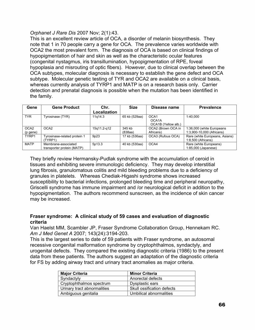
Orphanet J Rare Dis 2007 Nov; 2(1):43. This is an excellent review article of OCA, a disorder of melanin biosynthesis. They note that 1 in 70 people carry a gene for OCA. The prevalence varies worldwide with OCA2 the most prevalent form. The diagnosis of OCA is based on clinical findings of hypopigmentation of hair and skin as well as the characteristic ocular features (congenital nystagmus, iris transillumination, hypopigmentation of RPE, foveal hypoplasia and misrouting of optic fibers). However, due to clinical overlap between the OCA subtypes, molecular diagnosis is necessary to establish the gene defect and OCA subtype. Molecular genetic testing of TYR and OCA2 are available on a clinical basis, whereas currently analysis of TYRP1 and MATP is on a research basis only. Carrier detection and prenatal diagnosis is possible when the mutation has been identified in the family.
Gene Gene Product Chr. Localization
Size Disease name Prevalence
TYR Tyrosinase (TYR) 11q14.3 65 kb (529aa) OCA1 OCA1A OCA1B (Yellow alb.)
1:40,000
OCA2 (p gene)
OCA2 15q11.2-q12 345 kb (838aa)
OCA2 (Brown OCA in Africans)
1:36,000 (white Europeans 1:3,900-10,000 (Africans)
TYRP1 Tyrosinase-related protein 1 (TYRP1)
9p23 17 kb (536aa) OCA3 (Rufous OCA) Rare (white Europeans, Asians) 1:8,500 (Africans)
MATP Membrane-associated transporter protein (MATP)
5p13.3 40 kb (530aa) OCA4 Rare (white Europeans) 1:85,000 (Japanese)
They briefly review Hermansky-Pudlak syndrome with the accumulation of ceroid in tissues and exhibiting severe immunologic deficiency. They may develop interstitial lung fibrosis, granulomatous colitis and mild bleeding problems due to a deficiency of granules in platelets. Whereas Chediak-Higashi syndrome shows increased susceptibility to bacterial infections, prolonged bleeding time and peripheral neuropathy, Griscelli syndrome has immune impairment and /or neurological deficit in addition to the hypopigmentation. The authors recommend sunscreen, as the incidence of skin cancer may be increased. Fraser syndrome: A clinical study of 59 cases and evaluation of diagnostic criteria Van Haelst MM, Scambler JP, Fraser Syndrome Collaboration Group, Hennekam RC. Am J Med Genet A 2007; 143(24):3194-203. This is the largest series to date of 59 patients with Fraser syndrome, an autosomal recessive congenital malformation syndrome by cryptophthalmos, syndactyly, and urogenital defects. They compared the existing diagnostic criteria (1986) to the present data from these patients. The authors suggest an adaptation of the diagnostic criteria for FS by adding airway tract and urinary tract anomalies as major criteria.
Major Criteria Minor Criteria Syndactyly Anorectal defects Cryptophthalmos spectrum Dysplastic ears Urinary tract abnormalities Skull ossification defects Ambiguous genitalia Umbilical abnormalities
66
Laryngeal and tracheal anomalies Nasal anomalies Positive family history
Apical involvement with fibrous dysplasia: Implications for vision Cruz AA, Constanzi M, de Castro FA, dos Santos AC. Ophthal Plast Recontrs Surg 2007 Nov-Dec; 23(6):450-454. Patients with orbital Fibrous Dysplasia (FD) present with the classical signs of proptosis, dystopia and periorbital facial asymmetry. FD is a disorder of the adenyl cyclase system. This is a prospective case series of 21 patients with fibrous dysplasia with orbital involvement. Four of the patients had McCune Albright syndrome and 1 had Tuberous Sclerosis. Strabismus was not a frequent complication; 2/21(9.5%). Lacrimal duct obstruction was also infrequent; 1/21(4.76%) had epiphora. Of the 34 orbits affected, the roof was affected in 67.7%, whereas the floor was the least affected wall. Nineteen orbits showed radiologic signs of optic canal and/or superior orbital encasement. No patient showed any sign of trigeminal dysfunction or severe visual loss, despite long disease duration. The authors believe that patients with apical involvement should be carefully followed and have their apices decompressed only when their vision is clearly deteriorating. Their data does not support prophylactic optic canal decompression as a therapeutic measure. The authors believe the presence of intralesional cysts near the apex is clearly an indication for surgery. Ocular Clusterin expression in von Hippel-Lindau disease Zhou M, Shen D, Head JE, Chew EY et al. Mol Vision 2007 Nov; 13:129-26. Retinal hemangioblastoma/hemangioma is the most frequent and often earliest clinical manifestation of VHL disease. Although retinal hemangioblastoma associated with VHL usually presents at a relatively young age (median 25 years), the cumulative probability increases each decade of life, reaching 80% by the eighth decade. The majority of initial retinal hemangioblastoma associated with VHL disease are located in peripheral retina, while per papillary lesions are only reported in 10% of cases. Complications from retinal hemangioblastomas, even in optimally treated cases are visually significant. The probability of visual loss is age dependent. The life time cumulative probability of permanent visual loss is 60, with most of the risk (43%) falling with within the first 30 years of life. Clusterin (also termed apolipoprotein J) is a multifunctional glycoprotein. This is a retrospective case series investigating ocular clusterin expression in VHL in 9 eyes with retinal hemangioblastoma, one eye with VHL but without ocular disease, one surgically excised optic nerve with optic nerve hemangioblastoma, and 3 normal control eyes. Ocular specimen were evaluated by routine histology, immnohistochemistry for clusterin expression, and molecular detection of clusterin transcripts with ocular VHL hemangioblastomas compared with normal tissue from the same eye using microdissection and quantitative real time PCR. There was marked decrease of clusterin immunoreactivity in all retinal hemangioblastoma and the optic nerve hemangioblastoma , whereas positive clusterin reactivity of the vascular and glial components were similar to that of normal retina. PCR confirmed the decrease of
67
clusterin mRNA. Clusterin showed possible important functioning in tumor suppression by the VHL gene product and the potential to be a novel biomarker in retinal hemangioblastoma associated with VHL. Cionni endocapsular ring implantation in Marfan’s syndrome Bahar I, Kaiserman I, Rootman D. Br J Ophthalmol 2007 Nov; 91:1477-1480. This is a case series of 15 eyes of 12 patients (aged 19-56) with Marfan syndrome and subluxated lenses. A 2-eyelet Cionni ring and an AcrySOf foldable IOL were implanted in 13 eyes. Two eyes had trans-scleral IOL fixation in the ciliary sulcus. In all eyes, capsular bag centration was excellent. In this study the Cionni ring enable the capsular bag centration. However, the authors noted some intraoperative limitations. 1) With an unstable lens, a central capsulorhexis was at times difficult, and often small. 2) In cases with extensive subluxation, implantation could be difficult. 3)They noticed the capsular bag of the Marfan patient was sometimes smaller than that of normal eyes so that the standard Cionni ring may be too large for the kind of bag, and implanting it increases the risk of a rip or tear of the bag. Preoperative vision ranged from 20/50 to CF and improved to better than 20/40 in all eyes. They note that this procedure requires a highly skilled surgeon and cannot always be completed. .
RETINOBLASTOMA
68
Treatment of retinoblastoma: Current status and future perspectives. Rodriguez-Galindo C, Chantada GL, Haik BG, Wilson MW. Curr Treat Options Neurol 2007 Jul; 9(4):294-307. This is a good review article delineating the current treatment status for RB. They emphasize that the treatment must always be individualized. They review the various treatment modalities: surgery, focal therapy (photocoagulation, cryo, and thermotherapy), radiation therapy and chemotherapy. Each of the specific chemo agents is reviewed. They touch on the emerging therapies and provide an opinion statement. Eye size in retinoblastoma: MR imaging measurements in normal and affected eyes. De Graaf P, Knol DL, Moll AC, Imhof SM et al. Radiology 2007 Jul; 244(1):273-80 This is a retrospective study of 100 eyes (50 boys/50girls) with RB over a 12 year period. They used MR imaging to measure axial length (AL), equatorial diameter ED) and eye volume (EV). Normal eyes of patients with unilateral retinoblastoma served as controls. Eyes with Rb had significantly shorter AL’s and EDs and significantly smaller EVs than normal eyes. In those eyes with Rb, the larger the tumor volume, the smaller the eye. One hit, two hits, three hits, more? Genomic changes in the development of retinoblastoma. Corson TW, Gallie BL. Genes Chromosomes Cancer 2007 Jul; 46(7):617-34. RB is initiated by the loss of both alleles of the prototypic tumor suppressor gene RB1. However a large number of cytogenetic and comparative genomic hybridization studies have shown that the M1 and M2 mutational events are not the only genomic changes in RB. The authors review these subsequent changes which are likely crucial for tumor progression. The authors provide a comprehensive summary of the genomic evidence implicating gain of 1q, 2p, 6p, and 13q and loss of 16q in RB oncogenesis. They discuss the search for candidate oncogenes and tumor suppressor genes within these regions and their potential diagnostic, prognostic, and therapeutic implications. Outcomes of a two-drug chemotherapy regimen for intraocular retinoblastoma. Zage PE, Reitman AJ, Seshadri R, et al Pediatr Blood Cancer, 2007 Aug 29 [Epub ahead of print]
69
This was a prospective study of 21 patients enrolled in a protocol utilizing carboplatin and etoposide (six cycles) and focal therapy. The response rate after 6 cycles of chemotherapy was 85.4% Twenty –two eyes were enucleated, but only seven eyes received EBRT. The vision salvage rate without EBRT was 82.6% for eyes with Reese-Ellsworth groups I-IV tumors and 20% for eyes with group V tumors. The vision salvage rate without EBRT for eyes with Murphree groups A and B tumors was 77.3%, but was only 26.9% for eyes with groups C and D tumors. The authors believe that treatment of RB with carboplatin and etoposide plus focal therapy has excellent response rates and salvages globes and vision with minimal acute toxicity and minimal ERBT use in eyes with R_E groups I-IV or Murphree groups A and B tumors. A retrospective review of hearing in children with retinoblastoma treated with carboplatin-based chemotherapy. Lambert MP, Shields C, Meadows AT Pediatr Blood Cancer, 2007, Feb 2 [Epub ahead of print] This is a retrospective review of 258 children with RB of whom 164 received carboplatin as part of their chemo treatment. The children received carboplatin, vincristine, and etoposide (CEV) for 6 cycles. (The dosage of carboplatin was 18.6 mg/kg q 4 weeks.) One hundred and sixteen of the children had audiogram data. Age appropriate audiologic evaluation was performed prior to therapy, after three cycles of chemo and upon completion of the six cycles of chemotherapy. Of the 116 children, 14 (5.6%) had abnormal audiograms prior to initiation of chemo. Of the 14 audiograms that were abnormal initially, only 3 were persistently abnormal. Only one patient had progressive hearing loss. The authors note that ototoxicity is a potential concern; carboplatin in the treatment of RB does not appear to produce impairment. Screening can identify children who require frequent audiologic follow-up, but children with normal initial hearing tests are unlikely to develop hearing deficit as a result of therapy with carboplatin given at the doses in the above schedule. They recommend that these children be assessed clinically without additional monitoring beyond that which is part of routine pediatric care.
70
Secondary acute myeloid leukemia after etoposide therapy for retinoblastoma. Weintraub M, Revel-Vilk S, Charit M, et al Pediatr Hematol Oncol 2007, Vol 29; 9 This is a case report of a child diagnosed with RB at age 2 months with bilateral RB. The child was found to have partial mosaicism for R35X mutation of the RB gene. She was treated with chemotherapy (vincristine, carboplatinum, and etoposide); six cycles. She required 4 cryotherapy sessions for persistent lesions, with a complete response. At the age of 2 years; 22 months after the diagnosis of RB and 18 months after the last dose of etoposide, the patient was diagnosed with secondary M4 acute myeloid leukemia. Secondary AML may occur after therapy with topoisomerase II inhibitors (anthracyclines, epipodophyllotoxins) or with alkylating agents (cyclophosphamide, procarbazine). It occurs in both adults and children and is usually associated with a relatively short latency period and with cytogenetic aberrations in chromosome 11Q23, involving the mixed lineage leukemia (MLL) gene. The most common aberration seen is t(9;11). The presumed mechanism of topoisomerase-associated t AML is formation of translocations as a result of defective breakage repair after cleavage of DNA by topoisomerase II. From the archives of the AFIP. Pediatric orbit tumors and tumor-like lesions: Neuroepithelial lesions of the ocular globe and optic nerve. Chang EM, Specht CS, Schroeder JW. Radiographics 2007 Jul-Aug; 27(4):1159-86. This a superb review article. They describe the imaging features and pathologic bases of ocular and optic nerve neuroepithelial neoplasms in children (RB, pseudoretinoblastoma: PHPV, Coats disease, Toxocara; Medulloepithelioma, and Optic Nerve Glioma_ They discuss the differentiating features amongst these masses and discuss the differential diagnoses. They even discuss the treatment and prognosis for each. Trilateral Retinoblastoma. Antoneli CBG, Ribeiro K, Sakamoto LH, Chojniak MM, Novaes PE, Arias VE. Pediatr Blood Cancer 2006 Mar 29; [Epub ahead of print] This is a good review article on Tuberous Sclerosis (TSC), an autosomal dominant multisystem disorder. It occurs by spontaneous mutation in approximately 70% of affected. Mutations in the TSC1 gene, on chromosome 9q34, and TSC3 gene, on chromosome 16913.3, result in a similar phenotypic presentation. TSC1 is more common in familial cases and results in less severe disease. The pathogenesis of TSC lies in the expression and function of the gene products, tuberin in TSC2 and hamartin in TSC1, in tissue. The interaction between these two proteins as been shown to be
71
critical to multiple intracellular-signaling pathways, especially those in control of cell growth TSC is associated with both nonretinal and retinal lesions. Nonretinal abnormalities include iris depigmentation, eyelid angiofibromas, strabismus, cataracts, and coloboma. Hamartomas are the most common retinal finding and found in 40-50%. They are bilateral in 34-50%. Pigmentary retinal abnormalities are also frequently seen: punchedout areas of retinal depigmentation, plaque-like lesions, and pigment clumping may be seen. Molecular genetics of RB1 – the retinoblastoma gene Leiderman UYI, Kiss S, Mukai S. Semin Ophthalmol 2007; 22(4):247-54. This is an excellent review article. They discuss the function and structure of the RB1 gene and the biology of its protein product, pRB. They review the gene mutations in RB. The majority of germ line mutations harboring hereditary RB are nonsense or frame shift mutations. Genotype-Phenotype Correlates in Hereditary RB*
Mutation Resultant pRB Activity In Vivo RB Phenotype (Assuming Loss-of-Heterzygosity)
Nonsense† Nil Hereditary (Typically multi-focal, bilateral disease)
Frameshift† Nil Hereditary Aberrant slice mutation (without subsequent nonsense mutation)
Exonic – variable Intronic – variable, may yield reduced quantity of functional protein
Hereditary High to reduced penetrance and/or decreased expressivity
Missense and inframe+ Variable – reduced to normal quantities of stable transcript
Reduced penetrance and/or decreased expressivity
Mutations in RB1 promoter sequences
Variable – reduced to normal quantities of stable transcript
Reduced penetrance and/or decreased expressivity
* The above table provides general genotype-phenotype correlations; exceptions have been well described, and thus this information should not be used for the purpose of genetic counseling or determining prognostic outcomes. † Isolated, unilateral RB arising from nonsense or frameshift mutations has been shown to occur in the setting of somatic mosaicism, estimated to be present in at least 10% of families with hereditary Rb (Sippel et al., 1998) + Most clinically relevant mutations arise from amino acid insertion, deletion, or substitution with the A/B pocket (Otterson et al., 1997) Current management strategies for intraocular retinoblastoma Kim JW, Abramson DH, Dunkel IJ. Drugs 2007; 67(15):2173-85. During the last decade there has been a dramatic shift in the treatment for RB, emphasizing chemoreduction protocols and minimizing the use of external beam radiation. Age at the time of radiation is critical: those irradiated during the first year of life are 2 to 8 times more like to develop second cancers as those irradiated after year
72
one. Patients treated with radiation tend to develop brain tumors and sarcomas of the head and neck with the radiation field in the first 10 years of life, whereas germinal RB survivors who did not receive radiation develop epithelial cancers (lung, bladder, melanomas) out of the radiation field in adulthood. The authors note that although the chemo protocols may vary slightly between institutions, most are using a 3 drug regiment: carboplatin, vincristine, and etoposide. A rare complication of the chemoreduction is the development of second nonocular cancers, particularly hematological malignancies. The presence of subretinal or vitreous seeds is a common cause of treatment failure in chemoreduction. This review article emphasizes the importance and necessity of individualizing the treatment. Retinoblastoma: Review of Current Management Chintagumpala M, Chevez-Barrios P, Paysse EA, Plon SE, et al. The Oncologist 2007 Oct; 12(10):1237-1246. This is a nice general review article. The authors review the genetics and note that the presence of a mutation in blood is presumptive evidence of a germline or constitutional mutation. Once a germline mutation is identified then all siblings or offspring should be tested for the specific mutation and to determine the need for surveillance for RB. They review the vast improvement in the various treatment modalities. Children with the heritable form of RB are at high risk of developing subsequent malignancies, most commonly sarcomas. The risk is greater for those children with the heritable form of the disease who were exposed to ionizing radiation at less than one year of age. Conservative Treatments of Intraocular Retinoblastoma Rouic LL, Aerts I, Lévy-Gabriel C, Dendale R, et al. Ophthalmology 2008 Jan 24; [Epub ahead of print] This is a prospective nonrandomized trial of 83 children (113 eyes) with conservative treatment: chemotherapy (two cycles of carbolatin and etoposide) followed by local ocular therapy (thermotherapy, cryo-application, iodine 125 brachytherapy) and/or hemothermotherapy. The authors wished to determine if they could reduce the external beam irradiation from 50% to 20%. Tumor control was obtained in 84% of cases, with EBR necessary in only 13 eyes of 9 children (12%). Enucleation was necessary in 23 eyes (20%), five of which were because of complications. The authors were encouraged by these results achieving satisfactory tumor control and a low need for EBR.
73
NEURO-OPHTHALMOLOGY Adverse effects of apraclonidine used in the diagnosis of Horner’s syndrome in infants. Watts T, Satterfield D, Kim MK. J AAPOS 2007; 11: 282-83. The authors present an index case of a five-month-old infant girl along with four additional cases provided through the pediatric ophthalmology listserve. Three of these children had drowsiness and two cases required emergency admission for unresponsiveness. One case had associated bradycardia, hypertension, and decreased oxygen saturations. The authors recommend that apraclonidine be used with caution or not at all in infants under six months of age. If used the patient should be observed for a period of at least two hours after installation of the drops with admission to the pediatric ward if lethargy, bradycardia or reduced respiratory rate develop. Pediatric idiopathic intracranial hypertension Rangwala LM, Liu GT. Surv Ophthalmol 2007; 52: 597-617. This review article summarizes currently knowledge regarding pediatric idiopathic intracranial hypertension. The use of rigorous methodologies and standard definitions in recent studies has demonstrated distinct demographic trends: (1) The incidence of idiopathic intracranial hypertension seems to be increasing among adolescent children, and among older children its clinical picture is similar to that of adult idiopathic intracranial hypertension (female and obese). Within younger age groups there are more boys and nonobese children who may develop idiopathic intracranial hypertension. (2) The pathogenesis of the disease has yet to be elucidated. Idiopathic intracranial hypertension among young children has been associated with several new etiologies, including recombinant growth hormone and all-trans-retinoic acid. (3) More modern neuroimaging techniques such as MRI and MRI-venograms are being used to exclude intracranial processes. (4) Although most cases of pediatric idiopathic intracranial hypertension improve with medical treatment, those who have had visual progression despite medical treatment have undergone optic nerve sheath fenestration and lumboperitoneal shunting. (5) Because idiopathic intracranial hypertension in young children appears to be a different disorder than in adolescents and adults, separate diagnostic criteria for younger children are warranted. The authors propose new criteria for pediatric idiopathic intracranial hypertension in which children should have signs or symptoms consistent with elevated intracranial pressure, be prepubertal, have normal sensorium, can have reversible cranial nerve palsies, and have an opening cerebrospinal fluid pressure greater than 180 mm H2O if less than age 8 and papilledema is present, but greater than 250 mm H20 if age 8 or above or less than 8 without papilledema.
74
Neuro-ophthalmological management of optic pathway gliomas Lee AG. Neurosurg Focus 2007 Nov; 23(5):E1-6. Optic pathway gliomas are typically childhood tumors. About 30 of patients with OPGs have NF1. Their growth rates are variable but unpredictable. Dr. Lee recommends treatment on an individual basis. Close clinical neuro-ophthalmic follow-up and serial radiography (preferably MRI) are indicated. Only symptomatic and radiographically progressing OPGs require strong consideration for treatment. Both chemotherapy and radiation treatment can be considered, but chemo in patients <5 years old causes fewer side effects than RT. Tumor location defines prognosis, as ONGs have the lowest morbidity and mortality followed by OCGs and then hypothalamic glioma. The major symptom of OPG is visual loss, but the major cause of systemic complication is hypothalamic involvement (causing electrolyte abnormalities) or hydrocephalus. Resection is an option for ONGs, but is generally reserved for patients with poor vision, severe proptosis or severe pain in a blind eye. Resection is generally not an option for intrinsic chiasmal or retrochiasmal OPGs. Extrinsic or cystic components can be debulked surgically on an individual basis. Neuroradiographic, endocrinologic, and ophthalmic correlates of adverse developmental outcomes in children with optic nerve hypoplasia: A prospective study. Garcia-Fillon P, Epport K, Nelson M, Azen C et al. Pediatrics 2008 Mar; 121(3):e653-e659. The authors conducted a prospective study of 73 children who were diagnosed with optic nerve hypoplasia prior to 36 months of age. They found that 71% of subjects had developmental delay. Corpus callosum hypoplasia and hypothyroidism were significantly associated with poor developmental outcomes. The authors conclude that because there is this correlation with developmental delay, all children with optic nerve hypoplasia should have neuroradiologic and endocrinologic testing for risk factors in delay and developmental assessment so they may receive early intervention planning if necessary Cognitive visual impairment with good visual acuity in children with posterior periventricular white matter injury: A series of 7 cases. Saidkasimova S, Bennet D, Butler S, Dutton G. J AAPOS 2007 Oct. 11(5); 426 The authors present 7 pts with normal visual acuity, but with “cognitive” visual impairments characterized by difficulties navigating crowed visual scenes, seeing distant objects, finding a parent in a group or an item of clothing in a pile. These children get lost in complex visual environments, but do well in tasks against a plain background with minimal foreground clutter. On MRI, these seven had PVL with lesions in occipito-parietal periventricular white matter. The difficulties were consistent with dorsal stream dysfunction.
75
TRAUMA Unilateral retinal hemorrhages in shaken baby syndrome. Arlotti S., Forbes BJ, et al. J AAPOS 2007; 11: 175-6. The authors present data on 17 patients with documented shaken baby syndrome and unilateral retinal hemorrhages. Nine of the 17 had bilateral intracranial hemorrhages and unilateral retinal hemorrhage. All eight patients with unilateral intracranial hemorrhage had their unilateral retinal hemorrhage on the same side. The authors give data on the specifics of the cases and make the point that unilateral retinal hemorrhage does not rule out the diagnosis of shaken baby syndrome.
Management of canalicular lacerations: epidemiological aspects and experience with mini-monoka monocanalicular stent.
Naik MN, Kelapure A, Rath S, Honavar SG.
Am J Ophthalmol. 2008 Feb;145(2):375-380. Epub 2007 Dec 3.
PURPOSE: To report the epidemiological data, clinical profile, and surgical outcome of canalicular lacerations in an Indian population. DESIGN: Retrospective interventional case series.
METHODS: All patients who underwent canalicular laceration repair from July 1, 2002 to June 30, 2005 at a tertiary eye care center were retrospectively reviewed. Demographics, cause of eyelid injury, associated ocular injury, surgical management with Mini-Monoka monocanalicular stent, and its outcome were analyzed.
RESULTS: Sixty-six patients underwent eyelid laceration repair, of which 24 (36%) had involvement of the canalicular system. The mean age at presentation was 16 years (range, 10 months to 52 years); 20 (83.3%) patients were males. Mode of injury included the blouse-hook fastener in five (20.8%) breast-feeding infants, bicycle handle injury in four (16.7%) children, and metal rod injury in five (20.8%) adults. Lower canaliculus was involved in 13 (54.1%), upper in eight (33.3%), and both in three (12.5%) patients. Simultaneous globe injury was noted in six (25%) patients, five (83.3%) of which had upper canalicular involvement. Twenty-seven canalicular lacerations (24 patients) underwent stenting with the Mini-Monoka monocanalicular stent. Three (11.1%) stents extruded within one month. At the final follow-up (mean, 18.5 months), canalicular block was noted in two (10%) out of 20 patients; none had epiphora.
76
CONCLUSION: Canalicular involvement occurs in 36% of eyelid injuries. Injury by the "blouse-hook fastener" is unique to infants in the Indian context. Simultaneous globe injury is present in 25% of cases, especially when the upper canaliculus is involved. Mini-Monoka stent extrusions occur within one month. With an 11.1% extrusion rate, Mini-Monoka stents achieved good anatomical (90%) and functional (100%) success in the management of canalicular injury.
Traumatic pediatric cataract: a decade of follow-up after Artisan aphakia intraocular lens implantation.
Sinia M, Odenthal M, Wenniger-Prick L, Gortzak-Moorstein N, Volker-Dieben J.
J AAPOS 2007 Dec 11(6); 555.
Retrospective review of 5 children who had had the Artisan iris fixated lens implanted post cataract extraction secondary to penetrating ocular trauma. No pt had IOL dislocation, corneal decompensation, chronic uveitis CME or iris atrophy. The authors conclude this IOL isa useful alternative for traumatic aphakia without sufficient capsular support.
77
UVEITIS Th1 and Th2 responses on the ocular surface in uveitis identified by CCR4 and CCR5 conjunctival expression. Trinh L, Brignole-Baudouin F, Raphaël M, Dupont-Monod S, Cassoux N, Lehoang P, Baudouin C. Am J Ophthalmol. 2007 Oct;144(4):580-5. Epub 2007 Aug 8 PURPOSE: To investigate CC chemokine receptor 4 (CCR4) and CC chemokine receptor 5 (CCR5) expression, known to be related to the Th2 and Th1 inflammatory pathways, respectively, and human leukocyte antigen-D related (HLA-DR) antigens as hallmarks for ocular surface inflammation in patients with uveitis using conjunctival impression cytologic specimens. DESIGN: Case-controlled study. METHODS: Conjunctival impression cytologic specimens were obtained from patients with anterior uveitis (n = 26), and their inflammatory profile was compared with those of patients with vernal keratoconjunctivitis (VKC; n = 24), keratoconjunctivitis sicca (KCS; n = 17), and normal subjects (n = 17). Expressions of CCR4, CCR5, and HLA-DR were analyzed using flow cytometry and were expressed by determining the percentage of cells expressing the markers in the conjunctival epithelium. RESULTS: CCR4 was overexpressed in the uveitis group (mean, 19.8% +/- 19.7% of positive cells) and in the VKC group (24.7% +/- 20.1%). CCR5 was expressed only weakly in uveitis patients (6.4% +/- 13.1%) and in the normal subjects (2.4% +/- 2.4%). HLA-DR expression by conjunctival cells was increased in the uveitis patients (57.4% +/- 21.1%) and in the KCS group (52.4% +/- 12.1%) compared with the VKC group (23.9% +/- 26.8%; P < .001) and normal subjects (22.1% +/- 19.1%; P < .001). CONCLUSIONS: CCR4, classically related to the Th2 system, and HLA-DR both were overexpressed by the conjunctival epithelium in uveitis patients, whereas CCR5, related to the Th1 system, was expressed weakly in uveitis patients. These preliminary results seem to suggest an involvement of the Th2 system on the ocular surface in uveitis. Exploration of the ocular surface in uveitis may represent a new way to understand better the immune pathways involved in this complex disease. Pars Planitis in Children: Epidemiologic, Clinical and Theraputic Characteristics. JPOS 2007; 44:288-293. Pars planitis is an idiopathic form of intermediate uveitis characterized by minimal anterior chamber involvement, anterior vitreous cells, vitreous condensations, and
78
peripheral retinal vasculitis. The etiology is unknown. 30 eyes in 16 patients diagnosed between 1995 and 2005 in the Department of Pediatric Ophthalmology at Hospital Universitario La Paz, Madrid, Spain were reviewed retrospectively. Pars planitis was bilateral in 87.5% and more frequent in males (68.8%). Average age of onset was 9.2 years. Cataract formation was the most prevalent complication (36.7%). Decreased vision was the most frequent initial symptom in this group. Periocular steroids were used in 33.3% of cases and cryotherapy or laser photocoagulation in 16.7%. With adequate medical and surgical treatment, pars planitis has a good prognosis in most cases.
Infliximab to treat chronic noninfectious uveitis in children: retrospective case series with long-term follow-up.
Ardoin SP, Kredich D, Rabinovich E, Schanberg LE, Jaffe GJ.
Am J Ophthalmol. 2007 Dec;144(6):844-849. Epub 2007 Oct 22.
PURPOSE: To assess a response to infliximab therapy in childhood uveitis. DESIGN: Retrospective case series.
METHODS: We reviewed the course of 16 children with noninfectious uveitis treated with infliximab at an academic medical center. Outcome measures included incidence of uveitis recurrences, proportion of patients achieving zero or two-step decline in ocular inflammation, visual acuity, and proportion discontinuing topical glucocorticoids at zero, three, six, nine, and 12 months of therapy.
RESULTS: Of sixteen children (29 affected eyes) with median age 11 years, six had associated extraocular inflammatory conditions. Fifteen of 16 were treated with concomitant methotrexate. Median follow-up was 26 months and median maintenance infliximab dose was 8.2 mg/kg. The median interval between infliximab infusions was 5.6 weeks. At one year, 64% achieved zero ocular inflammation, and 79% had zero inflammation or a two-step decline in inflammation. Topical glucocorticoids were discontinued in 69%, and 58% remained free of uveitis recurrence at one year. Visual acuity remained stable. Infliximab was discontinued in two children, one because of inefficacy and the other because of parental concern about potential side effects. No adverse events occurred.
CONCLUSIONS: Sixteen children with chronic, noninfectious uveitis tolerated chronic methotrexate and infliximab therapy. Visual acuity remained stable, control of ocular inflammation improved, and reliance on topical glucocorticoids decreased. High infliximab doses and frequent dosing intervals were necessary to control uveitis.
79
Pars planitis: a 20-year study of incidence, clinical features, and outcomes.
Donaldson MJ, Pulido JS, Herman DC, Diehl N, Hodge D.
Am J Ophthalmol. 2007 Dec;144(6):812-817.
PURPOSE: To measure the incidence of pars planitis in a community-based population and to report clinical features, complications, and visual prognosis. DESIGN: Population-based, retrospective, 20-year cohort study.
METHODS: Multicenter study using the Rochester Epidemiology Project medical records linkage system, which allows analysis of almost all patients within Olmsted County, Minnesota, with a given medical condition. Databases were searched to identify all patients with pars planitis from January 1, 1985 through December 31, 2004. Forty-six eyes of 25 patients were evaluated.
RESULTS: Mean follow-up was 14.3 years. The incidence of pars planitis was 2.077 per 100,000 persons (95% confidence interval [CI], 1.43 to 2.62). The most common complications were epiretinal membrane (ERM) in 17 eyes (36%), cataract in 14 eyes (30.4%), and cystoid macular edema (CME) in 12 eyes (26.1%). Mean visual acuity after 10 years of follow-up was 20/30, with 18 of 24 patients maintaining a visual acuity of 20/40 or better. One-third of patients maintained normal visual acuity without requiring treatment.
CONCLUSIONS: The visual prognosis of pars planitis is relatively good, with 75% of patients maintaining a visual acuity of 20/40 or better after 10 years. Many patients with mild disease do not require treatment. A subset of patients, however, experience severe disease with severe vision loss despite treatment. The rate of smoking and multiple sclerosis in patients with pars planitis is much higher than that of the general population.
Posterior Scleritis in a 7-Month-Old Infant. Van der Maisen, K et al. JPOS 2007; 44: 377-378. Posterior scleritis in children is rare. Only 22 cases have been reported in the literature, non younger than five years of age.
80
This article describes a 7-month-old infant with posterior scleritis diagnosed on the basis of B-scan ultrasonography and computed tomography. The patient was initially diagnosed with preseptal cellulites and endophthalmitis. Posterior scleritis should be considered in the differential diagnosis of acute orbital inflammation in children younger than one year. SHORT-TERM SAFETY AND EFFICACY OF INTRAVITREAL TRIAMCINOLONE ACETONIDE FOR UVEITIC MACULAR EDEMA IN CHILDREN Ahmed Sallam MD FRCS; Richard M Comer MD FRCOphth; John H Chang MD; John R Grigg FRACO FRACS; Richard Andrews FRCOphth; Peter J McCluskey MD FRACO; Susan Lightman PhD FRCOphth Arch Ophthalmol 2008;126(2):200-205 This was a retrospective noncomparative interventional case series to evaluate the short-term safety and efficacy of intravitreal (IV) triamcinolone acetonide (TA) for treating pediatric cystoid macular edema (CME) secondary to noninfectious uveitis. Medical records of 15 consecutive children (16 eyes) with uveitic CME treated with IVTA (2 or 4 mg) were reviewed. Data collected included details of uveitis, CME, visual acuity, intraocular pressure, and cataract development. The median follow-up time was 16 months (range, 9-36 months). Resolution of CME was achieved in all treated eyes, with a median time for CME resolution at 3 weeks (range, 1-24 weeks). Mean improvement of visual acuity after IVTA was 0.6 logarithm of the minimum angle of resolution. CME relapsed in 5 eyes (31%) after a median time of 7 months (range, 3-13 months). The most common adverse effect was increased intraocular pressure, with an increase of more than 15 mm Hg in 5 eyes (31%). Steroid-induced cataract was observed in 6 of 11 phakic eyes (55%). The authors found that IVTA is efficacious in the treatment of uveitic CME in children and results in CME resolution and visual acuity improvement. As in adults, treatment in children may be associated with elevated intraocular pressure and cataract.
81
RETINA Vitreoretinal surgery after childhood ocular trauma. Sheard RM, Mireskandari K, Ezra E, Sullivan PM. Eye. 2007 Jun;21(6):793-8 This is a retrospective review of 61 children (age 16 years or less) undergoing vitreoretinal surgical procedures following ocular trauma at a tertiary referral centre. Twenty-eight children (45.9%) had open globe injuries (OGI) and 33 closed globe injuries (CGI, 54.1%). The mean age of children with OGI was 9.5 years and with CGI 12.3 years (P=0.0068). Forty-seven children had traumatic retinal detachments (77.1%), which in 17 cases were treated with conventional scleral buckling surgery and in 30 by vitrectomy. Retinal re-attachment was achieved after one procedure in 70.6% with scleral buckling and 46.7% with vitrectomy. Fourteen children (22.9%) had attached retinas but required vitrectomy for lens or cataract subluxation, vitreous hemorrhage intraocular foreign body or macular hole. After a mean follow-up of 19.6 months, the median visual acuity (VA) of the children improved from counting fingers at presentation to 6/36 at final review (P=0.0031). Traumatic retinal detachment requiring vitrectomy was associated with poor visual outcome (P=0.0003). Vitreoretinal intervention resulted in an improvement in vision in 32 children (57.1%) and stabilised 11 at their presenting acuity (19.6%). Two thirds of the children attained a final VA of 6/60 or better. Proliferative vitreoretinopathy was the cause of redetachment in 68.2% of cases and was significantly associated with a poor outcome (P<0.0001). Retinal function in infants with optic nerve hypoplasia: electroretinograms to large patterns and photopic flash. McCulloch DL, Garcia-Fillion P, van Boemel GB, Borchert MS. Eye. 2007 Jun;21(6):712-20. Optic nerve hypoplasia (ONH), which is defined as a congenital deficiency of retinal ganglion cells, may also involve more distal layers of the retina. The authors investigated electrophysiological function of the retina in ONH using electroretinograms (ERGs). ERGs were recorded from 48 subjects (3.5-35 months) with unilateral or bilateral ONH. Pattern reversal (4 degrees checks) was presented under chloral hydrate sedation, using an optical system to correct a cycloplegic refraction. A photopic flash stimulus was also used. Fundus photographs were used to measure the disk diameter/disk macula ratio (DD/DM), and to document other clinical signs. Eyes were classified as moderate (0.15-0.3) or severe (<0.15) ONH, and those with DD/DM greater than 0.3 were used as reference eyes. Pattern ERG recording was completed in 89 eyes and was detectable in 80% of eyes with ONH (61/76 tested) and in all 13
82
reference eyes. Photopic flash ERGs were of good quality in all eyes. The severity of ONH correlates with the amplitude of the photopic flash b-waves and with the amplitude of the N95 component of the pattern ERG (P<0.01). However, the ERGs to large patterns were well preserved (>3.5 microV) in 10 of 35 eyes with severe ONH. Tortuous retinal vessels in eyes with either moderate or severe ONH were associated with smaller amplitude photopic b-waves and markedly diminished or undetectable pattern ERGs. CONCLUSIONS: This study supports the hypothesis that retinal dysfunction distal to the ganglion cells is common in ONH, but is not predictable on the basis of ONH severity alone. Additionally, tortuous retinal vessels in ONH may be a sign associated with retinal dysfunction. Macular schisis and detachment associated with presumed acquired enlarged optic nerve head cups. Zumbro DS, Jampol LM, Folk JC, Olivier MM, Anderson-Nelson S. Am J Ophthalmol. 2007 Jul;144(1):70-74. Epub 2007 May 9. PURPOSE: To describe a clinical syndrome of macular schisis and detachment in patients with acquired optic nerve head cupping resulting from glaucoma. METHODS: Patients were included if they had optic nerve cupping and macular schisis with or without detachment with no other identifiable cause. The patients had to have no leakage on fluorescein angiography and no vitreous traction on examination or on optic coherence tomography (OCT). These patients were followed up and visual acuity, intraocular pressure, and the findings of serial fundus and OCT examinations were noted. RESULTS: Five patients had schisis with or without detachment of the macula with pronounced optic nerve head cupping. One patient had resolution of the macular fluid after filtering surgery for uncontrolled glaucoma. Two patients underwent a vitrectomy with intraocular gas and had almost total resolution of macular fluid and improved vision. CONCLUSIONS: Macular schisis and detachment can occur in patients with presumed enlarged optic nerve head cups in the absence of obvious congenital anomalies of the disk. The authors believe the cause is leakage of fluid from the vitreous through a tiny hole in the thin tissue of the cup. This is a similar mechanism to that seen in patients with optic pits. A vitrectomy or steps to reduce the intraocular pressure may result in resolution of the fluid and improved vision. Pediatric traumatic macular hole: Results of autologous plasmin enzyme-assisted vitrectomy. Wu WC, Drenser KA, Trese MT, Williams GA, Capone A. Am J Ophthalmol. 2007 Sep 15; [Epub ahead of print] PURPOSE: To review our experience with plasmin-assisted vitrectomy surgery for the treatment of pediatric macular holes.
83
METHODS: Thirteen pediatric patients aged one to 15 years with a traumatic macular hole underwent surgical repair. All patients underwent surgery between February 1997 and March 2005 with autologous plasmin enzyme-assisted vitrectomy. After induction of posterior vitreous detachment (PVD), vitrectomy with membrane peeling and gas or silicone oil injection were performed followed by prone positioning for seven days. Main outcome measures included anatomic closure rate, visual outcome, and ocular complications. RESULTS: The macular hole was closed successfully in 12 (92%) of 13 cases. Of the 12 patients for whom vision could be measured, 11 patients (92%) had visual acuity improvement of 2 or more lines and six patients (50%) achieved vision of 20/50 or better; all of the patients achieved vision better than 20/200. The visual improvement was statistically significant (P = .005, Wilcoxon signed-rank test). Surgical complications included cataract formation in one patient and retinal detachment in one patient. CONCLUSIONS: Autologous plasmin enzyme may be a helpful adjunct when performing vitrectomy for traumatic macular holes. Profile of the retina by optical coherence tomography in the pediatric age group. Gupta G, Donahue JP, You T. Am J Ophthalmol. 2007 Aug;144(2):309-10. PURPOSE: To establish normative values of the retina in the pediatric population using optical coherence tomography (OCT). METHODS: Prospective study examining macular thickness and nerve fiber layer thickness in children with no ocular disease. After clinical examination, patients meeting the inclusion and exclusion criteria underwent OCT scanning. RESULTS: Thirty-two eyes were examined for macular thickness and 25 eyes for nerve fiber layer thickness. Normative values are found in the Table. The average foveal thickness for children is 221 microns vs 182 microns in adults. CONCLUSION: This study demonstrates normative values of retinal thickness and retinal nerve fiber layer (RNFL) thickness in the pediatric age group. Children have slightly thicker maculas than adults; the RNFL thickness is comparable to adults. Relationship of axial length and retinal vascular caliber in children. Cheung N, Tikellis G, Saw SM, Amirul Islam FM, Mitchell P, Wang JJ, Wong TY. Am J Ophthalmol. 2007 Nov;144(5):658-662. Epub 2007 Sep 17. PURPOSE: Previous studies in older adults suggest that longer axial length is associated with narrower arteriolar caliber. In this study, we re-examined this relationship in a cohort of children, while controlling for the effects of ocular magnification. DESIGN: Cross-sectional study of 767 children aged 7 to 9 years. METHODS: Retinal vascular calibers were measured from retinal photographs using a computer-based program. Ocular magnification was corrected using the Bengtsson formula. Standardized examination of refraction and ultrasound ocular biometry was performed for all children.
84
RESULTS: In models that adjusted for age, gender, ethnicity, body mass index, blood pressure, and birth weight, longer axial length was associated strongly with narrower retinal arteriolar caliber (3.18-microm decrease per standard deviation increase in axial length; P < .001) and venular caliber (4.62-microm decrease standard deviation increase in axial length; P < .001) before correction for ocular magnification. However, after correction, these associations no longer were significant (0.44 microm; P = .31, change for arteriolar caliber; and 0.70 microm; P = .25, for venular caliber). CONCLUSIONS: Our study in children found no association between axial length and retinal vascular caliber after correcting for ocular magnification, suggesting that the previously reported association was likely related to differences in ocular magnification.
Paediatric vs adult retinal detachment. Rumelt S, Sarrazin L, Averbukh E, Halpert M, Eye. 2007 Dec;21(12):1473-8. This Israeli study evaluates the causes, incidences, characteristics, and treatment outcomes of paediatric vs adult retinal detachment 1980 and 2000. 136 patients out of 2408 consecutive retinal detachments (6.6%) occurred in children under the age of 18 years. Of them, 144 eyes of 127 children were treated and compared with a sample of 56 consecutive retinal detachments in 50 adults (over the age of 18 years). The parameters for comparison included cause, type of retinal detachment, its extent, macular involvement, number of tears, number and types of surgery, and the anatomic and functional surgical outcome. Rhegmatogenous RD was less common (P=0.004), and exudative RD was more common (P=0.021) in the paediatric group. Ocular trauma and ocular syndromes were more common in the paediatric group (P<0.001). Myopia, posterior vitreous detachment, and retinal detachment following cataract surgery were less common in the pediatric group compared with adults (P<0.001, <0.001, and 0.001, respectively). Initial and last visual acuity of >20/400, last visual acuity of >20/40, and retinal complete reattachment were higher in adults (P<0.001). The less successful functional and anatomical outcomes of retinal detachment surgery in children may reflect the different aetiologies and indicate the need for aetiology-specific treatment strategies according to each aetiology
The negative ERG: clinical phenotypes and disease mechanisms of inner retinal dysfunction
Audo I, Robson AG, Holder GE, Moore AT. Surv Ophthalmol 2008; 53: 16-40.
85
This review article summarizes current knowledge on normal retinal physiology, the investigative techniques used, and the range of clinical disorders in which there is predominantly inner retinal dysfunction. Inner retinal dysfunction is encountered in a number of retinal disorders, either inherited or acquired, as a primary or predominant defect. Fundus examination is rarely diagnostic in these disorders, although some show characteristic features, and careful electrophysiological assessment of retinal function is needed for accurate diagnosis. The ERG in inner retinal dysfunction typically shows a negative waveform with a preserved a-wave and a selectively reduced b-wave. Advances in retinal physiology and molecular genetics have led to a greater understanding of the pathogenesis of these disorders. This paper includes a detailed discussion of inherited disorders such as congenital stationary night blindness, X-linked retinoschisis, vitreoretinal dystrophies, systemic diseases such as neuronal ceroid lipofuscinosis and Refsum disease; and several acquired conditions.
NASOLACRIMAL
86
ORBIT Porous orbital implants, wraps, and PEG placement in the pediatric population after enucleation. Wang JK, Liao SL, Lin LL, Kao SC, Tseng HS. Am J Ophthalmol. 2007 Jul;144(1):109-116. Epub 2007 May 11. PURPOSE: To investigate complications of various porous orbital implants and wrapping materials in the pediatric population after enucleation. METHODS: Between November 1992 and November 2006, patients younger than 15 years old were collected for study participation. They underwent enucleation with porous orbital implants primarily or secondarily at National Taiwan University Hospital. The authors used the hydroxyapatite (HA), Medpor, and Bioceramic orbital implant. The HA implant was wrapped with four different materials: donor sclera, Lyodura, porcine sclera, and Vicryl mesh. A part of HA implants and all bioceramic implants were wrapped with Vicryl mesh, added anteriorly with scleral patch grafts. All Medpor implants were unwrapped. RESULTS: Forty-seven cases had more than a two-year follow-up. The exposure rates according to implants and wraps were: donor sclera-wrapped HA (two of nine, 22%), porcine sclera-wrapped HA (three of three, 100%), Vicryl mesh-wrapped HA (one of five, 20%), and unwrapped Medpor (one of four, 25%). No exposure was found in four Lyodura-wrapped HA implants, and 22 Vicryl mesh-wrapped HA and Bioceramic implants with anteriorly scleral coating. The exposure rate was lower in cases with implants wrapped by our method and Lyodura than in those with implants wrapped by other materials (P < .001). Of 47 patients, 20 (42.5%) were fitted with peg-coupled prostheses and all had good prosthetic movements subjectively. CONCLUSIONS: Different types of implants and wraps resulted in various exposure rates in the pediatric population. The modified wrapping technique may prevent porous implants from exposure in children. Microbiology of Pediatric Orbital Cellulitis. McKinley SH, Yen MT, Miller AM, Yen KG. Am J Ophthalmol. 2007 Aug 13; [Epub ahead of print] PURPOSE: To evaluate the microbiology of pediatric orbital cellulitis associated with sinusitis. METHODS: All pediatric patients treated for orbital cellulitis associated with sinusitis at Texas Children's Hospital between December 1, 2001 and September 30, 2005 were reviewed. Data collected included patient age, history, microbiology results, and surgical intervention. RESULTS: Thirty-eight cases were identified. Fifteen cases required medical management, whereas 23 patients received a combination of medical and surgical
87
intervention. Three patients had multiple surgical procedures performed. Of the procedures performed, four were sinus irrigation, 12 were sinusotomy and drainage, nine were orbitotomy with drainage of abscess, and one was craniotomy with drainage of abscess. Surgical aspirate specimens yielded a higher positive culture result rate with 9/9 of orbital abscesses and 13/16 of sinus aspirates demonstrating a positive yield. Two of the 27 blood cultures had a positive yield. Staphylococcus species was the most common organism isolated. Methicillin-resistant S. aureus (MRSA) represented 73% of S. aureus isolates. Streptococcus species was the next most common pathogen. Three cultures yielded Haemophilus species with one being positive for H. influenzae. CONCLUSIONS: Organisms responsible for causing pediatric orbital cellulitis are evolving, with Staphylococcus followed by Streptococcus species being the most common pathogens. The occurrence of MRSA in pediatric orbital cellulitis is increasing, and empiric antimicrobial therapy should be directed against these organisms if they are prevalent in the community. Sinus and orbital abscess aspirates yielded the greatest number of positive cultures, though these invasive surgical procedures should be performed only when clinically indicated.
Reappraisal of Astigmatism Induced by Periocular Capillary Hemangioma and Treatment with Intralesional Corticosteroid Injection
Avery H. Weiss, John P. Kelly
Ophthalmology 2008;115:390-397
Design: Retrospective, interocular comparison, interventional case series of thirteen infants with anisometropic astigmatism of at least 1.50 diopters induced by PCH. All infants had one or more intralesional corticosteroid injections between 2 and 10 months of age.
Results: In affected eyes, mean astigmatisms were 3.75 D (pretreatment) and 1.25 D (post treatment). Reduction of astigmatism was observed within 1 to 14 months after the injection. Despite reciprocal changes in astigmatism and spherical error, the amount of anisometropia remained constant.
Conclusion: Intralesional corticosteroid injections resulted in a 63% reduction in mean amount of astigmatism. The treatment effect was due to restoration of the spherical shape of the cornea. Astigmatism is the immediate indication for treatment of PCH with intralesional corticosteroids.
.
88
PLASTICS
Conjunctival epithelial changes induced by cilia in patients with epiblepharon or entropion.
Kim C, Shin YJ, Kim NJ, Khwarg SI, Hwang JM, Wee WR.
Am J Ophthalmol. 2007 Oct;144(4):564-9. Epub 2007 Aug 9.
PURPOSE: To evaluate the effect of mechanical trauma induced by cilia on the conjunctival epithelium in patients with epiblepharon or entropion and to evaluate changes in epithelium after surgical correction in patients with entropion. DESIGN: Case-control study.
METHODS: One hundred and seven eyes of 61 patients were enrolled in this study and were divided into three groups: the epiblepharon group (n = 59), the entropion group (n = 17), and the age-matched control group for the epiblepharon group (n = 31). Impression cytologic specimens were obtained from nasal and temporal bulbar conjunctiva of the epiblepharon and control groups immediately after the induction of general anesthesia. In the entropion group, these were obtained before and one month after surgical correction. Conjunctival changes were graded using the Tseng method and goblet cell densities were compared.
RESULTS: Cytologic scores were significantly higher in the epiblepharon group than in the control group (P < .001), and goblet cell densities of nasal and temporal conjunctiva were significantly lower in the epiblepharon group than in the control group (P = .044 and P = .018, respectively). In the entropion group, postoperative scores were significantly lower than preoperative scores in both conjunctival areas (P = .033 and P = .003, respectively). No statistically significant difference was found between nasal and temporal conjunctiva in the three groups.
CONCLUSIONS: The persistent mechanical trauma by cilia in patients with epiblepharon or entropion can induce squamous metaplasia of the conjunctival epithelium. However, these conjunctival changes can be reversed by surgical correction.
89
Superior oblique tendon damage resulting from eyelid surgery.
Kushner BJ, Jethani JN.
Am J Ophthalmol. 2007 Dec;144(6):943-948. Epub 2007 Oct 22.
PURPOSE: To describe the occurrence of superior oblique (SO) tendon damage resulting from upper eyelid surgery and to explain its cause and treatment. DESIGN: Retrospective, observational case series.
METHODS: An institution-based retrospective observational case series of seven patients in whom damage to the SO tendon secondary to eyelid surgery developed.
RESULTS: In four of the patients, ipsilateral SO palsy developed, and three patients, a Brown syndrome pattern developed. The causative eyelid procedures consisted of surgery to correct ptosis in four patients, tumor removal in two patients, and cosmetic blepharoplasty in one patient.
CONCLUSIONS: The SO tendon may be damaged as a result of eyelid surgery. The anatomy of the SO tendon should be kept in mind while performing surgery in the superomedial aspect of the upper eyelid.
LASH PTOSIS IN CONGENITAL AND ACQUIRED BLEPHAROPTOSIS Khurram J Malik MD; Michael S Lee MD; D J John Park MD; Andrew R Harrison MD Arch Ophthalmol 2007;125(12):1613-1615 The objective of this study was to determine the prevalence of lash ptosis (LP) in eyes with congenital and acquired blepharoptosis. Photographs of 228 eyes from 174 patients with congenital or acquired blepharoptosis were retrospectively graded for LP. The authors used a 4-point rating scale for LP, in which 0 indicates no LP; 1, minimal; 2, moderate; and 3, severe. A prospective evaluation of LP in 30 eyes from 15 patients without blepharoptosis (control eyes) was also performed. A total of 107 eyes (in 87 patients) demonstrated congenital blepharoptosis and 121 eyes (in 87 patients) had acquired blepharoptosis. A moderate to severe rating of LP (rating, ≥ 2) occurred in 60.7% of eyes with congenital blepharoptosis, 28.9% of eyes with acquired blepharoptosis, and 6.7% of control eyes. Lash ptosis (rating, ≥ 1) was present in 91.6% of eyes with congenital blepharoptosis, 83.5% of eyes with acquired blepharoptosis, and 33.3% of control eyes. The mean LP rating was 2.1 for eyes with congenital blepharoptosis, 1.3 for eyes with acquired blepharoptosis, and 0.6 for control eyes.
90
The authors conclude that lash ptosis was common in the patients with blepharoptosis. Moderate to severe LP occurred more commonly in all forms of blepharoptosis compared with normal eyes. There was more frequent and more severe LP demonstrated in eyes with congenital blepharoptosis. The Effect of Surgical Correction of Epiblepharon on Astigmatism in Children. Park, SW et al. JPOS 2008;45: 31-35. This study is a retrospective analysis of 56 patients who underwent an operation to repair epiblepharon. Astigmatism decreased from a preoperative mean of 1.34 D with-the-rule astigmatism to a postoperative mean of 1.10 D with-the-rule astigmatism in all patients and this reduction was statistically significant in the 5- to 7-year age group. The results obtained suggest that an epiblepharon repair should be considered in patients with amblyopia and epiblepharon prior to the general treatment of amblyopia.
91
GLAUCOMA Surgical outcome of primary developmental glaucoma: a single surgeon's long-term experience from a tertiary eye care centre in India. Mandal AK, Gothwal VK, Nutheti R. Eye. 2007 Jun;21(6):764-74 Six hundred and twenty-four eyes of 360 consecutive patients who underwent primary combined trabeculotomy-trabeculectomy (CTT) for PDG from January 1990 to June 2004 were studied. The main outcome measures were pre- and postoperative intraocular pressures (IOPs), corneal clarity, visual acuities, refractive errors, success rate, time of surgical failure, complications, and factors associated with poor outcome. IOP reduced from 28.1+/-7.5 to 14.9+/-5.9 mmHg (P<0.0001). Probability of success (IOP<21 mmHg) was 85.2, 80.4, 77.2, 72.6, 66.2, and 57.5% at first, second, third, fourth, fifth, and sixth years, respectively (Kaplan-Meier analysis). The mean follow-up period was 20.3+/-25.6 months (median, 6 months). Preoperatively, 243 eyes (67.5%) had significant corneal oedema. Postoperatively, normal corneal transparency was achieved in 162 eyes (46.0%). Data on Snellen visual acuity were available in 100 patients (27.8%). At the final follow-up visit, 42 patients (42.0%) had visual acuity > 20/60). Myopia (mean spherical equivalent, 6.1 D) was the most common (75.0%) refractive error. In multivariate analyses, failure increased by three-fold in the presence of preoperative IOP>35 mmHg and two-fold in cases with a history of prior glaucoma surgery. There were no major intraoperative complications, bleb-related infection, or endophthalmitis. The effects of sevoflurane and ketamine on intraocular pressure in children during examination under anesthesia. Blumberg D, Congdon N, Jampel H, Gilbert D, Elliott R, Rivers R, Munoz B, Quigley H. Am J Ophthalmol. 2007 Mar;143(3):494-9. Epub 2007 Jan 2. PURPOSE: We studied the effects on intraocular pressure (IOP) of anesthesia administered during examination under anesthesia (EUA) in children. METHODS: This randomized trial compared IOP after inhaled sevoflurane gas to that after intramuscular ketamine hydrochloride in children undergoing EUA. IOP was measured in 30 eyes with TonoPen XL (Mentor, Inc, Norwell, Massachusetts, USA) as soon as possible after anesthesia induction (T1) and two, four, six, and eight minutes thereafter. At the same times, we recorded systolic and diastolic blood pressure (SBP, DBP) and heart rate (HR). RESULTS: Compared with the mean IOP at T1, IOP in the sevoflurane group was significantly lower for all measurements from two to eight minutes thereafter (mean decrease in IOP: two minutes = 12%, four minutes = 19%; six minutes = 19%; eight minutes = 17%, all P < or = .01). In the ketamine group, mean IOP was not significantly changed from T1 through six minutes, whereas at eight minutes, it was 7% lower (P =
92
.03). SBP and DBP were significantly lower for sevoflurane than for ketamine at all measurements from two minutes onward, and HR was lower for sevoflurane than for ketamine at two, four, and six minutes. CONCLUSIONS: IOP measured after ketamine sedation is more likely to represent the awake IOP than that after sevoflurane anesthesia. Changes in SBP, DBP, and HR caused by sevoflurane suggest that hemodynamic alterations may underlie its effects on IOP. Effect on Intraocular Pressure of Extraocular Muscle Surgery for Thyroid-Associated Ophthalmopathy. Gomi CF, Yates B, Kikkawa DO, Levi L, Weinreb RN, Granet DB. Am J Ophthalmol. 2007 Sep 15; [Epub ahead of print] PURPOSE: To study the effect of extraocular muscle surgery on intraocular pressure (IOP) in patients with thyroid-associated ophthalmopathy. METHODS: The medical records of patients with restrictive myopathy secondary to thyroid-associated ophthalmopathy who underwent strabismus surgery from July 1, 1997 through July 31, 2003 were reviewed and analyzed retrospectively. Seventeen patients met the criteria and were included in this study. All patients were seen at the Thyroid Eye Center at the University of California, San Diego, a university-based tertiary referral center. The main outcome measure was IOP readings obtained before and after surgery in both primary gaze and upgaze. RESULTS: A statistically significant decrease in IOP in upgaze was noted after extraocular muscle recession. The mean IOP before surgery was 16.6 +/- 3.78 mm Hg in primary gaze and 23.2 +/- 7.27 mm Hg in upgaze. After strabismus surgery, the mean IOP after one month was 15.7 +/- 2.36 mm Hg (P = .215) in primary gaze and 18.9 +/- 2.96 mm Hg in upgaze (P = .001). CONCLUSIONS: Strabismus surgery resulted in a significant reduction in IOP in the early postoperative period in patients with restrictive myopathy secondary to thyroid-associated ophthalmopathy.
The effect of laryngeal mask airway insertion on intraocular pressure measurement in children receiving general anesthesia.
Watts P, Lim MK, Gandhewar R, Mukherjee A, Wintle R, Armstrong T, Zatman T, Jones R, Al Madfai H.
Am J Ophthalmol. 2007 Oct;144(4):507-10. Epub 2007 Aug 8.
93
PURPOSE: To study changes in intraocular pressure (IOP) in children while under general anesthesia before and after laryngeal mask airway (LMA) insertion. DESIGN: Prospective, comparative study. METHOD: IOP was measured in children after induction and one minute after LMA insertion. Children younger than 16 years who were scheduled to undergo elective ophthalmic surgery while receiving a general anesthetic were included. Children with a history of glaucoma or previous intraocular surgery were excluded. Data were collected on the age of the child, IOP, heart rate (HR), end tidal CO2, and blood pressure (BP) before and after LMA insertion. RESULTS: Sixty-six children with a mean age of 5.5 +/- 3.6 years (range, four months to 16 years) were included in the study. The mean IOP was 13.6 +/- 3.9 mm Hg and 13.6 +/- 3.6 mm Hg in right and left eyes, respectively, before LMA insertion and 15.5 +/- 3.8 mm Hg and 15.2 +/- 3.8 mm Hg in right and left eyes, respectively, after LMA insertion (P = .001). A decrease in BP was significantly associated with an increase in IOP (P = .008), and the interaction between the change in the BP, HR, and CO2 affected the change in IOP measured after insertion of the LMA (P = .04). There was no correlation between the age of the child and the change in IOP measured after insertion of the LMA. CONCLUSIONS: In our study, a small but significantly higher IOP was found after LMA insertion than before. It is recommended that the measurement of IOP in children receiving a general anesthetic is carried out before the insertion of the LMA.
94
CONGENITAL INFECTION Congenital lymphocytic choriomeningitis virus infection: Spectrum of disease. Bonthius DM, Wright R, Tseng B, Barton L, et al. Ann Neurol June 2007; [Epub ahead of print] The authors report on 20 cases of congenital LCMV infection prospectively followed for 11 years. Half (12/20) of the mothers had a known exposure to wild mice during pregnancy. The 20 children diagnosed with congenital LCMV had diverse clinical presenting signs with chorioretinitis being seen in each child. The next most common presenting sign was microcephaly (65%), seizures (20%), hydrocephalus (20%), and jitteriness in 15%. In all patients signs of neurological dysfunction were evident with 48 hours of birth. The combination of microcephaly and periventricular calcifications were the most common neuroimaging abnormality and all children with this combination had profound mental retardation, epilepsy, and CP. Cortical neuronal migration disturbances and cerebellar hypoplasia were seen in up to 50% and those with less severe neuroimaging abnormalities had better outcomes. LCMV is a human pathogen and an emerging neuroteratogen. With intrauterine infection, the virus target and damages the fetal brain and retina. LCMV must be included in the work up of “congenital infections.” Incidence of cytomegalovirus infection among the general population and pregnant women in the United States. Colugnati FAB, Straras SAS, Dollart SC, Cannon MJ. BMC Infect Dis. 2007 Jul 2; 7(1):71 [Epub ahead of print] The authors used catalytic models with age specific CMV seroprevalences as cumulative markers of past infection to derive estimates. Among the US population aged 12-49 the force of infection was 1.6 per 100 susceptible persons per year. The associated basic reproductive rate of 1.7 indicates that on average an infected person transmits CMV to nearly 2 susceptible people. The average age of CMV infection was 28.6 years. Force of infections significantly higher among non Hispanic Blacks and Mexican Americans than among non-Hispanic Whites. Force of infection was significantly higher in the low household income group. Based upon CMV incidence estimates, approximately 27,000 new CMV infections occur among seronegative pregnant women in the US each year. .
95
CLINICAL CARE
Exclusion of Students with conjunctivitis from school: Policies of State Departments of Health. JPOS 2007;44:101-105
The current state department of health recommendations from 43 responding states were tabulated regarding exclusion of students with conjunctivitis from school. These results were compared with current literature on infectious conjunctivitis. The responses were inconsistent and variable. Seventeen states gave multiple recommendations, which were often contradictory. Although there is no current consensus, the literature supports excluding children with conjunctivitis from school until they are asymptomatic. When patients are treated with fourth generation fluoroquinolones, the length of exclusion may be as little as 24 hours in cases of bacterial conjunctivitis and longer in cases of viral conjunctivitis. Following these guidelines may prevent epidemics of bacterial and viral conjunctivitis. The American Association for Pediatric Ophthalmology and Strabismus workforce and distribution project. Estes R, Estes D, et al. J AAPOS 2007; 11: 325-29. The AAPOS workforce database and the associated map files may be used to assist physicians with practice development and identification of potentially underserved areas. The study looked at the distribution of 749 active AAPOS members. This is a very informative article which provides a database for AAPOS member to million person ratio, population variables of zero to 20 year old age subgroups, identification of AAO members with a specified pediatric practice focus two are not AAPOS members, and many other useful facts. Recruitment and manpower in pediatric ophthalmology and strabismus. Simon JW, Bradfield Y, et al. J AAPOS 2007; 11: 336-40. This article addresses questions regarding the apparent declining interest in pediatric ophthalmology and strabismus as a career by ophthalmology graduates. Data from the San Francisco Matching Program from 2000 to 2005 were evaluated along with supplemental surveys. Evidence suggests that the number Fellowship positions filled has not decreased but the number of positions offered have increased over the last six years. Residents choose to pursue pediatric ophthalmology and strabismus based on their experiences in training and recruitment can be enhanced by pediatric ophthalmologists.
96
PEDIATRICS Visual Manifestations of Craniofrontonasal Dysplasia. JPOS 2007; 44:251-254. Cranifrontal dysplasia is a rare, inheritable syndrome with features of coronal craniosynostosis and frontonasal dysplasia. Clinical features include brachycephaly, frontal bossing, orbital hypertelorism, thick, curly hair, longitudinally grooved nails, and skeletal abnormalities. High prevelance of strabismus has previously been reported. A 44% prevalence of visual impairment was observed in the nine patients identified with this syndrome. More than half of these patients were identified with potentially correctable causes of visual loss, including amblyopia and anisometropia. Regular eye exams in these individuals are recommended in order to identify not only strabismus (88.9%) but other potentially amenable forms of visual impairment. Neuroradiographic, endocrinologic, and ophthalmic correlates of adverse developmental outcomes in children with optic nerve hypoplasia: A prospective study. Garcia-Fillon P, Epport K, Nelson M, Azen C et al. Pediatrics 2008 Mar; 121(3):e653-e659. The authors conducted a prospective study of 73 children who were diagnosed with optic nerve hypoplasia prior to 36 months of age. They found that 71% of subjects had developmental delay. Corpus callosum hypoplasia and hypothyroidism were significantly associated with poor developmental outcomes. The authors conclude that because there is this correlation with developmental delay, all children with optic nerve hypoplasia should have neuroradiologic and endocrinologic testing for risk factors in delay and developmental assessment so they may receive early intervention planning if necessary. Omega-3 polyunsaturated fatty acid intake and islet autoimmunity in children at increased risk for Type 1 diabetes. Norris J, Yin X, Lamb M, Barriga K et al. JAMA 2007 Sep; 298(12):1420-1428. Type 1 diabetes is an autoimmune disease that leads to the destruction of insulin producing beta cells in the pancreas. A previous retrospective study from Norway showed that children with diabetes were less likely to have been given Cod Liver Oil during infancy than children without diabetes. In this longitudinal observational study, children at risk for the development of type 1 diabetes were followed prospectively to see if there was a correlation between Omega-3 fatty acid intake and the development of islet autoimmunity. The investigators found that dietary intake with Omega-3 fatty
97
acid were associated with reduced risk of islet autoimmunity in children at increased genetic risk for type 1 diabetes. Prevalence and patterns of morphological abnormalities in patients with childhood cancer. Merks J, Ozgen H, Koster J, Zwinderman A et al. JAMA 2008 Jan; 299(1):61-69. In this fascinating study conducted in the Netherlands, the investigators examined 1,073 patients who are either long-term survivors of childhood cancer or patients with newly diagnosed cancer. A complete physical examination was performed looking for morphologic abnormalities. The control group of 1,007 school children of the same age served as match controls and received the exact same type of physical examination. They found that there were significantly more morphologic abnormalities in the patients with cancer than patients within the control group. Blepherofimosis was found to be associated with co-occurring morphologic abnormalities suggestive of new tumor predisposition syndromes. The authors conclude that there are most likely unrecognized tumor predispositions syndromes, waiting to be described by astute clinicians. Optical Coherence Tomography Applications in Pediatric Ophthalmology. Salchow, DJ and Hutcheson, KA. JPOS 2007;44:335-349. Invasive and contact diagnostic testing methods are difficult to utilize in children. Optical coherence tomography (OCT) has become a widely-used non-contact method to obtain cross-sections of various ocular structures. Normative values for children have been published and should be used when measuring macular and RNFL thickness, or when assessing the optic disc using OCT. Children older than 3 years can usually cooperate for OCT imaging. This review article describes the principles and clinical applications of OCT with special attention to pediatric ophthalmology. Assessment of Psychomotor Development Before and After Strabismus Surgery for Congenital Esotropia. Tukkers-van Aalst, FS et al. JPOS 2007;44:350-355. Infantile esotropia occurs in up to 30% of children with neurologic and developmental problems, but is rare among the strabismus population with an estimated prevalence of 0.27%. The appropriate age at which to perform surgery is
98

controversial, ranging from one to two years of age in the United States to four to five years of age in Europe. The study demonstrated that children with infantile esotropia had delayed motor and mental development compared with healthy children. After strabismus surgery, the children recovered mentally, but their motor delays persisted for months on testing with the Bayley scales. The results of this study demonstrate that early strabismus surgery (before two years of age) for children with infantile esotropia is beneficial for early brain development.
99
INFANTILE DISEASES Abnormal Head Posture in a Patient With Normal Ocular Motility: Sandifer Syndrome. Kostakis, A et al. JPOS 2008;45: 57-58. Sandifer syndrome consists of torticollis and dystonic body post- uring and movements in association with gasteroesophageal reflux with or without a hiatus hernia. Ophthalmologists may be the first clinician consulted to evaluate the head tilt, especially in the absence of neurologic features. Pediatric ophthalmologists in par- ticular should be aware of this syndrome and well-versed in the differential diagnosis of head tilt in the absence of ocular findings.
100
SYSTEMIC
Visual function and ocular features in children and adolescents with attention deficit hyperactivity disorder, with and without treatment with stimulants. Grönlund MA, Aring E, Landgren M, Hellström A. Eye. 2007 Apr;21(4):494-502 Detailed ophthalmologic evaluations without and with stimulants were performed in 42 children (37 boys) with AD/HD, mean age 12 years, and compared with a reference group (ref; n=50; mean age 11.9 years; 44 boys). In all, 83% had visual acuity of >0.8 (<0.1 logMAR) without treatment, 90% with stimulants (ref 98%; P=0.032 and n.s., respectively). Heterophoria was found in 29% without, and in 27% with, stimulants (ref 10%; P=0.038 and n.s., respectively) and subnormal stereovision (>60 s of arc) in 26% (ref 6%; P=0.016) without stimulants, and in 27%, with (P=0.014). Abnormal convergence (>6 cm or absent) was noted in 24% (ref 6%; P=0.031) without treatment and in 17%, with (n.s.). Astigmatism (> or =1.0 D) was observed in 24% (ref 6%; P=0.03), and signs of visuoperceptual problems in 21% (ref 2%; P=0.007). We found smaller optic discs (n=8/38) and neuroretinal rim areas (n=7/38) (P<0.0001) and decreased tortuosity of retinal arteries (n=6/34) (P=0.0002) than that of controls. CONCLUSIONS: Children with AD/HD had a high frequency of ophthalmologic findings, which were not significantly improved with stimulants. They presented subtle morphological changes of the optic nerve and retinal vasculature, indicating an early disturbance of the development of these structures. Ankylosing spondylitis. Braun J, Sieper J. Lancet. 2007;369:1379-1390. Excellent review article on AS discussing structural and functional impairments mostly of the spine. Little is said of the associated iritis. New treament options with TNF blockers is breakthrough for AS. Probiotics for prevention of necrotizing enterocolitis in preterm neonates with very low birthweight: a systematic review of randomised controlled trials. Deshande G, Rao S, Patole S. Lancet. 2007;369:1614-1620. Interesting literature review of controlled studies on the use of probiotics supplementation (live microbial supplements that colonize the gut, such as lactobacilli and bifidobacteria) to reduce the incidence of NEC in preterms. The risk of death was reduced and the time to full feeds was significantly shorter in the probiotic group through out the studies.
101
Acute and chronic complications of type 2 diabetes mellitus in children and adolescents. Phinhas-Hamiel O, Zeitler P. Lancet. 2007; 369:1823-1831. With the concern that type II diabetes is increase in adolescents, the authors did a literature search of articles describing the complications and the ages they were seen. One study with included retinopathy found a higher rate in type I diabetics, but that type II diabetics were found to have retinopathy sooner than type I, presumably because the type II diabetics had undiagnosed diabetes longer than type I. Another study of Pima Indians found a similar rate in both types and no one in this study developed retinopathy until after age 20.
Diagnosis of ocular toxocariasis by establishing intraocular antibody production.
de Visser L, Rothova A, de Boer JH, van Loon AM, Kerkhoff FT, Canninga-van Dijk MR, Weersink AY, de Groot-Mijnes JD.
Am J Ophthalmol. 2008 Feb;145(2):369-74. Epub 2007 Dec 3.
PURPOSE: To investigate the role of Toxocara canis in posterior uveitis of undetermined origin. DESIGN: Retrospective case-study.
METHODS: Paired ocular fluid (47 aqueous humor [AH] and two vitreous fluids) and serum samples of 37 adults and 12 children with undetermined posterior uveitis were retrospectively analyzed for intraocular IgG antibody production against Toxocara canis by enzyme-linked immunosorbent assay and Goldmann-Witmer coefficient (GWC) determination. Previous diagnostic investigation by polymerase chain reaction and GWC for Herpes simplex virus, Varicella zoster virus, and Toxoplasma gondii had not provided a cause of the posterior uveitis.
RESULTS: Three of 12 (25%) children showed intraocular IgG production against Toxocara canis. One child had vitritis, one presented with a low-grade uveitis and a peripheral retinal lesion, and the third had posterior uveitis and a chorioretinal scar. All three children had AH IgG titers exceeding those of the corresponding serum. In fact, two children had low Toxocara serum IgG titers (<1:32) and would have been considered seronegative upon routine serology screening. Intraocular antibody production against Toxocara canis was absent in all 37 adults, including five seropositive patients.
CONCLUSIONS: Our results indicate that ocular toxocariasis is mainly a pediatric disease. Serological screening is not informative for the diagnosis of intraocular
102

Toxocara infection. Toxocara GWC analysis, however, can be of value when diagnosing patients with posterior focal lesions or vitritis of unknown etiology.
103
VISUAL IMPAIRMENT Risk of Visual Impairment in Children with Congenital Toxoplasmic Retinochoroiditis. Tan HK, Schmidt D, Stanford M, Teär-Fahnehjelm K, Ferret N, Salt A, Gilbert R; The European Multicentre Study on Congenital Toxoplasmosis (EMSCOT). Am J Ophthalmol. 2007 Sep 12; [Epub ahead of print] PURPOSE: Reliable information is needed to counsel parents of children with congenital toxoplasmosis regarding the long-term risk of visual impairment resulting from ocular toxoplasmosis. METHODS: After three years of age, ophthalmologists reported the site of retinochoroidal lesions and visual acuity and parents reported visual impairment. An ophthalmologist predicted the child's vision based on the last retinal diagram. Selection biases were minimized by prospective enrollment and data collection, high rates of follow-up, and exclusion of referred cases. RESULTS: Two hundred and eighty-one of 284 infected children who underwent ophthalmic examinations were followed up to a median age of 4.8 years. One in six children (49/281; 17%) had at least one retinochoroidal lesion, two-thirds of whom (32/49; 65%) had a lesion at the posterior pole. In children with retinochoroiditis who had visual acuity measured after 3 years of age, 94% (31/33) had normal vision in the best eye (6/12 Snellen or better), as did 91% of those with a posterior pole lesion (21/23). Analyses based on affected eyes showed that 42% (29/69) had a posterior pole lesion, of which just more than half (15/29, 52%) had normal vision, as did 84% (16/19) of eyes with a peripheral lesion alone. Vision predicted by the ophthalmologist was moderately sensitive (59%) but overestimated impairment associated with posterior pole lesions. Of 44 children with information on acuity, four (9%) had bilateral visual impairment worse than 6/12 Snellen. CONCLUSIONS: Severe bilateral impairment occurred in 9% of children with congenital toxoplasmic retinochoroiditis. Half the children with a posterior pole lesion and one in six of those with peripheral lesions alone were visually impaired in the affected eye. Cause of Visual Impairment in Children: A Study of 3,210 Cases. JPOS 2007;44:232-240. This study evaluated 3,210 children with visual impairment referred to the Brazilian Association for the Visually Impaired People (Laramara) and the Low Vision Service of the Ophthalmic clinic of the University of Sao Paulo between March 1998 and September 2002. Gender representation was equivalent. Children were divided into visually impaired group (57%) and multiple disability group (43%). Causes of visual impairment included toxoplasmic macular retinochoroiditis, retinal dystrophies, ROP, optic atrophy, cortical visual impairment, congenital glaucoma, ocular malformation, and
104
congenital cataracts. The retina was the most frequently affected anatomic site in the visually impaired group (49.2%) and the optic nerve in the multiple disability group (39%). Primary, secondary, and tertiary prevention efforts for childhood blindness must be considered in Latin America. Cortical visual impairment: etiology, associated findings, and prognosis in a tertiary care setting. Khetpal V, Donahue SP. J AAPOS 2007; 11: 235-39. This is a retrospective chart review evaluating ophthalmic findings in addition to the etiology, prognosis, and associated neurologic concerns in children with cortical visual impairment (CVI). 98 patients were identified. The authors demonstrate the etiologies and major risk factors. The ophthalmic abnormalities included strabismus, nystagmus, optic atrophy, and significant refractive error. The majority of the patients showed no improvement and visual function over time. The authors note that patients showing the most improvement and visual function or those that had better initial acuity. Baby's Count: The national registry for children with visual impairments, birth to three years. Hatton DD, Schwietz E, et al. J AAPOS 2007; 11: 351-55. Babies Count is a national registry of children with visual impairment in United States, birth to three years of age. Data has been collected on 2155 children at their point of entry into specialized intervention programs. They registry provides a comprehensive set of data regarding the epidemiology of visual impairment. This article gives a breakdown on the specific forms of visual impairment. Recent trends in visual impairment and blindness in the UK. Bodeau-Livinec F, Surman G, Kaminski M, et al. Arch Dis Child. 2007. Retrospective review of data from the registration of visual impairment in south England for childred from 1984-1998. There was a total of 691 eligible children, 358 had visual impairment (VI) while 323 had severe visual impairment or blindness (SVI/BL). As expected, there was an inverse relationship with gestational age and birth weight. 55% of the VI children and 77% of the SVI/BL had other impairments; the association of impairments in children with VI decreased over the time period. Of the cohort, 19% had died (130) with half of those dying prior to age 5 years.
105
Leber congenital amaurosis - a model for efficient genetic testing of heterogeneous disorders: LXIV Edward Jackson Memorial Lecture.
Stone EM.
Am J Ophthalmol. 2007 Dec;144(6):791-811. Epub 2007 Oct 26.
PURPOSE: To critically evaluate our experience in molecular testing of Leber congenital amaurosis (LCA) and to use this information to devise a general approach to heterogeneous recessive disorders. Careful clinical and molecular characterization of large cohorts of patients affected with inherited eye diseases will be an essential step in the development of effective therapy for these diseases, especially when the therapy involves gene replacement. DESIGN: A molecular genetic case-control study.
METHODS: Six hundred forty-two unrelated individuals with the clinical diagnosis of LCA and 200 unrelated control individuals were screened for disease-causing sequence variations in eight genes using various combinations of single-strand conformational polymorphism analysis (SSCP), automated DNA sequencing, multiplex allele-specific ligation analysis (SNPlex), and high-density solid-phase single nucleotide polymorphism genotyping.
RESULTS: Four hundred forty instances of 189 different disease-causing sequence variations were observed in this study, 98 of which have not been previously reported. One hundred forty-six of the 189 variations (77%) were observed in only a single individual. The observed variations were not evenly distributed among the LCA patients or among the eight genes. Empirical analysis of this uneven distribution was used to devise a multi-platform mutation detection strategy that is four times more efficient than a more conventional strategy of completely sequencing all of the coding regions of all LCA genes in all subjects. Hardy-Weinberg analysis of the observed mutations suggests that these eight genes are collectively responsible for about 70% of the cases of LCA in North America. The carrier frequency of the most common LCA allele (an intron 26 variation in CEP290) was found to be 2/3,248, which suggests that the overall prevalence of LCA in this population is about 1/81,000. An allele-specific ligation assay (SNPlex) was designed to detect 68 of the most common LCA-causing alleles, and semi-quantitative analysis of the data from this assay also revealed examples of gene deletion and isodisomy in the cohort.
CONCLUSIONS: The data demonstrates that a tiered screening strategy combining allele-specific detection with automated DNA sequencing can increase the efficiency of autosomal recessive mutation detection four-fold when compared with DNA sequencing alone. However, the very high rate of unique mutations observed in this study (77%) suggests that DNA sequencing will remain an important part of the overall strategy if high sensitivity is to be achieved.
106
Childhood blindness: a UK epidemiological perspective
Rahi JS
Eye. 2007 Oct;21(10):1249-53
Of 439 children who were newly diagnosed as being severely visually impaired or blind in 2000, 54% were boys, 72 % were white, 24% weighed less than2500g at birth and 40% were in the most deprived national quintile. 73% had associated non-ophthalmic disorders/impairments. Those of low birth weight were more likely than those of normal birth weight to have severe visual impairment. There was no association with sex or ethnic group. The total annual age-specific incidence was highest in the first year of life (4 per 10,000), with a cumulative incidence by 16 years of 5.9 per 10,000. 10% of these children died within a year of diagnosis, 77% within the first year of life. Infant mortality was 119.3 per 10,000 of blind infants. A single anatomical site was reported to be effected in 77% of all children, with multiple main sites in the remaining children. The main disorders were cerebral visual impairment (48%), retinal disorders (29%) and disorders of the optic nerve (28%). Children of low birth weight were significantly more likely to have CVI that those of normal birth weight (60% vs. 43%).
107
NYSTAGMUS Clinical contrast sensitivity testing in patients with infantile nystagmus syndrome compared with age-matched controls. Hertle RW, Reese M. Am J Ophthalmol. 2007 Jun;143(6):1063-5. PURPOSE: To compare contrast sensitivity using a commercially available technique between patients with infantile nystagmus syndrome (INS) and controls. METHODS: The functional acuity contrast test (FACT) was used binocularly in patients with INS and aged-matched controls with best acuity under normal lighting using a progression of high-quality sine wave grating size changes with increasing spatial frequencies. RESULTS: Nine INS patients aged 5 to 11 years and 25 controls were tested. The INS patients' best binocular acuity ranged from 20/125 to 20/30, and that of the controls was 20/25 or better. When compared with age-matched controls, INS patients displayed deficiencies in contrast sensitivity syndrome using the FACT test. CONCLUSIONS: The FACT or a similar testing system may have potential as a commercially available outcome measure in clinical trials in patients with INS.
Outcome study of two standard and graduated augmented modified Kestenbaum surgery protocols for abnormal head postures in infantile nystagmus. Chang YH, Chang JH, Han SH, Lee JB. Binocul Vis Strabismus Q. 2007;22(4):235- Since Kestenbaum and Anderson, several ophthalmologists have reported the results of different surgical procedures for abnormal head posture in infantile nystagmus. In this study, the authors evaluated the surgical results of Parks' original 5-6-7-8 mm modified Kestenbaum procedure and their own 6-7-6-7 mm (medial rectus recessed or resected 6mm and the lateral rectus recessed or resected 7mm) modified Kestenbaum procedure. The medical records of 92 patients, who had modified Kestenbaum surgery (5-6-7-8 mm or 6-7-6-7 mm) at The Yonsei Medical Center in Seoul, Korea, from March 1991 to September 2001 with a follow-up period of more than 6 months, were reviewed retrospectively. They compared Parks' modified Kestenbaum surgery (5- 6-7-8 mm) performed on 51 patients with the modified Kestenbaum surgery (6-7-6-7 mm) on 41 patients. Each procedure was done with graded augmentation (0%, 20% or 30% augmentation) according to the amount of the face turn and the null point in electro-oculography. RESULTS: In the follow-up of an average 33 months, 45 out of 51 patients (88.2%) who underwent Parks' modified procedures showed face turn less than 10 degrees. In the follow-up of an average 29 months, 36 out of 41 patients (87.8%) with 6-7-6-7 mm procedure had face turn less than 10 degrees. The authors suggest that the 6-7-6-7 mm modified Kestenbaum procedure with a graded augmentation may be a
108

109
safe and efficient procedure to correct abnormal head posture in infantile nystagmus with a minimum decrease in ocular motility which can result after larger amounts of augmentation
MYOPIA