Embed Size (px)
Citation preview

When to Suspect Pulmonary Vasculitis: When to Suspect Pulmonary Vasculitis: Radiologic and Clinical CluesRadiologic and Clinical Clues
Eva Castañer, MD, et al, RadioGraphics 2010; 30:33–53
• The term pulmonary vasculitis refers to distinct disorders that are pathologically characterized by the destruction of blood vessels
• (1) Pulmonary vasculitis may be secondary to other conditions or constitute a primary and in most cases idiopathic disorder. Underlying conditions in the secondary vasculitides are infectious diseases, connective tissue diseases, malignancies, and hypersensitivity disorders
• (2) The primary vasculitides are rare; the overall annual incidence is approximately 20–100 cases per million and the prevalence is 150–450 cases per million

• ANCAs are antibodies against intracellular antigens found in neutrophils and monocytes.
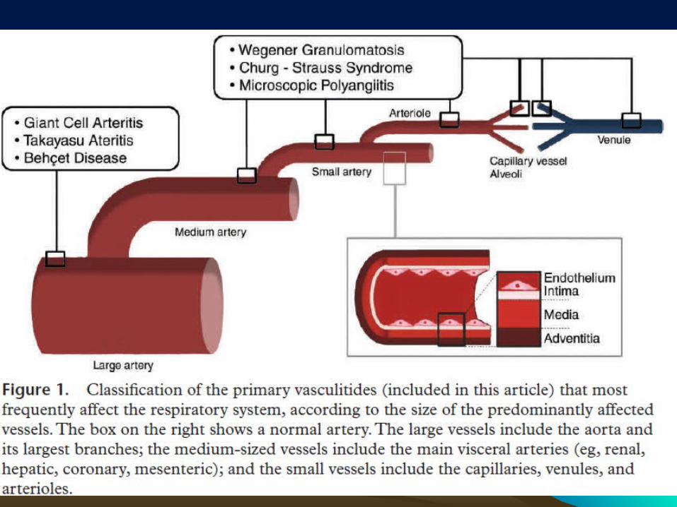
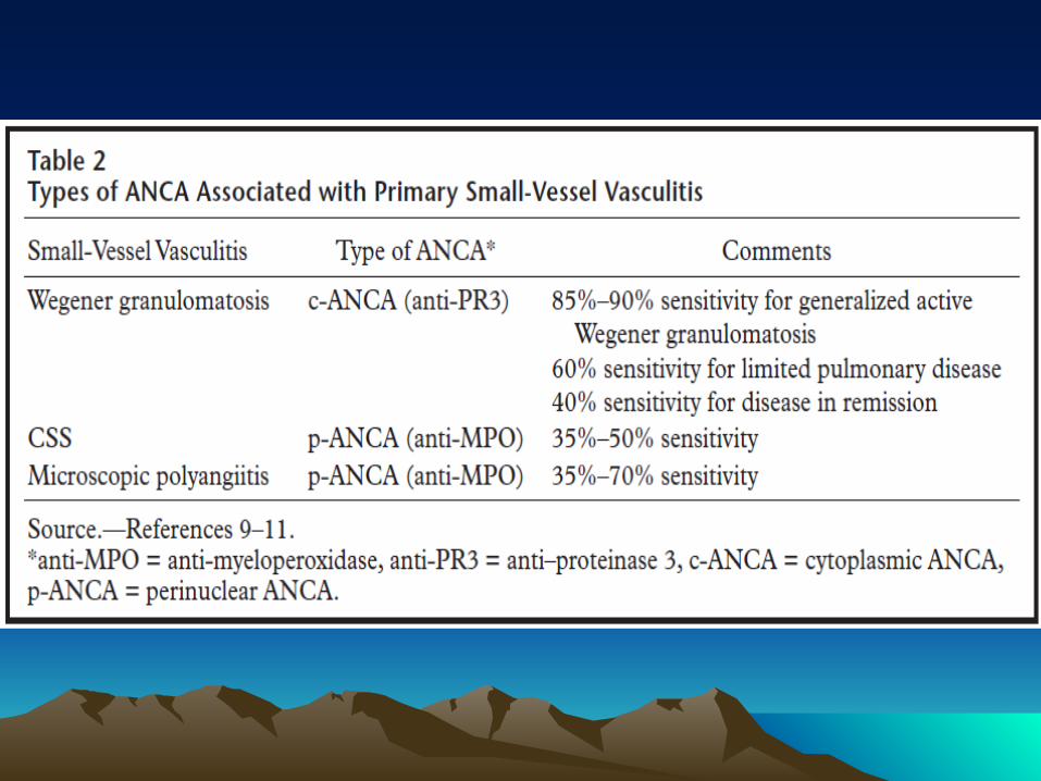
• The ANCA-associated vasculitides (Wegener granulomatosis, CSS, and microscopic polyangiitis) are grouped together because of common clinical features, histopathologic involvement of small vessels, similar response to immunosuppressive treatment, and ANCA positivity
• ANCA positivity is common in these entities but not universal; thus, ANCA negativity does not completely rule out these diseases


Large-Vessel Vasculitis-----Large-Vessel Vasculitis-----Takayasu ArteritisTakayasu Arteritis
• Takayasu arteritis affects almost exclusively patients younger than 40 years, involves primarily the aorta and its major branches, and generally spares the cranial arteries
• Takayasu arteritis is characterized by granulomatous inflammation of the arterial wall with marked intimal proliferation and fibrosis of the media and adventitia; these processes eventually lead to stenosis, occlusion, and, occasionally, poststenotic dilatations and aneurysm formation (when inflammation destroys the media)

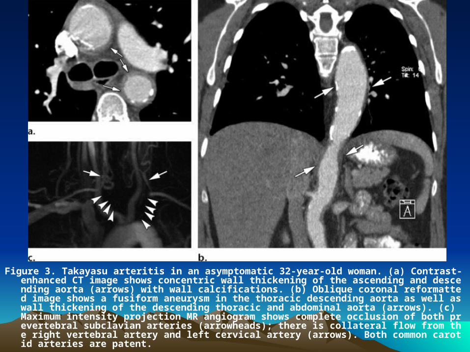
Figure 3. Takayasu arteritis in an asymptomatic 32-year-old woman. (a) Contrast-enhanced CT image shows concentric wall thickening of the ascending and descending aorta (arrows) with wall calcifications. (b) Oblique coronal reformatted image shows a fusiform aneurysm in the thoracic descending aorta as well as wall thickening of the descending thoracic and abdominal aorta (arrows). (c) Maximum intensity projection MR angiogram shows complete occlusion of both prevertebral subclavian arteries (arrowheads); there is collateral flow from the right vertebral artery and left cervical artery (arrows). Both common carotid arteries are patent.
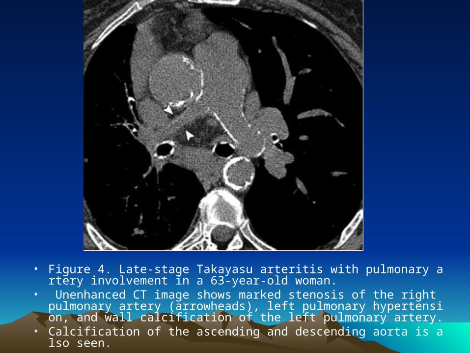
• Figure 4. Late-stage Takayasu arteritis with pulmonary artery involvement in a 63-year-old woman.
• Unenhanced CT image shows marked stenosis of the right pulmonary artery (arrowheads), left pulmonary hypertension, and wall calcification of the left pulmonary artery.
• Calcification of the ascending and descending aorta is also seen.

Giant Cell Arteritis
• GCA (temporal arteritis) is the most common vasculitis of large and medium-sized arteries, affecting almost exclusively individuals over 50 years of age
• The disease predominantly affects the extracranial carotid branches and the aorta and, rarely, the central pulmonary arteries
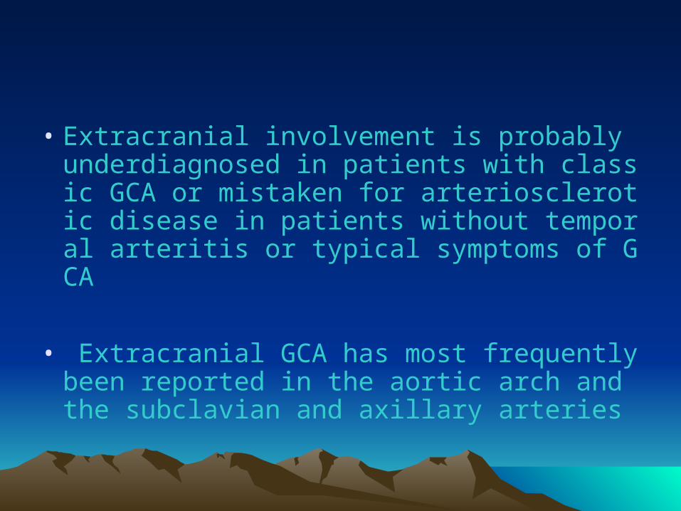
• Extracranial involvement is probably underdiagnosed in patients with classic GCA or mistaken for arteriosclerotic disease in patients without temporal arteritis or typical symptoms of GCA
• Extracranial GCA has most frequently been reported in the aortic arch and the subclavian and axillary arteries

• At histologic analysis, GCA appears similar to Takayasu arteritis
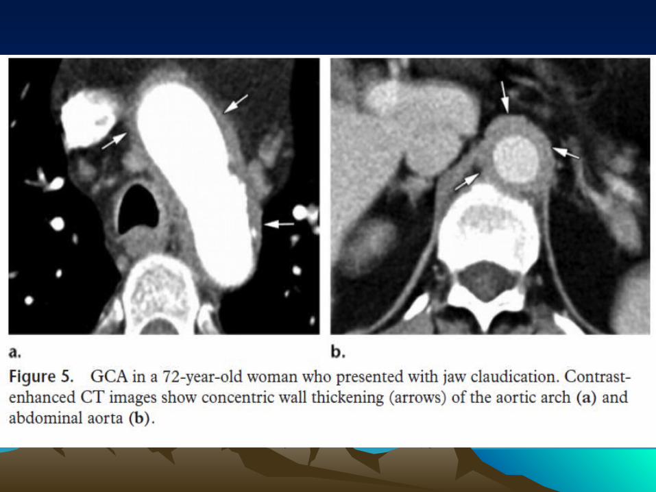
• The principal CT and MR imaging appearance of GCA is similar to that of Takayasu arteritis, with evidence of arterial wall thickening (Fig 5), stenosis, and aneurysm
• Aortic GCA typically remains asymptomatic during the early phase of the disease and causes serious late complications like aneurysms and dissections

Behcet DiseaseBehcet Disease
• Behcet disease is a chronic multisystemic vasculitis
• It is characterized by recurrent oral and genital ulcerations, ocular anomalies (uveitis), and additional clinical manifestations in multiple organ systems
• The disease usually manifests in the second or third decade of life, and the male-to-female ratio is reported to be almost equal

• The vasculitis can involve large, medium, and small vessels of both the arterial and venous circulation.
• The pulmonary arteries are the second most common site of arterial involvement, preceded by the aorta.
• Behcet disease is the most common cause of pulmonary artery aneurysm; the underlying pathophysiologic process is inflammation of the vasa vasorum of the tunica media with destruction of the elastic fibers and dilatation of the vessel lumen.

• Figure 6. Behcet disease in a 26-year-old woman who presented with dyspnea.
• (a) Chest radiograph shows increased size and opacity of the right interlobar and lower lobe pulmonary arteries as well as of the left pulmonary artery (arrows).
• (b) Chest radiograph obtained 6 months earlier shows normal findings.
• (c) Coronal MIP CT image, obtained with 10-mm section thickness, shows increased diameter of both interlobar and lower lobe pulmonary arteries. The aneurysms are thrombosed (arrows), with partial thrombosis on the right and complete thrombosis on the left (*).
• (d) CT image (lung window) shows subpleural wedge-shaped areas of increased opacity, which are suggestive of pulmonary infarction associated with pulmonary thromboembolism.
• (e) T2-weighted MR image (four-chamber view) shows a thrombus in the right atrium (*).
• (f) Coronal MIP CT image, obtained with 10-mm section thickness at the same level as in c 1 month after immunosuppressive treatment, shows a decrease in the size and in the extent of thrombosis of the bilateral pulmonary artery aneurysms.

Medium-sized Vessel Vasculitis
• The medium-sized vessels consist of the main visceral arteries (eg, renal, hepatic, coronary, and mesenteric arteries)
• Polyarteritis nodosa and Kawasaki disease are the two major forms of medium-sized vessel vasculitis
• Lung involvement is extremely rare in polyarteritis nodosa, and its presence argues against this entity
• Kawasaki disease usually occurs in children under 5 years of age

Small-Vessel VasculitisSmall-Vessel Vasculitis
• Small-vessel vasculitis is defined as vasculitis that affects vessels smaller than arteries, such as arterioles, venules, and capillaries
• The diagnosis of small-vessel vasculitis requires features of vasculitic involvement of capillaries and venules, such as purpura, glomerulonephritis, or pulmonary capillaritis
• Lung involvement is most commonly seen with primary, idiopathic, ANCA-associated smallvessel vasculitis

• ANCA-associated small-vessel vasculitides are the most common primary systemic small-vessel vasculitides in adults and include three major categories: Wegener granulomatosis, CSS, and microscopic polyangiitis.
• Wegener granulomatosis and CSS manifest with necrotizing granulomatous inflammation, whereas microscopic polyangiitis manifests with necrotizing inflammation without granulomatosis

Wegener GranulomatosisWegener Granulomatosis
• Wegener granulomatosis is the most common of the ANCA-associated vasculitides
• It is characterized clinically by the triad of upper airway disease (nasal, oral, or sinus inflammation), lower respiratory tract disease (airway or lung), and glomerulonephritis.

Nodules, Masses, and ConsolidationNodules, Masses, and Consolidation
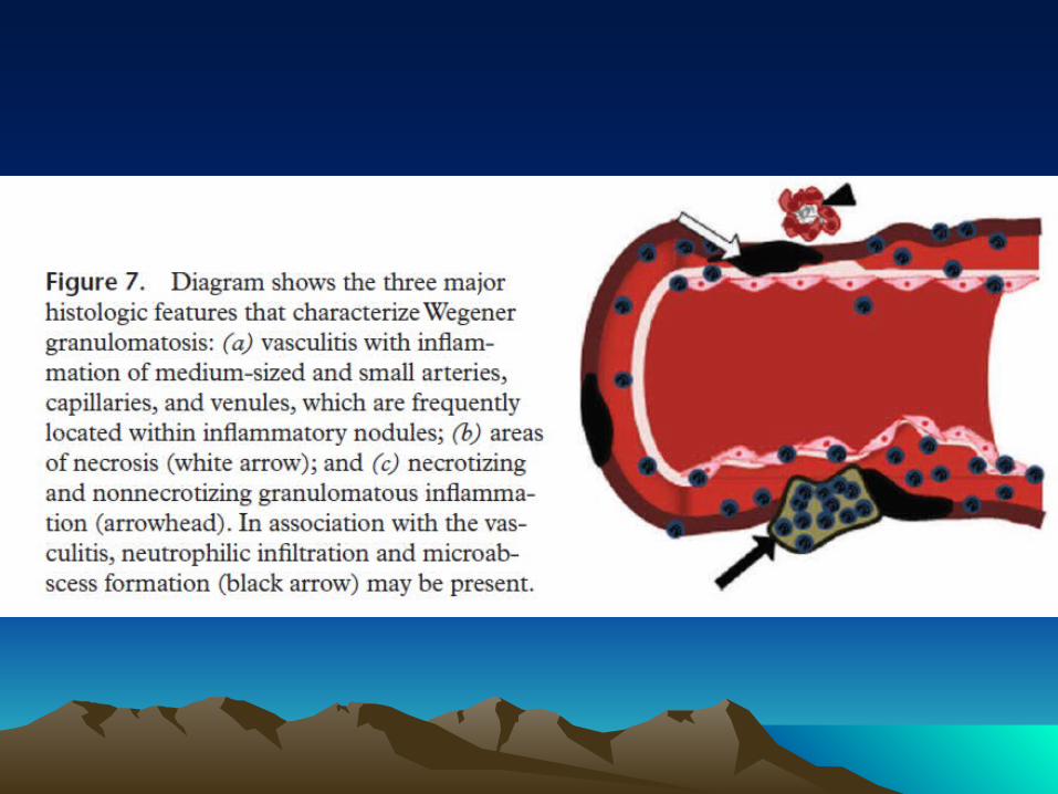
• At histopathologic analysis, nodules and masses in active pulmonary disease are composed of granulomatous tissue
• The characteristic granulomas of Wegener granulomatosis are confluent necrotizing lesions with a tendency toward cavitation

• Figure 8. Relapsing Wegener granulomatosis in a 57-year-old man who initially presented with malaise and recurrent episodes of epistaxis.
• (a, b) CT images (lung window) show irregular, thick-walled, cavitated masses in the right upper lobe
• (a) and left lower lobe (b). The patient responded satisfactorily to treatment, with complete resolution of the pulmonary masses. Two years later, the patient presented with arthralgias and hemoptysis.
• (c) Posteroanterior chest radiograph shows well-defined multiple bilateral nodules, some of which are cavitated, affecting predominantly the upper lobes. Despite immunosuppressive treatment, 3 months later the patient presented with acute shortness of breath and a cough.
• (d) Chest radiograph shows coalescence of the cavitated lesions, some of which demonstrate an air-fluid level secondary to infection.
• (e) CT image (lung window) shows bilateral fairly well-defined nodules and masses. Some lesions are cavitated and demonstrate air-fluid levels. Some of the cavities are thin walled.
• (f) CT image (lung window) obtained 1 year later shows a favorable response to treatment, with marked fibrotic reaction around the healing residual lesions.

• Figure 9. Wegener granulomatosis in a 76-year-old man who presented with otitis and arthralgias.
• Contrast-enhanced CT image shows a mass in the right lower lobe with a central low-attenuation area. The patient also had other bilateral noncavitated masses (not shown)
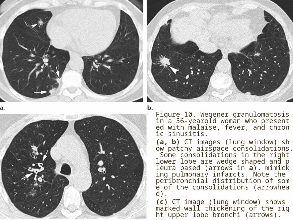
• Figure 10. Wegener granulomatosis in a 56-yearold woman who presented with malaise, fever, and chronic sinusitis.
• (a, b) CT images (lung window) show patchy airspace consolidations. Some consolidations in the right lower lobe are wedge shaped and pleura based (arrows in a), mimicking pulmonary infarcts. Note the peribronchial distribution of some of the consolidations (arrowhead).
• (c) CT image (lung window) shows marked wall thickening of the right upper lobe bronchi (arrows).

• Bronchial abnormalities (once regarded as an unusual CT finding), mainly bronchial wall thickening in the segmental and subsegmental bronchi (Fig 10c)
Tracheobronchial Involvement
Churg-Strauss SyndromeChurg-Strauss Syndrome
• CSS is characterized by the clinical triad of asthma, hypereosinophilia, and necrotizing vasculitis
• The diagnosis of CSS can be made if four or more of the following six findings are present: asthma, more than 10% eosinophilia in a differential white blood cell count, mononeuropathy or polyneuropathy attributable to systemic vasculitis, migratory or transient pulmonary opacities, paranasal sinus abnormalities, and extravascular eosinophils in a biopsy specimen.

• Relatively late age of onset (mean, 32 years) distinguishes the asthma in CSS from asthma in the general population
• The lung is the most commonly involved organ, followed by the skin
• Pulmonary hemorrhage and glomerulonephritis are much less common than in the other small vessel vasculitides
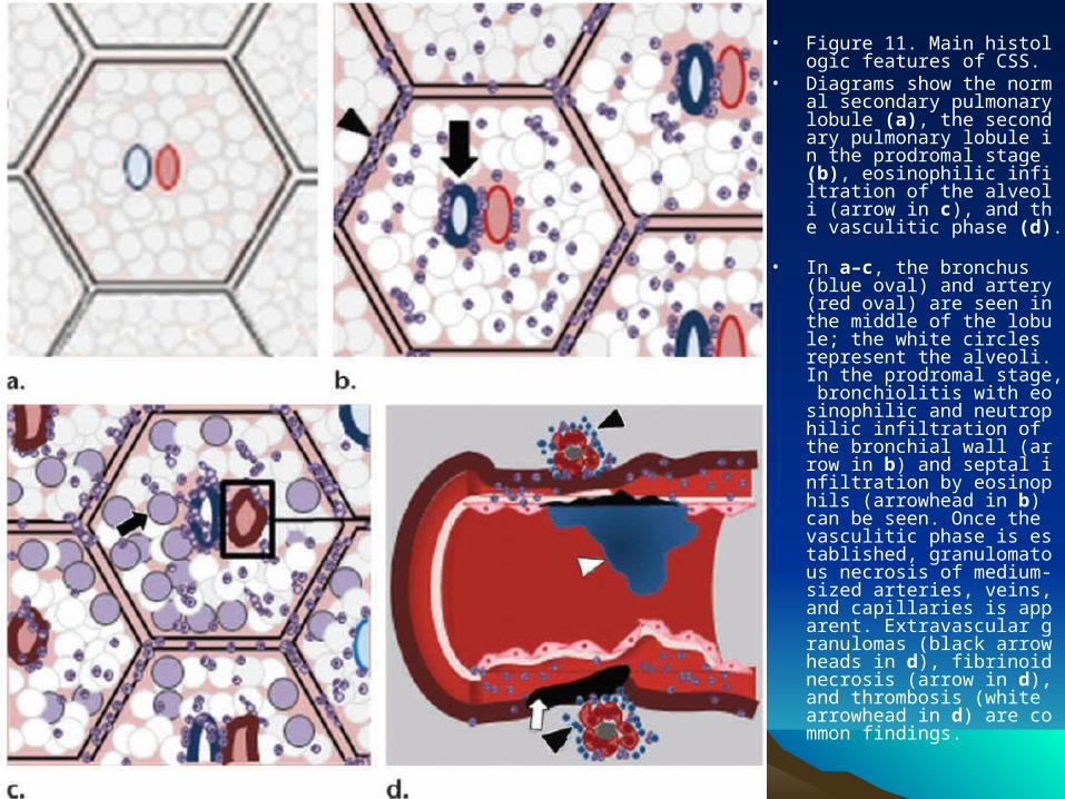
• Figure 11. Main histologic features of CSS.
• Diagrams show the normal secondary pulmonary lobule (a), the secondary pulmonary lobule in the prodromal stage (b), eosinophilic infiltration of the alveoli (arrow in c), and the vasculitic phase (d).
• In a–c, the bronchus (blue oval) and artery (red oval) are seen in the middle of the lobule; the white circles represent the alveoli. In the prodromal stage, bronchiolitis with eosinophilic and neutrophilic infiltration of the bronchial wall (arrow in b) and septal infiltration by eosinophils (arrowhead in b) can be seen. Once the vasculitic phase is established, granulomatous necrosis of medium-sized arteries, veins, and capillaries is apparent. Extravascular granulomas (black arrowheads in d), fibrinoid necrosis (arrow in d), and thrombosis (white arrowhead in d) are common findings.

• The most common lung radiographic manifestations of CSS consist of transient, bilateral, nonsegmental areas of consolidation without predilection for any lung zone (Fig 12a).

• Figure 12. CSS in a 38-year-old woman with asthma diagnosed 7 years before who presented with a 2-month history of fever and cough. She had a history of persistent eosinophilia and sinusitis.
• (a) Chest radiograph shows opacities in both lungs; the opacities spare the apices and costophrenic angles.
• (b) CT image (lung window) shows patchy areas of groundglass opacity in the right upper lobe (arrows) and dense consolidation in the left upper lobe.
• (c, d) CT images (lung window) show bilateral areas of consolidation; some are distributed along the periphery, whereas others are distributed along the bronchovascular bundles (arrowhead). Note the wall thickening of the right upper lobe bronchi (arrow in c). Thickening of the interlobular septa is also seen (arrow in d).
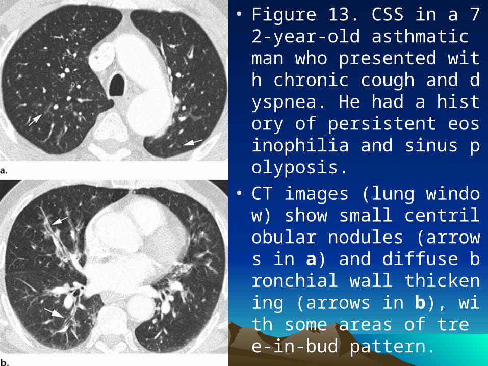
• Figure 13. CSS in a 72-year-old asthmatic man who presented with chronic cough and dyspnea. He had a history of persistent eosinophilia and sinus polyposis.
• CT images (lung window) show small centrilobular nodules (arrows in a) and diffuse bronchial wall thickening (arrows in b), with some areas of tree-in-bud pattern.
Microscopic PolyangiitisMicroscopic Polyangiitis
• Microscopic polyangiitis is a nongranulomatous necrotizing systemic vasculitis
• It is the most common cause of pulmonary-renal syndrome, a syndrome characterized by the coexistence of pulmonary hemorrhage and glomerulonephritis
• The diagnosis of microscopic polyangiitis should be suspected in patients with rapidly progressive glomerulonephritis in whom perinuclear ANCA is present and who show clinical and radiologic findings consistent with diffuse pulmonary hemorrhage
Diffuse Alveolar HemorrhageDiffuse Alveolar Hemorrhage
• DAH can be defined by the presence of hemoptysis, diffuse alveolar infiltrates, and a drop in hematocrit level.
• Symptoms include cough, hemoptysis, dyspnea, and anemia.
• However, chest radiographic and CT findings are nonspecific (the alveolar infiltrates can even sometimes be unilateral), and hemoptysis may be lacking (even in patients with sufficient hemorrhage to result in anemia).
• The diagnosis is usually made with bronchoscopy, by means of which serial bronchoalveolar lavage samples (from the same location) reveal an increasing red blood cell count.

• Whatever the underlying cause of DAH, the resultant hemorrhagic filling of the airspaces causes widespread radiographic shadowing, which ranges in intensity from vague ground-glass opacity to extensive intense consolidation
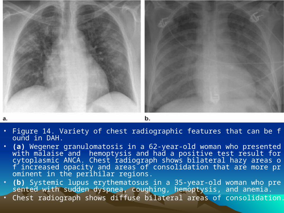
• Figure 14. Variety of chest radiographic features that can be found in DAH.• (a) Wegener granulomatosis in a 62-year-old woman who presented with m
alaise and hemoptysis and had a positive test result for cytoplasmic ANCA. Chest radiograph shows bilateral hazy areas of increased opacity and areas of consolidation that are more prominent in the perihilar regions.
• (b) Systemic lupus erythematosus in a 35-year-old woman who presented with sudden dyspnea, coughing, hemoptysis, and anemia.
• Chest radiograph shows diffuse bilateral areas of consolidation.
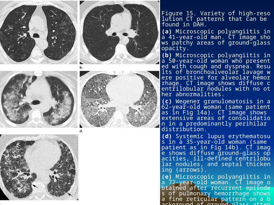
• Figure 15. Variety of high-resolution CT patterns that can be found in DAH.
• (a) Microscopic polyangiitis in a 41-year-old man. CT image shows patchy areas of ground-glass opacity.
• (b) Microscopic polyangiitis in a 50-year-old woman who presented with cough and dyspnea. Results of bronchoalveolar lavage were positive for alveolar hemorrhage. CT image shows diffuse centrilobular nodules with no other abnormalities.
• (c) Wegener granulomatosis in a 62-year-old woman (same patient as in Fig 14a). CT image shows extensive areas of consolidation in a predominantly perihilar distribution.
• (d) Systemic lupus erythematosus in a 35-year-old woman (same patient as in Fig 14b). CT image shows diffuse ground-glass opacities, ill-defined centrilobular nodules, and septal thickening (arrows).
• (e) Microscopic polyangiitis in a 72-year-old woman. CT image obtained after recurrent episodes of pulmonary hemorrhage shows a fine reticular pattern on a background of ground-glass attenuation, signs of pulmonary fibrosis with a peripheral honeycombing pattern (arrows), and traction bronchiectasis (arrowhead).

• The many different entities causing DAH may be classified into three groups:
• (a) ANCA-associated small-vessel vasculitis, which generally involves pulmonary capillaritis (Wegener granulomatosis, CSS, microscopic polyangiitis);
• (b) syndromes caused by immune deposits that can be detected with immunofluorescence, such as Goodpasture syndrome and systemic lupus erythematosus;
• (c) a large group of miscellaneous entities that includes diffuse alveolar damage, drug reactions (including cocaine inhalation), coagulopathies, infections, and idiopathic disease such as idiopathic pulmonary hemosiderosis

Diagnostic ApproachDiagnostic Approach
• The primary vasculitides are rare disorders, and their diagnoses are among the most demanding challenges in medicine because their signs and symptoms are nonspecific and overlap with those of infections, connective tissue diseases, and malignancies.
• An integrated clinical, radiologic, and sometimes histologic approach is needed.
• Symptoms such as uveitis, unusual rashes, arthritis, or “sinus troubles” must be remembered and considered relevant when the clinical presentation is an abnormal lung CT or chest radiographic finding, shortness of breath, or renal failure
• The presence of otherwise unexplained nodular or cavitary disease should raise the suspicion of vasculitis.
• Although infection and malignancy are the most common explanations, in the correct clinical setting vasculitis, particularly Wegener granulomatosis, should be strongly considered.

• CSS should be suspected when patchy ground-glass opacities or consolidations are seen in a patient with a history of asthma who also presents with eosinophilia.
• Differentiation between CSS and simple pulmonary eosinophilia or chronic eosinophilic pneumonia in an asthmatic patient is based on systemic manifestations of CSS (eg, peripheral neuropathy, rash) and the presence of perinuclear ANCA in serum.

• Radiologic signs of DAH are nonspecific and variable but must be considered in patients with otherwise unexplained alveolar infiltrates, particularly when seen with new-onset renal insufficiency or a connective tissue disease
ConclusionsConclusions
• The diagnosis of vasculitis is often delayed because a number of other disorders can mimic the clinical manifestations.
• Chest radiographs and especially CT are valuable for noninvasive diagnosis in patients with pulmonary vasculitis; certain radiologic signs in combination with clinical features enable an earlier diagnosis.

ThanksThanks !!!!!!