Embed Size (px)
Citation preview
GE J Port Gastrenterol. 2012;19(4):217---218
www.elsevier.pt/ge
ENDOSCOPIC SPOT
Whipple’s disease and giardiasis: An uncommon association
Doenca de whipple e giardíase: uma associacão incomum
Frederico Ferreira ∗, Helder Cardoso, Andreia Albuquerque, Fernando Magro,Guilherme Macedo
Gastroentrology Department, Hospital S. João and Faculty of Medicine, Porto, Portugal
Received 8 July 2011; accepted 10 January 2012Available online 29 June 2012
Whipple‘s disease is a rare systemic disorder with unspecificsigns and symptoms, that remains a diagnostic challenge.1
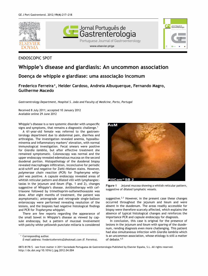
A 61-year-old female was referred to the gastroen-terology department due to abdominal pain, diarrhea andarthralgias. The investigation revealed anemia, hypoalbu-minemia and inflammatory markers’ elevation, with normalimmunological investigation. Fecal smears were positivefor Giardia lamblia, but after effective treatment sheremained symptomatic. Colonoscopy was normal and theupper endoscopy revealed edematous mucosa on the secondduodenal portion. Histopathology of the duodenal biopsyrevealed macrophages infiltration, inconclusive for periodicacid-schiff and negative for Ziehl---Nielsen stains. However,polymerase chain reaction (PCR) for Tropheryma whip-plei was positive. A capsule endoscopy revealed areas ofwhitish reticular pattern and dilated villi with lymphangiec-tasias in the jejunum and ileum (Figs. 1 and 2), changessuggestive of Whipple’s disease. Antibiotherapy with cef-triaxone followed by trimethoprim-sulfamethoxazole wasdone. After eight months of treatment, the patient wasasymptomatic; anterograde and retrograde single-balloonenteroscopy were performed revealing resolution of thelesions, and the biopsies had negative histological findingsand PCR for Tropheryma whipplei.
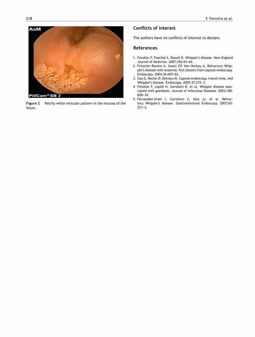
There are few reports regarding the appearance ofthe small bowel in Whipple’s disease as viewed by cap-sule endoscopy, but a pattern of mucosal involvementwith patchy white-yellowish punctate miliaria is considered
∗ Corresponding author.E-mail address: [email protected] (F. Ferreira).
Figure 1 Jejunal mucosa showing a whitish reticular pattern,suggestive of dilated lymphatic vessels.
suggestive.2,3 However, in the present case these changesoccurred throughout the jejunum and ileum and wereabsent in the duodenum. The areas readily accessible forbiopsy were therefore scarcely affected, which explains theabsence of typical histological changes and reinforces theimportance PCR and capsule endoscopy for diagnosis.
In conclusion, this case is original for the presence oflesions in the jejunum and ileum with sparing of the duode-num, rending diagnosis even more challenging. This patienthad also simultaneous infection with Giardia lamblia whichis an uncommon association, whose etiology is still a matterof debate.4,5
0872-8178/$ – see front matter © 2011 Sociedade Portuguesa de Gastrenterologia Published by Elsevier España, S.L. All rights reserved.http://dx.doi.org/10.1016/j.jpg.2012.04.004
218 F. Ferreira et al.
Figure 2 Patchy white reticular pattern in the mucosa of theileum.
Conflicts of interest
The authors have no conflicts of interest to declare.
References
1. Fenollar F, Puechal X, Raoult D. Whipple’s disease. New EnglandJournal of Medicine. 2007;356:55---66.
2. Fritscher-Ravens A, Swain CP, Von Herbay A. Refractory Whip-ple’s disease with anaemia: first lessons from capsule endoscopy.Endoscopy. 2004;36:659---62.
3. Gay G, Roche JF, Delvaux M. Capsule endoscopy, transit time, andWhipple’s disease. Endoscopy. 2005;37:272---3.
4. Fenollar F, Lepidi H, Gerolami R, et al. Whipple disease asso-ciated with giardiasis. Journal of Infectious Diseases. 2003;188:828---34.
5. Fernandez-Urien I, Carretero C, Sola JJ, et al. Refrac-tory Whipple’s disease. Gastrointestinal Endoscopy. 2007;65:521---2.


![Clase 6[1]. giardiasis](https://img.pdfslide.net/doc/110x75/58ab7bff1a28ab3e738b479b/clase-61-giardiasis-58abceb292378.jpg)