Embed Size (px)
Citation preview

Why do we move our eyes?
- Image stabilization in the presence of body movements.
- Information acquisition - bring objects of interest onto high acuity region in fovea.

Retinal structure

Cone Photoreceptors are densely packed in the central fovea

Visual Acuity matches photoreceptor density

Oculomotor Muscles
Muscles innervated by oculomotor, trochlear, and abducens (cranial) nerves from the oculomotor nuclei in the brainstem. Oculo-motor neurons: 100-600Hz vs spinal motorNeurons: 50-100Hz

Types of Eye Movement
Information Gathering StabilizingVoluntary (attention) Reflexive
Saccades vestibular ocular reflex (vor)new location, high velocity (700 deg/sec), body movementsballistic(?)
Smooth pursuit optokinetic nystagmus (okn)object moves, velocity, slow(ish) – typically whole field image motionup to 35 deg/sec
Vergencechange point of fixation in depthslow, disjunctive (eyes rotate in opposite directions)
(all others are conjunctive)Note: link between accommodation and vergenceFixation: period when eye is relatively stationary between saccades.

AccelerationDepth-dept gain, Precision in natural vision
Velocity
Acuity – babies

https://www.youtube.com/watch?v=KSJksSA6Q-A
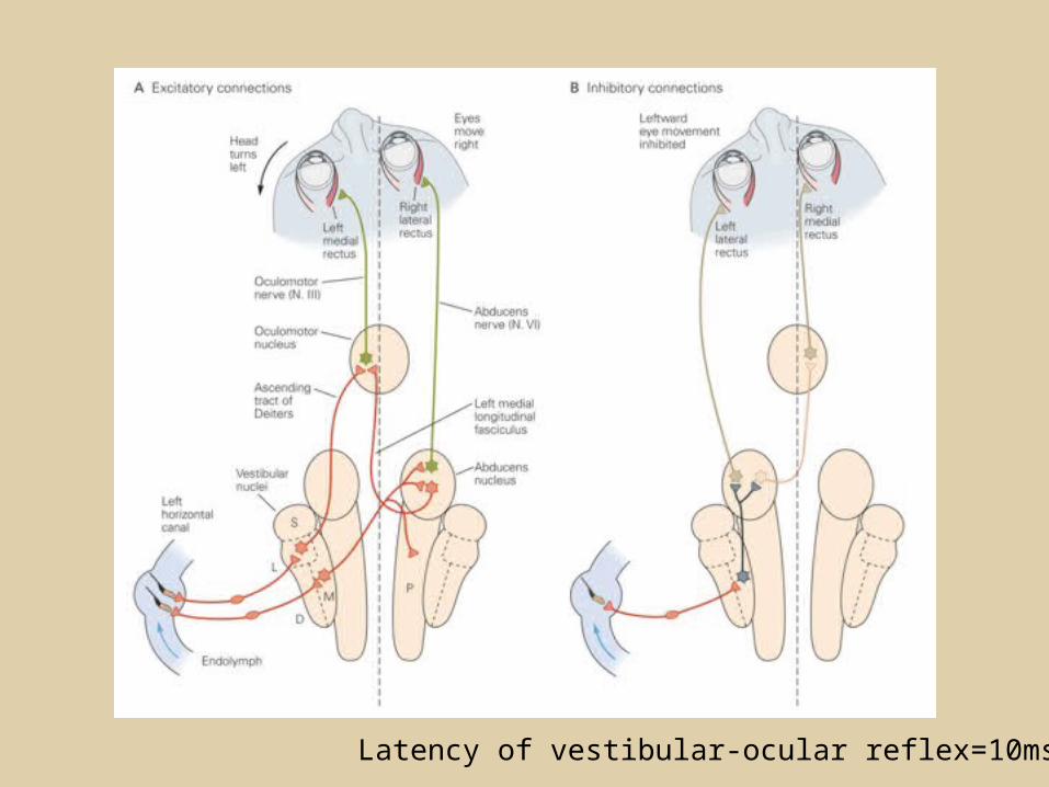
Latency of vestibular-ocular reflex=10msec

It is almost impossible to hold the eyes still.
Demonstration of VOR and its precision – sitting vs standing
Miniature eye movements
Slow driftMicro-saccadestremor


Saccade latency approx 200 msec, pursuit approx 100 – smaller when there is a context thatallows prediction.
Step-ramp allows separation of pursuit (slip) and saccade (displacement)


“main sequence”: duration = c Amplitude + bMin saccade duration approx 25 msec, max approx 200msec

Factors That Control Gaze.
- TASK Defines behavioral goals, what information is relevant.
- REWARDS Oculomotor circuitry sensitive to reward/subjective value of those goals.
- UNCERTAINTY Get information. Peripheral resolution/ working memory REDUCTION decay etc
- PRIORS/ Memory Gaze targeting reflects stored knowledge.
- IMAGE Salient properties eg high contrast/ spatial outliers

1. Neural activity related to saccade
2. Microstimulation generates saccade
3. Lesions impair saccade
Brain Circuitry for Saccades

Brain Circuitry for Pursuit

Eye Tracking Methods

Developments in Eye Tracking
Head fixed /restricted: Contact lenses: mirror / magnetic coils Early infra-red systems Dual Purkinje Image tracker
Head Free: Head mounted IR video-based systems Remote systems with head tracking
Scene camera
Difficulty: optical power of eye + observer movement

Why eye movements are hard to measure.
18mm
0.3mm = 1 deg visual angle
x a
tan(a/2) = x/da = 2 tan
-1 x/d
Visual Angle
d
1 diopter = 1/focal length in meters
55 diopters = 1/.018
A small eye rotation translates into a big change in visual angle

Early Methods:
“Barlow photographed a droplet of mercury
placed on the limbus. Translations of the head
were minimized by having subjects lie on a
stone slab with their heads wedged tightly
inside a rigid iron frame”
Kowler, 1990
Measuring Eye Movements

Early methods:
“The eye is first cocainized, then the lids should
be propped apart by some form of eye-lid
fastener, of which the best is probably that in
form of a wide-opening spring with tortoise-
shell grooves for the lids.”
Delabarre,
1898

Monitoring Eye Movements; YarbusMirror mounted on eye using suction. Light bounces off mirror and is recorded on film

• Non image-based eye trackers
– Electrical/analog
– Limbus
– Magnetic search coil
Non image-based Eye Trackers

EOG
EOG
The eye is a ‘dipole’ with ~millivolts voltage difference between the retina and the cornea.

ElectroOculoGram (EOG)
Use in clinic – head not fixed

By monitoring the ‘whites of the eye’ below the iris, it is possible to determine eye position.
Vertical eye movements cause both signals to increase (up) or decrease (down).
Horizontal eye movements cause differential illumination between the right and left sensors.
Limbus Trackers
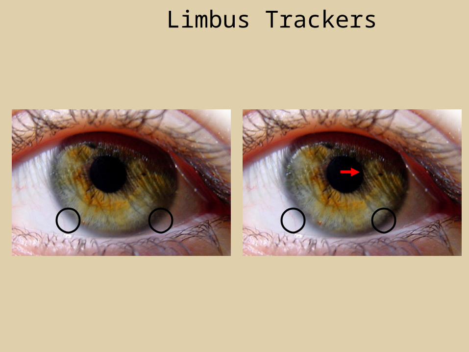
Limbus Trackers

Limbus

EOG and Limbus trackers
Good temporal resolution.
Lousy spatial resolution
High noise, drift
Mostly useful in clinic

Skalar search coils
Magnetic Search CoilsUsed for much animal work, though less so recently. Very high precision andaccuracy (few minutes of arc). Used in older human em literature.Can use similar methodology for head and hand (see Hayhoe lab)

• Image-based eye trackers
– Dual Purkinje
– Video based
Image-based Eye Trackers

Dual Purkinje Trackers
The ‘gold standard’ in eye trackers
Multiple reflections from the cornea and lens vary in a very well-defined way as the eye moves. By tracking the 1st and 4th reflections, the tracker can determine eye position with very high precision.Bill Geisler lab has a binocular tracker.
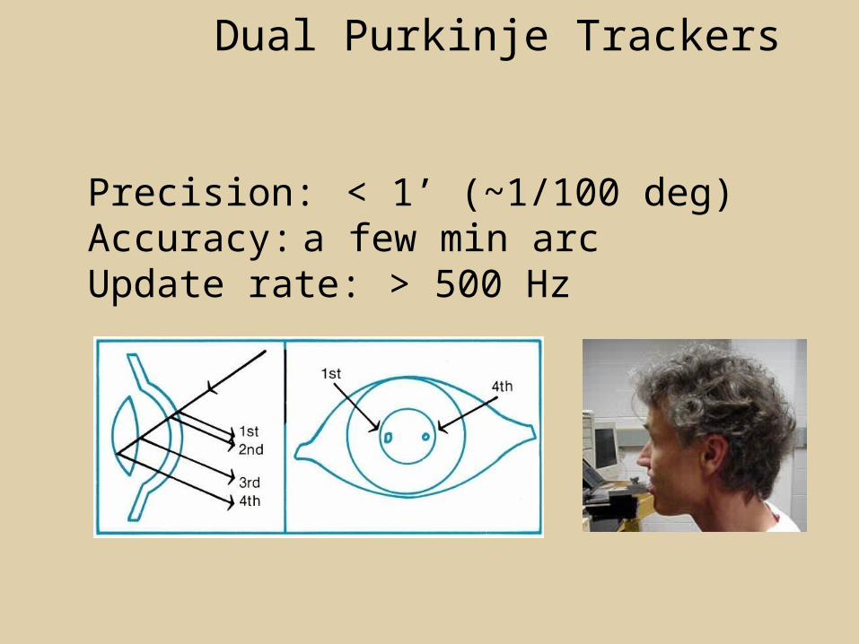
Dual Purkinje Trackers
Precision: < 1’ (~1/100 deg)Accuracy: a few min arcUpdate rate: > 500 Hz

Dual Purkinje Trackers
Usually requires bite bar but theoretically can get away with head rest.

• Video-based eye trackers:
–Head mounted
–Remote
Video-based Eye Trackers

Head mountedCamera on head views scene, another camera views eye.

Video-based Eye TrackersInfra-red video camera finds center of pupil and corneal reflection.Advantages: unconstrained viewing.Disadvantages: temporal resolution may be as low as 30 HzAccuracy never better than 0.5 deg.
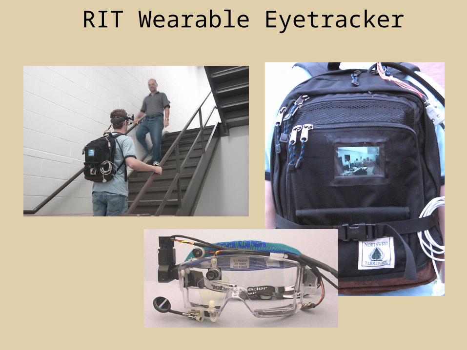
RIT Wearable Eyetracker

RIT Wearable Eyetracker


Build-up neurons in the intermediate layers of the SC are activeprior to a saccade.
Cell in the superifical layers get input from the retina. This may mediateVery fast saccades – sometimes called “express saccades”
Extent of buildup neuron activity reflects stimulus probability.
Express saccades might also reflect activity in buildup neurons.

LIP: Lateral Intra-parietal AreaTarget selection for saccades: cells fire before saccade to attended object
Posterior Parietal Cortex
reaching
grasping
Intra-Parietal Sulcus: areaof multi-sensory convergence
Visual stability

Model of saccade generation: target selection depends on expected value
Trommershauser, Glimcher, Gegenfurtner, 2009
Area LIP contains a reward expectation signal which modulates the gain of visual neurons in LIP.
Reward modulation of saccadic eye movements originates from dopaminergic input to caudate nucleus.

Relation between saccades and attention.
Saccade is always preceded by an attentional shiftHowever, attention can be allocated covertly to the peripheral retina without a saccade.
Pursuit movements also require attention.

Figure 8.18 The comparator
Visual Stability


A cross seen through an aperture that moves clockwise around the boundary. Alternatively, the aperture may be stationary, and the cross move behind it. Individual views, shown on the right, are ambiguous.
Observers have no trouble with this if they have an “internal model” or schema that readily allows interpretation of the sequence.


-Saccades/Smooth Pursuit
-Planning/ Error Checking
-relates to behavioral goals
Supplementary eye fields
A subset of SEF neurons and LFPs exhibited strong modulation following erroneous saccades to adistractor. Altogether, these results suggest that SEF plays a limited role in controlling ongoing visual search behavior, but may play a larger role in monitoring search performance.
Nearby Anterior Cingulate also involved in performance monitoring.

Motor neurons for the eye muscles are located in the oculomotor nucleus (III), trochlear nucleus (IV), and abducens nucleus (VI), and reach the extraocular muscles via the corresponding nerves (n. III, n. IV, n. VI).Premotor neurons for controlling eye movements are located in the paramedian pontine reticular formation(PPRF), the mesencephalic reticular formation (MRF), rostral interstitial nucleus of the medial longitudinal fasciculus (riMLF), the interstitial nucleus of Cajal (IC), the vestibular nuclei (VN), and the nucleus prepositus hypoglossi (NPH).
Motor neurons
Pre-motor neurons
Oculomotor nucleus
Trochlear
Abducens
H
V

Pulse-Step signal for a saccade

Brainstem circuits for saccades. Omnipause neurons (OPN) in the nucleus raphe interpositus (RIP) tonically inhibit excitatory burst neurons (EBN) located in the paramedian pontine reticular formation (PPRF). When OPNs pause, the EBNs emit a burst of spikes, which activate motor neurons (MN) in the abducens nucleus (VI) innervating the lateral rectus muscle. The burst also activates interneurons (IN) which activate motor neurons on the oculomotor nucleus (III) on the opposite side, innervating the medial rectus. Inhibitory burst neurons (IBN) show a pattern of activity similar to EBNs, but provide inhibitory inputs to decrease activation in the complementary circuits and antagonist muscles. Long-lead burst neurons (LLBN) showactivity long before movement onset, and provide an excitatory input to EBNs.

Brain areas involved in making a saccadic eye movementBehavioral goal: make a sandwich (learn how to make sandwiches) Frontal cortex.
Sub-goal: get peanut butter (secondary reward signal - dopamine - basal ganglia)
Visual search for pb: requires memory for eg color of pb or location (memory for visual properties - Inferotemporal cortex; activation of color - V1, V4)
Visual search provides saccade goal. LIP - target selection, also FEF
Plan saccade - FEF, SEF
Coordinate with hands/head
Execute saccade/ control time of execution: basal ganglia (substantia nigra pars reticulata, caudate)
Calculate velocity/position signal oculomotor nuclei
Cerebellum?

RF reticular formation VN vestibular nuclePN , pontine nucleii
CerebellumOV oculomotor vermis VPF ventral paraflocculus FN fastigial nucleus

otoliths
Rotational (semi-circular canals) translational (otoliths)

target selection
signals to muscles(forces)
inhibits SC
saccade decision
saccade command(where to go)
monitor/plan movements
Function of Different Areas
H
V


Smooth pursuit& Supplementary
Brain Circuitry for Pursuit
Velocity signal
Early motion analysis

Eye Movement Research
Consequences of image motion on visual acuity: stabilized images
Metrics of saccades/ pursuit/ vergence/vor
Constancy of visual direction
Eye movements in reading/ Cognitive role of eye movements
Active Vision/Natural tasks: Fixation patterns, eye/head/hand coordination
Language comprehension in visual context
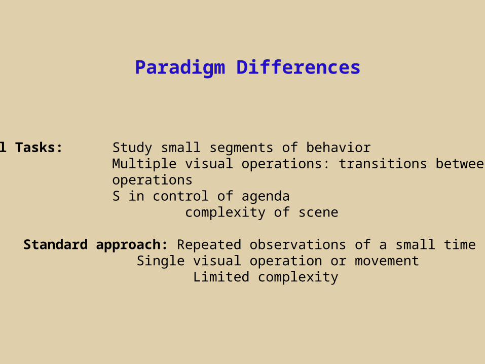
Paradigm Differences
Natural Tasks: Study small segments of behavior Multiple visual operations: transitions between operations S in control of agenda
complexity of scene
Standard approach: Repeated observations of a small time slice Single visual operation or movement
Limited complexity

Remote
Bright pupil Dark pupil

Video-based Eye Trackers

Video-based Eye Trackers

Dark pupil
Bright pupil
Bright-pupil; coaxial illumination

Two signals; pupil and corneal reflection - fairly robustto tracker movement wrt head.Lower temporal and spatial resolution than eg coils/DPI1 deg, 60-120 Hz
Embed eye image in video record to monitor quality of track.