Embed Size (px)
Citation preview


Why perform fetal monitoring
Identify the fetus in distress
To avert permanent fetal damage or death
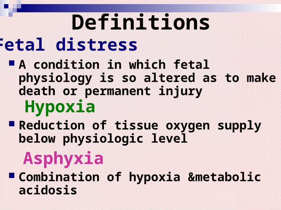
Definitions
A condition in which fetal physiology is so altered as to make death or permanent injury
Hypoxia Reduction of tissue oxygen supply below
physiologic level
Asphyxia Combination of hypoxia &metabolic
acidosis
Fetal distress
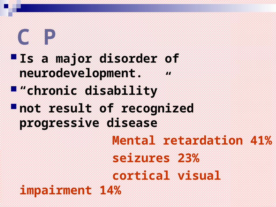
C P Is a major disorder of
neurodevelopment. “chronic disability” not result of recognized progressive
disease
Mental retardation 41%
seizures 23%
cortical visual impairment 14%
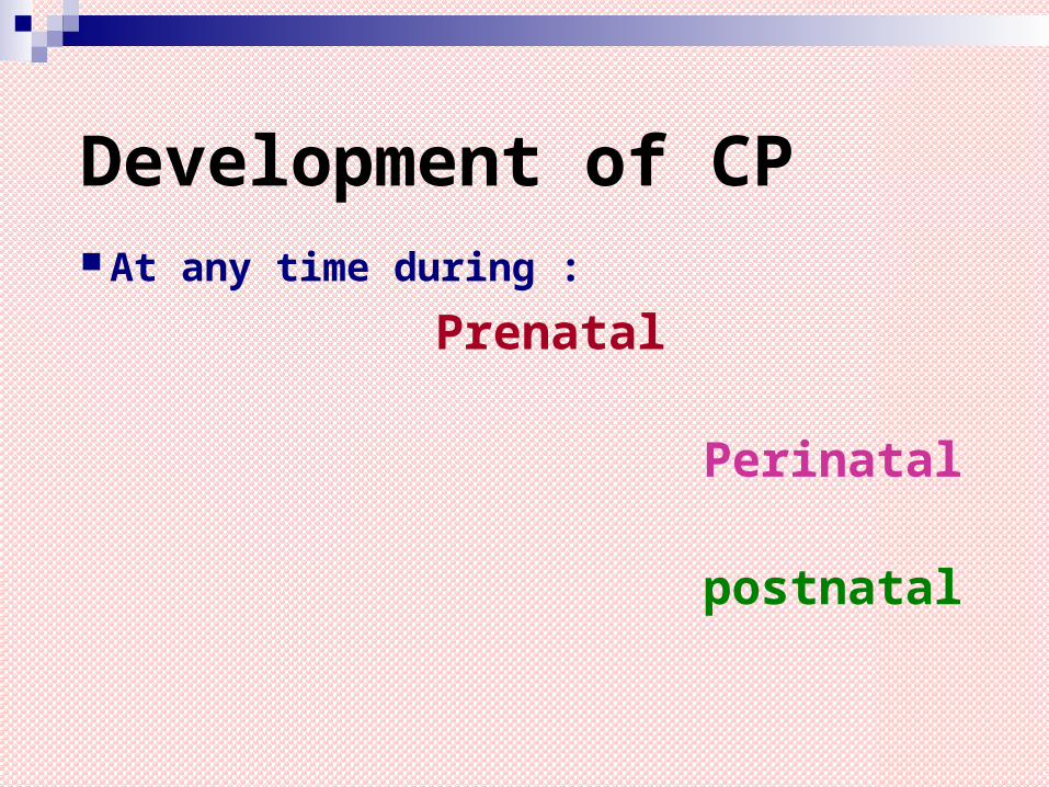
Development of CP At any time during :
Prenatal
Perinatal
postnatal

8.2- 9% of CP case were
potentially attributableto
birth asphyxia
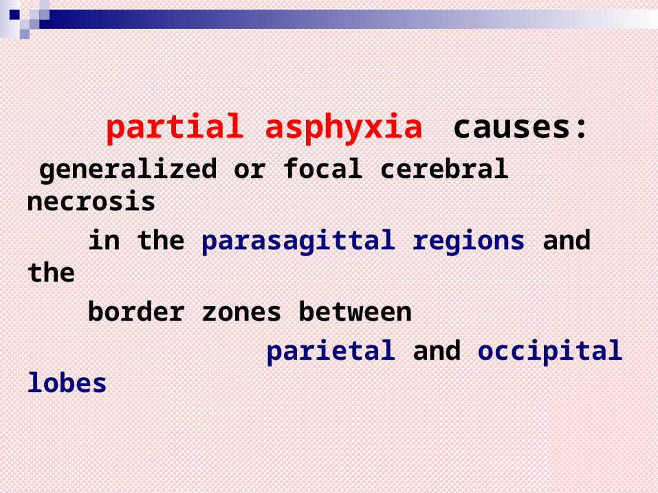
partial asphyxia causes: generalized or focal cerebral necrosis
in the parasagittal regions and the
border zones between
parietal and occipital lobes
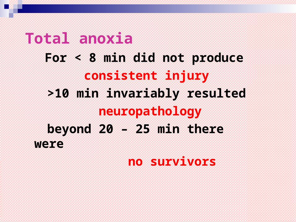
Total anoxia For < 8 min did not produce
consistent injury
>10 min invariably resulted
neuropathology
beyond 20 – 25 min there were
no survivors
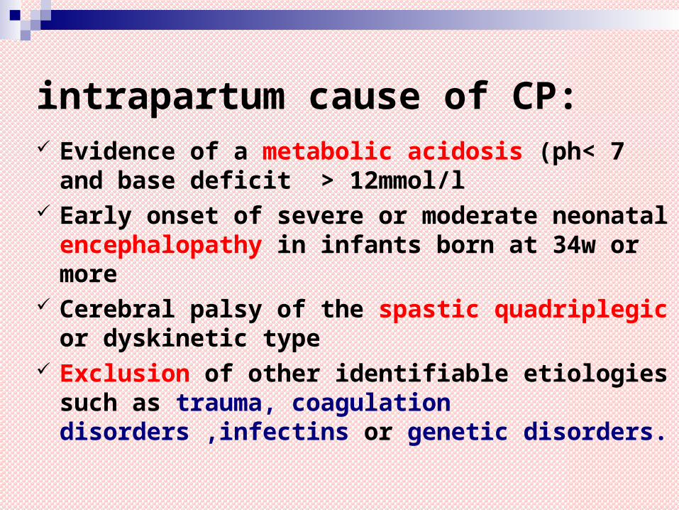
intrapartum cause of CP: Evidence of a metabolic acidosis (ph< 7 and
base deficit > 12mmol/l Early onset of severe or moderate neonatal
encephalopathy in infants born at 34w or more Cerebral palsy of the spastic quadriplegic or
dyskinetic type Exclusion of other identifiable etiologies such
as trauma, coagulation disorders ,infectins or genetic disorders.

Fetal Fetal
HeartHeart
raterate
Fetal Fetal
HeartHeart
raterate
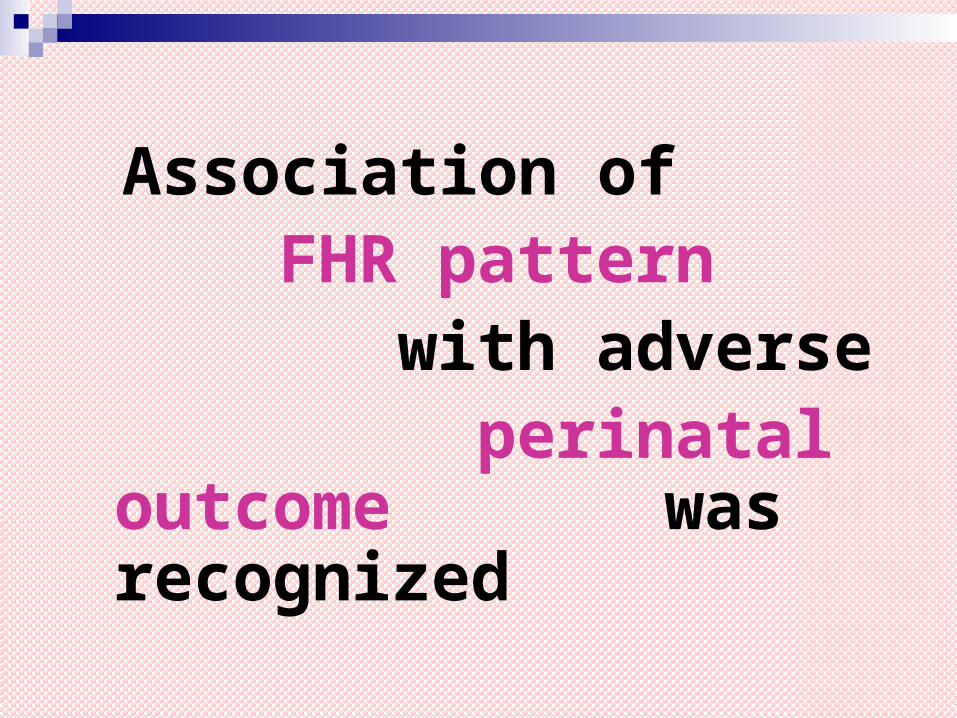
Association of FHR pattern with adverse perinatal outcome was recognized
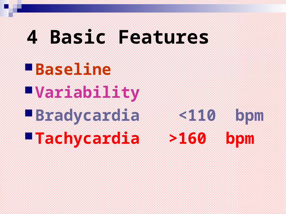
4 Basic Features
Baseline VariabilityBradycardia <110 bpm Tachycardia >160 bpm
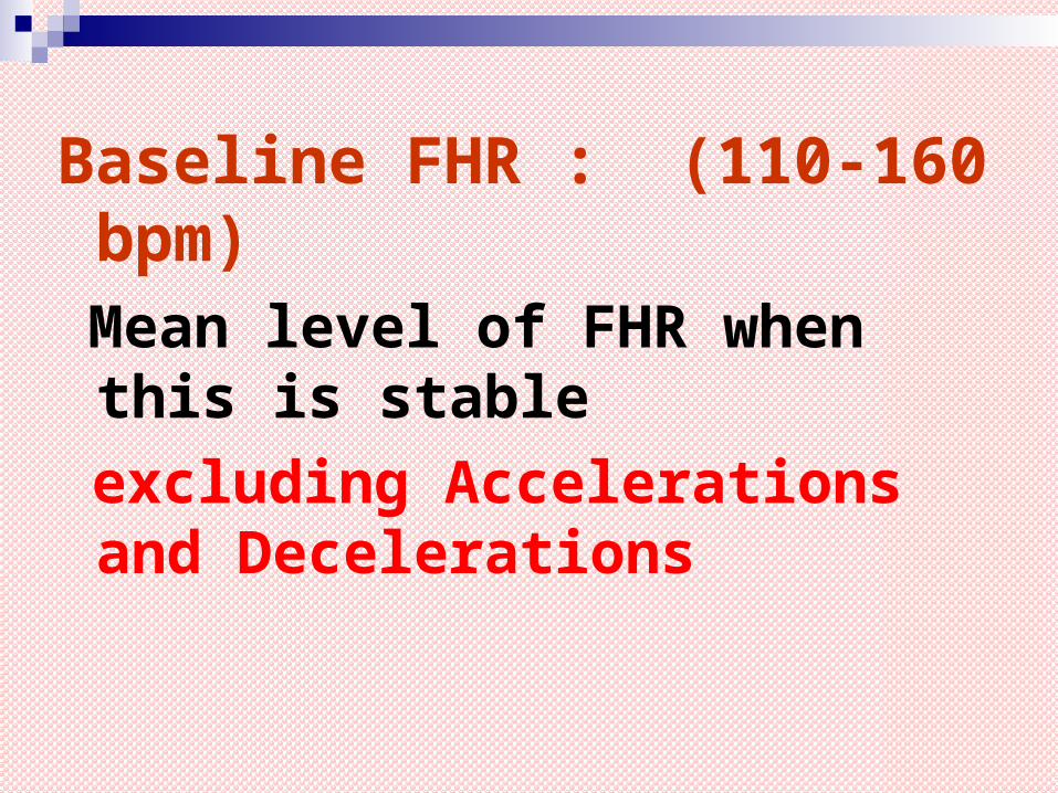
Baseline FHR : (110-160 bpm) Mean level of FHR when this is
stable
excluding Accelerations and Decelerations

Bradicardia
Maternal B blocker therapy Hypothermia Hpoglycemia Hypothyroidism Fetal cardiac conduction defect
(structural abn ,viral inf,)
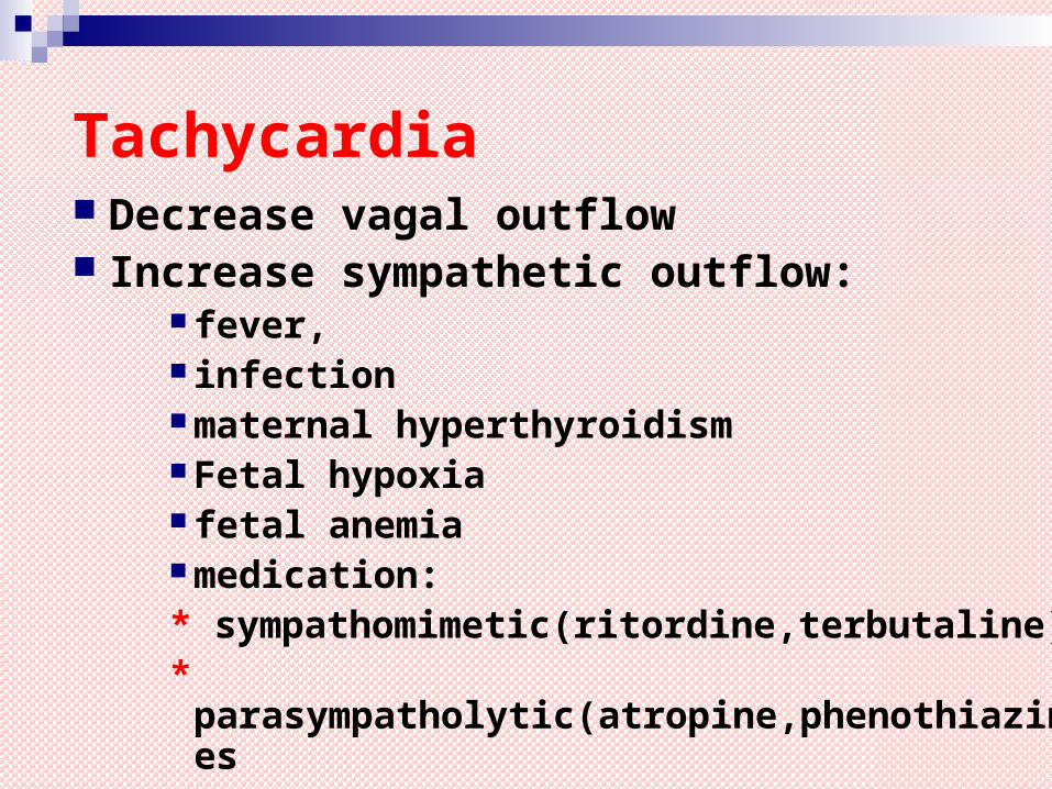
Tachycardia Decrease vagal outflow Increase sympathetic outflow:
fever, infection maternal hyperthyroidism Fetal hypoxia fetal anemia medication:* sympathomimetic(ritordine,terbutaline)* parasympatholytic(atropine,phenothiazines
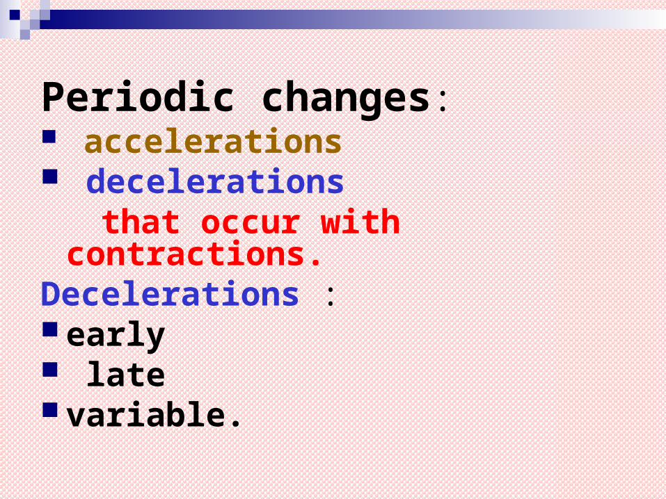
Periodic changes: accelerations decelerations that occur with contractions. Decelerations : early late variable.
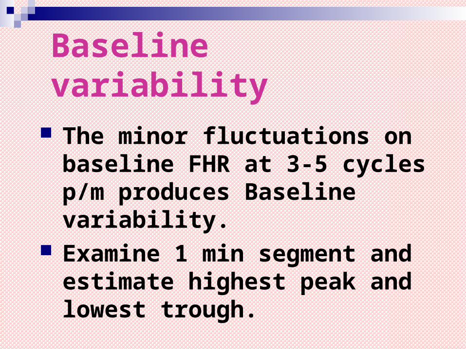
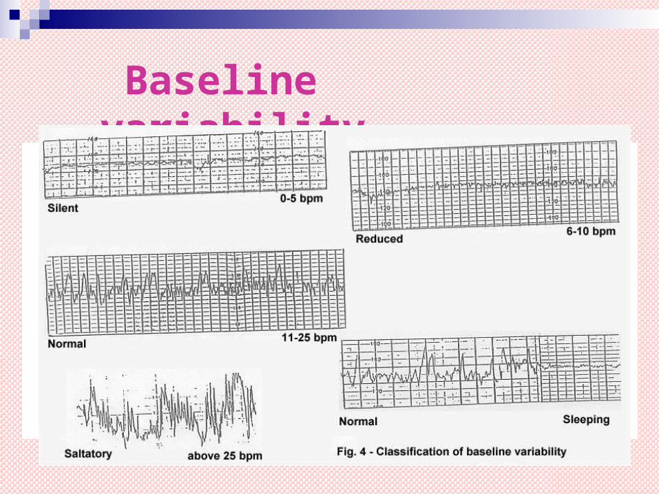
Baseline variability
The minor fluctuations on baseline FHR at 3-5 cycles p/m produces Baseline variability.
Examine 1 min segment and estimate highest peak and lowest trough.

Baseline Variability
Normal: Normal is more than or equal to 5 bpm. Non reassuring,Less than 5 bpm or
less, but less than 30 min
Abnormal: less than 5 bpm for 90 min or more.
Baseline variability
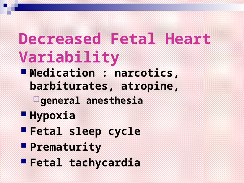
Decreased Fetal Heart Variability Medication : narcotics, barbiturates,
atropine,general anesthesia
Hypoxia Fetal sleep cycle Prematurity Fetal tachycardia
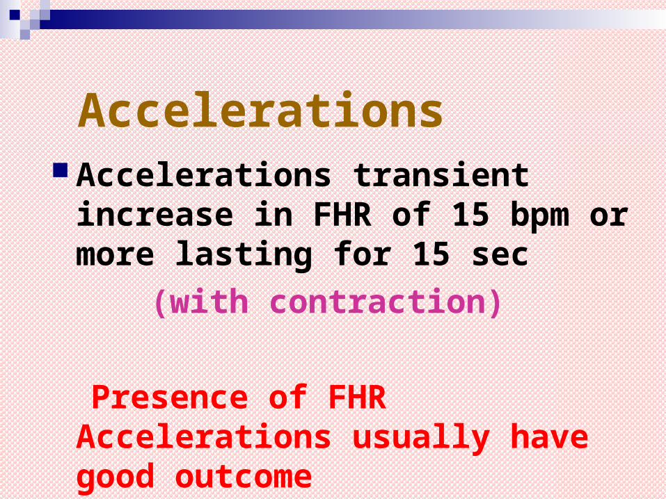
Accelerations Accelerations transient increase in
FHR of 15 bpm or more lasting for 15 sec
(with contraction)
Presence of FHR Accelerations usually have good outcome

Decelerations: transient slowing of FHR below the
baseline level of more than 15 bpm and lasting for 15 sec. Or more.
(with contraction)
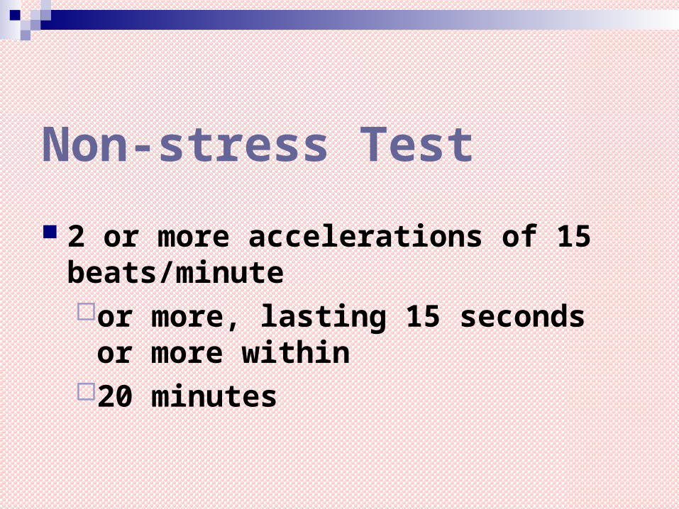
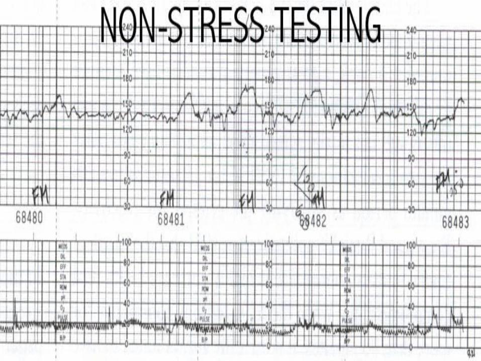
Non-stress Test
2 or more accelerations of 15 beats/minuteor more, lasting 15 seconds or more
within 20 minutes
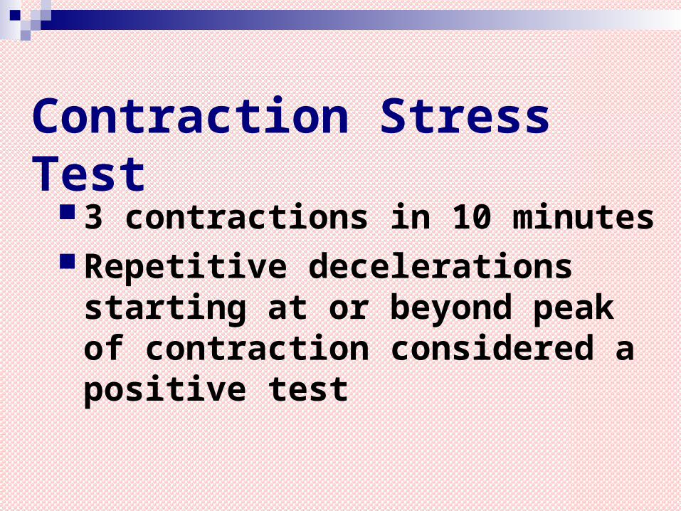
Contraction Stress Test 3 contractions in 10 minutes Repetitive decelerations starting at
or beyond peak of contraction considered a positive test

Accelerations

Early Decelerations
Head compression Begins on the onset of contraction
and returns to baseline as the contraction ends.
Should not be disregarded if they appear early in labor or Antenatal.


EARLY DECELERATION


Late Decelerations. Uniform periodic slowing of FHR with
the on set of the contractions often with slow return to the baseline.
Repetitive late decelerations increases risk of Umbilical artery acidosis and Apgar score of less than 7 at 5 mins and Increased risk of CP.

Late Decelerations• Due to acute and chronic uteroplacental
insufficiency Are precipitated by hypoxemia Associated with respiratory and
metabolic acidosis Common in patients with PIH, DM, IUGR
or other form of placental insufficiency.


Late Deceleration

Late Deceleration

Variable Decelerations Fetal heart rate deceleration is
variable in shape, onset, non-repetitive
Caused by compression of the umbilical cord
Often associated with oligohydramnios with or without rupture of membranes
Acidosis if prolonged and recurrent

Variable Decelerations

Variable Decelerations

Response to Variable Decelerations Change maternal position to right or
left side, or Trendelenburg Elevate presenting part (lift off cord) 100% oxygen Stop oxytocin Persistent Bradycardia: Deliver forceps
(if delivery imminent) or C-section (if delivery is not imminent)

Decelerations

Drop in FHR of 30 bpm or more lasting
for at least 2 mins Is pathological when it crosses
2 contractions in 3 mins Results in reduced of O2 transfer to
placenta Associated with poor neonatal outcome
Prolonged Deceleration

Prolonged DecelerationsCAUSES Cord prolapse
Maternal hypertension/hypotension
Uterine hypertonia
Epidural/spinal or pudendal anesthesia

Prolonged Deceleration

Sinusoidal pattern Baseline abnormality Smooth sine wave Amplitude 5-15beat/min(above but not
below the baseline). Frequency 2-5 cycle/min Little beat to beat variability Acceleration is absent

Associated with: hypoxia sever fetal anemia:(fetomaternal hemorrhage)
chorioamnionitis fetal sepsis administration of narcotic analgesia Cord compression hypovolemia Anaemia
Sinusoidal pattern


Sinusoidal pattern

Relationship between FHR and PH
No deceleration early deceleration pH >7.29 mild variable deceleration Severe variable late deceleration pH<7.15
No acceleration low pH
poor perinatal outcome

Biophysical Profile Fetal breathing : 30 seconds of sustained fetal breathing
in 30 minutes Fetal movement : 3 or more gross body movements in 30
minutes Fetal tone : one episode of limb motion from flexion
to extension to flexion Amniotic fluid : pocket of fluid measuring at least 2cm