Embed Size (px)
Citation preview

Wisconsin Emergency Wisconsin Emergency Response Guide forResponse Guide forClinical LaboratoriesClinical Laboratories
Developed: 2005 Updated: 2007, 2009
Wisconsin State Laboratory of Hygiene

BIOTERRORISM RESPONSE GUIDE FOR CLINICAL LABORATORIES
This Page Last Updated: August 22, 2005
CONTACTS Wisconsin State Laboratory of Hygiene 465 Henry Mall Madison, WI 53706
Emergency Response 24/7 Messaging Center: 608-263-3280 [You will receive a return call from WSLH staff within 10 minutes.] Please do not provide this number to patients or media. Wisconsin Division of Public Health 1 W. Wilson Street Madison, WI 53703 Emergency Response 24/7 Messaging Center: 608-258-0099 Please do not provide this number to patients or media. Your Local Health Department FBI Milwaukee Suite 600 330 East Kilbourn Avenue Milwaukee, Wisconsin 53202-6627 milwaukee.fbi.gov Available 24/7 (414) 276-4684 Your Local Law Enforcement Your Local Fire Department Poison Center Children’s Hospital of Wisconsin Poison Center, Milwaukee Emergency Phone: 1-800-222-1222

BIOTERRORISM RESPONSE GUIDE FOR CLINICAL LABORATORIES
This Page Last Updated: August 22, 2005
The following checklist represents recommendations for response to a bioterrorism event or the isolation of a potential priority agent of bioterrorism . Your laboratory may wish to modify or supplement this list.
When a bioterrorism event or isolation of a priority agent of bioterrorism is suspected or confirmed:
Notify internal staff per your laboratory’s procedures, e.g., infection control, administration, medical director, laboratory staff, safety. ___________________________________________________________ ___________________________________________________________
Notify the WSLH at 608-263-3280. Notify your Local Health Department (LHD) and/or the State
Division of Public Health (DPH). LHD Phone: _________________________ DPH Phone: 608-267-9003 (weekdays) / 608-258-0099 (after-hours)
Check the Health Alert Network (HAN) for information and updates at www.han.wisc.edu.
Implement your laboratory’s procedures for handling the suspect agent, e.g., potential staff exposures.
Locate/acquire shipping containers and forms for sample transport. Institute chain of custody on all samples related to the sample or
event, if appropriate. Arrange sample transport directly to the Wisconsin State
Laboratory of Hygiene (or other laboratory, if so directed by the Wisconsin State Laboratory of Hygiene staff).
Implement your institution’s communications/media policies If you expect additional samples related to this event, confirm your
laboratory’s readiness: Safety procedures and capabilities Specimen collection needs and resources Testing capabilities Potential needs and resources for reagents and materials Potential staffing needs and resources Sample transport supplies and procedures
After the event, assess the event response; identify areas that need improvement.

WISCONSIN EMERGENCY RESPONSE GUIDE FOR CLINICAL LABORATORIES
This Page Last Updated: February, 2009
Table of Contents
Section Content Version Date
Emergency Response Contact List 8-22-2005 Response Checklist 8-22-2005 Table of Contents 12--2009
I Background Information • Introduction • Using This Guide • Bioterrorism Events • Chemical Terrorism Events • Radiation Emergency Events • Laboratory Response Network (LRN) for
Bioterrorism • Laboratory Response Network (LRN) for
Chemical Terrorism • Wisconsin Clinical Laboratory Network
(WCLN)
12--2009
II Communications • Emergency Communication Plan for
Wisconsin Laboratories • Media/Press Relations • Result Reporting by the WSLH
8-22-2005
III Safety • Laboratory Safety • Summary of Biosafety Levels for Infectious
Agents • Biosafety Levels for Bioterrorism Agents • Bioterrorism Agent Clinical Summary • Risk Assessment • Laboratory Security • References
12--2009
WISCONSIN EMERGENCY RESPONSE GUIDE FOR CLINICAL LABORATORIES
This Page Last Updated: December, 2009
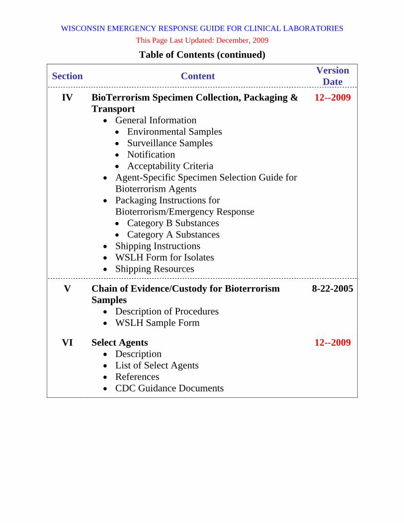
Table of Contents (continued)
Section Content Version Date
IV BioTerrorism Specimen Collection, Packaging & Transport
• General Information • Environmental Samples • Surveillance Samples • Notification • Acceptability Criteria
• Agent-Specific Specimen Selection Guide for Bioterrorism Agents
• Packaging Instructions for Bioterrorism/Emergency Response • Category B Substances • Category A Substances
• Shipping Instructions • WSLH Form for Isolates • Shipping Resources
12--2009
V Chain of Evidence/Custody for Bioterrorism Samples
• Description of Procedures • WSLH Sample Form
8-22-2005
VI Select Agents • Description • List of Select Agents • References • CDC Guidance Documents
12--2009

WISCONSIN EMERGENCY RESPONSE GUIDE FOR CLINICAL LABORATORIES
This Page Last Updated: December, 2009
Table of Contents (continued)
Section Content Version Date
VII Agent-Specific Laboratory Protocols • Bacillus anthracis • Botulinum toxin • Brucella species • Burkholderia mallei & pseudomallei • Coxiella burnetii • Francisella tularensis • Smallpox (Variola virus) • Staphylococcal Enterotoxin B • Yersinia pestis
8-22-2005 12--2009
8-22-2005 12--2009 12--2009
8-22-2005 8-22-2005 8-22-2005 12--2009
VIII Chemical Terrorism Specimen Collection, Packaging & Transport
• Collection & Labeling • Packaging & Documentation • Shipping • Chain of Custody for Chemical Terrorism
Samples • Chemical Terrorism Response Supplements
1-31-2007
8-22-2005
IX Radiological Emergencies 12--2009
X Shipping Repositories • Description • List of Sites and Contacts
8-22-2005
XI Wisconsin Health Alert Network (HAN) • Description • How to Register
12--2009
XII Reportable Diseases in Wisconsin 12--2009
XIII Resources • Technical Resources • Training Resources
12--2009

WISCONSIN EMERGENCY RESPONSE GUIDE FOR CLINICAL LABORATORIES
Section I - 1
This Page Last Updated: December, 2009
Introduction for Wisconsin Laboratories This document is intended to provide guidance to enable laboratories in Wisconsin to respond effectively to a bioterrorism event, chemical terrorism event, or other event of public health importance. An effective response requires that Wisconsin laboratories respond in a coordinated fashion. The core of the original guide was a CDC document that was developed by the Bioterrorism Preparedness and Response Program of the National Center for Infectious Diseases and the Laboratory Practice Training Branch, Division of Laboratory Systems, Public Health Practice Program Office, at the Centers for Disease Control and Prevention (CDC). The Wisconsin State Laboratory of Hygiene (WSLH) has updated and appended the document to provide current and Wisconsin-specific information for Wisconsin laboratories. Updates to this document will be provided as necessary. The clinical laboratory is likely to receive the first clinical specimens and isolate the first organisms from an unannounced bioterrorism event. It is therefore critical that clinical laboratories have the capability to rule out suspect isolates as bioterrorism priority agents or refer suspect isolates to appropriate laboratories for identification. This guide contains information, protocols and other resources to aid the clinical laboratory in the processes to rule out critical agents and refer the sample if critical agents cannot be ruled out. The American Society for Microbiology (ASM) is leading the development of sentinel laboratory protocols. This guide includes agent-specific protocols and other information for the CDC priority agents of bioterrorism, i.e., Bacillus anthracis, Yersinia pestis, Francisella tularensis, and Brucella species. Sentinel laboratory procedures are also available at the ASM website at http://www.asm.org/index.php?option=com_content&view=article&id=6342&Itemid=639. .
WISCONSIN EMERGENCY RESPONSE GUIDE FOR CLINICAL LABORATORIES
Section I - 2
This Page Last Updated: December, 2009
Using this Guide This guide is not intended to replace the standard operating procedures manual that your laboratory uses for testing and identification. Your laboratory must meet regulatory requirements to incorporate these protocols into your laboratory’s operations and processes. This guide is intended to be used as a reference by laboratory staff trained in bioterrorism response. We recommend that it be stored near the laboratory workbench, readily available to appropriate laboratory staff. Although this guide deals primarily with the response to bioterrorism events, it also contains information and resources for the response to chemical terrorism events and radiological emergencies. Much of the information provided for bioterrorism and chemical terrorism events can also be applied to other public health emergencies and outbreaks.

WISCONSIN EMERGENCY RESPONSE GUIDE FOR CLINICAL LABORATORIES
Section I - 3
This Page Last Updated: December, 2009
Bioterrorism Events Bioterrorism is defined as the “intentional use of microorganisms, or toxins derived from living organisms, to produce death or disease in humans, animals, or plants”. Bioterrorism events may occur as announced (overt) or as unannounced (covert) events. In an announced event, the notification that an agent had been released would prompt an immediate response by law enforcement and emergency response personnel. Public health officials would help evaluate the risk and control the spread of disease. Samples might be collected and would be sent directly to public health laboratories for testing. In an unannounced event, the release of the agent may go unnoticed for days or weeks, when an unusual isolate in a clinical laboratory or a cluster of illnesses would likely be the signal that an event had occurred. Clinical laboratories or hospital emergency departments may be the first to note unusual patterns of illness or unusual isolates. In an unannounced event, testing would likely be performed in clinical laboratories, with referral of suspect organisms to the public health laboratory for identification. Efforts to control the spread of disease would be delayed until the isolates or cluster of illnesses was noted and public health authorities notified.
Recognition of Overt vs. Covert Events
Responder Community
Public HealthCommunity
Health CareCommunity
Overt Event
Covert Event

WISCONSIN EMERGENCY RESPONSE GUIDE FOR CLINICAL LABORATORIES
Section I - 4
This Page Last Updated: December, 2009
Chemical Terrorism Events Chemical terrorism is defined as the intentional use of chemicals to scare, injure, or kill people. The dispersion methods used in a chemical terrorism event may be simple or complex (e.g., opening or spraying a container vs. exploding a bomb with chemicals inside). The chemicals used can contaminate people, air, water, food, or surfaces. In addition to chemical warfare agents, industrial chemicals and waste could also be used in a chemical terrorism event. While the time interval between release and development of symptoms in a biological event may take hours to weeks, the time interval following a chemical event would be much shorter, usually minutes to hours. While most biological agents are odorless and colorless, chemicals are likely to leave unusually colored residue or odors, and can be expected to affect plants, insects and animals in addition to people. Although hospital emergency departments would likely treat the victims of a chemical terrorism event, diagnostic testing would be performed by state or federal laboratories. The role of clinical laboratories in a chemical terrorism event would be the collection and referral of clinical specimens (i.e., blood, urine) for testing. The WSLH has developed the capability to test for selected chemical agents and provides consultation and support for specimen collection and transport. Chemical terrorism response protocols in this manual will be updated as necessary. In a chemical terrorism event, laboratories should contact the WSLH through the 24/7 emergency messaging service at 608-263-3280 for current guidance in specimen collection and transport.

WISCONSIN EMERGENCY RESPONSE GUIDE FOR CLINICAL LABORATORIES
Section I - 5
This Page Last Updated: December, 2009
Radiation Emergency Events Radiation emergency events may result from either accidental or deliberate releases of radioactive material. Accidental releases may be related to nuclear reactors, medical radiation therapy, industrial irradiators, lost or stolen radioactive sources, or transportation accidents. Deliberate radiation emergency events may result from the intentional release of radiological material, as a “dirty bomb”, detonation of a low-yield nuclear weapon, or an attack or sabotage of a nuclear facility. Additional information related to laboratory testing in response to a radiological event are being further developed by the Centers for Disease Control and Prevention and their partners. Additional information is available at http://emergency.cdc.gov/radiation/links.asp The instructions for collection, packaging, and transport of clinical specimens for chemical terrorism should be followed during a radiological event, according to the CDC. The WSLH recommends that current instructions should be obtained from 608-263-3280 before specimens are collected or transported in response to a radiation emergency. CDC would play a key role in protecting the public health during and after an emergency involving radiation or radioactive materials. Information to help prepare for a radiation emergency is available at http://www.bt.cdc.gov/radiation/ or http://emergency.cdc.gov/radiation/ Additional information on this topic will be included in this document as it becomes available.

WISCONSIN EMERGENCY RESPONSE GUIDE FOR CLINICAL LABORATORIES
Section I - 6
This Page Last Updated: December, 2009
The Laboratory Response Network (LRN) The Laboratory Response Network (LRN) is a collaborative, voluntary system of laboratories, established in 1999 by the Centers for Disease Control and Prevention (CDC) in partnership with the Association of Public Health Laboratories (APHL), federal agencies including the Federal Bureau of Investigation (FBI) and the Department of Defense (DOD), and state public health laboratories through the Bioterrorism Preparedness Initiative. The mission of the LRN is to “maintain an integrated national and international network of laboratories that are fully equipped to respond quickly to acts of chemical or biological terrorism, emerging infectious diseases, and other public health threats and emergencies.” The LRN is a unique asset in the nation's growing preparedness for biological and chemical terrorism. The LRN is comprised of both public and private laboratories, with a central role for state public health laboratories. Veterinary, agricultural, food, and water testing laboratories have been included in the LRN to enable a broad-based response to public health emergencies, including the capability for veterinary, food or water testing. In addition to the collaboration between laboratories, the LRN provides a linkage between local, state, and federal agencies. The linking of state and local public health laboratories, veterinary, agriculture, military, and water- and food-testing laboratories is unprecedented. A cornerstone of the LRN is timely and accurate testing and reporting by member laboratories that use consensus protocols and CDC-approved methods. LRN reference laboratories use secure CDC-approved protocols and reagents.

WISCONSIN EMERGENCY RESPONSE GUIDE FOR CLINICAL LABORATORIES
Section I - 7
This Page Last Updated: December, 2009
The Laboratory Response Network (LRN) for Biological Terrorism The current concept of the national LRN for Biological Terrorism consists of a three-tiered pyramid (replacing the original four-tiered pyramid (Level A, B, C, and D Laboratories), as described below: ∆ The base of the pyramid is comprised of the “Sentinel” Laboratories,
previously referred to as “Level A” laboratories. The role of Sentinel Laboratories is to recognize the agents of bioterrorism, perform testing to rule out the agents of bioterrorism, and refer suspect isolates to LRN Reference Laboratories. Sentinel Laboratories serve a critical role in the LRN, as they will likely be involved in early detection of an unannounced bioterrorism event. Nationally, Sentinel Laboratories are defined as laboratories that perform microbiology and operate at BioSafety Level 2 (BSL-2), but would adopt BioSafety Level 3 (BSL-3) practices when working with a suspected bioterrorism agent. Any clinical laboratories that perform bacteriology and are CLIA certified may be Sentinel Laboratories. Formal registration is not required to participate in the LRN as a Sentinel Laboratory.
∆ The second and third tiers of the pyramid have been consolidated under the term “Reference Laboratories”, previously referred to as “Level B” and “Level C” laboratories, respectively. The role of LRN Reference Laboratories is to provide confirmatory testing for the agents of bioterrorism. LRN Reference Laboratories are usually public health laboratories that have BSL-3 capabilities, can confirm the identification of bioterrorism agents using conventional and molecular methods, and have rapid methods capability, e.g., time-resolved fluorescence and molecular amplification. LRN Reference Laboratories also evaluate new tests and reagents as formal members of the LRN.
∆ The peak of the pyramid is comprised of National Laboratories, i.e., the CDC and the U.S. Army Medical Research Institute for Infectious Diseases (USAMRIID), previously termed “Level D” laboratories. The role of the National Laboratories is to provide definitive characterization of the agents of bioterrorism. These National Laboratories have highly specialized capabilities for isolation and identification and have Biosafety Level 4 (BSL-4) maximum containment facilities that are capable of handling highly infectious organisms such as smallpox and hemorrhagic fever viruses.

WISCONSIN EMERGENCY RESPONSE GUIDE FOR CLINICAL LABORATORIES
Section I - 8
This Page Last Updated: December, 2009
The Laboratory Response Network (LRN) for Bioterrorism (continued)
Original Concept of Current Concept of the Laboratory the Laboratory Response Network Response Network (LRN) (LRN)
Level C
Level B
Level A
Level D

WISCONSIN EMERGENCY RESPONSE GUIDE FOR CLINICAL LABORATORIES
Section I - 9
This Page Last Updated: December, 2009
Laboratory Network for Chemical Terrorism (The following is reprinted from information developed by the CDC.
Sixty-two state, territorial and metropolitan public health laboratories are members of the chemical component of the Laboratory Response Network (LRN). A designation of Level 1, 2, or 3 identifies laboratory capabilities and defines member network participation. (Please note that the level designations were changed in early 2005 so that laboratories previously designated “Level 1” are now “Level 3,” and laboratories previously designated “Level 3” are now “Level 1.”).
Level 3 Laboratories Although every network member participates in Level 3 activities, only 15 laboratories are designated as Level 3 laboratories. These 15 laboratories work with hospitals and other first responders within their jurisdiction to maintain competency in clinical specimen collection, storage, and shipment.
Level 2 Laboratories Thirty-seven laboratories are designated as Level 2 laboratories. Chemists in these laboratories are trained to detect exposure to a number of toxic chemical agents. Analysis of cyanide, nerve agents, and toxic metals in human samples are examples of Level 2 activities.
Level 1 Laboratories
Ten laboratories currently participate in Level 1 activities. These laboratories, which serve as surge-capacity laboratories for CDC, are able to detect not only the toxic chemical agents that Level 2 laboratories can detect, but also can detect exposure to an expanded number of chemicals, including mustard agents, nerve agents, and other toxic industrial chemicals.
For more information, visit http://emergency.cdc.gov/lrn/chemical.asp or http://emergency.cdc.gov/chemical/lab.asp.

WISCONSIN EMERGENCY RESPONSE GUIDE FOR CLINICAL LABORATORIES
Section I - 10
This Page Last Updated: December, 2009
The Wisconsin Clinical Laboratory Network (WCLN)
The Wisconsin Clinical Laboratory Network (WCLN) is a subset of the National Laboratory Response Network. The WCLN was previously known as the Wisconsin Laboratory Response Network (WLRN). The name was changed and the purpose was defined in writing in 2008. The Wisconsin State Laboratory of Hygiene (WSLH) is an LRN Reference Laboratory for Biological Terrorism and is the coordinating laboratory of the WCLN. The Milwaukee Health Department Bureau of Laboratories (MHDL) and Marshfield Clinic Research Foundation Laboratory (MCRF) serve as surge capacity LRN Reference Laboratories for bioterrorism. Additional LRN Reference Laboratories for bioterrorism may be incorporated into the WCLN as the network evolves and/or additional laboratories are identified and approved by the CDC. The WSLH is the only CDC-qualified laboratory for Chemical Terrorism Response in Wisconsin and currently serves as a Level One laboratory for chemical terrorism response. Sentinel Laboratories in Wisconsin are comprised of hospital-based and large clinical laboratories that perform microbiology. Although the description of LRN Sentinel Laboratories refers to biosafety level 2 (BSL-2) and the use of a biological safety cabinet (BSC), this criterion has not been applied for inclusion in the WCLN. In an unannounced bioterrorism event, the role of the clinical laboratory would be to report and refer suspect isolates to Wisconsin’s LRN Reference Laboratories. Clinical laboratories in Wisconsin should contact the WSLH for guidance on where and how to ship the specimen/isolate. The laboratory should contact the WSLH for guidance whenever a bioterrorism agent or agent requiring a higher biosafety level is suspected during the testing or identification process for bacteria or viruses. In an announced bioterrorism event, the role of the clinical laboratory would be to provide supportive testing for patients. Environmental specimens should be transported directly to the WSLH by the local health department, law enforcement, or other first responders. To avoid possible contamination of the laboratory, clinical laboratories in Wisconsin should not accept environmental specimens.

WISCONSIN EMERGENCY RESPONSE GUIDE FOR CLINICAL LABORATORIES
Section I - 11
This Page Last Updated: December, 2009
The Wisconsin Clinical Laboratory Network (WCLN) (continued)
In a chemical terrorism event, the role of the clinical laboratory would be the collection and packaging of blood and urine specimens for transport to the WSLH. Clinical laboratories should contact the WSLH for consultation and additional instructions for specimen collection and transport.

BIOTERRORISM RESPONSE GUIDE FOR CLINICAL LABORATORIES
Section II - 1
This Page Last Updated: August 22, 2005
Emergency Communication Plan for Wisconsin Clinical Laboratories No guide can cover all possibilities or provide all the necessary information for an emergency situation or event such as bioterrorism or chemical terrorism. Consequently, the WSLH has adopted a “just in case/just in time” approach for emergency laboratory communications in response to an emergency event or outbreak. The WSLH will provide training and information (e.g., this Guide, response exercises, etc.) to laboratories to establish and reinforce a basic foundation of knowledge about emergency response. In a bioterrorism event, chemical terrorism event, or other public health emergency, the WSLH will supplement that basic knowledge with event-specific instructions and information, updated as needed. Information relevant to laboratories will be provided in “real time” through the Laboratory Messaging System, posting on the HAN and/or Wisconsin State Laboratory of Hygiene website, and audioconferences if needed. This approach to emergency laboratory communications will enable an effective and coordinated laboratory response to an emergency.
Preparedness “Just In Case”
Response “Just in Time”

BIOTERRORISM RESPONSE GUIDE FOR CLINICAL LABORATORIES
Section II - 2
This Page Last Updated: August 22, 2005
Emergency Communication Plan for Wisconsin Clinical Laboratories (continued) WSLH Responsibilities: As a bioterrorism event, chemical terrorism event, or other public health emergency is identified, the WSLH will: • Provide guidance in the packaging and transport of samples. • Provide information on the situation and guidance to laboratories
immediately, using fax and e-mail, via the Wisconsin Laboratory Messaging System and posting on the WSLH website at http://www.slh.wisc.edu and/or the Health Alert Network (HAN) https://www.han.wisc.edu
• Provide an audioconference if necessary. • Provide updates and response recommendations to laboratories as the
situation develops. • Inform laboratories when the situation resolves. • Utilize additional communication capabilities as they are developed. • Provide state-of-the-art rapid testing and reporting or identify and facilitate
the transport of specimens to testing sites. Clinical Laboratory Responsibilities: Laboratory staff should contact the local and/or state public health department and the WSLH in the following circumstances: • Suspicion of a bioterrorism event • Suspicion of a chemical terrorism event • Suspicion of or inability to rule out agents of bioterrorism Laboratory staff are also encouraged to contact the WSLH for: • Consultation in cases of problematic isolates • Guidance in the transport of specimens/isolates related to diseases of
public health importance • Guidance in transfer or disposal of “Select Agents”. Laboratories should contact the Wisconsin Division of Public Health when they encounter suspect cases of emerging diseases of public health importance (e.g., Severe Acute Respiratory Syndrome “SARS”, possible human cases of avian influenza). To ensure a timely diagnosis of infectious and biological agents of high public health importance, specimens must be collected and transported to the WSLH on a priority basis, within 24 hours of collection.

BIOTERRORISM RESPONSE GUIDE FOR CLINICAL LABORATORIES
Section II - 3
This Page Last Updated: August 22, 2005
Emergency Communication Plan for Wisconsin Laboratories (continued) Media/Press Relations • Information regarding a terrorism event will be available at the CDC
website at http://www.cdc.gov and on the Wisconsin Health Alert Network (HAN) at https://www.han.wisc.edu
• Any release of information or responses to media or press inquiries should
follow your facility’s policies. • If the state Emergency Operations Center (EOC) is activated during an
event, all state-level information will be provided to the media at the EOC’s Joint Information Center. The information released from the EOC will be posted on the Health Alert Network by the Wisconsin Division of Public Health.

BIOTERRORISM RESPONSE GUIDE FOR CLINICAL LABORATORIES
Section II - 4
This Page Last Updated: August 22, 2005
Emergency Communication Plan for Wisconsin Laboratories (continued) Result Reporting by the Wisconsin State Laboratory of Hygiene (WSLH) • Environmental Specimens
Results of bioterrorism-related environmental specimen examination and testing are telephoned to Wisconsin Division of Public Health, who telephone the local health department and the submitter. Written reports are issued by the WSLH to the submitter, local health department, and Wisconsin Division of Public Health (WDPH). This reporting protocol will be followed for presumptive and definitive result reports.
• Referred Clinical Specimens Results of bioterrorism-related clinical specimen examination and testing are telephoned to Wisconsin Division of Public Health (WDPH) and to the submitting facility designated on the specimen submittal form. The WDPH telephones the local health department and the submitting physician. Written reports are issued by the WSLH to the submitting facility designated on the specimen submittal form, local health department, and WDPH. This reporting protocol will be followed for presumptive and definitive result reports.
• Referred Clinical Isolates
Results of bioterrorism-related isolates referred to the WSLH by other laboratories are telephoned to the Wisconsin Division of Public Health (WDPH) and to the submitting facility designated on the specimen submittal form. Written reports are issued by the WSLH to the submitting facility designated on the specimen submittal form, local health department, and WDPH. This reporting protocol will be followed for presumptive and definitive result reports.
• Presumptive Results
Positive results for some tests (e.g., PCR) are considered presumptive, pending results of definitive testing by the Centers for Disease Control and Prevention (CDC). In these cases, the results will be clearly indicated as presumptive.

WISCONSIN EMERGENCY RESPONSE GUIDE FOR CLINICAL LABORATORIES
Section III - 1
This Page Last Updated: December, 2009
Laboratory Safety • The appropriate combination of laboratory practices, equipment and
facilities are essential to reduce the risk of human exposure to infectious agents in the laboratory setting and to reduce the risk of contamination of the laboratory itself.
• Sentinel Laboratories should not under any circumstance accept
environmental samples from a possible bioterrorism event, especially powders, due to the risk of contaminating the laboratory. The local health department, law enforcement, or other first responders should contact the WSLH about transport of environmental specimens.
• If an environmental sample (e.g., powder) related to a possible bioterrorism event does arrive in the clinical laboratory, immediately isolate and contain the sample and notify the local health department and the WSLH.
• The laboratory should contact the WSLH immediately for guidance if a
bioterrorism agent or agent requiring a higher biosafety level is suspected at any time during the testing or identification process for bacteria or viruses.
• The summary of biosafety levels for infectious agents on the following
page can be used with a risk assessment to determine appropriate laboratory practices and equipment. Information about risk assessments is provided later in this section.

WISCONSIN EMERGENCY RESPONSE GUIDE FOR CLINICAL LABORATORIES
Section III - 2
This Page Last Updated: December, 2009
Summary of Recommended Biosafety Levels for Infectious Agents* * Adapted from “Biosafety in Microbiological and Biomedical Laboratories”, 5th Edition
BSL Agents Microbiology Practices
Safety Equipment (Primary Barriers)
Facilities (Secondary Barriers)
1 Not known to consistently cause disease in healthy adults
Standard Microbiological Practices*
None required Open bench top sink required
2 Associated with human disease;
Routes of transmission include percutaneous injury, ingestion, mucous membrane exposure
BSL-1 practice plus: Limited access;
Biohazard warning signs;
"Sharps" precautions; Biosafety manual
defining needed waste decontamination or medical surveillance policies
Primary barriers: Class I or II BSCs or
other physical containment devices for all manipulations of agents that cause splashes or aerosols of infectious material;
PPEs: Laboratory coats;
gloves; face protection as needed
BSL-1 plus: Autoclave available
3 Indigenous or exotic agents with potential aerosol transmission;
Disease may have serious or lethal consequences
BSL-2 practice plus: Controlled access; Decontamination of
all waste; Decontamination of
lab clothing before laundering;
Baseline serum
Primary barriers: Class I or II BCSs or
other physical containment devices for all open manipulations of agents;
PPEs: Protective lab clothing;
gloves; respiratory protection as needed
BSL-2 plus: Physical separation from
access corridors; Self-closing, double-
door access; Exhausted air not
recirculated; Negative airflow into
laboratory
4 Dangerous/exotic agents which pose high risk of life-threatening disease;
Aerosol-transmitted lab infections have occurred;
Or related agents with unknown risk of transmission
BSL-3 practices plus: Clothing change
before entering; Shower on exit; All material
decontaminated on exit from facility
Primary barriers: All procedures
conducted in Class III BSCs or Class I or II BSCs in combination with full-body, air-supplied, positive pressure personnel suit
BSL-3 plus: Separate building or
isolated zone; Dedicated supply and
exhaust, vacuum, and decontamination systems;
Other requirements
BSC=biological safety cabinet; BSL= biosafety level; PPE=personal protective equipment
Note: BSL-2 facility/laboratory with BSL-3 Practices usually indicates practices as described under “BSL-2” AND controlled access to the area when working with the agent, decontamination of all waste, all work performed in a BSC, using protective equipment, including disposable gloves, solid front gowns with cuffed sleeves, and respiratory protection when working with the agent in the BSC, but eye and face protection if splashes or sprays of hazardous material is anticipated.

WISCONSIN EMERGENCY RESPONSE GUIDE FOR CLINICAL LABORATORIES
Section III - 3
This Page Last Updated: December, 2009
Recommended Biosafety Levels for Most Commonly Encountered Agents of Bioterrorism (adapted from CDC and ASM at
www.bt.cdc.gov/documents/PPTResponse/table3bbiosafety.pdf and www.asm.org/images/pdf/BTtemplateRevised8-10-6.doc)
Biosafety Level Agent Specimen
Handling Culture
Handling Specimen Exposure/Risk Recommended Precautions
for Sentinel Laboratories
Bacillus anthracis 2 3 Blood, skin lesion exudates, CSF, pleural fluid, sputum; rarely urine & feces.
BSL2: Activities involving clinical material collection & diagnostic quantities of infectious cultures.
BSL3: Activities with high potential for aerosol or droplet production.
Brucella spp. 2 3 Blood, bone marrow, CSF, tissue, semen, occasionally urine.
BSL2: Activities limited to collection, transport & plating of clinical material.
BSL3: All activities involving manipulations of cultures.
Burkholderia mallei & pseudomallei
2 3 Blood, sputum, CSF, tissue, abscesses, and urine
BSL2: Activities limited to collection, transport & plating of clinical material.
BSL3: All activities involving manipulations of cultures.
Clostridium botulinum 2 3
Toxin may be present in food specimens, clinical material (serum, gastric & feces), & environmental samples (soil, surface water). TOXIN IS EXTREMELY POISONOUS!
BSL2: Activities with materials known or potentially containing toxin must be handled in a Class II BSC with lab coat, disposable gloves, & face shield (as needed).
BSL3: Activities with high potential for aerosol or droplet production.
Francisella tularensis 2 3
Skin lesion exudates, respiratory secretions, CSF, blood, urine, tissues from infected animals & fluids from infected arthropods.
BSL2: Activities limited to collection, transport & plating of clinical material.
BSL3: All activities involving manipulations of cultures.
Yersinia pestis 2 3 Bubo fluid, blood, sputum, CSF, feces, urine.
BSL2: Activities involving clinical material collection & diagnostic quantities of infectious cultures
BSL3: Activities with high potential for aerosol or droplet production.

WISCONSIN EMERGENCY RESPONSE GUIDE FOR CLINICAL LABORATORIES
Section III - 4
This Page Last Updated: December, 2009
Recommended Biosafety Levels for Less Commonly Encountered Agents of Bioterrorism (adapted from CDC, ASM, and BMBL at www.bt.cdc.gov/documents/PPTResponse/table3bbiosafety.pdf and www.asm.org/images/pdf/BTtemplateRevised8-10-6.doc and www.slh.wisc.edu/dotAsset/6796.pdf)
Biosafety Level Agent
Specimen Handling
Culture Handling
Specimen Exposure/Risk Recommended Precautions for Sentinel Laboratories
Alphaviruses 2 3
Blood, CSF. Tissue culture and animal inoculation studies should be performed at BSL-3 and are NOT Sentinel (Level A) laboratory procedures.
BSL-2: Activities involving clinical material collection and transport
Biosafety levels variable by agent; may require BSL3 or more.
Coxiella burnetii 2 3
Blood, tissue, body fluids, feces. Manipulation of tissues from infected animals and tissue culture should be performed at BSL-3 and are NOT Sentinel laboratory procedures.
BSL2: Activities limited to collection and transport of clinical material, including serological examinations.
BSL3: Activities involving inoculation, incubation, harvesting of eggs or cell cultures, animal necropsy, manipulation of infected tissues.
Smallpox 4 4 Lesion fluid or crusts, respiratory secretions, tissue
BSL-2: Packing and shipping. Do NOT put in cell culture. Contact public health and WSLH
Staphylococcal enterotoxin B 2 2
Toxin may be present in food specimens, clinical material (serum, gastric, urine, respiratory secretions, and feces), and isolates of S. aureus.
BSL-2: Activities involving clinical material collection and diagnostic quantities of infectious cultures
BSL3: Activities with high potential for aerosol or droplet production.
Viral Hemorrhagic Fever (VHF) 4 4 Blood, urine, respiratory, and throat
secretions, semen, and tissue BSL-2: Packing and shipping. Do NOT put in cell culture. Contact public health and WSLH

WISCONSIN EMERGENCY RESPONSE GUIDE FOR CLINICAL LABORATORIES
Section III - 5
This Page Last Updated: December, 2009
Risk Assessment
A risk assessment is a series of actions to identify hazards and to evaluate the probability that a problem will occur due to that hazard and the severity of the consequences if a problem did occur. Hazards may be physical, chemical or biological. Laboratory managers, supervisors and employees all share responsibility for the identification of hazards in the laboratory. The laboratory director in collaboration with the institution’s biosafety committee should conduct risk assessments at regular intervals, at least annually, and whenever a change occurs in the laboratory (e.g., move, renovation, new infectious agent, change in equipment, etc.).
The purpose of a risk assessment is to: • provide information to keep people safe • identify training needs • evaluate emergency plans • justify space, renovation, and equipment needs • evaluate procedural changes
Many of the elements that can comprise a risk assessment in the following list are part of the everyday operations of a laboratory: • Reviewing laboratory records:
• injury and illness reports • equipment maintenance records • employee training records • environmental monitoring records
• Inspecting the laboratory: • daily monitoring by employees • periodic “walk-throughs” • formal inspections by certifying agencies
• Reviewing published materials: • equipment manuals and product inserts • scientific journals • safety manuals and guidelines
• Observing laboratory operations: • new employees, procedures, or equipment • work-flow

WISCONSIN EMERGENCY RESPONSE GUIDE FOR CLINICAL LABORATORIES
Section III - 6
This Page Last Updated: December, 2009
Risk Assessment (continued) Performing a Risk Assessment Risk assessments should be performed as a systematic two-part process, using a checklist developed for or adapted to your laboratory. The first step in the process is to identify the hazards; the second step is to determine the degree of risk (probability of occurrence and severity of consequences) associated with each hazard. The WSLH provides a separate reference document “Laboratory Biosafety: Performing a Risk Assessment” to aid you in assessing your laboratory. This document is available at the WSLH website http://www.slh.wisc.edu/labupdates/wcln/index.dot. Step 1: Identify the Hazards Consider the following items as you assess your facility for hazards: • Physical facility
Air-flow Laboratory access Composition of ceiling, walls and floors Containment equipment Biosafety cabinets and fume hoods Aerosol-free centrifuge cups/carriers
• Personnel Experience & training Physical disabilities Immune status/Immunization records Pregnancy
• Agents worked with in the laboratory Pathogenicity/virulence Mode of transmission and transmissibility Available information, especially for new agents
• Types of procedures performed Aerosol generating Use of syringes and needles Temperature extremes Use of sterile techniques Methods that amplify organisms
Some examples of hazards that may be identified are: 1) poor work-flow; 2) poor techniques or practices when working in the biosafety cabinet; 3) failure to check condition of containment equipment; 4) spills and wet spots; 5) improper disposal of waste; and 6) poor placement of biosafety cabinet(s).

WISCONSIN EMERGENCY RESPONSE GUIDE FOR CLINICAL LABORATORIES
Section III - 7
This Page Last Updated: December, 2009
Performing a Risk Assessment (continued) Step 2: Determine the Degree of Risk for each Hazard Risk is influenced by a number of factors, including: • Pathogenicity and virulence of the agent, including the incidence and
severity of disease • Mode of transmission of the agent • Use of aerosol-generating procedures (most laboratory infections result
from aerosol transmission, so the potential for aerosols should be minimized)
• Environmental stability of the agent, including its ability to survive under varying conditions
• Concentration of the pathogen, including the sample type, the work to be performed, and the volume of sample being handled
• Form of the agent (liquid suspension, colonies on solid medium, dried) • Origin of the agent or sample, including the geographic or species origin • Availability of effective prophylaxis for the agent • Skill and experience of personnel, including housekeeping and other at-
risk personnel, and assessment of the need for additional training. In addition, you will want to take into account: • Applicable regulations • Safety features of your facility (air handling system, safety equipment
available, limitations for decontaminating waste) • Security/accessibility of your facility It may be useful to use a matrix format to evaluate the risk associated with each potential hazard.
Example of a Hazard/Risk Matrix Potential Hazard:
Severity of Consequences Probability of Occurrence Low Medium High
High Medium
Low
WISCONSIN EMERGENCY RESPONSE GUIDE FOR CLINICAL LABORATORIES
Section III - 8
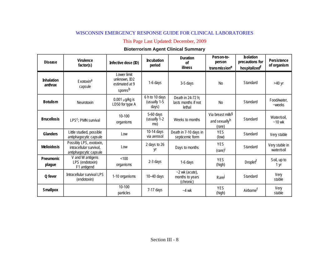
This Page Last Updated: December, 2009 Bioterrorism Agent Clinical Summary
Disease Virulence factor(s) Infective dose (ID) Incubation
period Duration
of illness
Person-to- person
transmissione
Isolation precautions for
hospitalizedf
Persistence of organism
Inhalation anthrax
Exotoxina capsule
Lower limit unknown, ID2 estimated at 9
sporesb
1-6 days 3-5 days No Standard >40 yr
Botulism Neurotoxin 0.001 µg/kg is
LD50 for type A 6 h to 10 days (usually 1-5
days)
Death in 24-72 h; lasts months if not
lethal No Standard Food/water,
~weeks
Brucellosis LPSc; PMN survival 10-100
organisms
5-60 days (usually 1-2
mo) Weeks to months
Via breast milkg and sexuallyh
(rare)
Standard Water/soil, ~10 wk
Glanders Little studied, possible antiphagocytic capsule
Low 10-14 days via aerosol
Death in 7-10 days in septicemic form
YES (low)
Standard Very stable
Melioidosis Possibly LPS, exotoxin,
intracellular survival, antiphagocytic capsule
Low 2 days to 26 yr Days to months
YES (rare)i
Standard Very stable in water/soil
Pneumonic plague
V and W antigens LPS (endotoxin)
F1 antigend
<100 organisms
2-3 days 1-6 days YES (high) Dropletf Soil, up to
1 yr
Q fever Intracellular survival LPS (endotoxin)
1-10 organisms 10–40 days ~2 wk (acute),
months to years (chronic)
Rarej Standard Very stable
Smallpox 10-100
particles 7-17 days ~4 wk YES
(high) Airbornef Very stable

WISCONSIN EMERGENCY RESPONSE GUIDE FOR CLINICAL LABORATORIES
Section III - 9
This Page Last Updated: December, 2009 Bioterrorism Agent Clinical Summary (continued)
Disease Virulence factor(s) Infective dose (ID) Incubation
period Duration
of illness
Person-to- person transmissione
Isolation precautions for hospitalizedf
Persistence of organism
Staphylococcal enterotoxin B Superantigen
0.0004 µg/kg incapacitation; LD50 is
0.02 µg/kg
3-12 h after inhalation Hours No Standard Resistant to
freezing
Tularemia Intracellular survival 10-50 organisms 2-10 days >2 wk Single case report
during autopsy Standard Moist soil, ~months
VHF Varies with virus 1-10 particles 4-21 days 7-16 days YES (moderate) Airborne and contactf Unstable aB. anthracis exotoxin or exotoxins consist of three components: the edema factor and lethal factor exert their effect within cells by interacting with a common transport protein designated “protective antigen” (so named because, when modified, it contributes to vaccine efficacy). Expression of toxic factors is mediated by one plasmid, and that of the capsule (D-glutamic acid polypeptide) is mediated by a second plasmid. Strains repeatedly subcultured at 42°C become avirulent as a result of losing virulence-determining plasmids, which is thought to be the basis for Pasteur’s attenuated anthrax vaccine used at Pouilly-le-Fort in 1881.
bThe estimate that nine inhaled spores would infect 2% of the exposed human population is based on data from Science 266:1202-1208, 1994. The dose needed to infect 50% of the exposed human population may be 8,000 or higher.
c The major virulence factor for brucellosis appears to be an endotoxic lipopolysaccharide (LPS) among smooth strains. Pathogenicity is related to an LPS containing poly Nformyl perosamine O chain, CuZn superoxide dismutase, erythrulose phosphate dehydrogenase, intracellular survival stress-induced proteins, and adenine and guanine monophosphate inhibitors of phagocyte functions.
d The V and W antigens and the F1 capsular antigens are only expressed at 7°C and not at the lower temperature of the flea (20 to 25°C). e Periods of communicability are as follows: for inhalation anthrax and botulism, none; no evidence of person-to-person transmission; pneumonic plague, 72 h following initiation of appropriate antimicrobial therapy or until sputum culture is negative; smallpox, approximately 3 weeks; usually corresponds with the initial appearance of skin lesions to their final disappearance and is most infectious during the first week of rash via inhalation of virus released from oropharyngeal lesion secretions of the index case; VHF, varies with virus, but at minimum, all for the duration of illness, and for Ebola/Marburg transmission through semen may occur up to 7 weeks after clinical recovery.
f Guidelines for isolation precautions in hospitals can be found in Infect. Control Hosp. Epidemiol. 17:5380, 1996, in addition to the standard precautions that apply to all patients.
g Published reports of possible transmission of brucellosis via human breast milk may be found in Int. J. Infect. Dis. 4:5556, 2000; Ann. Trop. Paediatr. 10:305307, 1990; J. Infect. 26:346348, 1993; and Trop. Geogr .Med. 40:151152, 1988.
h Published reports of possible sexual transmission of brucellosis can be found in Lancet i:773, 1983; Aten Primaria 8:165166, 1991; Lancet 337:848849, 1991; Lancet 347:1763, 1996; Lancet 337:1415, 1991; Infection 11:313314, 1983; and Lancet 348:615, 1996.
i See Lancet 337:12901291, 1991. j Published reports of possible sexual transmission of Q fever can be found in Clin. Infect. Dis. 22:10871088, 1996; and Clin. Infect. Dis. 33:399402, 2001.
WISCONSIN EMERGENCY RESPONSE GUIDE FOR CLINICAL LABORATORIES
Section III - 10
This Page Last Updated: December, 2009
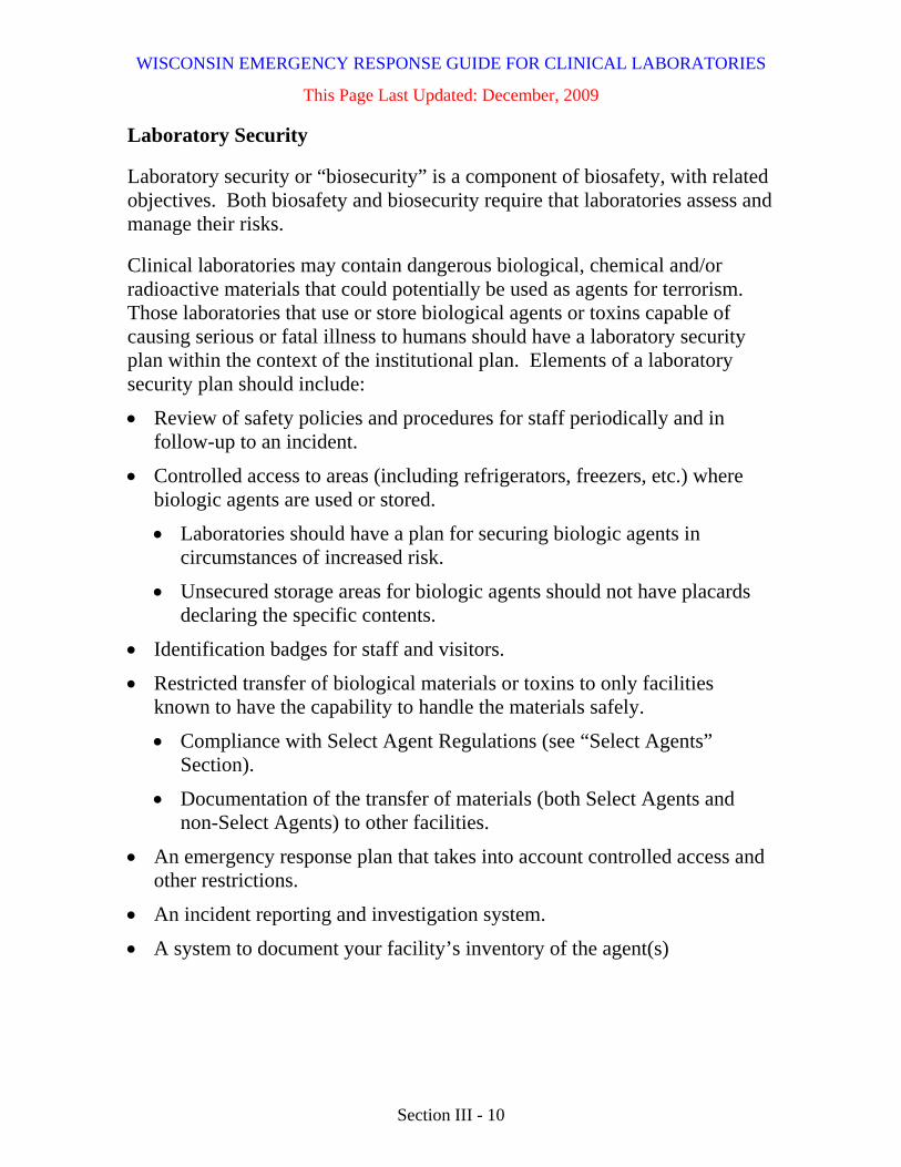
Laboratory Security
Laboratory security or “biosecurity” is a component of biosafety, with related objectives. Both biosafety and biosecurity require that laboratories assess and manage their risks.
Clinical laboratories may contain dangerous biological, chemical and/or radioactive materials that could potentially be used as agents for terrorism. Those laboratories that use or store biological agents or toxins capable of causing serious or fatal illness to humans should have a laboratory security plan within the context of the institutional plan. Elements of a laboratory security plan should include:
• Review of safety policies and procedures for staff periodically and in follow-up to an incident.
• Controlled access to areas (including refrigerators, freezers, etc.) where biologic agents are used or stored.
• Laboratories should have a plan for securing biologic agents in circumstances of increased risk.
• Unsecured storage areas for biologic agents should not have placards declaring the specific contents.
• Identification badges for staff and visitors.
• Restricted transfer of biological materials or toxins to only facilities known to have the capability to handle the materials safely.
• Compliance with Select Agent Regulations (see “Select Agents” Section).
• Documentation of the transfer of materials (both Select Agents and non-Select Agents) to other facilities.
• An emergency response plan that takes into account controlled access and other restrictions.
• An incident reporting and investigation system.
• A system to document your facility’s inventory of the agent(s)

WISCONSIN EMERGENCY RESPONSE GUIDE FOR CLINICAL LABORATORIES
Section III - 11
This Page Last Updated: December, 2009
References • Biosafety in Microbiological and Biomedical Laboratories, 5th Edition.
U.S. Department of Health and Human Services, Centers for Disease Control and Prevention, and National Institutes of Health. 2007. http://www.cdc.gov/od/ohs/biosfty/bmbl5/bmbl5toc.htm
• Primary Containment for Biohazards: Selection, Installation and Use
of Biological Safety Cabinets, 3rd Edition. U.S. Department of Health and Human Services Public Health Service, Centers for Disease Control and Prevention, and National Institutes of Health. September 2007. http://www.cdc.gov/OD/OHS/biosfty/primary_containment_for_biohazards.pdf
• Laboratory Safety, Management, and Diagnosis of Biological Agents
Associated with Bioterrorism, Cumitech 33. ASM Press. May be purchased at http://estore.asm.org/viewItemDetails.asp?ItemID=381 [previously included in this binder]
• Laboratory Biosafety: Performing a Risk Assessment. WSLH document available at the Wisconsin State Laboratory of Hygiene (WSLH) website http://www.slh.wisc.edu/labupdates/wcln/index.dot.
• Wisconsin Clinical Laboratory Network (WCLN) Additional WCLN materials are available at the Wisconsin State Laboratory of Hygiene (WSLH) website http://www.slh.wisc.edu/labupdates/wcln/index.dot

WISCONSIN EMERGENCY RESPONSE GUIDE FOR CLINICAL LABORATORIES
Section IV - 1
This Page Last Updated: December, 2009
Specimen Collection of Bioterrorism-Related Samples • General Information
• Environmental Samples
• Sentinel Laboratories should not accept environmental samples from a possible bioterrorism event, especially powders, due to the risk of accidental contamination of the laboratory. The local health department, law enforcement, or other first responders should contact the WSLH about transport of environmental specimens. • If an environmental sample (e.g., powder) related to a possible
bioterrorism event does arrive in the clinical laboratory, immediately isolate and contain the sample and notify the local health department and the WSLH.
• Acceptability Criteria • Contact your local public health department or the Wisconsin
Division of Public Health for assistance in assessing the situation.
• Surveillance Samples • Culturing of surveillance samples in your institution, including nasal
swabs for anthrax, may be performed after consultation with Wisconsin Division of Public Health, hospital infection control personnel and hospital or laboratory management. Under no circumstances should the results of a nasal swab culture collected for surveillance purposes for anthrax be used for guiding care of a patient.
• Notification • Notify the WSLH via the 24/7 emergency answering service (608-
263-3280) prior to submitting specimens for bioterrorism-related testing so that the laboratory can make the necessary arrangements.
• Chain of Custody
• If bioterrorism is suspected, chain of custody procedures should be instituted at specimen collection or as soon as bioterrorism is suspected. Chain of custody procedures are described in Section V of this guide.

WISCONSIN EMERGENCY RESPONSE GUIDE FOR CLINICAL LABORATORIES
Section IV - 2
This Page Last Updated: December, 2009
Instructions for Specimen Collection and Handling
Refer to the following table excerpted from “Sentinel Laboratory Guidelines for Suspected Agents of Bioterrorism” and the agent-specific sections for information and instructions on collection, handling and transport of specimens collected for the following bioterrorism agents:
• B. anthracis
• Botulinum toxin
• Brucella spp.
• Burkholderia mallei/pseudomallei
• Coxiella burnetii
• Francisella tularensis
• Staphylococcal enterotoxin B
• Yersinia pestis
WISCONSIN EMERGENCY RESPONSE GUIDE FOR CLINICAL LABORATORIES
Section IV - 3
This Page Last Updated: December, 2009
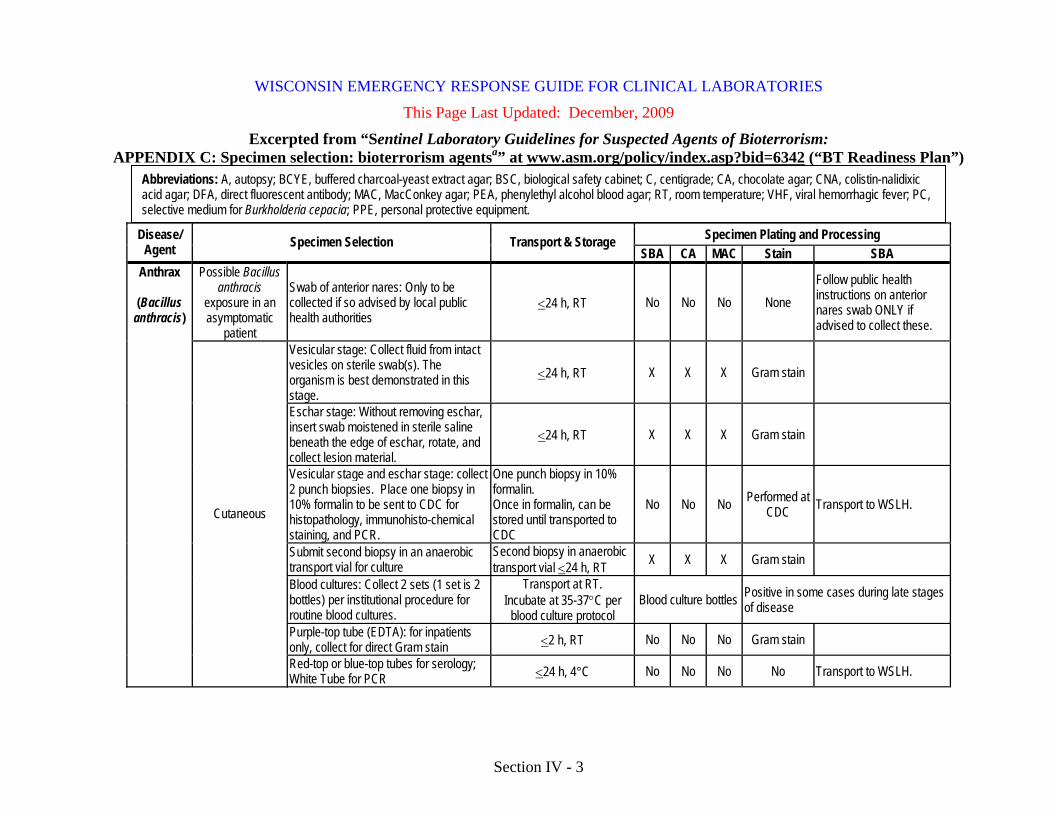
Excerpted from “Sentinel Laboratory Guidelines for Suspected Agents of Bioterrorism: APPENDIX C: Specimen selection: bioterrorism agentsa” at www.asm.org/policy/index.asp?bid=6342 (“BT Readiness Plan”)
Specimen Plating and Processing Disease/ Agent Specimen Selection Transport & Storage
SBA CA MAC Stain SBA Possible Bacillus
anthracis exposure in an asymptomatic
patient
Swab of anterior nares: Only to be collected if so advised by local public health authorities
<24 h, RT No No No None Follow public health instructions on anterior nares swab ONLY if advised to collect these.
Vesicular stage: Collect fluid from intact vesicles on sterile swab(s). The organism is best demonstrated in this stage.
<24 h, RT X X X Gram stain
Eschar stage: Without removing eschar, insert swab moistened in sterile saline beneath the edge of eschar, rotate, and collect lesion material.
<24 h, RT X X X Gram stain
Vesicular stage and eschar stage: collect 2 punch biopsies. Place one biopsy in 10% formalin to be sent to CDC for histopathology, immunohisto-chemical staining, and PCR.
One punch biopsy in 10% formalin. Once in formalin, can be stored until transported to CDC
No No No Performed at CDC Transport to WSLH.
Submit second biopsy in an anaerobic transport vial for culture
Second biopsy in anaerobic transport vial <24 h, RT X X X Gram stain
Blood cultures: Collect 2 sets (1 set is 2 bottles) per institutional procedure for routine blood cultures.
Transport at RT. Incubate at 35-37°C per
blood culture protocol Blood culture bottles Positive in some cases during late stages
of disease
Purple-top tube (EDTA): for inpatients only, collect for direct Gram stain <2 h, RT No No No Gram stain
Anthrax
(Bacillus anthracis)
Cutaneous
Red-top or blue-top tubes for serology; White Tube for PCR <24 h, 4°C No No No No Transport to WSLH.
Abbreviations: A, autopsy; BCYE, buffered charcoal-yeast extract agar; BSC, biological safety cabinet; C, centigrade; CA, chocolate agar; CNA, colistin-nalidixic acid agar; DFA, direct fluorescent antibody; MAC, MacConkey agar; PEA, phenylethyl alcohol blood agar; RT, room temperature; VHF, viral hemorrhagic fever; PC, selective medium for Burkholderia cepacia; PPE, personal protective equipment.

WISCONSIN EMERGENCY RESPONSE GUIDE FOR CLINICAL LABORATORIES
Section IV - 4
This Page Last Updated: December, 2009
(cont.) Excerpted from “Sentinel Laboratory Guidelines for Suspected Agents of Bioterrorism: APPENDIX C: Specimen selection: bioterrorism agentsa” at www.asm.org/policy/index.asp?bid=6342 (“BT Readiness Plan”)
Specimen Plating and Processing Disease/ Agent Specimen Selection Transport & Storage
SBA CA MAC Stain Other Stool: Collect 5-10 g in a clean, sterile, leakproof container. <24 h, 4°C Inoculate routine stool plating
media plus CNA or PEA. Minimal recovery
Blood cultures: Collect 2 sets (1 set is 2 bottles) per institutional procedure for routine blood cultures.
Transport at RT. Incubate at 35-37°C per blood
culture protocol. Blood culture bottles Positive in late stages of disease
Purple-top tube (EDTA): for inpatients only, collect for direct Gram stain <2 h, RT No No No Gram
stain
Gastro- intestinal
Red-top or blue-top tubes for serology; White Tube for PCR <24 h, 4°C No No No No
Sputum: Collect expectorated specimen into a sterile, leakproof container. <24 h, 4°C X X X Gram
stain Minimal recovery
Pleural fluid: Collect specimen into sterile, leakproof container. <24 h, 4°C X X X Gram
stain Save excess (if any) for PCR.
Blood cultures: Collect 2 sets (1 set is 2 bottles) per institutional procedure for routine blood cultures.
Transport at RT. Incubate at 35-37°C per blood
culture protocol. Blood culture bottles Positive in late stages of disease
Purple-top tube (EDTA): For inpatients only, collect for direct Gram stain.
<2 h, RT No No No Gram
stain
Inhalation
Red-top or blue-top tubes for serology; White Tube for PCR <24 h, 4°C No No No No
Cerebrospinal fluid culture: Aseptically collect CSF per institutional procedure. <24 h, RT X X Gram
stain
May be seen in late stages of disease; consider adding broth medium such as brain heart infusion.
Anthrax
(Bacillus
anthracis)
Meningitis
Blood cultures: Collect 2 sets (1 set is 2 bottles) per institutional procedure for routine blood cultures.
Transport at RT. Incubate at 35-37°C per blood culture protocol.
Blood culture bottles Positive in late stages of disease

WISCONSIN EMERGENCY RESPONSE GUIDE FOR CLINICAL LABORATORIES
Section IV - 5
This Page Last Updated: December, 2009
(cont.) Excerpted from “Sentinel Laboratory Guidelines for Suspected Agents of Bioterrorism: APPENDIX C: Specimen selection: bioterrorism agentsa” at www.asm.org/policy/index.asp?bid=6342 (“BT Readiness Plan”)
Disease/ Agent Specimen Selection Transport &
Storage Specimen Handling
Clinical syndrome
Specimen type Foodborne Infant Wound
Intention- al release (airborne)
Specimen(s) of choice for confirming botulism: 1. Serum 2. Wound/tissue 3. Stool 4. Incriminated food
Enema fluid 20 ml X X
X 4°C Purge with a minimal amount of sterile nonbacteriostatic water to minimize dilution of toxin.
Food sample 10-50g X X X 4°C
Foods that support C. botulinum growth will have a pH of 3.5-7.0; most common pH is 5.5-6.5. Submit food in original container, placing individually in leakproof sealed transport devices.
Gastric fluid 20 ml X,A
4°C Collect up to 20 ml.
Intestinal fluid 20 ml A A
4°C Autopsy: Intestinal contents from various areas of the small and
large intestines should be provided. Nasal swab
(anaerobic swab)
X RT For aerosolized botulinum toxin exposure, obtain nasal cultures
for C. botulinum and serum for mouse toxicity testing.
Serum 15-20 mls
X,A
X X 4°C
Serum should be obtained as soon as possible after the onset of symptoms and before antitoxin is given. Whole blood (30 ml [3 red-top or gold-top tubes]) is required for mouse toxicity testing. In infants, serum is generally not useful, since the toxin is quickly absorbed before serum can be obtained.
Stool >25 g X X X X 4°C
Botulism has been confirmed in infants with only “pea-size” stools. Please note: Anticholinesterase given orally, as in patients with myasthenia gravis, has been shown to interfere with toxin testing.
Vomitus 20 ml X
4°C Collect up to 20 ml.
Botulism
(botulinum
toxin)
Wound, tissue - anaerobic
swab or transport system
`
Anaerobic swab or
transport system Transport at RT
Exudate, tissue, or swabs must be collected and transported in an anaerobic transport system. Samples from an enema or feces should also be submitted, since the wound may not be the source of botulinum toxin.

WISCONSIN EMERGENCY RESPONSE GUIDE FOR CLINICAL LABORATORIES
Section IV - 6
This Page Last Updated: December, 2009
(cont.) Excerpted from “Sentinel Laboratory Guidelines for Suspected Agents of Bioterrorism: APPENDIX C: Specimen selection: bioterrorism agentsa” at www.asm.org/policy/index.asp?bid=6342 (“BT Readiness Plan”)
Specimen Plating and Processing Disease/ Agent Specimen Selection Transport &
Storage SBA CA MAC Stain Other Serum: Collect 10-12 cc (ml) of acute-phase specimen as soon as possible after disease onset. Follow with a convalescent-phase specimen obtained 21 days later.
Transport in <2 h, at RT. Store at -20°C.
Specimen should be stored and shipped frozen at -20°C to State Laboratory or other LRN Reference laboratory.
Serologic diagnosis: 1. Single titer:>1:160 2. 4-fold rise 3. IgM NOTE: B. canis does not cross-react with standard serologic reagents.
Blood: Collect 2 sets (1 set is 2 bottles) per institutional procedure for routine blood cultures.
Transport at RT. Incubate at 35-37°C
Blood culture bottles: Subculture at 5 days and hold 21 days.
Blood culture isolation rates vary from 15-70% depending on methods and length of incubation. Cultures should be manipulated in aBSC. PPE includes gloves, gown, mask, and protective faceshield. All cultures should be taped shut during incubation.
X X
Acute, subacute, or chronic
Bone marrow, spleen, or liver: Collect per institution’s surgical/pathology procedure.
<24 h, RT Hold cultures for at least 7 days.
Gram stain
Cultures should be manipulated in a BSC. PPE includes gloves, gown, mask, and protective faceshield. All cultures should be taped shut during incubation.
X X Cerebrospinal fluid culture: Aseptically collect CSF per institutional procedure.
<24 h, RT Hold cultures for at least 7 days. Gram
stain
Cultures should be manipulated in a BSC. PPE includes gloves, gown, mask, and protective faceshield. All cultures should be taped shut during incubation. Consider adding broth medium such as brain heart infusion.
Brucellosis
(Brucella melitensis, B. abortus,
B. suis, B. canis)
Meningitis
Cerebrospinal fluid for antibody testing -20°C
Specimen should be stored and shipped frozen at -20C or lower temperature to State Laboratory or other LRN Reference laboratory.
None

WISCONSIN EMERGENCY RESPONSE GUIDE FOR CLINICAL LABORATORIES
Section IV - 7
This Page Last Updated: December, 2009 (cont.) Excerpted from “Sentinel Laboratory Guidelines for Suspected Agents of Bioterrorism:
APPENDIX C: Specimen selection: bioterrorism agentsa” at www.asm.org/policy/index.asp?bid=6342 (“BT Readiness Plan”) Specimen Plating and Processing Disease/
Agent Specimen Selection Transport & Storage SBA CA MAC PC Stain Other
Possible Burkholderia pseudomallei or Burkholderia mallei exposure in asymptomatic patient
No cultures or serology indicated
Follow public health instructions if advised to collect specimens.
Bone marrow Transport within <2 h at RT. Store <24 h at 4°C
X Gram stain
B. pseudomallei is a small gram-negative bacillus that may demonstrate bipolar morphology on stain.
B. mallei is a small gram-negative coccobacillus. Incubation should be at 35 to 37°C, ambient atmosphere; CO2 incubation is acceptable.
Blood culture bottles OR
Collect lysis-centrifugation (e.g., Isolator) blood cultures and
plate to:
Melioidosis and glanders
(Burkholderia pseudomallei
and Burkholderia mallei)
Clinical illness
Blood cultures: Collect 2 sets (1 set is 2 bottles) per institutional procedure for routine blood cultures OR collect lysis-centrifugation (e.g., Isolator) blood cultures.
Transport at RT. Incubate at 35-37°C per blood culture protocol.
X
Cultures should be manipulated in a BSC. PPE includes gloves, gown, mask, and protective faceshield. All cultures should be taped shut during incubation. Incubation should be at 35 to 37°C, ambient atmosphere; CO2 incubation is acceptable.

WISCONSIN EMERGENCY RESPONSE GUIDE FOR CLINICAL LABORATORIES
Section IV - 8
This Page Last Updated: December, 2009 (cont.) Excerpted from “Sentinel Laboratory Guidelines for Suspected Agents of Bioterrorism:
APPENDIX C: Specimen selection: bioterrorism agentsa” at www.asm.org/policy/index.asp?bid=6342 (“BT Readiness Plan”) Specimen Plating and Processing Disease/
Agent Specimen Selection Transport & Storage SBA CA MAC PC Stain Other
Respiratory specimens, abscess material, wound specimens, urine
Transport within <2 h, at RT. Store <24 h, at 4°C.
X X X X Gram stain
If the laboratory has B. cepacia selective agar medium, it has been shown useful in isolation of B. pseudomallei for specimens in which indigenous microflora is likely to be encountered. Ashdown medium is a selective medium specifically designed for recovery of B. pseudomallei. This medium is not likely to be available in most Sentinel Laboratories. Incubation should be at 35 to 37°C, ambient atmosphere; CO2 incubation is acceptable.
Melioidosis and glanders
(Burkholderia pseudomallei
and Burkholderia mallei)
(continued)
Clinical illness (continued)
Serum: Red-top or gold-top tube for both acute and convalescent (obtained 14 days after the acute specimen)
Transport within ~6 h, at 4°C. Store at -20°C to -70°C.
Obtain if serologic diagnosis of B. pseudomallei infection is being considered.

WISCONSIN EMERGENCY RESPONSE GUIDE FOR CLINICAL LABORATORIES
Section IV - 9
This Page Last Updated: December, 2009
(cont.) Excerpted from “Sentinel Laboratory Guidelines for Suspected Agents of Bioterrorism: APPENDIX C: Specimen selection: bioterrorism agentsa” at www.asm.org/policy/index.asp?bid=6342 (“BT Readiness Plan”)
Disease/Agent Specimen Selection Transport &
Storage Specimen Handling
Serum: Collect 10 ml of serum (red-top, tiger-top, or gold-top tube) as soon as possible after onset of symptoms (acute) and with a follow-up specimen (convalescent) at >14 days for serological testing.
Transport within ~6 h, at 4°C. Store at -20°C to -70°C
Blood: Collect blood in EDTA (lavender) or sodium citrate (blue) and maintain at 4°C for storage and shipping for PCR or special cultures. If possible, collect specimens prior to antimicrobial therapy.
Transport within ~6 h, at 4°C. Store at 4°C.
Q. fever
(Coxiella burnetii)
Tissue, body fluids, others, including cell cultures and cell supernatants: Specimens can be kept at 2-8°C if transported within 24 h. Store frozen at -70°C or on dry ice.
Transport within <24 h, at 2-8°C. Store at -70°C or on dry ice.
Do not attempt tissue culture isolation, as that could result in a very unsafe situation in which there is a significant amount of infectious organism. Sentinel laboratories should consult with State Public Health Laboratory Director (or designate) prior to or concurrent with testing if C. burnetii is suspected by the attending physician. Serology is available through commercial reference as well as public health laboratories.

WISCONSIN EMERGENCY RESPONSE GUIDE FOR CLINICAL LABORATORIES
Section IV - 10
This Page Last Updated: December, 2009
(cont.) Excerpted from “Sentinel Laboratory Guidelines for Suspected Agents of Bioterrorism: APPENDIX C: Specimen selection: bioterrorism agentsa” at www.asm.org/policy/index.asp?bid=6342 (“BT Readiness Plan”)
Specimen Plating and Processing Disease/ Agent Specimen Selection Transport &
Storage SBA CA MAC Stain Other Possible Francisella tularensis exposure in asymptomatic patient
No cultures or serology indicated Follow public health instructions if advised to
collect specimens.
Conjunctival scraping <24 h, 4°C X X X
Gram stain; prepare smears for DFA referral.
Add a BCYE plate and a plate selective for Neisseria gonorrhoeae such as modified Thayer-Martin. Manipulate cultures in a BSC. PPE includes gloves, gown, mask, and protective faceshield. All cultures should be taped shut during incubation.
Lymph node aspirate: Flushing with 1.0 ml of sterile saline may be needed to obtain material.
Transport at RT, 4°C if transport is delayed. Store at <24 h, 4°C.
X X X
Gram stain; prepare smears for DFA referral.
Add a BCYE plate and a plate selective for Neisseria gonorrhoeae such as modified Thayer-Martin. Manipulate cultures in a BSC. PPE includes gloves, gown, mask, and protective faceshield. All cultures should be taped shut during incubation.
Tularemia
(Francisella tularensis)
Oculo-glandular
Blood cultures: Collect 2 sets (1 set is 2 bottles) per institutional procedure for routine blood cultures. Growth is more likely from aerobic bottle.
Transport at RT. Incubate at 35-37°C per blood culture protocol.
Blood culture bottles; subculture the broth to BCYE plate and incubate aerobically.
Manipulate cultures in a BSC. PPE includes gloves, gown, mask, and protective faceshield. All cultures should be taped shut during incubation.
WISCONSIN EMERGENCY RESPONSE GUIDE FOR CLINICAL LABORATORIES
Section IV - 11
This Page Last Updated: December, 2009
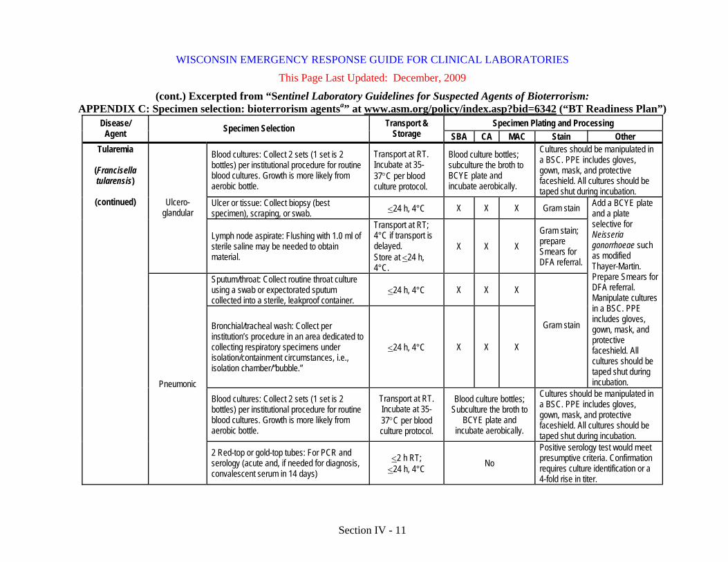
(cont.) Excerpted from “Sentinel Laboratory Guidelines for Suspected Agents of Bioterrorism: APPENDIX C: Specimen selection: bioterrorism agentsa” at www.asm.org/policy/index.asp?bid=6342 (“BT Readiness Plan”)
Specimen Plating and Processing Disease/ Agent Specimen Selection Transport &
Storage SBA CA MAC Stain Other
Blood cultures: Collect 2 sets (1 set is 2 bottles) per institutional procedure for routine blood cultures. Growth is more likely from aerobic bottle.
Transport at RT. Incubate at 35-37°C per blood culture protocol.
Blood culture bottles; subculture the broth to BCYE plate and incubate aerobically.
Cultures should be manipulated in a BSC. PPE includes gloves, gown, mask, and protective faceshield. All cultures should be taped shut during incubation.
Ulcer or tissue: Collect biopsy (best specimen), scraping, or swab. <24 h, 4°C X X X Gram stain Ulcero-
glandular
Lymph node aspirate: Flushing with 1.0 ml of sterile saline may be needed to obtain material.
Transport at RT; 4°C if transport is delayed. Store at <24 h, 4°C.
X X X Gram stain; prepare Smears for DFA referral.
Sputum/throat: Collect routine throat culture using a swab or expectorated sputum collected into a sterile, leakproof container.
<24 h, 4°C X X X
Bronchial/tracheal wash: Collect per institution’s procedure in an area dedicated to collecting respiratory specimens under isolation/containment circumstances, i.e., isolation chamber/“bubble.”
<24 h, 4°C X X X
Gram stain
Add a BCYE plate and a plate selective for Neisseria gonorrhoeae such as modified Thayer-Martin. Prepare Smears for DFA referral. Manipulate cultures in a BSC. PPE includes gloves, gown, mask, and protective faceshield. All cultures should be taped shut during incubation.
Blood cultures: Collect 2 sets (1 set is 2 bottles) per institutional procedure for routine blood cultures. Growth is more likely from aerobic bottle.
Transport at RT. Incubate at 35-37°C per blood culture protocol.
Blood culture bottles; Subculture the broth to
BCYE plate and incubate aerobically.
Cultures should be manipulated in a BSC. PPE includes gloves, gown, mask, and protective faceshield. All cultures should be taped shut during incubation.
Tularemia
(Francisella tularensis)
(continued)
Pneumonic
2 Red-top or gold-top tubes: For PCR and serology (acute and, if needed for diagnosis, convalescent serum in 14 days)
<2 h RT; <24 h, 4°C No
Positive serology test would meet presumptive criteria. Confirmation requires culture identification or a 4-fold rise in titer.

WISCONSIN EMERGENCY RESPONSE GUIDE FOR CLINICAL LABORATORIES
Section IV - 12
This Page Last Updated: December, 2009
(cont.) Excerpted from “Sentinel Laboratory Guidelines for Suspected Agents of Bioterrorism: APPENDIX C: Specimen selection: bioterrorism agentsa” at www.asm.org/policy/index.asp?bid=6342 (“BT Readiness Plan”)
Specimen Selection
Disease/Agent Specimen type Foodborne
Airborne (intentional
release)
Transport and Storage Specimen Handling
Serum – 10 ml X X 2-8oC
1. Obtain as soon as possible after the onset of symptoms to detect the toxin. 2. Also collect 7-14 days after onset of illness to compare acute and convalescent antibody titers. 3. Do not send whole blood, since hemolysis during transit will compromise the quality of the specimen.
Nasal swab - dacron or
rayon swab X 2-8oC
Collect a nasal swab within 24 h of exposure by rubbing a dry, sterile swab (Dacron or rayon) on the mucosa of the anterior nares. Place in protective transport tube.
Induced respiratory secretions
X 2-8oC Collect sputum induced by instilling 10-25 ml of sterile saline into nasal passages into a sterile screw-top container.
Urine – 20-30 ml X X 2-8oC Collect into a sterile, leakproof container with screw-top lid.
Stool or gastric
aspirate – 10-50 g
X X 2-8oC Collect into a sterile, leakproof container with screw-top lid.
Postmortem 10 g X X 2-8oC
Obtain specimens of the intestinal contents from different levels of the small and large bowel. Place 10 g of specimen into a sterile, leakproof container with screw-top lid. Obtain serum as previously described.
Culture isolate X X 2-8oC Send S. aureus isolate for toxin testing on appropriate agar slant.
Staphylococcal enterotoxin B
(From
Staphylococcus aureus)
Food specimen X X 2-8oC
Food should be left in its original container if possible or placed in sterile unbreakable containers and labeled carefully. Place containers individually in leakproof containers (i.e., sealed plastic bags) to prevent cross-contamination during shipment. Empty containers with remnants of suspected contaminated foods can be examined.
WISCONSIN EMERGENCY RESPONSE GUIDE FOR CLINICAL LABORATORIES
Section IV - 13
This Page Last Updated: December, 2009
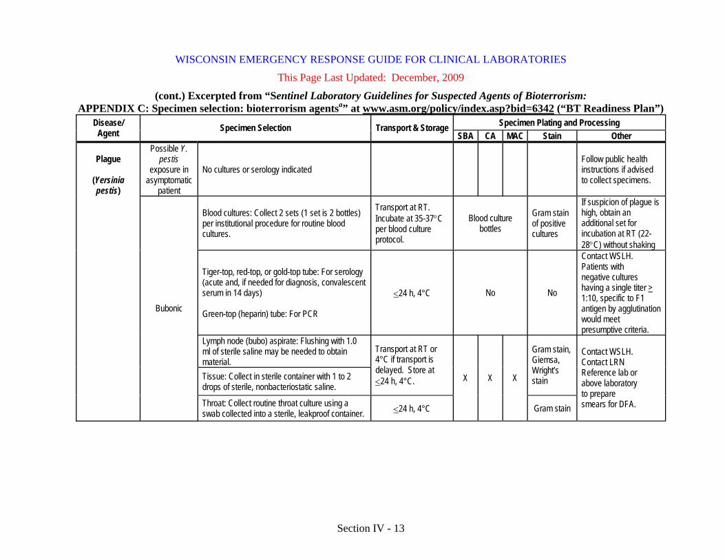
(cont.) Excerpted from “Sentinel Laboratory Guidelines for Suspected Agents of Bioterrorism: APPENDIX C: Specimen selection: bioterrorism agentsa” at www.asm.org/policy/index.asp?bid=6342 (“BT Readiness Plan”)
Specimen Plating and Processing Disease/ Agent Specimen Selection Transport & Storage
SBA CA MAC Stain Other Possible Y.
pestis exposure in
asymptomatic patient
No cultures or serology indicated Follow public health instructions if advised to collect specimens.
Blood cultures: Collect 2 sets (1 set is 2 bottles) per institutional procedure for routine blood cultures.
Transport at RT. Incubate at 35-37°C per blood culture protocol.
Blood culture bottles
Gram stain of positive cultures
If suspicion of plague is high, obtain an additional set for incubation at RT (22-28°C) without shaking
Tiger-top, red-top, or gold-top tube: For serology (acute and, if needed for diagnosis, convalescent serum in 14 days) Green-top (heparin) tube: For PCR
<24 h, 4°C No No
Contact WSLH. Patients with negative cultures having a single titer > 1:10, specific to F1 antigen by agglutination would meet presumptive criteria.
Lymph node (bubo) aspirate: Flushing with 1.0 ml of sterile saline may be needed to obtain material. Tissue: Collect in sterile container with 1 to 2 drops of sterile, nonbacteriostatic saline.
Transport at RT or 4°C if transport is delayed. Store at <24 h, 4°C.
Gram stain, Giemsa, Wright’s stain
Plague
(Yersinia pestis)
Bubonic
Throat: Collect routine throat culture using a swab collected into a sterile, leakproof container. <24 h, 4°C
X X X
Gram stain
Contact WSLH. Contact LRN Reference lab or above laboratory to prepare smears for DFA.

WISCONSIN EMERGENCY RESPONSE GUIDE FOR CLINICAL LABORATORIES
Section IV - 14
This Page Last Updated: December, 2009
(cont.) Excerpted from “Sentinel Laboratory Guidelines for Suspected Agents of Bioterrorism: APPENDIX C: Specimen selection: bioterrorism agentsa” at www.asm.org/policy/index.asp?bid=6342 (“BT Readiness Plan”)
Specimen Plating and Processing Disease/ Agent Specimen Selection Transport &
Storage SBA CA MAC Stain Other Sputum/throat: Collect routine throat culture using a swab or expectorated sputum collected into a sterile, leakproof container. Bronchial/tracheal wash: Collect per institution’s procedure in an area dedicated to collecting respiratory specimens under isolation/containment circumstances, i.e., isolation chamber/“bubble.”
<24 h, 4°C X X X Gram stain Contact WSLH. Contact LRN Reference lab or above laboratory to prepare smears for DFA.
Blood cultures: Collect 2 sets (1 set is 2 bottles) per institutional procedure for routine blood cultures.
Transport at RT. Incubate at 35-37°C per blood culture protocol.
Blood culture bottles
Gram stain of positive cultures
If suspicion of plague is high, obtain an additional set for incubation at RT (22-28°C) without shaking.
Pneumonic
Tiger-top, red-top, or gold-top tube: For serology (acute and, if needed for diagnosis, convalescent serum in 14 days)
Green-top (heparin) tube: For PCR
<24 h, 4°C No No
Contact WSLH. Patients with negative cultures having a single titer, >1:10, specific to F1 antigen by agglutination would meet presumptive criteria.
Blood cultures: Collect 2 sets (1 set is 2 bottles) per institutional procedure for routine blood cultures.
Transport at RT. Incubate at 35-37°C per blood culture protocol.
Blood culture bottles
Gram stain of positive cultures
If suspicion of plague is high, obtain an additional set for incubation at RT (22-28°C) without shaking.
Tiger-top, red-top, or gold-top tube: For serology (acute and, if needed for diagnosis, convalescent serum in 14 days)
Green-top (heparin) tube: For PCR
<24 h, 4°C No No
Contact WSLH. Patients with negative cultures having a single titer, >1:10, specific to F1 antigen by agglutination would meet presumptive criteria.
Plague
(Yersinia pestis)
(continued)
Meningitis
Cerebrospinal fluid Transport at RT. Store incubated
at 35-37°C. X X Gram stain Can add broth culture at RT
(22-28°C) without shaking.

WISCONSIN EMERGENCY RESPONSE GUIDE FOR CLINICAL LABORATORIES
Section IV - 15
This Page Last Updated: December, 2009
Bioterrorism/Emergency Response Packaging Instructions Note: If a Chain of Custody/Evidence form is required, refer to information in Section V of this guide prior to packaging.
The shipper bears the responsibility for appropriate packaging of samples and risks significant fines and other penalties for inappropriate packaging. Training in sample packaging is required by regulations for anyone involved in the transport of laboratory samples that meet the definition of “dangerous goods”. Training is available from a number of sources (see “References” at the end of this section), including the “Packaging Clinical Laboratory Samples for Domestic Transport” manual that was distributed to Wisconsin Sentinel Laboratories and local health departments. The following pages provide specific instructions for packaging laboratory samples for transport to the Wisconsin State Laboratory of Hygiene for bioterrorism or other public health emergency response. Packaging samples for chemical terrorism response is included in the chemical terrorism section of this guide. If you do not have a specimen shipping kit, you can contact one of the local health department repository sites to get one of Wisconsin Emergency Response Shipping Kits. These shippers are stored at most local public health departments for use in emergency or outbreak response. A listing of repository sites with contact information is posted on the HAN (in the “Laboratory” topic area, under “Repository”) and is also available later in this section. Your laboratory is expected to make arrangements to collect the shipper from the local health department repository site. The shippers are class 6.2 “infectious substance” shippers but can also be used for shipping Biological substance, Category B specimens. The shippers are appropriate for shipping samples at ambient temperature, refrigerated temperatures, or dry ice temperatures. These kits are not intended for routine specimen submission.

WISCONSIN EMERGENCY RESPONSE GUIDE FOR CLINICAL LABORATORIES
Section IV - 16
This Page Last Updated: December, 2009
Instructions for Packaging "Biological Substance, Category B" Samples to the Wisconsin State Laboratory of Hygiene via Dunham Express for Emergency Response Note: If a Chain of Custody/Evidence form is required, refer to Section V.
• “Biological Substance, Category B” samples include specimens collected
directly from the patient (e.g., swabs, urine, feces, CSF, blood, etc.) and isolates, unless they are suspected to contain unusually high risk agents (i.e., meet criteria for a Category A Infectious Substance).
• All specimens must be “triple-packaged.” • There are no quantity restrictions for Biological Substance, Category B samples
when using ground transportation. • Wrap the closure of each specimen container (the “primary container”, e.g.,
tubes, vials, urine cup, etc.) with tape to prevent loosening. • Wrap each container in enough absorbent material to absorb the entire specimen
and cushion it from breakage. • Place wrapped specimens in zip-lock bag (“secondary container”). • Place one requisition form for each sample in the outer sleeve of each bag. • If you are not using a Wisconsin Emergency Response Shipper Kit
• Place the bagged specimens inside the styrofoam shipper or inside the plastic can in the styrofoam box, if provided in shipper.
• Include sufficient cushioning material to prevent breakage during transport. • If specimens must be kept cold, place frozen kool-packs outside the plastic
can (if included) or specimen bag. • If including dry ice, place dry ice outside the plastic can (if included) or
specimen bag. • If you are using the Wisconsin Emergency Response Shipper Kit:
• Place the bagged specimens into the foam liner inside the plastic can. • If the specimen(s) will not fit into the foam liner, remove the foam and
replace with padding to prevent breakage of the specimen container. • Screw the cap onto the plastic can; place the can into the styrofoam box. • If specimens must be kept cold, place frozen kool-packs outside plastic can. • If including dry ice, place dry ice outside the plastic can. • Place cardboard tray on the plastic can; place the styrofoam lid on the box. • Fold (but do not seal) the plastic liner of the box. Close and tape the flaps of
the box. If dry ice was included, do NOT tape all seams of the box.

WISCONSIN EMERGENCY RESPONSE GUIDE FOR CLINICAL LABORATORIES
Section IV - 17
This Page Last Updated: December, 2009
Instructions for Packaging "Biological Substance, Category B" Samples to the Wisconsin State Laboratory of Hygiene via Dunham Express for Emergency Response (continued) Package Labeling • Attach the “Biological Substance, Category B / UN3373” label to the outside of
the box. • If the package contains dry ice, attach the diamond-shaped “Class 9” dry ice
label and the “Dry Ice/UN 1845/Net Quantity __kg” label to the outside of the box, write in the approximate weight of dry ice on the label.
• Complete the “Emergency Contact Label” with the name and telephone number of a person who has knowledge of the contents of the package.
• Address the package as follows: Rapid Response – BT/Outbreak Wisconsin State Laboratory of Hygiene-Communicable Diseases 465 Henry Mall Madison, WI 53706 • Circle “BT” on the label if the specimen is bioterrorism-related, • Circle “Outbreak” on the label if the specimen is outbreak-related
• Instructions for contacting and shipping specimens via Dunham Express are provided later in this section.

WISCONSIN EMERGENCY RESPONSE GUIDE FOR CLINICAL LABORATORIES
Section IV - 18
This Page Last Updated: December, 2009
Instructions for Packaging "Infectious Substances, affecting Humans" (Category A Samples) to the Wisconsin State Laboratory of Hygiene via Dunham Express for Emergency Response • Category A “Infectious substances” include isolates from inoculated cultures,
forensic specimens for which a criminal investigation or legal action is expected, and patient specimens that are suspected to contain “high-risk” agents such as smallpox, ebola, and lassa viruses.
• All specimens must be “triple-packaged.” Laboratories must use certified packaging obtained from a manufacturer of Class 6.2 packaging.
• There are no quantity restrictions for Category A Infectious Substances when using ground transportation.
• If the substance is listed as a Select Agent, you must contact the CDC to receive approval for shipping and notify the WSLH prior to shipping.
• Wrap the closure of each specimen container (the “primary container”, e.g., tubes, vials, etc.) with tape to prevent loosening.
• Wrap each primary container in enough absorbent material to absorb the entire specimen and cushion against breakage.
• Place specimen container in zip-lock bag; place one requisition form for each sample in the outer sleeve of each bag.
• Place the bagged specimens into the plastic can; if the bagged specimen(s) will not fit into the foam liner which may be included in the can, remove the foam and replace with padding to prevent breakage of the specimen container. Screw the cap onto the plastic can.
• Place the can into the outer shipping container (e.g., styrofoam box). • Attach an itemized list of the contents in a ziplock bag to the plastic can.
• The itemized list should include the number of sample containers, the type of sample (e.g., broth culture, cell culture isolate), and the agent suspected or confirmed to be in the sample.
• If specimens must be kept cold, place frozen kool-packs outside the plastic can. • If including dry ice, place dry ice outside the plastic can. • Place the lid on the box. • Fold the plastic liner (if provided in shipper) of the box, but do not seal it. • Close and tape the flaps of the cardboard box. If dry ice was included, do NOT
tape all seams of the box.

WISCONSIN EMERGENCY RESPONSE GUIDE FOR CLINICAL LABORATORIES
Section IV - 19
This Page Last Updated: December, 2009
Instructions for Packaging "Infectious Substances, affecting Humans" (Category A Samples) to the Wisconsin State Laboratory of Hygiene via Dunham Express for Emergency Response (continued) • Attach diamond-shaped and rectangular “Infectious Substance” labels,
“Infectious substance, affecting Humans, UN2814 Net Quantity __ml” label; write in the total quantity of the infectious substance.
• If the package contains dry ice, attach diamond-shaped “Class 9” dry ice label, and the “Dry Ice/UN 1845/Net Quantity __kg” label; write in the approximate weight of dry ice in the package.
• Provide emergency contact information to include a name and a (24/7) telephone number of a person who has knowledge of the package contents.
• Address the package as follows: Rapid Response – BT/Outbreak Wisconsin State Laboratory of Hygiene-Communicable Diseases 465 Henry Mall Madison, WI 53706 • Circle “BT” on the label if the specimen is bioterrorism-related, • Circle “Outbreak” on the label if the specimen is outbreak-related,.
• Attach the “shipping paper” or “dangerous goods form” (DGF) to the outside of the shipping container. If more than 1 page, the pages must be numbered and the first page must specify the total number of pages.
• Retain 1 copy of the shipping paper or DGF on file at your facility for 2 years.
• Retain Emergency Response Information at the emergency contact telephone number provided. “Material Safety Data Sheets for Infectious Microorganisms are available from Health Canada Office of Biosafety at http://www.phac-aspc.gc.ca/msds-ftss/.
• Information required is a description of the dangerous goods; immediate hazards to health; risk of fire or explosion; precautions to be taken in the event of an accident or incident; immediate methods for handling fires; initial methods for handling spills or leaks in the absence of a fire; and preliminary first aid measures.
Instructions for contacting and shipping specimens via Dunham Express are provided on the next page.

WISCONSIN EMERGENCY RESPONSE GUIDE FOR CLINICAL LABORATORIES
Section IV - 20
This page last Updated: December, 2009
Instructions for Shipping of Emergency Response "Biological, Substance, Category B" Samples and “Infectious Substances, affecting Humans” (Category A) Samples to the Wisconsin State Laboratory of Hygiene via Dunham Express
• The WSLH has arranged a contract with Dunham Express for shipment of samples for emergency response to the State Laboratory of Hygiene, with charges billed to the WSLH.
• Specimens will be delivered to the WSLH the following day, Monday through Friday. If you must ship on a Friday, contact the WSLH Emergency Answering Service at 608-263-3280 for instructions
• Complete the package preparation before the courier arrives. • Contact the Dunham Express office in your area (list below); calls are answered
24 hours a day, 7 days a week. Appleton area: Call 920-722-6360 Eau Claire area: Call 715-874-4595 LaCrosse area: Call 608-779-4588 Madison area: Call 608- 242-1000 Milwaukee area: Call 414-435-0002 Niagara area: Call 715-251-1909 Wausau area: Call 715-355-0400
• Information which you must provide to Dunham Express when you call: • Confirm the pick-up time and request "Overnight Service". • Give the Dunham Express office the following information:
The State Lab Rapid Response account number: 7263 The account name: State Lab Rapid Response Your name, phone number, and pickup address Other location information (e.g., room number) Destination: Rapid Response-BT/Outbreak
Wisconsin State Lab of Hygiene 465 Henry Mall, Madison, WI 53706
Shipment description, if asked: Specimens for delivery Other Information: Specify delivery option as “Overnight Service”
• Contact the WSLH Emergency Answering Service at 608-263-3280 for instructions if you need assistance or believe that special arrangements are necessary.

WISCONSIN EMERGENCY RESPONSE GUIDE FOR CLINICAL LABORATORIES
Section IV - 21
This Page Last Updated: December, 2009
WSLH REQUISITION FORM FOR ISOLATE REFERRALS*
Wisconsin Emerging Infections/ Emergency Response Referral (rev.09/2003)
PATIENT INFORMATION SUBMITTING LABORATORY
Patient Name (Last, First):
WSLH Agency Number If Known
Patient Address: Agency Name & Telephone Number
Patient City: State: Zip: Agency Address
Age or Date of Birth:
City, State, Zip Code of Agency
Patient Sex: M / F
Outbreak/Study: Emergency Response WSLH Billing Account 72150
Your Specimen Number:
Date Original Specimen Collected: Original Specimen Type:
____________________________ ___Blood ___Wound (Site:_________________)
___CSF ___Respiratory (Source:__________)
___Urine ___Other (Site: __________________)
Organism Suspected: __________________________________
Isolation and Identification History: Media Isolated On: ______________________________
ID Method: ___MIDI ___MicroScan (version ______)
___VITEK (version ______) ___API (version ____) ___Other test (Specify:_________________________)
Additional Tests Performed: ________________________________________________
Laboratory Contact: ___________________________________Phone: _________________
WSLH Test Code: 624 Referred Culture, Public Health rDNA Analysis (not reported)
* You may substitute a standard WSLH requisition form or the Wisconsin Emerging Infections (WEIP) requisition form.
_______________________________________________________________________________________ WISCONSIN STATE LAB OF HYGIENE USE ONLY

WISCONSIN EMERGENCY RESPONSE GUIDE FOR CLINICAL LABORATORIES
Section IV - 22
This Page Last Updated: December, 2009
Shipping Resources • Containers
• The following list of commercial sources of transport containers is not all-inclusive. This listing is not intended to be an endorsement or non-endorsement of any companies.
• Saf-T-Pak, Inc., website http://www.saftpak.com • EXAKT Technologies, Inc., website http://www.exaktpak.com/ • Therapak Corporation, website http://www.therapak.com • Air Sea Atlanta, website http://www.airseaatlanta.com • All-Pak, Inc., website http://www.all-pak.com • DG Supplies, Inc., website http://www.dgsupplies.com • Federal Industries Corporation, website http://www.chem-tran.com • ICC The Compliance Center Inc., website
http://www.thecompliancecenter.com • Inmark, Inc., website http://www.inmarkinc.com
Training • The following list of websites includes commercial sources of training for
shipping. This listing is not all-inclusive and is not intended to be an endorsement or non-endorsement of any companies or products.
• http://www.phmsa.dot.gov/hazmat/training-outreach • http://www.saftpak.com • http://www.highqllc.com • http://www.dgitraining.com/menu.htm • http://cargopak.com • “Packaging Clinical Laboratory Samples for Domestic Transport”
training manual prepared by the Wisconsin State Laboratory of Hygiene

BIOTERRORISM RESPONSE GUIDE FOR CLINICAL LABORATORIES
Section V - 1
This Page Last Updated: August 22, 2005
Chain of Custody Documentation for Bioterrorism-Related Samples The recommendations for chain of custody documentation in this section are based on the current understanding of Wisconsin State Laboratory of Hygiene staff regarding chain of custody requirements. “Chain of custody” or “chain of evidence” documentation is a chronological record of who had custody of or access to evidence from acquisition to disposal. Laboratories should institute chain of custody for a sample at any point that terrorism or a CDC priority agent of bioterrorism is suspected or recognized. All samples and property associated with a terrorism event are potential evidence in the legal and criminal proceedings that may result from the event. Laboratories must ensure that the processes and documentation related to forensic specimens meet legal requirements. Preparing for Chain of Custody Sample Storage: Laboratories should identify a location that can be locked and secured with limited/controlled access for sample storage in the appropriate conditions. This requires that you identify a storage location for room temperature, refrigerator temperature, and freezer temperature storage. Multiple “lockboxes” that can be stored in these locations, with identified key-holders may meet the storage requirement. Ensuring that Laboratory Testing Procedures and Records Meet Legal Requirements: Laboratory procedures involved in testing the sample or an isolate cultured from a sample must be well-defined and well-documented and must follow the laboratory’s standard operating procedures in compliance with regulations (e.g., CAP, CLIA). For example:
• All laboratory protocols and procedures must be accurately and appropriately documented, including procedures for sample receipt, identification, numbering, testing, and storage.
• Test records related to the sample must be complete with dates, personnel and must meet the criteria established in the laboratory’s standard operation procedures manual.

BIOTERRORISM RESPONSE GUIDE FOR CLINICAL LABORATORIES
Section V - 2
This Page Last Updated: August 22, 2005
Chain of Custody Documentation for Bioterrorism-Related Samples (continued)
• All test records should be documented in permanent ink; any corrections should be made by “strike-throughs” with initials of the person making the correction. No erasures or use of correcting fluid should be allowed.
When to Initiate Chain of Custody Documentation:
• Chain of custody documentation should be initiated:
• At sample collection if it is suspected or announced that a terrorism event has occurred;
• As soon as it is recognized that an event has occurred or that an isolate may be a priority agent of bioterrorism, even if chain of custody documentation is not initiated at sample collection;
• When it is suspected or confirmed that an isolate may be a priority agent of bioterrorism.
• Chain of custody documentation cannot be established retrospectively. How to Initiate Chain of Custody Documentation: At Sample Collection:
• Verify patient identification at sample collection, if possible.
• Label the sample with patient identification, date and time of specimen collection, and the identification of the person collecting the sample.
• Apply a tamper-evident seal to the properly labeled sample.
• The tamper-evident seal may be applied directly to the specimen container or may be applied to a plastic zip-lock bag that the specimen container is placed inside.
• To apply the seal, place a single, unbroken strip of waterproof, tamper-evident forensic evidence tape over the seal of the specimen container or bag. The individual placing the evidence tape must initial and date the tape with their initial half on the container or bag and half on the evidence tape.
• Begin a chain of custody form by completing the patient identification, date, time, and reason for sample collection, type of sample, and the identification and signature of the person collecting the sample.
BIOTERRORISM RESPONSE GUIDE FOR CLINICAL LABORATORIES
Section V - 3
This Page Last Updated: August 22, 2005
Chain of Custody Documentation for Bioterrorism-Related Samples (continued) At Sample Receipt: • When receiving a sample with chain of custody documentation, the recipient
should document a description of the sample received, from whom it was received, and both parties should sign the form.
• If the person delivering the sample has a chain of custody form, entries should be completed on that form; the laboratory may institute a new form.
Sample Storage: • The sample and the chain of custody form must be stored in a secured
location with controlled access, in appropriate conditions (e.g., temperature). Maintaining Chain of Custody Documentation: • The chain of custody form must be signed and dated by both parties when
transferring custody within the laboratory, from the initial receipt of the evidence, through the processing, storage, and release of the evidence to a law enforcement official.
• An entry should be completed on the chain of custody form each time a person handles the specimen. Each person who takes control of the specimen, removes an aliquot of the specimen, or manipulates it in any manner must complete the chain of custody form with the appropriate information.
• When the specimen is transferred to another individual, both individuals should complete the entry on the form.
• When the specimen is stored in the secure location (e.g., a lockbox), that should also be documented on the chain of custody form.
Sample Aliquotting or Splitting • In the event that custodianship of the evidence is split, due to sampling of a
specimen or the transfer of one or more items, a chain of custody form must be initiated, maintained and transferred with each portion of evidence.

BIOTERRORISM RESPONSE GUIDE FOR CLINICAL LABORATORIES
Section V - 4
This Page Last Updated: August 22, 2005
Chain of Custody Documentation for Bioterrorism-Related Samples (continued) Sample Transport • If the sample is to be transported, a chain of custody form (or copy of the
form) must remain with the sample; the original or a copy should be retained in the laboratory with a description of the transport method, and date and time of transport.
• The courier may sign the chain of custody form, but it is believed that this is not necessary when a commercial courier or USPS is used, as long as the sample has an intact evidence seal.
Records Retention • The chain of custody must be documented until the specimen is released or
discarded.
• The laboratory should maintain originals of all chain of custody documentation and provide copies to law enforcement official upon transfer of evidence.
• Chain of custody records and sample test records should be maintained by the laboratory for an undefined period; at this time, there is no defined interval after which they can be destroyed or disposed of.
• The chain of custody documentation should be considered confidential/classified information; it should be maintained in a secure location.

BIOTERRORISM RESPONSE GUIDE FOR CLINICAL LABORATORIES
Section V - 5
WSLH CHAIN OF CUSTODY FORM
Original Agency & Specimen/Case ID: Current Agency & Specimen/Case ID: Description of sample: Received From (print/sign): Received By (print/sign):
Date: Time:
Reason:
Condition: Cracked/Broken Improperly Sealed Good Condition Other Comments:
Received From (print/sign): Received By (print/sign):
Date: Time:
Reason:
Condition: Cracked/Broken Improperly Sealed Good Condition Other Comments:
Received From (print/sign): Received By (print/sign):
Date: Time:
Reason:
Condition: Cracked/Broken Improperly Sealed Good Condition Other Comments: Received From (print/sign): Received By (print/sign):
Date: Time:
Reason:
Condition: Cracked/Broken Improperly Sealed Good Condition Other Comments:

WISCONSIN EMERGENCY RESPONSE GUIDE FOR CLINICAL LABORATORIES
Section VI - 1
This Page Last Updated: December, 2009
Select Agents
Select agents are pathogens and toxins that have the potential to pose a severe threat to public health and safety, to animal and plant health, or to animal and plant products. The U.S. Department of Health and Human Services (HHS) has responsibility for those agents that could impact public health and safety; the U.S. Department of Agriculture (USDA) has responsibility for those agents that could impact animal and plant health and products. Agents that are subject to regulation by both agencies are termed “overlap select agents and toxins”.
The National Select Agents Registry (NSAR) Program oversees activities related to the possession of biological agents and toxins that have the potential to pose a severe threat to public, animal or plant health, or to animal or plant products. The NSAR website (http://www.selectagents.gov/) provides forms, guidance, frequently asked questions, and current regulations regarding Select Agents.
The list of the specific agents and toxins that are designated as “Select Agents” and the regulatory requirements for possession, use, and transfer of these agents have been published in the Code of Federal Regulations (CFR) (7CFR Part 331, 9CFR Part 121, and 42 CFR part 73). The CDC has developed guidance documents for the HHS select agents (see “References” of this section). A list of Select Agents is available later in this section and at http://www.selectagents.gov/Select%20Agents%20and%20Toxins.html. Additional information about Select Agents is available at http://www.selectagents.gov/index.html
In brief, regulations governing Select Agents require the following:
• Laboratories that store or handle “Select Agents”, as defined by The Public Health Security and Bioterrorism Preparedness and Response Act of 2002, must meet specific security and documentation requirements. • Institutions are required to notify the U.S. Department of Health and
Human Services or the U.S. Department of Agriculture if they possess certain pathogens or toxins.
• Institutions that possess these “select agents” must have a facility security plan, personnel security policy, controlled access to areas that contain select agents, procedures to account for Select Agents (including receipt and transfer), an emergency response plan, and an incident reporting system.

WISCONSIN EMERGENCY RESPONSE GUIDE FOR CLINICAL LABORATORIES
Section VI - 2
This Page Last Updated: December, 2009
Select Agents (continued) Most clinical or diagnostic laboratories do not store Select Agents and are not Select Agent Registered Laboratories, but may encounter them in the course of testing for diagnosis, verification or proficiency testing. The information that follows is intended to assist these laboratories. If your laboratory does identify or encounter a Select Agent, • Contact Dr. David Warshauer, PhD, at the WSLH (608-265-9115) or the
WSLH Emergency Answering Service (608-263-3280) for guidance.
• Refer to the document “APHIS/CDC Form 4, Report of Identification of Select Agents or Toxin)” and guidance for completing the form, both available at http://www.selectagents.gov/Forms.html.
• Following is an overview of the required actions when select agents are identified by the clinical laboratory: • The laboratory must report the identification of a select agent or toxin
from a diagnostic or verification specimen within 7 days. Between the time of identification and the time of transfer or
destruction, the agent must be secured against theft, loss or release. Any theft, loss or release must be reported. The laboratory that identified the agent or toxin bears the primary
responsibility for reporting the identification and the disposition of the agent or toxin on Form 4. o If your laboratory forwarded the material to a reference
laboratory for identification, the reference laboratory should contact your laboratory to verify the disposition (e.g., disposal) of the material and to determine if there were any potential exposures, but they will file the report. • If there was a potential exposure in your laboratory, your
laboratory must file a Form 3 (Report of Theft, Loss, or Release) and do appropriate follow up of employees, even though the final identification was performed by the reference laboratory.
o If the identification was performed in your laboratory, your laboratory is required to file the Form 4 report.
o We recommend that your laboratory maintain documentation of your work with, communications about, and disposal of the agent or toxin, even though you are not required to file a Form 4.

WISCONSIN EMERGENCY RESPONSE GUIDE FOR CLINICAL LABORATORIES
Section VI - 3
This Page Last Updated: December, 2009
Select Agents (continued) • The laboratory must report the identification of a select agent or toxin
from proficiency testing material within 90 days of receipt of the sample.
• In addition to the 7 or 90 day reporting regulations: The select agents and toxins included in the following list must be reported immediately (e.g. via telephone, fax, or email) upon identification/encounter AND must be reported in writing (APHIS/CDC Form 4) within 7 days: Notify the CDC (telephone 404-718-2000, facsimile 404-718-2096,
or email [email protected]) for HHS and Overlap Select Agents and Toxins or
Notify APHIS (telephone 301-734-5960, facsimile 301-734-3652, or email [email protected]) for USDA Plant Protection and Quarantine Select Agents and Toxins
In addition to reporting the identification of a select agent or toxin using the APHIS/CDC Form 4, the following select agents and toxins are required to be immediately (i.e. within 24 hours) reported to APHIS or CDC (e.g. via telephone, fax, or email):
Select Agents and Toxins Requiring Immediate Notification
African horse sickness virus African swine fever virus Avian influenza virus (highly pathogenic) Bacillus anthracis Botulinum neurotoxins Bovine spongiform encephalopathy agent Brucella melitensis Classical swine fever virus Ebola virus Foot-and-mouth disease virus Francisella tularensis Hendra virus Lassa fever virus Marburg virus Nipah virus Peronosclerospora philippinensis (Peronosclerospora sacchari) Phoma glycinicola (formerly Pyrenochaeta glycines)
Ralstonia solanacearum race 3; biovar 2 Rathayibacter toxicus Rift Valley fever virus Rinderpest virus Sclerophthora rayssiae var zeae South American Hemorrhagic Fever viruses (Junin; Machupo; Sabia; Flexal; Guanarito) Swine vesicular disease virus Synchytrium endobioticum Variola major virus (Smallpox virus) Variola minor (Alastrim) Venezuelan equine encephalitis virus Virulent Newcastle disease virus Xanthomonas oryzae Xylella fastidiosa (citrus variegated chlorosis strain) Yersinia pestis
WISCONSIN EMERGENCY RESPONSE GUIDE FOR CLINICAL LABORATORIES
Section VI - 4
This Page Last Updated: December, 2009
Select Agents (continued) • In addition to notification as previously described, laboratories must:
• Secure the identified select agent or toxin against theft, loss, or release until it is destroyed or transferred.
• If recovered from a diagnostic or verification specimen, transfer the select agent or toxin to a facility registered for that agent, or destroy the agent on-site by a recognized sterilization or inactivation process within 7 days.
• If recovered from proficiency testing material, transfer the select agent or toxin to a facility registered for that agent or destroy the agent on-site by a recognized sterilization or inactivation process within 90 days.
• The laboratory may retain the agent only if the facility is registered for that agent.
• Retain copies of documents for a minimum of three years. • If transferring the select agent to a facility registered for that agent,
refer to the guidance document “Guidance Document for Completion of APHIS/CDC Form 2”. Prior approval from CDC or USDA must be received before a
transfer can be made. In all cases, the laboratory must:
Submit completed documentation forms. Keep copies of the completed forms for a minimum of three
years. • Facilities that offer a Select Agent for transportation must develop and
implement a security plan that includes a) Personnel security; b) Unauthorized access; and c) En route security. • Guidance in the development of a security plan for Select Agents is
available from the U.S. Department of Transportation at their website: https://hazmatonline.phmsa.dot.gov/services/publication_documents/ESRequirements.pdf
• CDC/APHIS also provide a guidance document entitled “Select Agents and Toxins Security Information Document” at their website: http://www.selectagents.gov/resources/Security%20Information%20Document.pdf

WISCONSIN EMERGENCY RESPONSE GUIDE FOR CLINICAL LABORATORIES
Section VI - 5
This Page Last Updated: December, 2009
Select Agents (continued)
HHS AND USDA Select Agents And Toxins 7 CFR Part 331, 9 CFR Part 121, and 42 CFR Part 73
HHS Select Agents AND TOXINS Abrin Botulinum neurotoxins Botulinum neurotoxin producing species of Clostridium Cercopithecine herpesvirus 1 (Herpes B virus) Clostridium perfringens epsilon toxin Coccidioides posadasii/Coccidioides immitis Conotoxins Coxiella burnetii Crimean-Congo haemorrhagic fever virus Diacetoxyscirpenol Eastern Equine Encephalitis virus Ebola virus Francisella tularensis Lassa fever virus Marburg virus Monkeypox virus Reconstructed replication competent forms of the 1918 pandemic influenza virus containing any portion of the coding regions of all eight gene segments (Reconstructed1918 Influenza virus) Ricin Rickettsia prowazekii Rickettsia rickettsii Saxitoxin Shiga-like ribosome inactivating proteins Shigatoxin South American Haemorrhagic Fever viruses Flexal, Guanarito, Junin, Machupo, Sabia Staphylococcal enterotoxins T-2 toxin Tetrodotoxin Tick-borne encephalitis complex (flavi) viruses Central European Tick-borne encephalitis Far Eastern Tick-borne encephalitis Kyasanur Forest disease Omsk Hemorrhagic Fever Russian Spring and Summer encephalitis Variola major virus (Smallpox virus) Variola minor virus (Alastrim) Yersinia pestis
OVERLAP Select Agents AND TOXINS Bacillus anthracis Brucella abortus Brucella melitensis Brucella suis Burkholderia mallei (formerly Pseudomonas mallei) Burkholderia pseudomallei (formerly Pseudomonas pseudomallei) Hendra virus Nipah virus Rift Valley fever virus Venezuelan Equine Encephalitis virus USDA Select Agents AND TOXINS African horse sickness virus African swine fever virus Akabane virus Avian influenza virus (highly pathogenic) Bluetongue virus (exotic) Bovine spongiform encephalopathy agent Camel pox virus Classical swine fever virus Ehrlichia ruminantium (Heartwater) Foot-and-mouth disease virus Goat pox virus Japanese encephalitis virus Lumpy skin disease virus Malignant catarrhal fever virus (Alcelaphine herpesvirus type 1) Menangle virus Mycoplasma capricolum subspecies capripneumoniae (contagious caprine pleuropneumonia) Mycoplasma mycoides subspecies mycoides small colony (Mmm SC) (contagious bovine pleuropneumonia) Peste des petits ruminants virus Rinderpest virus Sheep pox virus Swine vesicular disease virus Vesicular stomatitis virus (exotic): Indiana subtypes VSV-IN2, VSV-IN3 Virulent Newcastle disease virus 1

WISCONSIN EMERGENCY RESPONSE GUIDE FOR CLINICAL LABORATORIES
Section VI - 6
This Page Last Updated: December, 2009
Select Agents (continued) List of HHS and USDA Select Agents and Toxins
USDA PLANT PROTECTION AND QUARANTINE (PPQ) SELECT AGENTS AND TOXINS Peronosclerospora philippinensis (Peronosclerospora sacchari) Phoma glycinicola (formerly Pyrenochaeta glycines) Ralstonia solanacearum race 3, biovar 2 Rathayibacter toxicus Sclerophthora rayssiae var zeae Synchytrium endobioticum Xanthomonas oryzae Xylella fastidiosa (citrus variegated chlorosis strain)
Permissible Amounts of Toxins The following toxins are not regulated if the amount under the control of a principal investigator, treating physician, veterinarian, commercial manufacturer, or distributor does not, at any time, exceed the amount listed below:
Toxin AmountAbrin 100 mgBotulinum neurotoxins 0.5 mgClostridium perfringens epsilon toxin 100 mgConotoxin 100 mgDiacetoxyscirpenol (DAS) 1000 mgRicin 100 mgSaxitoxin 100 mgShiga-like ribosome inactivating proteins 100 mgShigatoxin 100 mgStaphylococcal enterotoxins 5 mgT-2 toxin 1000 mgTetrodotoxin 100 mg

WISCONSIN EMERGENCY RESPONSE GUIDE FOR CLINICAL LABORATORIES
Section VI - 7
This Page Last Updated: December, 2009
Select Agents (continued)
Exclusions for Attenuated Strains Selected attenuated strains are not subject to the Select Agent Regulations if used in basic or applied research, as positive controls, for diagnostic assay development, or for the development of vaccines and therapeutics. A list of the exclusions is available at http://www.selectagents.gov/Select%20Agents%20and%20Toxins%20Exclusions.html An individual or entity that possesses, uses, or transfers an excluded attenuated strain will be subject to the regulations if there is any reintroduction of factor(s) associated with virulence or other manipulations that modify the attenuation such that virulence is restored or enhanced.

WISCONSIN EMERGENCY RESPONSE GUIDE FOR CLINICAL LABORATORIES
Section VI - 8
This Page Last Updated: December, 2009
Select Agents (continued) References • Biosafety in Microbiological and Biomedical Laboratories, 5th Edition.
U.S. Department of Health and Human Services, Centers for Disease Control and Prevention, and National Institutes of Health. February 2007. http://www.cdc.gov/od/ohs/biosfty/bmbl5/bmbl5toc.htm
• CDC National Select Agent Registry website http://www.selectagents.gov/
• Guidance Documents: • Reporting the Identification of a Select Agent or Toxin in a Clinical
or Diagnostic Laboratory. http://www.selectagents.gov/CDForm.html
• Request Permission to Transfer Select Agents and Toxins. http://www.selectagents.gov/TransferForm.html
• Application for Laboratory Registration for Possession, Use, and Transfer of Select Agents and Toxins. http://www.selectagents.gov/RegistrationForm.html
• “Morbidity and Mortality Weekly Report”, December 6, 2002, Volume 51, No. RR-19, “Laboratory Security and Emergency Response Guidance for Laboratories Working with Select Agents”, available at http://www.cdc.gov/mmwr/PDF/RR/RR5119.pdf
• Possession, Use, and Transfer of Select Agents and Toxins; Final Rule, Federal Register, Friday, March 18, 2005, Part IV, Department of Health and Human Services, 42 CFR Parts 72 and 73, Office of the Inspector General, 42 CFR Part 1003, http://www.selectagents.gov/resources/42_cfr_73_final_rule.pdf
• Hazardous materials Transportation: Enhanced Security Requirements. U.S. Department of Transportation. https://hazmatonline.phmsa.dot.gov/services/publication_documents/ESRequirements.pdf
• Additional Information on CDC’s Select Agent Program http://emergency.cdc.gov/cotper/dsat/pdf/DSAT-AAG-080408.pdf

GUIDANCE DOCUMENT FOR REPORTING THE IDENTIFICATION OF A SELECT AGENT OR TOXIN
(APHIS/CDC FORM 4)
FORM APPROVED
OMB NO. 0579-0213 OMB NO. 0920-0576
EXP DATE 12/31/2011
INTRODUCTION The U.S. Departments of Health and Human Services (HHS) and Agriculture (USDA) published final rules (7 CFR 331, 9 CFR 121, and 42 CFR 73), which implement the provisions of the Public Health Security and Bioterrorism Preparedness and Response Act of 2002 (Public Law 107-188) setting forth the requirements for possession, use, and transfer of select agents and toxins. The select agents and toxins identified in the final rules have the potential to pose a severe threat to public health and safety, to animal and plant health, or to animal and plant products. Responsibility for providing guidance on this form was designated to the Centers for Disease Control and Prevention (CDC) by the HHS Secretary and to the Animal and Plant Health Inspection Service (APHIS) by the USDA Secretary. In order to minimize the reporting burden to the public, APHIS and CDC have developed a common reporting form for this data collection.
Clinical or diagnostic laboratories and other entities that have identified select agents and toxins contained in a specimen presented for diagnosis, verification, or proficiency testing are required by regulation (7 CFR 331, 9 CFR 121, and 42 CFR 73) within 7 calendar days after identification of the select agent or toxin contained in a specimen presented for diagnosis or verification or within 90 days of receipt for proficiency testing must report this identification to APHIS or CDC. In addition to the reporting requirement, the identified select agent or toxin must be secured against theft, loss, or release during the period between identification and final disposition. Within 7 calendar days after identification of the select agent or toxin contained in a specimen presented for diagnosis or verification or 90 days of receipt for proficiency testing, the identified select agent or toxin must be transferred in accordance with 7 CFR 331.16, 9 CFR 121.16 or 42 CFR 73.16 or destroyed on-site by a recognized sterilization or inactivation process. The select agent or toxin may be retained only if the entity is currently registered for the select agent and toxin identified. If the select agent or toxin is retained, the entity may need to amend its certificate of registration to reflect the addition of the agent and maintain records associated with any intra-entity transfers. To report the identification of a select agent, the Responsible Official or Facility Director must submit this form (APHIS/CDC Form 4) to either APHIS or CDC:
Animal and Plant Health Inspection Service Centers for Disease Control and Prevention Agricultural Select Agent Program Division of Select Agents and Toxins 4700 River Road Unit 2, Mailstop 22, Cubicle 1A07 1600 Clifton Road NE, Mailstop A-46
Riverdale, MD 20737 Atlanta, GA 30333 FAX: 301-734-3652 FAX: 404-718-2096 E-mail: [email protected] Email: [email protected]
The following select agents and toxins contained in a specimen presented for diagnosis or verification are required to be immediately reported to APHIS or CDC: African horse sickness virus Phoma glycinicola (formerly Pyrenochaeta glycines) African swine fever virus Ralstonia solanacearum race 3, biovar 2 Avian influenza virus (highly pathogenic) Rift Valley fever virus Bacillus anthracis Rinderpest virus Botulinum neurotoxins Sclerophthora rayssiae var zeae Bovine spongiform encephalopathy agent South American Hemorrhagic Fever viruses (Junin, Machupo, Brucella melitensis Sabia, Flexal, Guanarito) Classical swine fever virus Swine vesicular disease virus Foot-and-mouth disease virus Synchytrium endobioticum Francisella tularensis Variola major virus (Smallpox virus) Ebola virus Variola minor (Alastrim) Hendra virus Venezuelan equine encephalitis virus Lassa fever virus Virulent Newcastle disease virus Marburg virus Xanthomonas oryzae Nipah virus Xylella fastidiosa (citrus variegated chlorosis strain) Peronosclerospora philippinensis (Peronosclerospora sacchari) Yersinia pestis Any known select agent or toxin seized by a Federal law enforcement agency will be excluded from the requirements of the regulations during the period between seizure of the agent and the transfer or destruction of such agent provided that (1) as soon as practicable, the Federal law enforcement agency transfers the seized agent to an entity registered for that agent or destroys the agent by a recognized sterilization or inactivation process; (2) the Federal law enforcement agency secures the seized agent against theft, loss, or release; and (3) the Federal law enforcement agency reports the seizure of the agent by submitting this form. PURPOSE The purpose of this form is to report select agents or toxins contained in specimens presented for diagnosis, verification, or proficiency testing as defined under 7 CFR 331.1, 9 CFR 121.1 or 42 CFR 73.1 and seizure of select agents or toxins by federal law enforcement agencies. A copy of the completed form and attachments must be maintained by the entity for three years.
.
(CDC Adobe Acrobat 9.0 Electronic Version, 1/2009)

INSTRUCTIONS Diagnosis and verification 1. The reference laboratory (laboratory that confirms the identification of the select agent) completes Section 1 within seven calendar
days after identification for all entities in possession of the specimen or isolate at the time of the identification. Additional copies of Section C are available at http://www.selectagents.gov, http://www.aphis.usda.gov/programs/ag_selectagent/index.html and http://www.cdc.gov/od/sap. a. For registered entities, the information provided for this form should match the information submitted for the entity’s certificate
of registration. b. Please provide all information as it relates to the case. For example, the case (e.g., patient) generates multiple specimens
(e.g., tissue, fluid) and/or multiple specimen types that are cultured on various media (e.g., 15 blood agar plates) would be listed as 1 case for block 15. Attach additional sheets if necessary.
c. Indicate the disposition of materials generated from the case (e.g., specimens and cultures) in block 17. 2. To request prior authorization to transfer select agent(s) or toxin(s) identified for research purposes, APHIS/CDC Form 2, “Request
to Transfer Select Agents and Toxins,” must be submitted to either APHIS or CDC. To ensure that your entity receives authorization from APHIS or CDC to transfer the select agent or toxin, you need to verify that the recipient is registered for that agent.
3. Less stringent reporting may be required based on extraordinary circumstances (e.g., agricultural emergencies, widespread outbreaks, endemic areas).
Proficiency testing 1. Complete section 2 within 90 calendar days of receipt. For registered entities, the information provided for this form should match
the information submitted for the entity’s certificate of registration. 2. To request prior authorization to transfer select agent(s) or toxin(s) identified, APHIS/CDC Form 2, “Request to Transfer Select
Agents and Toxins,” must be submitted to either APHIS or CDC. To ensure that your entity receives authorization from APHIS or CDC to transfer the select agent or toxin, you need to verify that the recipient is registered for that agent.
3. A select agent or toxin that is contained in a specimen for proficiency testing may be transferred without prior authorization from APHIS or CDC provided that, at least seven calendar days prior to the transfer, the sender reports to APHIS or CDC the select agent or toxin to be transferred and the name and address of the recipient (See 7 CFR 331.16, 9 CFR 121.16 and 42 CFR 73.16).
Reporting seized select agents or toxins by federal law enforcement agencies 1. Complete section 3 within seven calendar days after seizure and/or final disposition of select agents or toxins. 2. For registered entities, the information provided for this form should match the information submitted for the entity’s certificate of
registration.
OBTAINING EXTRA COPIES OF THIS FORM To obtain additional copies of this form, contact APHIS at (301) 734-5960 or CDC at (404) 718-2000. This guidance document and form are also available at http://www.selectagents.gov, http://www.aphis.usda.gov/programs/ag_selectagent/index.html and http://www.cdc.gov/od/sap.
.
(CDC Adobe Acrobat 9.0 Electronic Version, 1/2009)

REPORT OF THE IDENTIFICATION OF A SELECT AGENT OR TOXIN
(APHIS/CDC FORM 4)
FORM APPROVED
OMB NO. 0579-0213 OMB NO. 0920-0576
EXP DATE 12/31/2011 Read all instructions carefully before completing the form. Answer all items completely and type or print in ink. The form must be signed and submitted to either APHIS or CDC:
Animal and Plant Health Inspection Service Centers for Disease Control and Prevention Agricultural Select Agent Program Division of Select Agents and Toxins 4700 River Road Unit 2, Mailstop 22, Cubicle 1A07 1600 Clifton Road NE, Mailstop A-46
Riverdale, MD 20737 Atlanta, GA 30333 FAX: 301-734-3652 FAX: 404-718-2096 E-mail: [email protected] Email: [email protected]
SECTION 1 – TO BE COMPLETED BY REFERENCE LABORATORY SECTION A – REFERENCE LABORATORY INFORMATION
1. Entity name: 2. Entity registration number: _______________________ Clinical/diagnostic laboratory
3. Address (NOT a post office address): 4. City: 5. State: 6. Zip Code:
7. Responsible Official or Facility Director name First: MI: Last:
8. Telephone #:
9. FAX #: 10. E-mail address:
SECTION B – SELECT AGENTS AND TOXINS IDENTIFIED FROM CLINICAL/DIAGNOSTIC SPECIMENS 11. Select agent or toxin being reported:
12. Date(s) agent was identified:
13. Type of sample analyzed: Clinical/diagnostic sample Environmental sample Isolate Other (specify): ______________________
14. Original source of sample: Human Animal (species: _________________________________________________) Plant (species: ___________________________________) Other (specify): ____________________________________ 15. Provide a summary of the methodologies used to identify the select agent or toxin including specimen type(s), media, total quantity, and if the source expected to provide additional specimens (see instructions): 16. Was there a possibility that personnel in your laboratory were exposed to the select agent or toxin while working with this sample? No Yes (If Yes, please complete APHIS/CDC Form 3.) 17. Disposition of select agent or toxin:
Transferred to a registered entity (Give entity name and APHIS/CDC registration number. Include a copy of the approved APHIS/CDC Form 2, “Request to Transfer Select Agents and Toxins”):____________________________________________________________________________
Destroyed on site: Autoclaving Chemical (Describe:_________________) Incineration Irradiation Other:______________ Date select agent or toxin was destroyed: ____________
Retained and/or transferred via intra-entity transfer to (Give name of Principal Investigator and/or Amendment #): ___________________________ Date select agent or toxin was transferred: ____________
SECTION C – SAMPLE PROVIDER 18. Has the sender(s) of the sample been notified of the identification of the select agent or toxin? No Yes
NOTE: Please complete Section C for each laboratory that was in possession of the sample or isolate. (Attach additional sheets if necessary.) 19. Entity name: 20. Entity registration number: _______________________
Clinical/diagnostic laboratory 21. Address (NOT a post office address): 22. City: 23. State: 24. Zip Code:
25. Responsible Official (RO) or facility director First: MI: Last:
26. Telephone #:
27. FAX #: 28. E-mail address:
29. Was there a possibility of an exposure while working with this sample? No Yes (If Yes, please complete APHIS/CDC Form 3.)
30. Disposition of select agent or toxin: Destroyed on site Retained Transferred to a registered entity (Provide entity name if different than Block 1): _______________________________________
I hereby certify that the information contained on this form is true and correct to the best of my knowledge. I understand that if I knowingly provide a false statement on any part of this form, or its attachments, I may be subject to criminal fines and/or imprisonment. I further understand that violations of 7 CFR 331, 9 CFR 121, or 42 CFR 73 may result in civil or criminal penalties, including imprisonment.
Signature of Responsible Official/Facility Director: _________________________________________________ Date: _____________________________
.
(CDC Adobe Acrobat 9.0 Electronic Version, 1/2009)

SECTION 2 – TO BE COMPLETED BY LABORATORY THAT RECEIVED PROFICIENCY TESTING
SECTION A – LABORATORY INFORMATION 31. Entity name: 32. Entity registration number:
33. Address (NOT a post office address): 34. City: 35. State: 36. Zip Code:
37. Responsible Official or Facility Director name First: MI: Last:
38. Telephone #:
39. FAX #: 40. E-mail address:
41. Was there a possibility of an exposure while working with this sample? No Yes (If Yes, please complete APHIS/CDC Form 3.)
SECTION B – SELECT AGENTS AND TOXINS IDENTIFIED FROM PROFICIENCY TESTING 42. Select agent and strain designation (if known) or toxin being reported:
43. Total quantity identified:
44. Location where proficiency testing was conducted Building: Room:
45. BSL of laboratory or PPQ containment designation:
46. Name of laboratory test that proficiency test was designed to assess:
47. Date obtained from sponsor:
48. Sponsor/entity that you received select agent or toxin from: College of American Pathologists Registered entity (Entity name, APHIS or CDC registration number):________________________________________________________________ Other (Explain):
49. Disposition of select agent or toxin: Transferred to a registered entity (Give entity name and APHIS/CDC registration number. Include a copy of the approved APHIS/CDC Form 2,
“Request to Transfer Select Agents and Toxins”):____________________________________________________________________________ Destroyed on site: Autoclaving Chemical (Describe:_________________) Incineration Irradiation Other:______________
Date select agent or toxin was destroyed: ____________ Retained and/or transferred via intra-entity transfer to (Give name of Principal Investigator and/or Amendment #): ___________________________
Date select agent or toxin was transferred: ____________ I hereby certify that the information contained on this form is true and correct to the best of my knowledge. I understand that if I knowingly provide a false statement on any part of this form, or its attachments, I may be subject to criminal fines and/or imprisonment. I further understand that violations of 7 CFR 331, 9 CFR 121, or 42 CFR 73 may result in civil or criminal penalties, including imprisonment.
Signature of Responsible Official/Facility Director: _________________________________________________ Date: _____________________________
.
(CDC Adobe Acrobat 9.0 Electronic Version, 1/2009)

SECTION 3 – TO BE COMPLETED BY FEDERAL LAW ENFORCEMENT AGENCY
SECTION A – FEDERAL LAW ENFORCEMENT INFORMATION 50. Name of federal law enforcement agent First: MI: Last:
51. Telephone #:
52. Badge #: 53. E-mail address:
54. Select agent and strain designation (if known) or toxin being seized:
55. Total quantity identified:
SECTION B – ENTITY INFORMATION 56. Disposition of select agent or toxin:
Transferred to a registered entity (Give entity name and APHIS/CDC registration number.):_______________________________________________ Destroyed on site: Autoclaving Chemical (Describe:_________________) Incineration Irradiation Other:______________
Date select agent or toxin was destroyed: ____________ 57. Entity name: 58. Entity registration number:
59. Address (NOT a post office address): 60. City: 61. State: 62. Zip Code:
63. Responsible Official name First: MI: Last:
64. Telephone #:
65. FAX #: 66. E-mail address:
67. Select agent and strain designation (if known) or toxin being seized:
68. Total quantity identified:
I hereby certify that the information contained on this form is true and correct to the best of my knowledge. I understand that if I knowingly provide a false statement on any part of this form, or its attachments, I may be subject to criminal fines and/or imprisonment. I further understand that violations of 7 CFR 331, 9 CFR 121, or 42 CFR 73 may result in civil or criminal penalties, including imprisonment.
Signature of Agent: ______________________________________________________ Date: _________________________
Public reporting burden: Public reporting burden of providing this information is estimated to average1 hour per response, including the time for reviewing instructions, searching existing data sources, gathering and maintaining the data needed, and completing and reviewing the collection of information. An agency may not conduct or sponsor, and a person is not required to respond to a collection of information unless it displays a currently valid OMB control number. Send comments regarding this burden estimate or any other aspect of this collection of information, including suggestions for reducing this burden to CDC/ATSDR Reports Clearance Officer; 1600 Clifton Road NE, MS D-74, Atlanta, Georgia 30333; ATTN: PRA (0920-0576). APHIS/CDC Form 4 (12/31/2011)
.
(CDC Adobe Acrobat 9.0 Electronic Version, 1/2009)

GUIDANCE DOCUMENT FOR REQUEST TO TRANSFER SELECT AGENTS AND TOXINS
(APHIS/CDC FORM 2)
FORM APPROVED
OMB NO. 0579-0213 OMB NO. 0920-0576
EXP DATE 12/31/2011
INTRODUCTION
The U.S. Departments of Health and Human Services (HHS) and Agriculture (USDA) published final rules (7 CFR 331, 9 CFR 121, and 42 CFR 73), which implement the provisions of the Public Health Security and Bioterrorism Preparedness and Response Act of 2002 (Public Law 107-188) setting forth the requirements for possession, use, and transfer of select agents and toxins. The select agents and toxins identified in the final rules have the potential to pose a severe threat to public health and safety, to animal and plant health, or to animal and plant products. Responsibility for providing guidance on this form was designated to the Centers for Disease Control and Prevention (CDC) by the HHS Secretary and to the Animal and Plant Health Inspection Service (APHIS) by the USDA Secretary. In order to minimize the reporting burden to the public, APHIS and CDC have developed a common reporting form for this data collection.
A select agent or toxin may only be transferred under the conditions described in 7 CFR 331.16, 9 CFR 121.16, and 42 CFR 73.16 and must be authorized by APHIS or CDC prior to transfer. To request approval, the recipient’s Responsible Official (RO) must submit this form (APHIS/CDC Form 2) to either APHIS or CDC:
Animal and Plant Health Inspection Service Centers for Disease Control and Prevention Agricultural Select Agent Program Division of Select Agents and Toxins 4700 River Road Unit 2, Mailstop 22, Cubicle 1A07 1600 Clifton Road NE, Mailstop A-46
Riverdale, MD 20737 Atlanta, GA 30333 FAX: 301-734-3652 FAX: 404-718-2096 E-mail: [email protected] Email: [email protected]
PURPOSE
The purpose of this form is to request prior authorization of a transfer of select agent(s) or toxin(s) and to provide a method for the documentation of the transfer. The form must be completed for each transfer of select agents or toxins and maintained for three years.
INSTRUCTIONS
1. Prior to transferring a select agent or toxin, the recipient RO must complete section 1, sign and date at the bottom of the page, and send the completed form to APHIS or CDC for transfer approval. For registered entities, the information provided for this form must match the information submitted in the entity’s certificate of registration. a. Transfer of select agents or toxins may require the intended recipient to obtain a valid USDA and/or PHS permit prior to the
transfer (See 7 CFR Part 330.200, 9 CFR Part 122.2, and 42 CFR Part 71.54) The application and instructions for obtaining USDA transport or import permits are available through the APHIS website at: http://www.aphis.usda.gov/vs/ncie/ or the PPQ website at: http://www.aphis.usda.gov/ppq/permits/ or by calling 301-734-5960. The application and instructions for obtaining PHS import permits are available through the CDC website at: http://www.cdc.gov/od/eaipp/ or by calling 404-718-2077. A copy of the APHIS and/or PHS permit should be included with the transfer request.
b. Clinical and diagnostic laboratories that transfer select agents and toxins after identification (See 7 CFR 331, 9 CFR 121, and 42 CFR 73) are required to submit this form for approval prior to transferring the select agent or toxin for research purposes to a registered entity (see also APHIS/CDC Form 4, “Report of the Identification of a Select Agent or Toxin”).
c. The agency receiving the form (APHIS or CDC) will review the request and approve or disapprove the transfer. The agency will return the form to the recipient RO and will send a copy of the form to the sender. The transfer must be completed within 30 days of issuance of the Transfer Authorization.
2. When the sender receives the Form 2 with CDC or APHIS authorization for transfer, the sender must complete Section 2 and sign and date at the bottom of Section 2. a. For block 25 (“Characterization of agent”), please provide characterization of agent (e.g., strain designation, GenBank
Accession number, publication citation, molecular characterization data, etc.). If unknown, indicate “not known” for block. b. For block 36 (“Name of carrier”), please indicate the method of shipment (e.g., Fed-Ex delivery or hand-delivered by sender,
recipient, or federal law enforcement agency. For hand-deliveries, please include the name of the individual). c. If the sender has a suspicion that the agent may not be used for the requested purpose, then the sender should consult with
APHIS or CDC prior to the transfer. Select agents and toxins must be packaged, labeled, and shipped in accordance with all federal and international regulations. It is highly recommended that the sender utilize a method for tracking the movement of the select agents and toxins being shipped.
d. The sender must place one copy of page 2 of the Form in the shipment and send one copy of page 2 of the form to CDC or APHIS.
3. Upon receipt of the shipment, the recipient’s RO must complete Section 3 and send one copy of page 2 of the form to the sender and one copy to APHIS or CDC within 2 business days of receipt. If the select agent or toxin has not been received within 48 hours after the expected delivery time or the package received containing select agents or toxins has been damaged to the extent that a release of the select agent or toxin may have occurred, the recipient’s RO must immediately report to APHIS or CDC and complete APHIS/CDC Form 3, “Report of Theft, Loss, or Release of Select Agents and Toxins.” A copy of the completed form must be maintained for 3 years. NOTE: If the transfer does not occur within 30 days of authorization, the recipient RO completes block 39 of Section 3 and sends the completed form to APHIS or CDC.
OBTAINING EXTRA COPIES OF THIS FORM
To obtain additional copies of this form, contact APHIS at (301) 734-5960 or CDC at (404) 718-2000. This guidance document and form are also available at http://www.selectagents.gov, http://www.aphis.usda.gov/programs/ag_selectagent/index.html and http://www.cdc.gov/od/sap.

REQUEST TO TRANSFER SELECT AGENTS AND TOXINS
(APHIS/CDC FORM 2)
FORM APPROVED
OMB NO. 0579-0213 OMB NO. 0920-0576
EXP DATE 12/31/2011
Read all instructions carefully before completing the report. Answer all items completely and type or print in ink. This report must be signed and submitted to either APHIS or CDC:
Animal and Plant Health Inspection Service Centers for Disease Control and Prevention Agricultural Select Agent Program Division of Select Agents and Toxins 4700 River Road Unit 2, Mailstop 22, Cubicle 1A07 1600 Clifton Road NE, Mailstop A-46
Riverdale, MD 20737 Atlanta, GA 30333 FAX: 301-734-3652 FAX: 404-718-2096 E-mail: [email protected] Email: [email protected]
SECTION 1 – TO BE COMPLETED BY RECIPIENT
SECTION A – RECIPIENT INFORMATION 1. Entity name: 2. Entity registration number:
3. Address (NOT a post office address): 4. City: 5. State: 6. Zip Code:
7. Principal Investigator name First: MI: Last:
8. a. APHIS Permit #: b. US PHS#:
9. Responsible Official name First: MI: Last:
10. Telephone #:
11. FAX #: 12. E-mail address:
SECTION B – SENDER INFORMATION 13. Entity name: 14. Entity registration number: ______________________________
Clinical/diagnostic laboratory Other: _______________________________________
15. Address (NOT a post office address): 16. City: 17. State: 18. Zip Code:
19. Responsible Official (RO) or facility director First: MI: Last:
20. Telephone #:
21. FAX #: 22. E-mail address:
SECTION C – LIST OF SELECT AGENTS AND TOXINS REQUESTED (attach additional sheets if necessary)
23. Select agents and/or toxins to be transferred:
A
B
C
D
E
F
I hereby certify that the information contained in Section 1 on this form is true and correct to the best of my knowledge. I understand that if I knowingly provide a false statement on any part of this form, or its attachments, I may be subject to criminal fines and/or imprisonment. I further understand that violations of 7 CFR 331, 9 CFR 121, and 42 CFR 73 may result in civil or criminal penalties, including imprisonment.
Signature of Responsible Official: _________________________________________________ Title: ____________________________________
Typed or printed name of Responsible Official: _______________________________________ Date: ____________________________________

REQUEST TO TRANSFER SELECT AGENTS AND TOXINS
(APHIS/CDC FORM 2)
FORM APPROVED
OMB NO. 0579-0213 OMB NO. 0920-0576
EXP DATE 12/31/2011
Read all instructions carefully before completing the report. This report must be signed and submitted to either APHIS or CDC:
Animal and Plant Health Inspection Service Centers for Disease Control and Prevention Agricultural Select Agent Program Division of Select Agents and Toxins 4700 River Road Unit 2, Mailstop 22, Cubicle 1A07 1600 Clifton Road NE, Mailstop A-46
Riverdale, MD 20737 Atlanta, GA 30333 FAX: 301-734-3652 FAX: 404-718-2096 E-mail: [email protected] Email: [email protected]
APHIS/CDC AUTHORIZATION NUMBER: ______________________ EXPIRATION DATE: _________________________
SECTION 2 – TO BE COMPLETED BY SENDER
SECTION D – LIST OF SELECT AGENTS AND TOXINS SHIPPED (attach additional sheets if necessary)
24. Select agents and/or toxins: 25. Characterization of agent: 26.
Number of vials:
27. Form (powder/liquid/
slant):
28. Volume or wt of vial contents
(e.g., mL, mg, ng):
A
B
C
D
E
F
SECTION E – SHIPPING INFORMATION 29. Recipient Notified of Expected Shipment Date: First: MI: Last:
30. Date of notification:
31. Type of notification: E-mail Fax Telephone
32. Name of individual who packaged shipment: First: MI: Last:
33. Number of packages shipped:
34. Shipment Date:
35. Package description (size, shape, description of packaging including number and type of inner packages): 36. Name of carrier (If hand-delivered, please indicate and include name of individual):
37. Airway bill number/bill of lading number/tracking number:
I hereby certify that the select agents and/or toxins were packaged, labeled, and shipped in accordance with all federal and international regulations and information contained on in Section 2 of this form is true and correct to the best of my knowledge. I understand that if I knowingly provide a false statement on any part of this form, or its attachments, I may be subject to criminal fines and/or imprisonment. I further understand that violations of 7 CFR 331, 9 CFR 121, and 42 CFR 73 may result in civil or criminal penalties, including imprisonment.
Signature of Sender: ____________________________________________________________ Title: ________________________________________________
Typed or printed name of Sender: ___________________________________________________________ Date: _____________________________________
SECTION 3 – TO BE COMPLETED BY RECIPIENT 38. Name of individual who received shipment: First: MI: Last:
39. Transfer Did Not Occur Transfer Occurred/Date of Receipt:
40. The agents/toxins listed in Section was received: Yes If no, explain discrepancy in separate attachment.
41. Shipment was packaged, labeled, and shipped in accordance with regulations: Yes If no, explain discrepancy in separate attachment.
I hereby certify that the information contained in Section 3 on this form is true and correct to the best of my knowledge. I understand that if I knowingly provide a false statement on any part of this form, or its attachments, I may be subject to criminal fines and/or imprisonment. I further understand that violations of 7 CFR 331, 9 CFR 121, and 42 CFR 73 may result in civil or criminal penalties, including imprisonment.
Signature of Responsible Official: __________________________________________________ Title: _______________________________________________
Typed or printed name of Responsible Official: _________________________________________________ Date: ______________________________________ Public reporting burden: Public reporting burden of this collection of information is estimated to average 1.5 hours per response, including the time for reviewing instructions, searching existing data sources, gathering and maintaining the data needed, and completing and reviewing the collection of information. An agency may not conduct or sponsor, and a person is not required to respond to a collection of information unless it displays a currently valid OMB control number. Send comments regarding this burden estimate or any other aspect of this collection of information, including suggestions for reducing this burden to CDC/ATSDR Reports Clearance Officer; 1600 Clifton Road NE, MS D-74, Atlanta, Georgia 30333; ATTN: PRA (0920-0576). APHIS/CDC FORM 2 (12/31/2011)

BASIC DIAGNOSTIC TESTING
PROTOCOLS FOR LEVEL A LABORATORIES
FOR THE PRESUMPTIVE IDENTIFICATION OF
Bacillus anthracis
CDC Centers for Disease Control
and Prevention
ASM American Society for Microbiology
APHL Association of Public Health Laboratories
ban.asm.la.cp.031802 3/18/02 Page 1 of 22

Credits: Bacillus anthracis
Subject Matter Experts, ASM James W. Snyder, Ph.D. University of Louisville, School of Medicine Daniel S. Shapiro, M.D. Boston Medical Center Mary J. R. Gilchrist, Ph.D. University of Iowa Subject Matter Experts, CDC Robbin S. Weyant, Ph.D. Centers for Disease Control and Prevention Tanja Popovic, M.D., Ph.D. Centers for Disease Control and Prevention Subject Matter Expert, USAMRIID John W. Ezzell, Jr., Ph.D. United States Army Medical Research Institute of Infectious Diseases Technical Editor Stephen A. Morse, M.S.P.H., Ph.D. Centers for Disease Control and Prevention
ban.asm.la.cp.031802 3/18/02 Page 2 of 22

Table of Contents: Bacillus anthracis I. General information
A. Description of organism B. History C. Geographic distribution D. Clinical presentation
II. Procedures
A. General B. Precautions C. Specimens
1. Acceptable specimens 2. Rejection criteria 3. Specimen transport and storage
D. Materials E. Quality control F. Stains and smears: Gram stain G. Cultures
1. Inoculation and plating procedure 2. Incubation 3. Colony characteristics
H. Motility test 1. Purpose 2. Wet mount procedure 3. Motility medium test 4. Interpretation of results 5. Quality control 6. Resolving out-of-control results
I. Interpretation and reporting 1. Presumptive identification criteria 2. Reporting/action
III. References IV. Appendix:
A. India ink stain B. Nasal specimens for screening C. Change record
ban.asm.la.cp.031802 3/18/02 Page 3 of 22

I. General information
A. Description of organism
Bacillus anthracis is an aerobic, spore-forming, nonmotile, large, gram-positive rod.
B. History
The first recorded description of anthrax is in the Book of Genesis, wherein the disease was referred to as the fifth plague (1491 BC) and was responsible for killing Egyptian cattle. Additional descriptions of anthrax affecting both animals and humans have been recorded in the early literature of Hindus, Greeks, and Romans. A pandemic, referred to as the “black bane,” swept through Europe in the 17th century and was responsible for many animal and human deaths. The human form of disease was later referred to as the “malignant pustule.”
During the 19th century, several distinguished microbiologists characterized the pathologic basis of the disease and attempted to develop a vaccine to combat the problem of anthrax in the livestock industry. The first field-tested vaccine, developed by Pasteur in 1881 as an attenuated spore vaccine, was followed in 1939 by Sterne’s development of a vaccine consisting of a spore suspension of an avirulent, nonencapsulated live strain of B. anthracis. The vaccine remains in use today for the vaccination of livestock.
The first reports of outbreaks associated with occupational cutaneous and respiratory anthrax occurred in the mid-1800s in the industrialized parts of Europe, namely England and Germany. Cutaneous forms of the disease resulted from handling hides, wool, and hair; aerosol-creating activities such as carding wool (“woolsorter’s disease”) or handling contaminated sacks of dried bones in the production of bonemeal were responsible for the respiratory form of the disease. Early in the 20th century, in the United States, anthrax occurred in individuals who handled materials that had been woven from contaminated animal fibers. Since that time, the number of reported cases in developed countries has steadily declined. This decline can be attributed to the administration of a cell-free anthrax vaccine in individuals who work in a high-risk industry, a decrease in importation of potentially contaminated animal products, improved animal husbandry, and the emphasis placed on the practice of hygiene in industry.
C. Geographic distribution
Although anthrax can be found globally in temperate zones, it is more often a risk in countries with less standardized and less effective public health programs. Areas currently listed as high risk are South and Central America, Southern and Eastern Europe, Asia, Africa, the Caribbean, and the Middle East. Anthrax infrequently occurs in livestock and wildlife in North America; however, anthrax outbreaks have been reported among deer from Texas and among wood buffalo in the Northwest Territory in Canada. Animal infections in the United States are reported most often in Texas and North and South Dakota.
ban.asm.la.cp.031802 3/18/02 Page 4 of 22

D. Clinical presentation
Humans can become infected with B. anthracis by handling products or consuming undercooked meat from infected animals. Infection may also result from inhalation of B. anthracis spores from contaminated animal products, such as wool, or the intentional release of spores. Human-to-human transmission has not been reported. Three forms of anthrax occur in humans: cutaneous, gastrointestinal, and inhalational.
1. Cutaneous anthrax: Cutaneous infections occur when the bacterium or spore enters
a cut or abrasion on the skin. Skin infection begins as a raised itchy bump or papule that resembles an insect bite. Within 1 to 2 days, the bump develops into a fluid-filled vesicle, which ruptures to form a painless ulcer (called an eschar), usually 1 to 3 cm in diameter, with a characteristic black necrotic (dying) area in the center. Pronounced edema is often associated with the lesions because of the release of edema toxin by B. anthracis. Lymph glands in the adjacent area may also swell. Approximately 20% of untreated cases of cutaneous anthrax result in death either because the infection becomes systemic or because of respiratory distress caused by edema in the cervical and upper thoracic regions. Deaths are rare following appropriate antibiotic therapy, with lesions becoming sterile within 24 h and resolving within several weeks.
2. Gastrointestinal anthrax: The gastrointestinal form of anthrax may follow the
consumption of contaminated meat from infected animals and is characterized by an acute inflammation of the intestinal tract. Initial signs of nausea, loss of appetite, vomiting, and fever are followed by abdominal pain, vomiting of blood, and severe bloody diarrhea. The mortality rate is difficult to determine for gastrointestinal anthrax, but is estimated to be 25 to 60% if not treated.
3. Inhalational anthrax: Inhalational anthrax results from inhaling B. anthracis spores
and is most likely following an intentional aerosol release of B. anthracis. After an incubation period of 1 to 6 days (depending on the number of inhaled spores), disease onset is gradual and nonspecific. Fever, malaise, and fatigue may be present initially, sometimes in association with a nonproductive cough and mild chest discomfort. These initial symptoms are often followed by a short period of improvement (ranging from several hours to days), followed by the abrupt development of severe respiratory distress with dyspnea (labored breathing), diaphoresis (perspiration), stridor (high- pitched whistling respiration), and cyanosis (bluish skin color). Shock and death usually occur within 24 to 36 h after the onset of respiratory distress, and in later stages, mortality approaches 100% despite aggressive treatment. Physical findings are usually nonspecific. The chest X-ray is often pathognomonic (disease -specific), revealing a widened mediastinum with pleural effusions, but typically without infiltrates.
ban.asm.la.cp.031802 3/18/02 Page 5 of 22

II. Procedures: Bacillus anthracis A. General: The procedures described below function to rule out or presumptively identify
B. anthracis from clinical specimens or isolates.
B. Precautions: These procedures should be performed in microbiology laboratories that use Biological Safety Level-2 (BSL-2) practices.
C. Specimens
1. Acceptable specimens: Collect other specimens if/as clinically indicated (e.g.,
cerebrospinal fluid [CSF], lymph node biopsy). Refer to Appendixes for information on nasal specimens for screening.
a. Cutaneous anthrax
(1) Vesicular stage: Aseptically collect vesicular fluid on sterile swabs from previously unopened vesicles. Note: The anthrax bacilli are most likely to be seen by Gram stain in the vesicular stage.
(2) Eschar stage: Collect eschar material by carefully lifting the eschar’s outer edge; insert a sterile swab, then slowly rotate for 2-3 sec beneath the edge
of the eschar without removing it.
b. Gastrointestinal anthrax (1) Blood cultures: Collect appropriate blood volume and number of sets per
laboratory protocol. In later stages of disease (2-8 days post-exposure) blood cultures may yield the organism, especially if obtained before antibiotic treatment.
(2) Stool: Transfer ≥5 g of stool directly into a clean, dry, sterile, wide-mouth, leak-proof container.
(3) Rectal swab: For patients unable to pass a specimen, obtain a rectal swab by carefully inserting a swab 1 inch beyond the anal sphincter.
c. Inhalational anthrax (1) Blood cultures: Collect appropriate blood volume and number of sets per laboratory protocol. (2) Sputum: Collect >1 ml of a lower respiratory specimen into a sterile container.
Inhalational anthrax usually does not result in sputum formation.
2. Rejection criteria: Use standard laboratory criteria. 3. Specimen transport and storage: Refer to Shipping Procedure.
a. Swabs: Transport directly to laboratory at room temperature. For transport time >1 h, transport at 2-8°C.
b. Stool: Transport unpreserved stool to laboratory within 1 h. For transport time >1h, transport at 2-8°C.
c. Sputum: Transport in sterile, screw-capped container at room temperature when transport time is <1 h. For transport time >1 h, transport at 2-8°C.
ban.asm.la.cp.031802 3/18/02 Page 6 of 22

d. Blood culture: Transport directly to laboratory at room temperature. D. Materials
1. Reagents
a. Gram stain reagents b. Catalase reagent (3% hydrogen peroxide)
c. Motility media (or slide, coverslips, saline for wet mount) d. Sterile saline e. India ink (an optional test, refer to Appendix) 2. Media a. 5% sheep blood agar (SBA) or equivalent
b. Chocolate agar (CA) c. MacConkey agar (MAC)
d. Phenyl ethyl alcohol agar (PEA) e. Blood culture bottles
f. Tubed motility media g. Tryptic soy broth (TSB), or equivalent h. Thioglycolate broth or equivalent 3. Equipment/miscellaneous
a. Blood culture instrument (optional) b. Light microscope with 10X, 40X and 100X objectives and 10X eyepiece c. Microscope slides and coverslips d. Disposable bacteriologic inoculating loops e. Incubator, 35-37oC, ambient preferred (CO2 enriched is acceptable)
E. Quality control: Document all quality control results for the following tests per standard
laboratory procedure/protocol.
F. Stains and smears: Gram stain 1. Procedure: Perform Gram stain procedure/QC per standard laboratory protocol. 2. Interpretation
a. B. anthracis is a large gram-positive rod (1-1.5 X 3-5 µm). b. Blood and impression smears: Vegetative cells seen on Gram stain of blood and
impression smears are in short chains of 2-4 cells that are encapsulated, which may be seen on the Gram stain as clear zones around the bacilli. Spores are not present in clinical samples unless exposed to low CO2 levels, such as those found in ambient atmosphere; higher CO2 levels within the body inhibit sporulation. The presence of large encapsulated gram-positive rods in the blood is strongly presumptive for B. anthracis identification. Refer to Fig. A2.
c. Growth on SBA or equivalent medium: B. anthracis forms oval, central-to-subterminal spores (1 X 1.5 µm) on SBA that do not cause significant swelling of the cell; frequently occur as long chains of bacilli. However, cells from growth on SBA
ban.asm.la.cp.031802 3/18/02 Page 7 of 22

regardless of the incubation conditions (ambient atmosphere or CO2 enriched) are not encapsulated. Refer to Fig. A3a and Fig. A3b.
G. Cultures
1. Inoculation and plating procedure: Inoculate and streak the following media for isolation of the respective specimen types. Note: Standard media should be used according to normal laboratory procedures.
a. Blood cultures: Process following routine laboratory protocol. b. Cutaneous swab specimens: Plate directly on media used routinely for surface
wounds such as SBA, MAC, and broth enrichment, and prepare smears for staining. Note: B. anthracis does not grow on MAC.
c. Stool: Plate directly on appropriate media, such as PEA, SBA, and MAC. d. Sputum specimens: Plate directly on media used routinely, such as SBA, MAC, and
CA, and prepare smears for staining. 2. Incubation
a. Temperature: 35-37°C b. Atmosphere: Ambient preferred, CO2 is acceptable. c. Length of incubation: Hold primary plates for at least 3 days; read daily. Examine
plates within 18-24 h of incubation. Growth of B. anthracis may be observed as early as 8 h after incubation.
3. Colony characteristics of B. anthracis
a. After incubation of SBA plates for 15-24 h at 35-37°C, well isolated colonies of B. anthracis are 2-5 mm in diameter. The flat or slightly convex colonies are irregularly round, with edges that are slightly undulate (irregular, wavy border), and have a ground-glass appearance. There may be often comma-shaped projections from the colony edge, producing the "Medusa-head" colony. Refer to Fig. A4.
b. B. anthracis colonies on SBA usually have a tenacious consistency. When teased with a loop, the growth will stand up like beaten egg white; refer to Fig. A5. In contrast to colonies of B. cereus and B. thuringiensis, colonies of B. anthracis are not β-hemolytic; refer to Fig. A6. However, weak hemolysis may be observed under areas of confluent growth in aging cultures and should not be confused with β-hemolysis.
c. When examining primary growth media, it is important to compare the extent of growth on SBA plates with that on MAC. B. anthracis grows well on SBA but does not grow on MAC.
d. B. anthracis grows rapidly; heavily inoculated areas may show growth within 6-8 h and individual colonies may be detected within 12-15 h. This trait can be used to isolate B. anthracis from mixed cultures containing slower-growing organisms.
H. Motility test: Wet mount or motility medium 1. Purpose: Used to determine motility of suspected isolates. B. anthracis is nonmotile.
Two methods are given, the wet mount and the motility medium test.
ban.asm.la.cp.031802 3/18/02 Page 8 of 22

2. Wet mount procedure a. Deliver 2 drops (approximately 0.1 ml) of TSB, or equivalent, into a sterile glass tube.
Using an inoculating loop, transfer a portion of the suspect colony from a 12-20 h culture and suspend the growth in the broth medium.
b. Alternatively, a loopful of medium from a fresh broth culture can be used. c. Transfer 10 µl of the suspension to a microscope slide and overlay with a coverslip.
d. Examine slide under a microscope using the 40X objective (total magnification 400X; may also be viewed at 1000X with oil objective).
e. Discard slide(s) following standard laboratory procedures, such as into 0.5% hypochlorite solution.
3. Motility medium test procedure
a. Using a sterile inoculating needle, remove a portion of growth from an isolated, suspect colony after 18-24 h incubation.
b. Inoculate the motility medium by carefully stabbing the needle 3-4 cm into the medium and then drawing the needle directly back out so that a single line of inoculum can be observed.
c. Incubate the tube at 35-37°C in ambient atmosphere for 18-24 h.
4. Interpretation of motility results: Lack of motility is unusual among Bacillus spp. and is therefore useful in the preliminary identification of B. anthracis isolates.
a. Wet mount (1) Positive result: Motile organisms will be observed moving throughout the
suspension. Observe that the movement may be sluggish/slower than that of the positive controls .
(2) Negative result: Nonmotile organisms either do not move or move with Brownian motion.
b. Motility test (1) Positive result: Motile organisms will form a diffuse growth zone around the
inoculum stab. (2) Negative result: Nonmotile organisms, such as B. anthracis, will form a single
line of growth that does not deviate from the original inoculum stab.
5. Quality control a. Positive control strain: Pseudomonas aeruoginosa ATCC 35032 or laboratory-
validated equivalent will demonstrate motility. b. Negative control strain: Acinetobacter spp. ATCC 49139 or laboratory-validated
equivalent will show no motility c. Method controls: Perform the test with fresh cultures of the control strains
using the same method as with unknowns. Control strains should be assayed on each day of testing.
6. Resolving out-of-control results
a. Check media, reagents, controls and equipment; replace or correct as appropriate. Document corrective actions and repeat test.
b. Check purity and identity of control strains and repeat testing.
ban.asm.la.cp.031802 3/18/02 Page 9 of 22
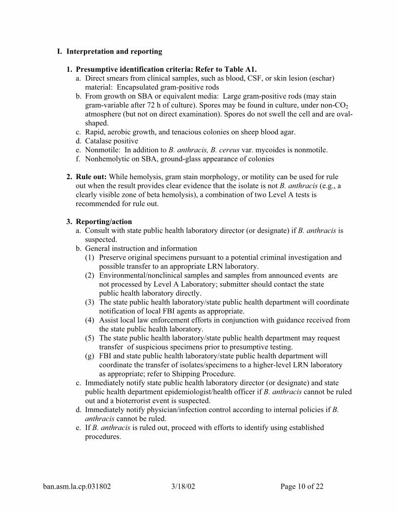
I. Interpretation and reporting
1. Presumptive identification criteria: Refer to Table A1. a. Direct smears from clinical samples, such as blood, CSF, or skin lesion (eschar)
material: Encapsulated gram-positive rods b. From growth on SBA or equivalent media: Large gram-positive rods (may stain
gram-variable after 72 h of culture). Spores may be found in culture, under non-CO2 atmosphere (but not on direct examination). Spores do not swell the cell and are oval-shaped.
c. Rapid, aerobic growth, and tenacious colonies on sheep blood agar. d. Catalase positive e. Nonmotile: In addition to B. anthracis, B. cereus var. mycoides is nonmotile. f. Nonhemolytic on SBA, ground-glass appearance of colonies
2. Rule out: While hemolysis, gram stain morphology, or motility can be used for rule out when the result provides clear evidence that the isolate is not B. anthracis (e.g., a clearly visible zone of beta hemolysis), a combination of two Level A tests is recommended for rule out.
3. Reporting/action a. Consult with state public health laboratory director (or designate) if B. anthracis is
suspected. b. General instruction and information
(1) Preserve original specimens pursuant to a potential criminal investigation and possible transfer to an appropriate LRN laboratory.
(2) Environmental/nonclinical samples and samples from announced events are not processed by Level A Laboratory; submitter should contact the state public health laboratory directly.
(3) The state public health laboratory/state public health department will coordinate notification of local FBI agents as appropriate.
(4) Assist local law enforcement efforts in conjunction with guidance received from the state public health laboratory.
(5) The state public health laboratory/state public health department may request transfer of suspicious specimens prior to presumptive testing.
(g) FBI and state public health laboratory/state public health department will coordinate the transfer of isolates/specimens to a higher-level LRN laboratory
as appropriate; refer to Shipping Procedure. c. Immediately notify state public health laboratory director (or designate) and state
public health department epidemiologist/health officer if B. anthracis cannot be ruled out and a bioterrorist event is suspected.
d. Immediately notify physician/infection control according to internal policies if B. anthracis cannot be ruled.
e. If B. anthracis is ruled out, proceed with efforts to identify using established procedures.
ban.asm.la.cp.031802 3/18/02 Page 10 of 22

III. References
Brachman, P.S., and A.M. Friedlander. Anthrax, p. 729-739. In S.A. Plotkin and E.A. Mortimer, Jr. (ed), Vaccines. W.B. Saunders, Philadelphia, PA.
Cieslak, T.J. and E.M. Eitzen, Jr. 1999. Clinical and epidemiologic principles of anthrax. Emerg. Infect. Dis. 5:552-555.
Dutz, W. and E. Kohout. 1971. Anthrax. Pathol. Annu. 6:209-248.
Gilchrist, M.J.R., W.P. McKinney, J.M. Miller, and A.S. Weissfeld. 2000. Cumitech 33, Laboratory Safety, management , and diagnosis of biological agents associated with bioterrorism. Coordinating ed., J.W. Snyder. ASM Press, Washington, D.C.
Lew, D. P. 2000. Bacillus anthracis (Anthrax). p. 2215-2220. In G. L. Mandell, J.E. Bennett, and R. Dolin (ed), Principles and Practice of Infectious Disease, 5th ed. Churchill Livingston, Philadelphia, PA.
Logan, N.A. and P.C. Turnbull. 1999. Bacillus and recently derived genera, p. 357- 369. In P.R. Murray, E.J. Baron, M.A. Pfaller, F.C. Tenover, and R.H. Yolken (ed) manual of Clinical Microbiology, 7th ed. American Society for Microbiology, Washington, D.C.
ban.asm.la.cp.031802 3/18/02 Page 11 of 22

ban.asm.la.cp.031802 3/18/02 Page 12 of 22

Figure A2. Gram stain of B. anthracis in rhesus monkey blood, magnification 1000X
ban.asm.la.cp.031802 3/18/02 Page 13 of 22

Figure A3a. Gram stain of B. anthracis from SBA, magnification 1000X
ban.asm.la.cp.031802 3/18/02 Page 14 of 22
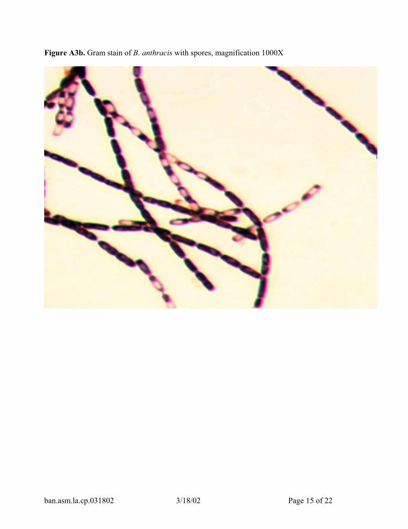
Figure A3b. Gram stain of B. anthracis with spores, magnification 1000X
ban.asm.la.cp.031802 3/18/02 Page 15 of 22

Figure A4. B. anthracis colony morphology; overnight cultures on SBA
ban.asm.la.cp.031802 3/18/02 Page 16 of 22

Figure A5. Tenacious colonies of B. anthracis on SBA
ban.asm.la.cp.031802 3/18/02 Page 17 of 22

Figure A6. B. anthracis and B. cereus colony morphology; overnight cultures of B. cereus (left side of plate) and B. anthracis (right side) on SBA.
ban.asm.la.cp.031802 3/18/02 Page 18 of 22

Table A1. Presumptive identification of Bacillus anthracis Lab Level
Type of sample
Presumptive identification
Characteristic Method
A
Clinical sample
1. Gram-positive rods AND
2. Capsule
Gram stain India ink stain
A
Isolate
1 Spore-former AND
2. Colony morphology AND
3. Nonhemolytic AND
4. Nonmotile
Gram stain Observation on SBA Observation on SBA Motility medium or Wet mount
ban.asm.la.cp.031802 3/18/02 Page 19 of 22

IV. Appendices
A: Appendix A: India ink stain
1. Purpose: This optional test is used to improve visualization of encapsulated B. anthracis in clinical samples such as blood, blood culture bottles, or cerebrospinal fluid (CSF). This is not an ASM recommended procedure; performance of the test requires staff that are trained/ experienced with this procedure.
2. Quality control
a. Positive control strain: Klebsiella pneumoniae (or laboratory validated equivalent) will demonstrate a well-defined clear zone on SBA.
b. Negative control strain: E. coli ATCC 25922 (or laboratory validated equivalent) will demonstrate no clear zone.
c. Method controls: Perform the test with suspensions of fresh cultures of the control strains. Control strains should be assayed on each day of testing.
d. Resolving out-of-control results: Check media, reagents, controls and equipment; replace or correct as appropriate. Document corrective actions and repeat test.
3. Procedure
a. For the controls, transfer a small amount of growth (1 mm diameter) from each control SBA plate (positive control = Klebsiella pneumoniae; negative control = Escherichia coli ATCC 25922) into 0.5 ml saline and mix.
b. For the unknowns, take 100 µl of sample (blood, CSF). Transfer 5-10 µl of unknown sample or control to a slide. Place a coverslip on the drop, and then add 5-10 µl of India ink to the edge of the coverslip. After the ink diffuses across the slide, view the cells using 100X oil immersion objective with oil on top of the coverslip.
4. Interpretation a. Positive result: The capsule will appear as a well-defined clear zone around the
cells. b. Negative result: No zone will be present.
5. Reporting/actions
a. Clinical specimens with encapsulated (visualized with India Ink), gram-positive rods provide a presumptive identification of B. anthracis; it does not confirm B. anthracis.
b. Every effort should be made to obtain an isolate for continued testing and referral to state public health laboratory.
6. Limitations a. Interpretation of results requires trained/experienced staff.
b. A negative test result should not be used to rule out B. anthracis.
ban.asm.la.cp.031802 3/18/02 Page 20 of 22

B. Appendix B: Nasal specimens for screening Bacillus anthracis
1. General: Nasal specimens (nares culture) should ONLY be used to support a confirmed exposure to B. anthracis or during an ongoing epidemiologic investigation. Gram stain of nasal specimens for B. anthracis spores are not recommended. Refer to limitations section below.
2. Materials: Swab (Dacron, rayon or other synthetic swabs are preferred over cotton) and transport medium for culture.
3. Procedure
a. Selection (1) The specimen of choice is a swab specimen taken at least 1 cm inside the nares. (2) Lesions in the nose require samples from the advancing margin of the lesions.
b. Method (1) Carefully insert the moistened swab (saline, sterile water) at least 1 cm into the nares. (2) Firmly sample the inside of the nares by rotating the swab and leaving it in place for 10 to 15 sec. (3) Withdraw the swab, insert it into its transport container, and submit the sampling unit to the laboratory for culture.
c. Labeling (1) Label the swab container with patient information. (2) Indicate, if possible, the degree or likelihood of exposure.
d. Transport (1) Transport the specimen to the laboratory as soon as possible. (2) Do not refrigerate specimens for culture. e. Culture: Heat Shock
(1) Remove the swab from transport container and place it into 1.5 ml of sterile saline or a nutrient broth such as trypticase soy broth, brain heart infusion broth, or equivalent. Vigorously twist the swab, and recap the tube. (2) Leave the swab in the tube. Place the broth suspension into a 65oC water bath for 30 min.
(3) Plate 100-200 µl of broth on 5% sheep blood agar plate and incubate at 35-37oC for 18-24 h. Many B. anthracis will have visible growth in 12-18 h. Observe for characteristics of B. anthracis.
4. Interpretation: Observe colony morphology for typical Bacillus colonies, look for lack of hemolysis, perform Gram stain, and evaluate for B. anthracis characteristics as described in the Level A laboratory protocol.
5. Reporting: If B. anthracis cannot be ruled out, submit the isolate to the state public health laboratory/department for confirmation. Refer to Level A reporting section.
6. Limitations: Nasal cultures taken to evaluate for the presence of anthrax spore have not been evaluated for sensitivity or specificity. Nasopharyngeal and throat
ban.asm.la.cp.031802 3/18/02 Page 21 of 22
specimens are not recommended for anthrax screens and should not be submitted. Nasal cultures are NOT recommended for screening those who are asymptomatic and
without known exposure.
7. Procedure Notes a. Anterior nares cultures, without an indication of the presence of a lesion, are
routinely examined only for presence of Staphylococcus aureus and β-hemolytic streptococci. Because of the unknown sensitivity of this method for detecting B. anthracis spores, interpret negative results with caution. b. Anterior nares cultures cannot be used to predict a subsequent infection with B.
anthracis, and should not be submitted in lieu of blood and other appropriate specimens from symptomatic patients.
c. Anaerobic cultures are not done on nasal specimens. B. anthracis produces spores in culture only when grown in air.
d. Nasal swabs may also be plated directly onto sheep blood agar prior to or without heat shocking, however normal nasal flora may overgrow very low numbers of Bacillus colonies.
e. Pediatric needs: Use the same procedure substituting a small fine-wire or nasopharyngeal swab to sample the anterior nares.
C. Appendix C: Change record 1. 18 Mar. 2002 (ban.asm.cp.la.021402) a. Recommend use of TSB, or equivalent, for wet mount procedure (II. H. 2. a.) b. Added criteria for rule out at Level A (II. I. 2.) 2. 06 Nov. 2001 (ban.asm.cp.la.110501) a. Figure A1 and text (II. F. 2. c.) revised to state “frequently” occur as long chains of bacilli. b. File name, date, page information added to footer. c. Change record added to appendices 3. 29 Oct. 2001 (ban.asm.cp.la.102901f) a. Nasal swab procedure added. b. File name, date, page information deleted from footer (an error). 4. 24 Oct. 2001 (ban.asm.cp.la.102401f): Original ASM/APHL/CDC Level A procedure for B. anthracis loaded to ASM public site.
ban.asm.la.cp.031802 3/18/02 Page 22 of 22

Revised 7.30.03
SENTINEL LABORATORY GUIDELINES FOR
SUSPECTED AGENTS OF BIOTERRORISM
Botulinum Toxin
American Society for Microbiology
2
Credits: Botulinum Toxin Subject Matter Experts, ASM Daniel S. Shapiro, M.D. Lahey Clinic Burlington, MA Alice S. Weissfeld, Ph.D. Microbiology Specialists Incorporated Houston, TX ASM Laboratory Protocol Working Group Vickie Baselski, Ph.D. University of Tennessee at Memphis Memphis, TN Roberta B. Carey, Ph.D. Peter H. Gilligan, Ph.D. University of North Carolina Hospitals/Clinical Microbiology and Immunology Labs Chapel Hill, NC Larry Gray, Ph.D. TriHealth Laboratories and University of Cincinnati College of Medicine Cincinnati, OH Rosemary Humes, MS, MT (ASCP) SM Association of Public Health Laboratories Silver Spring, MD Karen Krisher, Ph.D. Clinical Microbiology Institute Portland (Wilsonville), OR Judith Lovchik, Ph.D. Public Health Laboratories, NYCDOH New York, NY Chris N. Mangal, MPH Association of Public Health Laboratories Silver Spring, MD

3
Susan E. Sharp, Ph.D. Kaiser Permanente Portland, OR David Welch, Ph.D. Medical Microbiology Consulting Dallas, TX Mary K. York, Ph.D. MKY Microbiology Consultants Walnut Creek, CA Coordinating Editor: James W. Snyder, Ph.D. University of Louisville Louisville, KY

4
Table of Contents: Botulinum toxin
I. General Information A. Description of Organism B. History C. Geographic Distribution D. Clinical Presentation
II. Procedure
A. General B. Precautions C. Specimens
1. Acceptable specimens a. Feces b. Enema c. Gastric aspirate or vomitus d. Serum e. Tissue or exudates f. Postmortem g. Food specimens h. Swab samples (environmental or clinical) i. Environmental samples
(i) Soil (ii) Water
2. Specimen handling 3. Rejection criteria
D. Materials 1. Supplies
E. Shipping F. Limitations
III. References IV. Appendices
A. Suggested specimens based on form of botulism B. Specimen-related information

5
I. GENERAL INFORMATION
A. Description of organism
Clostridium botulinum is a group of anaerobic organisms, commonly found in soils and aquatic habitats throughout the world, that are alike only in that they are clostridia and produce antigenically distinct neurotoxins with similar pharmacologic actions. C. botulinum organisms are straight to slightly curved, gram-positive (in young cultures), motile, anaerobic rods, 0.5 to 2.0 µm in width and 1.6 to 22.0 µm in length, with oval, subterminal spores. The seven types of C. botulinum (A to G) are distinguished by the antigenic characteristics of the neurotoxins they produce. Human botulism is primarily caused by the strains of C. botulinum that produce toxin types A, B, and E, but rare cases of type F have been reported. Also, rare cases of human botulism by C. butyricum-like and C. baratii-like organisms have been reported to produce botulinum type E and F toxins, respectively. C. botulinum type G, which has been reclassified as Clostridium argentinense, has at the case report level been suggested as a cause of sudden, unexpected death, but a clearly causal relationship has not been established.
B. History
Worldwide, sporadic cases and small outbreaks occur where food products are prepared or preserved by methods that do not destroy the spores and permit toxin formation. Cases rarely result from commercially processed products, but outbreaks have occurred through cans that were damaged after processing. The potential for intentional poisoning with botulinum toxin is now a realistic threat. Inhalation and foodborne botulism are the likely forms of disease following a bioterrorist event. It is estimated that as little as 1 g of aerosolized botulinum toxin has the potential to kill at least 1.5 million people. Botulinum toxin is absorbed through the lungs and into the bloodstream. Three cases of human inhalational botulism were reported in 1962 in veterinary technicians in Germany who were working with aerosolized botulinum toxin in animals. Symptoms occurred approximately 72 h after exposure.
Past efforts to weaponize botulinum toxin include a U.S. weapons program beginning in World War II and ending after the 1972 Biological and Toxin Weapons Convention, research conducted in the former Soviet Union and Iraq as late as the 1990s, and the attempted use of aerosolized botulinum toxin in Japanese cities by the Aum Shinrikyo cult on at least three occasions in the 1990s.
Contamination of a municipal water supply is unlikely, since it would require a large quantity of toxin. Moreover, toxin is naturally inactivated in freshwater within 3 to 6 days and rapidly (within 20 min) inactivated by standard municipal potable water treatments.
In April 2002, the FDA approved the use of botulinum toxin type A for cosmetic purposes. Therapeutic botulinum toxin contains about 0.3% of the estimated lethal human inhalation dose and only 0.005% of the estimated lethal human oral dose. Therefore, this form of toxin is not likely to be used as a bioweapon (see Suggested Reading).
C. Geographic distribution
C. botulinum spores are ubiquitous in soil worldwide. Approximately 100 cases are reported in the U.S. each year. Five western states (California, Washington, Colorado, Oregon, and

6
Alaska) have accounted for more than half of all reported foodborne outbreaks since 1950. Alaska alone accounts for 16% of these outbreaks, due in great part to the consumption of fermented seafood, seals, whales, and other mammal meat products contaminated with toxin-producing clostridia.
D. Clinical presentation
Four distinct forms of botulism have occurred in humans: (i) foodborne, (ii) wound, (iii) infant, and (iv) child or adult non-foodborne. Foodborne botulism results from the ingestion of food containing preformed toxin. Wound botulism is caused by organisms that multiply and produce toxin in a contaminated wound, most commonly in injection drug users. Infant botulism is due to the endogenous production of toxin by germinating spores of C. botulinum in the intestine of the infant. Child or adult botulism is represented by those cases in which no food vehicle can be identified, there is no evidence of wound botulism, and there is the possibility of intestinal colonization in a person older than 1 year of age. Important epidemiologic features and some clinical characteristics distinguish the types of botulism that cause human illness.
The clinical syndrome of botulism is dominated by the neurologic symptoms and signs resulting from a toxin-induced blockade of the voluntary motor and autonomic cholinergic junctions. Incubation periods for foodborne botulism are reported to be as short as 6 h or as long as 10 days, but generally the time between toxin ingestion and onset of symptoms ranges from 18 to 36 h. The ingestion of other bacteria or their toxins in improperly preserved food or changes in bowel motility are likely to account for the abdominal pain, nausea, vomiting, and diarrhea that often precede or accompany the neurologic symptoms of foodborne botulism. Dryness of the mouth, inability to focus to a near point (prompting the patient to complain of "blurred vision"), and diplopia (double vision) are usually the earliest neurologic complaints. If the disease is mild, no other symptoms may develop, and the initial symptoms will gradually resolve. The person with mild botulism may not come to medical attention. In more severe cases, however, these initial symptoms may be followed by voice impairment (dysphonia, dysarthria), difficulty swallowing (dysphagia), and peripheral muscle weakness. If illness is severe, respiratory muscles become involved, leading to respiratory failure and death unless supportive care is provided. Recovery follows the regeneration of new neuromuscular connections. A 2- to 8-week duration of respiratory support is common, although patients have required respiratory support for up to 7 months before the return of muscular function. Death occurs in 5 to 10% of cases of foodborne botulism; early deaths result from a failure to recognize the severity of disease or from secondary pulmonary or systemic infections, whereas deaths after 2 weeks are usually from the complications of long-term mechanical respiratory management.
Animal studies have shown that botulinum toxins produce similar effects whether inhaled or ingested. Presumably the gastrointestinal symptoms present in foodborne botulism would be absent following inhalation. The onset of symptoms of inhalational botulism in animals extends from 24 h to 2 days, depending on the extent of exposure.
The administration of antitoxin is the only specific therapy available for botulism, and evidence suggests that it is effective only if given very early in the course of neurologic dysfunction. Thus, the diagnosis of this illness cannot await the results of studies that may be

7
long delayed and only confirmatory in some cases. The diagnosis and the decision to treat should be made on the basis of the case history and physical findings.
Botulism is not transmitted from person to person. However, even a single diagnosis should be considered a possible public health emergency situation due to the possibility of other common source cases. Since there are cases that are acquired in the absence of bioterrorism, clinicians should look for clusters of cases of an acute onset, afebrile, symmetric, descending flaccid paralysis that begins in the bulbar muscles and includes dilated pupils and dry mucous membranes but normal mental status and an absence of sensory changes. Botulism needs to be differentiated from other neurological diseases, including Landry-Guillain-Barre syndrome, tick paralysis, myasthenia gravis, and Lambert-Eaton syndrome. Once a presumptive clinical diagnosis is made, an intense epidemiologic investigation should ensue to identify other related cases.
II. PROCEDURE
A. General. Laboratory Response Network (LRN) Level A (Sentinel) laboratory procedures are designed to ensure the proper collection and distribution of appropriate specimens to designated testing laboratories.
B. Precautions 1. The suspicion of botulism is a public health emergency: notify both local public
health officials and the state public health laboratory for approval to submit samples for testing. Submit specimens without delay.
2. DO NOT attempt to culture, identify the organism, or attempt to perform toxin analysis. 3. Sentinel laboratories should not accept environmental or animal specimens; such
specimens should be forwarded directly to the state health laboratory.
C. Specimen
1. Acceptable specimens (for testing at Level C [LRN Reference] laboratories) a. Feces. Place into sterile unbreakable container and label carefully.
Confirmatory evidence of botulism may be obtained from 10- to 50-g quantities (walnut size); botulism has been confirmed in infants with only "pea-size" stool samples.
b. Enema. Place approximately 20 ml into a sterile unbreakable container and label carefully. If an enema must be given because of constipation, a minimal amount of fluid (preferably sterile, nonbacteriostatic water) should be used to obtain the specimen so that the toxin will not be unnecessarily diluted.
c. Gastric aspirate or vomitus. Place approximately 20 ml into a sterile unbreakable container and label carefully.
d. Serum. Use red top or serum separator tubes to obtain serum (no anticoagulant). Samples should be obtained as soon as possible after the onset of symptoms and before antitoxin is given. Enough blood should be collected to provide at least 10 ml of serum for mouse toxicity tests (usually 20 ml of whole blood); serum volumes less than 3 ml will provide inconclusive results. Whole blood should not be sent, because it typically undergoes excessive hemolysis during transit.

8
e. Tissue or exudates. Place into sterile unbreakable container and label carefully. Specimens should be placed in anaerobic transport media and sent to the appropriate laboratory for attempted isolation of C. botulinum.
f. Postmortem. Obtain specimens of intestinal contents from different levels of small and large intestines. Place approximately 10 g per specimen into a sterile unbreakable container and label carefully. Obtain gastric content, serum, and tissue specimens if or as appropriate.
g. Food specimens. Foods should be left in their original containers if possible, or placed in sterile unbreakable containers and labeled carefully. Place containers individually in leakproof containers (e.g., sealed plastic bags) to prevent cross-contamination during shipment. Empty containers with remnants of suspected foods can be examined.
h. Swab samples (environmental or clinical). Send clinical swabs in an anaerobic transport medium. Environmental swabs (from which spores may be isolated) may be sent in plastic containers without any medium. Swabs may be shipped at room temperature or refrigerated. Collect three to four swabs from each potential site. NOTE: Sentinel laboratories should not accept environmental (or animal) specimens: such specimens should be directly forwarded to the appropriate LRN Reference laboratory.
i. Environmental samples. Collect a sample in the size indicated below for each possible location. (i) Soil (50 to 100 g)
(ii) Water (~100 ml) NOTE: Sentinel laboratories should not accept environmental (or animal) specimens. Such specimens should be directly forwarded to the appropriate LRN Reference laboratory.
2. Specimen handling a. Store all specimens at 4°C and ship on cold packs as soon as possible. b. Submit to a LRN Reference laboratory as soon as possible.
3. Rejection criteria a. Incomplete documentation. All specimens must include the sender's name and
telephone number to contact for the preliminary report and additional information. b. Improper packaging/shipping c. Lack of prior approval. Do not ship specimens to higher-level LRN laboratories
without prior approval.
D. Materials
1. Supplies a. Anaerobic transport vials b. Leakproof containers (i.e., sealed plastic bags and other plastic containers) c. Packaging materials (Refer to ASM procedure on Transportation and Transfer of
Biological Agents http://www.asm.org/Policy/index.asp?bid=6342)
E. Shipping. Refer to ASM procedure on Transportation and Transfer of Biological Agents http://www.asm.org/Policy/index.asp?bid=6342. Complete and attach appropriate documentation.

9
F. Submitting samples to LRN Reference laboratory 1. Submit samples to a laboratory approved to perform testing as directed by the state
public health laboratory. Note that not all states currently have testing capability. 2. Toxin testing is performed primarily by a mouse toxicity and toxin neutralization
method. Final results may be expected after 3 days. G. Limitations. If the patient has been taking any medication that might interfere with toxin
assays or culturing of the stool, the laboratory should be notified. For example, it has been demonstrated that anticholinesterase drugs given orally to patients for myasthenia gravis can interfere with mouse botulinum toxin assays of stool extracts.
III. REFERENCES
1. Allen, S. D., C. L. Emery, and D. M. Lyerly. 2003. Clostridium, p. 835-856. In P. R. Murray, E. J. Baron, M. A. Pfaller, F. C. Tenover, and R. H. Yolken (ed.), Manual of clinical microbiology, 8th ed. ASM Press, Washington, D.C.
2. Arnon, S. S. 1992. Infant botulism, p. 1095-1102. In R. Feigen and J. Cherry (ed.),
Textbook of pediatric infectious diseases. W. B. Saunders, Philadelphia, Pa. 3. Cato, E. P., W. L. George, and S. M. Finegold. 1986. Genus Clostridium,
Praemozski 1880, 23AL, p. 1141-1200. In P. H. A. Sneath, N. S. Mair, M. E. Sharp, and J. G. Holt (ed.), Bergey’s manual of systematic bacteriology, vol. 2.Williams & Wilkins, Baltimore, Md.
4. Centers for Disease Control and Prevention. 1976. Follow-up: botulism associated
with commercial cherry peppers. Morb. Mortal. Wkly. Rep. 25:148. 5. Centers for Disease Control and Prevention. 1998. Botulism in the United States,
1899-1996. Handbook for epidemiologists, clinicians, and laboratory workers. Centers for Disease Control and Prevention, Atlanta, Ga.
6. Danzig, R., 1996. Biological warfare: a nation at risk–a time to act. Strategic Forum
58:1-4. 7. Eitzen, E., J. Pavlin, T. Cieslak, G. Christopher, and R. Culpepper. July 1998.
Medical management of biological casualties handbook, 3rd ed. U.S. Army Medical Research Institute of Infectious Diseases, Fort Detrick, Md.
8. Ekeus, R. 1991. Report of the Secretary General on the status of the implementation of
the Special Commission’s plan for the ongoing monitoring and verification of Iraq’s compliance with relevant parts of Sector C of Security Council Resolution 687, United Nation’s Special Commission (UNSCOM). United Nations, New York, N.Y.
9. Hatheway, C. L. 1995. Botulism: the present status of the disease. Curr. Top.
Microbiol. Immunol. 195:55-75.

10
10. Hughes, J. M., J. R. Blumenthal, M. H. Merson, G. L. Lombard, V. R. Dowell, Jr.,
and E. J. Gangarosa. 1981. Clinical features of types A and B foodborne botulism. Ann. Intern. Med. 95:442-445.
11. Long, S. S., J. L. Gajewski, L. W. Brown, and P. H. Gilligan. 1985. Clinical,
laboratory, and environmental features of infant botulism in southeastern Pennsylvania. Pediatrics 75:935-941.
12. Merson, M. H., and V. R. Dowell, Jr. 1973. Epidemiologic, clinical and laboratory
aspects of wound botulism. N. Engl. J. Med. 289:1105-1110.
13. Shapiro, R. L., C. Hatheway, S. Becher, and D. L. Swerdlow. 1997. Botulism surveillance and emergency response. A public health strategy for a global challenge. JAMA 278:433-435.
14. Shapiro, R. L., C. Hatheway, and D. L. Swerdlow. 1998. Botulism in the United
States: a clinical and epidemiologic review. Ann. Intern. Med. 129:221-228.
15. St. Louis, M. E., S. H. Peck, D. Bowering, G. B. Morgan, J. Blatherwick, S. Banerjee, G. D. Kettyls, W. A. Black, M. E. Milling, A. H. Hauschild et al. 1988. Botulism from chopped garlic: delayed recognition of a major outbreak. Ann. Intern. Med. 108:363-368.
16. Tackett, C. O., W. X. Shandera, J. M. Mann, N. T. Hargrett, and P. A. Blake.
1984. Equine antitoxin use and other factors that predict outcome in type A foodborne botulism. Am. J. Med. 76:794-798.
17. Terranova, W., J. G. Breman, R. P. Locey, and S. Speck. 1978. Botulism type B:
epidemiologic aspects of an extensive outbreak. Am. J. Epidemiol. 108:150-156.
18. Woodruff, B. A., P. M. Griffin, L. M. McCroskey, J. F. Smart, R. B. Wainwright, R. G. Bryant, L. C. Hutwagner, and C. L. Hatheway. 1992. Clinical and laboratory comparison of botulism from toxin types A, B, and E in the United States 1975-1988. J. Infect. Dis. 166:1281-1286.
Suggested Reading
1. Infectious Diseases Society of America. 2002. Bioterrorism information and resources. http://www.idsociety.org.
2. Infectious Diseases Society of America. Botulism, 20 February 2003.
http://www.idsociety.org/bt/biotemplate.cfm?template=bot_summary.htm.

11
IV. APPENDICES
Appendix A. Suggested specimens based on form of botulism
1. Foodborne a. Clinical material. Serum, gastric contents, vomitus, stool, return from sterile water
enema or saline enema b. Autopsy samples. Intestinal contents and gastric contents (serum if available) c. Food samples
2. Infant
a. Feces b. Return from sterile water or sterile saline enema c. Serum. Although circulating toxin may be detected in infants with botulism, it is rare.
Shipment of other specimens should not be delayed while waiting for serum collection. d. Postmortem samples. Intestinal contents from different levels of small and large
intestine e. Food and environmental samples (as appropriate for the investigation)
3. Wound
a. Serum b. Exudate, tissue, or swab samples of wound transported in an anaerobic transport
medium c. Feces or return from sterile water enema (wound may not be source) d. An isolate of suspected C. botulinum (maintain under anaerobic conditions)
4. Intentional toxin release (inhalational or ingested)
a. Serum b. Feces or return from sterile water enema c. Food, solid or liquid d. Environmental or nasal swabs e. Gastric aspirate
Appendix B. Specimen-related information
1. Food a. Foods most likely to allow growth of C. botulinum will have a pH range of 3.5 to 7.0;
the most common pH is 5.5 to 6.5. However, suspected foods, regardless of pH, can be examined, since localized environmental conditions may be present that may support the growth of C. botulinum.
b. Botulinum toxin in commercial products is rare. The state public health laboratory should notify the FDA at (301) 443-1240 if a commercial product is suspected of containing botulinum toxin.
2. Feces. C. botulinum has been isolated from stools following antitoxin treatment.
Revised 15 October 2004
SENTINEL LABORATORY GUIDELINES
FOR
SUSPECTED AGENTS OF BIOTERRORISM
Brucella species
ASM American Society for Microbiology

Credits: Brucella Species ASM Subject Matter Experts Peter H. Gilligan, Ph.D., ABMM Clinical Microbiology-Immunology Laboratories University of North Carolina Hospitals Chapel Hill, NC Mary K. York, Ph.D., ABMM Clinical Microbiology Consultant Walnut Creek, CA ASM Laboratory Protocol Working Group Vickie Baselski, Ph.D. University of Tennessee at Memphis Memphis, TN Roberta B. Carey, Ph.D. Karen Krisher, Ph.D., D (ABMM) Clinical Microbiology Institute Wilsonville, OR Judith Lovchik, Ph.D. Public Health Laboratories, NYCDOH New York, NY Larry Gray, Ph.D. TriHealth Laboratories and University of Cincinnati College of Medicine Cincinnati, OH Rosemary Humes, MS, MT (ASCP) SM Association of Public Health Laboratories Washington, DC Chris N. Mangal, MPH Association of Public Health Laboratories Washington, DC Michael A. Saubolle Ph.D. D(ABMM) Banner Good Samaritan Medical Center Laboratory Sciences of Arizona Banner Health System

3
Phoenix, AZ Daniel S. Shapiro, M.D. Lahey Clinic Burlington, MA Susan E. Sharp, Ph.D. Kaiser Permanente Portland, OR Alice Weissfeld, Ph.D. Microbiology Specialists, Inc. Houston, TX David Welch, Ph.D. Medical Microbiology Consulting Dallas, TX Coordinating Editor James W. Snyder, Ph.D. University of Louisville Louisville, KY

4
Table of Contents: Brucella Species
I. General Information A. Description of organism B. History C. Geographic distribution D. Clinical presentation
II. Procedures A. General B. Precautions C. Specimen D. Materials
1. Media 2. Reagents 3. Equipment and supplies
E. Quality Control F. Stains and smears G. Further testing H. Interpretation and reporting
1. Major characteristics 2. Presumptive identification
3. Reporting/appropriate action I. Limitations
III. References IV. Appendix
A. Change record
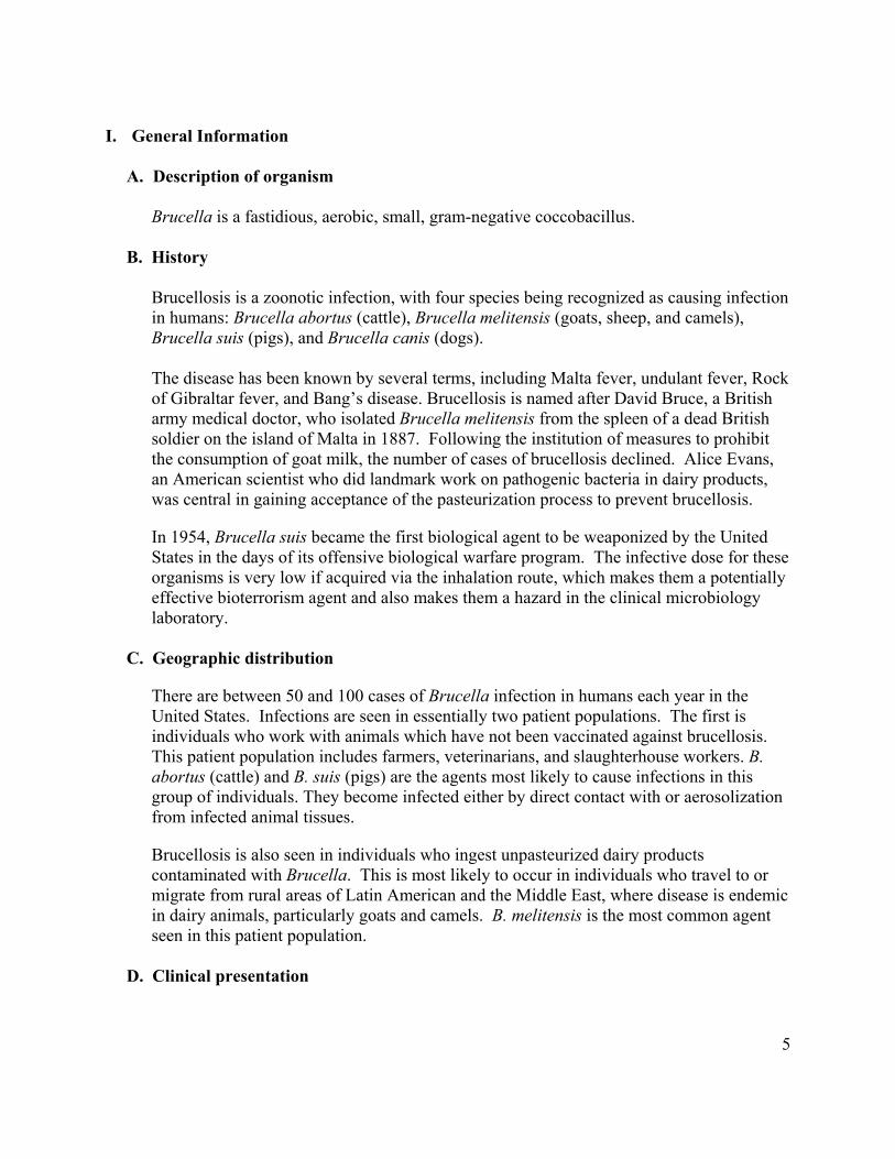
5
I. General Information
A. Description of organism
Brucella is a fastidious, aerobic, small, gram-negative coccobacillus.
B. History
Brucellosis is a zoonotic infection, with four species being recognized as causing infection in humans: Brucella abortus (cattle), Brucella melitensis (goats, sheep, and camels), Brucella suis (pigs), and Brucella canis (dogs). The disease has been known by several terms, including Malta fever, undulant fever, Rock of Gibraltar fever, and Bang’s disease. Brucellosis is named after David Bruce, a British army medical doctor, who isolated Brucella melitensis from the spleen of a dead British soldier on the island of Malta in 1887. Following the institution of measures to prohibit the consumption of goat milk, the number of cases of brucellosis declined. Alice Evans, an American scientist who did landmark work on pathogenic bacteria in dairy products, was central in gaining acceptance of the pasteurization process to prevent brucellosis.
In 1954, Brucella suis became the first biological agent to be weaponized by the United States in the days of its offensive biological warfare program. The infective dose for these organisms is very low if acquired via the inhalation route, which makes them a potentially effective bioterrorism agent and also makes them a hazard in the clinical microbiology laboratory.
C. Geographic distribution
There are between 50 and 100 cases of Brucella infection in humans each year in the United States. Infections are seen in essentially two patient populations. The first is individuals who work with animals which have not been vaccinated against brucellosis. This patient population includes farmers, veterinarians, and slaughterhouse workers. B. abortus (cattle) and B. suis (pigs) are the agents most likely to cause infections in this group of individuals. They become infected either by direct contact with or aerosolization from infected animal tissues.
Brucellosis is also seen in individuals who ingest unpasteurized dairy products contaminated with Brucella. This is most likely to occur in individuals who travel to or migrate from rural areas of Latin American and the Middle East, where disease is endemic in dairy animals, particularly goats and camels. B. melitensis is the most common agent seen in this patient population.
D. Clinical presentation

6
Brucella can cause both acute and chronic infections. The symptoms of brucellosis are non-specific and systemic, with fever, sweats, headache, anorexia, back pain, and weight loss being frequent. The chronic form of the disease can mimic miliary tuberculosis with suppurative lesions in the liver, spleen, and bone. The organism is often included in the differential diagnosis of fevers of unknown origin. It has a mortality of 5% in untreated individuals.

7
II. Procedures: Brucella species A. General: The procedures described below function to rule out suspected Brucella
species using specimens and isolates.
B. Precautions: All patient specimens should be handled while wearing gloves and gowns and working in a biosafety cabinet. Subcultures should be performed in a biosafety cabinet and incubated in 5 to 10% CO2. Plates should be taped shut, and all further testing should be performed only in the biosafety cabinet.
C. Specimens
1. Blood or bone marrow 2. Spleen, liver, joint fluid or abscesses are occasionally sources of Brucella ssp. 3. Serum (at least 1ml)-For serologic diagnosis, an acute-phase specimen should be
collected as soon as possible after onset of disease. A convalescent-phase specimen should be collected > 14 days after the acute specimen.
D. Materials
1. Media a. Blood, bone marrow or joint fluid culture. Choose one of the following.
i. Standard liquid blood culturing system ii. Biphasic system such as
a. Septi-Chek BBL Septi-Chek (B-D Microbiology Systems, Cockeysville, MD)
b. PML biphasic (PML Microbiologicals, Inc., Wilsonville, OR)
iii. Lysis-centrifugation system of ISOLATOR (Wampole Laboratories, Cranbury, NJ
b. Media for subculturing of positive blood culture bottles
i. Sheep blood agar (BAP) ii. Chocolate agar (CHOC) iii. MacConkey (MAC) or EMB agar
2. Reagents
a. Gram stain b. Catalase (3% hydrogen peroxide) c. Oxidase (0.5 tetramethyl-p-phenylenediamine) d. Urea agar (Christensen’s) or rapid urea disks (3) (Remel, Inc; Key Scientific; or Hardy Diagnostics) e. Culture of Staphylococcus aureus ATCC 25923
2. Equipment and supplies
a. Blood culture instrument (optional)

8
b. 35°C incubator with 5-10% CO2 c. Light microscope with 100X objective and 10X eyepiece d. Microscope slides, coverslips, disposable bacteriologic inoculating loops
Disclaimer: Names of vendors or manufacturers are provided as examples of suitable product sources; inclusion does not imply endorsement by the American Society for Microbiology, or any other contributor.
E. Quality Control: Perform quality control of media and reagents according to package inserts, NCCLS document M22-A2, and CLIA standards, using positive and negative controls. Do not use Brucella spp. as a control organism, due to its infectious nature.
F. Stains and smears:
B. Procedures: 1. Aseptically inoculate liquid blood culture bottles with maximum amount of blood or body fluid per manufacturers’ instructions. Incubate at 35°C. (See below for subculture method).
i. Incubate non-automated broth blood cultures for 21 days, with blind subculturing every 7 days, followed by terminal subculturing of negative blood cultures and holding sealed plates for 7 additional days. ii. Incubate automated systems for 10 days and perform terminal subcultures at 7 days to increase yield (10).
NOTE: Isolation of Brucella is often delayed compared to other bloodstream pathogens, with peak isolation occurring at 3 to 4 days compared to 6 to 36 h for most other pathogens. Although incubation time of 21 days with weekly or terminal blind subculture are advocated, careful studies in Brucella-endemic areas using the BACTEC 9240 system (B D Division Instrument Systems, Sparks, MD) suggest that a maximal incubation time of 10 days is sufficient for reliable recovery of this organism, with 93% of 97 patient isolates being detected in 5 days (1). For the BacT Alert system (Biomerieux Inc., Hazelwood, MO), terminal subcultures at 7 days increased yield (10). Lysis-centrifugation has been shown to be less sensitive than broth-based systems for pediatric specimens (11). There is very limited published data with the ESP system (TREK Diagnostics, Westlake OH), so its effectiveness in the recovery of Brucella is unknown (10).
2. For tissues, inoculate BAP, CHOC and MAC or EMB and incubate for up to 7 days at 35°C in a humidified incubator with 5 to 10% CO2. Humidity may be maintained by placing a pan of water in the bottom of the incubator or by wrapping the plates with gas permeable tape.
NOTE: Brucella has been responsible for many laboratory-acquired infections (4,6,8). If Brucella is suspected or the Gram stain shows a small, gram-negative coccobacillus, avoid aerosols and perform subcultures in a biosafety cabinet. Plates should be taped

9
shut, and all further testing should be performed only in the biosafety cabinet, using Biosafety level III practices (2).
3. Gram stain suspicious colonies or positive blood culture bottles.
Brucellae are small (0.4 by 0.8 µm), gram-negative coccobacilli that can be visualized directly from positive blood culture bottles or Gram stains of colonies from primary media. (See Figure 1).
4. Subculture suspicious blood cultures to BAP, CHOC and MAC or EMB.
5. Incubate plates at 35°C in a humidified incubator with 5 to 10% CO2.
Figure 1. Gram stain shows a small, gram-negative coccobacillus.
G. Further testing 1. Perform the following biochemical tests in a biological safety cabinet if the above
criteria are met. All reactions are positive for Brucella spp. a. Oxidase b. Catalase c. Urea
i. Observe for color change to pink at 15 min, 2 h and up to 72 h. ii. Reactions of small numbers of strains are delayed up to 72 h on Christensen’s
agar (See Figure 2).

10
WARNING: The identification of Brucella species should not be attempted with commercial identification systems.
2. Haemophilus can be confused with Brucella; however Haemophilus do not grow on BAP. When in doubt, differentiate between these two genera by performing a satellite test. Inoculate a blood agar plate, followed by cross-streaking or spotting with Staphylococcus aureus ATCC 25923. After 24-48 h of incubation in 5% CO2, Haemophilus demonstrate satellite growth around the S. aureus, while Brucella growth is not limited to the area around the staphylococcus.
3. Other organisms that can be confused with Brucella species because they are urease
positive are Oligella ureolytica (usually found only in the urine), Psychrobacter phenylpyruvicus, Psychrobacter immobilis, and Bordetella bronchiseptica (motile) (5,9) (See Table 1).
Figure 2. Weak urea reaction of B. melitensis at 24 h. on Christensens’s agar

11
H. Interpretation and reporting
1. Major characteristics of Brucella a. Small, gram-negative coccobacilli b. Grows only in aerobic blood culture bottles after 2-4 days. c. Grows as typical colonies on BAP and CHOC within 48 h. Isolates typically do
not grow on MAC or EMB, although pinpoint colonies have been infrequently observed on these media after extended incubation times (7 days).
i. Colony morphology on BAP: Brucella will appear as punctate colonies after 48 h (See Figure 3)
ii. Colonies are non-pignmented and non-hemolytic d. Positive for oxidase, catalase, and urea
NOTE: Confirmatory identification is made by agglutination with specific antiserum, generally in a reference or public health laboratory.
2. Presumptive identification of Brucella species
a. Brucella species will grow on subculture after 48 h of incubation in 5 to 10% CO2 on CHOC and BAP.
b. The organism does not show typical gram negative rod colony morphology on MAC within 48 hours, which will allow it to be separated from some other gram-negative coccobacilli.
c. The colonies typically show “dust-like” growth after overnight incubation, and a minimum of 48 h is necessary to get sufficient growth for further identification.
d. Colonies are smooth, convex, and raised with an entire edge (i.e., they have no distinguishing features). (See Figure 3)
Figure 3. Colony of Brucella on subculture at 48 h on BAP

12
3. Reporting/appropriate action
a. Level A laboratories should consult with state public health laboratory director (or designate) prior to or concurrent with testing if Brucella species is suspected by the physician.
b. Immediately notify state public health laboratory director (or designate) and state public health department epidemiologist/health officer if Brucella cannot be ruled out and a bioterrorist event is suspected. The state public health laboratory/state public health department will notify local FBI agents as appropriate.
c. Immediately notify physician/infection control according to internal policies if Brucella species cannot be ruled out.
d. Preserve original specimens pursuant to a potential criminal investigation and possible transfer to an appropriate Laboratory Response Network (LRN) laboratory. FBI and state public health laboratory/state public health department will coordinate the transfer of isolates/specimens to a higher-level LRN laboratory as appropriate.
e. Obtain guidance from the state public health laboratory as appropriate (e.g., requests from local law enforcement or other local government officials).
f. If Brucella species is ruled out, proceed with efforts to identify using established procedures.
g. Do not process nonclinical (environmental or animal specimens). Restrict processing to human clinical specimens only. Nonclinical specimens should be directed to the state public health laboratory.
I. Limitations
1. B. abortus, B. melitensis, and B. suis are all oxidase-positive organisms. B. canis isolates may be oxidase-variable.
2. Using the Christensen’s tube test, urea hydrolysis can be observed in as early as 15 min
incubation with B. suis strains and within 1 day of incubation with most strains of B. abortus, and B. melitensis. Some B. melitensis strains take even longer to be positive.
3. Do not attempt to identify tiny gram-negative rods that do not grow on MAC or EMB
using a commercial identification system because of their lack of accuracy and danger of aerosols.
4. Because there are a number of urea-positive, fastidious tiny gram-negative rods, the
definitive identification of Brucella is generally performed by a reference or state health department laboratory. However, isolation of an organism with the characteristics of Brucella listed in this procedure from a blood or normally sterile site is most likely Brucella.
5. In vitro susceptibility testing is not helpful. Tetracyclines (doxycycline) are the most
active drugs and should be used in combination with streptomycin (or gentamicin or rifampin, if streptomycin is unavailable) to prevent relapse.
13
III. References
1. Bannatyne, R. M., M. C. Jackson, and Z. Memish. 1997. Rapid detection of Brucella bacteremia by using the BACTEC 9240 system J. Clin. Microbiol. 35:2673–2674.
2. Department of Health and Human Services. 1999. Biosafety in microbiology and biomedical laboratories, 4th ed. U.S. Department of Health and Human Services. Stock no. 017-040-00547-4.
3. Ederer, G. M., J. H. Chu, and D. J. Blazevic. 1971. Rapid test for urease and
phenylalanine deaminase production. Appl. Microbiol. 21:545.
4. Fiori, P. L., S. Mastrandrea, P. Rappelli, and P. Cappuccinelli. 2000. Brucella abortus infection acquired in microbiology laboratories. J. Clin. Microbiol. 38:2005–2006.
5. Schreckenberger, P. C., and A. von Graevenitz. 1999. Acinetobacter,
Achromobacter, Alcaligenes, Moraxella, Methylobacterium, and other nonfermentative gram-negative rods, p. 539–560. In P. R. Murray, E. J. Baron, M. A. Pfaller, F. C. Tenover, and R. H. Yolken (ed.), Manual of clinical microbiology, 7th ed. ASM Press, Washington, D.C.
6. Sewell, D. L. 1995. Laboratory-associated infections and biosafety. Clin. Microbiol.
Rev. 8:389–405.
7. Shapiro, D. S., and J. D. Wong. 1999. Brucella, p. 625–631. In P. R. Murray, E. J. Baron, M. A. Pfaller, F. C. Tenover, and R.H. Yolken (ed.), Manual of clinical microbiology, 7th ed. ASM Press, Washington, D.C.
8. Staszkiewicz, J., C. M. Lewis, J. Colvile, M. Zervos, and J. Band. Outbreak of
Brucella melitensis among microbiology laboratory workers in a community hospital. J. Clin. Microbiol. 29:287–290.
9. Weyant, R. S., C. W. Moss, R. E. Weaver, D. G. Hollis, J. G. Jordan, E. C. Cook,
and M. I. Daneshvar. 1995. Identification of unusual pathogenic Gram-negative aerobic and facultatively anaerobic bacteria, 2nd ed. Williams & Wilkins, Baltimore, MD.
10. Yagupsky, P. 1999. Detection of brucellae in blood cultures. J. Clin. Microbiol.
37:3437–3442.

14
11. Yagupsky, P., N. Peled, J. Press, O. Abramson, and M. Abu-Rashid. 1997. Comparison of BACTEC 9240 Peds Plus medium and Isolator 1.5 Microbial Tube for the detection of Brucella melitensis from blood cultures. J. Clin. Microbiol. 35:1382–1384.

Flowchart
SAFETY: As soon as Brucella is suspected, perform ALL further Work in a BioSafety Cabinet (BSL3)
Major Characteristics of Brucella Species Morphology: Small (0.4 x 0.8um), Gram-negative coccobacillus
Visible on Gram stain of positive blood culture broth
THINK BRUCELLA
Growth: Subculture positive aerobic blood culture bottle to: Sheep Blood Agar (BAP) Chocolate Agar (CHOC)
Incubate in 5 – 10 % CO2 at 35o C.
Spot BAP with S. aureus ATCC 25923 for satellite test
Note poorly growing colonies after 24 hours incubation on BAP and CHOC Incubate plates for at least 2 additional days if no
growth in 24 hours. Organism does not grow on MAC
Consider Haemophilus
Is the organism satelliting the S. aureus at 24 – 48 hours? YES
YES
YES
Urea Positive?
Oxidase Positive and Catalase Positive?
Think Francisella
(see procedure)
NO
NO
SEND TO LOCAL OR STATE PUBLIC HEALTH LABORATORY
Inform physician that Brucella species cannot be ruled out.
Antimicrobial therapy: Rifampin or Streptomycin plus Doxycycline
NO See written procedure

Table 1. Differentiation of Brucella from other urea-positive, oxidase-positive gram-negative coccobacillia
Brucellaa EO-2 , EO-4 Psychrobacter
immobilis
Psychrobacter phenylpyruvicus
Oligella ureolyticaa
Actinobacillus spp.a
Bordetella bronchiseptica
Ralstonia paucula (IV c2)
Bordetella hinzii
Haemophilus spp.c
Gram stain morphology
tiny ccb, stains faintly
small ccb, rods E0-2 in packets
ccb tiny ccb ccb, rods ccb, rods ccb, rods ccb
Catalase + + + + v + + v
Oxidase + + + + + + + v
Ureab + v + + + + 14% pos v
Motility - - - +,delayed - + + -
PDAa - - + + - V - -
Nitrate + v 68% + + V - NA
Nitrite - v - + - - - NA
TSI Alkaline Alkaline Alkaline Alkaline Acid/Acid Alkaline Alkaline No growth
MAC-48 h -,poor -, poor -,poor -,poor -,poor + + -
a Reactions extracted from references 7 and 9; NA, not applicable; v, variable; ccb, coocbacilli; PDA is phenylalanine deaminase. TSI is triple sugar iron agar;
MAC is Mac Conkey agar. O. ureolytica is primarily a uropathogen. A. actinomycetemcomitans is urea -negative and rarely oxidase-positive. Urea-positive
Actinobacillus are from animal sources.
b Use rapid urea test to increase sensitivity.
c Only grows on chocolate; or on blood agar associated with staphylococcus colony.

17
IV. Appendix A: Change record 1. 29 May 2002 a. Revised Table 1. 2. 15 October 2004 a. Made title change to “Sentinel Laboratory Guidelines for Suspected Agents of Bioterrorism” b. Made contact changes for Lovchik, Saubolle, Shapiro, Welch, on Credits page. b. Made changes to H.1.c. and H.2.b. to clarify growth of Brucella.

1
28 February 2008
SENTINEL LABORATORY GUIDELINES FOR
SUSPECTED AGENTS OF BIOTERRORISM
Burkholderia mallei and B. pseudomallei
American Society for Microbiology

2
Credits: Burkholderia mallei and B. pseudomallei Subject Matter Experts, ASM: Peter H. Gilligan, Ph.D. University of North Carolina Hospitals/Clinical Microbiology & Immunology Labs Chapel Hill, NC Mary K. York, Ph.D. MKY Microbiology Consultants Walnut Creek, CA ASM Sentinel Laboratory Guideline Working Group Vickie Baselski, Ph.D. University of Tennessee at Memphis Memphis, TN Roberta B. Carey, Ph.D. Karen Krisher, Ph.D., D (ABMM) Clinical Microbiology Institute Wilsonville, OR Judith Lovchik, Ph.D. Indiana State Department of Health Indianapolis, IN Larry Gray, Ph.D. TriHealth Laboratories and University of Cincinnati College of Medicine, and Clinical Microbiology Laboratory Consultants, LLC Cincinnati, OH Rosemary Humes, MS, MT (ASCP) SM Association of Public Health Laboratories Silver Spring, MD Chris N. Mangal, MPH Association of Public Health Laboratories Silver Spring, MD Daniel S. Shapiro, M.D. Lahey Clinic Burlington, MA

3
Susan E. Sharp, Ph.D. Kaiser Permanente Portland, OR Alice Weissfeld, Ph.D. Microbiology Specialists, Inc. Houston, TX David Welch, Ph.D. Laboratory Corporation of America Dallas, TX Coordinating Editor: James W. Snyder, Ph.D. University of Louisville Louisville, KY

4
Table of Contents: Burkholderia mallei and B. pseudomallei
I. General Information A. Description of Organisms B. History C. Geographic Distribution D. Clinical Presentation E. Treatment and Protection
II. Procedure
A. General B. Precautions C. Specimen D. Materials E. Quality Control F. Stains and Smears G. Cultures H. Interpretation and Reporting
III. References

5
I. GENERAL INFORMATION
A. Description of organisms Burkholderia mallei is a nonmotile, aerobic gram-negative coccobacillus, which may or may not be oxidase positive or grow on MacConkey agar. Burkholderia pseudomallei is an oxidase-positive, aerobic gram-negative bacillus that is straight or slightly curved. The organism will grow on most standard laboratory media, such as sheep blood and chocolate and MacConkey agars, and it produces a characteristic musty odor (13). The recent sequencing of the genomes of these two organisms suggest that B. mallei is a recently evolved clone of B. pseudomallei. B. mallei has a smaller genome which makes it much less environmentally adaptable (14).
B. History
Burkholderia mallei is the etiologic agent of glanders, a febrile illness typically seen in equines: i.e., horses, mules, and donkeys (2). During World War I, the German military used this agent as a biological weapon against horses and mules, the primary form of transportation during that conflict (4). Naturally occurring human infection is likely due to exposure to an infected animal. The last naturally acquired human case of glanders in the United States was seen in 1945 (2). A case of laboratory-acquired glanders occurred in 2000 (2). Burkholderia pseudomallei is an environmental organism found in soil and water and is most likely obtained naturally by direct contact with, or aerosols from, environmental sources. For many years, this bacterial species as well as B. mallei were classified as a member of the genus Pseudomonas, but in 1992, they were reclassified into the genus Burkholderia (28). B. pseudomallei was first reported as causing human infections in 1911 by Whitmore from individuals living in Rangoon, Burma (now Yangon, Myanmar) (27) and in earlier medical literature was called “Whitmore’s disease.” These patients were described as septicemic with widespread abscesses in the lungs, liver, spleen, and kidneys. Burkholderia was one of the first organisms reported as a cause of infection in intravenous drug users (27). Because the organism was thought to cause a glanders-like illness in humans, it was called “pseudomallei” by Stanton and Fletcher (8). The glanders-like disease in humans due to B. pseudomallei is now referred to in the medical literature as “melioidosis,” from the Greek word “melis,” which was the term for distemper in donkeys (19). It was found to cause disease in soldiers from both Australia and the United States during the Vietnam War and has been referred to as the “Vietnam time bomb” because the disease, much like tuberculosis, can reactivate after remaining latent for decades (8). Two organisms which are very similar to B. pseudomallei phenotypically have recently been described in the literature (9,10). Burkholderia thailandensis is an environmental organism found in rice paddy water and soil in Thailand. It has been shown to be of comparatively low virulence in animal models and has been infrequently reported to be a cause of human disease (9,10). In the clinical laboratory, it is most easily differentiated from B. pseudomallei on the basis of its

6
ability to assimilated arabinose and test for which B. pseudomallei is negative. The second organism is Burkholderia oklahomensis. It has been reported from two cases in the US and it has been found to be essentially avirulent in animal models (9). These two organisms will not be differentiated from B. pseudomallei by the algorithm supplied in this protocol. However given the rarity of isolation of these two organisms in clinic settings, it is unlikely that they will be encountered in critically ill individuals.
C. Geographic distribution
B. mallei was eradicated from the United States and Western Europe due to a program of compulsory slaughter of infected or seropositive horses or other animals. Equines) are the primary reservoir of the rare cases of glanders still seen in Eastern Europe, the Middle East, Asia, and Africa (2).
Melioidosis is a disease endemic in the tropical regions of the world, with the majority of cases in the medical literature being reported from rice-growing regions of Southeast Asia and the tropical, northern regions of Australia. The organism has been detected in very high concentrations in water found in rice paddies in both Vietnam and Thailand (13). There are data to suggest that this organism is also endemic in both the Philippines and the Indian subcontinent (8). There is little known about the prevalence of this organism in tropical regions of Africa and the Americas although cases have been recently documented in both Brazil and Honduras (3,18). This paucity of knowledge may be due to the inability to recognize this organism in part due to poor performance of commercial identification systems (11,17). Infections with this organism in the United States and Western Europe are almost certainly imported from regions of endemicity, and imported cases are well documented in the medical literature (13). Laboratory-acquired infections have also been reported (8). The recognition of multiple cases of melioidosis in North America or Western Europe in patients without an appropriate travel history requires a thorough investigation of the possibility of a bioterrorism attack.
D. Clinical presentation
1. Glanders. Glanders can present as either a cutaneous or systemic disease (2). The incubation period is typically 1 to 14 days.
Cutaneous. Patients with the cutaneous infection will have nodules with accompanying localized lymphadenitis.
Systemic. The systemic illness usually manifests itself either as broncho- or lobar pneumonia. Bacteremia may also occur, resulting in lesions being seen in the liver and spleen. Infection in humans with B. mallei is often fatal without antimicrobial treatment.
2. Melioidosis. The illness can manifest as an asymptomatic, acute, subacute, or chronic process.

7
Asymptomatic infection. Serosurveillance indicates that the majority of those infected remain asymptomatic.
Acute infection. The typical presentation of acute infection is pneumonia. The incubation period of this infection is 2 to 5 days. The disease presents with high fever, dyspnea, and pleuritic chest pain. Sputum is usually purulent, and hemoptysis may be observed. The most severe manifestation of acute melioidosis is septicemic pneumonia. Mortality in those patients is approximately 40%; patients with fulminant septicemia, as evidenced by >100 CFU of B. pseudomallei per ml or blood culture showing growth in the first 24 h of incubation, have a mortality approaching 90% (8). Genitourinary infections are well described, and given the number of prostatic infections detected, all male patients with melioidosis should have their abdomens imaged, for example, by computerized tomography (CT) (7). Neurological melioidosis exists, but rather than presenting as a meningitis, the disease is more consistent with a brainstem encephalitis displaying peripheral weakness or flaccid paralysis (7).
Subacute infection. Subacute infections can mimic those of Mycobacterium tuberculosis. Patients can have low-grade fevers, malaise, anorexia, and weight loss, which occur over a period of months (19). Like M. tuberculosis, B. pseudomallei can survive within phagocytes (20) and produce nodular or cavitary lesions visible on a chest radiograph. The disease may lay quiescent for many years only to later reactivate, a more common finding than reinfection (6). Reactivation is most likely to occur in immunosuppressed individuals.
Chronic infection. Chronic infection is similar to miliary tuberculosis in that the infection is disseminated, and granulomatous lesions can be seen in a variety of tissues. Patients may have minimal symptoms, but most have symptoms similar to miliary tuberculosis, including fever, cough, and weight loss (19).
Risk factors for developing melioidosis include alcoholism, diabetes, renal failure, or penetrating wounds (13). The role of inoculum size in impacting whether infections are subacute, acute, fulminant, or chronic is unknown. Person-to-person spread has been documented via direct contact, but there are no data to suggest that person-to-person spread occurs via the respiratory route.
B. pseudomallei may also cause a disease similar to glanders in animals, with animal-to-animal spread being documented (19).
E. Treatment 1. Burkholderia mallei. There is essentially no clinical experience with treating B.
mallei with modern antimicrobial agents. It is likely that treatment strategies used for B. pseudomallei will be effective against B. mallei. For prophylaxis, limited animal studies suggest that both ciprofloxacin and doxycycline could be used (12).
2. Burkholderia pseudomallei. There is fairly extensive experience treating B. pseudomallei infections. For acute or chronic infections, parenteral administration of imipenem, meropenem or ceftazidime for 2 to 4 weeks followed by oral therapy with
8
amoxicillin-clavulanate or a combination of doxycycline and trimethoprim-sulfamethoxazole for 3 to 6 months is recommended (15). Because resistance has developed with ceftazidime therapy, combination therapy is usually recommended for initial treatment. There are no data published on prophylactic agents for B. pseudomallei, although it is likely that the agents used for oral eradication therapy would be useful prophylactically. Of note, B. pseudomallei organisms are frequently resistant to aminoglycosides, first and second generation cephalosporins, and fluoroquinolone antimicrobials in vitro, and so drugs in these classes, would likely not be good prophylactic agents.
3. There is no vaccine currently available for either B. mallei or B. pseudomallei.
II. PROCEDURES
A. General The procedures described below are to be used to rule out the presence of B. mallei and B. pseudomallei in clinical specimens or as isolates. These procedures will not differentiate B. pseudomallei from B. thailandensis or B. oklahomensis. Because the 2 species share >99% homology at the nucleotide level, differentiation between them at the molecular level is challenging (1). Biochemical differentiation can be more productive since they have unique characteristics.
B. Precautions 1. Sentinel laboratories should not accept environmental or animal specimens; such
specimens should be forwarded directly to the State Public Health Laboratory. (A list of State Public Health Laboratory contacts can be downloaded at http://www.aphl.org/programs/emergency_preparedness/Documents/EPrep110707_SPHL_contact_list.pdf)
2. Laboratory-acquired infections have been documented. All patient specimens and culture isolates should be handled while wearing gloves and gowns in a biosafety cabinet. Plates should be taped shut when incubating.
C. Specimens 1. Blood or bone marrow
2. Sputum or bronchoscopically obtained specimens
3. Abscess material and wound swabs
4. Urine
5. Serum (1 ml). Both acute- and convalescent-phase (obtained 14 days after the acute-phase specimen) specimens should be collected if serologic diagnosis of B. pseudomallei infection is being considered. Currently, no serology for B. mallei is available in the United States.
D. Materials
1. Blood and bone marrow cultures can be done using: a. Standard automated blood culture system

9
b. Lysis centrifugation system
2. Media for isolation from other clinical specimens: a. Chocolate agar (CHOC)
b. Sheep blood agar (SBA)
c. MacConkey agar (MAC)
d. Selective agars for Burkholderia pseudomallei including Burkholderia cepacia selective agars (13, 25)
3. Reagents a. Gram stain reagents
b. Oxidase reagent
c. Hydrogen peroxide (3%) for catalase test
d. Spot indole reagent (5% p-dimethylaminobenzaldehyde or 1% paradimethylaminocinnamaldehyde in 10% [vol/vol] concentrated HCl.)
e. Colistin (10 µg) or polymyxin B (300 U) disk
f. Motility semisolid medium with 2,3,5-triphenyltetrazolium chloride (TTC) indicator (Remel, Inc.:Lenexa Ks) or broth for wet mount motility.
g. Optional reagents
i. Arginine dihydrolase and Moeller base control
ii. Triple sugar iron agar (TSI) or Kligler’s iron agar (KIA)
iii. Nitrate test
a. Nitrate broth with gas indicator tube
b. Nitrite reduction reagent 1 (sulfanilic acid)
c. Nitrite reduction reagent 2 (dimethyl-α-naphthylamine)
d. Zinc dust
h. Alternately, API 20NFT, also called 20NE (BioMerieux, Durham, N.C.), or Vitek 1 gram-negative identification panels (BioMerieux) can be used for preliminary identification of Burkholderia psuedomallei but not B. mallei with reasonable accuracy. For a review of the performance of commercial systems, see reference 24. NOTE: While only the API 20NFT and the Vitek system have been studied extensively (16, 22), other systems that have arginine as one of the biochemical tests may work well. B. pseudomallei is in the database of the Microscan overnight system, but not the rapid, gram-negative rod panel, although the sensitivity and specificity of the overnight product have not been studied extensively. Burkholderia pseudomallei is not in the Phoenix (BD) database and has been reported to call the isolates B. cepacia (21).

10
4. Equipment and supplies
1. Blood culture instrument (optional)
2. 35°C (and 42°C [optional]) incubators
3. Light microscope with ×100 objective and ×10 eyepiece
4. Microscope slides, disposable bacteriologic inoculating loops
5. Glass tubes, sterile pipettes
6. Biological safety cabinet (BSC)
E. Quality control Document all quality control (QC) for the following tests per standard laboratory procedure/protocol.
F. Stains and smears. Gram stain
1. Procedure. Perform Gram stain procedure/QC per standard laboratory protocol.
2. Interpretation a. B. mallei is a small gram-negative coccobacillus.
b. B. pseudomallei is a small gram-negative rod (Fig. 1).
c. The organisms may be observed in direct gram stain from respiratory specimens or abscess material/wounds. They can also be seen in smears of positive blood culture bottles.
G. Cultures 1. Inoculation and plating procedures. Inoculate and streak the following media
for isolation of the respective specimen types. NOTE: Standard media should be used according to normal laboratory procedures.
a. Blood cultures. Process according to standard laboratory procedure.
b. Respiratory specimens, abscess material/wounds. Plate directly onto SBA and MacConkey agar; a standard enrichment broth can be used for wound/abscess material. Ashdown medium (13) is a selective medium specifically designed for recovery of B. pseudomallei. This medium is not likely to be available in most Sentinel laboratories. If available, Burkholderia cepacia selective agars can also be used to for the isolation of B. pseudomallei (25). It is unlikely that BCSA selective medium could be used for isolation of B. mallei since isolates are typically not resistant to aminoglycosides, a selective agent in this medium (26).
For improved isolation, a colistin disk or polymyxin B disk may be placed in the initial inoculation area of the SBA if isolation of Burkholderia spp. is specifically requested.

11
2. Incubation a. Temperature. 35 to 37°C
b. Atmosphere. Ambient; CO2 acceptable
c. Length of incubation. Hold primary plates for a minimum of 5 days; read daily. B. pseudomallei will reliably grow with 5 days of incubation from blood cultures, so extended incubation of either broth or plated blood cultures (lysis-centrifugation) is not necessary. B. mallei will not grow as rapidly as B. pseudomallei and may require extended incubation.
3. Growth and colony characteristics
All cultures suspected of containing B. mallei or B. pseudomallei should be handled in a biological safety cabinet.
a. Fulminant sepsis. The recovery of B. pseudomallei from blood culture within the first 24 h of incubation indicates fulminant sepsis, which has a very high (90%) mortality rate.
b. Growth characteristics
1. B. mallei i. On SBA, the organism shows smooth, gray, translucent colonies in 2 days,
without pigment or distinctive odor. B. mallei will grow without any inhibition around the colistin or polymyxin B disk.
ii. Colonies may or may not be present on MacConkey agar.
2. B. pseudomallei i. On SBA, the organism often reveals small, smooth creamy colonies in the
first 1 to 2 days, which gradually change after a few days to dry, wrinkled colonies similar to Pseudomonas stutzeri. Colonies are neither yellow nor violet pigmented. B. pseudomallei will grow without any inhibition around the colistin or polymyxin B disk.
ii. Colonies are present on both SBA and MacConkey agars.
iii. B. pseudomallei often produces a distinctive musty or earthy odor that is very pronounced on opening a petri dish growing the microorganism or even opening an incubator door when a positive plate is present. “Sniffing” of plates containing B. pseudomallei is dangerous and should not be done. However, the odor will be apparent without sniffing.
4. Screening tests a. The following biochemical tests can be used to rule out an isolate as B.
mallei: oxidase, indole, catalase (not needed if isolate is growing on MacConkey agar), resistance to colistin or polymyxin B, and motility. Isolates suspected to be B. mallei based on colony and Gram stain morphology should have these tests performed.

12
b. The following biochemical tests can be used to rule out an isolate as B. pseudomallei: oxidase, indole, and resistance to colistin or polymyxin B. Isolates suspected to be B. pseudomallei based on colony, odor, and Gram stain morphology should have these tests performed.
1. Catalase test. Perform catalase test/QC following standard laboratory procedure, if the isolate is not growing well on MacConkey agar in 48 h: B. mallei and B. pseudomallei are catalase positive.
2. Oxidase test. Perform oxidase test/QC following standard laboratory procedure: B. mallei may be oxidase positive or negative. B. pseudomallei is oxidase positive.
3. Indole test. Perform indole test/QC following standard laboratory procedure: B. mallei and B. pseudomallei are indole negative.
4. Colistin or polymyxin B resistance
i. Procedure a. Streak either an SBA or Mueller-Hinton agar plate with growth, using
a swab dipped into a broth culture corresponding to a no. 0.5 McFarland turbidity standard.
b. Place disk in the inoculated area of the plate.
c. Incubate for 24 to 48 h.
d. Examine for zone of inhibition around the disk.
ii. Interpretation 1. No zone around the disk indicates resistance to polymyxin B or
colistin. Burkholderia, Chromobacterium violaceum, and some Vibrio, and Ralstonia are resistant; most Pseudomonas species are susceptible.
2. B. mallei and B. pseudomallei are resistant to polymyxin B and colistin.
3. As an alternative, growth on B. cepacia selective agars or modified Thayer Martin may substitute for the disk test, because these media contain polymyxin B or colistin. However, the lack of growth on these media should be confirmed by the disk test.

13
iii. Quality control (5)
Pseudomonas aeruginosa
ATCC 27853 14-18 mm
Polymyxin B: 300 U
on Mueller Hinton
agar Escherichia coli ATCC 25922 13-19 mm
Pseudomonas aeruginosa
ATCC 27853 11-17 mm
Colistin: 10 µg
on Mueller Hinton
agar Escherichia coli ATCC 25922 11-17 mm
5. Motility test
a. Procedure 1. The motility test should be performed if the isolate has the colony
morphology and Gram stain reaction of B. mallei and is resistant to colistin or polymyxin B. Because of the danger of laboratory-acquired infection, the wet mount motility should not be performed; the tube test is recommended.
2. Inoculate medium with a stab down the center of the tube, to within 0.5 in. from the bottom of the tube.
3. Incubate tubes at 30°C.
b. Interpretation
1. A diffusible red-colored growth spreading away from the stab line indicates motility.
2. B. mallei is nonmotile, and B. pseudomallei is motile.
c. Quality control 1. Since motility can be difficult to demonstrate among glucose-
nonfermenting rods, use Pseudomonas aeruginosa ATCC 27853 as the positive control for the test.
2. Klebsiella pneumoniae ATCC 10031 is nonmotile.
6. Additional screening tests
If available, TSI or KIA, arginine dihydrolase, and nitrate may be performed to further exclude similar organisms.
a. TSI or KIA slant

14
1. Procedure Perform TSI or KIA QC following standard laboratory procedure.
2. Interpretation B. mallei and B. pseudomallei are glucose-nonfermenting rods or coccobacilli, and the results that will be observed will be an alkaline (red or no change) in tube butt/no H2S (no black color). B. pseudomallei may or may not produce an acid (yellow) slant, due to oxidation of lactose.
b. Arginine dihydrolase with Moeller’s base control
1. Procedure i. Inoculate a tube of both arginine dihydrolase and Moeller’s base
with an isolated colony of the suspected isolate.
ii. Overlay the contents of both tubes with sterile mineral oil or Vaspar, liquid paraffin, or petroleum jelly, maintained at 56°C in liquid form.
iii. Tighten caps on tubes.
iv. Incubate for 1 to 2 days at 35°C.
2. Interpretation i. Compare colors in the Moeller’s base control to that in the tube
containing arginine.
a. Positive result. Arginine tube is clearly purple, while Moeller base control has not changed color or has become yellow.
b. Negative result. No difference between the tube containing arginine and the tube containing base.
ii. B. mallei and B. pseudomallei are positive for arginine dihydrolase.
3. Quality control using glucose-nonfermenting strains:
i. Positive control strain. Pseudomonas aeruginosa ATCC 27853
ii. Negative control strain. Acinetobacter lwoffi ATCC 15309
4. This test is available in many identification systems used routinely in laboratories.
c. Nitrate reduction
1. Procedure
i. Inoculate the nitrate broth with 1 to 2 isolated colonies.
ii. Incubate aerobically at 35 to 37°C for up to 48 h.

15
iii. After 24 h of incubation, observe for turbidity and gas in the Durham tube.
a. Do not add reagents if there is no visible growth in the tube.
b. If gas is present and the isolate is glucose nonfermenting (alkaline butt in TSI or KIA), do not add reagents, as the test is positive.
c. If there is growth and no gas production, remove 1 ml of broth from tube with a sterile pipette. Place broth in a small glass tube.
i. Add 1 or 2 drops of nitrite-reduction reagents 1 and 2 to broth. If a red color forms after the addition of nitrite-reduction reagents 1 and 2, the organism is positive for nitrate reductase activity.
ii. If no red color forms after 10 minutes, add a pinch of zinc dust. If red color forms after addition of zinc dust, nitrate has not been reduced. If no color change is observed after addition of zinc dust, nitrate has been reduced to nitrogen gas.
d. If the test is negative after 24 h (i.e., a red color is observed after addition of zinc dust), the remaining broth should be reincubated and retested at 48 h.
2. Interpretation i. B. mallei typically reduces nitrate without gas and will give a red
color with the addition of reagents 1 and 2 and is therefore nitrate reductase positive without gas.
ii. B. pseudomallei typically reduces nitrate to nitrogen gas and is therefore nitrate reductase positive with gas. B. pseudomallei often produces only one small bubble of gas.
3. Quality control i. Positive control strains. Pseudomonas aeruginosa ATCC 27853
reduces nitrate to nitrogen gas. Escherichia coli 25922 reduces nitrate to nitrite with no gas.
ii. Negative control strain. Acinetobacter lwoffi ATCC 15309. No red color develops until the zinc dust is added.
H. Susceptibility testing Due to the danger in working with B. mallei and B. pseudomallei, susceptibility testing should be performed only in laboratories with biosafety level 3 containment and personnel precautions. Routine antimicrobial susceptibility testing is not recommended.

16
I. Interpretation and reporting
1. Presumptive identification criteria
a. Burkholderia mallei- see figure 3 i. Gram stain reaction. Small, straight or slightly curved gram-negative
coccobacilli. Cells are arranged in pairs, parallel bundles, or the Chinese-letter form.
ii. Colony characteristics. Colonies are gray, translucent, and have no pigment or distinctive odor. They may or may not grow on MacConkey agar.
iii. Oxidase variable, catalase positive, indole negative, nonmotile, colistin resistant.
iv. Optional: TSI-alkaline/alkaline, KIA-alkaline/alkaline, arginine dihydrolase positive, and nitrate reductase positive without gas.
v. Kit identification. There is only one study that describes the reliability of commercial systems for the identification B. mallei. Both API 20 NE and RapID NF Plus (Remel, Lenexa, KS) either incorrectly or failed to identify 23 B. mallei isolates. (11) Therefore isolates that screen as potentially B. mallei and are not identified by commercial systems should be referred to the LRN Reference Laboratory for identification.
b. Burkholderia pseudomallei – see Figure 4 i. Gram stain reaction. Small, straight, or slightly curved gram-negative
rod; may demonstrate bipolar morphology in direct specimens (Fig. 1).
ii. Colony characteristics. Colonies are initially cream colored. After 3 or 4 days of incubation, B. pseudomallei colonies will have a dry, wrinkled appearance on MacConkey agar (Fig. 2). The colonies will also emit a strong, musty or dirt-like odor.
iii. Oxidase positive, indole negative, motile, colistin resistant
iv. Optional: TSI-acid or alkaline/alkaline, KIA-acid or alkaline/alkaline, arginine dihydrolase positive, and nitrate reductase positive with gas.
v. In many cases a Kit identification is performed before B. pseudomallei is suspected (see Figure 5 for guidelines for supplemental tests when kits are used). Initial studies suggested that the API 20E and 20NE systems and VITEK 1 (but NOT VITEK 2) (BioMerieux, Durham, N.C.) were reasonably reliable systems for the identification of B. pseudomallei (16, 22). More recent studies suggest that the API 20NE is not as reliable for identification of B. pseudomallei when a greater variety of isolates were tested (11,17). In a recent study, when colormeteric rather than fluorometric based identification system was used in the VITEK 2, the identification of B. pseudomallei was still not

17
particularly accurate (23). These data suggest that organisms identified as B. pseudomallei by commercial systems should be referred to an LRN Reference Laboratory for confirmation. In addition glucose non-fermenters which screen as potential B. pseudomallei using the above listed screening tests and give “no identification” by commercial systems should also be referred. Finally, some strains of B. pseudomallei have been misidentified by the API 20E and 20NE systems as C. violaceum (16, 22). Therefore, strains that are not violet colored but are identified as C. violaceum by the API system should also be referred to a LRN Reference laboratory for confirmation.
2. Referral of presumptive B. mallei or B. pseudomallei isolates to LRN Reference laboratory
a. When to refer: i. Naturally occurring cases of B. mallei are extremely rare in humans and
should be referred to LRN Reference laboratories in all cases.
ii. Naturally occurring cases of B. pseudomallei may be observed in any of the following situations. Confirmation of the identification of these isolates may be requested from a LRN Reference laboratory, but they are unlikely to represent a bioterrorism event. Patients who cannot be classified into any of the following patient populations may represent a bioterrorism event.
1. Patients with acute infection who have a recent history of travel to the region of endemicity. This includes Southeast Asia (in particular Thailand, Vietnam, Mynamar, or Taiwan), the Philippines, the Indian subcontinent, or the northern coast of Australia. Recent US cases have also been imported from Honduras and cases have been reported in Brazil so B. pseudomallei may also be considered, for biothreat reasons, as endemic in tropical regions of Central and South America.
2. Recent immigrants or visitors from the region of endemicity.
3. Patients with recent onset of diabetes, renal failure, or immunosuppressed states who have traveled in the region of endemicity mentioned above even if that travel occurred decades before.
4. Individuals who work with animals (such as zoo employees) that have recently been imported from regions of endemicity.
5. Individuals who work in laboratories where they may be exposed to this organism.
b. Whom to notify and when to notify them: i. Further epidemiologic investigation is needed whenever a presumptive
identification of B. mallei or B. pseudomallei is made.

18
ii. Within the hospital setting, the infectious disease service and/or infection control department should be notified so further investigation of the patient’s history can be made so that naturally occurring infections can be ruled out.
iii. The State Laboratory Director (or designate) should be notified of the presumptive identification of B. mallei or B. pseudomallei.
iv. When referring the isolate to the LRN Reference laboratory, the Reference Laboratory should provide the referring laboratory with guidance and recommendations for retaining the specimen or isolate. Once the identification is confirmed, and the isolate is confirmed as either B. mallei or B. pseudomallei, the Sentinel Lab is required to destroy it (e.g. autoclaving). In particular the appropriate material, including blood culture bottles, tubes and plates, and actual clinical specimens (aspirates, biopsies, sputum specimens) should be saved until the Reference Laboratory confirms the identification.
v. Any environmental specimens collected for detection of B. mallei or B. pseudomallei as part of an epidemiologic/criminal investigation should be referred directly to the State Public Health Laboratory.
vi. The State Public Health Laboratory Director will coordinate notification of the Centers for Disease Control and Prevention and State and Federal law enforcement agencies.

19
Figure 1
Gram stain of B. pseudomallei in a blood culture (8)
Gram stain of B. pseudomallei from a colony on blood agar (13)

20
Figure 2 B. pseudomallei colonies on MacConkey agar (13)
B. pseudomallei colonies on blood agar (13)
B. pseudomallei colonies on Ashdown medium agar (13)

21
III. References 1. Bauernfeind, A., C. Roller, D. Meyer, R. Jungwirth, and I. Schneider. 1998. Molecular procedure for rapid detection of Burkholderia mallei and Burkholderia
pseudomallei. J. Clin. Microbiol. 36:2737-2741 2. Centers for Disease Control and Prevention. 2000. Laboratory-acquired human
glanders—Maryland, May 2000. Morb. Mortal. Wkly. Rep. 49:532-535. 3. Centers for Disease Control and Prevention. 2006. Imported meliodosis- South
Florida, 2005. Morb. Mortal. Wkly. Rep. 55:873-876. 4. Christopher, G. W., T. J. Cieslak, J. A. Pavlin, and E. M. Eitzen, Jr. 1997. Biological
warfare: a historical perspective. JAMA 278:412-417. 5. Clinical and Laboratory Standards Institute. 2005. Performance standards for antimicrobial
susceptibility testing. Seventeenth informational supplement. CLSI document M100-S15. Clinical and Laboratory Standards Institute, Wayne, Pa.
6. Currie, B. J., D. A. Fisher, N. M. Anstey and S. P. Jacups. 2000. Melioidosis: acute and chronic disease, relapse and re-activation. Trans. R. Soc. Trop. Med. Hyg. 94:301-304.
7. Currie, B. J., D. A. Fisher, D. M. Howard, J. N. Burrow, D. Lo, S. Selva-Nayagam, N. M. Anstey, S. E. Huffam, P. L. Snelling, P. J. Marks, D. P. Stephens, G. D. Lum, S. P. Jacups, and V. L. Krause. 2000. Endemic melioidosis in tropical northern Australia: a 10-year prospective study and review of the literature. Clin. Infect. Dis. 31:981-986.
8. Dance, D. A. B. 1991. Melioidosis: the tip of the iceberg? Clin. Microbiol. Rev. 4:52- 60.
9. DeShazer, D. 2007. Virulence of clinical and environmental isolates of Burkholderia oklahomensis and Burkholderia thailandensis in hamsters and mice. FEMS Microbiol Lett 277:64-69
10. Glass M.B., A. G. Steigerwalt, J. G. Jordan, P.P. Wilkins, J.E. Gee 2006. Burkholderia oklahomensis sp. nov., a Burkholderia pseudomallei-like species formerly known as the Oklahoma strain of Pseudomonas pseudomallei. Int J Syst Evol Microbiol. 56: 2171-2176
11. Glass, M. B., and T. Popovic. 2005. Preliminary Evaluation of the API 20NE and RapID NF Plus Systems for Rapid Identification of Burkholderia pseudomallei and B. mallei. J. Clin. Microbiol. 43: 479-483
12. Gilligan, P. H. 2002. Therapeutic challenges posed by bacterial bioterrorism threats. Curr. Opin. Microbiol. 5:489-495.
13. Gilligan, P. H., G. Lum, P. Vandamme, and S. Whittier. 2003. Burkholderia, Stenotrophomonas, Ralstonia, Brevundimonas, Comamonas, and Acidovorax. In P. R. Murray, E. J. Baron, M. A. Pfaller, F. C. Tenover, and R. H. Yolken (ed.), Manual of clinical microbiology, 8th ed. ASM Press, Washington, DC. p. 729-748.
14. Holden MT, R.W. Titball , S.J. Peacock , A.M. Cerdeño-Tárraga , T. Atkins, L.C. Crossman, T. Pitt, C. Churcher K. Mungall, S.D. Bentley, M. Sebaihia, N.R. Thomson, N. Bason, I.R. Beacham, K. Brooks, K.A. Brown, N.F. Brown, G.L. Challis I. Cherevach, T. Chillingworth, A. Cronin, B. Crossett, P. Davis, D. DeShazer, T. Feltwell, A.Fraser, Z. Hance, H. Hauser, S. Holroyd, K. Jagels, K.E. Keith, M. Maddison, S. Moule, C. Price, M.A. Quail, E. Rabbinowitsch, K. Rutherford, M. Sanders, M. Simmonds, S. Songsivilai, K. Stevens, S. Tumapa, M. Vesaratchavest, S. Whitehead, C. Yeats, B. G. Barrell, P.C. Oyston, and J. Parkhill. 2004. Genomic plasticity of the causative agent of melioidosis, Burkholderia pseudomallei. Proc Natl Acad Sci USA 101:14240-5.

22
15. Hsueh, P. R., L. J. Teng, L. N. Lee, C. J. Yu, P. C. Yang, S. W. Ho, and K. T. Luh. 2001. Melioidosis: an emerging infection in Taiwan? Emerg. Infect. Dis. 7:428-433.
16. Inglis, T. J. J., D. Chiang, G. S. H. Lee, and L. Chor-Kiang. 1998. Potential misidentification of Burkholderia psuedomallei by API 20NE. Pathology 30:62-64.
17. Inglis, T. J. J., A. Merritt, G. Chidlow, M. Aravena-Roman, and G. Harnett. 2005. Comparison of diagnostic laboratory methods for identification of Burkholderia pseudomallei. J. Clin. Microbiol. 43: 2201-2206.
18. Inglis T. J. J., D.B. Rolim, And A. De Queiroz Sousa. 2006. Melioidosis in the Americas. Am J Trop Med Hyg, 75: 947 - 954.
19. Ip, M., L. G. Osterberg, P. Y. Chau, and T. A. Raffin. 1995. Pulmonary meliodosis. Chest 108:1420-1424.
20. Jones, A. L., T. J. Beveridge, and D. E. Woods. 1996. Intracellular survival of Burkholderia pseudomallei. Infect. Immun. 64:782-790.
21. Koh, T. H., L. S Yong Ng, J. L. Foon Ho, L.-H. Sng, G. C. Y Wang, and R.Valentine Tzer Pin Lin. 2003. Automated identification systems and Burkholderia pseudomallei. J. Clin. Microbiol. 41: 1809.
22. Lowe, P., C. Engler, and R. Norton. 2002. Comparison of automated and nonautomated systems for identification of Burkholderia pseudomallei. J. Clin. Microbiol. 40:4625-4627.
23. Lowe, P., H. Haswell, and K. Lewis. 2006. Use of various common isolation media to evaluate the new VITEK 2 colorimetric GN card for identification of Burkholderia pseudomallei. J. Clin. Microbiol. 44: 854-856.
24. O'Hara, C. M. 2005. Manual and automated instrumentation for identification of Enterobacteriaceae and other aerobic gram-negative bacilli. Clin. Microbiol. Rev. 18: 147-162.
25. Peacock S. J., G. Chieng, A. C. Cheng, D. A. B. Dance, P. Amornchai, G. Wongsuvan, N. Teerawattanasook, W.Chierakul, N. P. J. Day, and V. Wuthiekanun. 2005. Comparison of Ashdown's Medium, Burkholderia cepacia medium, and Burkholderia pseudomallei selective agar for clinical isolation of Burkholderia pseudomallei J. Clin. Microbiol. 43: 5359-5361.
26. Thibault FM, E. Hernandez, D.R. Vidal, M. Girardet, and J.D. Cavallo. 2004. Antibiotic susceptibility of 65 isolates of Burkholderia pseudomallei and Burkholderia mallei to 35 antimicrobial agents. J Antimicrob Chemother. Dec;54(6):1134-8.
27. Whitmore, A. 1913. An account of a glanders-like disease in Rangoon. J. Hyg. 13:1-34. 28. Yabuuchi, E., Y. Kosako, H. Oyaizu, I. Yano, H. Hotta, Y. Hashimoto, T. Ezaki, and
M. Arakawa. 1992. Proposal of Burkholderia gen. nov; and transfer of seven species of the Pseudomonas homology group II to the new genus, with the type species Burkholderia cepacia (Palleroni and Holmes 1981) comb. nov. Microbiol. Immunol. 36:1251-1275.

23
Figure 3. Burkholderia mallei Flowchart
Sentinel Laboratory
Morphology: Gram-negative coccobacilli or small rod Growth: Poor growth at 24 h; better growth of gray, translucent colonies at 48 h on SBA; may or may not grow on MAC; no distinctive odor Reactions: Oxidase-variable, variable growth on MAC, indole negative, catalase positive
Submit to LRN Reference laboratory or perform additional testing.
Indole negative, catalase positive, no pigment
No
Polymyxin B or colistin: no zone
No Not Burkholderia mallei or B. pseudomallei
Not B. mallei. May be B. pseudomallei
No
Consider fermenters and poorly staining gram-positives.
TSI or KIA butt: red (no change) TSI or KIA slant: red
Nonmotile
Report: Possible Burkholderia mallei submitted to LRN Reference laboratory. Additional screening test: B. mallei is arginine positive, unlike many other Burkholderia spp. (Test can be in kit identification systems.) It is nitrate positive without gas and does not grow at 42°C in 48 h.
No Consider Acinetobacter or Brucella
Yes
Yes
Yes
Yes
Yes

24
Figure 4. Burkholderia pseudomallei Flowchart Sentinel Laboratory
Morphology: Gram-negative rod, small, straight or slightly curved, may demonstrate bipolar morphology at 24 h and peripheral staining, like endospores, when cultures are older. Growth: Poor growth at 24 h, good growth of white colonies at 48 h on SBA, may develop wrinkled colonies in time, nonpigmented. Often demonstrates strong characteristic musty, earthy odor. Reactions: Oxidase-positive, growth on MacConkey in 48 h, indole negative
Results are consistent with indole-negative Vibrio spp., nonpigmented Chromobacterium violaceum, or Burkholderia spp. Submit to LRN reference laboratory or do additional testing.
Oxidase positive and indole negative
Growth on MacConkey
No
Polymyxin B or colistin: no zone or growth on B. cepacia selective agars
No Rule out other agents, such as Burkholderia mallei, Brucella, and Francisella.
Not Burkholderia
No Consider C. violaceum or indole-negative Vibrio spp.
TSI or KIA butt: red (no change) TSI or KIA slant: variable
Report: Possible Burkholderia pseudomallei submitted to LRN Reference laboratory Additional screening test: B. pseudomallei and B. mallei are arginine positive, unlike other Burkholderia. (Test can be in kit identification systems.) Both are nitrate positive, but only B. pseudomallei produces gas. Unlike B. mallei, B. pseudomallei grows at 42°C in 48 h and is motile. Neither species is hemolytic on blood agar, which easily differentiates them from hemolytic Pseudomonas aeruginosa.
Yes
Yes
Yes
Yes
Yes

25
Figure 5. Burkholderia pseudomallei Flowchart
Sentinel Laboratory doing automated or kit methods
Growth: Poor growth at 24 h, good growth of white colonies at 48 h on SBA, growth on MacConkey in 48 hr; may develop wrinkled colonies in time, nonpigmented. Often demonstrates strong characteristic musty, earthy odor. Reaction: arginine positive on kit
Results are consistent with indole-negative Vibrio spp., nonpigmented Chromobacterium violaceum, or Burkholderia pseudomallei.
Non-hemolytic on BAP and non-pigmetned
Oxidase-positive Indole-negative
No
Polymyxin B or colistin: no zone or growth on B. cepacia selective agars
no Consider other organisms
Consider Pseudomonas aeruginosa, Vibrio and other non-fermenting rods.
No Consider C. violaceum or indole-negative Vibrio spp.
TSI or KIA butt: red (no change) TSI or KIA slant: variable
Report: Possible Burkholderia pseudomallei submitted to LRN Reference laboratory. Additional screening test: B. pseudomallei is nitrate positive, and produces gas. B. pseudomallei grows at 42°C in 48 h and is motile. Note: rare Achromobacter may be resistant to polymyxin B or colistin and share these positive reactions.
yes
Yes
Yes
Yes
No Consider other non-fermenting rods.

6.26.03
SENTINEL LABORATORY GUIDELINES FOR
SUSPECTED AGENTS OF BIOTERRORISM
Coxiella burnetii
American Society for Microbiology

2
Credits: Coxiella burnetii Subject Matter Expert, ASM David F. Welch, Ph.D., D(ABMM) Medical Microbiology Consulting Dallas, TX ASM Laboratory Protocol Working Group Vickie Baselski, Ph.D. University of Tennessee at Memphis Memphis, TN Roberta B. Carey, Ph.D. Peter H. Gilligan, Ph.D. University of North Carolina Hospitals/Clinical Microbiology and Immunology Labs Chapel Hill, NC Larry Gray, Ph.D. TriHealth Laboratories and University of Cincinnati College of Medicine Cincinnati, OH Rosemary Humes, MS, MT(ASCP)SM Association of Public Health Laboratories Silver Spring, MD Karen Krisher, Ph.D., D (ABMM) Clinical Microbiology Institute Portland (Wilsonville), OR Judith Lovchik, Ph.D. Public Health Laboratories, NYSDOH New York, NY Chris N. Mangal, MPH Association of Public Health Laboratories Silver Spring, MD Daniel S. Shapiro, M.D. Lahey Clinic Burlington, MA

3
Susan E. Sharp, Ph.D. Kaiser Permanente Portland, OR Alice Weissfeld, Ph.D. Microbiology Specialists, Inc. Houston, TX Mary K. York, Ph.D. MKY Microbiology Consultants Walnut Creek, CA Coordinating Editor: James W. Snyder, Ph.D. University of Louisville Louisville, KY

4
Table of Contents: Coxiella burnetii
I. General Information A. Description of Organism B. History C. Geographic Distribution D. Clinical Presentation E. Treatment
II. Procedures
A. Laboratory Diagnosis B. Note to Virology Laboratories C. Specimens
III. Referral
IV. References

5
I. GENERAL INFORMATION
A. Description of organism Coxiella burnetii is a pleomorphic coccobacillus that is gram-negative, obligately intracellular, and 0.3 to 0.7µm long. There is a sporelike form, the small cell variant, which is remarkably stable in extracellular environments. A large cell variant also exists that is the vegetative, metabolically active form. Mixtures of both forms are found in phagolysosomes. There is phase variation, similar to that in Salmonella, in which the lipopolysaccharide (LPS) varies chemically as either the virulent, phase I “smooth” type LPS, or the phase II “rough” LPS, associated with avirulent C. burnetii. C. burnetii is phylogenetically related to Pseudomonas, Francisella, and Legionella, within the Legionella group of the γ-Proteobacteria subdivision. It is more distantly related to Rickettsia (5). B. History A febrile illness among slaughterhouse workers was identified in Queensland, Australia, in 1935, and called “Query (Q)” fever because its etiology was unknown. The causative agent was originally named Rickettsia diaporica and then Rickettsia burnetii (7). Frank MacFarlane Burnet was a virologist who worked on the Australian cases. Also, in 1935, at the Rocky Mountain Laboratory in Hamilton, Montana, Herold R. Cox, a rickettsiologist, was among a group who investigated a tick-borne agent that resulted in a laboratory-acquired infection. It was designated the “Nine Mile agent,” based on the source of the ticks being near Nine Mile creek. The Q fever agent and Nine-Mile agent were proven to be identical, and the agent was subsequently renamed Coxiella burnetii in honor of these two investigators (6, 9). Q fever is a zoonotic disease, especially of parturient goats, sheep, or cattle and occasionally domestic cats. Aerosolized contaminated dust particles are the source of human infection, and the infectious dose is very low. Infection may be acquired less commonly through ingestion of contaminated milk. Although ticks can be infected with C. burnetii, they do not represent a major vector of human disease, and human-to-human transmission is rare. On a worldwide basis, C. burnetii is the leading cause of overt laboratory-acquired infections among viral, rickettsial, and chlamydial agents. Most of these have involved work with pregnant sheep in research laboratories (4).
C. Geographic distribution
FIG. 1. Reported human Q fever cases, 1948-1977 and 2000
8[2]
3[1]
1[2]
RI, 1[2]
20[6]
[1]
2[1]
7[1]
3[1]
3[1]
[2]
[1]14[7]
118[1]
90[3]
785[11]3[1]
54
10 3
6
41
1 1121
6
1
11
DE, 1
MD, 3
CT, 7[1]

6
Active surveillance of Q fever has been inconsistent; however, Figure 1 shows the number of cases reported to the Centers for Disease Control and Prevention (CDC) by State Health Departments from 1948 to 1977, followed in brackets by the number of cases identified in 2000 by the CDC and physician network sources (2, 8). This serves to illustrate the distribution that has been experienced historically with cases of Q fever, as well as the more recent distribution of the disease. In conjunction with the emerging infections network of the Infectious Diseases Society of America, the CDC accounted for 45 cases of Q fever in the United States during 2000 (Q fever again became reportable in 1999), but this is still believed to have underrepresented the prevalence of disease. Where there has been active surveillance for the disease, it has been found. D. Clinical presentation The symptoms of Q fever are generally nonspecific. There are multiple presentations, most commonly pneumonia (47 to 63%), hepatitis (60%), or fever only (14%) (9). It is estimated that self-limited febrile illness may, in fact, be the most common form of the disease. The incubation period is 2 to 3 weeks. The organisms proliferate in the lung following inhalation of contaminated aerosols and then invade the bloodstream. Acute Q fever is characterized by sudden onset of high fever, headache, myalgias, arthralgias, cough, and, less frequently, rash or a meningeal syndrome. In addition to radiographic manifestations of pneumonia, patients often have elevated liver enzyme levels and erythrocyte sedimentation rates and thrombocytopenia. Development of chronic Q fever is a more serious disease, which can occur up to 20 years after the initial infection. The major complication of chronic Q fever is endocarditis. Overall, the mortality rate of Q fever is low, approximately 2.4% (10), but it may be as high as 65% among those with chronic Q fever (9).
Several potential bioterrorism agents could present as community-acquired pneumonias, including those causing tularemia, plague, anthrax, or Q fever. The milder forms of pneumonic tularemia could be clinically indistinguishable from Q fever, whereas plague or anthrax would typically follow a more rapidly fulminate course. In addition to atypical pneumonia as part of a clinical algorithm leading to consideration of Q fever the following should suggest Q fever (3): influenza-like illness during periods of low influenza activity; hepatitis without markers for hepatitis A, B, or C; and fever of unknown origin in children.
E. Treatment Acute Q fever is most effectively treated when doxycycline is administered within 3 days after onset of the illness. Chronic Q fever endocarditis carries a poor prognosis and is much more difficult to treat. Combination long-term therapy with doxycycline and hydroxychloroquine or doxycycline with a fluoroquinolone is currently recommended (1).

7
II. PROCEDURES
A. General Since this organism is an obligate intracellular organism, it cannot be cultured on routine bacteriologic media. The laboratory diagnosis of Q fever is based mainly on serologic testing. Antibody responses are measured against phase I and II antigens of C. burnetii. Patients with acute Q fever typically produce an antibody response primarily to C. burnetii phase II antigen, while chronic C. burnetii infections typically elicit a higher antibody response to phase I antigen. The diagnosis can be confirmed by (i) demonstration of fourfold or greater changes in antibody titer between paired acute- and convalescent-phase serum samples by immunofluorescence antibody testing, (ii) detection of C. burnetii by polymerase chain reaction or immunohistochemical staining of biopsy material from affected organs, or (iii) culture of this material (3). The demonstration of a single positive immunoglobulin G (IgG) or IgM titer (as defined by the testing laboratory) in clinically compatible cases defines a probable case of Q fever. A suggestive clue to the diagnosis of acute Q fever hepatitis is the presence of doughnut granulomas in liver (6), and a clue to the diagnosis of chronic Q fever is culture-negative endocarditis (3). B. Precautions Because of the highly infectious nature of this organism (Biosafety Level 3 [BSL-3]), specimens from suspected cases of Q fever should be immediately forwarded to a Local or State Health Department for isolation and identification. Due to the extreme infectivity of C. burnetii, Level A (Sentinel) laboratories should not attempt to culture this organism, but should be aware of the potential for inadvertent isolation of C. burnetii in cell culture systems designed for virus isolation (Fig. 2). C. burnetii can be inadvertently isolated in conventional cell cultures in a wide variety of cell lines, including all fibroblast cell lines. After an incubation period of 5 to 15 days, C. burnetii-infected cells are detectable as cytoplasmic inclusions (Fig. 2, arrows).

8
FIG. 2. Fibroblast L929 cell line infected with Coxiella burnetii.
Special decontamination procedures are necessary for surfaces potentially contaminated with C. burnetii. Household bleach solutions may be ineffective. Minor spills should be covered with absorbent paper, such as paper towels, and then flooded with 70% ethanol or 5% MicroChem-Plus (a dual quaternary ammonium compound), which should be allowed to act for 30 min before cleanup. Spills that involve samples with high concentrations of organisms, involve organic matter, or occur in areas of lower temperatures (e.g., refrigerators or freezers), should be exposed to disinfectant solution for 1 h before cleanup.
C. Specimens
1. Acceptable specimens. NOTE: Level A (Sentinel) laboratories should not accept environmental or animal specimens; such specimens should be forwarded directly to the State Health Laboratory.
a. Serum. Collect serum (red-top or serum separator tube [SST], tiger-top tube) as soon as possible after onset of symptoms (acute phase) and with a follow-up specimen (convalescent phase) at >14 days for serological testing.
b. Blood. Collect blood in EDTA (lavender) or sodium citrate (blue) and maintain at 4oC for storage and shipping for PCR or special cultures. If possible, collect specimens prior to antimicrobial therapy.
c. Tissue, body fluids, and others, including cell cultures and cell supernatants. Specimens can be kept at 2 to 8oC if transported within 24 h. Store frozen at –70oC or on dry ice.
9
2. Specimen handling a. In conjunction with instructions from the State Public Health Laboratory, arrange for
immediate shipment at 2 to 8oC to an appropriate higher-level Laboratory Response Network (LRN) laboratory.
b. Follow infectious substance regulations for packing and shipping. c. Level A (Sentinel) laboratories should not accept environmental or animal
specimens; such specimens should be forwarded directly to the State Health Laboratory.
3. Rejection criteria a. Incomplete documentation. All specimens must include the sender’s name and
telephone number to contact for the preliminary report and additional information. b. Improper packaging/shipping c. Lack of prior approval. Do not ship specimens to Level B or C (LRN Reference)
laboratories without prior approval.
D. Reporting 1. Level A (Sentinel) laboratories should consult with the State Public Health Laboratory
Director (or designate) prior to or concurrent with testing if C. burnetii is suspected by the attending physician.
2. Serology is available through commercial reference as well as public health laboratories. Report positive results to the patient’s physician and hospital infection control and public health officials.
ACKNOWLEDGMENT Didier Raoult kindly provided Fig. 2. III. REFERENCES
1. Centers for Disease Control and Prevention. 2002. Q fever: California, Georgia,
Pennsylvania and Tennessee, 2000-2001. Morb. Mortal. Wkly. Rep. 51:924-927.
2. D’Angelo, I. J., L. J. Baker, and E. F. Schlosser. 1979. Q fever in the United States. J. Infect. Dis. 139:613-615.
3. Fournier, P.-E., T. G. Marrie, and D. Raoult. 1998.Diagnosis of Q fever. J. Clin.
Microbiol. 36:1823-1834.
4. Hall, C. J., S. J. Richmond, E. O. Caul, N. H. Pearce, and I. A. Silver. 1982. Laboratory outbreak of Q fever acquired from sheep. Lancet i:1004-l006.
5. Heinzen, R. H. 1997. Intracellular development of Coxiella burnetii, p 99-130. In B.
Anderson, H. Friedman, and M. Bendinelli. (ed.), Rickettsial infection and immunity. Plenum Press, New York, N.Y.
10
6. Maurin, M., and D. Raoult. 1999. Q fever. Clin. Microbiol. Rev. 12:518-553.
7. McDade, J. E. 1990. Historical aspects of Q fever, p. 5-21. In T. J. Marrie (ed.), Q fever, vol. I. The disease. CRC Press, Boca Raton, Fla.
8. McQuiston, J. H., J. E. Childs, and H. A. Thompson, 2002. Q fever. J. Am. Vet. Med.
Assoc. 221:796-799.
9. Raoult, D., J.-L. Mege, and T. Marrie. 2001. Q fever: queries remaining after decades of research, p. 29-56. In W. M. Scheld, W. A. Craig, and J. M. Hughes (ed.), Emerging infections 5, ASM Press, Washington, D.C.
10. Tissot-Dupont, H., D. Raoult, P. Brouqui, F. Janbon, D. Peyramond, P. J.Weiller, C.
Chicheportiche, M. Nezri, and R. Poirier. 1992. Epidemiologic features and clinical presentation of acute Q fever in hospitalized patients--323 French cases. Am. J. Med. 93:427-434.

BASIC PROTOCOLS FOR LEVEL A LABORATORIES
FOR THE PRESUMPTIVE IDENTIFICATION OF
Francisella tularensis
CDC Centers for Disease Control
and Prevention
ASM American Society for Microbiology
APHL
Association of Public Health Laboratories
ftu.asm.cp.121301 12/13/01 Page 1 of 13

Credits: Francisella tularensis Subject Matter Expert, CDC May C. Chu, Ph.D. Centers for Disease Control and Prevention Subject Matter Expert, ASM Roberta B. Carey, Ph.D., D (ABMMM) Loyola University Medical Center David F. Welch, Ph.D., D (ABMMM) University of Texas Southwestern Medical Center Acknowledgments Thomas J. Quan, Ph.D. Centers for Disease Control and Prevention As well as: Zenda L. Berrada, Holly B. Bratcher, Leon G. Carter, Devin W. Close, Katie L. Davis, Todd S. Deppe, Kiyotaka R. Tsuchiya, Betty A. Wilmoth, Brook M. Yockey, and David T. Dennis Technical Editor Stephen A. Morse, M.S.P.H., Ph.D. Centers for Disease Control and Prevention
ftu.asm.cp.121301 12/13/01 Page 2 of 13

Table of Contents: Francisella tularensis I. General information
A. Description of organism B. History C. Geographic distribution D. Clinical presentation
II. Procedures A. General
B. Precautions C. Specimen 1. Acceptable specimens 2. Specimen handling 3. Rejection criteria D. Materials
1. Media 2. Reagents 3. Equipment/supplies
E. Quality control F. Stains and smears G. Cultures H. Biochemical reactions/tests I. Interpretation and reporting 1. Suspected criteria 2. Reporting/appropriate action J. Limitations
III. References
ftu.asm.cp.121301 12/13/01 Page 3 of 13

I. General information
A. Description of organism
Francisella tularensis (F. tularensis) is a tiny, pleomorphic, nonmotile, gram-negative, facultative intracellular coccobacillus (0.2 to 0.5 µm by 0.7 to 1.0 µm). It is a fastidious organism and may require cysteine supplementation for good growth on general laboratory media.
B. History
A plague-like disease in California ground squirrels was described by McCoy in 1911. The causative agent was named Bacterium tularense (McCoy, 1912). The human disease was recognized and described by Edward Francis (Francis, 1922) as tularemia, and the agent was renamed Francisella tularensis in his honor. Tularemia is a disease of wild animals. Ticks, mosquitoes, and biting flies have been implicated as vectors of tularemia bacteria that infect animals and humans. Contaminated hay, water, infected carcasses, chronically infected animals, and aerosolized particles have been documented as sources of infection. F. tularensis is one of the most infectious bacteria known and can cause severe illness and death in humans (Overholt, 1961).
C. Geographic distribution
Reported cases of tularemia, 1990 to 1996. (Note: NR – None Reported)
ftu.asm.cp.121301 12/13/01 Page 4 of 13

D. Clinical presentation
The symptoms of tularemia are not unique. The incubation period is 2 to 10 days. There are multiple presentations, most commonly as ulceroglandular disease (45 to 80%). The bacteria replicate in the skin at the localized site of penetration, where an ulcer usually forms. From the penetration site(s), bacteria are transported by the lymphatic system to regional nodes and then may be disseminated to blood and other sites. Onset is sudden: typically, the patient has a temperature of 38 to 40oC accompanied by chills, headache, generalized body aches (often prominent in the low back), coryza, pharyngitis, cough, and chest pain or tightness. Without treatment, nonspecific symptoms usually persist for several weeks, and sweats, chills, progressive weakness, and weight loss characterize the illness. Other forms of tularemia may be complicated by bacteremic spread, leading variously to tularemic pneumonia, sepsis, and meningitis (rare).
ftu.asm.cp.121301 12/13/01 Page 5 of 13

II. Procedures: Francisella tularensis
A. General: The procedures described below function to rule out suspected F. tularensis using specimens or isolates.
B. Precautions: These procedures should be performed in microbiology laboratories that
use Biological Safety Level-2 (BSL-2) practices; use of biological safety cabinet is recommended. Because of the highly infectious nature of this organism (BSL-3) the state public health laboratory/department should be consulted immediately if tularemia is suspected.
C. Specimen
1. Acceptable specimens
a. Blood culture: Collect appropriate blood volume and number of sets per established laboratory protocol.
b. Biopsied tissue or scraping of an ulcer is preferable; a swab of the ulcer is an acceptable alternative.
c. Aspirate of involved tissue. 2. Specimen handling
a. Blood: Transport directly to laboratory at room temperature. Hold at room temperature until placed onto the blood culture instrument or incubator. Do not refrigerate. Follow established laboratory protocol for processing blood cultures.
b. Biopsy: Submit tissue, scraping or aspirate in a sterile container. For small tissue samples, add several drops of sterile normal saline to keep the tissue moist. Transport at room temperature for immediate processing. If processing of specimen is delayed, keep specimen chilled (2-8oC).
c. Swabs: Obtain a firm sample of the advancing margin of the lesion. If using a swab transport carrier, the swab should be reinserted into the transport package and the swab fabric moistened with the transport medium inside the packet. Transport at 2-8oC, room temperature is acceptable. If processing of specimen is delayed, keep specimen chilled (2-8oC).
3. Rejection criteria
a. Use established laboratory criteria. b. Dried specimens should be referred to your state public health laboratory. c. Environmental/nonclinical samples and samples from announced events are not
processed by Level A laboratory; submitter should contact the state public health laboratory directly.
D. Materials
1. Media
a. General nutrient rich agar: Sheep blood agar (SBA) or equivalent
ftu.asm.cp.121301 12/13/01 Page 6 of 13

b. Cysteine-supplemented agar: Chocolate agar (CA), Thayer-Martin (TM) agar,
buffered charcoal yeast extract (BCYE), or other similar agar c. Selective agar: MacConkey agar or eosin methylene blue (EMB) d. Thioglycolate broth e. Blood culture, standard blood culture system
2. Reagents
a. Catalase reagent (3% hydrogen peroxide) b. Gram stain reagents c. Oxidase reagent d. XV or Staphylococcus aureus ATCC #25923 for satellite test e. Beta-lactamase test (e.g., Cefinase test reagent) f. Urease test (e.g., Christensen agar or biochemical kit)
3. Equipment/supplies
a. Microscope slides b. Heat source for fixing slides: Burner (gas, alcohol) or heat block c. Staining rack for slides d. Microscope with high power and oil immersion objectives e. Bacteriologic loops, sterile f. Incubator: 35-37oC, ambient atmosphere, CO2 is acceptable
Disclaimer: Names of vendors or manufacturers are provided as examples of suitable product sources; inclusion does not imply endorsement by the Centers for Disease Control and Prevention, the Department of Health and Human Services, the Federal Bureau of Investigation (FBI), the American Society for Microbiology, the Association of Public Health Laboratories, or any other contributor.
E. Quality control: Perform quality control of media and reagents according to package
inserts, NCCLS document M22-A2, and CLIA standards, using positive and negative controls appropriate for each media and reagent. Document all quality control results according to standard laboratory practices.
F. Stains and smears: Gram stain
1. Procedure: Perform Gram stain procedure/quality control per standard laboratory protocol.
2. Characteristics: Staining of F. tularensis often reveals the presence of tiny, 0.2-0.5-µm X 0.7-1.0 µm, pleomorphic, poorly staining, gram-negative coccobacilli seen mostly as single cells (Fig. A1). The gram stain interpretation may be difficult because the cells are minute and faintly staining. F. tularensis cells are smaller than Haemophilus influenzae. Bipolar staining is not a distinctive feature of F. tularensis cells.
3. Additional work: Another smear may be prepared for referral to your state public health laboratory.
ftu.asm.cp.121301 12/13/01 Page 7 of 13

Figure A1. Gram stain of F. tularensis, X1000
G. Cultures 1. Use established inoculation and plating procedures. For tissues, use established
laboratory procedure to inoculate media (e.g., grind, touch-preparation, or a sterile wood stick). Tape plates shut in 2 places to prevent inadvertent opening (alternate to taping is acceptable).
2. Incubation of cultures. a. Temperature: 35-37°C b. Atmosphere: Ambient, use of 5% CO2 is acceptable. c. Length of incubation: Hold primary plates for 5 days. If it is known that patient
has been treated with bacteriostatic antibiotics, then hold plates for up to 7 days to allow bacteria recovery time.
3. Characteristics: F. tularensis grows in commercial blood culture media. These organisms require cysteine supplementation; therefore, F. tularensis may at first grow on SBA, but upon subsequent passage will fail to grow on standard SBA. On cysteine supplemented agar plates, it is a gray-white, opaque colony, usually too small to be seen at 24 h on most general media such as CA, TM, and BCYE. After incubation for 48 h or more, colonies are about 1-2 mm in diameter, white to grey to bluish-grey, opaque, flat, with an entire edge, smooth, and have a shiny surface (Fig. A2a and A2b). F. tularensis will not grow on MacConkey or EMB plates.
H. Biochemical reactions/tests
1. Procedure: Use established laboratory procedures for catalase, oxidase, beta-lactamase, XV (or satellite), and urease tests.
2. Interpretation: According to established laboratory practice. 3. Additional notes: Commercial biochemical identification systems are not
recommended at this stage.
ftu.asm.cp.121301 12/13/01 Page 8 of 13

Figure A2a. F. tularensis SCHU strain on 6% sheep blood agar (SBA), 72 h. Note its inability to grow well on SBA.
Figure A2b. F. tularensis SCHU strain on chocolate agar, 72 h
I. Interpretation and reporting (Fig. A3)
1. Suspected criteria: Any isolate, from the respiratory tract, blood, or lymph node, containing the major characteristics noted below should be suspected as F. tularensis. Warning: Refer to J. Limitations. a. Tiny, poorly staining gram-negative coccobacilli seen mostly as single cells (Fig.
A1); morphology on Gram stain may be indistinct because the cells are so small; pinpoint colonies on chocolate agar and often on SBA at 24 h and more visible (1-2 mm) colonies after 48 h (Fig. A2a and A2b).
b. No growth on MacConkey/EMB c. Oxidase negative d. Catalase weakly positive e. Beta-lactamase positive f. Satellite or XV test negative g. Urease test negative
ftu.asm.cp.121301 12/13/01 Page 9 of 13

2. Reporting/appropriate action a. Level A laboratories should consult with state public health laboratory director (or
designate) prior to or concurrent with testing if F. tularensis is suspected by the physician.
b. Immediately notify state public health laboratory director (or designate) and state public health department epidemiologist/health officer if F. tularensis cannot be ruled out and a bioterrorist event is suspected. The state public health laboratory/state public health department will notify local FBI agents as appropriate.
c. Immediately notify physician/infection control according to internal policies if F. tularensis cannot be ruled out. d. Preserve original specimens pursuant to a potential criminal investigation and
possible transfer to an appropriate Laboratory Response Network (LRN) laboratory. FBI and state public health laboratory/state public health department will coordinate the transfer of isolates/specimens to a higher-level LRN laboratory as appropriate. Start chain of custody documentation if appropriate.
e. Obtain guidance from the state public health laboratory as appropriate (e.g., requests from local law enforcement or other local government officials).
f. If F. tularensis is ruled out, proceed with efforts to identify using established procedures.
J. Limitations
1. The identification of F. tularensis should not be attempted with commercial
identification systems because of the potential of generating aerosols and the high probability of misidentification.
2. Wild-type F. tularensis will grow initially on SBA but will grow poorly or not at all
upon subsequent passages. Cysteine-enriched media (CA, TM, BYCE) would support growth of subcultures.
3. The most common misidentification of F. tularensis is Haemophilus influenzae
(satellite or XV positive) and Actinobacillus species (beta-lactamase negative). Identification of isolates by using commercial identification systems is not recommended due to the high probability of misidentification. The Vitek NHI panel may give as high as 99% confidence to the identification of Actinobacillus actinomycetemcomitans with strains of F. tularensis.
ftu.asm.cp.121301 12/13/01 Page 10 of 13

ftu.asm.cp.121301 12/13/01 Page 11 of 13

III. References
Burke D.S. 1977. Immunization against tularemia: analysis of the effectiveness of live Francisella tularensis vaccine in prevention of laboratory-acquired tularemia. J. Infect. Dis. 135:55-60. Clarridge J.E. III, T. J. Raich, A. Sjosted, G. Sandstrom, R. O. Darouiche, R. M. Shawar, P. R. Georghiou, C. Osting, Vo. Lan. 1996. Characterization of two unusual clinically significant Francisella strains. J. Clin. Microbiol. 34 :1995-2000. Francis E. 1922. Tularemia Francis 1921. Hygienic Laboratory Bulletin no. 130. Jellison W. L. 1972. Tularemia: Dr. Edward Francis and his first 23 isolates of Francisella tularensis. Bull. History Med. 96:477-85. McCoy G.W., C. W. Chapin. 1912. V. Bacterium tularense the cause of a plague-like disease of rodents. Public Health Bull. 53:17-23. Overholt E. L., W. D. Tigertt, P. J. Kadull, M. K. Ward, N. D. Charkes, R. M. Rene, T. E. Salzman, M. Stephens. 1961. An analysis of forty-two cases of laboratory-acquired tularemia. Am. J. Med. 30:785-806. Sawyer W. D., H. G. Dangerfield, A. Hogge, D. Crozier. 1966. Antibiotic prophylaxis and therapy of airborne tularemia. Bacteriol. Rev. 30:542-8. Taylor J.P., G. R. Istre, T. C. McChesney, F. T. Satalowich, R. L. Parker, L. M. McFarland. 1991. Epidemiologic characteistics of human tularemia in the Southwest-Central states, 1981-1987. Am. J. Epidemiol. 133:1032-8. Wong J. D., D. S. Shapiro. 1999. Francisella, p. 647–51. In Manual of Clinical Microbiology, Murray P. R. (editor-in-chief), 7th ed.
ftu.asm.cp.121301 12/13/01 Page 12 of 13

IV. Appendix: Change record
1. 13 Dec. 2001, (ftu.la.cp.121301) a. Section II. I. 2. c. was revised, added the word “out” to “ . . . ruled out”. b. Change record added.
2. 10 Dec. 2001, (ftu.la.cp.120701): Level A procedure loaded to ASM site.
ftu.asm.cp.121301 12/13/01 Page 13 of 13

Final 01/23/04
SENTINEL LABORATORY GUIDELINES FOR
SUSPECTED AGENTS OF BIOTERRORISM
Staphylococcal Enterotoxin B
American Society for Microbiology

2
Credits: Staphylococcal Enterotoxin B Subject Matter Expert, ASM Roberta B. Carey, Ph.D. Associate Professor, Pathology Director, Clinical Microbiology Laboratory Loyola University Medical Center Maywood, IL ASM Laboratory Protocol Working Group Vickie Baselski, Ph.D. University of Tennessee at Memphis Memphis, TN Peter H. Gilligan, Ph.D. University of North Carolina Chapel Hill, NC Larry Gray, Ph.D. TriHealth Laboratories and University of Cincinnati College of Medicine Cincinnati, OH Rosemary Humes, MS, MT (ASCP) SM Association of Public Health Laboratories Washington, DC Karen Krisher, Ph.D. Clinical Microbiology Institute Wilsonville, OR Judith Lovchik, Ph.D. University of Maryland Medical System Baltimore, MD Chris N. Mangal, MPH Association of Public Health Laboratories Washington, DC Daniel S. Shapiro, M.D. Boston Medical Center Boston, MA

3
Susan E. Sharp, Ph.D. Kaiser Permanente Portland, OR James W. Snyder, Ph.D. University of Louisville Louisville, KY Alice Weissfeld, Ph.D. Microbiology Specialists, Inc. Houston, TX David Welch, Ph.D. Laboratory Corporation of America Dallas, TX Mary K. York, Ph.D. MKY Microbiology Consultants Walnut Creek, CA

4
Table of Contents: Staphylococcal Enterotoxin B
I. General Information A. Description of Organism B. History C. Geographic Distribution D. Clinical Presentation E. Treatment and Protection
II. Procedure
A. General B. Precautions C. Specimen D. Materials E. Test F. Shipping G. Interpretation and Reporting H. Limitations
III. References

5
I. GENERAL INFORMATION
A. Description of organism Staphylococcal enterotoxin B (SEB) is one of several exotoxins produced by Staphylococcus aureus. S. aureus is a ubiquitous, nonmotile, gram-positive coccus found on the skin and mucous membranes of humans and animals. It is identified by its ability to produce catalase and coagulase. S. aureus produces a variety of extracellular proteins and enzymes, which act as virulence factors for the organism. These include toxic shock syndrome toxin 1 (TSST-1), exfoliative toxins (ETA and ETB), leukocidin, and staphylococcal enterotoxins (SEA, SEB, SEC, SED, SEE, SEG, SHE, and SEI). The eight staphylococcal exotoxins are characterized as enterotoxins, because they exert their effect on the intestinal tract when ingested. SEB has a molecular mass of 28 kDa and is heat stable and soluble in water. SEB has a broad spectrum of biological activity, and depending on the portal of entry (e.g., gastrointestinal, respiratory, or mucosal), the toxin will elicit a different clinical syndrome.
B. History
SEB is the enterotoxin that most commonly causes classic food poisoning. Usually multiple people are affected after ingesting the same foods at a picnic or a restaurant. SEB also causes nonmenstrual toxic shock syndrome. In addition, it has been studied as a biological weapon, because of its stability and the fact that it can be easily aerosolized. It was part of the U.S. stockpile of bioweapons prior to its destruction in 1972. This enterotoxin produces a debilitating syndrome after inhalation that resolves after 2 weeks. However, inhalation of high concentrations of the enterotoxin can result in multiorgan system failure and death. The effective dose capable of incapacitating 50% of the exposed population is 0.0004 µg/kg of body weight, and the 50% lethal dose (LD50) is 0.02 µg/kg. The effects of SEB when delivered by aerosol exposure are mediated by the stimulation of T lymphocytes in the host’s immune system. The toxin binds to the major histocompatibility class II molecules, which stimulates proliferation of T lymphocytes and release of various cytokines. Therefore, SEB is classified as a “superantigen,” similar to the streptococcal pyrogenic exotoxins and toxic shock syndrome toxin 1 (TSST-1).
C. Geographic distribution
S. aureus bacteria and the enterotoxins they excrete are found worldwide. The actual incidence is unknown, because patients with mild forms of the illness do not seek treatment. For symptomatic patients with food poisoning, treatment is empiric, and other causes of gastroenteritis may mimic SEB-induced illness.
D. Clinical presentation
1. Ingestion exposure. The patient may exhibit a sudden onset of symptoms 1 to 8 h after ingestion of the enterotoxin. These patients present with nausea, vomiting, abdominal cramping, and diarrhea. The patient may appear dehydrated, depending on the severity of the illness. Physical examination may reveal hypotension (low blood pressure), tachycardia (rapid heart beat), and hyperperistalsis (active bowel motility). No blood is detectable in the stool. No fever or respiratory symptoms are seen with foodborne intoxication. Infants and debilitated persons can be more severely affected.

6
In an outbreak, a careful patient history would reveal a common location where all of the patients shared food or drink that had been improperly refrigerated, stored, or handled. For reimbursement purposes, staphylococcal food poisoning is classified as an ICD-9-CM code of 005 or ICD-10 code of A05.0.
2. Inhalation exposure. From 3 to 12 h after aerosol exposure to the inhaled form of the
enterotoxin, there will be an abrupt onset of high fever (103 to 106°F) that lasts for 2 to 5 days, chills, headache, myalgia, and nonproductive cough persisting for up to 4 weeks. The lungs are clear, with no consolidation or effusion. Some patients complain of shortness of breath and retrosternal chest pain. In heavier exposures, there may be pulmonary edema or signs of adult respiratory distress syndrome (ARDS) with cough and frothy sputum.
Patients presenting with fever, myalgias, nonproductive cough, and headache may resemble those infected with influenza, adenovirus, parainfluenza, or mycoplasma. Early clinical manifestations of inhaled SEB may be confused with inhalation anthrax or pneumonia caused by tularemia, plague, or Q fever. The progression of respiratory symptoms stabilizes with SEB intoxication, while the illness continues to become more severe in the other infections if left untreated.
In inhalation exposure to SEB, numerous patients of all ages would display symptoms within a short period of time. There would be a common geographic history among them, such as everyone being at the same athletic event or office building.
3. Mucosal exposure. Toxic shock syndrome was initially associated with tampon use, especially super-absorbent tampons. Since their removal from the commercial market, there is less risk. Wound infections with S. aureus that produce SEB may also lead to toxic shock syndrome. These isolated cases should not be construed as a bioterrorism event.
E. Treatment and protection
Treatment is supportive, and the disease is usually self-limiting. If the patient is severely dehydrated, intravenous fluids should be administered. For patients with pulmonary symptoms, humidified oxygen and pain medication are appropriate. Intubation may be required following significant inhalation exposure.
Antibiotics have not been demonstrated to have any efficacy in SEB intoxication, and steroids have not been shown to be effective in treating the pulmonary edema. There is no vaccine or antitoxin available to treat SEB before or after exposure; however, passive immunotherapy can decrease mortality of inhalation exposure if given within 4 to 8 h of exposure. Experiments with animal models show a favorable response to agents that down-regulate the expression of cytokines and other mediators involved in the development of toxic shock.

7
The enterotoxin affects only the person who ingested or inhaled the toxin. The S. aureus bacterium does not infect the person, and there is no risk of acquiring the toxin from person to person. No isolation precautions are needed.
A military chemical protective mask is effective against inhalation of the toxin.
II. PROCEDURE
A. General 1. The diagnosis of SEB intoxication is primarily clinical, with confirmation by
epidemiologic assays of tissue or body fluids. 2. Laboratory findings are not very helpful in the diagnosis of SEB intoxication. 3. Testing is currently performed in selected Laboratory Response Network (LRN)
Reference laboratories. 4. LRN Sentinel (formerly Level A) laboratory guidelines are designed to ensure the
proper collection and distribution of appropriate specimens to designated LRN Reference (formerly Level B/C) laboratories.
B. Precautions
1. Sentinel laboratories should not accept environmental (including food samples) or animal specimens for testing; such specimens should be forwarded directly to the next level LRN Reference laboratory.
2. These procedures should be performed in LRN Reference laboratories with Biological Safety Level 2 (BSL-2) facilities that follow BSL-3 safety guidelines.
3. Sentinel laboratories should not attempt to perform toxin analysis. 4. Health care workers should exercise standard precautions. Contaminated articles can
be disinfected with 0.05% hypochlorite solution (1 tablespoon of bleach per gallon of water) for 10 to 15 min.
C. Specimen
1. Acceptable specimens (for testing at an LRN Reference laboratory). NOTE: Sentinel laboratories should not accept environmental (including food samples) or animal specimens for testing; such specimens should be forwarded directly to the next level LRN Reference laboratory. Exposure to SEB as a result of a bioterrorist event may include exposure to both the organism S. aureus and the enterotoxin or exposure to the enterotoxin only. Specimens may be tested for both the presence of enterotoxin and the bacterium.
a. Serum. Serum is the preferred specimen for testing for inhalation SEB intoxication
by detecting antibodies to SEB. Use a red-top or serum separator-type (SST) tube to obtain serum. The tube must be free of anticoagulants. Samples should be obtained as soon as possible after the onset of symptoms to detect the toxin. Approximately 10 ml of blood should be drawn to provide 5 ml of serum. Serum should also be collected 7 to 14 days after onset of illness to compare acute- and convalescent-

8
phase antibody titers. Do not send whole blood, since hemolysis during transit will compromise the quality of the specimen. Label completely.
b. Culture isolate. If an isolate of S. aureus is recovered from a specimen, it may be sent for toxin testing on an appropriate agar slant that supports its growth or a transport swab. Label completely.
c. Food specimens. Sentinel laboratories should forward these specimens directly to an LRN Reference laboratory. Foods should be left in their original containers if possible or placed in sterile unbreakable containers. Place containers individually in leakproof containers (i.e., sealed plastic bags) to prevent cross-contamination during shipment. Empty containers with remnants of suspected contaminated foods can be examined. Label completely.
d. Environmental samples. Sentinel laboratories should forward these specimens directly to an LRN Reference laboratory. Paper, powder, swabs, wipes, water, and soil can be sent for SEB testing. Label completely.
e. Other patient specimens 1. Nasal swab. Collect a nasal swab within 24 h of exposure by rubbing a dry,
sterile swab (Dacron or rayon) on the mucosa of the anterior nares. Place in protective transport tube and label completely.
2. Induced respiratory secretions. Sputum induced by instilling 10 to 25 ml of sterile saline into the nasal passages should be collected into a sterile screw-top container. Seal tightly and label completely.
3. Urine. A 20- to 30-ml urine sample should be collected from the patient into a sterile screw-top container as soon as possible. Seal the container tightly and label completely.
4. Stool/gastric aspirate. A 10- to 50-g sample of stool should be placed in a sterile leakproof container with a screw-top lid. Close securely and label completely.
5. Postmortem. Obtain specimens of the intestinal contents from different levels of the small and large bowel. Place 10 g of specimen into a sterile unbreakable container and label completely. Obtain serum as previously described.
2. Specimen handling
a. In conjunction with instructions from the State Public Health Laboratory, arrange for immediate shipment at 2 to 8oC to the appropriate LRN Reference laboratory.
b. Follow infectious substance regulations for packing and shipping. [Refer to ASM Guideline on Packing and Shipping Infectious Substances, Diagnostic Specimens, and Biological Agents (http://www.asm.org/index.asp?bid=6342)].
c. Sentinel laboratories should not accept environmental (including food samples) or animal specimens for testing; such specimens should be forwarded directly to the next level LRN Reference laboratory.
3. Rejection criteria
a. Incomplete documentation. All specimens must include the sender’s name and a telephone number to contact for the preliminary report and additional information.
b. Improper packaging/shipping

9
c. Lack of prior approval. Do not ship specimens to LRN Reference laboratories without prior approval.
D. Materials 1. Medium. Use appropriate agar to grow and ship the S. aureus isolate. 2. Supplies
a. Leakproof containers b. Serum collection tubes (red-top or SST) c. Packaging materials. Approved packaging and labels
E. Test
1. Since there are no Food and Drug Administration (FDA)-approved toxin assays for clinical use, specimens must be shipped to appropriate LRN Reference laboratories. Currently 50 laboratories in 37 states have the capacity of performing the assay.
2. The testing method is the time-resolved fluorescence (TRF) immunoassay, a solid-phase, noncompetitive sandwich ELISA of capture antibody, antigen, and detection antibody.
3. The turnaround time is 3 to 3.5 h. 4. Mouse assays are no longer performed to detect SEB.
F. Shipping
1. Locate the closest LRN Reference laboratory that performs the assay to detect SEB, and notify them that you would like to ship material for testing.
2. Refer to ASM Guideline on Packing and Shipping Infectious Substances, Diagnostic Specimens, and Biological Agents (http://www.asm.org/index.asp?bid=6342). Send specimen with complete documentation according to the directions from the receiving laboratory.
G. Reporting
1. Follow institutional reporting protocols if associated with a possible bioterrorist attack. 2. If a cluster of patients presents with similar symptoms, either gastrointestinal or
pulmonary, notify institutional infection control and/or the State Public Health Laboratory in accordance with your facility’s regulations.
H. Limitations
1. SEB can be identified in nasal swabs collected 12 to 24 h after exposure to respiratory aerosols. Samples taken more than 24 h after exposure may not contain detectable levels of toxin.
2. Data from rabbit studies showed that SEB is transient in the serum. By the time symptoms are noted in a patient, toxin concentrations are below detectable levels. Exposure to toxin can be detected by comparing acute- and convalescent-phase antibody titers in serum.
3. SEB accumulates in urine and may be detected for several hours after exposure.

10
III. REFERENCES
1. Dinges, M. M., P. M. Orwin, and P. M. Schlievert. 2000. Exotoxins of Staphylococcus aureus. Clin. Microbiol. Rev. 13:16-34.
2. Greenfield, R. A., B. R. Brown, J. B. Hutchins, J. J. Iandolo, R. Jackson, L. N. Slater,
and M. S. Bronze. 2002. Microbiological, biological, and chemical weapons of warfare and terrorism. Am. J. Med. Sci. 323:326-340.
3. Hale, M. L., S. B. Margolin, T. Krakauer, C. J. Roy, and B. G. Stiles. 2002.
Pirfenidone blocks the in vitro and in vivo effects of staphylococcal enterotoxin B. Infect. Immun. 70:2989-2994.
4. Mattix, M. E., R. E. Hunt, C. L. Wilhelmsen, A. J. Johnson, and W. B. Baze. 1995.
Aerosolized staphylococcal enterotoxin B-induced pulmonary lesions in rhesus monkeys (Macaca mulatta). Toxicol. Pathol. 23:262-268.
5. Ulrich, R. G., S. Sidell, T. J. Taylor, C. L. Wilhelmsen, and D. R. Franz. 1997.
Staphylococcal enterotoxin B and related pyrogenic toxins, p. 621-631. In Textbook of military medicine. Part I. Warfare, weaponry and the casualty, vol. 3. U.S. Government Printing Office, Washington, D.C.
6. Williams, J. 2001. CBRNE—Staphylococcal enterotoxin B.
http://www.emedicine.com/

16 December 2009
SENTINEL LABORATORY GUIDELINES FOR
SUSPECTED AGENTS OF BIOTERRORISM
Yersinia pestis
American Society for Microbiology

2
Credits: Yersinia pestis
ASM Subject Matter Experts
Susan E. Sharp, Ph.D. Kaiser Permanente Portland, OR
Michael A. Saubolle Ph.D. D(ABMM) Banner Good Samaritan Medical Center Laboratory Sciences of Arizona Banner Health System Phoenix, AZ
ASM Laboratory Protocol Working Group
Vickie Baselski, Ph.D. University of Tennessee at Memphis Memphis, TN
Roberta B. Carey, Ph.D.
Peter H. Gilligan, Ph.D. University of North Carolina Hospitals/Clinical Microbiology & Immunology Labs Chapel Hill, NC
Karen Krisher, Ph.D., D(ABMM) Clinical Microbiology Institute Wilsonville, OR
Judith Lovchik, Ph.D. Public Health Laboratories, NYCDOH New York, NY
Larry Gray, Ph.D. TriHealth Laboratories and University of Cincinnati College of Medicine Cincinnati, OH
Rosemary Humes, MS, MT (ASCP) SM Association of Public Health Laboratories Silver Spring, MD
Chris N. Mangal, MPH Association of Public Health Laboratories Silver Spring, MD
3
Daniel S. Shapiro, M.D. Lahey Clinic Burlington, MA
Alice Weissfeld, Ph.D. Microbiology Specialists, Inc. Houston, TX
David Welch, Ph.D. Medical Microbiology Consulting Dallas, TX
Mary K. York, Ph.D. MKY Microbiology Consultants Walnut Creek, CA
Coordinating Editor
James W. Snyder, Ph.D. University of Louisville Louisville, KY

4
Table of Contents: Yersinia Pestis
I. General Information A. Description of Organism B. History C. Geographic Distribution D. Clinical Presentation
II. Procedures A. General B. Precautions C. Specimen D. Materials E. Quality Control F. Stains and Smears G. Cultures H. Biochemical Reactions/Tests I. Interpretation J. Miscellaneous testing K. Limitations L. Reporting/Action M. Therapy
III. Flow Chart
IV. References
V. Appendix A. Change Record

5
I. GENERAL INFORMATION
A. Description of Organism Yersinia pestis is a nonmotile, slowgrowing, facultative organism classified in the family Enterobacteriaceae. It appears as plump, gramnegative coccobacilli that are seen mostly as single cells or pairs, which may exhibit bipolar staining from a direct specimen if stained with Wright’s or Giemsa stains. This appearance has been referred to as “safety pinlike.”
B. History Y. pestis, the causative agent of plague, has a protracted history, being described in epidemics and pandemics since biblical times. In the Middle Ages, it was estimated to have killed up to 40% of the European population. In more recent history, pandemic plague began in China in the 1860s. It spread to Hong Kong by the 1890s and subsequently was spread by ship rats to the Americas, Africa, and other parts of Asia (Perry, 1997). As recently as the beginning of the 20thCentury, India suffered more than 10 million deaths from plague, and in the 1960s and 1970s, Vietnam was engrossed in a plague epidemic (Butler, 1983). Numerous references in art, literature, and monuments attest to the horrors and devastation associated with the plague bacillus. During 20042008, a total of 38 human cases of plague from the U.S. were reported to the Centers for Disease Control and Prevention (CDC, 2009).
C. Geographic Distribution Plague is a zoonotic disease transmitted ordinarily from animals and their infected fleas. To date, plague has not been transmitted east of the Rocky Mountains. However, cases acquired in the western United States have presented on the east coast (http://www.cdc.gov/mmwr/preview/mmwrhtml/mm5231a1.htm). Most cases occur in the late winter to summer months and are associated with flea contact (Lowell, 2005).
D. Clinical Presentation Humans can acquire plague through the bite of infected fleas, direct contact with contaminated tissue, or inhalation (Gage, 1998). The incubation period from fleas bite to symptomatic disease is 210 days (Gage, 2000). Clinically, plague may present in bubonic, septicemic, and pneumonic forms (Perry, 1997). Bubonic plague is characterized by sepsis that is accompanied by the sudden onset of fever, chills, weakness, headache, and the formation of painful buboes (swelling of regional lymph nodes of the groin, axilla, or neck). Septicemic plague is similar to bubonic plague, but lacks the swelling of the lymph nodes. Pneumonic plague, the most deadly form of the disease and the form that can be transmitted rapidly, presents as fever and lymphadenopathy with cough, chest pain, and often hemoptysis. Secondary pneumonia from hematogenous spread of the organisms can occur (secondary pneumonic plague). The organism can also occasionally be passed from human to human by close contact as in primary pneumonic plague (Campbell, 1998). Primary pneumonic plague is most likely the form that would be seen if Y. pestis were used in a bioterrorism event. This is due to the high

6
likelihood of aerosol delivery; however the communicability of this form of the disease would make control of this particular agent even more problematic.
II. PROCEDURES
A. General The procedures described below are intended to rule out Yersinia pestis from human specimens when examining isolates from cultures.
B. Precautions (BSL recommendations, etc) These procedures should be performed in microbiology laboratories that use Biological Safety Level2 (BSL2) practices which at a minimum have a biological safety cabinet. The CDC recommends that all manipulations be performed within a biological safety cabinet. Because of the infectious nature of this organism, the state public health laboratory/department should be consulted immediately if Y. pestis is suspected.
C. Specimens 1. Acceptable Specimens
Specimens of choice will be determined by the clinical presentation:
a. Lower respiratory tract (pneumonic): Bronchial wash or transtracheal aspirate (>1 ml). Sputum may be examined but this is not advised because of contamination by normal throat flora.
b. Blood (septicemic): Collect appropriate blood volume and number of sets per established laboratory protocol. Note: In suspected cases of plague, an additional blood or broth culture (general nutrient broth) should be incubated at room temperature (22–28 o C), temperature at which Y. pestis grows faster. Do not shake or rock the additional broth culture so that the characteristic growth formation of Y. pestis can be clearly visualized (see section G.3.b for description of growth characteristics).
c. Aspirate of involved tissue (bubonic) or biopsied specimen: Tissue or aspirates that can be obtained for culture include liver, spleen, bone marrow, lymph node, and/or lung. Note: Aspirates may yield little material; therefore, a sterile saline flush may be needed to obtain an adequate amount of specimen. Note: Aspirated specimens should not be submitted in a syringe with the needle attached. The needle should first be removed and the syringe capped prior to transport to the laboratory.
2. Rejection Criteria a. Use established laboratory criteria.
b. Dried specimens, i.e.; powders, etc., should be referred to the state public health laboratory for analysis. Environmental and nonclinical samples

7
should not be processed by Sentinel laboratories; submitter should contact the state public health laboratory directly.
3. Specimen transport and storage a. Respiratory/sputum: Transport specimens in sterile, screwcapped containers at room temperature. If it is known that material will be transported from 2–24 h after collection, then store container and transport at 2–8 o C.
b. Blood: Transport samples directly to the laboratory at ambient temperature. Hold them at ambient temperature until they are placed onto the blood culture instrument or incubator. Do not refrigerate. Follow established laboratory protocol for processing blood cultures.
c. Tissue aspirate/biopsy specimen: Submit tissue or aspirate in a sterile container. For small samples, add 1–2 drops of sterile normal saline to keep the tissue moist. Transport the sample at room temperature for immediate processing. Keep the specimen chilled if processing of the specimen will be delayed.
d. Swabs: A swab of tissue is not recommended. However, if a swab specimen is taken, the swab should be reinserted into the transport package for transport.
D. Materials 1. Media
a. General nutrient rich media: Sheep blood agar (SBA) or equivalent
b. General nutrient rich broth: Brain heart infusion (BHI) or equivalent
c. Selective agar: MacConkey (MAC) or Eosin methylene blue (EMB) agar
d. Blood culture, standard blood culture system
2. Reagents a. Gram stain reagents
b. WrightGiemsa or Wayson stain
c. Oxidase reagents
d. Catalase reagent (3% hydrogen peroxide)
e. Urease test (e.g., Christensen agar, biochemical kit)
3. Equipment/supplies a. Microscope slides

8
b. Heat source for fixing slides: Burner (gas, alcohol), heat block
c. Staining rack for slides
d. Microscope with high power and oil immersion objectives
e. Bacteriologic loops, sterile
f. Incubator: Ambient atmosphere, 28 o C and 35–37 o C
Disclaimer: Use of trade names and commercial sources is for identification only and does not imply endorsement by the American Society for Microbiology.
E. Quality Control Perform quality control of media and reagents according to package inserts, CLSI/NCCLS document M22A3, and CLIA standards, using positive and negative controls appropriate for each media and reagent. Document all quality control results according to standard laboratory practices.
F. Stains and smears: Gram stain 1. Gram stain
a. Procedure: Perform Gram stain procedure/quality control per standard laboratory protocol. Smears for staining may be prepared in order of likely positive results (i.e., cultures, bubo aspirates, tissue, blood, and sputum specimens).
b. Characteristics: Direct microscopic examination of specimens and cultures by Gram stain can provide a rapid presumptive identification. Stained specimens containing Y. pestis often reveal plump, gramnegative rods, 1–2 µm X 0.5 µm, that are seen mostly as single cells or pairs and short chains in liquid media (Fig. A1). Note: Patients with pneumonic plague may be secondarily infected with Streptococcus pneumoniae. Both of these organisms may be visualized in the sputum smears. It is imperative to evaluate such smears for the presence of gramnegative rods around the leukocytes (not necessarily intracellularly).
2. Other stains a. Presence of bipolarstaining bacterial cells in these smears should trigger the suspicion of plague. The Wright stain often reveals the bipolar staining characteristics of Y. pestis, whereas the Gram stain may not. The Wright Giemsa stains are the most reliable for accurately highlighting the bipolar staining characteristics of these gramnegative rods (Fig. A2). Note that in patients with overwhelming sepsis, bipolar staining rods may be detected in peripheral blood smears.
b. Wayson stain, another polychromatic stain, can be used instead of Wright Giemsa.

9
3. Additional work: Another smear may be prepared for referral to the state public health laboratory.
Figure A1. Gram stained touchprep of liver from Y. pestis infected mouse (1000x)
Figure A2. Giemsa stain of blood smear taken from septicemic patient containing Y. pestis (800X). Note: bipolar–staining “closed safety pin”shaped cells
G. Cultures 1. Inoculation and plating procedure
Use established inoculation and plating procedure. For tissues, use established laboratory procedure to inoculate media (e.g., grind, touchpreparation, or by using a sterile wood stick). Then, tape plates shut in 2 places (or use alternative method) to prevent inadvertent opening.
2. Incubation

10
a. Temperature: 2830°C (optimal); 35–37°C (grow more slowly).
b. Atmosphere: Ambient, use of 5% CO2 is acceptable.
c. Length of incubation: Hold primary plates for 5 days. Plates should be held for up to 7 days if the patient has been treated with bacteriostatic antibiotic.
3. Colony characteristics a. Agar plates: Y. pestis grows as graywhite, translucent colonies, usually too small to be seen as individual colonies at 24 h. After incubation for 48 h, colonies are about 1–2 mm in diameter, graywhite to slightly yellow, and opaque. Under 4X enlargement, after 48–72 h of incubation, colonies have a raised, irregular "fried egg" appearance, which becomes more prominent as the culture ages (Fig. A3a). Colonies also can be described as having a “hammered copper,” shiny surface (Fig. A3b). There is little or no hemolysis of the sheep red blood cells. Y. pestis will grow as small, non lactose fermenting colonies on MAC or EMB agar.
b. Broth tubes: Y. pestis grows in clumps that are typically described as flocculant” or “stalactite” in appearance when the broth culture is not shaken or mixed. At 24 h, the growth is seen as clumps that hang along the side of the tube. After 24 h the growth settles to the bottom of the tube described as “cotton fluff.”
Figure 2. 72 h Y. pestis culture exhibiting a “fried egg” appearance.
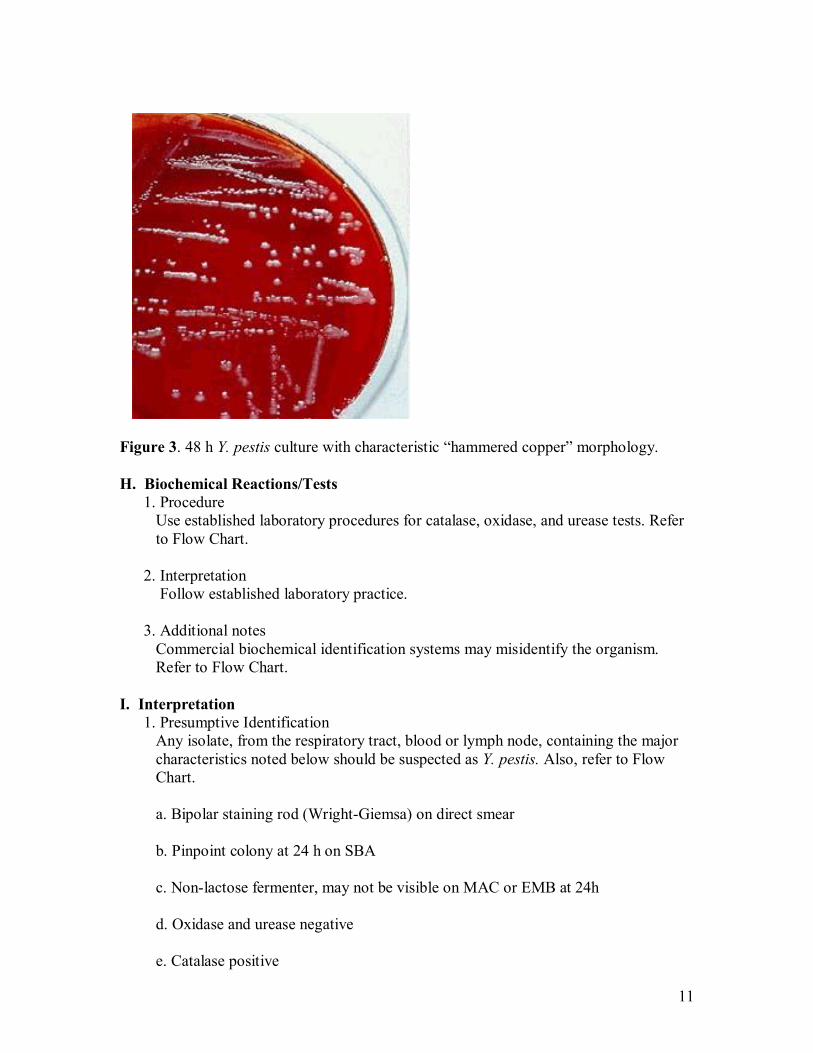
11
Figure 3. 48 h Y. pestis culture with characteristic “hammered copper” morphology.
H. Biochemical Reactions/Tests 1. Procedure Use established laboratory procedures for catalase, oxidase, and urease tests. Refer to Flow Chart.
2. Interpretation Follow established laboratory practice.
3. Additional notes Commercial biochemical identification systems may misidentify the organism. Refer to Flow Chart.
I. Interpretation 1. Presumptive Identification Any isolate, from the respiratory tract, blood or lymph node, containing the major characteristics noted below should be suspected as Y. pestis. Also, refer to Flow Chart.
a. Bipolar staining rod (WrightGiemsa) on direct smear
b. Pinpoint colony at 24 h on SBA
c. Nonlactose fermenter, may not be visible on MAC or EMB at 24h
d. Oxidase and urease negative
e. Catalase positive

12
f. Growth often better at 28 o C
J. Miscellaneous testing In an era of possible release of microorganisms as biological agents of terror, concern must be raised for sole reliance on diagnostic reagents targeting specific Y. pestis antigens or antibodies. Yersinia pestis has a genetic diversity that can potentially be manipulated to alter parameters for specific DNA sequences, thereby abrogating the use of available sensors. Thus, older methods relying on culture and overall phenotypic organism characteristics still play a major role in the recognition and diagnosis of Y. pestis. On the other hand, in nonbioterrorism situations, normal diagnosis of plague has been enhanced by more novel and rapid techniques (Anisimov, et. al., 2004).
1. Rapid diagnostic tests (RDTs) Several methods targeting the F1 antigen released by Y. pestis in situ (in bubo aspirates) have been described in the diagnosis of acute phase plague. These include enzymelinked immunosorbent assay (ELISA) and the immunogold chromatography dipstick assay. Bothe of theses assays are highly sensitive and specific and rapid within hours. PCR has also been developed detecting the F1 gene in bubo aprirates. This test is rapid with a moderate sensitivity. These tests are not normally readily available, especially in the U.S. (Rahalison, et. al., 2000; Stevenson, et. al., 2003; Butler, 2009).
A recent RDT dipstick method was described which had capability of detecting 0.5 ng/ml of Y. pestis F1 antigen, was rapid (15 minutes to complete), and had 100% sensitivity and specificity when tested against laboratory strains of Yersinia spp. (Chanteau, et. al., 2003). In clinical field trials the RDT detected 41.6% and 31% more positive clinical specimens than did traditional microbiologic and ELISA methods, respectively. The shelflife, unfortunately, was only 21 days. This RDT holds promise for rapid diagnosis for plague where larger volume testing is predicated and can be used in medically resourcepoor areas lacking technical or adequately trained staff support.
Several polymerase chain reaction (PCR) detection methods have been described for detection of Y. pestis genetic material, but most have not been well studied against human clinical specimens (Hinnebusch and Schwan, 1993; Stevenson, et. al., 2003). However, recently a PCR method targeting a 501bp fragment of the Y. pestis caf1 gene was evaluated with human specimens (Rahalison, et. al., 2000). Its sensitivity reached 89% in cultureproven patients with plague and 80.7% in patients diagnosed using F1 antigen detection methods. At this time, PCR methods are not recommended for routine diagnosis of plague.
2. Serology As with any tests requiring initiation of antibody production, serologies may be useful for epidemiologic studies or for retrospective confirmation of Y. pestis infection. They are, however, of little value for routine diagnosis of acute phase disease. Confirmation requires at least a fourfold rise in antibody titer to the F1

13
antigen (CDC, 1994), whereas a single titer rise to >1:10 is diagnostic (Butler, 2009).
K. Limitations 1. Y. pestis will grow on general nutrientrich media, but its growth rate is slower
than that of most other bacteria; therefore, its presence may be masked by organisms that replicate faster.
2. Bipolar staining of cells is not an exclusive feature limited to Y. pestis. Yersinia spp., enteric bacteria, and other gramnegative organisms, particularly Pasteurella spp., can exhibit the same staining characteristic.
3. Characteristic clumped growth in unshaken broth culture is not an exclusive feature of Y. pestis. Some Y. pseudtuberculosis and Streptococcus pneumoniae can exhibit the same growth features.
4. Some of the automated identification systems do not identify Y. pestis adequately. Y. pestis have been falsely identified as Y. pseudotuberculosis, Shigella, H2S negative Salmonella, or Acinetobacter (Wilmoth, et. al., 1996). Y. pestis is alkaline slant/acid butt in triple sugar iron. In most conventional biochemical or commercial identification systems, the organism appears relatively inert, making further biochemical testing of little value. A list of manual and automated identification tests with Y. pestis in their databases can be found in a review article on the commercial identification of gram negative bacilli (O’Hara, 2005).
5. Because of the limitations in identification of Y. pestis using methods typically found in microbiology laboratories, a high level of suspicion is essential. For blood isolates in particular, isolation of any Yersina spp or H2Snegative Salmonella may be an indication to evaluate the clinical condition of the patient to determine if plague is a possibility. Isolation of Shigella from blood is highly unlikely and should immediately raise suspicion. Similarly, isolation of Acinetobacter from a case of severe communityacquired pneumonia or sepsis is also unlikely.
L. Reporting/Action 1. Sentinel laboratories should consult with state public health laboratory director (or
designate) prior to or concurrent with testing if Y. pestis is suspected by the physician (Fig. A4).
2. Immediately notify state public health laboratory director (or designate) and state public health department epidemiologist/health officer if Y. pestis cannot be ruled out. The state public health laboratory/state public health department will notify law enforcement officials (state and federal).
3. Immediately notify physician/infection control according to internal policies if Y. pestis cannot be ruled out.

14
4. Preserve original specimens pursuant to a potential criminal investigation and possible transfer to an appropriate LRN laboratory. The FBI and state public health laboratory/state public health department will coordinate the transfer of isolates/specimens to a higherlevel LRN laboratory as appropriate. Start chain of custody documentation if appropriate.
5. Obtain guidance from the state public health laboratory as appropriate (e.g., requests from local law enforcement or other local government officials).
6. If Y. pestis is ruled out, proceed with efforts to identify using established procedures.
M. Therapy Streptomycin has historically been the preferred treatment for plague and is FDA approved for this purpose. .Streptomycin may not be widely and immediately available. The aminoglycosides, tetracyclines, and chloramphenicol are antimicrobials routinely used to treat plague. Among newer classes of antimicrobials, Y. pestis is susceptible to fluoroquinolones in vitro, and fluoroquinolones are efficacious in treating experimental plague in animals ( http://www.bt.cdc.gov/agent/plague/trainingmodule/5/06.asp).
Antimicrobial therapy for specific patient populations are shown below (Gerald E. Maloney – author,
Antimicrobial therapy for specific patient populations are shown below (Gerald E Maloney author, http://emedicine.medscape.com/article/830118treatment emedicine from WebMD, accessed 9/28/09).
Doxycycline Several different controlled and retrospective trials have established efficacy as treatment for brucellosis. Because of concerns regarding treatment failures, combination therapy with rifampin or an aminoglycoside now is recommended, although it remains approved for use as monotherapy.
Adult 200 mg/d PO, usually divided into 100 mg PO bid; may be administered IV if needed; duration is 36 wk
Pediatric 5 mg/kg/d PO for 3 wk
Rifampin Used in combination therapy with doxycycline, TMPSMZ, or gentamicin for treatment of brucellosis.
Adult 600900 mg PO/IV qd

15
Pediatric 1020 mg/kg PO/IV qd; not to exceed 600 mg
Sulfamethoxazole/trimethoprim Used as adjunctive therapy with gentamicin in treating infection in children <8 y; used as monotherapy or combined with rifampin or gentamicin to treat infection in pregnant females. Inhibits bacterial growth by inhibiting synthesis of dihydrofolic acid.
Adult 1 double strength tab PO bid (160/800) 810 mg/kg IV divided q6, 8, or 12h
Pediatric 5 mL/10 kg (40/200) PO bid
Gentamicin Aminoglycosides have been used for several years to treat brucellosis; studies to date have shown gentamicin to be the preferred aminoglycoside to treat infection as combined therapy with either TMPSMZ or doxycycline in children. Adult dose is either oncedaily dosing or a multipledaily dose.
Adult Oncedaily dose: 5.1 mg/kg IV/IM qd Multipledaily dose: 2 mg/kg loading dose, IV followed by 1.7 mg/kg IV/IM q8h; continue for 5 d
Pediatric 5 mg/kg IM for 5 d, in combination with either doxycycline or TMPSMZ
Streptomycin Has been used for several years to treat brucellosis; used in combination with doxycycline, especially for spondylitis or sacroiliitis; augments bacteriocidal action of other agents used to treat brucellosis.
Adult 15 mg/kg IM; not to exceed 1 g/d IM qd for 3 wk
Pediatric 2040 mg/kg IM qd; not to exceed 1 g qd
Note: CLSI document M100S15 outlines MIC interpretive standards for Y. pestis antimicrobial susceptibility testing (CLSI, 2005). This document also recommends extreme caution when testing this organism and outlines biological safety practiced that must be adhered to. The document also warns that although in vitro studies have shown Y. pestis to be susceptible to βlactam antibiotics, these antibiotics should not be reported as susceptible.

CDC Contact Numbers: English—8882462675 Español—8882462857 TTY—8668742646
Recommended Websites: http://www.asm.org http://www.cdc.gov http://www.bt.cdc.gov/lrn http://www.bt.cdc.gov/training/index.asp http://www.bt.cdc.gov/agent/agentlist.asp
Yersinia pestis: Sentinel Laboratory Flowchart
Morphology: Facultative, bipolar, 0.5 by 1.0 to 2.0 um, gram negative rods. Growth: Slow growing, pinpoint (12mm), graywhite to opaque, colonies on sheep blood agar after 24 h. Nonlactose fermenter, +/ growth on MAC/EMB at 24 h.
Oxidase: Negative Catalase: positive Urea: Negative Indole: Negative
WARNING: Automated identification systems often key out as nonY.pestis (e.g., Shigella, H2Snegative Salmonella, Acinetobacter and Y.pseudotuberculosis).
Yes (features present)
No (features not present)
Report: Y. pestis is ruled out; continue identification per laboratory procedures.
Report: Suspect, could not rule out; refer to state public health laboratory for confirmation.
III. REFERENCES
1. Anisimov A. P., L. E. Lindler, G. B. Pier. 2004. Intraspecific diversity of Yersinia pestis . Clin Microbiol Rev 17:434464.
2. Butler T. 1983. Plague and other Yersinia infections, p. 163–188. In: Greenough WB III, Merigan TC (eds), Current topics in infectious diseases. Plenum Medical Book and Company, New York.
3. Butler, T. 2009, Plague into the 21 st century. Clin Infect Dis. 49:736742.
4. Campbell G. L., D. T. Dennis. 1998. Plague and other Yersinia infections, p. 975– 983. In: Kasper DL, et al., (ed). Harrison’s principles of internal medicine. 14th ed. McGrawHill, New York, NY.
5. CDC. Human plague United States, 19931994. 1994. MMWR Morb Mortal Wkly Rep. 43:242.
6. CDC. Notifiable disease/deaths in selected cities weekly information. MMWR Morb Mortal Wkly Rep 58(37):10461057.
7. Chanteau S, Rahalison L, Ralafiarisoa L, Foulon J, Ratsitorahina M, Ratsifasoamanana L, Carniel E, Nato F. 2003. Development and testing of a rapid diagnostic test for bubonic and pneumonic plague. Lancet 361:211216.
8. Gage K. L. 1998. Plague. In: L. Colliers, A. Balows, M. Sussman, W. J. Hausles (ed). Topley and Wilson’s microbiology and microbiological infections, Vol. 3, p. 885903. Edward Arnold Press, London.
9. Gage K.L., Dennis, DT, Orloski, KA, et al. 2000. Cases of catassociated human plague in the western US, 19771998. Clin Infect Dis; 30:893900.
10. Hinnebusch, BJ, Schwan, TG. 1993. New method for plague surveillance using polymerase chain reaction to detect Yersinia pestis in fleas. J Clin Microbiol 31:1511 1514.
11. Inglesby T. V., D. T. Dennis, D. A. Henderson J. G. Bartlett, M. S. Ascher, E. Eitzen, A. D. Fine, A. M. Friedlander, J. Hauer, J. F. Koerner, M. Layton, J. McDade, M. T. Osterholm, T. O'Toole, G. Parker, T. M. Perl, P. K. Russell, M. SchochSpana, K. Tonat. 2000. Plague as a biological weapon: medical and public health management. Working Group on Civilian Biodefense JAMA, 283: 22812290.
11. O’Hara, C.M. 2005. Manual and automated instrumentation for identification of Enterobacteriaceas and other aerobic bramnegative bacilli. Clin Microbiol Rev 18:147 163.

18
12. Lowell, L.L., Wagner, D.M., Atshaber B, et al. 2005. Identifying sources of human exposure to plague. J Clin Microbiol 43:650656.
13. O’Hara, C.M. 2005.Manual and automated instrumentation for identification of Enterobacteriasceas and other aerobic gram negative bacilli. Clin Microbiol Rev 18:147 163.
14. Perry R. D., J. D. Fetherston. 1997. Yersinia pestis—etiologic agent of plague. Clin Microbiol Rev. 10:35–66.
15. Rahalison L, Vololonirina E, Ratsitorahina M, Chanteau S. 2000. Diagnosis of bubonic plague by PCR in Madagascar under field conditions. J Clin Microbiol. 38:2603.
16. Stevenson HL, Bai Y, Kosoy MY, Montenieri JA, Lowell JL, Chu MC, Gage KL. 2003. Detection of novel Bartonella strains and Yersinia pestis in prairie dogs and their fleas (Siphonaptera: Ceratophyllidae and Pulicidae) using multiplex polymerase chain reaction. J Med Entomol. 40:32937.4 August 20054 August 2005
17. Wilmoth B. A., M. C. Chu, T. J. Quan. 1996. Identification of Yersinia pestis by BBL Crystal Enteric Nonfermentor identification system. J Clin Microbiol. 43:2829– 2830.
Three references removed: 1. Bible and Chen, 1976. 2. Brubaker, 1972.
Koneman, et al., 19
ASM acknowledges CDC for the use of its photos.

19
V. APPENDIX A. Change Record 1. 29 September 2009 a. Under I.B. History, updated information added on number of Plague cases (38) in the U.S. between 20042008. b. Under I.C. Geographic Distribution, information added on when most cases occur (late winter to summer months). c. Under I.D. Clinical Presentation, information added about the incubation period (210 days). d. Under II.D. Materials, new disclaimer added. e. Under II.J. Miscellaneous testing, updated information added regarding Rapid Diagnostic Tests. f. Under II.J. Miscellaneous testing, updated information added regarding the single titer rise (to >1:10). g. Under II.M. Therapy, updated information added about antimicrobial therapy for specific patient populations. h. Under III, References, new references added (see 3, 6, 9, 12); two references removed. 2. 04 August 2005 a. Title Change to “Sentinel Laboratory Guidelines for Suspected Agents of Bioterrorism” b. Sentinel used instead of Level A throughout Guideline. c. On Credits page, new contact information for Lovchik, Saubolle, Shapiro and Welch. d. Under I. A. Description, added additional description of organism when stained with Wright’s or Giemsa stains. e. Under I. B. History, information added on the number of Plague cases (112) in eleven western states between 19982002. f. Under I. C. Geographic Distribution, added MMWR article about cases acquired in the western U.S. that have presented on the east coast. g. Under I. D. Clinical Presentation, added information about aerosol delivery. h. Under II. B. Precautions, added recommendation that all manipulations be performed in a biological safety cabinet at a minimum. i. Under II. C. 1. c. Specimens, added additional note regarding aspirated specimens. j. Under II. C. 1. c. Specimens, added lymph nodes to list of biopsies/aspirates that could be sampled. k. Under II. C. 2. b. Rejection Criteria, added example (powder) to dried specimens. l. Under II. C. 3. Changed title of section to “Specimen Treatment and Storage.” m. Under II. F. 2. Added: Note that in patients with overwhelming sepsis, bipolar staining rods may be detected in peripheral blood smears. n. Figure A. I, photo is 800x. o. Figure A. II., photo is 800x, and added additional note, “closed safety pin shaped cells.”

20
p. Under II. H. Biochemical Reactions/Tests, added, “refer to flow chart.” q. Under II. I. Interpretation, added new subsection, “Presumptive Identification.” r. Under II., new section J. added, “Miscellaneous Testing” including information on rapid diagnostic tests and serology. s. Under II. K. Limitations, new reference to O’Hara article sited. t. Under II. K. Limitations, added 5, emphasizing the need to have a high level of suspicion. u. Under II, new section M. added, “Therapy.” v. Under III. References, 10 new references added (#1, 3, 5, 6, 7, 9, 10, 11, 13, 14) and three references removed.

BIOTERRORISM RESPONSE GUIDE FOR CLINICAL LABORATORIES
Section VIII - 1
This Page Last Updated: January, 2007
Collection, Packaging and Transport of “Biological Substance Category B” Specimens to the Wisconsin State Laboratory of Hygiene via Dunham Express for Chemical Terrorism Response
Contact the WSLH through the 24/7 emergency messaging service at 608-263-3280 for current guidance in specimen collection and transport for a chemical terrorism event. Collecting Specimens • Required Specimens
Urine – Collect at least 25 mL in a screw-capped plastic container. Properly label the specimen (see labeling instructions and attached illustrated job aide) and freeze as soon as possible using a -70°C freezer or dry ice. If a -70°C freezer or dry ice is not available, use a -20°C freezer with the understanding that it will take longer to completely freeze the urine specimen. For pediatric patients, collect urine only unless otherwise instructed. If other than “clean catch”, note method of collection on specimen cup and Clinical Sample Evidence/Chain of Custody Form (e.g., obtained by catheterization).
Whole Blood – Fill three 4-mL or larger purple top (EDTA) non-gel tubes (unopened). If using 3 mL purple top tubes, fill four tubes (unopened).
Whole Blood – Fill one 3 mL or larger gray or green-top non-gel tube (unopened).
• Order of collection – Label the first purple-top tube of whole blood collected with a “1” using waterproof ink. Label the second purple-top tube of whole blood collected with a“2”, and the third purple top tube with a “3”.
• Blanks – For each lot number of tubes and urine cups used for collection, please provide two empty unopened purple-top tubes, two empty unopened green- or gray- top tubes, and two empty unopened urine cups to serve as blanks for measuring background contamination. Label these as “blank” using waterproof ink.

BIOTERRORISM RESPONSE GUIDE FOR CLINICAL LABORATORIES
Section VIII - 2
This Page Last Updated: January, 2007
Collection, Packaging and Transport of “Biological Substance Category B” Specimens to the Wisconsin State Laboratory of Hygiene via Dunham Express for Chemical Terrorism Response
Labeling Specimen Containers • Follow your facility’s procedures for proper specimen labeling. Label
specimens with labels generated by your facility. The label may include the medical records number, specimen identification number, collector’s initials and date and time of collection. The collector’s initials and date and time of collection will allow law enforcement officials to trace the specimen to the collector should investigations lead to legal action and the collector has to testify that he or she collected the specimen.
• If you use bar-coded labels, place the labels on the blood tubes and urine cups so that when these containers are upright, the bar code looks like a ladder.
• Information provided on the labels may prove helpful in correlating the results obtained from CDC’s Rapid Toxic Screen and subsequent analysis with the people from whom the specimens were collected.
• Maintain a list of names with corresponding specimen identification numbers at the collection site to enable results to be reported to the patient.
Shippers If you do not have a specimen shipping kit, you can contact one of the local health department repository sites to get one of Wisconsin Emergency Response Shipping Kits. These shippers are stored at most local public health departments for use in emergency or outbreak response. A listing of repository sites with contact information is posted on the HAN (in the “Laboratory” topic area, under “Repository”) and is available elsewhere in this guide. Your laboratory is expected to make arrangements to collect the shipper from the local health department repository site. The shippers are class 6 “infectious substance” shippers but can also be used for shipping Biological Substance Category B specimens. The shippers are appropriate for shipping samples at ambient temperature, refrigerated temperatures, or dry ice temperatures. These kits are not intended for routine specimen submission.

BIOTERRORISM RESPONSE GUIDE FOR CLINICAL LABORATORIES
Section VIII - 3
This Page Last Updated: January, 2007
Collection, Packaging and Transport of “Biological Substance Category B” Specimens to the Wisconsin State Laboratory of Hygiene via Dunham Express for Chemical Terrorism Response
Packaging • Package and ship the specimens as Biological Substance Category B
specimens.
• All specimens must be “triple-packaged.”
• There is no quantity restriction of samples for ground transport.
• Blood tubes Use a gridded box, or individually wrap each blood tube with absorbent
material to prevent contact between tubes during transport. If using a gridded box, wrap the box in absorbent material. Place the wrapped gridded box, or individually wrapped blood tubes in a
leak proof, sealable biohazard bag or. Note: If wrapping the tubes individually, each biohazard bag should contain all of the blood tubes for one patient.
Place enough absorbent material into the sealable biohazard bag to absorb the entire contents of the blood specimens.
Tubes that are labeled as “blank” should also be put in a separate sealable bag, but it is not necessary to include absorbent material.
After sealing the biohazard bags, place a single, unbroken strip of waterproof, tamper-evident forensic evidence tape over the closure of the bag. The individual placing the evidence tape must initial the tape with their initial that is half on the bag and half on the evidence tape.

BIOTERRORISM RESPONSE GUIDE FOR CLINICAL LABORATORIES
Section VIII - 4
This Page Last Updated: January, 2007
Collection, Packaging and Transport of “Biological Substance Category B” Specimens to the Wisconsin State Laboratory of Hygiene via Dunham Express for Chemical Terrorism Response
Packaging (continued)
• Urine cups Use a gridded box, or individually wrap each urine cup with absorbent
material to prevent contact between cups during transport. If using a gridded box, wrap the box in absorbent material. Place the wrapped gridded box, or individually wrapped urine cups in a
leak proof, sealable biohazard bag. Note: If wrapping the cups individually, each biohazard bag should contain the urine from one patient.
Place enough absorbent material into the sealable biohazard bag to absorb the entire contents of the urine cup(s).
Urine cups that are labeled as “blank” should also be put in a separate, sealable bag, but it is not necessary to include absorbent material.
After sealing the biohazard bags, place a single, unbroken strip of waterproof, tamper-evident forensic evidence tape over the closure of the bag. The individual placing the evidence tape must initial the tape with their initial that is half on the bag and half on the evidence tape.
• Outer Container – Use a Styrofoam-insulated corrugated fiberboard container. You may use packaging materials you have on hand, but the completed package must be capable of passing the “drop test”. If you do not have an appropriate shipping container, you can acquire one from a Wisconsin Emergency Response Shipper Repository Site; instructions are provided elsewhere in this guide.
• Do not ship frozen urine cups and blood tubes in the same package. Blood tubes – ship cold (1-10 degrees C).
Place absorbent material on the bottom of the outer container. Add a layer of frozen cold packs. Place sealed secondary containers on top of the cold packs. Place additional absorbent material between the secondary containers to
reduce movement within the outer container. Place a layer of frozen cold packs on top of the secondary containers.

BIOTERRORISM RESPONSE GUIDE FOR CLINICAL LABORATORIES
Section VIII - 5
This Page Last Updated: January, 2007
Collection, Packaging and Transport of “Biological Substance Category B” Specimens to the Wisconsin State Laboratory of Hygiene via Dunham Express for Chemical Terrorism Response
Packaging (continued) Urine cups – ship to ensure specimens remain frozen or freeze while in transport.
Place absorbent material on the bottom of the outer container. Add a layer of dry ice, if available. Do not use large chunks of dry ice for
shipment, because it has a potential to shatter urine cups during transport. If dry ice is not available, use frozen cold packs. If using frozen cold packs, make sure that the urine is completely frozen before shipment, unless otherwise instructed.
Place additional absorbent material between the secondary containers to reduce the movement within the outer container.
Place another layer of dry ice if available or frozen cold packs.
• Preparing documentation
• Blood specimens The individual collecting the specimen must complete the WSLH
“Chemical Emergency Response: Clinical Sample Evidence/Chain-of-Custody Form” for the urine specimen of each patient. The form and instructions for completing the form are included later in this section.
Each person who takes control of the specimen must sign and date this form.
Place the completed form in a sealable plastic bag. Place the sealed plastic bag on top of the specimens before closing the
Styrofoam lid of the corrugated fiberboard container. Tape the box closed with filament shipping tape.

BIOTERRORISM RESPONSE GUIDE FOR CLINICAL LABORATORIES
Section VIII - 6
This Page Last Updated: January, 2007
Collection, Packaging and Transport of “Biological Substance Category B” Specimens to the Wisconsin State Laboratory of Hygiene via Dunham Express for Chemical Terrorism Response
• Preparing documentation (continued)
• Urine specimen The individual collecting the specimen must complete the WSLH
“Chemical Emergency Response: Clinical Sample Evidence/Chain-of-Custody Form” for the urine specimen of each patient. The form and instructions for completing the form are included later in this section.
Indicate in the comments section of the form how the sample was collected if other than a “clean catch” (e.g., obtained by catheterization).
Each person who takes control of the specimen must sign and date this form.
Place the completed form in a sealable plastic bag. Place the sealed plastic bag on top of the specimens before closing the
Styrofoam lid of the corrugated fiberboard container. Tape the box closed with filament shipping tape. If dry ice was
included, do NOT seal all edges of the container, as the package must allow the escape of carbon dioxide gas.
Package Labeling • Attach the “Biological Substance Category B” and “UN3373” label(s)
adjacent to one another on the outside of the box (This may be a single, combined label.).
• If the package contains dry ice, attach diamond-shaped “Class 9” dry ice label, and the “Dry Ice/UN 1845/Net Quantity __kg” label; write in the approximate weight of dry ice in the package and a “Frozen Medical Specimen” label.
• Provide an emergency contact information to include a name and a (24/7) telephone number of a person who has knowledge of the package contents.
• Address the package as follows: Wisconsin State Laboratory of Hygiene Attn: CT Rapid Response 2601 Agriculture Drive Madison, WI. 53718

BIOTERRORISM RESPONSE GUIDE FOR CLINICAL LABORATORIES
Section VIII - 7
This Page Last Updated: January, 2007
Collection, Packaging and Transport of “Biological Substance Category B” Specimens to the Wisconsin State Laboratory of Hygiene via Dunham Express for Chemical Terrorism Response
Shipping Instructions via Dunham Express • The WSLH has arranged a contract with Dunham Express for shipment of
samples for emergency response to the State Laboratory of Hygiene, with charges billed to the WSLH.
• Complete the package preparation before the courier arrives; charges will be billed to the Wisconsin State Laboratory of Hygiene.
• Contact the Dunham Express office in your area (list below); calls are answered 24 hours a day, 7 days a week. Appleton area: Call 920-722-6360 or 1-800-236-7128. Eau Claire area: Call 715-834-3200 or 1-800-236-7129. LaCrosse area: Call 608-779-4588. Madison area: Call (608) 242-1000. Milwaukee area: Call (414) 435-0002 or 1-800-236-7126. Niagara area: Call 715-251-1909 or 1-800-298-1909. Wausau area: Call 715-355-0400.
• Information which you must provide to Dunham Express when you call: • Confirm the pick-up time and request "Overnight Service". • Give the Dunham Express office the following information:
The State Lab Rapid Response account number: 7263 The account name: State Lab Rapid Response Your name, phone number, and pickup address, including room number Destination: CT Rapid Response
Wisconsin State Lab of Hygiene 2601 Agriculture Drive, Madison, WI 53718
(You must specify when you call that this package should NOT be delivered to the Henry Mall address, but should be delivered to this address.)
Shipment description, if asked: Specimens for delivery Other Information: Specify delivery option as “Overnight Service”
If it has been agreed that delivery of the package requires “stat” service, and the package is expected to arrive at the State lab AFTER normal business hours (7:00-4:30, Monday-Friday), instruct the Dunham Express driver to call the WSLH Emergency Answering Service at (608) 263-3280 when driver is 1 hour from the State Lab to arrange for receipt of specimen.

BIOTERRORISM RESPONSE GUIDE FOR CLINICAL LABORATORIES
Section VIII - 8
This Page Last Updated: January, 2007
Collection, Packaging and Transport of “Biological Substance Category B” Specimens to the Wisconsin State Laboratory of Hygiene via Dunham Express for Chemical Terrorism Response
Shipping Resources • Containers
• The following list of commercial sources of transport containers is not all-inclusive. This listing is not intended to be an endorsement or non-endorsement of any companies.
• Saf-T-Pak, Inc., website http://www.saftpak.com • EXAKT Technologies, Inc., website http://www.exaktpak.com/ • Therapak Corporation, website http://www.therapak.com • Thermal Insulated Systems, website http://www.thermalsystems.com • Air Sea Atlanta, website http://www.airseaatlanta.com • All-Pak, Inc., website http://www.all-pak.com • DG Supplies, Inc., website http://www.dgsupplies.com • Federal Industries Corporation, website http://www.chem-tran.com • ICC The Compliance Center Inc., website
http://www.thecompliancecenter.com • Inmark, Inc., website http://www.inmarkinc.com
Training • The following list of websites includes commercial sources of training for
shipping. This listing is not all-inclusive and is not intended to be an endorsement or non-endorsement of any companies or products.
• http://hazmat.dot.gov/training.htm • http://www.saftpak.com • http://www.highqllc.com • http://www.dgitraining.com/menu.htm • http://cargopak.com • “Packaging Clinical Laboratory Samples for Domestic Transport”
training manual prepared by the Wisconsin State Laboratory of Hygiene
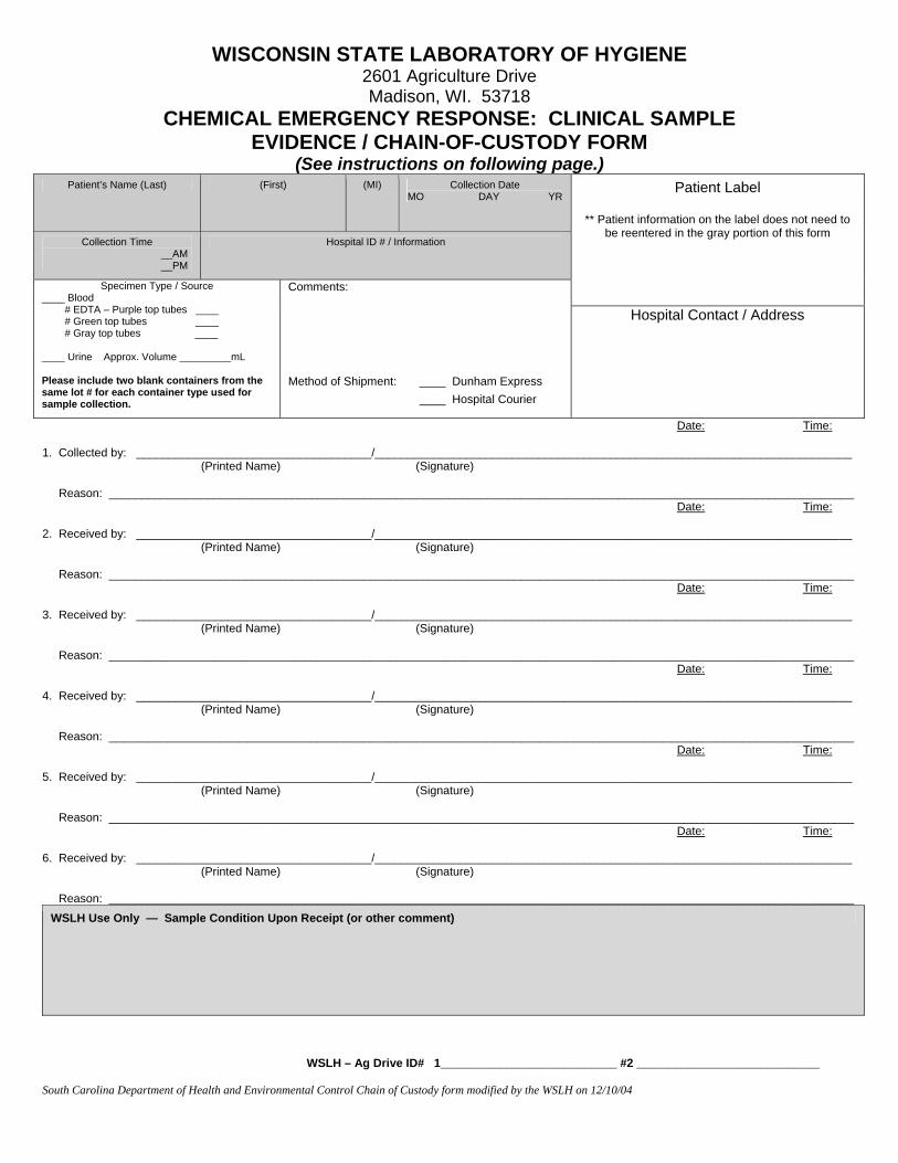
WISCONSIN STATE LABORATORY OF HYGIENE 2601 Agriculture Drive Madison, WI. 53718
CHEMICAL EMERGENCY RESPONSE: CLINICAL SAMPLE EVIDENCE / CHAIN-OF-CUSTODY FORM
(See instructions on following page.) Patient’s Name (Last) (First) (MI) Collection Date
MO DAY YR
Collection Time __AM __PM
Hospital ID # / Information
Patient Label
** Patient information on the label does not need to be reentered in the gray portion of this form
Specimen Type / Source ____ Blood # EDTA – Purple top tubes ____ # Green top tubes ____ # Gray top tubes ____ ____ Urine Approx. Volume _________mL Please include two blank containers from the same lot # for each container type used for sample collection.
Comments: Method of Shipment: ____ Dunham Express
____ Hospital Courier
Hospital Contact / Address
Date: Time: 1. Collected by: ____________________________________/_________________________________________________________________________ (Printed Name) (Signature) Reason: __________________________________________________________________________________________________________________ Date: Time: 2. Received by: ____________________________________/_________________________________________________________________________ (Printed Name) (Signature) Reason: __________________________________________________________________________________________________________________ Date: Time: 3. Received by: ____________________________________/_________________________________________________________________________ (Printed Name) (Signature) Reason: __________________________________________________________________________________________________________________ Date: Time: 4. Received by: ____________________________________/_________________________________________________________________________ (Printed Name) (Signature) Reason: __________________________________________________________________________________________________________________ Date: Time: 5. Received by: ____________________________________/_________________________________________________________________________ (Printed Name) (Signature) Reason: __________________________________________________________________________________________________________________ Date: Time: 6. Received by: ____________________________________/_________________________________________________________________________ (Printed Name) (Signature) Reason: __________________________________________________________________________________________________________________
WSLH Use Only — Sample Condition Upon Receipt (or other comment)
WSLH – Ag Drive ID# 1___________________________ #2 ____________________________ South Carolina Department of Health and Environmental Control Chain of Custody form modified by the WSLH on 12/10/04

INSTRUCTIONS FOR COMPLETING CHAIN OF CUSTODY FORMS:
NOTE – Two Chain of Custody Forms are required for each patient: one for the blood samples and one for the urine sample. A. Collector insures that patient information and date and time of
specimen collection appears on the Chain of Custody Form by affixing a patient label in the indicated area. If a patient label is not available, complete the gray shaded area on the Chain of Custody Form.
B. Collector completes Specimen Type / Source information on the Chain of Custody Form.
C. Collector begins the “chain of custody” by providing printed name, signature, date, time and reason for specimen collection on line 1 of the Chain of Custody Form. Each person who takes control of the specimens after the sample collector continues the chain by providing the appropriate information on the Chain of Custody Form.
D. Place completed Chain of Custody Forms in a plastic zip lock bag on top of the appropriate specimens before closing the lid of the shipping container.
Section VIII-10

CDC Specimen-Collection Protocol for a Chemical-Exposure Event
11/2006
Collect blood and urine samples for each person involved in the chemical-exposure event. Note: For children, collect only urine samples unless otherwise directed by CDC.
For each person, collect blood in glass or plastic tubes in the following order:1st: collect specimens in three (3) EDTA (purple-top) 4 mL or larger plastic or glass tubes; 2nd: collect another specimen in one (1) gray- or green-top tube. Collect the specimens by following the steps below:
Blood-Sample Collection
Urine-Sample CollectionFor each person, collect 25 mL- 50 mL of urine in a screw-cap urine cup.
Label the urine cup with the appropriate bar-coded label as shown. Indicate on the cup how the sample was collected if the method was other than "clean catch" (i.e., catheterization).
Place bar-coded labels on all cups so that when the cup is upright, the barcode looks like a ladder.
Freeze samples (optimally at -70°C).
For detailed instructions see CDC's Shipping Instructions for Specimens Collected from People Who May Have Been Exposed to Chemical-Terrorism Agents.
3 Place bar-coded labels on each tube, so that when the tubes are upright, the barcode looks like a ladder.
Mix contents of tubes by inverting them 5 or 6 times.
7
1
Tube #1
7
2
Tube #2
7
3
Tube #3
Label tubes in order of collection. #1, #2, #3
2
Tube #1
7
11
22
04
13
23
TL
C
TLC
Tube #2
7
11
22
04
13
23
TL
C
TLC
Tube #3
7
11
22
04
13
23
TL
C
TLC
Mix contents of the tube by inverting it 5 or 6 times.5
TLC
11
22
04
13
23
TL
C
Place bar-coded labels on the tube, so that when the tube is upright, the barcode looks like a ladder.6
Store samples at 1°C to 10°C.
After collecting samples in the purple-top tubes, collect one (1) sample in a gray- or green-top tube (gray-top tube shown). Allow the tube to fill to its stated capacity.
4
Do not use gel separators.
7 7 7
1
Do not use gel separators.
Collect a minimum of 12 mL of blood in three (3) 4 mL or larger glass or plastic tubes. If using 3 mL tubes, use four tubes.
Do not freeze.
Store samples at 1°C to 10°C. Do not freeze.

Pictorial aid to the Guidelines for Collection, Transport and Shipping of Biological Substance Category B Specimens for Chemical Terrorism Response — Packaging and Shipping of Blood.
This job aid is meant to be used in conjunction with the above mentioned guidelines document NOT as a substitute.
Use a gridded box or individually wrap each blood tube to prevent contact between the tubes.
Once the bag is sealed place an unbroken strip of evidence tape over the seal of the bag. Then initial the tape w/ initials half on and half off the tape.
NOTE: tubes labeled as “blank” should be wrapped and placed into a separate sealable plastic bag. It is not necessary to include absorbent material.
Place the box or tubes in a sealable biohazard bag. Place enough absorbent material into the bag to absorb the entire contents of the tubes. If wrapping tubes individually, each bag should contain ALL of the blood tubes for ONE patient.
START
After all the blood has been properly bagged and sealed w/ evidence tape, prepare the shippers.
Place absorbent material such as a Chux® pad in the bottom of the shipper.
Place frozen cold packs in a single layer on top of the absorbent material.
Place the bagged blood samples in the shipper using plenty of cushioning material to minimize shifting in transit.
Place frozen cold packs on top of the bag(s).
Place the completed Clinical Sample Evidence / Chain of Custody form in the sealable plastic bag and place on top of the cold packs inside the shipper.
Place the Styrofoam lid on the top and close the shipper. Secure the top of the box with filament tape. Place contact name, shipping address and phone number in the upper left corner and address the shipment to: WSLH Attn: CT Rapid Response 2601 Agriculture Drive Madison, WI. 53718
Place UN3373 and “Biological Substance Category B” labels on the shipper.
END

Pictorial aid to the Guidelines for Collection, Packaging and Transport of Biological Substance Category B Specimens for Chemical Terrorism Response — Packaging and Shipping of Urine.
This job aid is meant to be used in conjunction with the above mentioned guidelines document NOT as a substitute.
Use a gridded box or individually wrap each urine cup to cushion the cups in transit.
Once the bag is sealed place an unbroken strip of evidence tape over the seal of the bag. Then initial the tape w/ initials half on and half off the tape.
NOTE: Cups labeled as “blank” should be wrapped and placed into a separate sealable plastic bag. It is not necessary to include absorbent material.
Place the gridded box or cup in a sealable biohazard bag. Place enough absorbent material into the bag to absorb the entire contents of the urine. If packaging the urine cups individually, each bag should contain ONE patient’s urine specimen.
START
After all the urine cups have been properly bagged and sealed w/ evidence tape, prepare the shippers. Place absorbent material such as a Chux® pad in the bottom of the shipper.
Place dry ice in a single layer on top of the absorbent material. Then place the bagged urine cup(s) in the shipper using plenty of cushioning material to minimize shifting in transit. Fill the rest of the way with more dry ice. Do NOT use large chunks of dry ice.
Place a Class 9 label on the shipper. The label must show the amount of dry ice in the container, the
Place the
completed Clinical Sample Evidence / Chain of Custody form in a sealable plastic bag and place on top of the dry ice inside the shipper.
Place the Styrofoam lid on the top and close the shipper. Secure the top of the box with filament tape. Do not seal all the edges of the box as the package must allow the escape of CO2 gas. Place contact name, address, and phone number in the upper left corner of shipper and address the shipment to:
WSLH Attn: CT-Rapid Response 2601 Agriculture Drive Madison, WI. 53718
Place UN3373 and “Biological Substance Category B” labels on the shipper.
proper name (either “Carbon Dioxide, Solid”, or “Dry Ice”), and show the designation “UN1845”. Label or write “Frozen Medical Specimen” on the outside of the container.
END
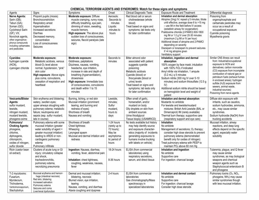
CHEMICAL TERRORISM AGENTS AND SYNDROMES: Watch for these signs and symptomsAgents Signs Symptoms Onset Clinical Diagnostic Tests Exposure Route and Treatment Differential diagnosis Nerve Agents:Sarin (GB);Tabun (GA); Soman (GD);Cyclohexyl Sarin(GF); VX;Novichok agents,other organophos-phorus compoundsincluding carbamatesand pesticides
Pinpoint pupils (miosis)BronchoconstrictionRespiratory arrestHypersalivationIncreased secretionsDiarrheaDecreased memory,
concentrationLoss of consciousnessSeizures
Moderate exposure: Diffusemuscle cramping, runny nose,difficulty breathing, eye pain,dimming of vision, sweating,muscle tremors.
High exposure: The above plussudden loss of consciousness,seizures, flaccid paralysis (latesign)
Aerosols: Seconds to
minutesLiquids: minutes tohours
Red blood cell or serumcholinesterase (wholeblood)
Treat based on signs andsymptoms; lab tests onlyfor later confirmation
Inhalation and dermal absorptionAtropine (2mg) IV; repeat q 5 minutes, titrate
until effective, average dose 6 to >15 mg– use IM in the field before IV access(establish airway for oxygenation)
Pralidoxime chloride (2-PAMCl) 600-1800mg IM or 1.0 g IV over 20-30 minutes(maximum 2 g IM or IV per hour)
Additional doses of atropine and 2-PAMCldepending on severity
Diazepam or lorazepam to prevent seizuresif >4 mg atropine given
Ventilatory support
Poisoning fromorganophosphate andcarbamate pesticides mayoccur as a result ofoccupational exposure
Cyanide poisoningMyasthenia gravis
Cyanides:hydrogen cyanide(HCN),cyanogen chloride
Moderate exposure:Metabolic acidosis, venousblood-O2 level abovenormal, hypotension, “pink”skin color
High exposure: Above signsplus coma, convulsions,cessation of respiration andheartbeat
Moderate exposure: Giddiness, palpitationsDizziness, nausea, vomiting,
headache, eye irritation,increase in rate and depth ofbreathing (hyperventilation),drowsiness
High exposure: Immediate lossof consciousness, convulsionsand death within 1 to 15minutes
Seconds tominutes
Bitter almond odorassociated with patientsuggests cyanidepoisoning
Metabolic acidosisCyanide (blood) or
thiocyanate (blood orurine) levels
Treat based on signs andsymptoms; lab tests onlyfor later confirmation
Inhalation, ingestion and dermalabsorption
100% oxygen by face mask; intubationwith 100% FiO2 if indicated
Amyl nitrite via inhalation, 1 ampule(0.2 mL) q 5 minutes
Sodium nitrite (300 mg IV over 5-10minutes) and sodium thiosulfate (12.5 gIV)
Additional sodium nitrite should be basedon hemoglobin level and weight ofpatient
Similar CNS illness can resultfrom: Industrial/occupationalexposure to HCN andderivatives; carbon monoxide(CO) exposure from incompletecombustion of natural gas orpetroleum fuels (exhaust fumesin enclosed areas); hydrogensulfide (H2S) exposure fromsewers, animal waste,industrial sources)
Poisoning from nerve agents
Vesicants/BlisterAgents:sulfur mustard,lewisite,nitrogen mustard,mustard lewisite,phosgene-oxime
Skin erythema and blistering;watery, swollen eyes;upper airways sloughing withpulmonary edema; metabolicfailure; neutropenia andsepsis (esp. sulfur mustard,late in course)
Burning, itching, or red skinMucosal irritation (prominent
tearing, and burning andredness of eyes)
Shortness of breathNausea and vomiting
Lewisite,minutes;Sulfurmustard,hours todays
Often smell of garlic,horseradish, and/ormustard on body
Oily droplets on skin fromambient sources
Urine thiodiglycolTissue biopsy (USAMRICD)
Inhalation and dermal absorptionMustards no antidoteFor lewisite and lewisite/mustard
mixtures: British Anti-Lewisite (BAL orDimercaprol) IM (rarely available)
Thermal burn therapy; supportive care(respiratory support and eye care)
Diffuse skin exposure withirritants, such as caustics,sodium hydroxides, ammonia,etc., may cause similarsyndromes.
Sodium hydroxide (NaOH) fromtrucking accidents
Pulmonary/Choking Agents:phosgene,chlorine,diphosgene,chloropicrin,oxides of nitrogen,sulfur dioxide
Pulmonary edema with somemucosal irritation (greaterwater solubility of agent =greater mucosal irritation)leading to ARDS or non-cardiogenic pulmonaryedema
Pulmonary infiltrate
Shortness of breathChest tightnessWheezingLaryngeal spasmMucosal and dermal irritation and
redness
1-24 hours(rarely up to72 hours);May beasymptoma-tic period ofhours
No tests available but historymay help identify sourceand exposure character-istics (majority of incidentsgenerating exposures tohumans involve truckingwith labels on vehicle)
InhalationNo antidoteManagement of secretions; O2 therapy;
consider high dose steroids to preventpulmonary edema (demonstratedbenefit only for oxides of nitrogen)
Treat pulmonary edema with PEEP tomaintain PO2 above 60 mm Hg
Mucosal irritation, airwayreactions, and deep lungeffects depend on the specificagent, especially watersolubility
Ricin (castor beanoil extract)
Clusters of acute lung or GIinjury; circulatory collapseand shock,tracheobronchitis,pulmonary edema,necrotizing pneumonia
Ingestion: Nausea, diarrhea,vomiting, fever, abdominal pain
Inhalation: chest tightness,coughing, weakness, nausea,fever
18-24 hours
8-36 hours
ELISA (from commerciallaboratories) usingrespiratory secretions,serum, and direct tissue
Inhalation and IngestionNo antidoteSupportive careFor ingestion: charcoal lavage
Tularemia, plague, and Q fevermay cause similarsyndromes, as may biologicalweapons and chemicalweapon agents such asStaphylococcal enterotoxin Band phosgene
T-2 mycotoxins:Fusarium,Myrotecium,Trichoderma,Verticimonosporium,Stachybotrys
Mucosal erythema and hemorr-hage (intestinal necrosis)
Red skin, blisteringIncreased salivationPulmonary edemaSeizures and comaLiver/renal dysfunction
Dermal and mucosal irritation;blistering, necrosis
Blurred vision, eye irritation,tearing
Nausea, vomiting, and diarrheaAtaxia coughing and dyspnea
2-4 hours ELISA from commerciallaboratoriesGas chromatography/Mass
spectroscopy inspecialized laboratories
Inhalation and dermal contactNo antidoteSupportive careFor ingestion: charcoal lavageConsider high dose steroids
Pulmonary toxins (O3, NOx,phosgene, NH3) may causesimilar syndromes thoughwith less mucosal irritation.
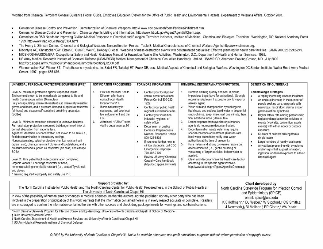
Modified from Chemical Terrorism General Guidance Pocket Guide, Employee Education System for the Office of Public Health and Environmental Hazards, Department of Veterans Affairs. October 2001.
• Centers for Disease Control and Prevention. Demilitarization of Chemical Weapons. http:// www.cdc.gov/nceh/demil/articles/initialtreat.htm.• Centers for Disease Control and Prevention. Chemical Agents Listing and Information. http://www.bt.cdc.gov/Agent/AgentlistChem.asp.• Committee on R&D Needs for Improving Civilian Medical Response to Chemical and Biological Terrorism Incidents, Institute of Medicine. Chemical and Biological Terrorism. Washington, DC: National Academy Press.
1999 http://www.nap.edu/catalog/6364.html• The Henry L. Stimson Center. Chemical and Biological Weapons Nonproliferation Project. Table E: Medical Characteristics of Chemical Warfare Agents http://www.stimson.org. • Macintyre AG, Christopher GW, Eitzen E, Gum R, Weir S, DeAtley C, et al. Weapons of mass destruction events with contaminated casualties: Effective planning for health care facilities. JAMA 2000;283:242-249.• NIOSH/OSHA/USCG/EPA. Occupational Safety and Health Guidance Manual for Hazardous Waste Site Activities. Washington, D.C.: Department of Health and Human Services. 1985.• US Army Medical Research Institute of Chemical Defense (USAMRICD) Medical Management of Chemical Casualties Handbook. 3rd ed. USAMRICD: Aberdeen Proving Ground, MD. July 2000.
http://ccc.apgea.army.mil/products/handbooks/mmccthirdeditionjul2000.pdf• Wannemacher RW, Wiener ST. Trichothecene mycotoxins. In: Sidell FR, Takafuji ET, Franz DR, eds. Medical Aspects of Chemical and Biological Warfare. Washington DC:Borden Institute, Walter Reed Army Medical
Center. 1997. pages 655-676.
UNIVERSAL PERSONAL PROTECTIVE EQUIPMENT (PPE)*
Level A: Maximum protection against vapor and liquids.Environment known to be immediately dangerous to life andhealth (harm occurs within 30 minutes).Fully encapsulating, chemical-resistant suit, chemically resistantgloves and boots, and a pressure-demand supplied air respirator(air hose) and escape self-contained breathing apparatus(SCBA)
Level B: Minimum protection exposure to unknown hazards.Full respiratory protection is required but danger to skin/risk ofdermal absorption from vapor is less.Agent not identified, or concentration not known to be safe (i.e.,field decontamination or ambulatory setting).Nonencapsulating, splash-protective chemical resistant suit(splash suit), chemical resistant gloves and boots/shoes, and apressure-demand supplied air respirator (air hose) and escapeSCBA
Level C: Until patient/victim decontamination completed.Organic vapor/P11 cartridge respirator or hood,nonencapsulating chemically-resistant (i.e., coated Tyvek) suitand gloves* Training required to properly and safely use PPE
NOTIFICATION PROCEDURES
1. First call the local HealthDirector; after hourscontact local HealthDirector via 911.
2. If criminal activity issuspected, call your locallaw enforcement and theFBI.
3. Alert local HAZMAT teamvia fire department at 911.
FOR MORE INFORMATION
• Contact your local poisoncontrol center or NationalPoison Control 800-222-1222
• Contact your public healthregional surveillance team
• Contact your institutionindustrial hygienist orsafety officer
• Department of JusticeDomestic PreparednessNational Response Hotline800-424-8802
• If you need further help inclinical diagnosis, call CDCEmergency Response770-488-7100
• Review US Army ChemicalCasualty Care handbook(http://ccc.apgea.army.mil)
UNIVERSAL DECONTAMINATION PROTOCOL
1. Remove clothing quickly and seal in plasticimpervious bags (save for authorities). Stronglyrecommended even if exposure only to vapor oraerosol agent.
2. Wash skin and shampoo with hypoallergenicliquid soap and copious tepid water in sequentialsteps of rinse, soap, rinse, wait one minute, thenfinal additional rinse (20 minutes).
3. Latent response from cyanide or pulmonaryagents do not require decontamination.
4. Decontamination waste water may requirespecial collection or treatment. (Discuss withlocal water authorities; notify local waterauthorities at the time of an event.)
5. Pure metals and strong corrosives require drydecontamination (i.e., gentle brushing orvacuuming of larger particles) before water isapplied.
6. Clean and decontaminate the healthcare facilityaccording to the specific agent involved.http://www.bt.cdc.gov/Agent/AgentlistChem.asp
DETECTION OF OUTBREAKS
Epidemiologic Strategies• A rapidly increasing disease incidence• An unusual increase in the number of
people seeking care, especially withneurologic, respiratory, dermal and/orgastrointestinal symptoms
• HIgher attack rate among persons whohad attendance at similar activities orevents (work site, convention, sportsevents) with either indoor or outdoorexposure.
• Clusters of patients arriving from asingle locale
• Large numbers of rapidly fatal cases• Any patient presenting with symptoms
and/or signs that suggest inhalation,ingestion, or dermal exposure to a toxicchemical agent
Support provided by:The North Carolina Institute for Public Health and The North Carolina Center for Public Health Preparedness, in the School of Public Health at
The University of North Carolina at Chapel HillIn view of the possibility of human error or changes in medical sciences, neither the authors, nor the publisher, nor any other party who has beeninvolved in the preparation or publication of this work warrants that the information contained herein is in every respect accurate or complete. Readersare encouraged to confirm the information contained herein with other sources and check drug package inserts for warnings and contraindications.
Chart developed by:North Carolina Statewide Program for Infection Control
and Epidemiology (SPICE)email: [email protected]
KK Hoffmann,* DJ Weber,* W Stopford,† CG Smith,‡J Newmark,§ BI Maliner,§ EP Clontz,* WA Rutala*
* North Carolina Statewide Program for Infection Control and Epidemiology, University of North Carolina at Chapel Hill School of Medicine† Duke University Medical Center‡ North Carolina Department of Health and Human Services and University of North Carolina at Chapel Hill§ US Army Medical Research Institute of Chemical Defense
© 2002 by the University of North Carolina at Chapel Hill. Not to be used for other than non-profit educational purposes without written permission of copyright owner.

CHEMICAL WEAPONS AND WAR AGENTSQUICK REFERENCE GUIDE
TYPE OFAGENT NERVE AGENTS ASPHYXIANTS CHOKING AGENTS VESICANTS
ChemicalName
MilitaryCode
ChemicalName
MilitaryCode
ChemicalName
MilitaryCode(s)
ChemicalName
MilitaryCode
MAJORCHEMICALS Tabun
SarinSomanO-ethyl-S
GAGBGDVX
CyanideCyanogen ClArsine
ACCKSA
Chlorine gasPhosgeneTear GasVomiting gasCapsaicin
CLCG
CR CS CN CADM DC DAPepper spray
MustardLewisitePhosgene oximeBis-2-chloro
H, HDL
CX
T
MODE OFACTION
Cholinergic crisis
Respiratory failure viamuscle paralysis &obstruction from airwaysecretions
Hypoxemia
Cellular hypoxia, failure touse O2 at the tissue level.Does not effect hemoglobinO2 binding.
Mucosal irritation
Incapacitating agents.Causes respiratory distressand severe mucosalirritation.
Caustic/Corrosive
Severe burns and irritationof mucous membranes andskin.
SYMPTOMS SyncopeMuscle paralysisBronchospasmBronchiole secretionsBradycardia
SyncopeSeizuresCardiac arrest
LaryngospasmBronchospasmPulmonary edema
Burns to skin, mouth andairwayHypovolemic shock
Emergency contact telephone numbers:
Poison Center 1-800-222-1222Wisconsin Emergency Management 24-hr 1-800-943-0003Wisconsin Dept of Health & Family Svcs 1-608-258-0099
1

CHEMICAL WEAPONS AND WAR AGENTS
CLINICALSIGNS NERVE AGENTS ASPHYXIANTS CHOKING AGENTS VESICANTS
Onset Coma/Apnea Flaccid Paralysis Eyes Nose/Oral Respiratory Cardiac Gastrointestinal Skin/Muscles Seizures Oximetry
Immediate or delayedYesYesMiosis/LacrimationSecretions/RhinitisSecretions/Apnea/BronchospasmBradycardia/HypertensionVomiting/DiarrheaFasciculation/DiaphoresisYesLow
ImmediateYesYesRed retinaRed mucosaHyperpnea/Clear chestHypotension/Cardiac arrestNot directly affectedNot cyanoticYesNormal
ImmediatePossibleNoConjunctivitis/LacrimationIrritation/RhinitisChoking/Stridor/BronchospasmNot directly affectedNot directly affectedMay see cyanosisUnlikelyMay be low
Immediate or delayedPossibleNoBurns/PainConjunctivitisBurns/Irritation/Clear if dermalHypotensive if volume lossesGI bleeding possibleBurns/BlistersUnlikelyLow if respiratory burns
RESPIRATORY SUPPORT IS THE MOST CRUCIAL INTERVENTION FOR VICTIMS REGARDLESS OF THE SPECIFIC AGENT
High flow oxygenSodium bicarbonate
TREATMENTS& ANTIDOTES
ATROPINE (secretions,bronchospasm, bradycardia)Adult 1-2 mg; PED: 0.25-0.5 mgprn IV or Nebulized. High dosesor continual IV may be needed.
BENZODIAZEPENE (seizures,agitation)Adult 2-5 mg; PED: 0.1 mg/kgprn IV, IM
2-PAM (reverse bond to enzyme)15 mg per kg to 600 mg MAX q6hr IV, IM
Lily Antidote Kit forsymptomatic patientsInhaled Nitrite1 ampule every 5 minutes untilIV startedIV Nitrite0.2 cc per kg (MAX 10 cc) IVtimes oneIV Thiosulfate2 cc per kg (MAX 50 cc) IVtimes one
High flow oxygen BAL if Lewisite
2.5 mg per kg IM (max 75 mg)
Prepared by Drs. Ernest Stremski and David Gummin, Medical Directors, The Children’s Hospital of Wisconsin, Poison Center
Printed by the Wisconsin Department of Health and Family ServicesNovember 2001
2

CHEMICAL WEAPONS AND WAR AGENTSQUICK REFERENCE GUIDE
PROTECTION FOR RESPONDERS
Wear the highest level protective equipment that is immediately available.
Beware of disoriented, combative victims and secondary attacks.
Avoid contaminating emergency vehicles and treatment areas.
CONTAINMENT
SECURE THE SCENE
Control crowds & onlookersMove victims UPWIND, UPHILL, OUTSIDE exposure areaStay > 300 ft from perimeters of contaminated areaPrevent cross-contamination and tracking
PROTECT TREATMENT AREAS & HOSPITALS
Establish DIRTY & CLEAN treatment zonesProtect staff with GLOVES, MASKS, EYE SHIELDS,OUTERWEARRemove victims’ outerwear, jewelry, etc. and store in sealedplastic bagsStore bagged contaminated items OUTSIDEVENTILATE to outside only
DECONTAMINATION
CLEANING EQUIPMENT
WaterSoap and waterHousehold bleach, undiluted (5.25%)
DECONTAMINATING VICTIMS
If liquids on skin & clothing: Remove clothing and flush bodywith water. Wrap hair in wet towels to stop volatilizationIf vapor exposure: Remove outer clothing prior to transport
3

CHEMICAL WEAPONS AND WAR AGENTSQUICK REFERENCE GUIDE
TRIAGE
Expectant Fatal
No cardiac activity
Needs Immediate Treatment
SeizuresSlow heart rate
Respiratory distressDermal contamination
Life-threatening injuries
Can Wait for Treatment
AlertWell aerated
No respiratory distressNo severe injuries/burns, etc
Minimal Treatment Needed
WalkingTalking
Alert & OrientedNo dermal contamination
No injuries
ADDITIONAL RESOURCES
Jane’s CHEM-BIO Handbook. Jane’s Information Group, 1-703-683-3700 or http://[email protected] ARMY Chemical Terrorism Fact Sheets: http://chppm-www.apgea.army.mil/dts/dtchemfs.htmUS ARMY Chemical Casualty Fact Book: http://www.vnh.org/CHEMCASU/titlepg.htmlUS ARMY Biologic Agent Information Papers: http://www.vnh.org/CHEMCASU/titlepg.htmlBiologic Readiness. A Template for Hospital Readiness: http://www.cdc.gov/ncidod/hip/Bio/13apr99APIC-CDCBioterrorism.PDFMedical Management of Radiologic Casualties Handbook: http://www.afrri.usuhs.mil/www/outreach/pdf/radiologicalhandbooksp99-2.pdf
Emergency contact telephone numbersPoison Center 1-800-222-1222Wisconsin Emergency Management 24-hr 1-800-943-0003Wisconsin Dept of Health & Family Svcs 1-608-258-0099
Prepared by: Drs. Ernest Stremski and David Gummin, Medical Directors, The Children’s Hospital of Wisconsin, Poison Center
Printed by the Wisconsin Department of Health and Family ServicesNovember 2001
4

WISCONSIN EMERGENCY RESPONSE GUIDE FOR CLINICAL LABORATORIES
Section IX - 1
This Page Last Updated: December, 2009
Radiological Emergency Events Radiation emergency events may result from either accidental or deliberate releases of radioactive material. Accidental releases may be related to nuclear reactors, medical radiation therapy, industrial irradiators, lost or stolen radioactive sources, or transportation accidents. Deliberate radiation emergency events may result from the intentional release of radiological material, as a “dirty bomb”, detonation of a low-yield nuclear weapon, or an attack or sabotage of a nuclear facility. Additional information related to laboratory testing in response to a radiological event is being further developed by the Centers for Disease Control and Prevention and their partners. Additional information is available at http://emergency.cdc.gov/radiation/links.asp The instructions for collection, packaging, and transport of clinical specimens for chemical terrorism (Section VIII of this manual) should be followed during a radiological event, according to information provided by the CDC. The WSLH recommends that current instructions should be obtained from 608-263-3280 before specimens are collected or transported in response to a radiation emergency. CDC would play a key role in protecting the public health during and after an emergency involving radiation or radioactive materials. Information to help prepare for a radiation emergency is available at http://www.bt.cdc.gov/radiation/ Additional information on this topic will be included in this document as it becomes available.

BIOTERRORISM RESPONSE GUIDE FOR CLINICAL LABORATORIES
Section IX - 1
This Page Last Updated: August 22, 2005
Description and Instructions for Use of the Wisconsin Emergency Response Shipper Kits Wisconsin Emergency Response Shipping Kits are stored at most local public health departments for use in emergency or outbreak response. These kits are intended for use by local health departments, first responders, clinical laboratories, or others in need of a kit to transport emergency response or outbreak specimens to the Wisconsin State Laboratory of Hygiene. These kits are not intended for routine specimen submission. A listing of repository sites with contact information is posted on the HAN (in the “Laboratory” topic area, under “Repository”) and is available near the end of this section. If your laboratory has a sample related to an outbreak or emergency response, you can contact one of the local health department repository sites to get one of the shippers. Your laboratory is expected to make arrangements to collect the shipper from the local health department repository site. The shippers are class 6 “infectious substance” shippers but can also be used for shipping diagnostic specimens. The shippers are appropriate for shipping samples at ambient temperature, refrigerated temperatures, or dry ice temperatures. The shipper includes: • A reminder for the local health department to notify the WSLH for a
replacement kit when this kit is used or distributed for use • Instructions for packaging and shipping a number of sample types • Selected forms
• A shipping form for infectious substances • A chain of custody form • WSLH requisition forms
• Packaging materials • Forensic evidence tape • Zip-lock specimen bags • Absorbent material for specimens • Kool-packs (must be frozen prior to use) • Amber bottle (for powder and/or chemical specimens)
• Labels (select the appropriate labels according to the instructions)
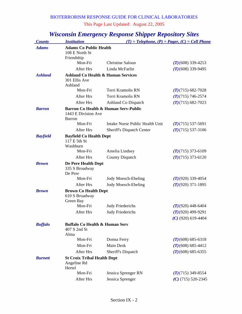
BIOTERRORISM RESPONSE GUIDE FOR CLINICAL LABORATORIES
Section IX - 2
This Page Last Updated: August 22, 2005
Wisconsin Emergency Response Shipper Repository Sites County Institution (T) = Telephone, (P) = Pager, (C) = Cell Phone Adams Adams Co Public Health 108 E North St Friendship Mon-Fri Christine Saloun (T) (608) 339-4253 After Hrs Linda McFarlin (T) (608) 339-9495 Ashland Ashland Co Health & Human Services 301 Ellis Ave Ashland Mon-Fri Terri Kramolis RN (T) (715) 682-7028 After Hrs Terri Kramolis RN (T) (715) 746-2574 After Hrs Ashland Co Dispatch (T) (715) 682-7023 Barron Barron Co Health & Human Serv-Public 1443 E Division Ave Barron Mon-Fri Intake Nurse Public Health Unit (T) (715) 537-5691 After Hrs Sheriff's Dispatch Center (T) (715) 537-3106 Bayfield Bayfield Co Health Dept 117 E 5th St Washburn Mon-Fri Amelia Lindsey (T) (715) 373-6109 After Hrs County Dispatch (T) (715) 373-6120 Brown De Pere Health Dept 335 S Broadway De Pere Mon-Fri Jody Moesch-Ebeling (T) (920) 339-4054 After Hrs Jody Moesch-Ebeling (T) (920) 371-1895 Brown Brown Co Health Dept 610 S Broadway Green Bay Mon-Fri Judy Friederichs (T) (920) 448-6404 After Hrs Judy Friederichs (T) (920) 499-9291 (C) (920) 619-4404 Buffalo Buffalo Co Health & Human Serv 407 S 2nd St Alma Mon-Fri Donna Ferry (T) (608) 685-6318 Mon-Fri Main Desk (T) (608) 685-4412 After Hrs Sheriff's Dispatch (T) (608) 685-6355 Burnett St Croix Tribal Health Dept Angeline Rd Hertel Mon-Fri Jessica Sprenger RN (T) (715) 349-8554 After Hrs Jessica Sprenger (C) (715) 520-2345

BIOTERRORISM RESPONSE GUIDE FOR CLINICAL LABORATORIES
Section IX - 3
This Page Last Updated: August 22, 2005
Wisconsin Emergency Response Shipper Repository Sites County Institution (T) = Telephone, (P) = Pager, (C) = Cell Phone Burnett Burnett Co Health Dept 7410 County Road K # 280 Siren Mon-Fri Carol A Larson RN (T) (715) 349-7600 After Hrs Burnett Co Dispatch (T) (715) 349-2128 Calumet Calumet Co Health Dept 206 Court St Chilton Mon-Fri Bonnie Kolbe RN (T) (920) 849-1432 After Hrs Bonnie Kolbe RN (T) (920) 853-7032 (C) (920) 849-2329 Chippewa Chippewa Co Public Health 711 N Bridge St Rm 222 Chippewa Falls Mon-Fri Debbie Odden RN (T) (715) 726-7900 After Hrs On Call Public Health Nurse (T) (715) 726-7901 Clark Clark Co Public Health 517 Court St Rm 105 Neillsville Mon-Fri Kathy Germain (T) (715) 743-5110 Mon-Fri Deanna Wolus (T) (715) 743-5106 After Hrs Kathy Germain (T) (715) 223-4486 Columbia Columbia Co Health Dept 2652 Murphy Rd Portage Mon-Fri Susan Lorenz (T) (608) 742-9265 After Hrs Susan Lorenz (T) (608) 617-9108 Crawford Crawford Co Public Health 111 W Dunn St Prairie du Chien Mon-Fri Gloria Wall (T) (608) 326-0230 After Hrs Gloria Wall (T) (608) 874-4004 (C) (608) 306-1300 Dane Dane Co Div of Public Health 1202 Northport Dr Rm 240 Madison Mon-Fri Julie Halvorsen (T) (608) 242-6487 (C) (920) 650-1337 After Hrs Judy Howard (T) (608) 301-0110 (C) (608) 332-6334
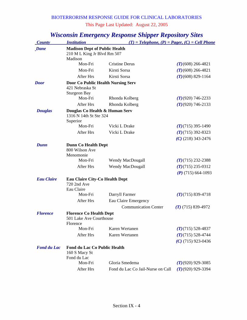
BIOTERRORISM RESPONSE GUIDE FOR CLINICAL LABORATORIES
Section IX - 4
This Page Last Updated: August 22, 2005
Wisconsin Emergency Response Shipper Repository Sites County Institution (T) = Telephone, (P) = Pager, (C) = Cell Phone Dane Madison Dept of Public Health 210 M L King Jr Blvd Rm 507 Madison Mon-Fri Cristine Derus (T) (608) 266-4821 Mon-Fri Kirsti Sorsa (T) (608) 266-4821 After Hrs Kirsti Sorsa (T) (608) 829-1164 Door Door Co Public Health Nursing Serv 421 Nebraska St Sturgeon Bay Mon-Fri Rhonda Kolberg (T) (920) 746-2233 After Hrs Rhonda Kolberg (T) (920) 746-2133 Douglas Douglas Co Health & Human Serv 1316 N 14th St Ste 324 Superior Mon-Fri Vicki L Drake (T) (715) 395-1490 After Hrs Vicki L Drake (T) (715) 392-8323 (C) (218) 343-2476 Dunn Dunn Co Health Dept 800 Wilson Ave Menomonie Mon-Fri Wendy MacDougall (T) (715) 232-2388 After Hrs Wendy MacDougall (T) (715) 235-0312 (P) (715) 664-1093 Eau Claire Eau Claire City-Co Health Dept 720 2nd Ave Eau Claire Mon-Fri Darryll Farmer (T) (715) 839-4718 After Hrs Eau Claire Emergency Communication Center (T) (715) 839-4972 Florence Florence Co Health Dept 501 Lake Ave Courthouse Florence Mon-Fri Karen Wertanen (T) (715) 528-4837 After Hrs Karen Wertanen (T) (715) 528-4744 (C) (715) 923-0436 Fond du Lac Fond du Lac Co Public Health 160 S Macy St Fond du Lac Mon-Fri Gloria Smedema (T) (920) 929-3085 After Hrs Fond du Lac Co Jail-Nurse on Call (T) (920) 929-3394

BIOTERRORISM RESPONSE GUIDE FOR CLINICAL LABORATORIES
Section IX - 5
This Page Last Updated: August 22, 2005
Wisconsin Emergency Response Shipper Repository Sites County Institution (T) = Telephone, (P) = Pager, (C) = Cell Phone Forest Forest Co Health Dept 200 E Madison Ave Crandon Mon-Fri Linda Kortbein (T) (715) 478-3371 After Hrs Linda Kortbein (T) (715) 478-3371 Grant Grant Co Health Dept 111 S Jefferson St Lancaster Mon-Fri Jeff Kindrai (T) (608) 723-6416 After Hrs Jeff Kindrai (T) (608) 778-2489 Green Green Co Health Dept N3150 Highway 81 Monroe Mon-Fri Vicki Evenson (T) (608) 328-9390 After Hrs Dawn Knaus (T) (608) 329-5155 (C) (608) 214-1533 After Hrs Vicki Evenson (T) (608) 897-2952 (C) (608) 558-1644 Green Lake Green Lake Co Health Serv 500 Lake Steel St Green Lake Mon-Fri Public Health Nurse (T) (920) 294-4070 After Hrs Sheriff's Department (T) (920) 294-4000 Iowa Iowa Co Health Dept 1205 N Bequette St Ste 1 Dodgeville Mon-Fri Ann Thompson (T) (608) 935-2810 After Hrs June E Meudt (T) (608) 553-7574 Iron Iron Co Health Dept 502 Copper St Hurley Mon-Fri Norene Gilbertson RN (T) (888) 561-2191 After Hrs Sheriff's Department (T) (715) 561-3800 Jackson Jackson Co Public Health 420 Hwy 54 W Black River Falls Mon-Fri John Mitchell (T) (715) 284-4301 Mon-Fri Ellen Moldenhauer (T) (715) 284-4301 After Hrs Sheriff's Dispatch (T) (715) 284-5357 Jefferson Jefferson Co Health Agency N3995 Annex Rd Jefferson Mon-Fri Tim Anderson (T) (920) 674-7275 After Hrs Tim Anderson (T) (920) 478-2338
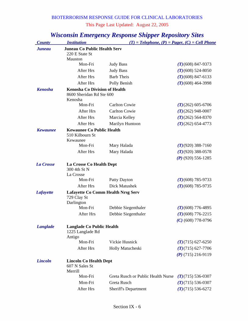
BIOTERRORISM RESPONSE GUIDE FOR CLINICAL LABORATORIES
Section IX - 6
This Page Last Updated: August 22, 2005
Wisconsin Emergency Response Shipper Repository Sites County Institution (T) = Telephone, (P) = Pager, (C) = Cell Phone Juneau Juneau Co Public Health Serv 220 E State St Mauston Mon-Fri Judy Bass (T) (608) 847-9373 After Hrs Judy Bass (T) (608) 524-8050 After Hrs Barb Theis (T) (608) 847-6133 After Hrs Polly Benish (T) (608) 464-3998 Kenosha Kenosha Co Division of Health 8600 Sheridan Rd Ste 600 Kenosha Mon-Fri Carlton Cowie (T) (262) 605-6706 After Hrs Carlton Cowie (T) (262) 948-0007 After Hrs Marcia Kelley (T) (262) 564-8370 After Hrs Marilyn Huntoon (T) (262) 654-4773 Kewaunee Kewaunee Co Public Health 510 Kilbourn St Kewaunee Mon-Fri Mary Halada (T) (920) 388-7160 After Hrs Mary Halada (T) (920) 388-0578 (P) (920) 556-1285 La Crosse La Crosse Co Health Dept 300 4th St N La Crosse Mon-Fri Patty Dayton (T) (608) 785-9733 After Hrs Dick Matushek (T) (608) 785-9735 Lafayette Lafayette Co Comm Health Nrsg Serv 729 Clay St Darlington Mon-Fri Debbie Siegenthaler (T) (608) 776-4895 After Hrs Debbie Siegenthaler (T) (608) 776-2215 (C) (608) 778-0796 Langlade Langlade Co Public Health 1225 Langlade Rd Antigo Mon-Fri Vickie Husnick (T) (715) 627-6250 After Hrs Holly Matucheski (T) (715) 627-7706 (P) (715) 216-9119 Lincoln Lincoln Co Health Dept 607 N Sales St Merrill Mon-Fri Greta Rusch or Public Health Nurse (T) (715) 536-0307 Mon-Fri Greta Rusch (T) (715) 536-0307 After Hrs Sheriff's Department (T) (715) 536-6272

BIOTERRORISM RESPONSE GUIDE FOR CLINICAL LABORATORIES
Section IX - 7
This Page Last Updated: August 22, 2005
Wisconsin Emergency Response Shipper Repository Sites County Institution (T) = Telephone, (P) = Pager, (C) = Cell Phone Manitowoc Manitowoc Co Health Dept 823 Washington St Manitowoc Mon-Fri James J Blaha (T) (920) 683-4155 After Hrs James J Blaha (T) (920) 371-2964 Marathon Marathon Co Health Dept 1220 Lake View Dr Ste 200 Wausau Mon-Fri Ruth Klee Marx (T) (715) 261-1904 After Hrs Julie Willems Van Dijk (T) (715) 573-9901 Marinette Marinette Co Public Health 2500 Hall Ave Ste C Marinette Mon-Fri Health Office (T) (715) 732-7670 After Hrs Sheriff's Dept Dispatch (T) (715) 732-7600 After Hrs Mary Mursau (C) (715) 923-3276 Marquette Marquette Co Health Dept 480 Underwood Ave Montello Mon-Fri Health Officer (T) (608) 297-9116 After Hrs Marquette Co Sheriff Dept (T) (608) 297-2115 To contact Health (C) (877) 627-6767 Milwaukee North Shore Health Dept 4800 W Green Brook Dr Brown Deer Mon-Fri Staff (T) (414) 371-2980 After Hrs Staff (T) (414) 870-0290 Milwaukee Cudahy Health Dept 5050 S Lake Dr Cudahy Mon-Fri Carol Wantuch (T) (414) 769-2239 (P) (414) 601-2371 (C) (414) 324-1398 After Hrs Carol Wantuch (T) (414) 571-6994 (P) (414) 601-2371 (C) (414) 324-1398 Milwaukee Franklin Health Dept 9229 W Loomis Rd Franklin Mon-Fri William M. Wucherer (T) (414) 425-9101 After Hrs Franklin Police-
-Non Emergency Dispatch (T) (414) 425-2522

BIOTERRORISM RESPONSE GUIDE FOR CLINICAL LABORATORIES
Section IX - 8
This Page Last Updated: August 22, 2005
Wisconsin Emergency Response Shipper Repository Sites County Institution (T) = Telephone, (P) = Pager, (C) = Cell Phone Milwaukee Greendale Health Dept 5650 Parking St Greendale Mon-Fri Susan Shepeard (T) (414) 423-2110 After Hrs Susan Shepeard (T) (414) 421-5832 Milwaukee Greenfield Health Dept 7325 W Forest Home Ave Greenfield Mon-Fri Elizabeth Anderson (T) (414) 329-5275 Milwaukee Hales Corners Health Dept 5635 S New Berlin Rd Hales Corners Mon-Fri Debra J Persak (T) (414) 529-6155 After Hrs Debra J Persak (T) (262) 784-7369 (C) (262) 719-3990 Milwaukee Milwaukee City Health Dept-Health Serv 841 N Broadway 3rd Fl Milwaukee Mon-Fri Sanjib Bhattacharyya Ph.D. (T) (414) 286-5702 (P) (414) 407-0116 After Hrs A. Singh (P) (414) 407-3959 (C) (414) 708-4532 After Hrs S. Gradus (P) (414) 205-1491 (C) (414) 708-2910 Milwaukee Oak Creek Health Dept 8640 S Howell Ave Oak Creek Mon-Fri Deborah Schier (T) (414) 768-6525 After Hrs Police Department (T) (414) 766-7600 After Hrs Deborah Schier (T) (414) 761-9225 (C) (414) 324-1540 Milwaukee Shorewood Health Dept 3930 N Murray Ave Shorewood Mon-Fri Cynthia Tomasello (T) (414) 847-2710 After Hrs Cynthia Tomasello (T) (414) 788-7018 Milwaukee South Milwaukee Health Dept 2424 15th Ave South Milwaukee Mon-Fri Jacqueline Ove (T) (414) 768-8055 After Hrs Jacqueline Ove (T) (414) 571-5447 (C) (414) 526-3679

BIOTERRORISM RESPONSE GUIDE FOR CLINICAL LABORATORIES
Section IX - 9
This Page Last Updated: August 22, 2005
Wisconsin Emergency Response Shipper Repository Sites County Institution (T) = Telephone, (P) = Pager, (C) = Cell Phone Milwaukee St Francis Health Dept 4235 S Nicholson Ave St Francis Mon-Fri Kathy Scott (T) (414) 978-2133 After Hrs Kathy Scott (C) (414) 588-4063 Milwaukee West Allis Health Dept 7120 W National Ave West Allis Mon-Fri Sally Nusslock (T) (414) 302-8600 After Hrs Sally Nusslock (T) (414) 324-1613 Monroe Monroe Co Health Dept 14301 County Trunk B/A-18 Sparta Mon-Fri Sharon Nelson (T) (608) 269-8666 After Hrs On-Call Nurse (T) (608) 269-2117 Oconto Oconto Co Public Health Serv Courthouse 501 Park Ave Oconto Mon-Fri Debra Konitzer (T) (920) 834-7000 (P) (920) 373-9075 (C) (920) 373-3314 After Hrs Debra Konitzer (T) (920) 846-2536 (P) (920) 373-9075 (C) (920) 373-3314 Oneida Oneida Co Health Dept 1 Courthouse Sq Rhinelander Mon-Fri Linda Conlon (T) (715) 369-6111 After Hrs Linda Conlon (T) (715) 493-0533 Outagamie Appleton Health Dept 100 N Appleton St Appleton Mon-Fri Kurt Eggebrecht (T) (920) 832-6429 Mon-Fri Appleton Health Dept (T) (920) 832-6429 After Hrs Dispatch After Hours Call List (T) (920) 832-5500 Outagamie Outagamie Co Public Health Div 401 S Elm St Appleton Mon-Fri Melody Bockenfeld (T) (920) 832-5100 After Hrs On-Call (P) (920) 616-9413

BIOTERRORISM RESPONSE GUIDE FOR CLINICAL LABORATORIES
Section IX - 10
This Page Last Updated: August 22, 2005
Wisconsin Emergency Response Shipper Repository Sites County Institution (T) = Telephone, (P) = Pager, (C) = Cell Phone Ozaukee Ozaukee Co Public Health Dept 121 W Main St Port Washington Mon-Fri Scott Visely (T) (262) 284-8170 After Hrs Sheriff's Dept - Page on call nurse (T) (262) 284-5575 (C) (262) 377-5820 Pepin Pepin Co Public Health Dept 740 7th Ave W Durand Mon-Fri Sue Kunferman (T) (715) 672-8961 After Hrs On Call Nurse (T) (715) 495-7631 Polk Polk Co Health Dept 100 Polk County Plz Ste 180 Balsam Lake Mon-Fri Public Health Nurse (T) (715) 485-8500 Mon-Fri Bonnie Leonard (T) (715) 485-8500 Mon-Fri Gretchen Sampson (T) (715) 485-8506 After Hrs Home Care Nurse on Call (T) (715) 485-8500 Portage Portage Co Health & Human Serv 817 Whiting Ave Stevens Point Mon-Fri Public Health Staff (T) (715) 345-5350 Mon-Fri Faye Tetzloff (T) (715) 345-5350 After Hrs Sheriff's Dept (T) (715) 346-1400 Price Price Co Health Dept 104 S Eyder Ave Phillips Mon-Fri Tracy Ellis (T) (715) 339-3054 After Hrs Sheriffs Dispatch (T) (715) 339-3011 Racine Western Racine Co Health Dept 156 E State St Burlington Mon-Fri Camille Gerou (T) (262) 763-4930 After Hrs Cheryl Mazmanian (P) (262) 576-8371 Racine Caledonia Mt Pleasant Health Dept 6922 Nicholson Rd Caledonia Mon-Fri Judith Price (T) (262) 835-6429 Mon-Fri Public Health Nurse (T) (262) 835-6429 After Hrs Police Dispatch (T) (262) 835-6131

BIOTERRORISM RESPONSE GUIDE FOR CLINICAL LABORATORIES
Section IX - 11
This Page Last Updated: August 22, 2005
Wisconsin Emergency Response Shipper Repository Sites County Institution (T) = Telephone, (P) = Pager, (C) = Cell Phone Racine Racine Health Dept 730 Washington Ave Racine Mon-Fri Robert C Bagley (T) (262) 636-9429 Mon-Fri Julie Kinzelman (T) (262) 636-9501 After Hrs Julie Kinzelman (T) (262) 554-7802 After Hrs Robert C Bagley (T) (262) 632-0453 Richland Richland Co Health Dept 221 W Seminary St Richland Center Mon-Fri Marianne Stanek RN (T) (608) 647-8821 After Hrs Marianne Stanek RN (T) (608) 647-4964 (C) (608) 604-2286 Rock Beloit City Health Dept 100 State St Beloit Mon-Fri Jackie Phillips (T) (608) 364-6637 (C) (608) 751-3082 Mon-Fri Linda Dalton (T) (608) 364-6627 (C) (608) 751-5482 After Hrs Linda Dalton (T) (815) 389-1944 (C) (608) 751-5482 After Hrs Jackie Phillips (T) (815) 624-2337 (C) (608) 751-3082 Rock Rock Co Health Dept 3328 N US Highway 51 Janesville Mon-Fri Timothy Banwell (T) (608) 757-5441 After Hrs Rock Co 911 Center (T) (608) 751-0400 Rusk Rusk Co Health Dept 311 Miner Ave E Ste C220 Ladysmith Mon-Fri Judy Bishop (T) (715) 532-2289 Mon-Fri Margaret “Kayo” Nash (T) (715) 532-2299 After Hrs Rusk Co Dispatch (T) (715) 532-2189 Saint Croix St Croix Co DHHS/Public Health 1445 N 4th St New Richmond Mon-Fri Health Officer or Environmental Hlth Specialist (T) (715) 246-8263 After Hrs Ed Thurman (T) (715) 247-2592 (C) (651) 238-5939 After Hrs Barbara Nelson (T) (715) 265-4804

BIOTERRORISM RESPONSE GUIDE FOR CLINICAL LABORATORIES
Section IX - 12
This Page Last Updated: August 22, 2005
Wisconsin Emergency Response Shipper Repository Sites County Institution (T) = Telephone, (P) = Pager, (C) = Cell Phone Sauk Sauk Co Public Health 505 Broadway St Ste 372 Baraboo Mon-Fri Timothy R Stieve (T) (608) 355-4419 (P) (800) 780-2152 (C) (608) 963-2291 After Hrs Timothy R Stieve (T) (608) 355-4419 (P) (800) 780-2152 (C) (608) 963-2291 Sawyer Sawyer Co Health Dept 10610 Main St Hayward Mon-Fri Eileen Simak (T) (715) 634-4874 After Hrs Sheriff's Dept (T) (715) 634-4858 Shawano Shawano Co Public Health Serv 311 N Main St Shawano Mon-Fri Janet Lewellyn (T) (715) 526-4808 After Hrs Janet Lewellyn (T) (715) 526-3332 (C) (920) 371-1892 Sheboygan Sheboygan Co Public Health 1011 N 8th St Sheboygan Mon-Fri Shirley Rohde (T) (920) 459-3265 After Hrs Sheriff's Dispatch (T) (920) 459-3112 Taylor Taylor Co Nursing Serv 224 S 2nd St Medford Mon-Fri Health Dept (T) (715) 748-1410 After Hrs Patty Krug (T) (715) 748-4226 (C) (715) 360-6139 Trempealeau Trempealeau Co Health Dept 36245 Main St Whitehall Mon-Fri Christine Hovell (T) (715) 538-2311 After Hrs Christine Hovell (T) (608) 582-3508 (C) (608) 484-0098 Vernon Vernon Co Health Dept E7410 County Highway BB Viroqua Mon-Fri Elizabeth A Johnson (T) (608) 637-5251 After Hrs Elizabeth A Johnson (T) (608) 637-5251 After Hrs Vernon Co Sheriffs Dept (T) (608) 637-2123
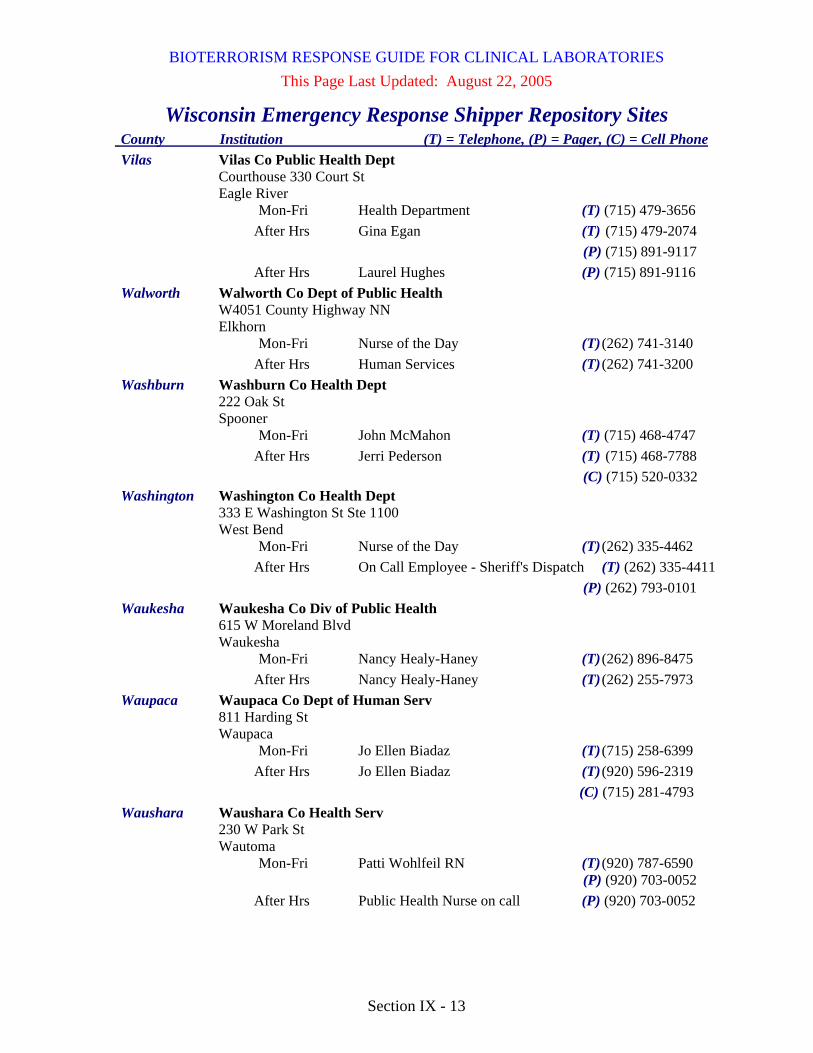
BIOTERRORISM RESPONSE GUIDE FOR CLINICAL LABORATORIES
Section IX - 13
This Page Last Updated: August 22, 2005
Wisconsin Emergency Response Shipper Repository Sites County Institution (T) = Telephone, (P) = Pager, (C) = Cell Phone Vilas Vilas Co Public Health Dept Courthouse 330 Court St Eagle River Mon-Fri Health Department (T) (715) 479-3656 After Hrs Gina Egan (T) (715) 479-2074 (P) (715) 891-9117 After Hrs Laurel Hughes (P) (715) 891-9116 Walworth Walworth Co Dept of Public Health W4051 County Highway NN Elkhorn Mon-Fri Nurse of the Day (T) (262) 741-3140 After Hrs Human Services (T) (262) 741-3200 Washburn Washburn Co Health Dept 222 Oak St Spooner Mon-Fri John McMahon (T) (715) 468-4747 After Hrs Jerri Pederson (T) (715) 468-7788 (C) (715) 520-0332 Washington Washington Co Health Dept 333 E Washington St Ste 1100 West Bend Mon-Fri Nurse of the Day (T) (262) 335-4462 After Hrs On Call Employee - Sheriff's Dispatch (T) (262) 335-4411 (P) (262) 793-0101 Waukesha Waukesha Co Div of Public Health 615 W Moreland Blvd Waukesha Mon-Fri Nancy Healy-Haney (T) (262) 896-8475 After Hrs Nancy Healy-Haney (T) (262) 255-7973 Waupaca Waupaca Co Dept of Human Serv 811 Harding St Waupaca Mon-Fri Jo Ellen Biadaz (T) (715) 258-6399 After Hrs Jo Ellen Biadaz (T) (920) 596-2319 (C) (715) 281-4793 Waushara Waushara Co Health Serv 230 W Park St Wautoma Mon-Fri Patti Wohlfeil RN (T) (920) 787-6590 (P) (920) 703-0052 After Hrs Public Health Nurse on call (P) (920) 703-0052
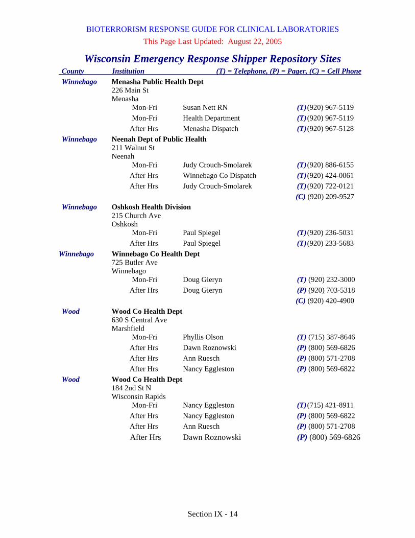
BIOTERRORISM RESPONSE GUIDE FOR CLINICAL LABORATORIES
Section IX - 14
This Page Last Updated: August 22, 2005
Wisconsin Emergency Response Shipper Repository Sites County Institution (T) = Telephone, (P) = Pager, (C) = Cell Phone Winnebago Menasha Public Health Dept 226 Main St Menasha Mon-Fri Susan Nett RN (T) (920) 967-5119 Mon-Fri Health Department (T) (920) 967-5119 After Hrs Menasha Dispatch (T) (920) 967-5128 Winnebago Neenah Dept of Public Health 211 Walnut St Neenah Mon-Fri Judy Crouch-Smolarek (T) (920) 886-6155 After Hrs Winnebago Co Dispatch (T) (920) 424-0061 After Hrs Judy Crouch-Smolarek (T) (920) 722-0121 (C) (920) 209-9527 Winnebago Oshkosh Health Division 215 Church Ave Oshkosh Mon-Fri Paul Spiegel (T) (920) 236-5031 After Hrs Paul Spiegel (T) (920) 233-5683 Winnebago Winnebago Co Health Dept 725 Butler Ave Winnebago Mon-Fri Doug Gieryn (T) (920) 232-3000 After Hrs Doug Gieryn (P) (920) 703-5318 (C) (920) 420-4900 Wood Wood Co Health Dept 630 S Central Ave Marshfield Mon-Fri Phyllis Olson (T) (715) 387-8646 After Hrs Dawn Roznowski (P) (800) 569-6826 After Hrs Ann Ruesch (P) (800) 571-2708 After Hrs Nancy Eggleston (P) (800) 569-6822 Wood Wood Co Health Dept 184 2nd St N Wisconsin Rapids Mon-Fri Nancy Eggleston (T) (715) 421-8911 After Hrs Nancy Eggleston (P) (800) 569-6822 After Hrs Ann Ruesch (P) (800) 571-2708 After Hrs Dawn Roznowski (P) (800) 569-6826

WISCONSIN EMERGENCY RESPONSE GUIDE FOR CLINICAL LABORATORIES
Section XI-1
This Page Last Updated: December, 2009
Wisconsin’s Health Alert Network (HAN) The Wisconsin Health Alert Network (HAN) is a secure internet site that provides a communications system for Wisconsin’s Public Health departments, hospitals, clinics, emergency rooms, laboratories, law enforcement, EMS and other health partners. The HAN is funded by a grant from the CDC to improve the communications infrastructure for Wisconsin public health agencies and their partners. It is part of a larger communication & information system - the Public Health Information Network or “PHIN”, which also includes the National Electronic Disease Surveillance System (NEDSS) for secure data transfer.
• Structure of the HAN The HAN is organized into the following “topic areas”: • AVR (Analysis, Visualization, and Reporting) • Chronic Disease Prevention & Health Promotion • Communicable • Communications • Contracting • Emergency Medical Services and Injury Prevention • Environmental • Epidemiology, Surveillance and Reporting • Family and Community Health • Flood Resources • Food Safety & Recreational Licensing • Integrated Data Systems • Laboratory • Preparedness • Wisconsin Public Health Within each topic area are “subtopics”, which may be organized into further subtopics.
• HAN registration provides access to: • Laboratory-specific information: You can access laboratory-specific
information in the “Laboratory” topic area, including a listing of shipper repository sites, sentinel laboratories, the Wisconsin Emergency Response Guide for Clinical Laboratories, Packaging Clinical Laboratory Samples for Domestic Transport, and various reports.
• Health Alerts and Advisories: You will see Health Alerts and Health Advisories on the HAN “Home” page (where you enter the HAN after logging in).
WISCONSIN EMERGENCY RESPONSE GUIDE FOR CLINICAL LABORATORIES
Section XI-2
This Page Last Updated: December, 2009
• Health Alerts are indicated by a bright RED header. Health Alerts are of immediate importance and a response or action may be required.
• Health Advisories are indicated by a dark BLUE header. Health Advisories are informational in nature and do not require an immediate response or action.
• Resources that are neither Alerts nor Advisories can be accessed from the top navigation bar under My HAN, then My HAN Additions on the sidebar.
• HAN Emergency Alert System (“Command Caller”): Once registered on the HAN, you can be included in groups (e.g., “laboratories”) that are created for emergency notifications by multiple contact methods, based on your profile information, like a personal “call-down tree”. • Command Caller is a communication tool to provide 24/7 capability
for emergency messages. • The system utilizes multiple channels (i.e., voicemail, fax, text
paging, wireless PDA, satellite phone, email, etc., depending on how your profile is set up)
• Command Caller can contact ~2000 individuals in 21 minutes • Note: The Command Caller feature can enhance the WSLH
capability to provide emergency messages to laboratories on a 24/7 basis as an adjunct to the current Laboratory Messaging System that uses email and fax. To utilize the Command Caller feature, the WSLH must be able to identify laboratory users of the HAN; registration instructions to enable this are included in the following description.
• Note 2: The Command Caller feature is expected to be discontinued when another system is fully operational.
• Individualized HAN Notifications: You can “personalize” the HAN for your use, by specifying that you want to be notified by email, including how often you want to receive notifications, when any of the following are updated: • HAN Site changes • Upcoming events • HAN content changes • Subscribed topic content • HAN training events You can also create “shortcuts” to areas that you use frequently.

WISCONSIN EMERGENCY RESPONSE GUIDE FOR CLINICAL LABORATORIES
Section XI-3
This Page Last Updated: December, 2009 • Registration: To become a HAN user, you must login to the HAN website
at https://www.han.wisc.edu, click on “Register” on the top menu bar, then complete a registration form, and receive approval by a HAN administrator. Detailed instructions and a tutorial for using the HAN are available at http://hanplus.wisc.edu/tutorial/ (HAN registration is not required for access to the tutorial.) The following pages contain detailed registration instructions for laboratorians that have been adapted from the tutorial.
• Logging off: To end your current HAN session, click on the Log Out button on the top navigation bar. If you are logged in to the HAN without any activity for more than 15 minutes, your session will time out and you will be prompted to re-enter your username and password.
HAN Registration Instructions (excerpted from “Tutorial” at http://hanplus.wisc.edu/tutorial/) An individual who wishes to access the HAN’s resources and features must obtain a Wisconsin User ID through the Web Access Management System (WAMS) and be approved for HAN access (a two-step approval process).
Go to the HAN and click on the Register tab: https://www.han.wisc.edu/ First obtain a Wisconsin User ID – click on Wisconsin UserID and Password (WAMS) to get to the link to begin registration.

WISCONSIN EMERGENCY RESPONSE GUIDE FOR CLINICAL LABORATORIES
Section XI-4
This Page Last Updated: December, 2009
HAN Registration Instructions (continued)
That link takes you to the WAMS Self Registration page at https://on.wisconsin.gov/WAMS/SelfRegController
Read the information on the page and then click on “Accept”
IMPORTANT: your WAMS account is NOT active until you activate it via the e-mail you receive from [email protected] after you submit the
registration form.
Fill out ONLY the spaces that are marked with the RED asterisk (*); you will have the opportunity to add more contact information in your HAN User Profile
NAME & E-MAIL

WISCONSIN EMERGENCY RESPONSE GUIDE FOR CLINICAL LABORATORIES
Section XI-5
This Page Last Updated: December, 2009
HAN Registration Instructions (continued)
USER NAME & PASSWORD
SECRET QUESTION FOR ACCOUNT RECOVERY
Click on “Submit”
Verify your e-mail address: a box will pop open and ask you to double check that you have entered the correct e-mail address

WISCONSIN EMERGENCY RESPONSE GUIDE FOR CLINICAL LABORATORIES
Section XI-6
This Page Last Updated: December, 2009 You will then receive the following message to complete PART TWO
of the process:
NOTE: IF you receive a message that says:
It means that you most likely already have a Wisconsin User ID –
contact the WAMS help desk at: https://on.wisconsin.gov/WAMS/FormattedEmail
The next step is what most people forget to do
IMPORTANT: your WAMS account is NOT active until you activate it via the e-mail you receive from [email protected] once you submit the
registration form.

WISCONSIN EMERGENCY RESPONSE GUIDE FOR CLINICAL LABORATORIES
Section XI-7
This Page Last Updated: December, 2009
HAN Registration Instructions (continued) Check your E-Mail – You should have one from [email protected] called
“State of Wisconsin Self-Registration
Open the E-mail and follow the instructions:
When you click on the link from the e-mail or cut and paste it into your Web Browser the following page will open. Enter the Username and Password you just created:

WISCONSIN EMERGENCY RESPONSE GUIDE FOR CLINICAL LABORATORIES
Section XI-8
This Page Last Updated: December, 2009
HAN Registration Instructions (continued)
Once you do that you will see the following message (your name will be in bold, rather than “HAN Administrator”)
Go to the HAN Login Tab (https://www.han.wisc.edu/mod_logn/) or simply close
the extra browser window and return to the HAN Registration page.
Enter your Wisconsin User ID and Password into the spaces provided:
You will then see the following screen to confirm your WAMS information:

WISCONSIN EMERGENCY RESPONSE GUIDE FOR CLINICAL LABORATORIES
Section XI-9
This Page Last Updated: December, 2009
HAN Registration Instructions (continued)
Click Next to begin the HAN Registration process:
Select your Organization from the pull down menu (sorted alphabetically or
hierarchically), or type part of the Organization name into the search bar and click “filter” as seen below. Please select the organization with the word “Lab” in the organization name.
For example, if your laboratory is a part of “ABC Hospital Medical Center”, you would select your primary organization as “ABC Hospital Medical Center Laboratory”. This allows the WSLH to identify you as a laboratorian, but you still retain your identity as a part of your organizational/institution.
NOTE: The WSLH recommends that laboratorians who are registering on the HAN should select a primary organization that includes the word “Lab” in the organization name. We have created “Lab” organizations for the organizations we have identified. Type in the word “lab” and click the “filter” button. The box will then display a “drop-down” list of laboratory organizations. If your organization does not appear on the list, please select the
organization “LABORATORIES”. We will contact you and create a new laboratory primary organization as necessary. Please do not check the box for “Don’t see your organization?”
If you have any questions about identifying yourself as a laboratory user of the HAN, please email [email protected] .
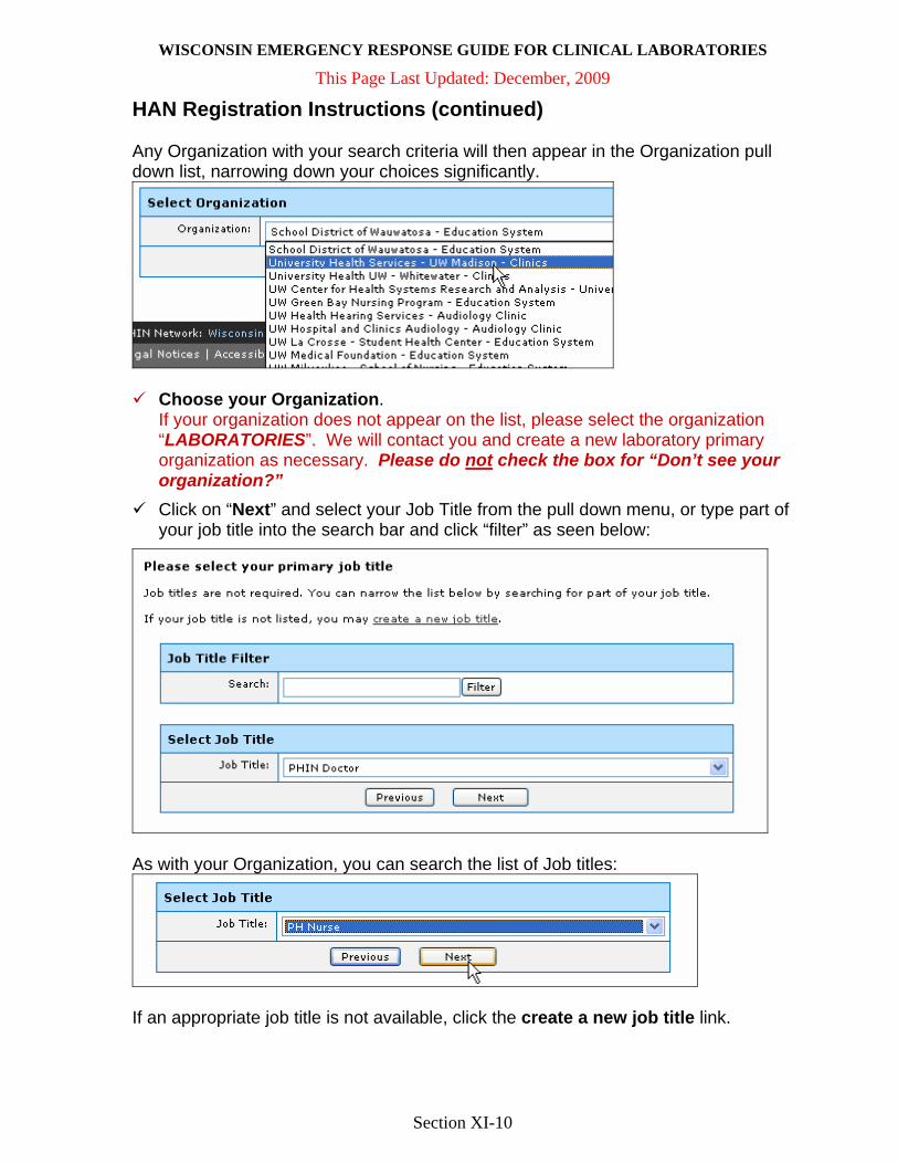
WISCONSIN EMERGENCY RESPONSE GUIDE FOR CLINICAL LABORATORIES
Section XI-10
This Page Last Updated: December, 2009
HAN Registration Instructions (continued) Any Organization with your search criteria will then appear in the Organization pull down list, narrowing down your choices significantly.
Choose your Organization. If your organization does not appear on the list, please select the organization “LABORATORIES”. We will contact you and create a new laboratory primary organization as necessary. Please do not check the box for “Don’t see your organization?”
Click on “Next” and select your Job Title from the pull down menu, or type part of your job title into the search bar and click “filter” as seen below:
As with your Organization, you can search the list of Job titles:
If an appropriate job title is not available, click the create a new job title link.

WISCONSIN EMERGENCY RESPONSE GUIDE FOR CLINICAL LABORATORIES
Section XI-11
This Page Last Updated: December, 2009
HAN Registration Instructions (continued)
Next you will be asked to enter your phone numbers. Remember that the HAN Emergency Broadcast System uses these numbers to contact you in the event of an emergency. Attempts to contact you will only be made at the numbers you enter below. Click “Next” when you have added your numbers.
Enter the numbers that you would like to be used and provide the priority usage (“Command Caller Rank”) for each.
It is recommended that you list your direct office number (not a number answered by a receptionist) and that you include your extension if the main number is always answered by an auto attendant.
The Command Caller system will not use any phone numbers that do not have a Command Caller Rank.
You can also designate whether you wish this information to be available to all HAN users (public) or not (private); this does not affect access by Command Caller.
Pagers listed in the “Phone Numbers” section will receive only numeric messages; to receive text messages, pagers should be listed in the “E-mail Addresses” section.
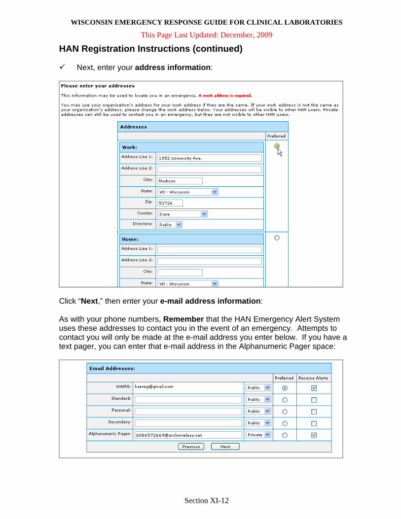
WISCONSIN EMERGENCY RESPONSE GUIDE FOR CLINICAL LABORATORIES
Section XI-12
This Page Last Updated: December, 2009
HAN Registration Instructions (continued)
Next, enter your address information:
Click “Next,” then enter your e-mail address information: As with your phone numbers, Remember that the HAN Emergency Alert System uses these addresses to contact you in the event of an emergency. Attempts to contact you will only be made at the e-mail address you enter below. If you have a text pager, you can enter that e-mail address in the Alphanumeric Pager space:

WISCONSIN EMERGENCY RESPONSE GUIDE FOR CLINICAL LABORATORIES
Section XI-13
This Page Last Updated: December, 2009
HAN Registration Instructions (continued) At this point you will be able to select any Digests you wish to receive. As with all other information in your User Profile, you will be able to edit this in the future if you wish. For more information on HAN Digests, please view the HAN Tutorial Page: http://hanplus.wisc.edu/tutorial/myhan.htm
Optional: You will now be asked to fill out information for the Wisconsin Emergency Assistance Volunteer Registry (WEAVR).
IF you choose YES in the above step, you will be asked to fill out information requested by WEAVR. The WCLN has not yet established a laboratory volunteer registry in WEAVR. IF you selected NO to the WEAVR registry, you will be taken to the “Skills” inventory. This is also optional, however, the HAN Emergency Broadcast System will use this information in times of emergency to seek out HAN Users with specific skills.
Once you have completed your Skills Inventory, click “Next.”

WISCONSIN EMERGENCY RESPONSE GUIDE FOR CLINICAL LABORATORIES
Section XI-14
This Page Last Updated: December, 2009
HAN Registration Instructions (continued)
The last step is the Registration Confirmation. If all the information is correct, click on “Submit”. If not, you are able to change your information by clicking on the “modify” links next to the appropriate category.
REMEMBER, you will be able to update this information in the future via the My HAN tab.
You will then receive a confirmation message indicating you have completed the registration application process. HAN accounts are generally approved or denied within 24 hours. If you register on a Friday or a Holiday, it may be longer. In the meantime, feel free to browse the HAN Tutorial to get a feel for the functionality: http://hanplus.wisc.edu/tutorial/

WISCONSIN EMERGENCY RESPONSE GUIDE FOR CLINICAL LABORATORIES
Section XII - 1
This Page Last Updated: December, 2009
Reportable Diseases in Wisconsin Laboratories are required to report to public health all suspect or confirmed cases of selected diseases that are defined by state statute. Reporting requirements are available at http://www.legis.state.wi.us/rsb/code/dhs/dhs145.pdf. A list of Wisconsin’s “reportable diseases” is available at http://dhs.wisconsin.gov/forms/F4/F44151.doc. Laboratory reports may be provided to public health via:
Paper-based reporting, using the case report form (Form 4151 or other required form), available at http://dhs.wisconsin.gov/forms/F4/F44151.doc. This completed form should be provided to local public health or the state epidemiologist, as directed on the form.
Electronic reporting, using:
• Automated laboratory reporting, or “ELR”, which is available to a limited number of laboratories that are capable of automated reporting from their computer system directly to the Wisconsin Electronic Disease Surveillance System (“WEDSS”).
• Web-based laboratory reporting, or “WLR”, which is available to all laboratories in Wisconsin, allows individuals to enter patient and laboratory data via the web, after which the data is provided directly to WEDSS. WLR can be accessed from the WSLH website at http://www.slh.wisc.edu/labupdates/reports/index.dot, where you click on the box that says “Click here to access Web-based Laboratory Reporting (WLR) of Reportable Diseases”.
o To receive authorization for you and your facility to use WLR, contact [email protected].
Once entered into WEDSS, reports are available to authorized local and state public health staff.
In addition to the paper-based, automated electronic, and web-based electronic reporting, immediate telephone notification is required for diseases and agents that are considered to be of urgent public health importance, classified as “Category 1” on the list of “Reportable Diseases”.

WISCONSIN EMERGENCY RESPONSE GUIDE FOR CLINICAL LABORATORIES
Section XII - 2
This Page Last Updated: December, 2009
Diseases Reportable at the State and National Level
1. Wisconsin Disease Surveillance Category I:
The following diseases are of urgent health importance and shall be reported IMMEDIATELY by telephone or fax to the patient’s local health officer upon identification of a case or a suspected case. In addition to the immediate report, complete and mail an Acute and Communicable Diseases Case Report (DPH 4151) to the address on the form, or enter the data into the Wisconsin Electronic Disease Surveillance System, within 24 hours. Category I Diseases Any illness caused by an agent that is foreign, exotic or unusual to Wisconsin, and that has public health implications4 Anthrax 1,4,5 Botulism 1,4 Botulism, infant 1,2,4 Cholera 1,3,4 *Diphtheria 1,3,4,5 *Haemophilus influenzae invasive disease, (including epiglottitis) 1,2,3,5 Hantavirus infection 1,2,4,5 *Hepatitis A 1,2,3,4,5 *Measles 1,2,3,4,5 Meningococcal disease 1,2,3,4,5 Outbreaks, foodborne or waterborne1,2,3,4 Outbreaks, suspected, of other acute or occupationally−related diseases *Pertussis (whooping cough) 1,2,3,4,5 Plague 1,4,5 *Poliovirus infection (paralytic or nonparalytic) 1,4,5 Rabies (human) 1,4,5 Ricin toxin 4,5 *Rubella 1,2,4,5 *Rubella (congenital syndrome) 1,2,5 Severe Acute Respiratory Syndrome−associated Coronavirus (SARS−CoV) 1,2,3,4 Smallpox 4,5 Tuberculosis 1,2,3,4,5 Vancomycin−intermediate Staphylococcus aureus (VISA) and Vancomycin−resistant Staphylococcus aureus (VRSA) infection 1,4,5 Yellow Fever 1,4

WISCONSIN EMERGENCY RESPONSE GUIDE FOR CLINICAL LABORATORIES
Section XII - 3
This Page Last Updated: December, 2009
Diseases Reportable at the State and National Level (continued)
2. Wisconsin Disease Surveillance Category II:
The following diseases shall be reported to the local health officer on an Acute and Communicable Disease Case Report (DPH 4151) or by other means or by entering the data into the Wisconsin Electronic Disease Surveillance System within 72 hours of the identification of a case or suspected case. Category II Diseases Arboviral disease1,2,4 Babesiosis 4,5 Blastomycosis 5 Brucellosis 1,4 Campylobacteriosis (campylobacter infection) 3,4 Chancroid 1,2,4,5 Chlamydia trachomatis infection 1,2,4,5 Cryptosporidiosis
1,2,3,4 Cyclosporiasis 1,4,5 Ehrlichiosis (anaplasmosis)1,5 E. coli 0157:H7, other Shiga toxin−producing E. coli (STEC), enteropathogenic E. coli, enteroinvasive E. coli, and enterotoxigenic E. coli 1,2,3,4 Giardiasis 3,4 Gonorrhea 1,2,4,5 Hemolytic Uremic Syndrome 1,2,4 *Hepatitis B 1,2,3,4,5 Hepatitis C 1,2 Hepatitis D 2,3,4,5 Hepatitis E 3,4 Histoplasmosis 5 Influenza−associated pediatric death 1,2 Influenza A virus infection, novel subtypes1,2 Kawasaki disease 2 Legionellosis 1,2,4 Leprosy (Hansen Disease) 1,2,3,4,5 Leptospirosis 4 Listeriosis 2,4 Lyme disease 1,2 Lymphocytic Choriomeningitis Virus (LCMV) infection 4 Malaria 1,2,4 Meningitis, bacterial (other than Haemophilus influenzae, meningococcal or streptococcal, which are reportable as distinct diseases) 2

WISCONSIN EMERGENCY RESPONSE GUIDE FOR CLINICAL LABORATORIES
Section XII - 4
This Page Last Updated: December, 2009
Diseases Reportable at the State and National Level (continued)
2. Wisconsin Disease Surveillance Category II (continued): *Mumps 1,2, 4,5 Mycobacterial disease (nontuberculous) Pelvic inflammatory disease2,5 Psittacosis 1,2,4 Q fever 4,5 Rheumatic fever (newly diagnosed and meeting the Jones criteria) 5 Rocky Mountain spotted fever 1,2,4,5 Salmonellosis 1,3,4 Shigellosis 1,3,4 Streptococcal disease (all invasive disease caused by Groups A and B Streptococci) Streptococcus pneumoniae invasive disease (invasive pneumococcal)1 Syphilis 1,2,4,5 *Tetanus 1,2,5 Toxic shock syndrome 1,2 Toxic substance related diseases:
Infant methemoglobinemia Lead intoxication (specify Pb levels) Other metal and pesticide poisonings
Toxoplasmosis Transmissible spongiform encephalopathy (TSE, human; CJD) Trichinosis 1,2,4 Tularemia 4 Typhoid Fever 1,2,3,4 *Varicella (chickenpox) 1,3,5 Vibriosis1,3,4 Yersiniosis 3,4
3. Wisconsin Disease Surveillance Category III: The following diseases shall be reported to the state epidemiologist on an AIDS Case Report (DPH 4264) or a Wisconsin Human Immunodeficiency Virus (HIV) Infection Confidential Case Report (DPH 4338) or by other means within 72 hours after identification of a case or suspected case.
Acquired Immune Deficiency Syndrome (AIDS) 1,2,4 Human Immunodeficiency Virus (HIV) infection 2,4 CD4 + T−lymphocyte < 200/uL, or CD4 + T−lymphocyte percentage of total lymphocytes of < 14

WISCONSIN EMERGENCY RESPONSE GUIDE FOR CLINICAL LABORATORIES
Section XII - 5
This Page Last Updated: December, 2009
Diseases Reportable at the State and National Level (continued) Key: * For diseases preceded by an (*), indicate immunization history in the
“Immunization data” box in the “Morbidity data” section. 1 Infectious diseases designated as notifiable at the national level. 2 Wisconsin or CDC follow−up form is required. Local health departments
have templates of these forms in the Epinet manual. 3 Risk assessment by local health department is needed to determine if
patient or member of patient’s household is employed in food handling, day care, or health care.
4 Case investigation by local health department is needed. 5 Immediate treatment is recommended, i.e., antibiotic or biologic for the
patient, or contact, or both.

WISCONSIN EMERGENCY RESPONSE GUIDE FOR CLINICAL LABORATORIES
Section XIII - 1
This Page Last Updated: December, 2009
Technical Resources • CDC homepage
www.cdc.gov
• CDC Bioterrorism homepage www.bt.cdc.gov
• CDC Smallpox homepage http://www.bt.cdc.gov/agent/smallpox/index.asp
• American Society for Microbiology (ASM) website http://www.asm.org/ Sentinel Level Clinical Microbiology Laboratory Guidelines and
Laboratory Preparedness Plan Template (BT Readiness Plan) at
http://www.asm.org/index.php?option=com_content&view=article&id=6342&title=Sentinel%20Level%20Clinical%20Microbiology%20Laboratory%20Guidelines
• Infection Control in Healthcare Settings http://www.cdc.gov/ncidod/dhqp/index.html
• Biosafety in Microbiological and Biomedical Laboratories, 5th Ed. U.S. Department of Health and Human Services, Centers for Disease Control and Prevention, and National Institutes of Health. Feb 2007. http://www.cdc.gov/od/ohs/biosfty/bmbl5/bmbl5toc.htm
• Association for Professionals in Infection Control and Epidemiology (APIC) www.apic.org
Training Resources • For a listing of training resources, refer to the “Continuing Education
Resources for Wisconsin Laboratories” at your institution and at the WSLH website http://www.slh.wisc.edu/dotAsset/15881.pdf
Wisconsin Clinical Laboratory Network (WCLN) • WCLN materials are available on the Health Alert Network (HAN) at
http://www.han.wisc.edu and at the Wisconsin State Laboratory of Hygiene (WSLH) website http://www.slh.wisc.edu/labupdates/wcln/index.dot