Embed Size (px)
Citation preview

"With ordinary talent and "With ordinary talent and extraordinary perseverance, all extraordinary perseverance, all things are attainable."things are attainable."- Thomas E. Buxton
"Achievement is connected "Achievement is connected with action, with action, not in genes..…!”not in genes..…!”- Conrad Hilton
Alcoholic liver disease
• Excessive alcohol consumption is the leading cause of liver disease.
• Alcoholic liver disease comprises of three main stages
1. Hepatic steatosis
2. Alcoholic hepatitis
3. Cirrhosis
Hepatic steatosis
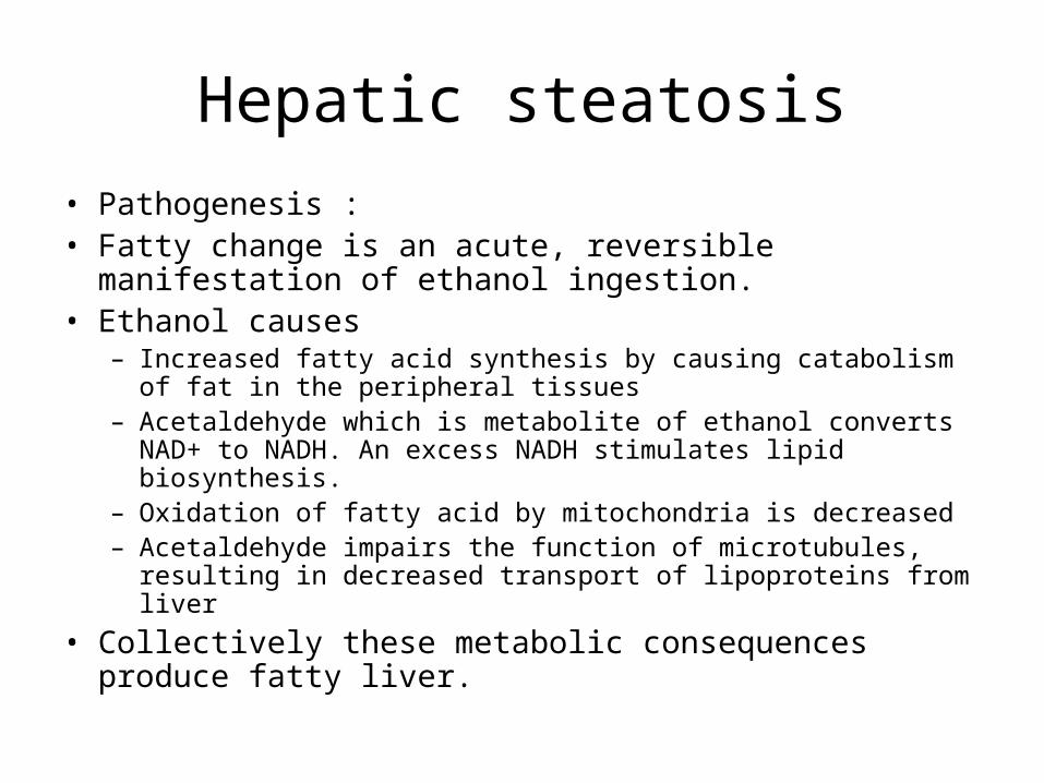
• Pathogenesis :• Fatty change is an acute, reversible manifestation of
ethanol ingestion.• Ethanol causes
– Increased fatty acid synthesis by causing catabolism of fat in the peripheral tissues
– Acetaldehyde which is metabolite of ethanol converts NAD+ to NADH. An excess NADH stimulates lipid biosynthesis.
– Oxidation of fatty acid by mitochondria is decreased– Acetaldehyde impairs the function of microtubules, resulting in
decreased transport of lipoproteins from liver
• Collectively these metabolic consequences produce fatty liver.
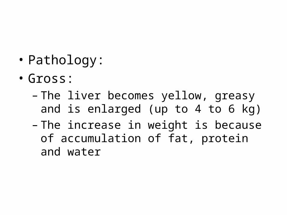
• Pathology:
• Gross:– The liver becomes yellow, greasy and is
enlarged (up to 4 to 6 kg)– The increase in weight is because of
accumulation of fat, protein and water
Alcoholic Fatty Liver
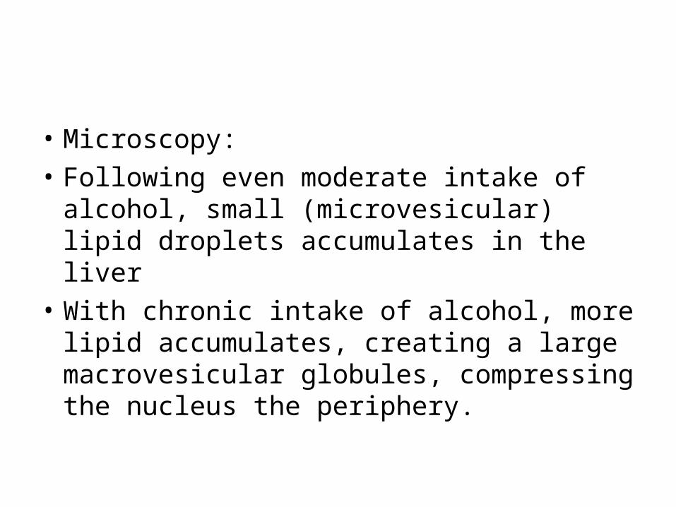
• Microscopy:
• Following even moderate intake of alcohol, small (microvesicular) lipid droplets accumulates in the liver
• With chronic intake of alcohol, more lipid accumulates, creating a large macrovesicular globules, compressing the nucleus the periphery.
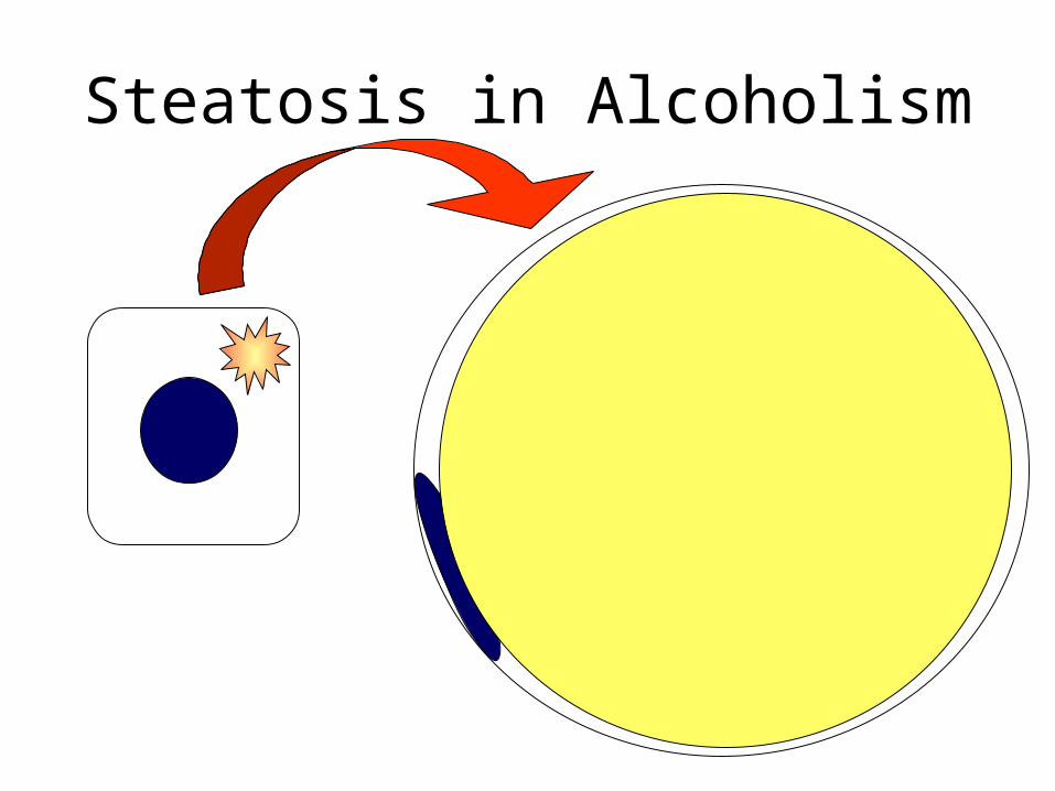
Steatosis in Alcoholism

Alcoholic Fatty Liver
• Clinical features of alcoholic steatosis– Hepatomegaly – Mild elevation of serum bilirubin, alkaline
phasphatase and gamma GT
Alcoholic hepatitis
• Is characterized by1. Hepatocyte swelling and necrosis
2. Mallory bodies
3. Neutrophilic inflammatory response
4. Perivenular fibrosis
• Hepatocyte swelling and necrosis:– Single or scattered foci of cells undergo
swelling (ballooning degeneration) and necrosis
• Mallory bodies:– Scattered hepatocytes accumulate cytokeratin
intermediate filaments and other proteins– Visible as eosinophilic cytoplasmic inclusions
in degenerating hepatocytes
• Neutrophilic reaction:– Neutrophils accumulate around the
degenerating hepatocytes, particularly those having Mallory bodies.
– Lymphocytes and macrophages also enter portal tracts and spill into parenchyma
• Fibrosis :– Commonly seen in the form of sinusoidal and
perivenular fibrosis– Occasionaly periportal fibrosis may
predominate– Fibrosis mainly occurs because of the
activation of sinusoidal stellate cells and portal tract fibroblasts
• Clinical features:– Malaise, anorexia, weight loss, upper
abdominal discomfort, tender hepatomegaly.– Laboratory findings:
• Hyperbilirubinemia• Elevated ALP,GGT, moderate elevation of AST • Neutrophilic leucocytosis
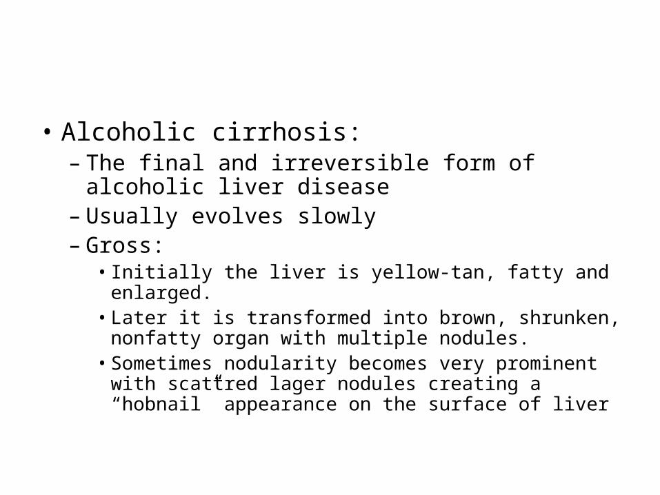
• Alcoholic cirrhosis:– The final and irreversible form of alcoholic
liver disease– Usually evolves slowly– Gross:
• Initially the liver is yellow-tan, fatty and enlarged.• Later it is transformed into brown, shrunken,
nonfatty organ with multiple nodules.• Sometimes nodularity becomes very prominent
with scattred lager nodules creating a “hobnail” appearance on the surface of liver

Normal Liver
Cirrhosis
Micronodular cirrhosis:
Alcoholic Cirrhosis
• Microscopy:– Initially fibrous septae are very delicate and
extend through sinusoids from central to portal regions as well as from portal tract to portal tract.
– As the fibrous septae dissect and surround nodules, the liver becomes more fibrotic, loses fat, and shrinks in size. (Laennec cirrhosis)
– Bile stasis may be seen.
Normal Liver Histology
Cirrhosis
Fibrosis
Regenerating Nodule
Liver Biopsy – Cirrhosis
Cirrhosis in Alcoholism
• Clinical features:– Features are similar to other forms of cirrhosis.– Malaise, weakness, weight loss, loss of appetite– Jaundice, ascites, and peripheral edema– Features of portal hypertension
• Laboratory findings:– Hyperbilirubinemia, elevated serum aminotransferase,
alkaline phasphatase, hypoproteinemia and anaemia
Alcoholic Liver Damage

Liver abscesses
Introduction• Liver abscesses can result from bacterial infection
(pyogenic abscess) or from Entamoeba histolytica. • Pyogenic abscesses have a high mortality rate of
40%. • Liver abscesses generally result from spread of infection
from : the digestive tract via the portal vein, from biliary disease or by direct extension from an adjacent infection.
• Risk factors include: Biliary disease Trauma Diabetes Malignancy.
Aetiology of liver abscess• Enteric Gram-negative bacilli (aerobes
and anaerobes) are frequently cultured.
• Many of the causative organisms originate in the gastrointestinal tract:Escherichia coli Klebsiella pneumoniae Bacteroides spp. Enterococcus spp. Anaerobic Streptococcus spp. Streptococcus ‘milleri’ group.
Diagnosis of liver abscessSigns and symptoms include:Fever Anorexia Nausea Weight loss Weakness Upper right quadrant pain Jaundice is rare until a late stage of the
infection.
Laboratory diagnosis• Diagnostic investigations include:• Culture of aspirated material (under ultrasound
guidance) is the most useful diagnostic test • With the advent of modern systems and improved
media, particularly for the recovery of anaerobic organisms, blood culture is often helpful.
• Imaging– CT is the most useful imaging technique, with
ultrasound effective for lesions more than a couple of centimetres in diameter.
Amoebic liver abscess
• Amebiasis is a disease caused by a one-celled parasite called Entamoeba histolytica .
• Mode of transmission: feco-oral with ingestion of amoebic cysts.
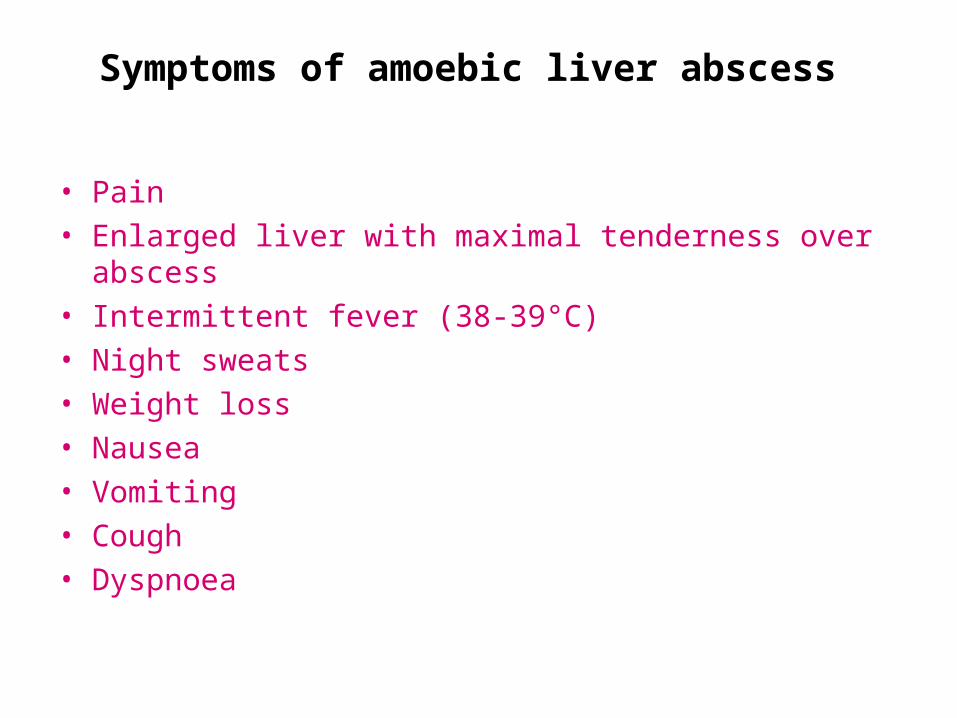
Symptoms of amoebic liver abscess
• Pain • Enlarged liver with maximal tenderness over abscess • Intermittent fever (38-39°C) • Night sweats • Weight loss • Nausea • Vomiting • Cough • Dyspnoea
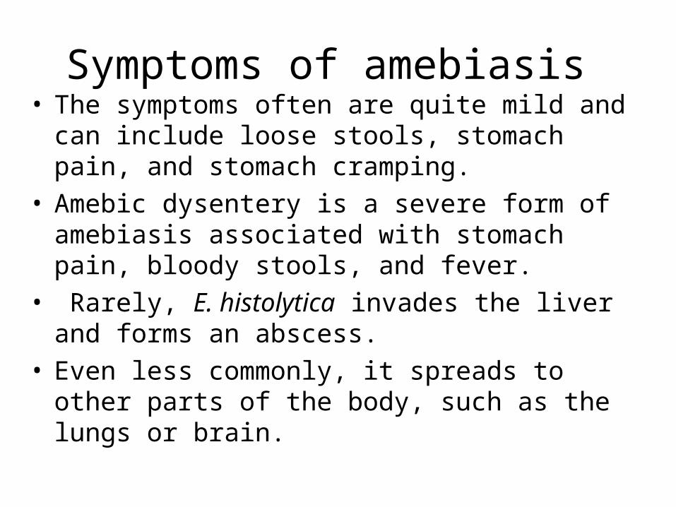
Symptoms of amebiasis • The symptoms often are quite mild and can
include loose stools, stomach pain, and stomach cramping.
• Amebic dysentery is a severe form of amebiasis associated with stomach pain, bloody stools, and fever.
• Rarely, E. histolytica invades the liver and forms an abscess.
• Even less commonly, it spreads to other parts of the body, such as the lungs or brain.
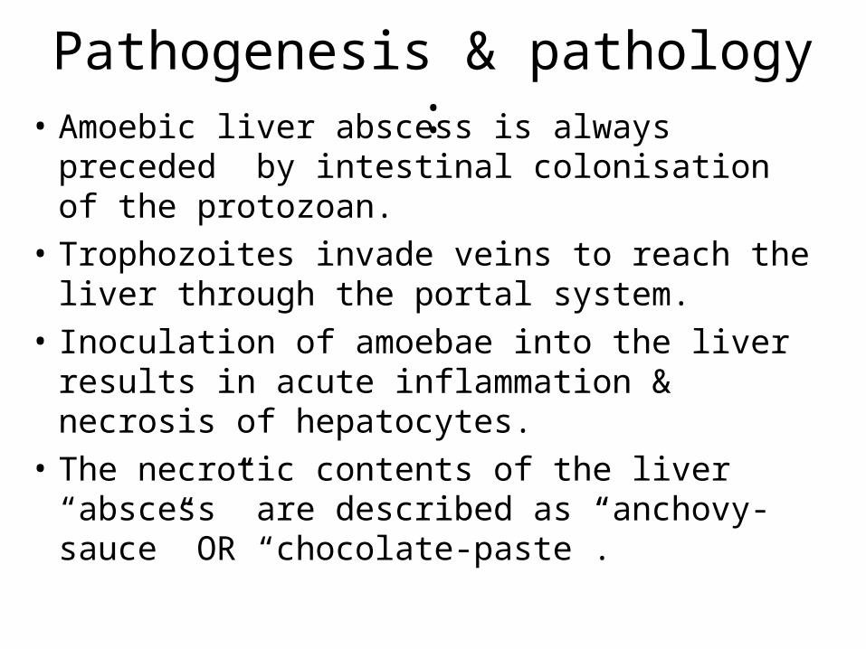
Pathogenesis & pathology :• Amoebic liver abscess is always preceded
by intestinal colonisation of the protozoan.• Trophozoites invade veins to reach the liver
through the portal system.• Inoculation of amoebae into the liver results
in acute inflammation & necrosis of hepatocytes.
• The necrotic contents of the liver “abscess” are described as “anchovy-sauce” OR “chocolate-paste”.
Trophozoites of entamoeba histolytica:
• The liver parenchyma is replaced by necrotic tissue surrounded by a thin rim of congested hepatic tissue, having a “shaggy” appearance due to fibrin.
Complications of amoebic liver abscess(ALA):
• ALA has a high mortality rate when associated with other-organ involvement.
• The abscess can rupture into :– the pleural space,– lung,– peritoneal cavity,– pericardial cavity and– the sub-phrenic space forming amoebic
abscesses in these sites.

‘‘Time’ is the best kept Time’ is the best kept secret of the rich..!secret of the rich..!
– Jim Rohn