Embed Size (px)
Citation preview
US 27457 Level 3 Credits 4
Name
Workbook Describe the anatomy and physiology of systems and associated organs of the
human body

Careerforce – Issue 1.3 – May 2014 27457 Describe the anatomy and physiology of systems and associated organs 2
US 27457
Level 3
Credits 4
Creative Commons
This work is licenced under a Creative Commons Attribution-NonCommercial Licence. You are free to
copy, distribute and transmit the work and to adapt the work. You must attribute Careerforce as the
author. You may not use this work for commercial purposes. For more information contact Careerforce
www.careerforce.org.nz
Careerforce – Issue 1.3 – May 2014 27457 Describe the anatomy and physiology of systems and associated organs 3
Contents
Before you start 4
Anatomy and physiology 7
Cardiovascular system 8
Respiratory system 12
Musculo-skeletal system 15
Endocrine system 21
Nervous system 24
Digestive system 30
Careerforce – Issue 1.3 – May 2014 27457 Describe the anatomy and physiology of systems and associated organs 4
Before you start
Welcome to this workbook for:
Describe the anatomy and physiology of systems and associated organs of the
human body
Unit Standard 27457.
For this unit standard you will have:
This workbook.
A trainee assessment.
In this workbook you will learn more about:
The cardiovascular system
The respiratory system
Muscles and bones
The function of hormones
The nervous system
The digestive system
How to use this workbook
This is your workbook to keep – make it
your own by writing in it.
Use highlighters to identify important ideas.
Do the learning activities included
throughout this workbook. Write your
answers in the spaces provided.
You might find it helpful to discuss your
answers with colleagues or your
supervisor.
Finish this workbook before you start on
the assessment.
When you see a
sticky note like
this, it gives a tip
or hint.
When you see a
sticky note like this, it
gives a tip or a hint.
Careerforce – Issue 1.3 – May 2014 27457 Describe the anatomy and physiology of systems and associated organs 5
Workbook activities
Stop – check what you know about this topic
You will see this stop symbol in places where you
are asked to stop and think about what you know
and:
Record your current knowledge
or impressions.
Check your knowledge.
This stop provides a reference point to return
to later. Stop activities have blue shading like this.
Rewind
When you see this rewind symbol, go back to:
Think about what you know.
Check your knowledge.
This rewind gives you an opportunity to add to,
change or confirm some of your initial thoughts
and ideas. Rewind activities have green shading
like this.
Learning activities
You will come across learning activities as you
work through this workbook.
These activities help you understand and apply
the information that you are learning about.
Learning activities have yellow shading like this.
The glossary and study hints book has study hints
for all trainees. It also explains key words and
phrases from the compulsory unit standards for
Foundation Skills and Core Competencies.
You can download it from www.careerforce.org.nz
or order it from http://shop.careerforce.org.nz
Careerforce – Issue 1.3 – May 2014 27457 Describe the anatomy and physiology of systems and associated organs 6
Before you go any further in this workbook, think about...
The human body
What is the difference between anatomy and physiology?
What part of the body controls the circulation and pressure of blood?
What is the function of alveoli?
What kind of functions does the nervous system control automatically?
Careerforce – Issue 1.3 – May 2014 27457 Describe the anatomy and physiology of systems and associated organs 7
Anatomy and physiology
In the living human, anatomy and physiology are inseparable.
Anatomy is the study, classification and description of the shape, structure, positions
(location) and interrelation of the systems and associated organs of the human body.
Physiology is the study of the mechanical, physical, and biochemical functions of the
systems and associated organs of the human body.
The human body is organised on several levels, from the simplest to the most complex:
1 Cells are the basic unit of life.
2 Tissues are clusters of cells that perform a similar function.
3 Organs are a group of tissues that perform a specific function.
4 Organ systems are groups of organs that perform a specific purpose.
These systems work together in an integrated way to help us function and to promote the
wellbeing of the entire body. The ability to balance these systems and maintain stability
is called homeostasis.
There are 11 organ systems in the human body:
Cardiovascular (circulatory)
Respiratory
Skeletal
Muscular
Endocrine
Nervous
Digestive
Immune
Integumentary (skin)
Reproductive
Excretory (urinary)
This workbook focuses on the first seven of these systems.
Careerforce – Issue 1.3 – May 2014 27457 Describe the anatomy and physiology of systems and associated organs 8
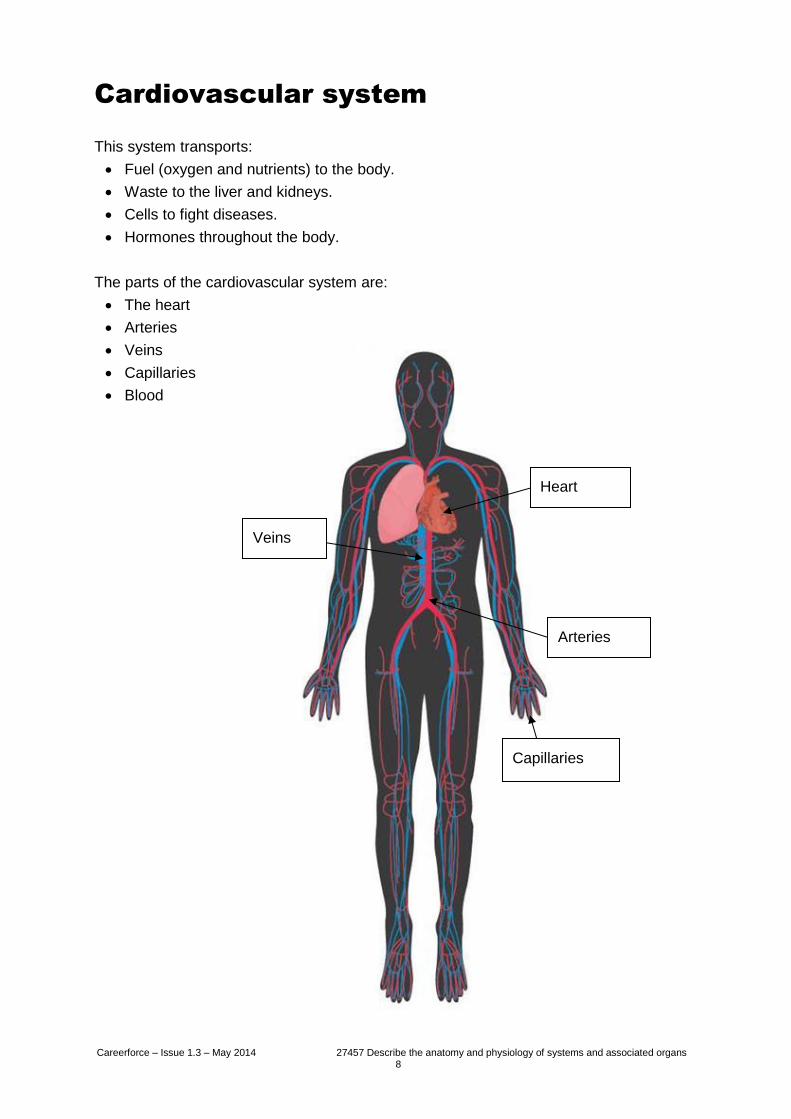
Cardiovascular system
This system transports:
Fuel (oxygen and nutrients) to the body.
Waste to the liver and kidneys.
Cells to fight diseases.
Hormones throughout the body.
The parts of the cardiovascular system are:
The heart
Arteries
Veins
Capillaries
Blood
Heart
Arteries
Veins
Capillaries
Careerforce – Issue 1.3 – May 2014 27457 Describe the anatomy and physiology of systems and associated organs 9
The cardiovascular system includes:
The pulmonary circulation – a “loop” that goes through the lungs to oxygenate the
blood.
The systemic circulation – a loop that goes through the rest of the body to provide
the oxygenated blood.
The coronary circulation – the heart receives blood under the highest possible
pressure to meet the demands of active cardiac muscle tissue.
The pulmonary circulation transports oxygen-depleted blood away from the heart to the
lungs and returns oxygenated blood back to the heart.
The systemic circulation transports oxygenated blood away from the heart to the rest of
the body and returns oxygen-depleted blood back to the heart.
Both the pulmonary and systemic loops begin and end at the heart and blood travels
through both loops in sequence. Blood that has gone through the pulmonary circuit will
go through the systemic circuit before returning to the pulmonary circuit.
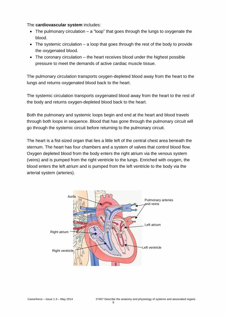
The heart is a fist-sized organ that lies a little left of the central chest area beneath the
sternum. The heart has four chambers and a system of valves that control blood flow.
Oxygen depleted blood from the body enters the right atrium via the venous system
(veins) and is pumped from the right ventricle to the lungs. Enriched with oxygen, the
blood enters the left atrium and is pumped from the left ventricle to the body via the
arterial system (arteries).
Left ventricle
Left atrium
Aorta
Right ventricle
Right atrium
Pulmonary arteries and veins
Careerforce – Issue 1.3 – May 2014 27457 Describe the anatomy and physiology of systems and associated organs 10
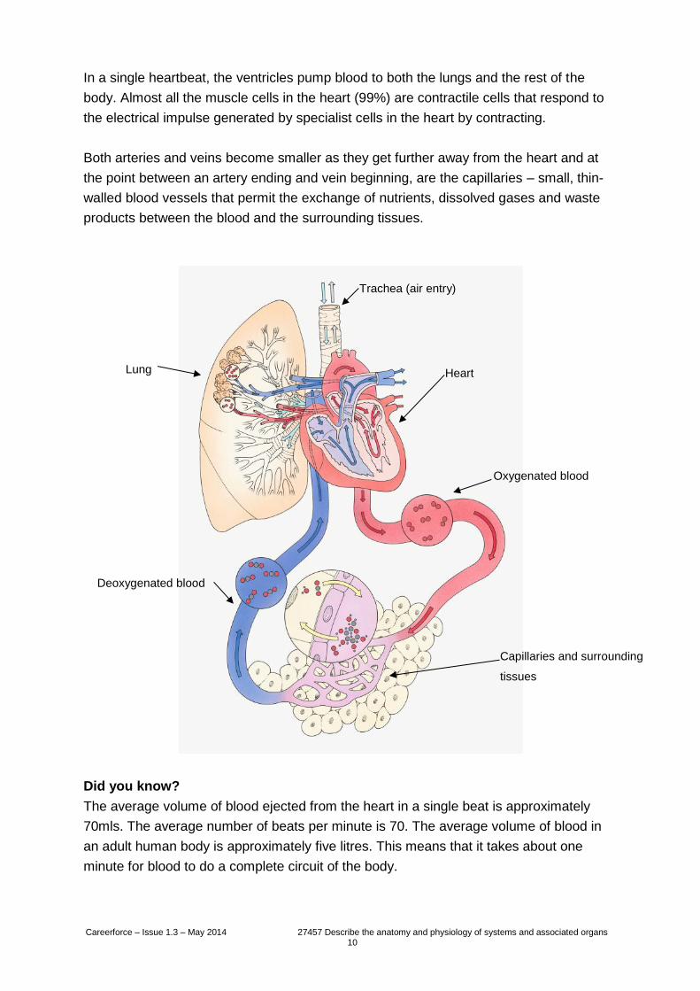
In a single heartbeat, the ventricles pump blood to both the lungs and the rest of the
body. Almost all the muscle cells in the heart (99%) are contractile cells that respond to
the electrical impulse generated by specialist cells in the heart by contracting.
Both arteries and veins become smaller as they get further away from the heart and at
the point between an artery ending and vein beginning, are the capillaries – small, thin-
walled blood vessels that permit the exchange of nutrients, dissolved gases and waste
products between the blood and the surrounding tissues.
Did you know?
The average volume of blood ejected from the heart in a single beat is approximately
70mls. The average number of beats per minute is 70. The average volume of blood in
an adult human body is approximately five litres. This means that it takes about one
minute for blood to do a complete circuit of the body.
Capillaries and surrounding
tissues
Oxygenated blood
Heart Lung
Deoxygenated blood
Trachea (air entry)
Careerforce – Issue 1.3 – May 2014 27457 Describe the anatomy and physiology of systems and associated organs 11
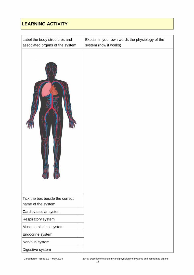
LEARNING ACTIVITY
Label the body structures and
associated organs of the system
Explain in your own words the physiology of the
system (how it works)
Tick the box beside the correct
name of the system:
Cardiovascular system
Respiratory system
Musculo-skeletal system
Endocrine system
Nervous system
Digestive system
Careerforce – Issue 1.3 – May 2014 27457 Describe the anatomy and physiology of systems and associated organs 12
Respiratory system
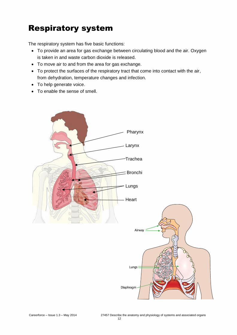
The respiratory system has five basic functions:
To provide an area for gas exchange between circulating blood and the air. Oxygen
is taken in and waste carbon dioxide is released.
To move air to and from the area for gas exchange.
To protect the surfaces of the respiratory tract that come into contact with the air,
from dehydration, temperature changes and infection.
To help generate voice.
To enable the sense of smell.
Pharynx
Larynx
Trachea
Bronchi
Lungs
Heart
Careerforce – Issue 1.3 – May 2014 27457 Describe the anatomy and physiology of systems and associated organs 13
The respiratory system is divided into upper and lower sections.
The upper respiratory system includes the nose and pharynx (the area shared by both
the digestive and respiratory systems). This is where air first enters the body and the
process of warming, filtering and humidification begins.
The lower respiratory system includes the larynx (voice box), trachea (windpipe),
bronchi, bronchioles and alveoli of the lungs.
The larynx connects the pharynx to the trachea. It is involved in breathing, sound
production and protecting the trachea from aspiration. If the larynx is stimulated by the
touch of food or liquid, the coughing reflex is triggered.
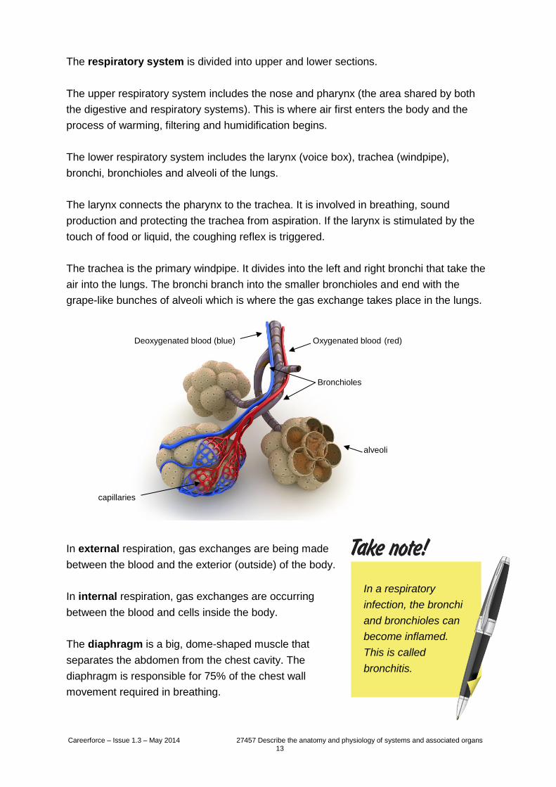
The trachea is the primary windpipe. It divides into the left and right bronchi that take the
air into the lungs. The bronchi branch into the smaller bronchioles and end with the
grape-like bunches of alveoli which is where the gas exchange takes place in the lungs.
In external respiration, gas exchanges are being made
between the blood and the exterior (outside) of the body.
In internal respiration, gas exchanges are occurring
between the blood and cells inside the body.
The diaphragm is a big, dome-shaped muscle that
separates the abdomen from the chest cavity. The
diaphragm is responsible for 75% of the chest wall
movement required in breathing.
In a respiratory
infection, the bronchi
and bronchioles can
become inflamed.
This is called
bronchitis.
alveoli
capillaries
Bronchioles
Oxygenated blood (red) Deoxygenated blood (blue)
Careerforce – Issue 1.3 – May 2014 27457 Describe the anatomy and physiology of systems and associated organs 14
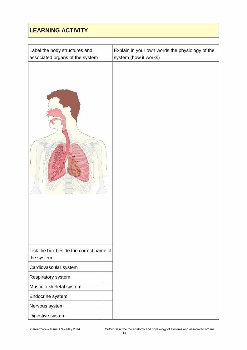
LEARNING ACTIVITY
Label the body structures and
associated organs of the system
Explain in your own words the physiology of the
system (how it works)
Tick the box beside the correct name of
the system:
Cardiovascular system
Respiratory system
Musculo-skeletal system
Endocrine system
Nervous system
Digestive system
Careerforce – Issue 1.3 – May 2014 27457 Describe the anatomy and physiology of systems and associated organs 15
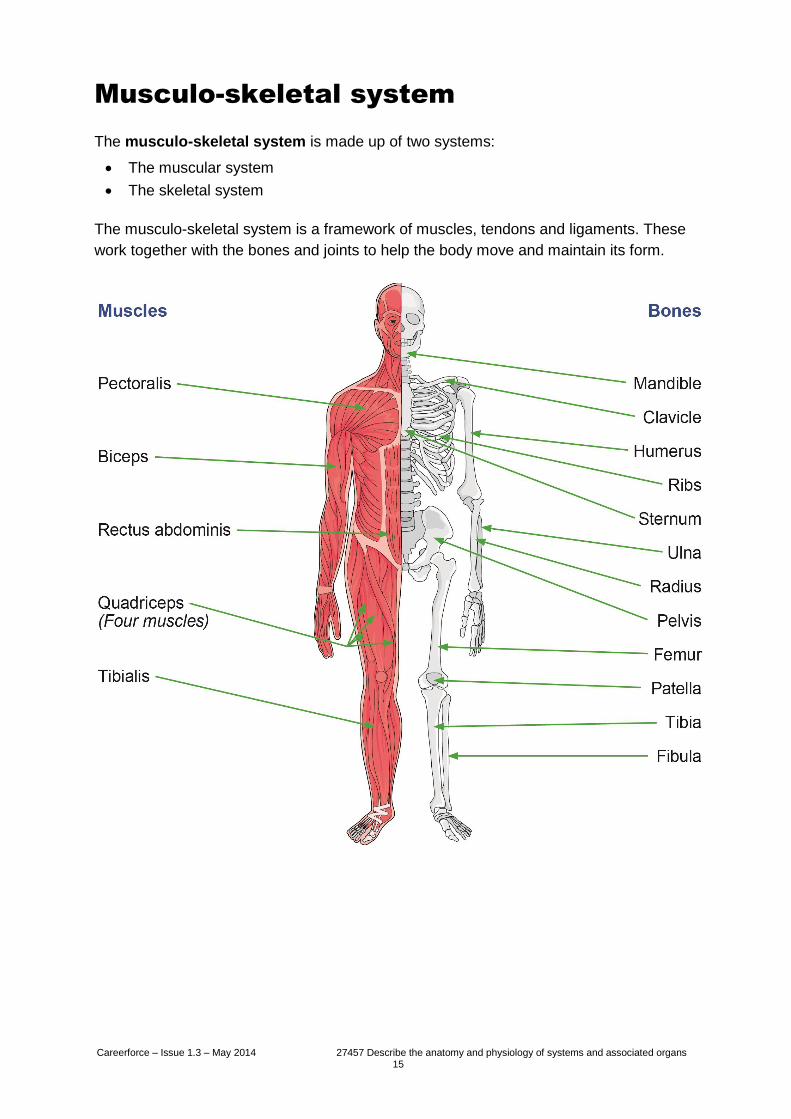
Musculo-skeletal system
The musculo-skeletal system is made up of two systems:
The muscular system
The skeletal system
The musculo-skeletal system is a framework of muscles, tendons and ligaments. These
work together with the bones and joints to help the body move and maintain its form.
Careerforce – Issue 1.3 – May 2014 27457 Describe the anatomy and physiology of systems and associated organs 16
The muscular system
Muscles are made of an elastic type of tissue and there are thousands of small fibres
that make up each muscle.
There are three types of muscle in the human body.
Smooth muscles are also called involuntary muscles. These muscles are found in the
stomach, digestive system, bladder, uterus and eyes. Smooth muscle controls the
movement of food through the digestive tract and the emptying of the bowels and
bladder.
Cardiac muscles, also called the myocardium, form the walls of the heart. The muscles
contract to pump blood to the rest of the body and then relax to allow the chambers to fill
with blood again. Cardiac muscles are also involuntary and their movement depends on
an electrical impulse being delivered from specialised cells called pacemakers.
Skeletal muscles are voluntary muscles and are attached to bones by tendons.
There are approximately 700 skeletal muscles and all of them are connected directly or
indirectly with the skeletal system to allow the body to move. Some of the biggest and
most powerful skeletal muscles are in the back, near the spine. These muscles keep the
body upright.
Muscles have different functions, including:
Producing body movements.
Stabilising body position.
Moving and storing substances.
Producing heat.
Careerforce – Issue 1.3 – May 2014 27457 Describe the anatomy and physiology of systems and associated organs 17
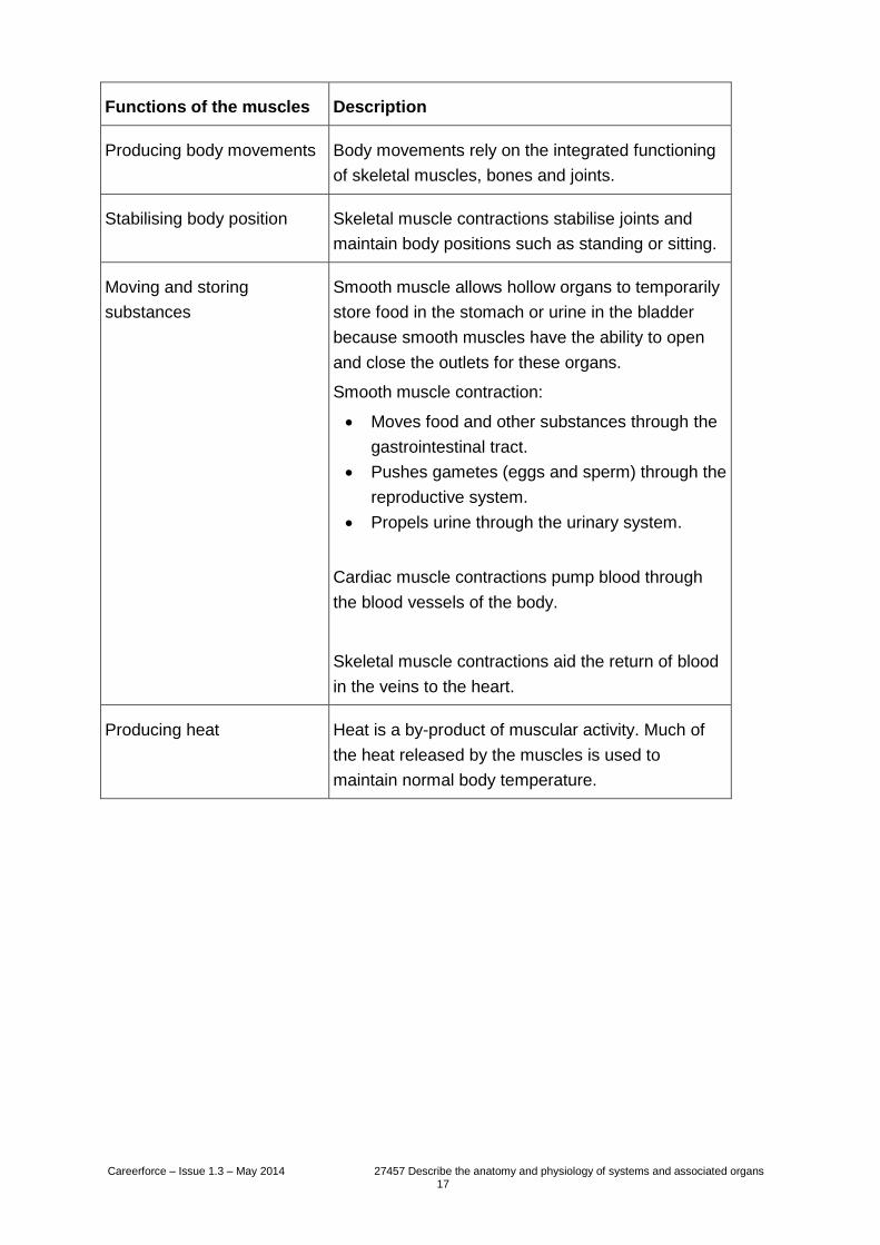
Functions of the muscles Description
Producing body movements Body movements rely on the integrated functioning
of skeletal muscles, bones and joints.
Stabilising body position Skeletal muscle contractions stabilise joints and
maintain body positions such as standing or sitting.
Moving and storing
substances
Smooth muscle allows hollow organs to temporarily
store food in the stomach or urine in the bladder
because smooth muscles have the ability to open
and close the outlets for these organs.
Smooth muscle contraction:
Moves food and other substances through the
gastrointestinal tract.
Pushes gametes (eggs and sperm) through the
reproductive system.
Propels urine through the urinary system.
Cardiac muscle contractions pump blood through
the blood vessels of the body.
Skeletal muscle contractions aid the return of blood
in the veins to the heart.
Producing heat Heat is a by-product of muscular activity. Much of
the heat released by the muscles is used to
maintain normal body temperature.
Careerforce – Issue 1.3 – May 2014 27457 Describe the anatomy and physiology of systems and associated organs 18
Bones
There are 206 bones in the human body.
Bones provide support and shape for our bodies.
The skull protects the brain and forms the shape of our face.
The spinal cord, which acts as the pathway for messages between the brain and the
body, is protected by the spinal column which also helps to hold the body in an
upright position.
The ribs form a cage that protects the heart, lungs, liver, and spleen; the pelvis helps
protect the bladder, intestines, and in women, the reproductive organs.
The long bones in our legs and arms are very strong and are designed to support
weight.
Bones are made up of living cells, which constantly grow and reshape themselves
throughout our life. As we age, this process slows down.
Calcium helps make bones strong which allows them to support our weight.
Bones store calcium. Bones also release calcium into the bloodstream when it's
needed by other parts of the body.
Bones are attached to other bones by long, fibrous straps called ligaments.
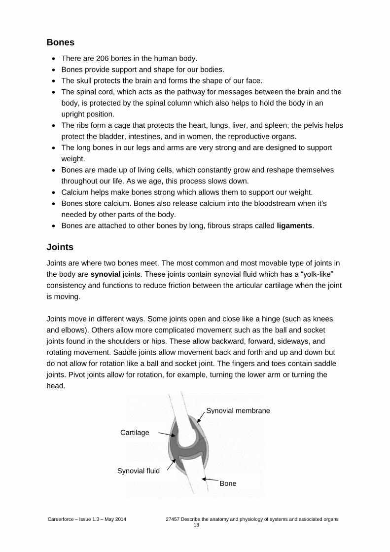
Joints
Joints are where two bones meet. The most common and most movable type of joints in
the body are synovial joints. These joints contain synovial fluid which has a “yolk-like”
consistency and functions to reduce friction between the articular cartilage when the joint
is moving.
Joints move in different ways. Some joints open and close like a hinge (such as knees
and elbows). Others allow more complicated movement such as the ball and socket
joints found in the shoulders or hips. These allow backward, forward, sideways, and
rotating movement. Saddle joints allow movement back and forth and up and down but
do not allow for rotation like a ball and socket joint. The fingers and toes contain saddle
joints. Pivot joints allow for rotation, for example, turning the lower arm or turning the
head.
Synovial membrane
Bone
Cartilage
Synovial fluid
Careerforce – Issue 1.3 – May 2014 27457 Describe the anatomy and physiology of systems and associated organs 19
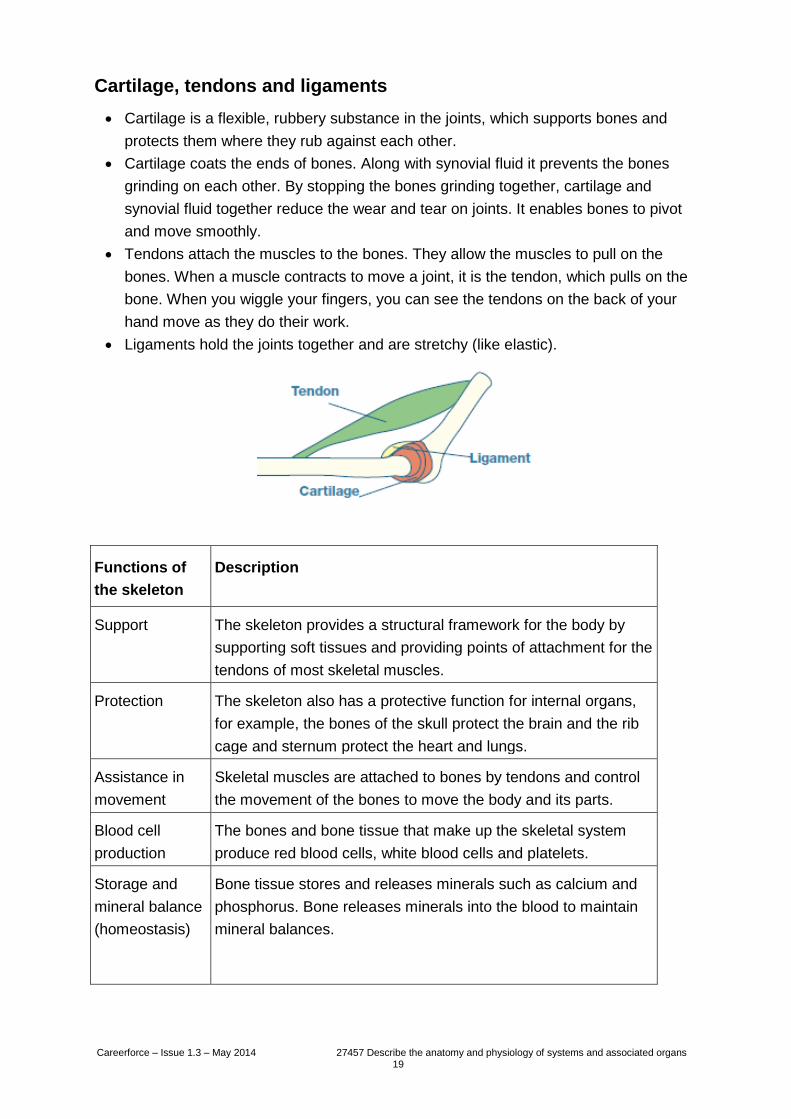
Cartilage, tendons and ligaments
Cartilage is a flexible, rubbery substance in the joints, which supports bones and
protects them where they rub against each other.
Cartilage coats the ends of bones. Along with synovial fluid it prevents the bones
grinding on each other. By stopping the bones grinding together, cartilage and
synovial fluid together reduce the wear and tear on joints. It enables bones to pivot
and move smoothly.
Tendons attach the muscles to the bones. They allow the muscles to pull on the
bones. When a muscle contracts to move a joint, it is the tendon, which pulls on the
bone. When you wiggle your fingers, you can see the tendons on the back of your
hand move as they do their work.
Ligaments hold the joints together and are stretchy (like elastic).
Functions of
the skeleton
Description
Support
The skeleton provides a structural framework for the body by
supporting soft tissues and providing points of attachment for the
tendons of most skeletal muscles.
Protection
The skeleton also has a protective function for internal organs,
for example, the bones of the skull protect the brain and the rib
cage and sternum protect the heart and lungs.
Assistance in
movement
Skeletal muscles are attached to bones by tendons and control
the movement of the bones to move the body and its parts.
Blood cell
production
The bones and bone tissue that make up the skeletal system
produce red blood cells, white blood cells and platelets.
Storage and
mineral balance
(homeostasis)
Bone tissue stores and releases minerals such as calcium and
phosphorus. Bone releases minerals into the blood to maintain
mineral balances.
Careerforce – Issue 1.3 – May 2014 27457 Describe the anatomy and physiology of systems and associated organs 20
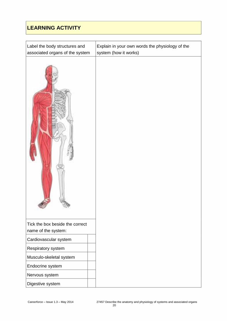
LEARNING ACTIVITY
Label the body structures and
associated organs of the system
Explain in your own words the physiology of the
system (how it works)
Tick the box beside the correct
name of the system:
Cardiovascular system
Respiratory system
Musculo-skeletal system
Endocrine system
Nervous system
Digestive system
Careerforce – Issue 1.3 – May 2014 27457 Describe the anatomy and physiology of systems and associated organs 21
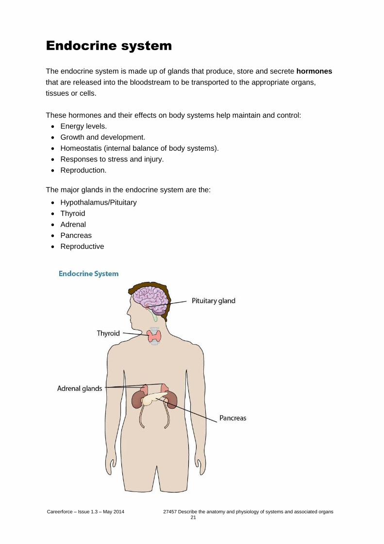
Endocrine system
The endocrine system is made up of glands that produce, store and secrete hormones
that are released into the bloodstream to be transported to the appropriate organs,
tissues or cells.
These hormones and their effects on body systems help maintain and control:
Energy levels.
Growth and development.
Homeostatis (internal balance of body systems).
Responses to stress and injury.
Reproduction.
The major glands in the endocrine system are the:
Hypothalamus/Pituitary
Thyroid
Adrenal
Pancreas
Reproductive
Careerforce – Issue 1.3 – May 2014 27457 Describe the anatomy and physiology of systems and associated organs 22
The pituitary gland is called the “master gland” but is, in fact, controlled by the
hypothalamus. Working together, these glands control many other functions of the
endocrine system. Some of the hormones secreted are:
Follicle-stimulating hormone which stimulates the production of follicles in a
woman’s ovaries.
Leutinising hormone that triggers ovulation.
Antidiuretic hormone that helps to prevent excess water loss by reducing the amount
excreted by the kidneys.
Endorphins that reduce the sensitivity of pain receptors.
The thyroid gland secretes thyroid hormones that regulate metabolism (body
temperature and weight), blood pressure, heart rate and the rate at which food is
converted into energy.
The thyroid uses iodine – a mineral found in some foods and in iodised salt – to make its
hormones, the two most important of which are thyroxine and triiodothyronine.
The adrenal glands are positioned above the kidneys. One of the hormones produced is
adrenaline, which is released in response to stress factors such as anger or injury.
Adrenaline increases the heart rate and speeds up the rate of breathing, to allow extra
oxygen to be circulated through the body.
The pancreas secretes two main hormones, insulin and glucagon, that regulate blood
sugar levels. Insulin reduces the level of sugar (glucose) by stimulating cells in the liver,
muscle and fat tissue to take glucose from the blood and store it as glycogen. Glucagon
raises the level by releasing sugar that has been stored.
Reproductive glands produce secrete hormones such as testosterone in males and
oestrogen in females. Testosterone controls things like male body shape, facial hair,
depth of voice and sexual drive. Oestrogen controls the female body shape and
development of breasts.
Careerforce – Issue 1.3 – May 2014 27457 Describe the anatomy and physiology of systems and associated organs 23
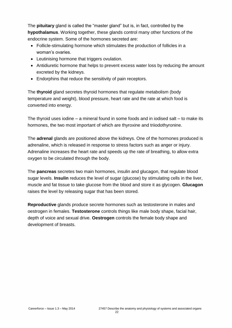
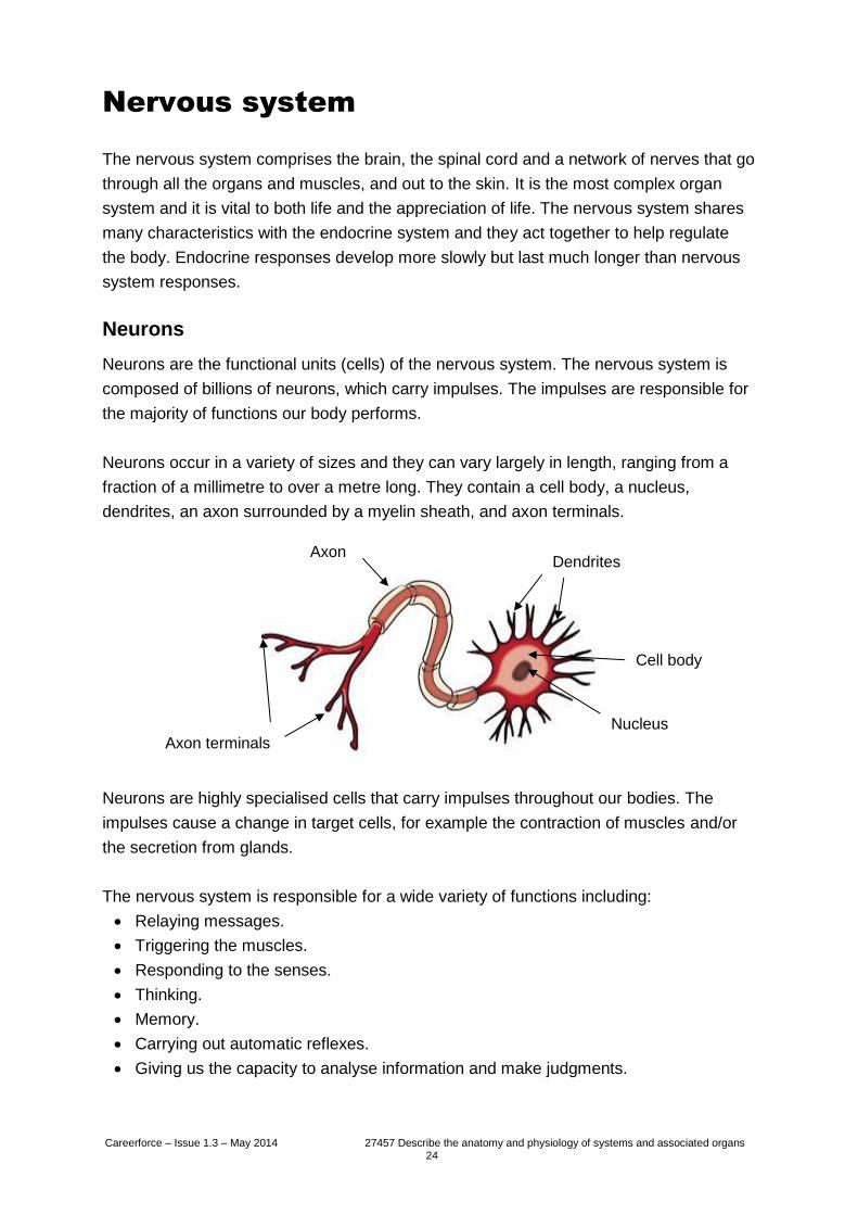
LEARNING ACTIVITY
Next to each named organ, write one thing that a hormone released from this organ can
influence.
Careerforce – Issue 1.3 – May 2014 27457 Describe the anatomy and physiology of systems and associated organs 24
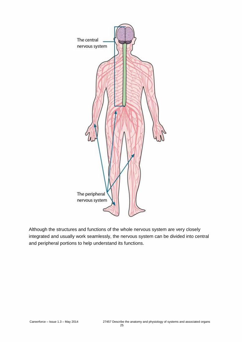
Nervous system
The nervous system comprises the brain, the spinal cord and a network of nerves that go
through all the organs and muscles, and out to the skin. It is the most complex organ
system and it is vital to both life and the appreciation of life. The nervous system shares
many characteristics with the endocrine system and they act together to help regulate
the body. Endocrine responses develop more slowly but last much longer than nervous
system responses.
Neurons
Neurons are the functional units (cells) of the nervous system. The nervous system is
composed of billions of neurons, which carry impulses. The impulses are responsible for
the majority of functions our body performs.
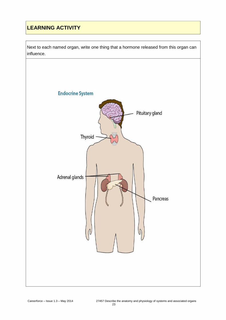
Neurons occur in a variety of sizes and they can vary largely in length, ranging from a
fraction of a millimetre to over a metre long. They contain a cell body, a nucleus,
dendrites, an axon surrounded by a myelin sheath, and axon terminals.
Neurons are highly specialised cells that carry impulses throughout our bodies. The
impulses cause a change in target cells, for example the contraction of muscles and/or
the secretion from glands.
The nervous system is responsible for a wide variety of functions including:
Relaying messages.
Triggering the muscles.
Responding to the senses.
Thinking.
Memory.
Carrying out automatic reflexes.
Giving us the capacity to analyse information and make judgments.
Dendrites Axon
Axon terminals
Cell body
Nucleus
Careerforce – Issue 1.3 – May 2014 27457 Describe the anatomy and physiology of systems and associated organs 25
Although the structures and functions of the whole nervous system are very closely
integrated and usually work seamlessly, the nervous system can be divided into central
and peripheral portions to help understand its functions.
Careerforce – Issue 1.3 – May 2014 27457 Describe the anatomy and physiology of systems and associated organs 26
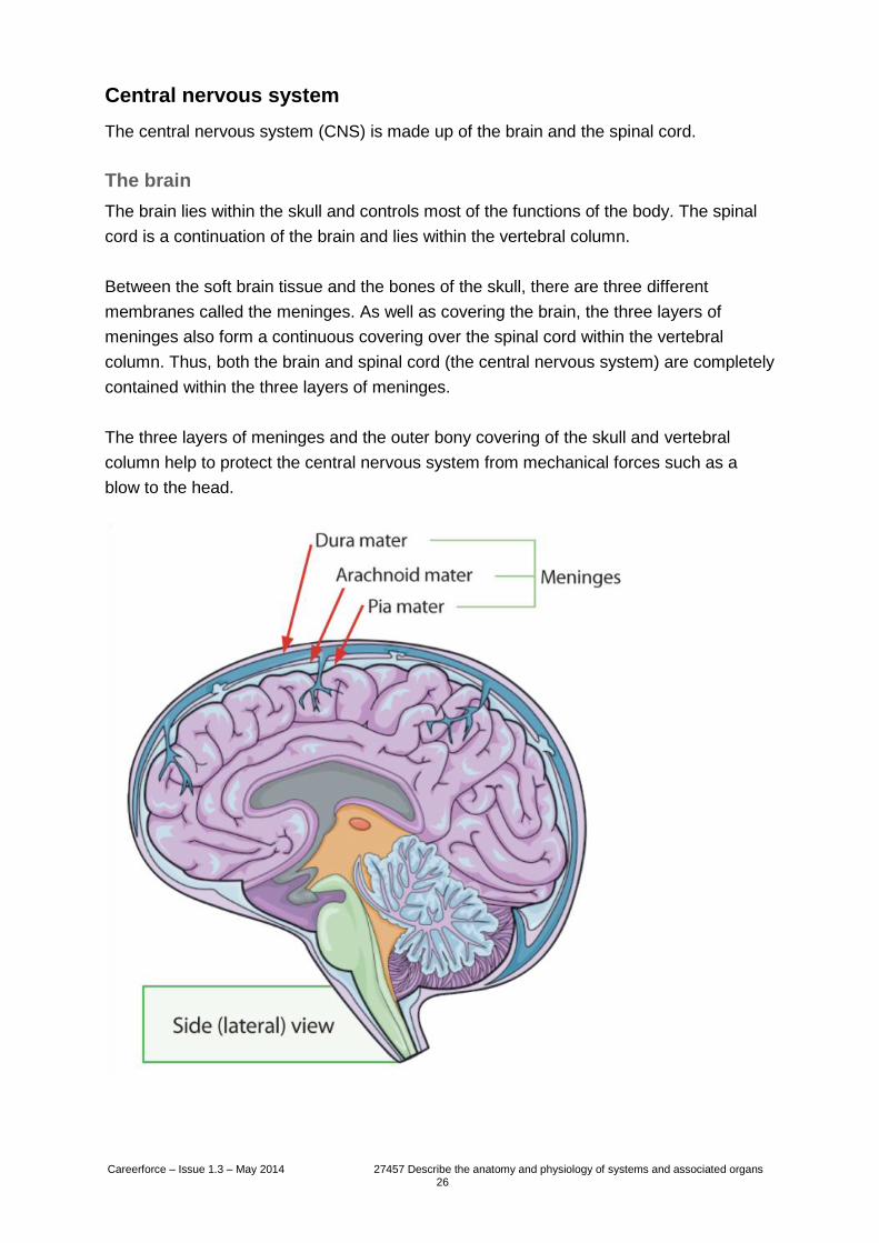
Central nervous system
The central nervous system (CNS) is made up of the brain and the spinal cord.
The brain
The brain lies within the skull and controls most of the functions of the body. The spinal
cord is a continuation of the brain and lies within the vertebral column.
Between the soft brain tissue and the bones of the skull, there are three different
membranes called the meninges. As well as covering the brain, the three layers of
meninges also form a continuous covering over the spinal cord within the vertebral
column. Thus, both the brain and spinal cord (the central nervous system) are completely
contained within the three layers of meninges.
The three layers of meninges and the outer bony covering of the skull and vertebral
column help to protect the central nervous system from mechanical forces such as a
blow to the head.
Careerforce – Issue 1.3 – May 2014 27457 Describe the anatomy and physiology of systems and associated organs 27
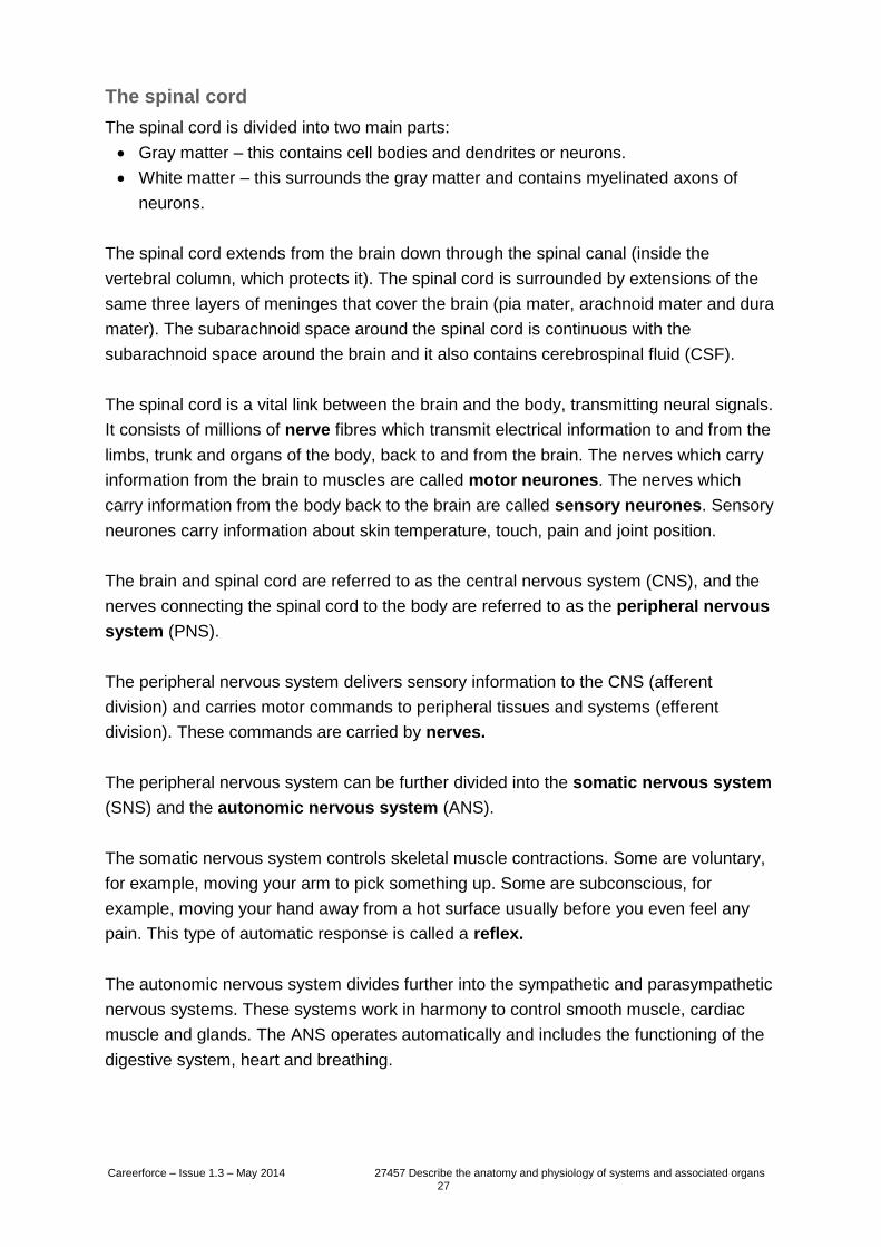
The spinal cord
The spinal cord is divided into two main parts:
Gray matter – this contains cell bodies and dendrites or neurons.
White matter – this surrounds the gray matter and contains myelinated axons of
neurons.
The spinal cord extends from the brain down through the spinal canal (inside the
vertebral column, which protects it). The spinal cord is surrounded by extensions of the
same three layers of meninges that cover the brain (pia mater, arachnoid mater and dura
mater). The subarachnoid space around the spinal cord is continuous with the
subarachnoid space around the brain and it also contains cerebrospinal fluid (CSF).
The spinal cord is a vital link between the brain and the body, transmitting neural signals.
It consists of millions of nerve fibres which transmit electrical information to and from the
limbs, trunk and organs of the body, back to and from the brain. The nerves which carry
information from the brain to muscles are called motor neurones. The nerves which
carry information from the body back to the brain are called sensory neurones. Sensory
neurones carry information about skin temperature, touch, pain and joint position.
The brain and spinal cord are referred to as the central nervous system (CNS), and the
nerves connecting the spinal cord to the body are referred to as the peripheral nervous
system (PNS).
The peripheral nervous system delivers sensory information to the CNS (afferent
division) and carries motor commands to peripheral tissues and systems (efferent
division). These commands are carried by nerves.
The peripheral nervous system can be further divided into the somatic nervous system
(SNS) and the autonomic nervous system (ANS).
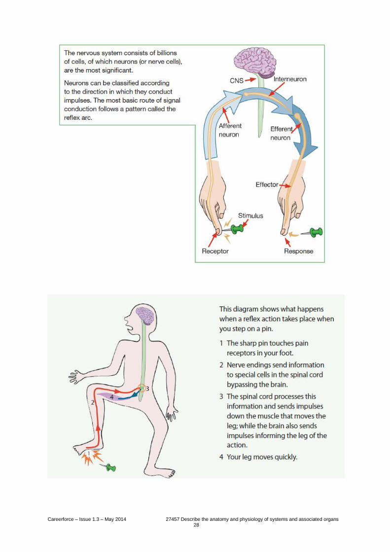
The somatic nervous system controls skeletal muscle contractions. Some are voluntary,
for example, moving your arm to pick something up. Some are subconscious, for
example, moving your hand away from a hot surface usually before you even feel any
pain. This type of automatic response is called a reflex.
The autonomic nervous system divides further into the sympathetic and parasympathetic
nervous systems. These systems work in harmony to control smooth muscle, cardiac
muscle and glands. The ANS operates automatically and includes the functioning of the
digestive system, heart and breathing.
Careerforce – Issue 1.3 – May 2014 27457 Describe the anatomy and physiology of systems and associated organs 28
Careerforce – Issue 1.3 – May 2014 27457 Describe the anatomy and physiology of systems and associated organs 29
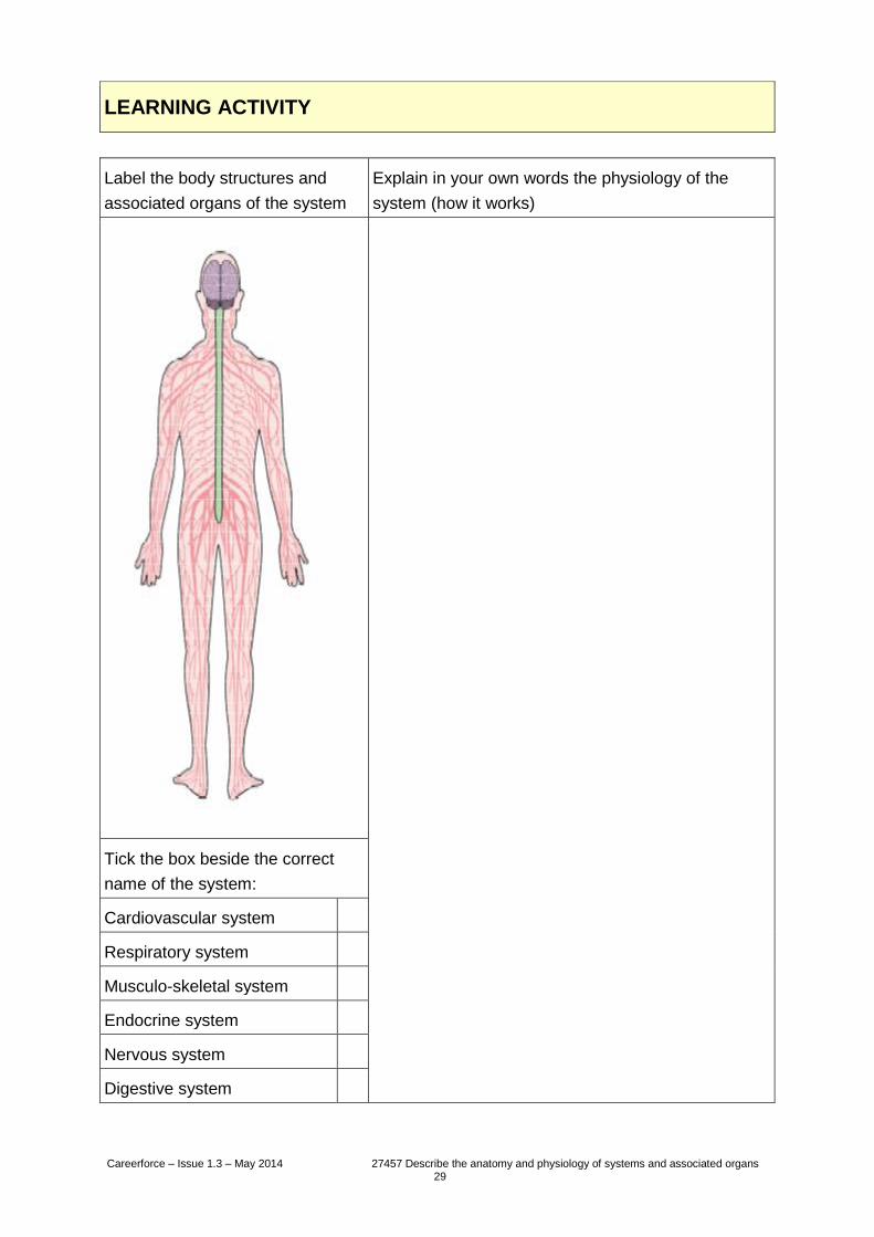
LEARNING ACTIVITY
Label the body structures and
associated organs of the system
Explain in your own words the physiology of the
system (how it works)
Tick the box beside the correct
name of the system:
Cardiovascular system
Respiratory system
Musculo-skeletal system
Endocrine system
Nervous system
Digestive system
Careerforce – Issue 1.3 – May 2014 27457 Describe the anatomy and physiology of systems and associated organs 30
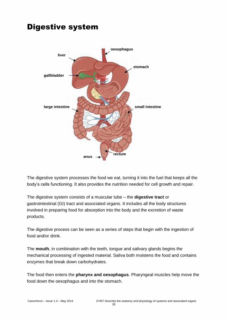
Digestive system
The digestive system processes the food we eat, turning it into the fuel that keeps all the
body’s cells functioning. It also provides the nutrition needed for cell growth and repair.
The digestive system consists of a muscular tube – the digestive tract or
gastrointestinal (GI) tract and associated organs. It includes all the body structures
involved in preparing food for absorption into the body and the excretion of waste
products.
The digestive process can be seen as a series of steps that begin with the ingestion of
food and/or drink.
The mouth, in combination with the teeth, tongue and salivary glands begins the
mechanical processing of ingested material. Saliva both moistens the food and contains
enzymes that break down carbohydrates.
The food then enters the pharynx and oesophagus. Pharyngeal muscles help move the
food down the oesophagus and into the stomach.
oesophagus
stomach
small intestine
rectum anus
large intestine
gallbladder
liver
Careerforce – Issue 1.3 – May 2014 27457 Describe the anatomy and physiology of systems and associated organs 31
The stomach stores and processes food. The mechanical breakdown of food is aided by
muscular contractions. Chemical breakdown happens through the addition of digestive
juices (acid and enzymes). Food may stay in the stomach for 3–4 hours. The thick,
partially digested food that passes from the stomach into the small intestine is called
chyme.
The small intestine plays a key role in the digestion and absorption of nutrients. The
vast majority (90%) of nutrient absorption happens in this section of the digestive tract
which measures an average six metres in length.
The duodenum is the part of the small intestine closest to the stomach and this is where
chyme is mixed with digestive secretions from the pancreas, liver and gallbladder.
The liver is the largest glandular organ of the body, and is divided into four lobes of
unequal size and shape. The liver lies on the right side of the abdominal cavity beneath
the diaphragm. Blood is carried to the liver via two large vessels called the hepatic artery
and the portal vein.
The liver has many functions which include:
Producing substances that break down fats.
Converting glucose to glycogen.
Making amino acids.
Filtering harmful substances from the blood (such as alcohol).
Storing vitamins and minerals.
The large intestine (or large bowel) is made up of the colon, caecum and rectum. It has
an average length of 1.5 metres and it completes most of the remaining 10% of nutrient
absorption. It is also responsible for the compaction of waste materials into faeces that
are expelled by defecation via the anus (the external sphincter muscle of the digestive
system).
The vomiting reflex
empties the contents
of the duodenum
back into the stomach
to be expelled along
with the stomach
contents.
Careerforce – Issue 1.3 – May 2014 27457 Describe the anatomy and physiology of systems and associated organs 32
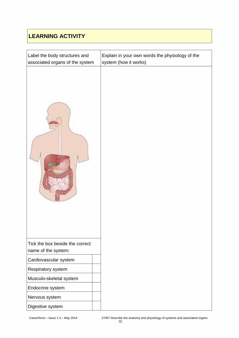
LEARNING ACTIVITY
Label the body structures and
associated organs of the system
Explain in your own words the physiology of the
system (how it works)
Tick the box beside the correct
name of the system:
Cardiovascular system
Respiratory system
Musculo-skeletal system
Endocrine system
Nervous system
Digestive system
Careerforce – Issue 1.3 – May 2014 27457 Describe the anatomy and physiology of systems and associated organs 33
Rewind to page 6...
What new information have you learned about the human body?
Careerforce – Issue 1.3 – May 2014 27457 Describe the anatomy and physiology of systems and associated organs 34
Completion and assessment
Congratulations!
You have come to the end of the workbook. Please check over all the activities in this
workbook to make sure you have completed them.
Your assessment is next.
You need to complete the assessment successfully to be credited with this unit standard.
Acknowledgements
Careerforce thanks the people who have contributed to this workbook by:
researching and validating content.
providing advice and expertise.
testing the activities.
sharing personal experiences.
appearing in photographs.
Creative Commons
This work is licenced under a Creative Commons Attribution-NonCommercial Licence. You are free to
copy, distribute and transmit the work and to adapt the work. You must attribute Careerforce as the
author. You may not use this work for commercial purposes. For more information contact Careerforce
www.careerforce.org.nz

Careerforce – Issue 1.3 – May 2014 27457 Describe the anatomy and physiology of systems and associated organs 35

Careerforce – Issue 1.3 – May 2014