Embed Size (px)
Citation preview
This is the html version of the file http://www.wcb.ny.gov/content/main/hcpp/ImpairmentGuidelines/2017DRAFTImpairmentGuide.pdf.Google automatically generates html versions of documents as we crawl the web.
Page 1
Workers’ CompensationGuidelines forDetermining Impairment
First Edition, September 1, 2017
Page 2
PREFACE
In April 2017, Governor Andrew M. Cuomo signed into law the 2017-2018 Executive Budget whichcontained several significant pieces of workers’ compensation reform. Included in the reforms wasWorkers’ Compensation Law (WCL) § 15(3)(x), which requires new Permanency ImpairmentGuidelines (“Guidelines”) to be adopted by January 1, 2018. The new Guidelines cover determinationsof permanency under WCL § 15(3)(a) through (v), which are also known as scheduled loss of use.
This document represents the Workers’ Compensation Board’s (“Board’s”) proposed Guidelines.Publication in the State Register commences a 45-day comment period (which expires on Monday,October 23, 2017). The Guidelines, along with regulations necessary to implement the Guidelines,are available on the Board’s website (http://www.wcb.ny.gov).
The publication of these proposed Guidelines on September 1, 2017, allows for a full and robust publiccomment period, and ensures that the regulatory process (including, if needed, a re-publication andsecond public comment period) can conclude prior to the mandatory implementation of new Guidelinesby January 1, 2018.
The Board strongly encourages the public, injured workers, employers, self-insured employers,insurance carriers, third-party administrators, attorneys, medical providers, and labor and businessorganizations to provide comment. The Board will evaluate all comments received, and will considernecessary revisions as the process advances.
The Board is committed to the timely and successful implementation of Guidelines that are, as thelegislation states, “reflective of advances in modern medicine that enhance healing and result in betteroutcomes” [WCL § 15(3)(x)]. It should be noted that these are proposed Guidelines, which are not ineffect.
We look forward to receiving your comments.
Page 3
ACKNOWLEDGEMENTS
The Board wishes to acknowledge the tremendous effort and significant contribution of the New YorkState Society of Orthopaedic Surgeons, which served as the Board’s consultant in developing theinitial draft permanent impairment guidelines, with special mention to Jeffrey Lozman, MD, JohnDiPreta, MD, and Executive Director, Babette Grey. On August 15, 2017, the Board, together with theOrthopaedic Society, conferred and consulted with a group of key stakeholders and their medicalexperts, as set forth in the 2017 legislation (WCL § 15[3][x]).
Also, thank you to the members of the stakeholder group, specifically the New York AFL-CIO, NYSBusiness Council, Medical Society of the State of New York, New York State Insurance Fund, ZurichInsurance on behalf of the insurance carriers, NYS Osteopathic Medical Society, NY Self-InsuranceAssociation, and the New York City Law Department. We thank all of the participants for theirsubstantive and constructive feedback to the initial draft. We look forward to their continued feedbackduring the public comment period on these proposed guidelines and the associated regulations.
Page 4
TABLE OF CONTENTS
CHAPTER 1: INTRODUCTION 1
1.1 Background and History ................................................................................................... 1
1.2 Nature of Schedule Loss of Use Awards .......................................................................... 2
1.3 Amenability ....................................................................................................................... 3
1.4 Using the Schedule Loss of Use Impairment Guidelines .................................................. 5
1.5 Medical Assessment ......................................................................................................... 6
1.5.1 Role of Examining Medical Providers and Independent Medical Examiners ..................... 6
1.5.2 Medical Impairment Evaluation ........................................................................................... 6
1.5.3 Range of Motion .................................................................................................................. 7
1.5.4 Muscle Strength ................................................................................................................... 8
1.5.5 Pain 8
1.6 Claimant Intake Form and Medical Assessment ............................................................. 10
1.7 Board Determination of Schedule Loss of Use ............................................................... 10
1.8 Prior Schedule Loss of Use Award ................................................................................. 11
1.9 WCL § 15(3)(v) ............................................................................................................... 11
CHAPTER 2: UPPER EXTREMITY - DIGITS 12
2.1 Objectives for Determining Impairment for Digits ............................................................ 12
2.2 Methods Available to Assess Permanent Impairment ..................................................... 12
2.3 Maximum Rating of Body Part ........................................................................................ 12
2.4 Thumb Range of Motion ................................................................................................. 13
2.4.1 IP Joint – Interphalangeal Joint ......................................................................................... 13
2.4.2 MCP – Metacarpophalangeal Joint ................................................................................... 13
2.4.3 CMC – Carpal-metacarpal Joint ........................................................................................ 13
2.5 Thumb Strength .............................................................................................................. 15
2.6 Thumb Pain .................................................................................................................... 16
2.7 Finger Range of Motion .................................................................................................. 17
2.7.1 Metacarpophalangeal (MCP) Joint - Range of Motion ...................................................... 17
2.7.2 Proximal Interphalangeal (PIP) Joint - Range of Motion ................................................... 17
2.7.3 Distal Interphalangeal (DIP) Joint - Range of Motion ........................................................ 17
2.8 Finger Strength ............................................................................................................... 18
2.9 Finger Pain ..................................................................................................................... 19
2.10 Loss of % Range of Motion Multiple Digits .................................................................... 20
Page 5
2.11 Multiple Digits - Strength............................................................................................... 20
2.12 Multiple Digits - Pain ..................................................................................................... 21
2.13 Special Condition Based Impairment ............................................................................ 21
2.14 Amputation ................................................................................................................... 23
2.14.1 Amputation, Digit – Single Digit ....................................................................................... 23
2.14.2 Amputation, Digit – Multiple Digits ................................................................................... 23
CHAPTER 3: UPPER EXTREMITY - HAND AND WRIST 24
3.1 Objectives for Determining Impairment for Hand and Wrist ............................................ 24
3.2 Methods Available to Assess Permanent Impairment ..................................................... 24
3.3 Hand & Wrist Impairment ............................................................................................... 25
3.4 Range of Motion Impairment .......................................................................................... 253.5 Muscle Strength ............................................................................................................. 26
3.6 Pain ............................................................................................................................... 27
3.7 Special Condition Based Impairment .............................................................................. 27
3.8 Amputation ..................................................................................................................... 27
CHAPTER 4: UPPER EXTREMITY – ELBOW 28
4.1 Objectives for Determining Impairment for Elbow ........................................................... 28
4.2 Methods Available to Assess Permanent Impairment ..................................................... 28
4.3 Elbow Range of Motion .................................................................................................. 30
4.4 Elbow Strength ............................................................................................................... 31
4.5 Elbow Pain ..................................................................................................................... 32
4.6 Elbow - Amputation ........................................................................................................ 32
CHAPTER 5: UPPER EXTREMITY - SHOULDER 33
5.1 Objectives for Determining Impairment for Shoulder ....................................................... 33
5.2 Methods Available to Assess Permanent Impairment ..................................................... 33
5.3 Shoulder Range of Motion .............................................................................................. 35
5.4 Shoulder Strength .......................................................................................................... 37
5.5 Shoulder Pain ................................................................................................................. 38
5.6 Special Condition Based Impairment .............................................................................. 38
5.7 Amputation - Shoulder .................................................................................................... 38
CHAPTER 6: LOWER EXTREMITY – HIP AND FEMUR 39
6.1 Objectives for Determining Impairment for Hip and Femur ............................................. 39
Page 6
6.2 Methods Available to Assess Permanent Impairment ..................................................... 39
6.3 Hip Range of Motion ....................................................................................................... 40
6.4 Hip Strength ................................................................................................................... 42
6.5 Hip Pain .......................................................................................................................... 43
6.6 Special Condition Based Impairment .............................................................................. 43
6.7 Hip Amputation ............................................................................................................... 43
CHAPTER 7: LOWER EXTREMITY - KNEE AND TIBIA 44
7.1 Objectives for Determining Impairment for Knee and Tibia ............................................. 44
7.2 Methods Available to Assess Permanent Impairment ..................................................... 44
7.3 Knee Range of Motion .................................................................................................... 46
7.4 Knee Strength ................................................................................................................ 46
7.5 Knee Pain ....................................................................................................................... 47
7.6 Special Condition Based Impairment .............................................................................. 48
7.7 Knee Amputation............................................................................................................48
CHAPTER 8: LOWER EXTREMITY - ANKLE AND FOOT 49
8.1 Objectives for Determining Impairment for Ankle and Foot.............................................49
8.2 Methods Available to Assess Permanent Impairment ..................................................... 49
8.3 Ankle and Foot Range of Motion .................................................................................... 50
8.4 Ankle and Foot Muscle Strength ..................................................................................... 51
8.5 Ankle and Foot Pain ....................................................................................................... 52
8.6 Special Condition Based Impairment..............................................................................52
8.7 Ankle and Foot Amputation ............................................................................................ 53
CHAPTER 9: LOWER EXTREMITY: GREAT AND LESSER TOES 54
9.1 Objectives for Determining Impairment for Great and Lesser Toes ................................. 54
9.2 Methods Available to Assess Permanent Impairment ..................................................... 54
9.3 Maximum Rating of Body Part ........................................................................................ 55
9.4 Single Toe Impairment ................................................................................................... 55
9.5 Great Toe Impairment .................................................................................................... 55
9.5.1 Range of Motion of the Great Toe ..................................................................................... 55
9.5.2 Great Toe Strength ............................................................................................................ 57
9.5.3 Great Toe Pain .................................................................................................................. 58
9.6 Lesser Toes Impairment ................................................................................................. 59
9.6.1 Lesser Toe Range of Motion ............................................................................................. 59
Page 7
9.6.2 Lesser Toe Strength .......................................................................................................... 59
9.6.3 Lesser Toe Pain ................................................................................................................. 60
9.7 Multiple Toe Injuries ....................................................................................................... 61
9.8 Amputation ..................................................................................................................... 61
CHAPTER 10: PERIPHERAL NERVE INJURIES AND COMPRESSION NEUROPATHIES 62
10.1 Objectives for Determining Impairment for Peripheral Nerve Injuries and CompressionNeuropathies ......................................................................................................................... 62
10.2 Methods Available to Assess Permanent Impairment ................................................... 62
10.3 Range of Motion ......................................................................................................... 65
10.4 Strength ...................................................................................................................... 65
10.5 Sensation .................................................................................................................... 67
10.6 Pain.............................................................................................................................67
10.7 Special Condition Based Impairment ......................................................................... 68
CHAPTER 11: VISUAL SYSTEM / AUDITORY SYSTEM / FACIAL SCARS ANDDISFIGUREMENT 70
11.1 Introduction - Visual System ......................................................................................... 70
11.1.1 Criteria and Methods for Evaluating PermanentImpairment .......................................... 70
11.2 Introduction - Auditory System ...................................................................................... 73
11.2.1 Occupational Loss of Hearing.......................................................................................... 73
11.2.2 Traumatic Loss of Hearing ............................................................................................... 73
11.3 Facial Scars and Disfigurement .................................................................................... 74
Page 8
1
Chapter 1: Introduction
1.1 Background and History
Permanent injuries are generally viewed as either a “schedule loss of use” award pursuant toWorkers’ Compensation Law (WCL) §15(3)(a-t) or “classification” pursuant WCL §15(3)(w).These Guidelines, as described below, pertain solely to the rendering of schedule loss of useawards.
1996 GuidelinesIn 1983, the New York State Legislature established a Temporary State Commission on Workers'Compensation and Disability Benefits to study and evaluate the systems of the New York StateWorkers' Compensation Board (Board). The Commission recommended that the Board publishuniform medical guidelines for the evaluation of functional impairments, to inform the generalpublic, and medical and legal practitioners. Thus, in June 1996, the Board published ImpairmentGuidelines.
2012 GuidelinesIn 2007, workers’ compensation reform legislation established durational limitations on non-schedule permanent partial disability awards based on a claimant’s loss of wage-earning capacity(LWEC). The 2012 Guidelines were thus developed to provide methods for evaluation of non-schedule permanent partial disabilities, and new sections were added for functional evaluationand the evaluation of loss of wage earning capacity.
The 2012 Guidelines retained the 1996 Guidelines with respect to Schedule Loss of Use.
2017 GuidelinesLegislation enacted in April 2017 [WCL§15(3)(x)] directed the Board to consult with“representatives of labor, business, medical providers, insurance carriers, and self-insuredemployers regarding revisions to permanency impairment guidelines, including permitting reviewand comment by such representatives’ chosen medical advisors…”, to adopt revised guidelinesfor the evaluation of medical impairment and determination of permanency with respect to injurieswhich are amenable to a schedule loss of use award pursuant to paragraphs (a) through (v) ofsubdivision 3 of section 15 of the WCL. As the law directs, these guidelines are to be “…reflectiveof advances in modern medicine that enhance healing and result in better outcomes.”[WCL§15(3)(x)]
The Board retained the New York State Society of Orthopaedic Surgeons as its medicalconsultant. The Board conferred and consulted with the stakeholders and their medical advisors,who reviewed the draft, and offered feedback. Following a review of that input, the Boardprepared the guidelines for publication as directed by law.
These Guidelines establish the methodology for evaluation of medical impairment, i.e. thepermanent residual physical deficit as it exists at the time of maximum medical improvement. Thefinding of permanency is to be made by the Board, based on the evidence of the permanentmedical impairment’s measured impact on the earning power of the disabled claimant.
These revised permanency Guidelines, contained herein, supersede those sections of theBoard’s 2012 Impairment Guidelines concerning medical evaluation of injuries amenable to a
Page 9
2
schedule loss of use (subchapter 1.5 and chapters 2, 3 and 4 of the 2012 Guidelines), as well asany other provisions of the 2012 Impairment Guidelines which are inconsistent with theseGuidelines. The methodology from the 2012 Guidelines involving the visual system, loss ofhearing, and facial scars and disfigurement, are placed herein as Chapter 11, in three sections,and are considered part of the 2018 Impairment Guidelines.
WCL § 15(3)(x) provides that “[a]s of January first, two thousand eighteen the 2012 permanencyGuidelines pertaining to paragraphs a through v of subdivision three of section fifteen of this articleare repealed, and shall have no effect. The Board shall train adjudication and other staff to ensuretimely and effective implementation.” As such, any schedule loss of use determinations made bythe Board on and after January 1, 2018 shall be in accordance with these Guidelines.
1.2 Nature of Schedule Loss of Use Awards
Unlike awards for non-schedule permanent impairments, schedule loss of use awards areindependent of the time a claimant actually loses from work, and are determined by the Workers’Compensation Law Judge (WCLJ), or, if agreed to, by a conciliator, based on a statutorilyprescribed formula calculated as a number of weeks of compensation. Schedule loss of useawards are only made for permanent impairments of an extremity, permanent loss of vision orhearing, or permanent facial disfigurement, as provided in Workers' Compensation Law Section15. The table of weeks by Percentage Loss of Use of Body Part is available in the 2012Impairment Guidelines (see page 43).
A schedule loss of use award is not intended to compensate a claimant for an injury sustained orfor pain and suffering. Rather, a schedule award compensates a claimant for the permanent lossof earning power resulting from the permanent residual physical deficit. See, e.g., Matter of PoliceDepartment, 2017 NY Wrk Comp G1028651 (Full Board), Matter of Marhoffer v Marhoffer , 220NY 543 (1917), Matter of Lamantia v. Midland El. Co., Inc ., 59 A.D.3d 892 (2009). Further, aschedule award "is not allocable to any particular time frame" [Matter of Miller v North SyracuseCent. School Dist.,1 AD3d 691 (2009)]. Ultimately, the amount of a schedule loss of use awardis a factual determination made by the Board based on (1) medical evidence of permanentresidual physical deficit which is consistent with these Guidelines, and (2) the impact on theclaimant’s earning power. In addition to the schedule set forth in WCL §15(3)(a-t), a claimant whoexperiences a protracted healing period is entitled to additional compensation as set forth inWCL§15(4-a).
Page 10
3
1.3 Amenability
A schedule loss of use award based on an injury to an extremity cannot be made unless thefollowing requirements have been met:
A. The claimant has reached maximum medical improvement, which occurs (a) when theclaimant has recovered from the injury to the greatest extent that is expected and (b)no further improvement in his or her condition is reasonably expected. The need forpalliative or symptomatic treatment does not preclude a finding of maximum medicalimprovement.
B. There is a permanent impairment of one or more extremity listed in paragraphs (a)through (l) of subdivision 3 of section 15 of the Workers' Compensation Law.
C. The impairment involves permanent residual physical deficits to soft tissue, bone,sensation, dexterity and power, and may also include atrophy, scarring, deformity,mobility defects and shortening.
D. If the same accident results in multiple injuries, either directly or consequentially, oneor more of which may not be the subject of a schedule award (e.g., back, neck, head,depression), all non-schedulable injuries have reached maximum medicalimprovement and are found by the medical provider to have fully resolved, withmedical evidence that no permanent residual impairment exists. Note: Where thereis permanent residual impairment in any non-schedulable injury site, the claim may beamenable to classification rather than schedule loss of use.
E. The permanent impairment of an extremity is not amenable to a permanent partialdisability classification pursuant to paragraph (w) of subdivision 3 of section 15 of theWorkers' Compensation Law.
Examples of permanent impairments of the extremities which are not amenable to aschedule award, and should result in permanent partial disability classification,include:
1. Progressive and severe painful conditions of the major joints of the extremitiessuch as the shoulders, elbows, hips and knees with one or more of thefollowing:
a. Objective findings of acute or persistent inflammation of one or morejoints such as swelling, effusion, change of color or temperature,tenderness, painful range of motion, etc.;
b. X-ray evidence of progressive and severe degenerative arthritis; and/orc. Minimal or no improvement after all modalities of medical and surgical
treatment have been exhausted.
2. Persistent painful condition of an extremity commonly affecting the distalextremities such as the hands and feet, with one or more of the following:
Page 11
4
a. Complex regional pain syndrome (reflex sympathetic dystrophy),Sudeck’s atrophy or persistent painful extremity syndrome;
b. Objective findings or persistent swelling, atrophy, dysesthesias,hypersensitivity or changes of skin color and temperature such asmottling;
c. X-ray evidence of osteoporosis; and/ord. Minimal or no reported improvement after claimant has undergone all
modalities of persistent pain treatment.
3. Mal-union of the long bones.
4. Aseptic necrosis of the head of the femur or other bones.
5. Severe and persistent instability of the knee joint or other major joints.
6. Advanced Paget’s Disease.
7. Tumors.
8. Caisson’s Disease involving the joints.
9. Persistent ulcerations, draining sinuses.
10. Recurrent dislocations (shoulders).
11. Amputees with neuromas or poorly healed stumps.
12. Failed joint replacement such as total hip, total knee and shoulderreplacements.
13. If a medical provider renders an opinion on permanent impairment of anextremity which includes an assessment of residual pain of three or greater ona scale of zero to five, the medical provider should strongly consider whetherthe claimant may have a related underlying condition that has yet to beresolved.
14. In the event of multiple injuries from the same accident or disablement, wherethere is an assessment of residual pain of three or greater in any of the sites,the medical provider should strongly consider whether the claimant may havea related underlying condition that has yet to be resolved.
15. Where the injury results in multiple schedules to major members, with two ormore significant schedule awards loss of use, the Board may consider medicalevidence that the multiple major member schedule constitutes ongoingsystemic injury which is more appropriate for classification.
To pursue a non-schedule permanent partial disability determination, parties may submit separateimpairment evaluations, and the Board will separately adjudicate the question of non-schedulepermanency.
Page 12
5
1.4 Using the Schedule Loss of Use Impairment Guidelines
These Guidelines are designed to be used by medical providers to assess and render a medicalopinion with regard to a claimant’s permanent residual physical deficit, which the Board willconsider when determining whether a schedule loss of use award is appropriate and the amountof the award. These Guidelines are divided into sections by the part of the body being assessed:Chapter 2: Digits; Chapter 3: Hand and Wrist; Chapter 4: Elbow; Chapter 5: Shoulder; Chapter 6:Hip and Femur; Chapter 7: Knee and Tibia; Chapter 8: Ankle and Foot; Chapter 9: Toes; Chapter
10: Peripheral Nerve Injuries and Compression Neuropathies; and Chapter 11: Visual System,Auditory System and Facial Scars/Disfigurement.
The structure of these Guidelines is different from the Board’s 2012 Impairment Guidelines inseveral ways. The majority of injuries will be assessed based on the permanent residualimpairment. Injuries are placed in one of three categories (Category A, Category B, and CategoryC) based on the permanent residual physical deficit that has or is expected to occur based on theseverity of the injury. Each category has a minimum and maximum impairment percentage [e.g.,Category A (0-30%), Category B (30-60%), Category C (60-90%), with cases of significantamputations at or near total loss (100%)]. Within each category, the medical evidence ofimpairment and its impact on earning power is determined by the Board. Digit and wrist injuriesare measured differently, given their elevated occupational significance. The method ofevaluation of evidence of how the injury impacts the claimant’s loss of earning power, is set forthin subchapter 1.6 below.
Medical providers evaluating a claimant located in New York, and medical providers located inNew York who perform evaluations, must be authorized by the Workers’ Compensation Board.For medical providers outside of New York, any evaluation performed must comport with theseGuidelines, including the use of any forms prescribed by the Chair.
Page 13
6
1.5 Medical Assessment
1.5.1 Role of Examining Medical Providers and Independent MedicalExaminers
Medical providers shall provide their professional opinion of the claimant’s medicalcondition and permanent residual physical deficit at the time of maximum medicalimprovement based on the criteria set forth in these Guidelines. It is the responsibility ofthe medical provider to submit medical evidence that the Board will consider in making alegal determination as to schedule loss of use.
Medical providers should not infer findings or manifestations that are not drawn from thephysical examination or test reports, but rather medical providers should look to theobjective findings of the physical examination and data as contained within the medicalrecords of the claimant. This methodology is intended to foster consistency, predictabilityand inter-rater reliability for determining permanent impairment.
1.5.2 Medical Impairment Evaluation
In order to prepare a report on permanent impairment, the medical provider should:
1. Review the Guidelines.2. Review the medical records.3. Perform a thorough history and physical examination and recount the
relevant medical history, examination findings and appropriate test results,validating if necessary with tools such as QuickDash, etc.
4. State the work related medical diagnosis(es) based upon the relevantmedical history, examination and test results.
5. Identify the affected body part or system.6. Prepare and attest to a medical opinion on impairment, which should
consist of the following elements:a. Whether claimant is at maximum medical improvement.b. Whether the claimant’s injury is permanent, and amenable to schedule
(see 1.3, above).c. Identification of the injury(ies) in terms of severity or category as
appropriate.d. For each injury, unless a special condition exists, the applicable
category of injury is selected. The medical provider then measures thepermanent residual physical deficit with respect to:
(1) range of motion (impairment of gait and deformity may also beassessed if expressly provided in the Guidelines for particulardiagnosis);
(2) strength and(3) pain.
In addition to considering range of motion, strength, and pain, theGuidelines for peripheral nerve injuries and compression neuropathies(Chapter 10), including finger injuries, also permit consideration for lossof sensation.
Page 14
7
1.5.3 Range of Motion
Whenever possible, any impairment of the range of motion of an injured extremity shouldbe assessed in contrast to the claimant’s uninjured contralateral limb.
• Range of Motion measurement procedure:
o Measure active, unassisted range of motion while the claimant is exerting fulleffort.
o Goniometric measurements are the preferred method for impairmentevaluation.
o Each range of motion measurement should be repeated three times. The threevalues for each motion should be within 10o of each other. Record the highestmeasurement of the three values. If the three measurements within a set arenot within 10o of each other, use the highest value for rating purposes andreport the values as inconsistent (e.g., shoulder flexion is measured as 90 o,80o and 78o, use 90o for rating purposes and report the other measurementsas inconsistent).
o Range of motion is measured starting from the defined 0o MeasurementPosition. The 0o Measurement Position is specified for each joint and plane ofmotion at the beginning of the relevant joint section.
o Measure and record the values for range of motion for each affected plane ofthe joint.
o Ankylosis is the fixation of a joint due to disease, injury or surgery. The rangeof ankylosis of a joint impairment rating is described in the respective sectionof each joint. This value is assigned in lieu of all other range of motion orankylosis values for that joint.
Page 15
8
1.5.4 Muscle Strength
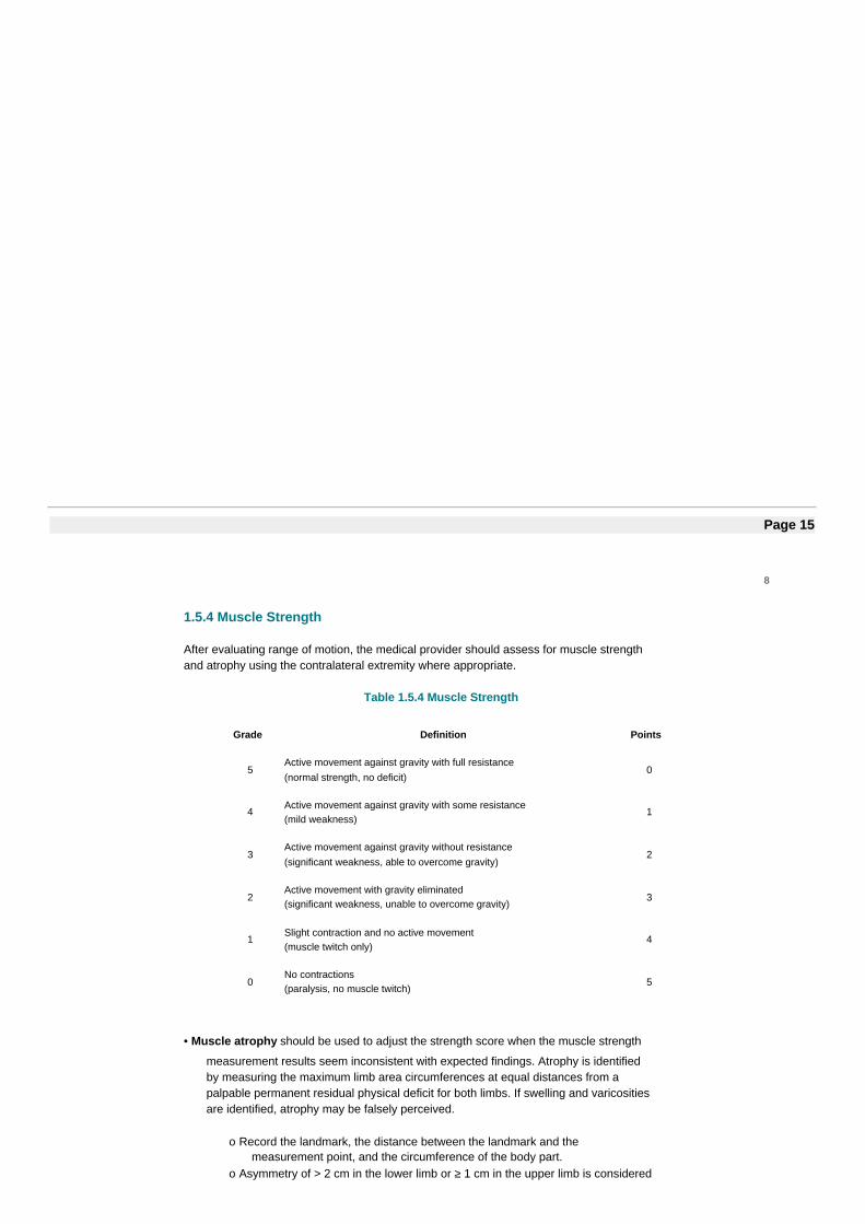
After evaluating range of motion, the medical provider should assess for muscle strengthand atrophy using the contralateral extremity where appropriate.
Table 1.5.4 Muscle Strength
Grade Definition Points
5Active movement against gravity with full resistance(normal strength, no deficit)
0
4Active movement against gravity with some resistance(mild weakness)
1
3Active movement against gravity without resistance(significant weakness, able to overcome gravity)
2
2Active movement with gravity eliminated(significant weakness, unable to overcome gravity)
3
1Slight contraction and no active movement(muscle twitch only)
4
0No contractions(paralysis, no muscle twitch)
5
• Muscle atrophy should be used to adjust the strength score when the muscle strength
measurement results seem inconsistent with expected findings. Atrophy is identifiedby measuring the maximum limb area circumferences at equal distances from apalpable permanent residual physical deficit for both limbs. If swelling and varicositiesare identified, atrophy may be falsely perceived.
o Record the landmark, the distance between the landmark and themeasurement point, and the circumference of the body part.
o Asymmetry of > 2 cm in the lower limb or ≥ 1 cm in the upper limb is considered
the minimum criteria for rating of atrophy. If measures are not possible (e.g.atrophy of the thenar muscles or shoulder), describe these findings within thephysical exam.
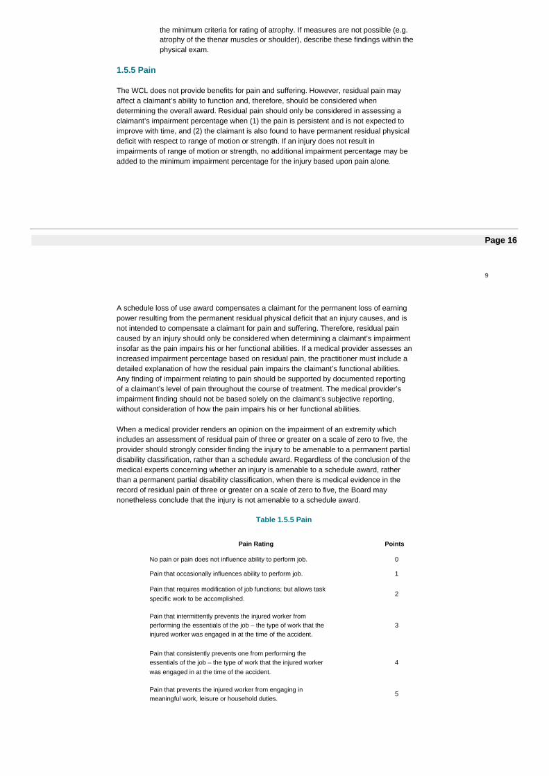
1.5.5 Pain
The WCL does not provide benefits for pain and suffering. However, residual pain mayaffect a claimant’s ability to function and, therefore, should be considered whendetermining the overall award. Residual pain should only be considered in assessing aclaimant’s impairment percentage when (1) the pain is persistent and is not expected toimprove with time, and (2) the claimant is also found to have permanent residual physicaldeficit with respect to range of motion or strength. If an injury does not result inimpairments of range of motion or strength, no additional impairment percentage may beadded to the minimum impairment percentage for the injury based upon pain alone.
Page 16
9
A schedule loss of use award compensates a claimant for the permanent loss of earningpower resulting from the permanent residual physical deficit that an injury causes, and isnot intended to compensate a claimant for pain and suffering. Therefore, residual paincaused by an injury should only be considered when determining a claimant’s impairmentinsofar as the pain impairs his or her functional abilities. If a medical provider assesses anincreased impairment percentage based on residual pain, the practitioner must include adetailed explanation of how the residual pain impairs the claimant’s functional abilities.Any finding of impairment relating to pain should be supported by documented reportingof a claimant’s level of pain throughout the course of treatment. The medical provider’simpairment finding should not be based solely on the claimant’s subjective reporting,without consideration of how the pain impairs his or her functional abilities.
When a medical provider renders an opinion on the impairment of an extremity whichincludes an assessment of residual pain of three or greater on a scale of zero to five, theprovider should strongly consider finding the injury to be amenable to a permanent partialdisability classification, rather than a schedule award. Regardless of the conclusion of themedical experts concerning whether an injury is amenable to a schedule award, ratherthan a permanent partial disability classification, when there is medical evidence in therecord of residual pain of three or greater on a scale of zero to five, the Board maynonetheless conclude that the injury is not amenable to a schedule award.
Table 1.5.5 Pain
Pain Rating Points
No pain or pain does not influence ability to perform job. 0
Pain that occasionally influences ability to perform job. 1
Pain that requires modification of job functions; but allows taskspecific work to be accomplished.
2
Pain that intermittently prevents the injured worker fromperforming the essentials of the job – the type of work that theinjured worker was engaged in at the time of the accident.
3
Pain that consistently prevents one from performing theessentials of the job – the type of work that the injured workerwas engaged in at the time of the accident.
4
Pain that prevents the injured worker from engaging inmeaningful work, leisure or household duties.
5
Page 17
10
1.6 Claimant Intake Form and Medical Assessment
The amount of a schedule loss of use award is a factual determination by the Board based onboth the permanent residual physical deficit caused by the injury, and other evidence relevant tothe claimant’s prospective loss of earning power, including an SLU-1 Intake Form.
A claimant must complete the SLU-1 Intake Form prior to undergoing an impairment evaluationat the time of maximum medical improvement, with the assistance of counsel if claimant isrepresented. The claimant will provide it to his/her medical provider at the time of the impairmentexamination so the treating medical provider can complete the remaining information.
1.7 Board Determination of Schedule Loss of Use
Upon submission of medical evidence of permanency, the Board will, in its discretion:
• Approve, if proper, a proposed stipulation of the parties (12 NYCRR Part 312);
• Approve, if proper, a formal stipulation of the parties (12 NYCRR §300.5);
• Propose a conciliation decision, that becomes final if no timely objection is received;
• Approve a waiver agreement per WCL § 32;
• Direct off calendar development of the record as appropriate and issue a reserved
decision; or
• Resolve a dispute through the formal hearing process.
Where the WCLJ makes a formal decision on the record, other than by agreement or stipulationof the parties, findings may include resolution of the following issues:
• Maximum medical improvement;
• Amenability to a schedule loss of use;
• Proper categorization of severity of injury(ies) as indicated by the opining medical
provider(s);
• Permanent residual physical deficit as measured by the opining medical provider(s);
and
• Level of permanent impact on earning power.
A schedule loss of use is a legal determination that sets forth a percentage loss of use, basedupon the evidence in the file, reflective of the judgment of the Board as to the permanent impacton the claimant’s earning power. Impact on earning power is not analogous to, or based upon,‘loss of wage earning capacity’, or ‘wage earning capacity’, as those terms are utilized withrespect to determinations of classifications per WCL § 15(3)(w), and § 15(5-a), respectively.
To ensure that the schedule loss of use award appropriately considers the claimant’s loss ofearning power, the Board has the discretion to add an additional value of up to 15% to the
Page 18
11
impairment finding, not to exceed 100% of the affected body part. When more than one digit isinvolved, the loss of earning power may be added to the combined impairment value (not toexceed the value of a hand).
The Board’s final determination of a schedule loss of a major member that is based on evaluationof more than one site of injury (for example, a single accident results in a leg injury that includesimpairment to a foot and a knee), will include only one evaluation of measurable impact onearning power, in addition to the aggregate medical impairment.
1.8 Prior Schedule Loss of Use Award
A claimant who previously received a schedule loss of use award may receive a schedule awardfor the same extremity based on a subsequent injury. The later schedule loss of use award shouldbe based on the claimant’s current permanent residual physical deficit, assessed pursuant tothese Guidelines, with a credit given for the prior schedule award, regardless of whether the priorschedule award was assessed based on these Guidelines, or earlier impairment Guidelines.Example: Prior schedule award of 10% of the leg based on a knee injury. After a subsequentwork-related knee injury has reached maximum medical improvement, claimant is found to havean overall 25% schedule loss of use of the leg. Claimant is awarded an additional 15% scheduleloss of use of the leg.
1.9 WCL § 15(3)(v)
Enabling legislation for these Guidelines set forth that they are applicable to permanencydeterminations “pertaining to paragraphs a through v of [15(3)]”. In the event of a finding oradditional compensation under WCL § 15(3)(v), the statute directs that “[s]uch additionalcompensation shall be determined in accordance with paragraph w of this subdivision.” Theappellate division, third department has held that the durational caps set forth in WCL § 15(3)(w)apply to WCL § 15(3)(v). Matter of Mancini v Office of Children & Family Servs .,___ AD3d ____(2017), 2017 NY Slip Op 05284. As such, findings under § 15(3)(v) are subject to durational caps.
Page 19
12
Chapter 2: Upper Extremity - Digits2.1 Objectives for Determining Impairment for Digits
Today’s work place often requires significant use of computers and other hand functions thatrequire a high level of finger and thumb motions. The Guidelines recognize that in the moderneconomy, with increased reliance on computers and other technology, digits have an elevatedimportance. Today a claimant with a digit injury may experience a more significant impact onhis/her earning potential.
The objective of any assessment system should be ease of use to obtain a reproducible resultthat accurately reflects the permanent residual physical deficit a claimant suffered as a result ofhis/her injury. To the degree possible, the assessment should be based on objective findingsdetermined by the history and physical examination, as well as the results of any appropriatediagnostic testing.
2.2 Methods Available to Assess Permanent Impairment
Determination of the degree of permanent residual physical deficit should be performed at thetime of maximum medical improvement, the point at which no further healing is expected.Maximum medical improvement should be determined based on the outcome of the clinicalcourse of treatment, the medical provider’s expertise and any further treatment options availableto the claimant. When evaluating the level of permanent residual physical deficit, the medicalprovider should consider expected/normal values and the contralateral extremity whereappropriate. The duration of time from the injury to maximum medical improvement may vary butin most cases is one year from the injury or last surgery.
The severity of the permanent residual physical deficit is not based on the mechanism of injury.It reflects the permanent residual physical deficit at the time of maximum medical improvementand may include physical damage to bone, muscles, cartilage, tendons, nerves, blood vessels,and other tissues.
The medical provider will determine permanent residual physical deficit associated with the rangeof motion, muscle strength and pain1. The result represents the medical provider’s assessmentof the claimant’s permanent residual impairment. This value will be utilized by the Board, inconjunction with the loss of earning power, to determine the final schedule loss of use award.
2.3 Maximum Rating of Body Part
These Guidelines are to be used for evaluating permanent residual physical deficits of the thumband fingers. Single digit loss/impairment must be determined based on the impairment to the digitalone and not as part of the hand. When multiple digit impairments are considered together inone comprehensive rating, the total impairment cannot exceed 100% of the next largest majormember. Therefore, the loss of multiple digits, resulting in conversion to a hand impairment, maynot exceed 100% schedule loss of use of the hand. The value of the range of motion, pain and
1 Special condition based impairments are calculated differently (see below).
Page 20
13
strength cannot exceed the value of an amputation at the same joint or, if multiple joints areaffected, 100% of the digit(s).
The conversion of digits to hand is shown in Table 2.3 below.
Table 2.3 Digit Conversion to Hand
Digit Conversion to Hand
Digit/Finger Digit (Statutory Weeks) Hand
Thumb 75 31%
Index 46 19%
Middle 30 12%
Ring 25 10%
Little 15 6%
2.4 Thumb Range of Motion
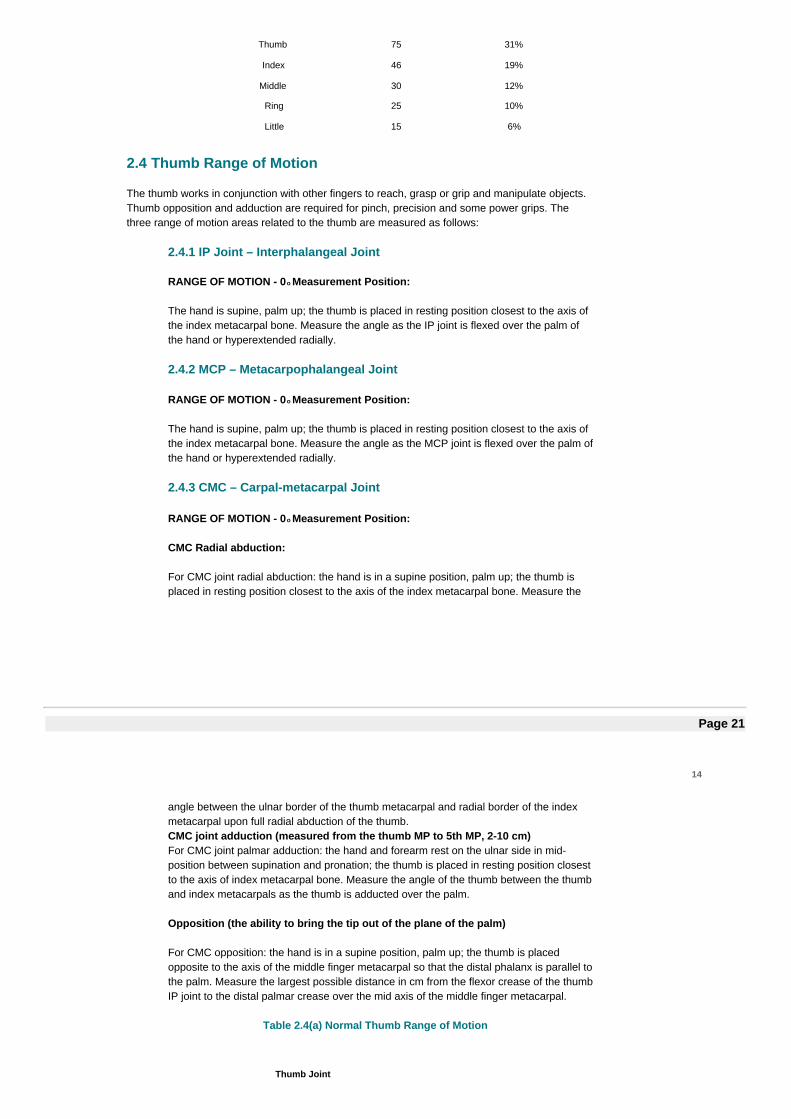
The thumb works in conjunction with other fingers to reach, grasp or grip and manipulate objects.Thumb opposition and adduction are required for pinch, precision and some power grips. Thethree range of motion areas related to the thumb are measured as follows:
2.4.1 IP Joint – Interphalangeal Joint
RANGE OF MOTION - 0o Measurement Position:
The hand is supine, palm up; the thumb is placed in resting position closest to the axis ofthe index metacarpal bone. Measure the angle as the IP joint is flexed over the palm ofthe hand or hyperextended radially.
2.4.2 MCP – Metacarpophalangeal Joint
RANGE OF MOTION - 0o Measurement Position:
The hand is supine, palm up; the thumb is placed in resting position closest to the axis ofthe index metacarpal bone. Measure the angle as the MCP joint is flexed over the palm ofthe hand or hyperextended radially.
2.4.3 CMC – Carpal-metacarpal Joint
RANGE OF MOTION - 0o Measurement Position:
CMC Radial abduction:
For CMC joint radial abduction: the hand is in a supine position, palm up; the thumb isplaced in resting position closest to the axis of the index metacarpal bone. Measure the
Page 21
14
angle between the ulnar border of the thumb metacarpal and radial border of the indexmetacarpal upon full radial abduction of the thumb.CMC joint adduction (measured from the thumb MP to 5th MP, 2-10 cm)For CMC joint palmar adduction: the hand and forearm rest on the ulnar side in mid-position between supination and pronation; the thumb is placed in resting position closestto the axis of index metacarpal bone. Measure the angle of the thumb between the thumband index metacarpals as the thumb is adducted over the palm.
Opposition (the ability to bring the tip out of the plane of the palm)
For CMC opposition: the hand is in a supine position, palm up; the thumb is placedopposite to the axis of the middle finger metacarpal so that the distal phalanx is parallel tothe palm. Measure the largest possible distance in cm from the flexor crease of the thumbIP joint to the distal palmar crease over the mid axis of the middle finger metacarpal.
Table 2.4(a) Normal Thumb Range of Motion
Thumb Joint
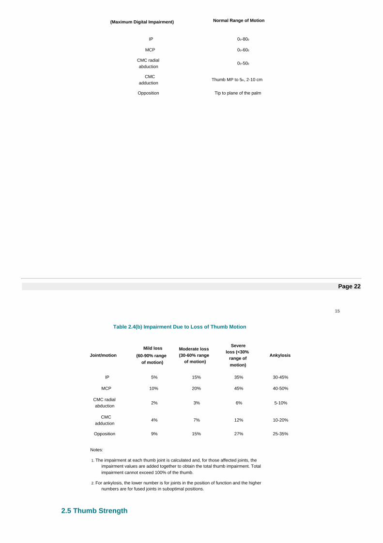
(Maximum Digital Impairment) Normal Range of Motion
IP 00-800
MCP 00-600
CMC radialabduction
00-500
CMCadduction
Thumb MP to 5th, 2-10 cm
Opposition Tip to plane of the palm
Page 22
15
Table 2.4(b) Impairment Due to Loss of Thumb Motion
Joint/motionMild loss
(60-90% rangeof motion)
Moderate loss(30-60% range
of motion)
Severeloss (<30%
range ofmotion)
Ankylosis
IP 5% 15% 35% 30-45%
MCP 10% 20% 45% 40-50%
CMC radialabduction
2% 3% 6% 5-10%
CMCadduction
4% 7% 12% 10-20%
Opposition 9% 15% 27% 25-35%
Notes:
1. The impairment at each thumb joint is calculated and, for those affected joints, theimpairment values are added together to obtain the total thumb impairment. Totalimpairment cannot exceed 100% of the thumb.
2. For ankylosis, the lower number is for joints in the position of function and the highernumbers are for fused joints in suboptimal positions.
2.5 Thumb Strength
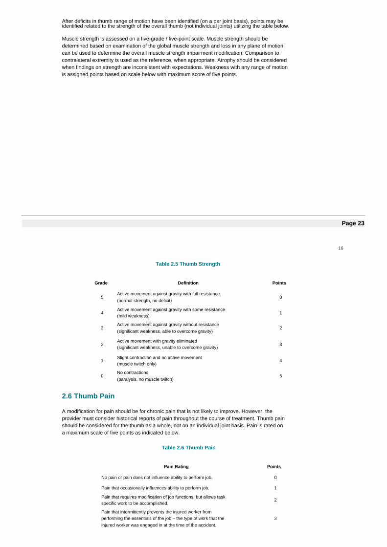
After deficits in thumb range of motion have been identified (on a per joint basis), points may beidentified related to the strength of the overall thumb (not individual joints) utilizing the table below.
Muscle strength is assessed on a five-grade / five-point scale. Muscle strength should bedetermined based on examination of the global muscle strength and loss in any plane of motioncan be used to determine the overall muscle strength impairment modification. Comparison tocontralateral extremity is used as the reference, when appropriate. Atrophy should be consideredwhen findings on strength are inconsistent with expectations. Weakness with any range of motionis assigned points based on scale below with maximum score of five points.
Page 23
16
Table 2.5 Thumb Strength
Grade Definition Points
5Active movement against gravity with full resistance(normal strength, no deficit)
0
4Active movement against gravity with some resistance(mild weakness)
1
3Active movement against gravity without resistance(significant weakness, able to overcome gravity)
2
2Active movement with gravity eliminated(significant weakness, unable to overcome gravity)
3
1Slight contraction and no active movement(muscle twitch only)
4
0No contractions(paralysis, no muscle twitch)
5
2.6 Thumb Pain
A modification for pain should be for chronic pain that is not likely to improve. However, theprovider must consider historical reports of pain throughout the course of treatment. Thumb painshould be considered for the thumb as a whole, not on an individual joint basis. Pain is rated ona maximum scale of five points as indicated below.
Table 2.6 Thumb Pain
Pain Rating Points
No pain or pain does not influence ability to perform job. 0
Pain that occasionally influences ability to perform job. 1
Pain that requires modification of job functions; but allows taskspecific work to be accomplished.
2
Pain that intermittently prevents the injured worker fromperforming the essentials of the job – the type of work that theinjured worker was engaged in at the time of the accident.
3
Pain that consistently prevents one from performing theessentials of the job – the type of work that the injured workerwas engaged in at the time of the accident.
4
Pain that prevents the injured worker from engaging inmeaningful work, leisure or household duties.
5
Page 24
17
2.7 Finger Range of Motion
The index through the small finger each have three joints, the metacarpophalangeal (MCP joint),the proximal interphalangeal (PIP) and the distal interphalangeal (DIP). The MCP joint enablesfinger flexion and extension and is important for both grip and pinch activities.
2.7.1 Metacarpophalangeal (MCP) Joint - Range of Motion
0o Measurement Position
Hand prone, fingers extended horizontal with palm and wrist. Measure the angle betweenthe metacarpal bone and the first phalanx as the MCP joint is flexed towards the palm orextended towards the dorsum of the hand.
2.7.2 Proximal Interphalangeal (PIP) Joint - Range of Motion
0o Measurement Position
Flexion: Hand prone, fingers extended horizontal with palm and wrist. Measure the anglebetween the metacarpal bone and the first phalanx as the PIP joint is flexed towards thepalm.
Extension: Hand prone, PIP joints fully flexed. Measure the angle as the PIP joint isextended towards horizontal position of the fingers.
2.7.3 Distal Interphalangeal (DIP) Joint - Range of Motion
0o Measurement Position
Flexion: Hand prone, fingers extended horizontal with palm and wrist. Measure the anglebetween the metacarpal bone and the first phalanx as the DIP joint is flexed towards thepalm.
Extension: Hand prone, DIP joints fully flexed. Measure the angle as the DIP joint isextended towards horizontal position of the fingers.
Table 2.7.3(a) Normal Finger Range of Motion
Finger Joint
(Maximum Digital Impairment)Normal Range of Motion
DIP (45%)0o Extension
70o Flexion
PIP (80%)0o Extension
100o Flexion
-20o Extension
MCP (100%) 90o Flexion
Page 25
18
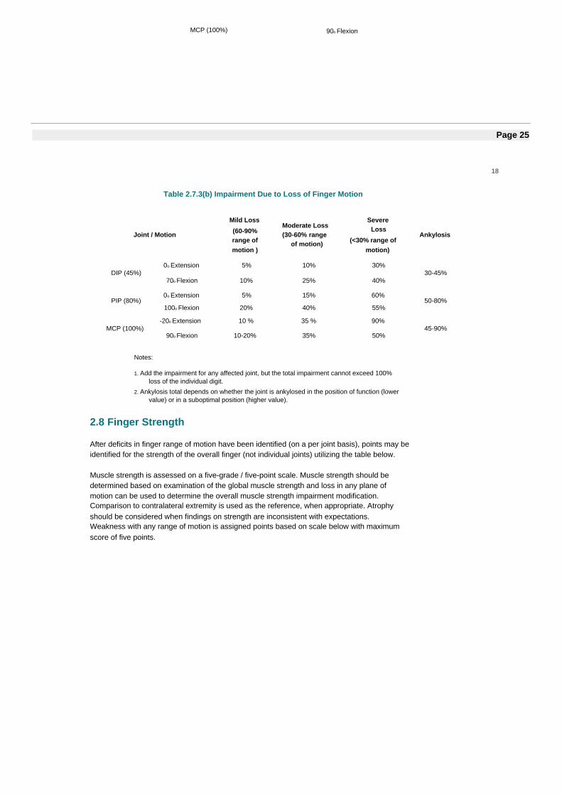
Table 2.7.3(b) Impairment Due to Loss of Finger Motion
Joint / Motion
Mild Loss(60-90%range ofmotion )
Moderate Loss(30-60% range
of motion)
SevereLoss
(<30% range ofmotion)
Ankylosis
DIP (45%)0o Extension 5% 10% 30%
30-45%70o Flexion 10% 25% 40%
PIP (80%)0o Extension 5% 15% 60%
50-80%100o Flexion 20% 40% 55%
MCP (100%)-20o Extension 10 % 35 % 90%
45-90%90o Flexion 10-20% 35% 50%
Notes:
1. Add the impairment for any affected joint, but the total impairment cannot exceed 100%loss of the individual digit.
2. Ankylosis total depends on whether the joint is ankylosed in the position of function (lowervalue) or in a suboptimal position (higher value).
2.8 Finger Strength
After deficits in finger range of motion have been identified (on a per joint basis), points may beidentified for the strength of the overall finger (not individual joints) utilizing the table below.
Muscle strength is assessed on a five-grade / five-point scale. Muscle strength should bedetermined based on examination of the global muscle strength and loss in any plane ofmotion can be used to determine the overall muscle strength impairment modification.Comparison to contralateral extremity is used as the reference, when appropriate. Atrophyshould be considered when findings on strength are inconsistent with expectations.Weakness with any range of motion is assigned points based on scale below with maximumscore of five points.
Page 26
19
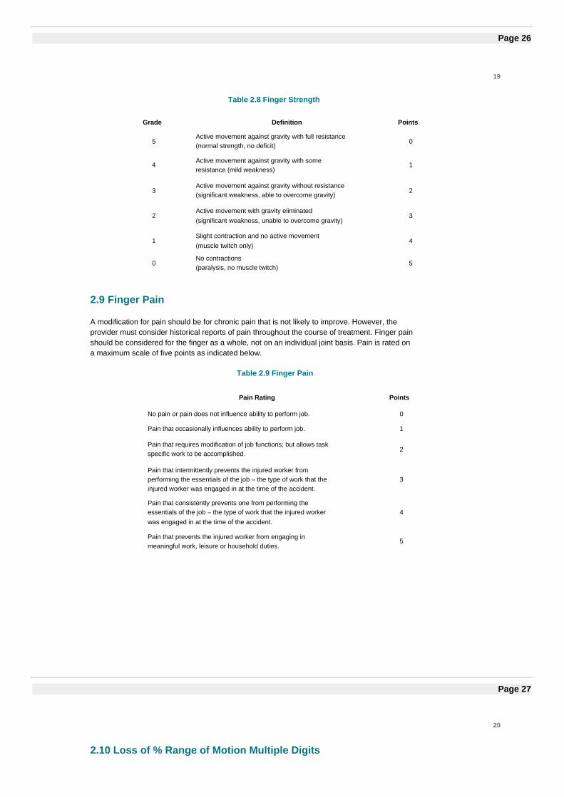
Table 2.8 Finger Strength
2.9 Finger Pain
A modification for pain should be for chronic pain that is not likely to improve. However, theprovider must consider historical reports of pain throughout the course of treatment. Finger painshould be considered for the finger as a whole, not on an individual joint basis. Pain is rated ona maximum scale of five points as indicated below.
Table 2.9 Finger Pain
Pain Rating Points
No pain or pain does not influence ability to perform job. 0
Pain that occasionally influences ability to perform job. 1
Pain that requires modification of job functions; but allows taskspecific work to be accomplished.
2
Pain that intermittently prevents the injured worker fromperforming the essentials of the job – the type of work that theinjured worker was engaged in at the time of the accident.
3
Pain that consistently prevents one from performing theessentials of the job – the type of work that the injured workerwas engaged in at the time of the accident.
4
Pain that prevents the injured worker from engaging inmeaningful work, leisure or household duties.
5
Grade Definition Points
5Active movement against gravity with full resistance(normal strength, no deficit)
0
4Active movement against gravity with someresistance (mild weakness)
1
3Active movement against gravity without resistance(significant weakness, able to overcome gravity)
2
2Active movement with gravity eliminated(significant weakness, unable to overcome gravity)
3
1Slight contraction and no active movement(muscle twitch only)
4
0No contractions(paralysis, no muscle twitch)
5
Page 27
20
2.10 Loss of % Range of Motion Multiple Digits
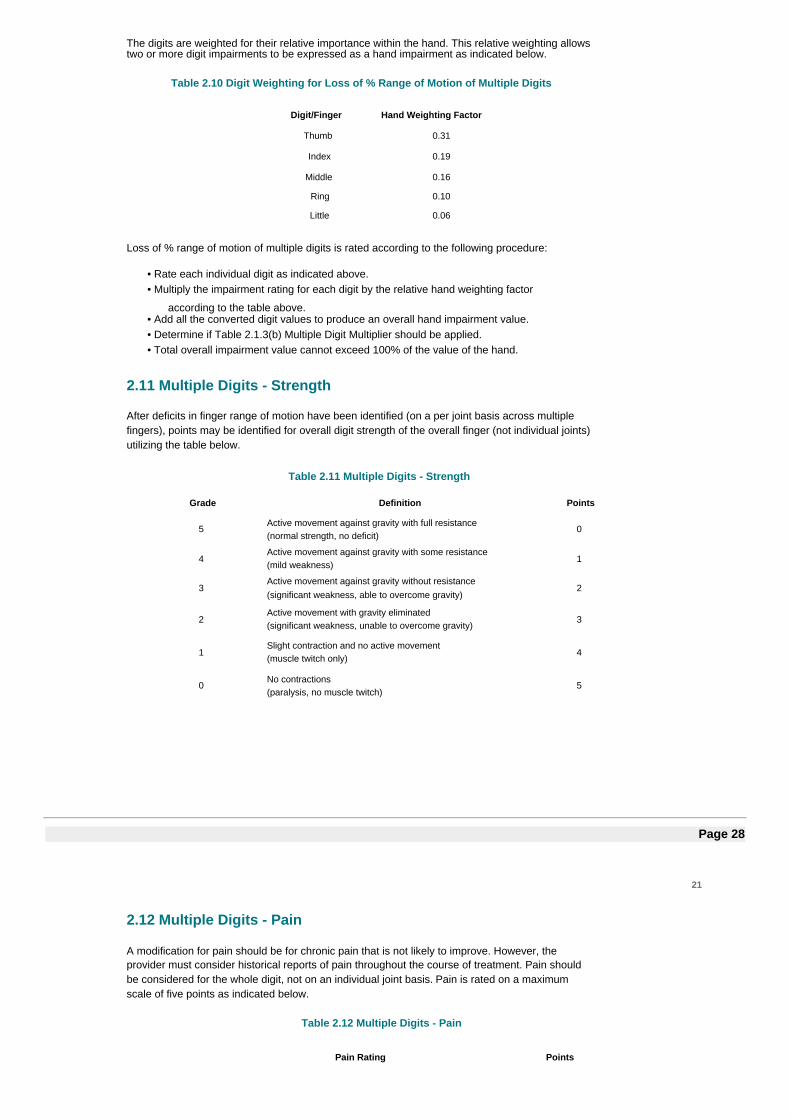
The digits are weighted for their relative importance within the hand. This relative weighting allowstwo or more digit impairments to be expressed as a hand impairment as indicated below.
Table 2.10 Digit Weighting for Loss of % Range of Motion of Multiple Digits
Digit/Finger Hand Weighting Factor
Thumb 0.31
Index 0.19
Middle 0.16
Ring 0.10
Little 0.06
Loss of % range of motion of multiple digits is rated according to the following procedure:
• Rate each individual digit as indicated above.• Multiply the impairment rating for each digit by the relative hand weighting factor
according to the table above.• Add all the converted digit values to produce an overall hand impairment value.• Determine if Table 2.1.3(b) Multiple Digit Multiplier should be applied.• Total overall impairment value cannot exceed 100% of the value of the hand.
2.11 Multiple Digits - Strength
After deficits in finger range of motion have been identified (on a per joint basis across multiplefingers), points may be identified for overall digit strength of the overall finger (not individual joints)utilizing the table below.
Table 2.11 Multiple Digits - Strength
Grade Definition Points
5Active movement against gravity with full resistance(normal strength, no deficit)
0
4Active movement against gravity with some resistance(mild weakness)
1
3Active movement against gravity without resistance(significant weakness, able to overcome gravity)
2
2Active movement with gravity eliminated(significant weakness, unable to overcome gravity)
3
1Slight contraction and no active movement(muscle twitch only)
4
0No contractions(paralysis, no muscle twitch)
5
Page 28
21
2.12 Multiple Digits - Pain
A modification for pain should be for chronic pain that is not likely to improve. However, theprovider must consider historical reports of pain throughout the course of treatment. Pain shouldbe considered for the whole digit, not on an individual joint basis. Pain is rated on a maximumscale of five points as indicated below.
Table 2.12 Multiple Digits - Pain
Pain Rating Points
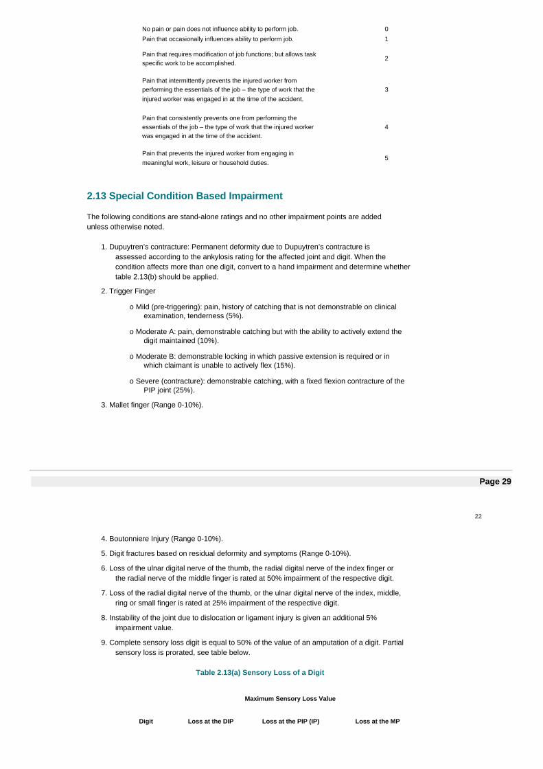
No pain or pain does not influence ability to perform job. 0
Pain that occasionally influences ability to perform job. 1
Pain that requires modification of job functions; but allows taskspecific work to be accomplished.
2
Pain that intermittently prevents the injured worker fromperforming the essentials of the job – the type of work that theinjured worker was engaged in at the time of the accident.
3
Pain that consistently prevents one from performing theessentials of the job – the type of work that the injured workerwas engaged in at the time of the accident.
4
Pain that prevents the injured worker from engaging inmeaningful work, leisure or household duties.
5
2.13 Special Condition Based Impairment
The following conditions are stand-alone ratings and no other impairment points are addedunless otherwise noted.
1. Dupuytren’s contracture: Permanent deformity due to Dupuytren’s contracture isassessed according to the ankylosis rating for the affected joint and digit. When thecondition affects more than one digit, convert to a hand impairment and determine whethertable 2.13(b) should be applied.
2. Trigger Finger
o Mild (pre-triggering): pain, history of catching that is not demonstrable on clinicalexamination, tenderness (5%).
o Moderate A: pain, demonstrable catching but with the ability to actively extend thedigit maintained (10%).
o Moderate B: demonstrable locking in which passive extension is required or inwhich claimant is unable to actively flex (15%).
o Severe (contracture): demonstrable catching, with a fixed flexion contracture of thePIP joint (25%).
3. Mallet finger (Range 0-10%).
Page 29
22
4. Boutonniere Injury (Range 0-10%).
5. Digit fractures based on residual deformity and symptoms (Range 0-10%).
6. Loss of the ulnar digital nerve of the thumb, the radial digital nerve of the index finger orthe radial nerve of the middle finger is rated at 50% impairment of the respective digit.
7. Loss of the radial digital nerve of the thumb, or the ulnar digital nerve of the index, middle,ring or small finger is rated at 25% impairment of the respective digit.
8. Instability of the joint due to dislocation or ligament injury is given an additional 5%impairment value.
9. Complete sensory loss digit is equal to 50% of the value of an amputation of a digit. Partialsensory loss is prorated, see table below.
Table 2.13(a) Sensory Loss of a Digit
Maximum Sensory Loss Value
Digit Loss at the DIP Loss at the PIP (IP) Loss at the MP
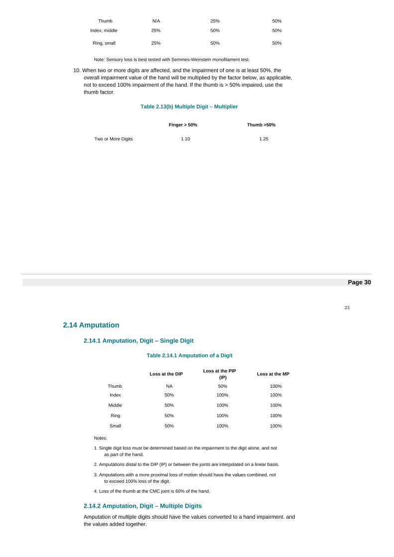
Thumb N/A 25% 50%
Index, middle 25% 50% 50%
Ring, small 25% 50% 50%
Note: Sensory loss is best tested with Semmes-Weinstein monofilament test.
10. When two or more digits are affected, and the impairment of one is at least 50%, theoverall impairment value of the hand will be multiplied by the factor below, as applicable,not to exceed 100% impairment of the hand. If the thumb is > 50% impaired, use thethumb factor.
Table 2.13(b) Multiple Digit – Multiplier
Finger > 50% Thumb >50%
Two or More Digits 1.10 1.25
Page 30
23
2.14 Amputation
2.14.1 Amputation, Digit – Single Digit
Table 2.14.1 Amputation of a Digit
Loss at the DIP Loss at the PIP(IP)
Loss at the MP
Thumb NA 50% 100%
Index 50% 100% 100%
Middle 50% 100% 100%
Ring 50% 100% 100%
Small 50% 100% 100%
Notes:
1. Single digit loss must be determined based on the impairment to the digit alone, and notas part of the hand.
2. Amputations distal to the DIP (IP) or between the joints are interpolated on a linear basis.
3. Amputations with a more proximal loss of motion should have the values combined, notto exceed 100% loss of the digit.
4. Loss of the thumb at the CMC joint is 60% of the hand.
2.14.2 Amputation, Digit – Multiple Digits
Amputation of multiple digits should have the values converted to a hand impairment. andthe values added together.
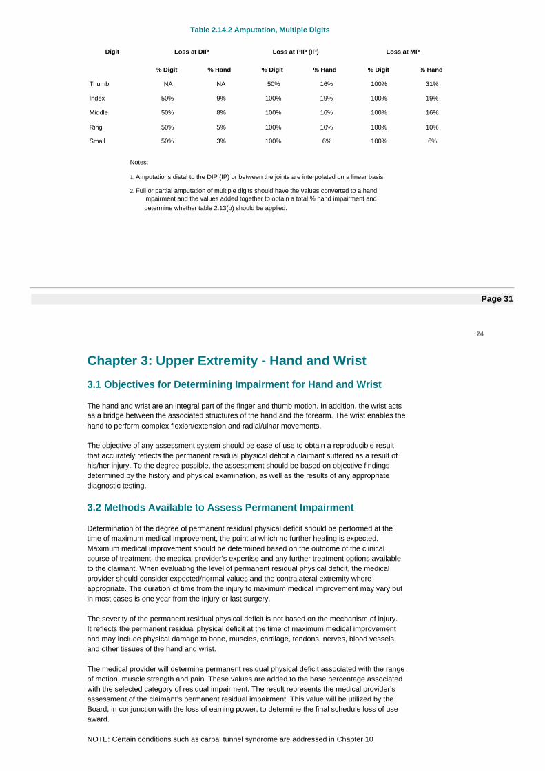
Table 2.14.2 Amputation, Multiple Digits
Digit Loss at DIP Loss at PIP (IP) Loss at MP
% Digit % Hand % Digit % Hand % Digit % Hand
Thumb NA NA 50% 16% 100% 31%
Index 50% 9% 100% 19% 100% 19%
Middle 50% 8% 100% 16% 100% 16%
Ring 50% 5% 100% 10% 100% 10%
Small 50% 3% 100% 6% 100% 6%
Notes:
1. Amputations distal to the DIP (IP) or between the joints are interpolated on a linear basis.
2. Full or partial amputation of multiple digits should have the values converted to a handimpairment and the values added together to obtain a total % hand impairment anddetermine whether table 2.13(b) should be applied.
Page 31
24
Chapter 3: Upper Extremity - Hand and Wrist
3.1 Objectives for Determining Impairment for Hand and Wrist
The hand and wrist are an integral part of the finger and thumb motion. In addition, the wrist actsas a bridge between the associated structures of the hand and the forearm. The wrist enables thehand to perform complex flexion/extension and radial/ulnar movements.
The objective of any assessment system should be ease of use to obtain a reproducible resultthat accurately reflects the permanent residual physical deficit a claimant suffered as a result ofhis/her injury. To the degree possible, the assessment should be based on objective findingsdetermined by the history and physical examination, as well as the results of any appropriatediagnostic testing.
3.2 Methods Available to Assess Permanent Impairment
Determination of the degree of permanent residual physical deficit should be performed at thetime of maximum medical improvement, the point at which no further healing is expected.Maximum medical improvement should be determined based on the outcome of the clinicalcourse of treatment, the medical provider’s expertise and any further treatment options availableto the claimant. When evaluating the level of permanent residual physical deficit, the medicalprovider should consider expected/normal values and the contralateral extremity whereappropriate. The duration of time from the injury to maximum medical improvement may vary butin most cases is one year from the injury or last surgery.
The severity of the permanent residual physical deficit is not based on the mechanism of injury.It reflects the permanent residual physical deficit at the time of maximum medical improvementand may include physical damage to bone, muscles, cartilage, tendons, nerves, blood vesselsand other tissues of the hand and wrist.
The medical provider will determine permanent residual physical deficit associated with the rangeof motion, muscle strength and pain. These values are added to the base percentage associatedwith the selected category of residual impairment. The result represents the medical provider’sassessment of the claimant’s permanent residual impairment. This value will be utilized by theBoard, in conjunction with the loss of earning power, to determine the final schedule loss of useaward.
NOTE: Certain conditions such as carpal tunnel syndrome are addressed in Chapter 10
Peripheral Nerve Injuries and Compression Neuropathies.
Page 32
25
3.3 Hand & Wrist Impairment
Normal hand function requires:
A full-length, opposable thumbFull length index, middle, ring and small fingersNormal, active range of motion of the jointsMuscle function, both extrinsic and intrinsicNormal, pain free sensation at the digit tipsNormal vascular supply
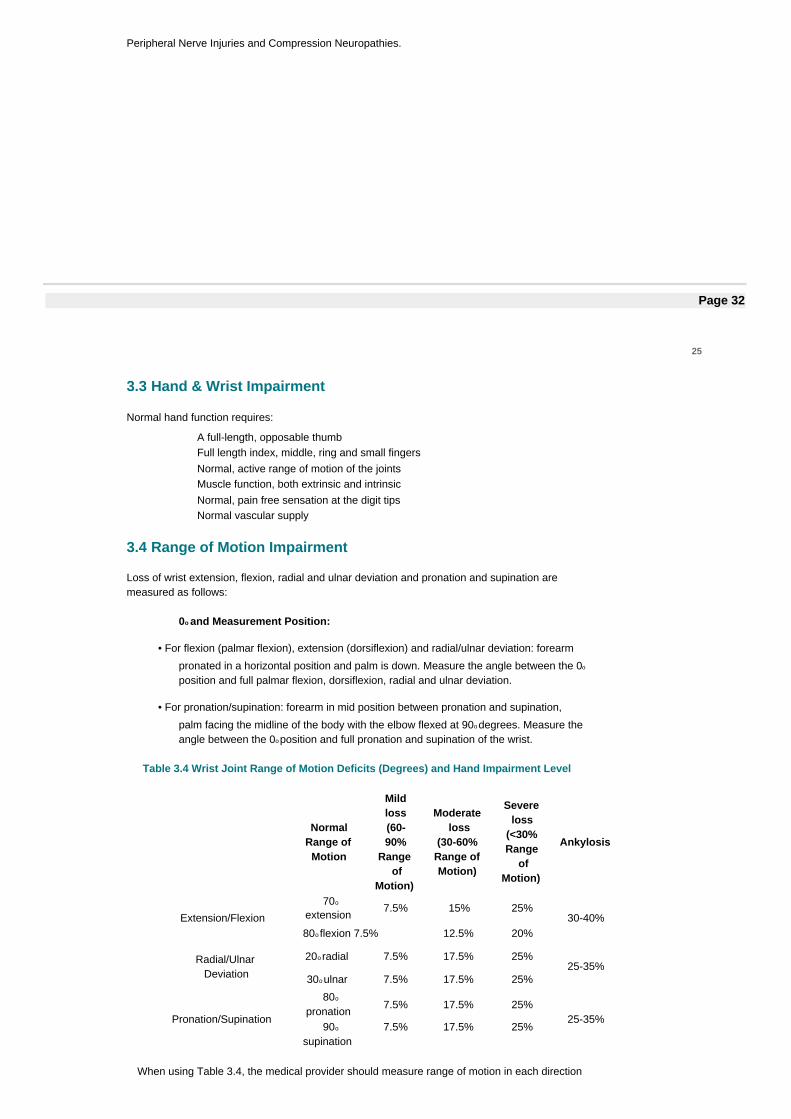
3.4 Range of Motion Impairment
Loss of wrist extension, flexion, radial and ulnar deviation and pronation and supination aremeasured as follows:
0o and Measurement Position:
• For flexion (palmar flexion), extension (dorsiflexion) and radial/ulnar deviation: forearm
pronated in a horizontal position and palm is down. Measure the angle between the 0o
position and full palmar flexion, dorsiflexion, radial and ulnar deviation.
• For pronation/supination: forearm in mid position between pronation and supination,
palm facing the midline of the body with the elbow flexed at 90o degrees. Measure theangle between the 0o position and full pronation and supination of the wrist.
Table 3.4 Wrist Joint Range of Motion Deficits (Degrees) and Hand Impairment Level
NormalRange of
Motion
Mildloss(60-90%
Rangeof
Motion)
Moderateloss
(30-60%Range ofMotion)
Severeloss
(<30%Range
ofMotion)
Ankylosis
Extension/Flexion
70o
extension7.5% 15% 25%
30-40%80o flexion 7.5% 12.5% 20%
Radial/UlnarDeviation
20o radial 7.5% 17.5% 25%25-35%
30o ulnar 7.5% 17.5% 25%
Pronation/Supination
80o
pronation7.5% 17.5% 25%
25-35%90o
supination7.5% 17.5% 25%
When using Table 3.4, the medical provider should measure range of motion in each direction
for each category and only apply the percentage associated with the motion direction that has
Page 33
26
the larger deficit. If the claimant has deficits in all three range of motion categories, theimpairment shall be based on the two categories where the claimant has the largest deficits.
Total impairment may not exceed 100% of the hand.
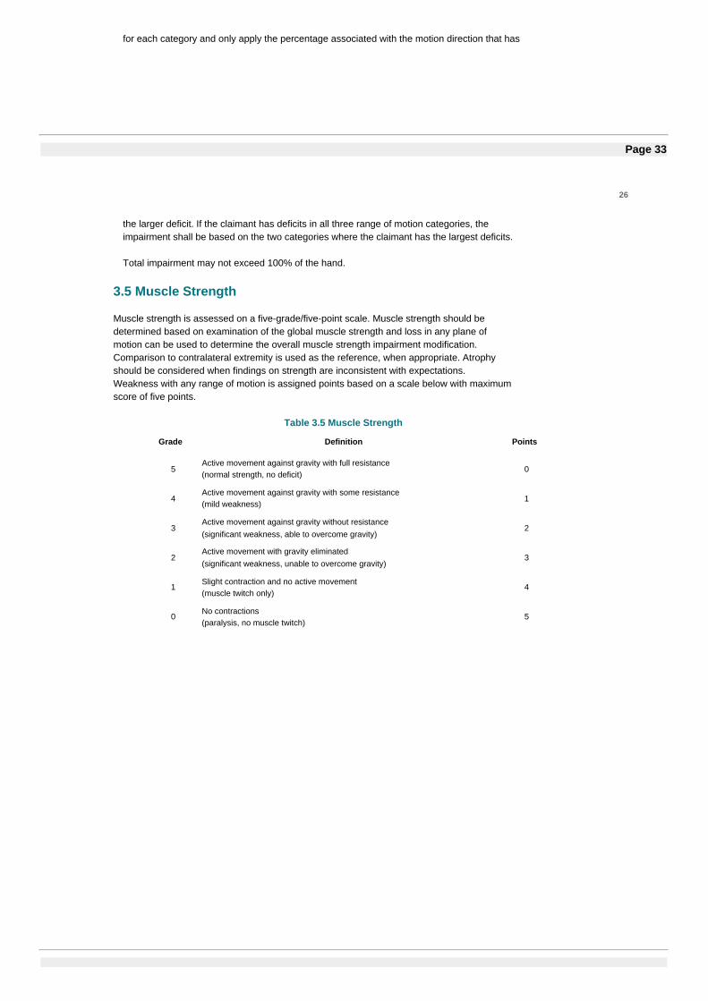
3.5 Muscle Strength
Muscle strength is assessed on a five-grade/five-point scale. Muscle strength should bedetermined based on examination of the global muscle strength and loss in any plane ofmotion can be used to determine the overall muscle strength impairment modification.Comparison to contralateral extremity is used as the reference, when appropriate. Atrophyshould be considered when findings on strength are inconsistent with expectations.Weakness with any range of motion is assigned points based on a scale below with maximumscore of five points.
Table 3.5 Muscle Strength
Grade Definition Points
5Active movement against gravity with full resistance(normal strength, no deficit)
0
4Active movement against gravity with some resistance(mild weakness)
1
3Active movement against gravity without resistance(significant weakness, able to overcome gravity)
2
2Active movement with gravity eliminated(significant weakness, unable to overcome gravity)
3
1Slight contraction and no active movement(muscle twitch only)
4
0No contractions(paralysis, no muscle twitch)
5
Page 34
27
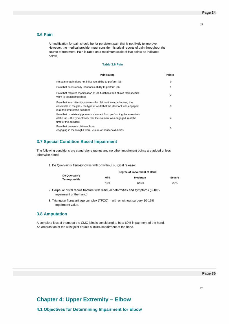
3.6 Pain
A modification for pain should be for persistent pain that is not likely to improve.However, the medical provider must consider historical reports of pain throughout thecourse of treatment. Pain is rated on a maximum scale of five points as indicatedbelow.
Table 3.6 Pain
Pain Rating Points
No pain or pain does not influence ability to perform job. 0
Pain that occasionally influences ability to perform job. 1
Pain that requires modification of job functions; but allows task specificwork to be accomplished.
2
Pain that intermittently prevents the claimant from performing theessentials of the job – the type of work that the claimant was engagedin at the time of the accident.
3
Pain that consistently prevents claimant from performing the essentialsof the job – the type of work that the claimant was engaged in at thetime of the accident.
4
Pain that prevents claimant fromengaging in meaningful work, leisure or household duties.
5
3.7 Special Condition Based Impairment
The following conditions are stand-alone ratings and no other impairment points are added unlessotherwise noted.
1. De Quervain’s Tenosynovitis with or without surgical release:
De Quervain’sTenosynovitis
Degree of Impairment of Hand
Mild Moderate Severe
7.5% 12.5% 20%
2. Carpal or distal radius fracture with residual deformities and symptoms (0-10%impairment of the hand).
3. Triangular fibrocartilage complex (TFCC) – with or without surgery 10-15%impairment value.
3.8 Amputation
A complete loss of thumb at the CMC joint is considered to be a 60% impairment of the hand.An amputation at the wrist joint equals a 100% impairment of the hand.
Page 35
28
Chapter 4: Upper Extremity – Elbow
4.1 Objectives for Determining Impairment for Elbow
The elbow plays an important role in positioning the hand and wrist to allows for functional use ofthe upper extremity. The most import function of the elbow joint is to position the hand, eithermoving the hand away from the body (elbow extension), towards the body (elbow flexion) or in amore precise hand movement (supination/pronation). The degree of impairment is dependent onmany factors including the residual permanent residual physical deficit, outcome of any treatmentand potential sequela from the injury or subsequent treatments.
The objective of any assessment system should be ease of use to obtain a reproducible resultthat accurately reflects the permanent residual physical deficit a worker suffered as a result ofhis/her injury. To the degree possible, the assessment should be based on objective findingsdetermined by the history and physical examination, as well as the results of any appropriatediagnostic testing.
4.2 Methods Available to Assess Permanent Impairment
Determination of the degree of permanent residual physical deficit should be performed at thetime of maximum medical improvement, the point at which no further healing is expected.Maximum medical improvement should be determined based on the outcome of the clinicalcourse of treatment, the medical provider’s expertise and any further treatment options availableto the claimant. When evaluating the level of permanent residual physical deficit, the medicalprovider should consider expected/normal values and the contralateral extremity whereappropriate. The duration of time from the injury to maximum medical improvement may vary butin most cases is one year from the injury or last surgery.
The severity of the permanent residual physical deficit is not based on the mechanism of injury.It reflects the permanent residual physical deficit at the time of maximum medical improvementand may include physical damage to bone, muscles, cartilage, tendons, nerves, blood vessels,and other tissues.
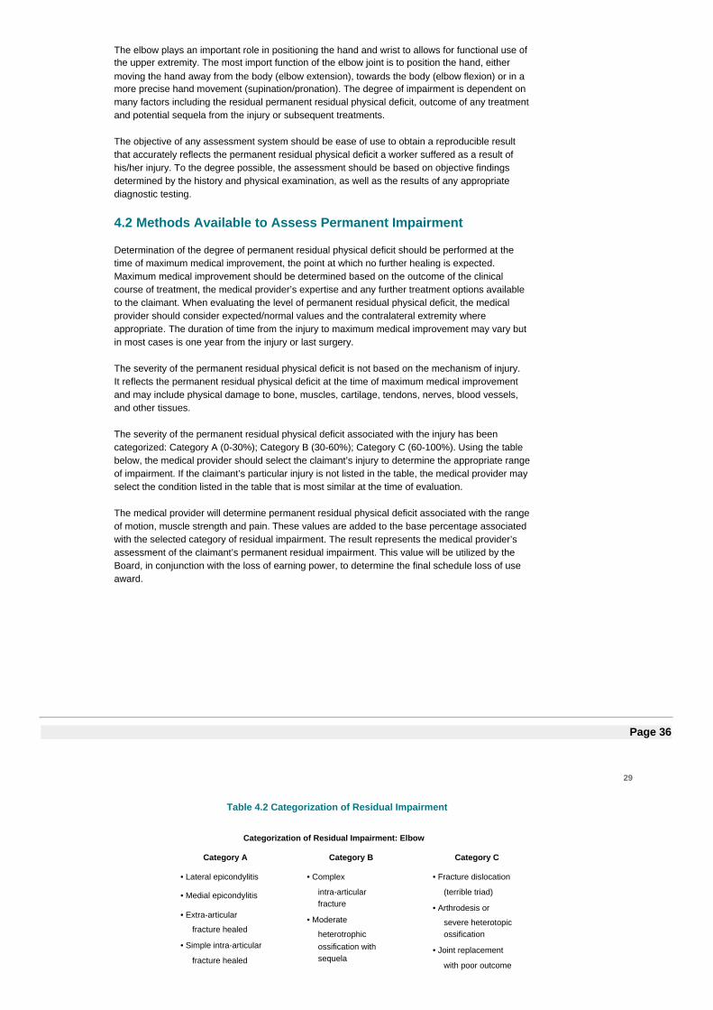
The severity of the permanent residual physical deficit associated with the injury has beencategorized: Category A (0-30%); Category B (30-60%); Category C (60-100%). Using the tablebelow, the medical provider should select the claimant’s injury to determine the appropriate rangeof impairment. If the claimant’s particular injury is not listed in the table, the medical provider mayselect the condition listed in the table that is most similar at the time of evaluation.
The medical provider will determine permanent residual physical deficit associated with the rangeof motion, muscle strength and pain. These values are added to the base percentage associatedwith the selected category of residual impairment. The result represents the medical provider’sassessment of the claimant’s permanent residual impairment. This value will be utilized by theBoard, in conjunction with the loss of earning power, to determine the final schedule loss of useaward.
Page 36
29
Table 4.2 Categorization of Residual Impairment
Categorization of Residual Impairment: Elbow
Category A Category B Category C
• Lateral epicondylitis
• Medial epicondylitis
• Extra-articular
fracture healed
• Simple intra-articular
fracture healed
• Complex
intra-articularfracture
• Moderate
heterotrophicossification withsequela
• Fracture dislocation
(terrible triad)
• Arthrodesis or
severe heterotopicossification
• Joint replacement
with poor outcome
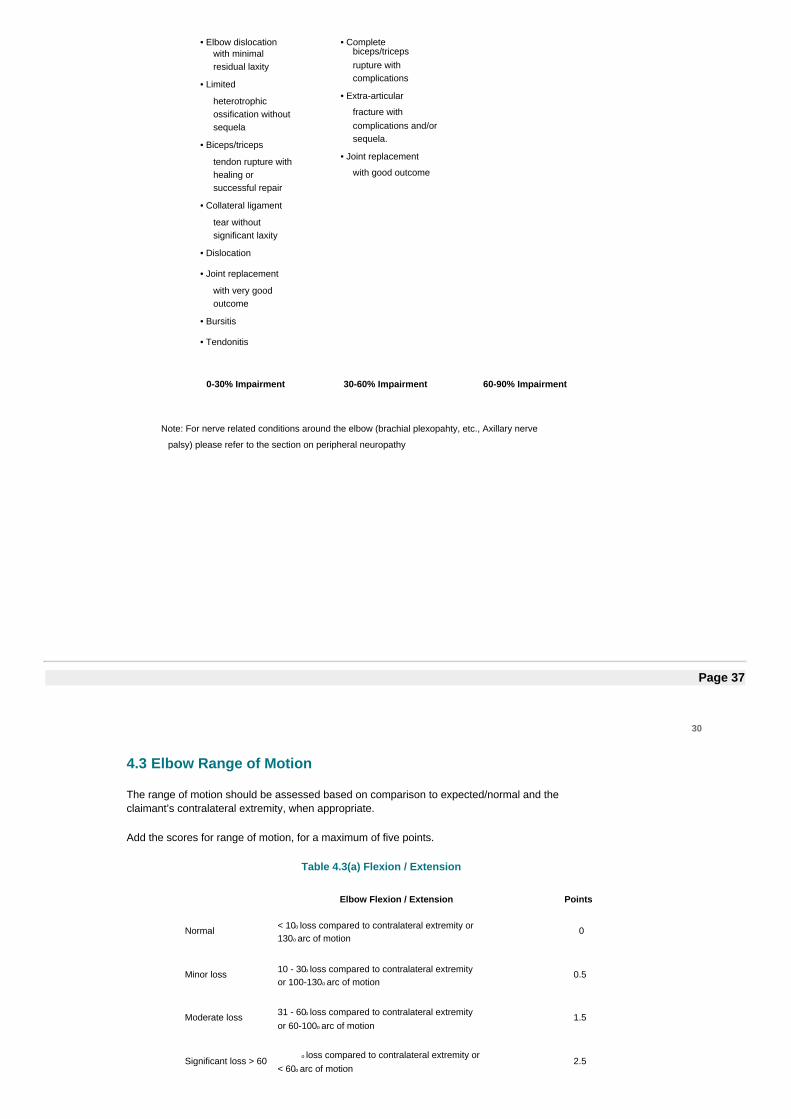
• Elbow dislocationwith minimalresidual laxity
• Limited
heterotrophicossification withoutsequela
• Biceps/triceps
tendon rupture withhealing orsuccessful repair
• Collateral ligament
tear withoutsignificant laxity
• Dislocation
• Joint replacement
with very goodoutcome
• Bursitis
• Tendonitis
• Completebiceps/tricepsrupture withcomplications
• Extra-articular
fracture withcomplications and/orsequela.
• Joint replacement
with good outcome
0-30% Impairment 30-60% Impairment 60-90% Impairment
Note: For nerve related conditions around the elbow (brachial plexopahty, etc., Axillary nerve
palsy) please refer to the section on peripheral neuropathy
Page 37
30
4.3 Elbow Range of Motion
The range of motion should be assessed based on comparison to expected/normal and theclaimant’s contralateral extremity, when appropriate.
Add the scores for range of motion, for a maximum of five points.
Table 4.3(a) Flexion / Extension
Elbow Flexion / Extension Points
Normal < 10o loss compared to contralateral extremity or130o arc of motion
0
Minor loss 10 - 30o loss compared to contralateral extremityor 100-130o arc of motion
0.5
Moderate loss 31 - 60o loss compared to contralateral extremityor 60-100o arc of motion
1.5
Significant loss > 60o loss compared to contralateral extremity or
< 60o arc of motion2.5
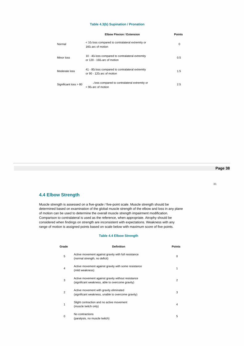
Table 4.3(b) Supination / Pronation
Elbow Flexion / Extension Points
Normal < 10o loss compared to contralateral extremity or160o arc of motion
0
Minor loss 10 - 40o loss compared to contralateral extremityor 120 - 160o arc of motion
0.5
Moderate loss 41 - 80o loss compared to contralateral extremityor 90 - 120o arc of motion
1.5
Significant loss > 80 o loss compared to contralateral extremity or< 90o arc of motion
2.5
Page 38
31
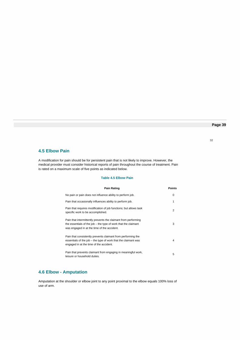
4.4 Elbow Strength
Muscle strength is assessed on a five-grade / five-point scale. Muscle strength should bedetermined based on examination of the global muscle strength of the elbow and loss in any planeof motion can be used to determine the overall muscle strength impairment modification.Comparison to contralateral is used as the reference, when appropriate. Atrophy should beconsidered when findings on strength are inconsistent with expectations. Weakness with anyrange of motion is assigned points based on scale below with maximum score of five points.
Table 4.4 Elbow Strength
Grade Definition Points
5Active movement against gravity with full resistance(normal strength, no deficit)
0
4Active movement against gravity with some resistance(mild weakness)
1
3Active movement against gravity without resistance(significant weakness, able to overcome gravity)
2
2Active movement with gravity eliminated(significant weakness, unable to overcome gravity)
3
1Slight contraction and no active movement(muscle twitch only)
4
0No contractions(paralysis, no muscle twitch)
5
Page 39
32
4.5 Elbow Pain
A modification for pain should be for persistent pain that is not likely to improve. However, themedical provider must consider historical reports of pain throughout the course of treatment. Painis rated on a maximum scale of five points as indicated below.
Table 4.5 Elbow Pain
Pain Rating Points
No pain or pain does not influence ability to perform job. 0
Pain that occasionally influences ability to perform job. 1
Pain that requires modification of job functions; but allows taskspecific work to be accomplished.
2
Pain that intermittently prevents the claimant from performingthe essentials of the job – the type of work that the claimantwas engaged in at the time of the accident.
3
Pain that consistently prevents claimant from performing theessentials of the job – the type of work that the claimant wasengaged in at the time of the accident.
4
Pain that prevents claimant from engaging in meaningful work,leisure or household duties.
5
4.6 Elbow - Amputation
Amputation at the shoulder or elbow joint to any point proximal to the elbow equals 100% loss ofuse of arm.

Page 40
33
Chapter 5: Upper Extremity - Shoulder
5.1 Objectives for Determining Impairment for Shoulder
The shoulder and elbow play important roles in positioning the hand in space to allow for functionaluse of the upper extremity. Injury can result in significant limitations and ability to perform workresponsibilities.
The objective of any assessment system should be ease of use to obtain a reproducible resultthat accurately reflects the permanent residual physical deficit a worker suffered as a result ofhis/her injury. To the degree possible, the assessment should be based on objective findingsdetermined by the history and physical examination, as well as the results of any appropriatediagnostic testing.
5.2 Methods Available to Assess Permanent Impairment
Determination of the degree of permanent residual physical deficit should be performed at thetime of maximum medical improvement, the point at which no further healing is expected.Maximum medical improvement should be determined based on the outcome of the clinicalcourse of treatment, the medical provider’s expertise and any further treatment options availableto the claimant. When evaluating the level of permanent residual physical deficit, the medicalprovider should consider expected/normal values and the contralateral extremity whereappropriate. The duration of time from the injury to maximum medical improvement may vary butin most cases is one year from the injury or last surgery.
The severity of the permanent residual physical deficit is not based on the mechanism of injury.It reflects the residual physical deficit at the time of maximum medical improvement and mayinclude physical damage to bone, muscles, cartilage, tendons, nerves, blood vessels, and othertissues.
The severity of the permanent residual physical deficit associated with the injury has beencategorized: Category A (0-30%), Category B (30-60%), Category C (60-100%). Using the tablebelow, the medical provider should select the claimant’s injury to determine the appropriate rangeof impairment. If the claimant’s particular injury is not listed in the table, the medical provider mayselect the condition listed in the table that is most similar at the time of evaluation.
The medical provider will determine permanent residual physical deficit associated with the rangeof motion, muscle strength and pain. These values are added to the base percentage associatedwith the selected category of residual impairment. The result represents the medical provider’sassessment of the claimant’s permanent residual impairment. This value will be utilized by theBoard, in conjunction with the loss of earning power, to determine the final schedule loss of useaward.
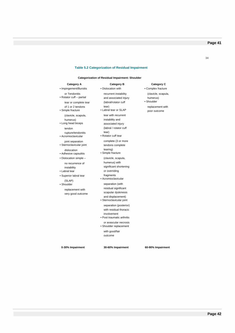
Page 41
34
Table 5.2 Categorization of Residual Impairment
Categorization of Residual Impairment: Shoulder
Category A Category B Category C• Impingement/Bursitis
or Tendonitis• Rotator cuff – partial
tear or complete tear
of 1 or 2 tendons• Simple fracture
(clavicle, scapula,
humerus)• Long head biceps
tendon
rupture/tendonitis• Acromioclavicular
joint separation• Sternoclavicular joint
dislocation• Adhesive capsulitis
• Dislocation simple –
no recurrence ofinstability
• Labral tear
• Superior labral tear
(SLAP)• Shoulder
replacement withvery good outcome
• Dislocation with
recurrent instabilityand associated injury
(labral/rotator cuff
tear)• Labral tear or SLAP
tear with recurrent
instability andassociated injury
(labral / rotator cufftear)
• Rotator cuff tear
complete (3 or moretendons complete
tearing)• Simple fracture
(clavicle, scapula,
humerus) withsignificant shortening
or overriding
fragments• Acromioclavicular
separation (with
residual significantscapular dyskinesis
and displacement)• Sternoclavicular joint
separation (posterior)
with residual thoracicinvolvement
• Post traumatic arthritis
or avascular necrosis• Shoulder replacement
with good/fairoutcome
• Complex fracture
(clavicle, scapula,humerus)
• Shoulder
replacement withpoor outcome
0-30% Impairment 30-60% Impairment 60-90% Impairment
Page 42
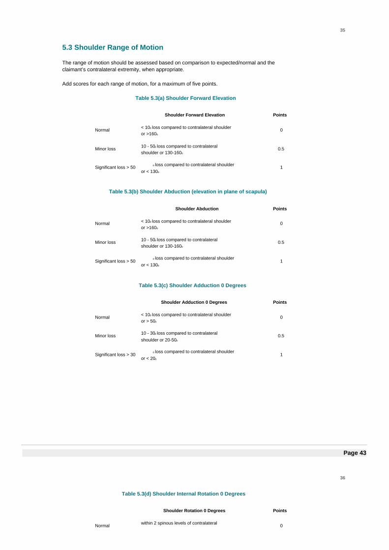
35
5.3 Shoulder Range of Motion
The range of motion should be assessed based on comparison to expected/normal and theclaimant’s contralateral extremity, when appropriate.
Add scores for each range of motion, for a maximum of five points.
Table 5.3(a) Shoulder Forward Elevation
Shoulder Forward Elevation Points
Normal < 10o loss compared to contralateral shoulderor >160o
0
Minor loss 10 - 50o loss compared to contralateralshoulder or 130-160o
0.5
Significant loss > 50 o loss compared to contralateral shoulderor < 130o
1
Table 5.3(b) Shoulder Abduction (elevation in plane of scapula)
Shoulder Abduction Points
Normal < 10o loss compared to contralateral shoulderor >160o
0
Minor loss 10 - 50o loss compared to contralateralshoulder or 130-160o
0.5
Significant loss > 50o loss compared to contralateral shoulder
or < 130o1
Table 5.3(c) Shoulder Adduction 0 Degrees
Shoulder Adduction 0 Degrees Points
Normal < 10o loss compared to contralateral shoulderor > 50o
0
Minor loss 10 - 30o loss compared to contralateralshoulder or 20-50o
0.5
Significant loss > 30o loss compared to contralateral shoulder
or < 20o1
Page 43
36
Table 5.3(d) Shoulder Internal Rotation 0 Degrees
Shoulder Rotation 0 Degrees Points
Normal within 2 spinous levels of contralateral 0
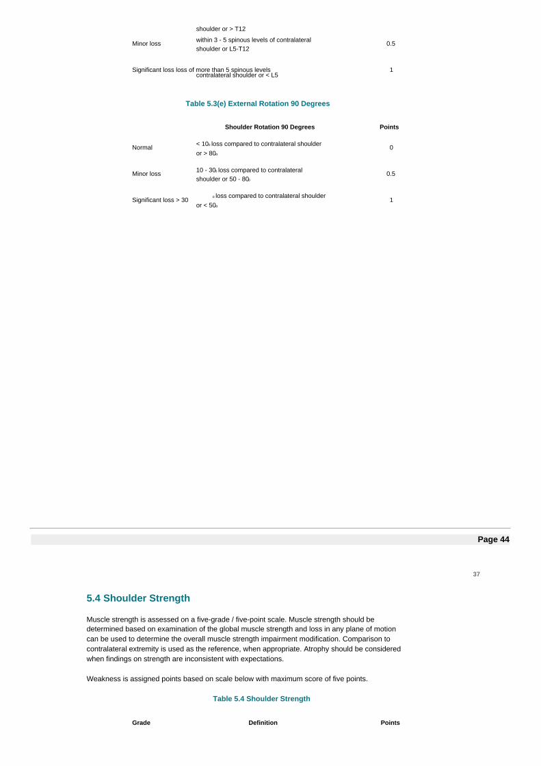
shoulder or > T12
Minor loss within 3 - 5 spinous levels of contralateralshoulder or L5-T12
0.5
Significant loss loss of more than 5 spinous levelscontralateral shoulder or < L5
1
Table 5.3(e) External Rotation 90 Degrees
Shoulder Rotation 90 Degrees Points
Normal < 10o loss compared to contralateral shoulderor > 80o
0
Minor loss 10 - 30o loss compared to contralateralshoulder or 50 - 80o
0.5
Significant loss > 30o loss compared to contralateral shoulder
or < 50o1
Page 44
37
5.4 Shoulder Strength
Muscle strength is assessed on a five-grade / five-point scale. Muscle strength should bedetermined based on examination of the global muscle strength and loss in any plane of motioncan be used to determine the overall muscle strength impairment modification. Comparison tocontralateral extremity is used as the reference, when appropriate. Atrophy should be consideredwhen findings on strength are inconsistent with expectations.
Weakness is assigned points based on scale below with maximum score of five points.
Table 5.4 Shoulder Strength
Grade Definition Points
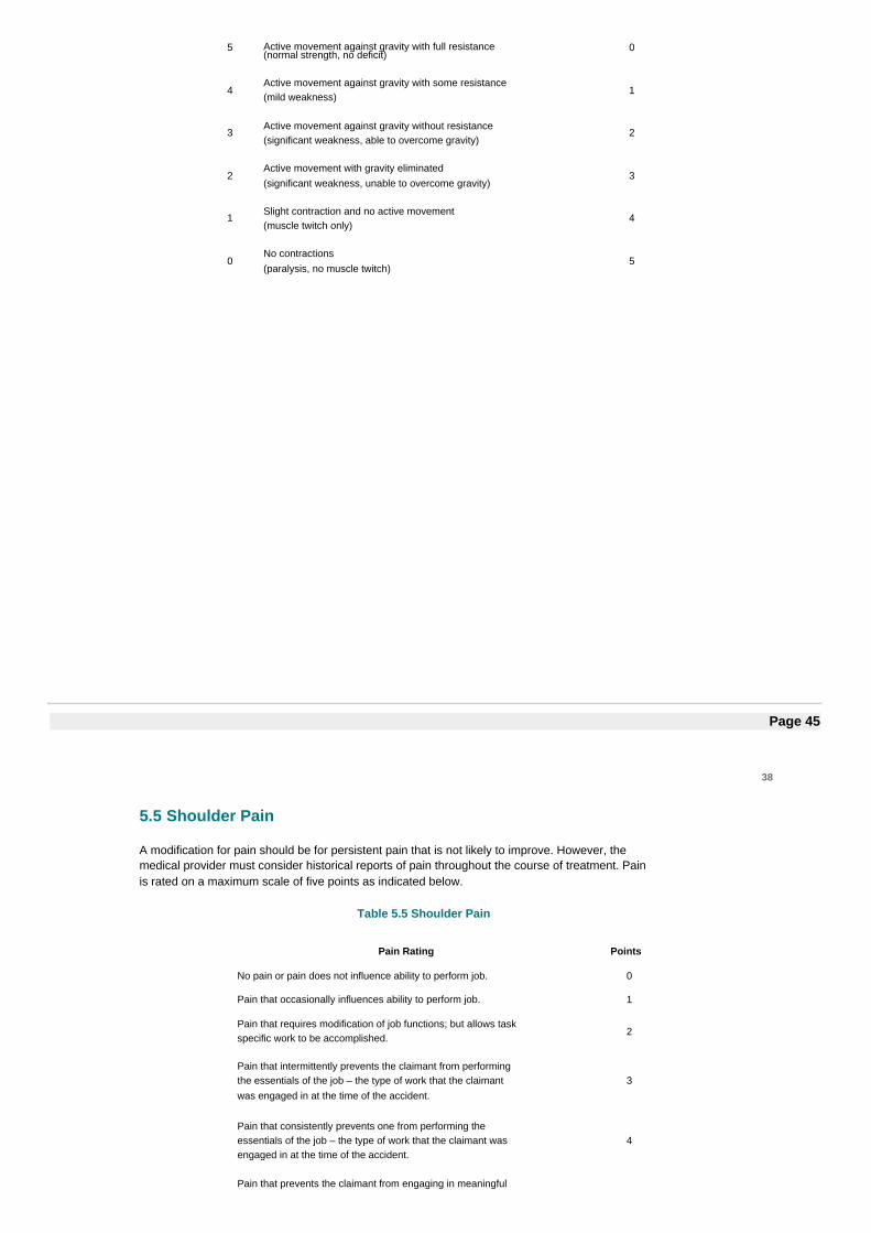
5 Active movement against gravity with full resistance(normal strength, no deficit)
0
4Active movement against gravity with some resistance(mild weakness)
1
3Active movement against gravity without resistance(significant weakness, able to overcome gravity)
2
2Active movement with gravity eliminated(significant weakness, unable to overcome gravity)
3
1Slight contraction and no active movement(muscle twitch only)
4
0No contractions(paralysis, no muscle twitch)
5
Page 45
38
5.5 Shoulder Pain
A modification for pain should be for persistent pain that is not likely to improve. However, themedical provider must consider historical reports of pain throughout the course of treatment. Painis rated on a maximum scale of five points as indicated below.
Table 5.5 Shoulder Pain
Pain Rating Points
No pain or pain does not influence ability to perform job. 0
Pain that occasionally influences ability to perform job. 1
Pain that requires modification of job functions; but allows taskspecific work to be accomplished.
2
Pain that intermittently prevents the claimant from performingthe essentials of the job – the type of work that the claimantwas engaged in at the time of the accident.
3
Pain that consistently prevents one from performing theessentials of the job – the type of work that the claimant wasengaged in at the time of the accident.
4
Pain that prevents the claimant from engaging in meaningful
work, leisure or household duties. 5
5.6 Special Condition Based Impairment
When two or more conditions (as designated by a “•” in Table 5.2) from Category A have beendiagnosed, the impairment value shall start at 10%, to be applied once to include all conditions.The medical provider may then add range of motion, muscle strength and pain, as appropriate,to arrive at the overall shoulder impairment (these measures may only be applied once).
5.7 Amputation - Shoulder
Amputation at the shoulder or elbow joint to any point proximal to the elbow equals 100% loss ofuse of arm.
Page 46
39
Chapter 6: Lower Extremity – Hip and Femur
6.1 Objectives for Determining Impairment for Hip and Femur
The hip joint with surrounding structures enables us to perform daily activities of walking,stair climbing and running. The hip is a ball and socket joint located where the femur meetsthe pelvis. The ball-and socket construct gives a wide range of motion to the hip second onlyto the shoulder.
The objective of any assessment system should be ease of use to obtain a reproducible resultthat accurately reflects the permanent residual physical deficit a claimant suffered as a result ofhis/her injury. To the degree possible, the assessment should be based on objective findingsdetermined by the history and physical examination, as well as the results of any appropriatediagnostic testing.
6.2 Methods Available to Assess Permanent Impairment
Determination of the degree of permanent residual physical deficit should be performed at thetime of maximum medical improvement, the point at which no further healing is expected.Maximum medical improvement should be determined based on the outcome of the clinicalcourse of treatment, the medical provider’s expertise and any further treatment options availableto the claimant. When evaluating the level of permanent residual physical deficit, the medicalprovider should consider expected/normal values and the contralateral extremity whereappropriate. The duration of time from the injury to maximum medical improvement may vary butin most cases is one year from the injury or last surgery.
The severity of the permanent residual physical deficit is not based on the mechanism of injury.It reflects the permanent residual physical deficit at the time of maximum medical improvementand may include physical damage to bone, muscles, cartilage, tendons, nerves, blood vessels,and other tissues.
The severity of the permanent residual physical deficit associated with the injury has been
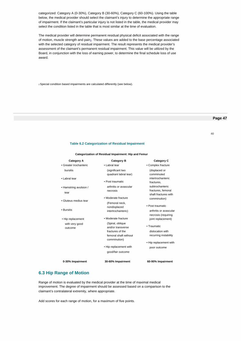
categorized: Category A (0-30%), Category B (30-60%), Category C (60-100%). Using the tablebelow, the medical provider should select the claimant’s injury to determine the appropriate rangeof impairment. If the claimant’s particular injury is not listed in the table, the medical provider mayselect the condition listed in the table that is most similar at the time of evaluation.
The medical provider will determine permanent residual physical deficit associated with the rangeof motion, muscle strength and pain2. These values are added to the base percentage associatedwith the selected category of residual impairment. The result represents the medical provider’sassessment of the claimant’s permanent residual impairment. This value will be utilized by theBoard, in conjunction with the loss of earning power, to determine the final schedule loss of useaward.
2 Special condition based impairments are calculated differently (see below).
Page 47
40
Table 6.2 Categorization of Residual Impairment
Categorization of Residual Impairment: Hip and Femur
Category A Category B Category C• Greater trochanteric
bursitis
• Labral tear
• Hamstring avulsion /
tear
• Gluteus medius tear
• Bursitis
• Hip replacement
with very goodoutcome
• Labral tear
(significant twoquadrant labral tear)
• Post traumatic
arthritis or avascularnecrosis
• Moderate fracture
(Femoral neck,nondisplacedintertrochanteric)
• Moderate fracture
(Spiral, obliqueand/or transversefractures of thefemoral shaft withoutcomminution)
• Hip replacement with
good/fair outcome
• Complex fracture
(displaced orcomminutedintertrochantericfractures,subtrochantericfractures, femoralshaft fractures withcomminution)
• Post traumatic
arthritis or avascularnecrosis (requiringjoint replacement)
• Traumatic
dislocation withrecurring instability
• Hip replacement with
poor outcome
0-30% Impairment 30-60% Impairment 60-90% Impairment
6.3 Hip Range of Motion
Range of motion is evaluated by the medical provider at the time of maximal medicalimprovement. The degree of impairment should be assessed based on a comparison to theclaimant’s contralateral extremity, where appropriate.
Add scores for each range of motion, for a maximum of five points.
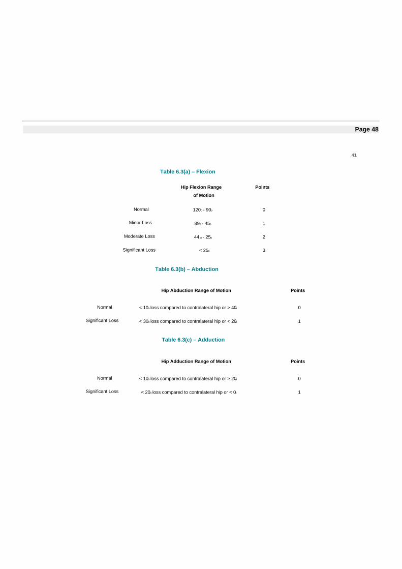
Page 48
41
Table 6.3(a) – Flexion
Hip Flexion Range
of Motion
Points
Normal 120o - 90o 0
Minor Loss 89o - 45o 1
Moderate Loss 44 o - 25o 2
Significant Loss < 25o 3
Table 6.3(b) – Abduction
Hip Abduction Range of Motion Points
Normal < 10o loss compared to contralateral hip or > 40o 0
Significant Loss < 30o loss compared to contralateral hip or < 20o 1
Table 6.3(c) – Adduction
Hip Adduction Range of Motion Points
Normal < 10o loss compared to contralateral hip or > 20o 0
Significant Loss < 20o loss compared to contralateral hip or < 0o 1
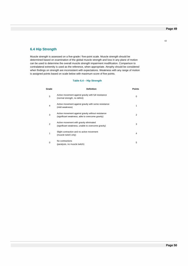
Page 49
42
6.4 Hip Strength
Muscle strength is assessed on a five-grade / five-point scale. Muscle strength should bedetermined based on examination of the global muscle strength and loss in any plane of motioncan be used to determine the overall muscle strength impairment modification. Comparison tocontralateral extremity is used as the reference, when appropriate. Atrophy should be consideredwhen findings on strength are inconsistent with expectations. Weakness with any range of motionis assigned points based on scale below with maximum score of five points.
Table 6.4 – Hip Strength
Grade Definition Points
5Active movement against gravity with full resistance(normal strength, no deficit)
0
4Active movement against gravity with some resistance(mild weakness)
1
3Active movement against gravity without resistance(significant weakness, able to overcome gravity)
2
2Active movement with gravity eliminated(significant weakness, unable to overcome gravity)
3
1Slight contraction and no active movement(muscle twitch only)
4
0No contractions(paralysis, no muscle twitch)
5
Page 50
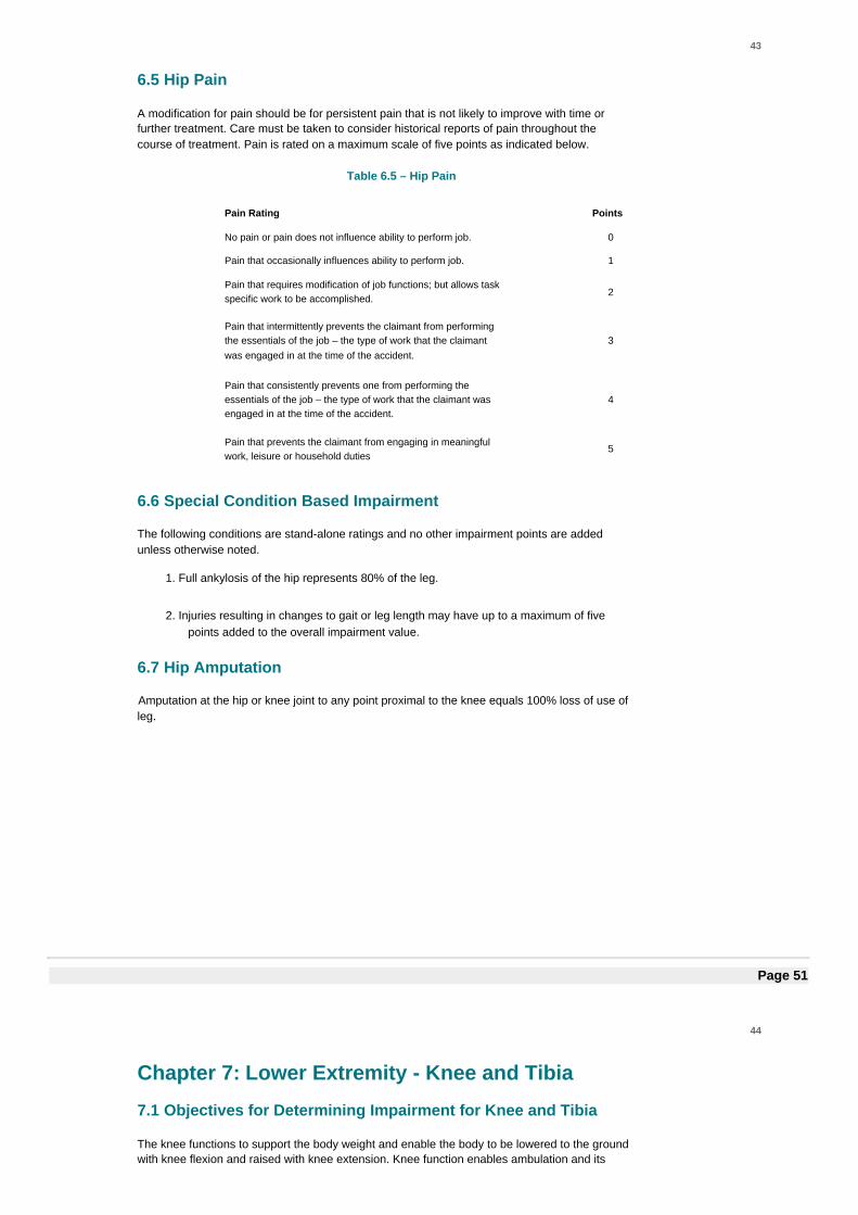
43
6.5 Hip Pain
A modification for pain should be for persistent pain that is not likely to improve with time orfurther treatment. Care must be taken to consider historical reports of pain throughout thecourse of treatment. Pain is rated on a maximum scale of five points as indicated below.
Table 6.5 – Hip Pain
Pain Rating Points
No pain or pain does not influence ability to perform job. 0
Pain that occasionally influences ability to perform job. 1
Pain that requires modification of job functions; but allows taskspecific work to be accomplished.
2
Pain that intermittently prevents the claimant from performingthe essentials of the job – the type of work that the claimantwas engaged in at the time of the accident.
3
Pain that consistently prevents one from performing theessentials of the job – the type of work that the claimant wasengaged in at the time of the accident.
4
Pain that prevents the claimant from engaging in meaningfulwork, leisure or household duties
5
6.6 Special Condition Based Impairment
The following conditions are stand-alone ratings and no other impairment points are addedunless otherwise noted.
1. Full ankylosis of the hip represents 80% of the leg.
2. Injuries resulting in changes to gait or leg length may have up to a maximum of fivepoints added to the overall impairment value.
6.7 Hip Amputation
Amputation at the hip or knee joint to any point proximal to the knee equals 100% loss of use ofleg.
Page 51
44
Chapter 7: Lower Extremity - Knee and Tibia
7.1 Objectives for Determining Impairment for Knee and Tibia
The knee functions to support the body weight and enable the body to be lowered to the groundwith knee flexion and raised with knee extension. Knee function enables ambulation and its
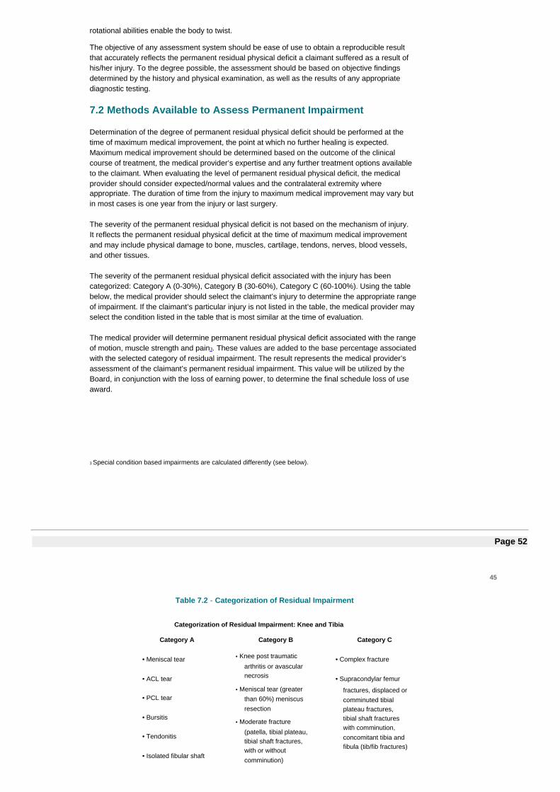
rotational abilities enable the body to twist.
The objective of any assessment system should be ease of use to obtain a reproducible resultthat accurately reflects the permanent residual physical deficit a claimant suffered as a result ofhis/her injury. To the degree possible, the assessment should be based on objective findingsdetermined by the history and physical examination, as well as the results of any appropriatediagnostic testing.
7.2 Methods Available to Assess Permanent Impairment
Determination of the degree of permanent residual physical deficit should be performed at thetime of maximum medical improvement, the point at which no further healing is expected.Maximum medical improvement should be determined based on the outcome of the clinicalcourse of treatment, the medical provider’s expertise and any further treatment options availableto the claimant. When evaluating the level of permanent residual physical deficit, the medicalprovider should consider expected/normal values and the contralateral extremity whereappropriate. The duration of time from the injury to maximum medical improvement may vary butin most cases is one year from the injury or last surgery.
The severity of the permanent residual physical deficit is not based on the mechanism of injury.It reflects the permanent residual physical deficit at the time of maximum medical improvementand may include physical damage to bone, muscles, cartilage, tendons, nerves, blood vessels,and other tissues.
The severity of the permanent residual physical deficit associated with the injury has beencategorized: Category A (0-30%), Category B (30-60%), Category C (60-100%). Using the tablebelow, the medical provider should select the claimant’s injury to determine the appropriate rangeof impairment. If the claimant’s particular injury is not listed in the table, the medical provider mayselect the condition listed in the table that is most similar at the time of evaluation.
The medical provider will determine permanent residual physical deficit associated with the rangeof motion, muscle strength and pain3. These values are added to the base percentage associatedwith the selected category of residual impairment. The result represents the medical provider’sassessment of the claimant’s permanent residual impairment. This value will be utilized by theBoard, in conjunction with the loss of earning power, to determine the final schedule loss of useaward.
3 Special condition based impairments are calculated differently (see below).
Page 52
45
Table 7.2 - Categorization of Residual Impairment
Categorization of Residual Impairment: Knee and Tibia
Category A Category B Category C
• Meniscal tear
• ACL tear
• PCL tear
• Bursitis
• Tendonitis
• Isolated fibular shaft
• Knee post traumatic
arthritis or avascularnecrosis
• Meniscal tear (greaterthan 60%) meniscusresection
• Moderate fracture
(patella, tibial plateau,tibial shaft fractures,with or withoutcomminution)
• Complex fracture
• Supracondylar femur
fractures, displaced orcomminuted tibialplateau fractures,tibial shaft fractureswith comminution,concomitant tibia andfibula (tib/fib fractures)
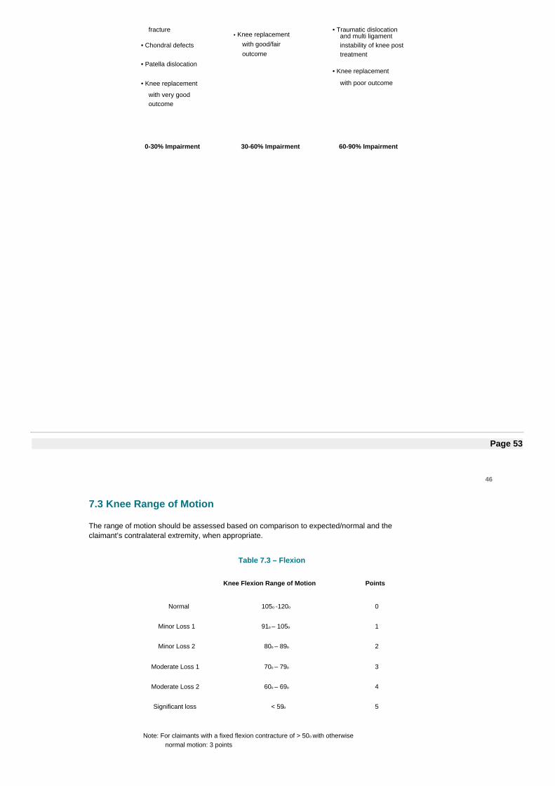
fracture
• Chondral defects
• Patella dislocation
• Knee replacement
with very goodoutcome
• Knee replacementwith good/fairoutcome
• Traumatic dislocationand multi ligamentinstability of knee posttreatment
• Knee replacement
with poor outcome
0-30% Impairment 30-60% Impairment 60-90% Impairment
Page 53
46
7.3 Knee Range of Motion
The range of motion should be assessed based on comparison to expected/normal and theclaimant’s contralateral extremity, when appropriate.
Table 7.3 – Flexion
Knee Flexion Range of Motion Points
Normal 105o -120o 0
Minor Loss 1 91o – 105o 1
Minor Loss 2 80o – 89o 2
Moderate Loss 1 70o – 79o 3
Moderate Loss 2 60o – 69o 4
Significant loss < 59o 5
Note: For claimants with a fixed flexion contracture of > 50o with otherwisenormal motion: 3 points
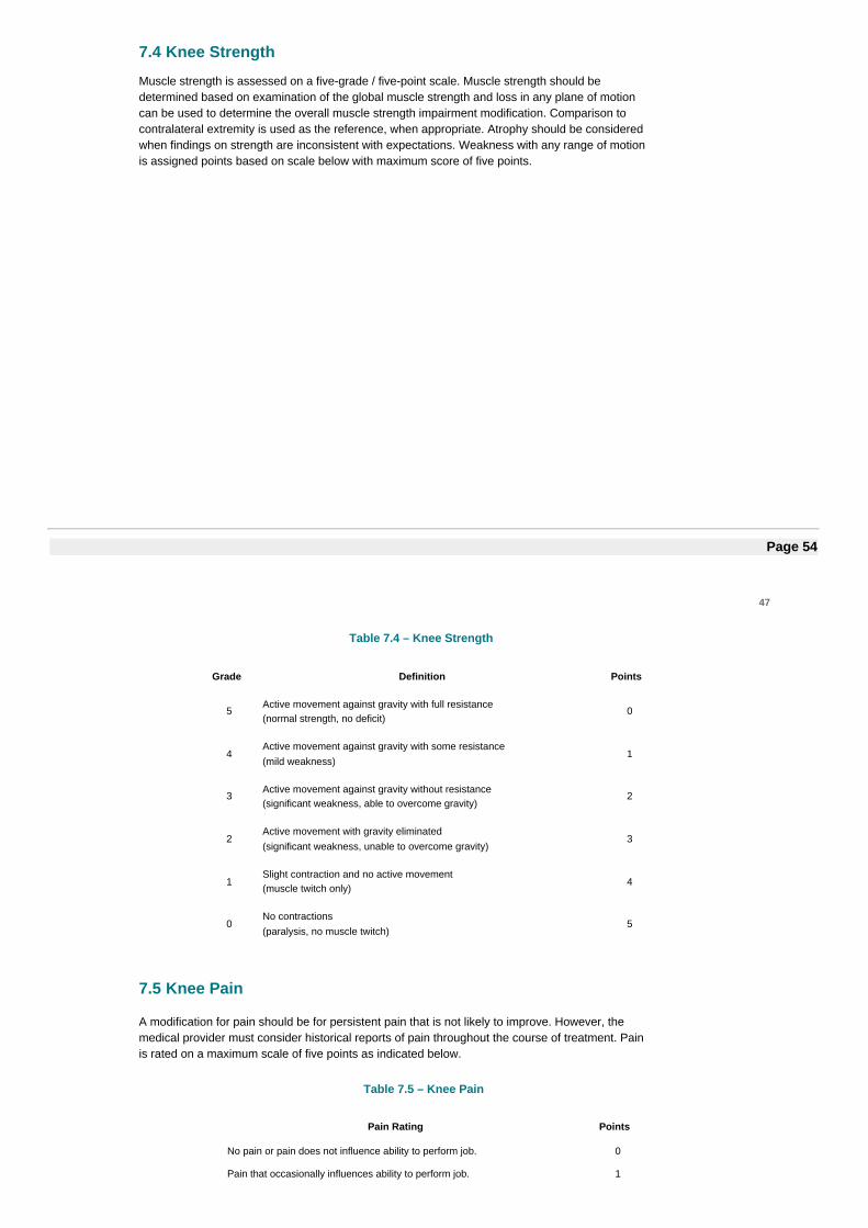
7.4 Knee Strength
Muscle strength is assessed on a five-grade / five-point scale. Muscle strength should bedetermined based on examination of the global muscle strength and loss in any plane of motioncan be used to determine the overall muscle strength impairment modification. Comparison tocontralateral extremity is used as the reference, when appropriate. Atrophy should be consideredwhen findings on strength are inconsistent with expectations. Weakness with any range of motionis assigned points based on scale below with maximum score of five points.
Page 54
47
Table 7.4 – Knee Strength
Grade Definition Points
5Active movement against gravity with full resistance(normal strength, no deficit)
0
4Active movement against gravity with some resistance(mild weakness)
1
3Active movement against gravity without resistance(significant weakness, able to overcome gravity)
2
2Active movement with gravity eliminated(significant weakness, unable to overcome gravity)
3
1Slight contraction and no active movement(muscle twitch only)
4
0No contractions(paralysis, no muscle twitch)
5
7.5 Knee Pain
A modification for pain should be for persistent pain that is not likely to improve. However, themedical provider must consider historical reports of pain throughout the course of treatment. Painis rated on a maximum scale of five points as indicated below.
Table 7.5 – Knee Pain
Pain Rating Points
No pain or pain does not influence ability to perform job. 0
Pain that occasionally influences ability to perform job. 1
Pain that requires modification of job functions; but allows taskspecific work to be accomplished.
2
Pain that intermittently prevents the claimant from performingthe essentials of the job – the type of work that the claimantwas engaged in at the time of the accident.
3
Pain that consistently prevents one from performing theessentials of the job – the type of work that the claimant wasengaged in at the time of the accident.
4
Pain that prevents the claimant from engaging in meaningfulwork, leisure or household duties.
5
Page 55
48
7.6 Special Condition Based Impairment
The following conditions are stand-alone ratings and no other impairment points are addedunless otherwise noted.
1. Instability of the knee with or without surgery, resulting from multiple causes includingligament laxity or others, is given up to an additional 5% impairment depending on severity.
2. Recurrent patella dislocation with or without surgery: Compare with the contralateral knee.If surgery was performed, evaluate after a minimum of twelve months after surgery. Insymptomatic cases without surgery, with objective findings (see New York Knee InjuryMedical Treatment Guidelines, section D.9), award a 15% impairment of the leg if nomuscle atrophy is present and 30% impairment of the leg if there is muscle atrophy.
7.7 Knee Amputation
Amputation at or above the knee joint equals 100% impairment of the leg.
Amputation at six inches or more below the knee joint equals 95% impairment of the leg.
For amputation of the lower extremity with a non-functional prosthesis with residual symptomsand complications, such as neuroma, phantom pain and persistent ulcers, consider classification.
Page 56
49
Chapter 8: Lower Extremity - Ankle and Foot
8.1 Objectives for Determining Impairment for Ankle and Foot
The ankle is a mortise joint formed by the distal tibia and fibula that provides lower limb stabilityand facilitates ambulation through movement of the foot. The foot acts during the gait cycle asboth a shock absorber during heel strike and rigid platform during toe off. Dorsiflexion of the ankleis required to climb and descend stairs while plantarflexion serves to elevate the body and depresspedals to operate vehicles or machinery.
The objective of any assessment system should be ease of use to obtain a reproducible resultthat accurately reflects the permanent residual physical deficit a worker suffered as a result ofhis/her injury. To the degree possible, the assessment should be based on objective findingsdetermined by the history and physical examination, as well as the results of any appropriatediagnostic testing.
8.2 Methods Available to Assess Permanent Impairment
Determination of the degree of permanent residual physical deficit should be performed at thetime of maximum medical improvement, the point at which no further healing is expected.Maximum medical improvement should be determined based on the outcome of the clinicalcourse of treatment, the medical provider’s expertise and any further treatment options availableto the claimant. When evaluating the level of permanent residual physical deficit, the medicalprovider should consider expected/normal values and the contralateral extremity whereappropriate. The duration of time from the injury to maximum medical improvement may vary butin most cases is one year from the injury or last surgery.
The severity of the permanent residual physical deficit is not based on the mechanism of injury.It reflects the permanent residual physical deficit at the time of maximum medical improvementand may include physical damage to bone, muscles, cartilage, tendons, nerves, blood vessels,and other tissues.
The severity of the permanent residual physical deficit associated with the injury has beencategorized: Category A (0-30%), Category B (30-60%), Category C (60-100%). Using the tablebelow, the medical provider should select the claimant’s injury to determine the appropriate rangeof impairment. If the claimant’s particular injury is not listed in the table, the medical provider mayselect the condition listed in the table that is most similar at the time of evaluation.
The medical provider will determine permanent residual physical deficit associated with the rangeof motion, muscle strength and pain4. These values are added to the base percentage associatedwith the selected category of residual impairment. The result represents the medical provider’sassessment of the claimant’s permanent residual impairment. This value will be utilized by theBoard, in conjunction with the loss of earning power, to determine the final schedule loss of useaward.
4 Special condition based impairments are calculated differently (see below).
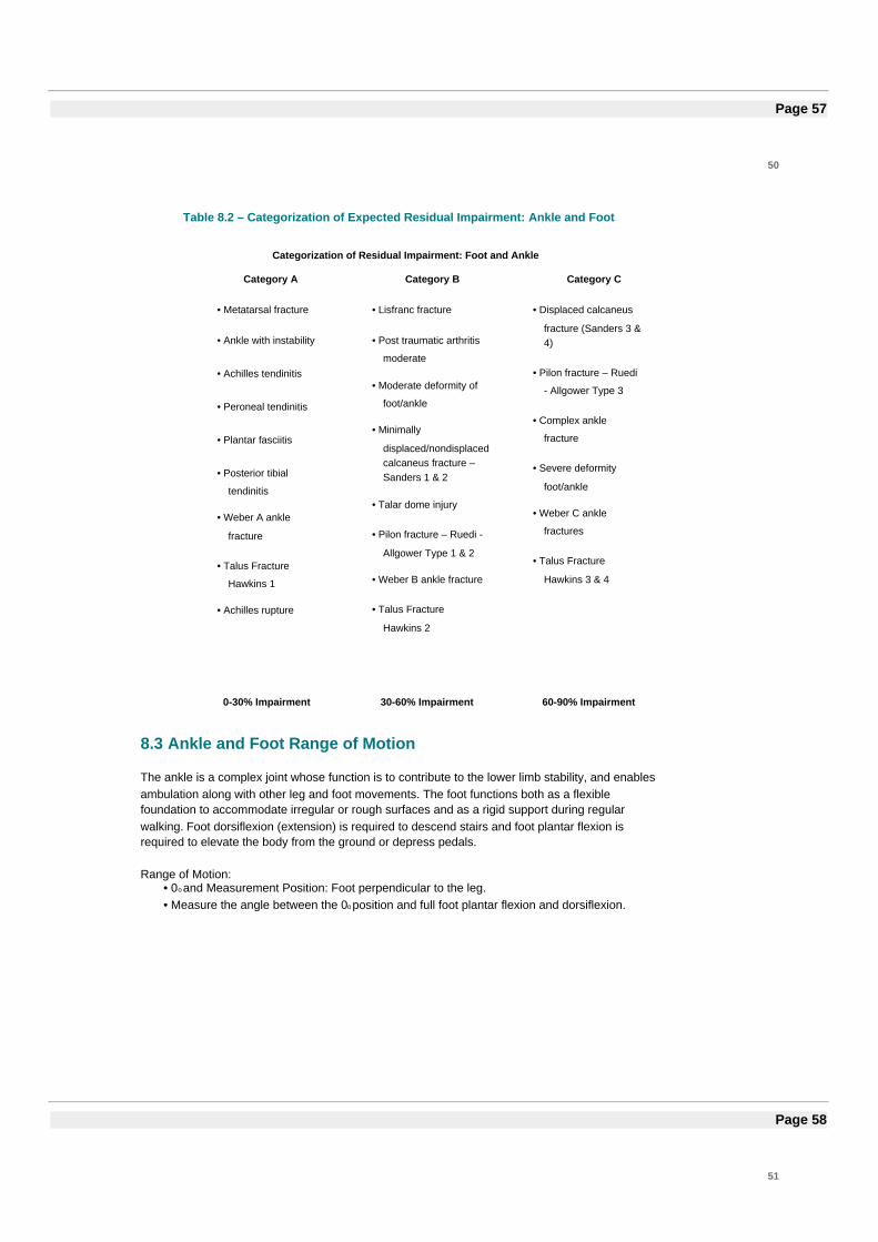
Page 57
50
Table 8.2 – Categorization of Expected Residual Impairment: Ankle and Foot
Categorization of Residual Impairment: Foot and Ankle
Category A Category B Category C
• Metatarsal fracture
• Ankle with instability
• Achilles tendinitis
• Peroneal tendinitis
• Plantar fasciitis
• Posterior tibial
tendinitis
• Weber A ankle
fracture
• Talus Fracture
Hawkins 1
• Achilles rupture
• Lisfranc fracture
• Post traumatic arthritis
moderate
• Moderate deformity of
foot/ankle
• Minimally
displaced/nondisplacedcalcaneus fracture –Sanders 1 & 2
• Talar dome injury
• Pilon fracture – Ruedi -
Allgower Type 1 & 2
• Weber B ankle fracture
• Talus Fracture
Hawkins 2
• Displaced calcaneus
fracture (Sanders 3 &4)
• Pilon fracture – Ruedi
- Allgower Type 3
• Complex ankle
fracture
• Severe deformity
foot/ankle
• Weber C ankle
fractures
• Talus Fracture
Hawkins 3 & 4
0-30% Impairment 30-60% Impairment 60-90% Impairment
8.3 Ankle and Foot Range of Motion
The ankle is a complex joint whose function is to contribute to the lower limb stability, and enablesambulation along with other leg and foot movements. The foot functions both as a flexiblefoundation to accommodate irregular or rough surfaces and as a rigid support during regularwalking. Foot dorsiflexion (extension) is required to descend stairs and foot plantar flexion isrequired to elevate the body from the ground or depress pedals.
Range of Motion:• 0o and Measurement Position: Foot perpendicular to the leg.• Measure the angle between the 0o position and full foot plantar flexion and dorsiflexion.
Page 58
51
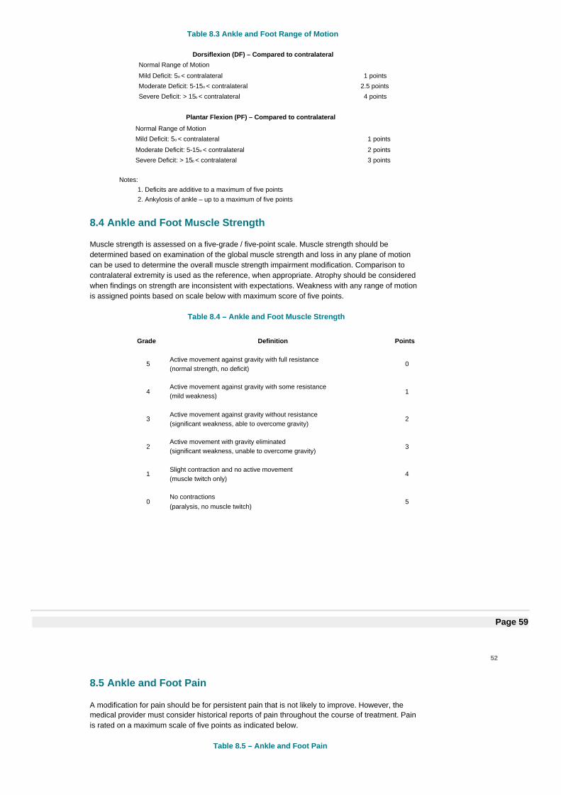
Table 8.3 Ankle and Foot Range of Motion
Dorsiflexion (DF) – Compared to contralateralNormal Range of Motion
Mild Deficit: 5o < contralateral 1 points
Moderate Deficit: 5-15o < contralateral 2.5 points
Severe Deficit: > 15o < contralateral 4 points
Plantar Flexion (PF) – Compared to contralateral
Normal Range of Motion
Mild Deficit: 5o < contralateral 1 points
Moderate Deficit: 5-15o < contralateral 2 points
Severe Deficit: > 15o < contralateral 3 points
Notes:1. Deficits are additive to a maximum of five points2. Ankylosis of ankle – up to a maximum of five points
8.4 Ankle and Foot Muscle Strength
Muscle strength is assessed on a five-grade / five-point scale. Muscle strength should bedetermined based on examination of the global muscle strength and loss in any plane of motioncan be used to determine the overall muscle strength impairment modification. Comparison tocontralateral extremity is used as the reference, when appropriate. Atrophy should be consideredwhen findings on strength are inconsistent with expectations. Weakness with any range of motionis assigned points based on scale below with maximum score of five points.
Table 8.4 – Ankle and Foot Muscle Strength
Grade Definition Points
5Active movement against gravity with full resistance(normal strength, no deficit)
0
4Active movement against gravity with some resistance(mild weakness)
1
3Active movement against gravity without resistance(significant weakness, able to overcome gravity)
2
2Active movement with gravity eliminated(significant weakness, unable to overcome gravity)
3
1Slight contraction and no active movement(muscle twitch only)
4
0No contractions(paralysis, no muscle twitch)
5
Page 59
52
8.5 Ankle and Foot Pain
A modification for pain should be for persistent pain that is not likely to improve. However, themedical provider must consider historical reports of pain throughout the course of treatment. Painis rated on a maximum scale of five points as indicated below.
Table 8.5 – Ankle and Foot Pain
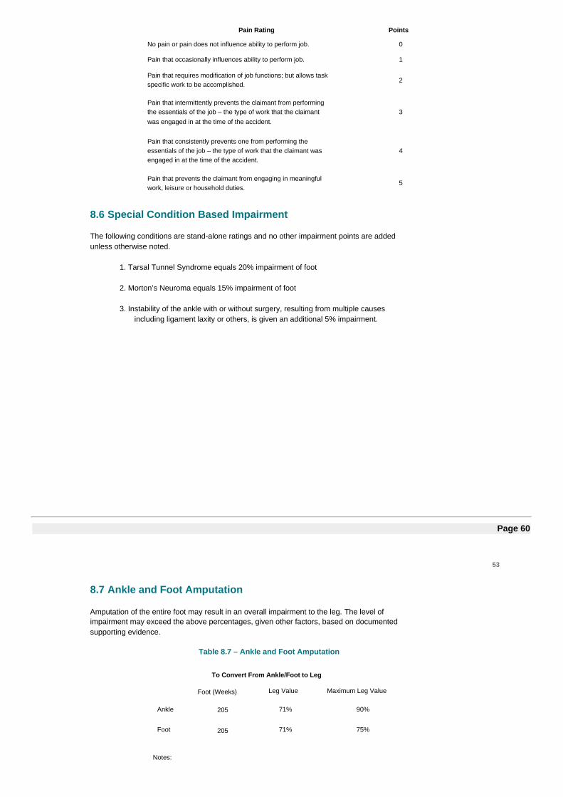
Pain Rating Points
No pain or pain does not influence ability to perform job. 0
Pain that occasionally influences ability to perform job. 1
Pain that requires modification of job functions; but allows taskspecific work to be accomplished.
2
Pain that intermittently prevents the claimant from performingthe essentials of the job – the type of work that the claimantwas engaged in at the time of the accident.
3
Pain that consistently prevents one from performing theessentials of the job – the type of work that the claimant wasengaged in at the time of the accident.
4
Pain that prevents the claimant from engaging in meaningfulwork, leisure or household duties.
5
8.6 Special Condition Based Impairment
The following conditions are stand-alone ratings and no other impairment points are addedunless otherwise noted.
1. Tarsal Tunnel Syndrome equals 20% impairment of foot
2. Morton’s Neuroma equals 15% impairment of foot
3. Instability of the ankle with or without surgery, resulting from multiple causesincluding ligament laxity or others, is given an additional 5% impairment.
Page 60
53
8.7 Ankle and Foot Amputation
Amputation of the entire foot may result in an overall impairment to the leg. The level ofimpairment may exceed the above percentages, given other factors, based on documentedsupporting evidence.
Table 8.7 – Ankle and Foot Amputation
To Convert From Ankle/Foot to Leg
Foot (Weeks) Leg Value Maximum Leg Value
Ankle 205 71% 90%
Foot 205 71% 75%
Notes:
1. At level of ankle = 90% impairment of leg2. Trans metatarsal amputation = 75% impairment of leg
Page 61
54
Chapter 9: Lower Extremity: Great and Lesser Toes
9.1 Objectives for Determining Impairment for Great and Lesser Toes
The toes, particularly the great toe, provides stability during regular walking and facilitates stance.They also provide support when standing on the toes for elevated reaching. When more thanone toe is injured, it may be considered as an overall impact on the foot.
The objective of any assessment system should be ease of use to obtain a reproducible resultthat accurately reflects the permanent residual physical deficit a claimant suffered as a result ofhis/her injury. To the degree possible, the assessment should be based on objective findingsdetermined by the history and physical examination, as well as the results of any appropriatediagnostic testing.
9.2 Methods Available to Assess Permanent Impairment
Determination of the degree of permanent residual physical deficit should be performed at thetime of maximum medical improvement, the point at which no further healing is expected.Maximum medical improvement should be determined based on the outcome of the clinicalcourse of treatment, the medical provider’s expertise and any further treatment options availableto the claimant. When evaluating the level of permanent residual physical deficit, the medicalprovider should consider expected/normal values and the contralateral extremity whereappropriate. The duration of time from the injury to maximum medical improvement may vary butin most cases is one year from the injury or last surgery.
The severity of the permanent residual physical deficit is not based on the mechanism of injury.
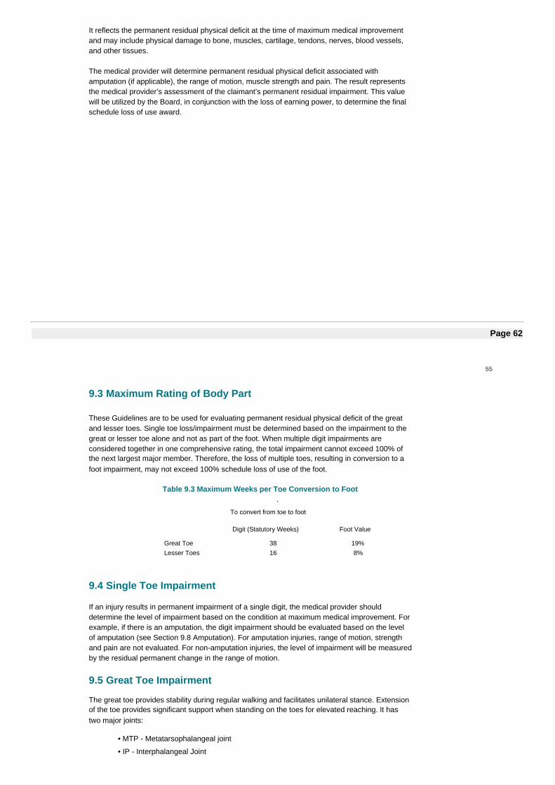
It reflects the permanent residual physical deficit at the time of maximum medical improvementand may include physical damage to bone, muscles, cartilage, tendons, nerves, blood vessels,and other tissues.
The medical provider will determine permanent residual physical deficit associated withamputation (if applicable), the range of motion, muscle strength and pain. The result representsthe medical provider’s assessment of the claimant’s permanent residual impairment. This valuewill be utilized by the Board, in conjunction with the loss of earning power, to determine the finalschedule loss of use award.
Page 62
55
9.3 Maximum Rating of Body Part
These Guidelines are to be used for evaluating permanent residual physical deficit of the greatand lesser toes. Single toe loss/impairment must be determined based on the impairment to thegreat or lesser toe alone and not as part of the foot. When multiple digit impairments areconsidered together in one comprehensive rating, the total impairment cannot exceed 100% ofthe next largest major member. Therefore, the loss of multiple toes, resulting in conversion to afoot impairment, may not exceed 100% schedule loss of use of the foot.
Table 9.3 Maximum Weeks per Toe Conversion to Foot.
To convert from toe to foot
Digit (Statutory Weeks) Foot Value
Great Toe 38 19%Lesser Toes 16 8%
9.4 Single Toe Impairment
If an injury results in permanent impairment of a single digit, the medical provider shoulddetermine the level of impairment based on the condition at maximum medical improvement. Forexample, if there is an amputation, the digit impairment should be evaluated based on the levelof amputation (see Section 9.8 Amputation). For amputation injuries, range of motion, strengthand pain are not evaluated. For non-amputation injuries, the level of impairment will be measuredby the residual permanent change in the range of motion.
9.5 Great Toe Impairment
The great toe provides stability during regular walking and facilitates unilateral stance. Extensionof the toe provides significant support when standing on the toes for elevated reaching. It hastwo major joints:
• MTP - Metatarsophalangeal joint
• IP - Interphalangeal Joint
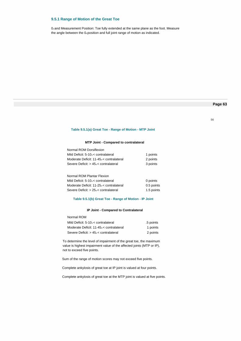
9.5.1 Range of Motion of the Great Toe
0o and Measurement Position: Toe fully extended at the same plane as the foot. Measurethe angle between the 0o position and full joint range of motion as indicated.
Page 63
56
Table 9.5.1(a) Great Toe - Range of Motion - MTP Joint
MTP Joint - Compared to contralateral
Normal ROM DorsiflexionMild Deficit: 5-10o < contralateral 1 pointsModerate Deficit: 11-45o < contralateral 2 pointsSevere Deficit: > 45o < contralateral 3 points
Normal ROM Plantar FlexionMild Deficit: 5-10o < contralateral 0 pointsModerate Deficit: 11-25o < contralateral 0.5 pointsSevere Deficit: > 25o < contralateral 1.5 points
Table 9.5.1(b) Great Toe - Range of Motion - IP Joint
IP Joint - Compared to Contralateral
Normal ROM
Mild Deficit: 5-10o < contralateral .5 pointsModerate Deficit: 11-45o < contralateral 1 points
Severe Deficit: > 45o < contralateral 2 points
To determine the level of impairment of the great toe, the maximumvalue is highest impairment value of the affected joints (MTP or IP),not to exceed five points.
Sum of the range of motion scores may not exceed five points.
Complete ankylosis of great toe at IP joint is valued at four points.
Complete ankylosis of great toe at the MTP joint is valued at five points.
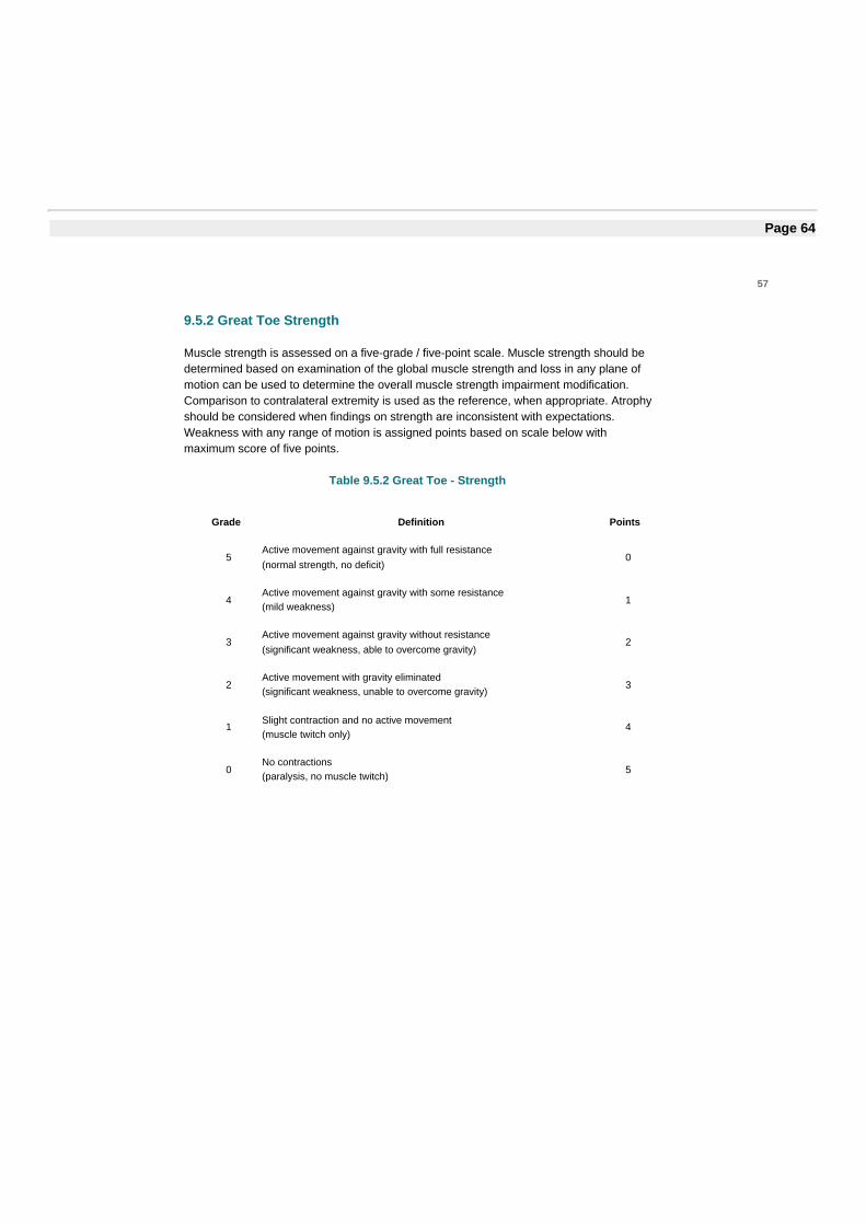
Page 64
57
9.5.2 Great Toe Strength
Muscle strength is assessed on a five-grade / five-point scale. Muscle strength should bedetermined based on examination of the global muscle strength and loss in any plane ofmotion can be used to determine the overall muscle strength impairment modification.Comparison to contralateral extremity is used as the reference, when appropriate. Atrophyshould be considered when findings on strength are inconsistent with expectations.Weakness with any range of motion is assigned points based on scale below withmaximum score of five points.
Table 9.5.2 Great Toe - Strength
Grade Definition Points
5Active movement against gravity with full resistance(normal strength, no deficit)
0
4Active movement against gravity with some resistance(mild weakness)
1
3Active movement against gravity without resistance(significant weakness, able to overcome gravity)
2
2Active movement with gravity eliminated(significant weakness, unable to overcome gravity)
3
1Slight contraction and no active movement(muscle twitch only)
4
0No contractions(paralysis, no muscle twitch)
5
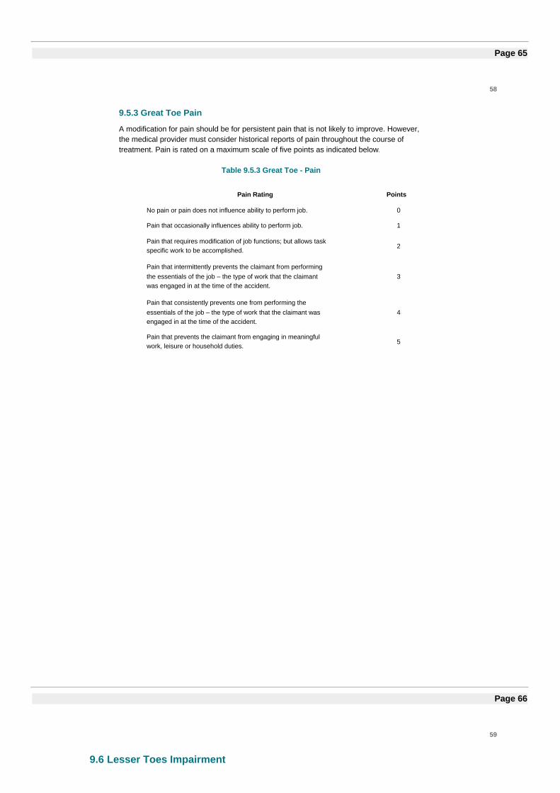
Page 65
58
9.5.3 Great Toe Pain
A modification for pain should be for persistent pain that is not likely to improve. However,the medical provider must consider historical reports of pain throughout the course oftreatment. Pain is rated on a maximum scale of five points as indicated below.
Table 9.5.3 Great Toe - Pain
Pain Rating Points
No pain or pain does not influence ability to perform job. 0
Pain that occasionally influences ability to perform job. 1
Pain that requires modification of job functions; but allows taskspecific work to be accomplished.
2
Pain that intermittently prevents the claimant from performingthe essentials of the job – the type of work that the claimantwas engaged in at the time of the accident.
3
Pain that consistently prevents one from performing theessentials of the job – the type of work that the claimant wasengaged in at the time of the accident.
4
Pain that prevents the claimant from engaging in meaningfulwork, leisure or household duties.
5
Page 66
59
9.6 Lesser Toes Impairment
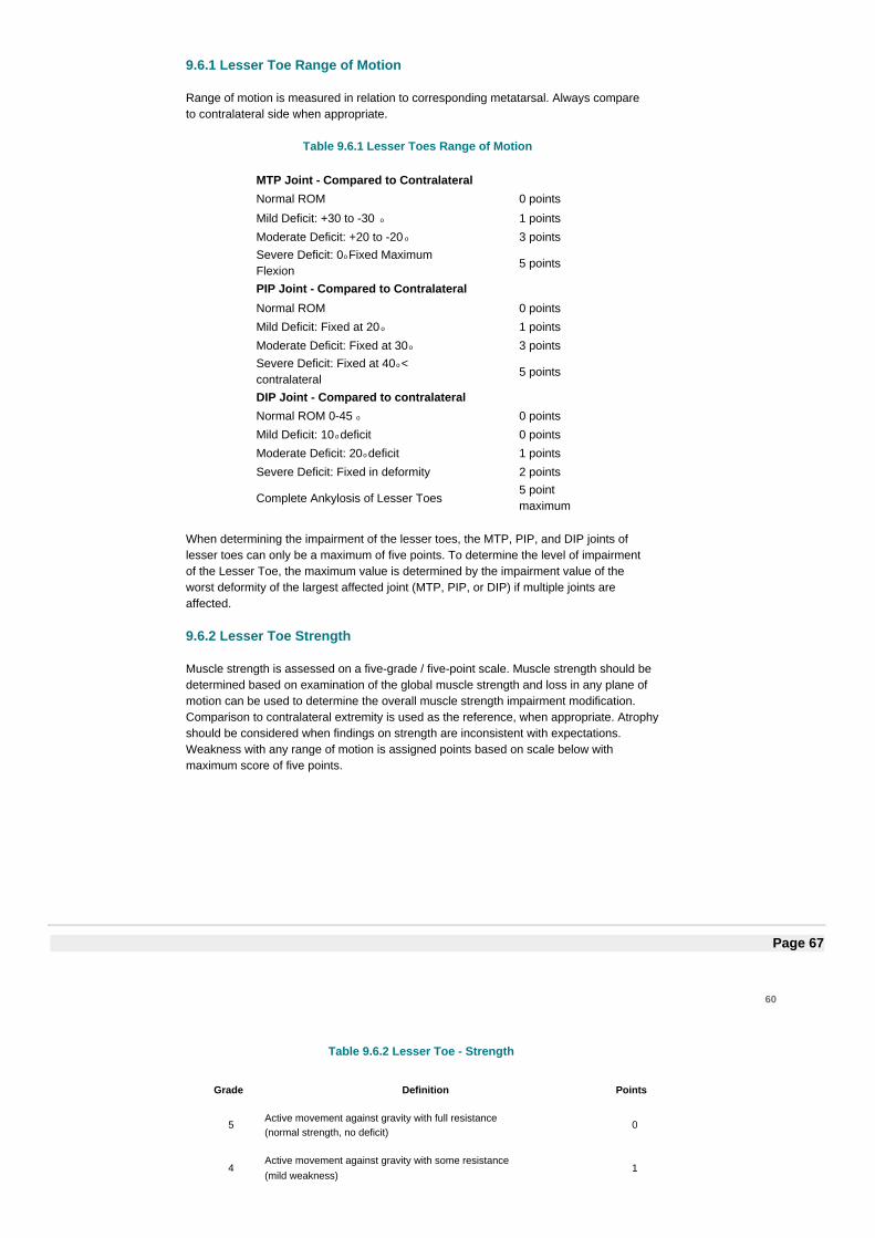
9.6.1 Lesser Toe Range of Motion
Range of motion is measured in relation to corresponding metatarsal. Always compareto contralateral side when appropriate.
Table 9.6.1 Lesser Toes Range of Motion
MTP Joint - Compared to ContralateralNormal ROM 0 points
Mild Deficit: +30 to -30 o 1 points
Moderate Deficit: +20 to -20 o 3 pointsSevere Deficit: 0o Fixed MaximumFlexion
5 points
PIP Joint - Compared to ContralateralNormal ROM 0 points
Mild Deficit: Fixed at 20 o 1 points
Moderate Deficit: Fixed at 30o 3 pointsSevere Deficit: Fixed at 40o <contralateral
5 points
DIP Joint - Compared to contralateralNormal ROM 0-45 o 0 points
Mild Deficit: 10 o deficit 0 points
Moderate Deficit: 20o deficit 1 points
Severe Deficit: Fixed in deformity 2 points
Complete Ankylosis of Lesser Toes5 pointmaximum
When determining the impairment of the lesser toes, the MTP, PIP, and DIP joints oflesser toes can only be a maximum of five points. To determine the level of impairmentof the Lesser Toe, the maximum value is determined by the impairment value of theworst deformity of the largest affected joint (MTP, PIP, or DIP) if multiple joints areaffected.
9.6.2 Lesser Toe Strength
Muscle strength is assessed on a five-grade / five-point scale. Muscle strength should bedetermined based on examination of the global muscle strength and loss in any plane ofmotion can be used to determine the overall muscle strength impairment modification.Comparison to contralateral extremity is used as the reference, when appropriate. Atrophyshould be considered when findings on strength are inconsistent with expectations.Weakness with any range of motion is assigned points based on scale below withmaximum score of five points.
Page 67
60
Table 9.6.2 Lesser Toe - Strength
Grade Definition Points
5Active movement against gravity with full resistance(normal strength, no deficit)
0
4Active movement against gravity with some resistance(mild weakness)
1
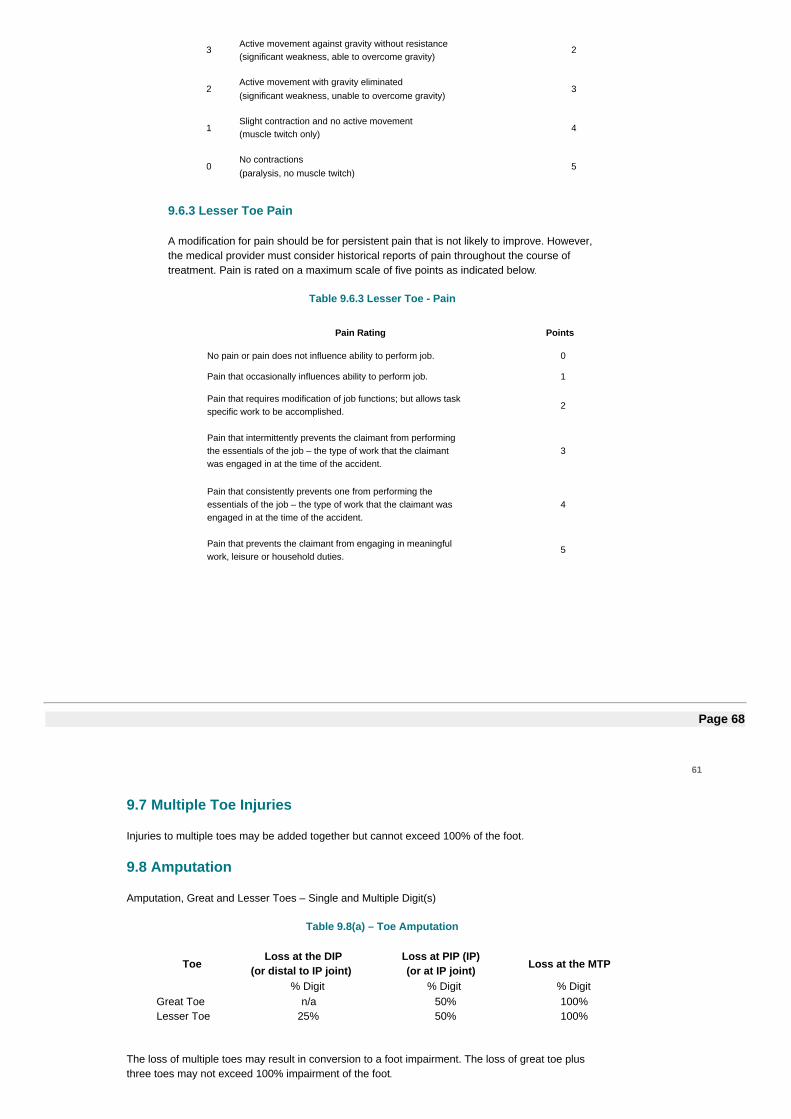
3Active movement against gravity without resistance(significant weakness, able to overcome gravity)
2
2Active movement with gravity eliminated(significant weakness, unable to overcome gravity)
3
1Slight contraction and no active movement(muscle twitch only)
4
0No contractions(paralysis, no muscle twitch)
5
9.6.3 Lesser Toe Pain
A modification for pain should be for persistent pain that is not likely to improve. However,the medical provider must consider historical reports of pain throughout the course oftreatment. Pain is rated on a maximum scale of five points as indicated below.
Table 9.6.3 Lesser Toe - Pain
Pain Rating Points
No pain or pain does not influence ability to perform job. 0
Pain that occasionally influences ability to perform job. 1
Pain that requires modification of job functions; but allows taskspecific work to be accomplished.
2
Pain that intermittently prevents the claimant from performingthe essentials of the job – the type of work that the claimantwas engaged in at the time of the accident.
3
Pain that consistently prevents one from performing theessentials of the job – the type of work that the claimant wasengaged in at the time of the accident.
4
Pain that prevents the claimant from engaging in meaningfulwork, leisure or household duties.
5
Page 68
61
9.7 Multiple Toe Injuries
Injuries to multiple toes may be added together but cannot exceed 100% of the foot.
9.8 Amputation
Amputation, Great and Lesser Toes – Single and Multiple Digit(s)
Table 9.8(a) – Toe Amputation
ToeLoss at the DIP
(or distal to IP joint)Loss at PIP (IP)(or at IP joint) Loss at the MTP
% Digit % Digit % DigitGreat Toe n/a 50% 100%Lesser Toe 25% 50% 100%
The loss of multiple toes may result in conversion to a foot impairment. The loss of great toe plusthree toes may not exceed 100% impairment of the foot.
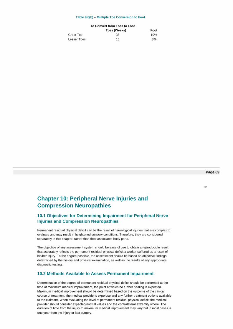
Table 9.8(b) – Multiple Toe Conversion to Foot
To Convert from Toes to FootToes (Weeks) Foot
Great Toe 38 19%Lesser Toes 16 8%
Page 69
62
Chapter 10: Peripheral Nerve Injuries andCompression Neuropathies
10.1 Objectives for Determining Impairment for Peripheral NerveInjuries and Compression Neuropathies
Permanent residual physical deficit can be the result of neurological injuries that are complex toevaluate and may result in heightened sensory conditions. Therefore, they are consideredseparately in this chapter, rather than their associated body parts.
The objective of any assessment system should be ease of use to obtain a reproducible resultthat accurately reflects the permanent residual physical deficit a worker suffered as a result ofhis/her injury. To the degree possible, the assessment should be based on objective findingsdetermined by the history and physical examination, as well as the results of any appropriatediagnostic testing.
10.2 Methods Available to Assess Permanent Impairment
Determination of the degree of permanent residual physical deficit should be performed at thetime of maximum medical improvement, the point at which no further healing is expected.Maximum medical improvement should be determined based on the outcome of the clinicalcourse of treatment, the medical provider’s expertise and any further treatment options availableto the claimant. When evaluating the level of permanent residual physical deficit, the medicalprovider should consider expected/normal values and the contralateral extremity where. Theduration of time from the injury to maximum medical improvement may vary but in most cases isone year from the injury or last surgery.
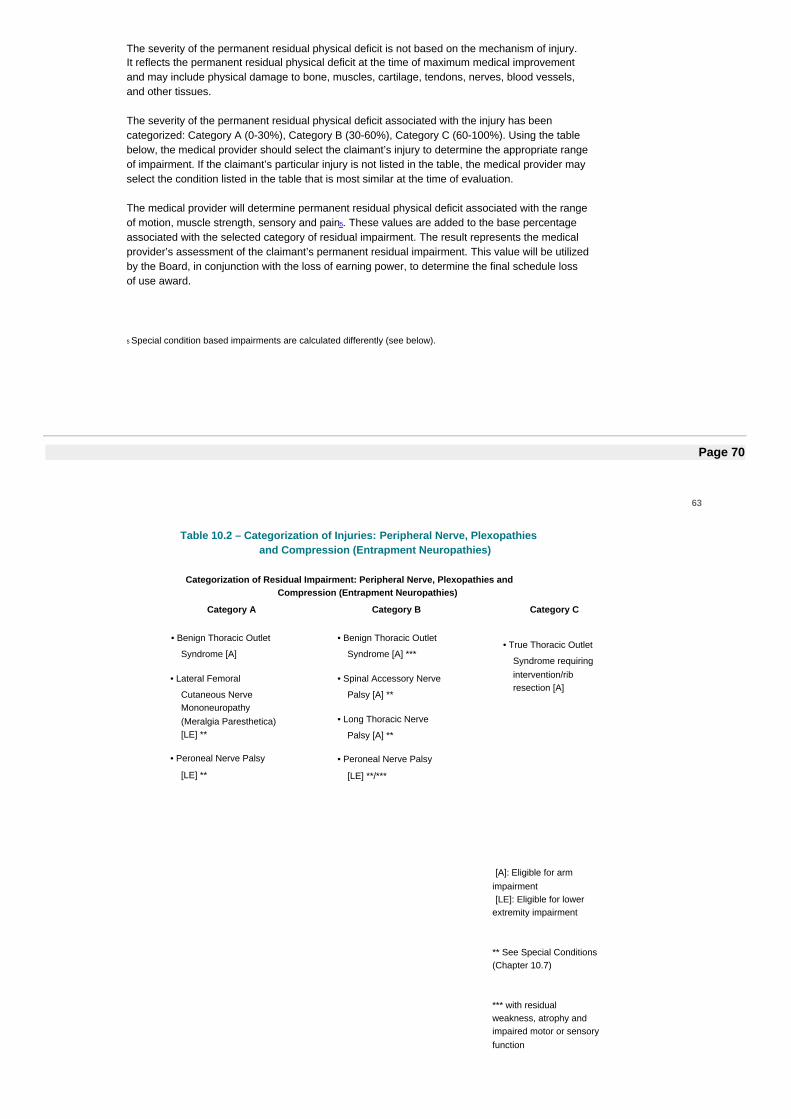
The severity of the permanent residual physical deficit is not based on the mechanism of injury.It reflects the permanent residual physical deficit at the time of maximum medical improvementand may include physical damage to bone, muscles, cartilage, tendons, nerves, blood vessels,and other tissues.
The severity of the permanent residual physical deficit associated with the injury has beencategorized: Category A (0-30%), Category B (30-60%), Category C (60-100%). Using the tablebelow, the medical provider should select the claimant’s injury to determine the appropriate rangeof impairment. If the claimant’s particular injury is not listed in the table, the medical provider mayselect the condition listed in the table that is most similar at the time of evaluation.
The medical provider will determine permanent residual physical deficit associated with the rangeof motion, muscle strength, sensory and pain5. These values are added to the base percentageassociated with the selected category of residual impairment. The result represents the medicalprovider’s assessment of the claimant’s permanent residual impairment. This value will be utilizedby the Board, in conjunction with the loss of earning power, to determine the final schedule lossof use award.
5 Special condition based impairments are calculated differently (see below).
Page 70
63
Table 10.2 – Categorization of Injuries: Peripheral Nerve, Plexopathiesand Compression (Entrapment Neuropathies)
Categorization of Residual Impairment: Peripheral Nerve, Plexopathies andCompression (Entrapment Neuropathies)
Category A Category B Category C
• Benign Thoracic Outlet
Syndrome [A]
• Lateral Femoral
Cutaneous NerveMononeuropathy(Meralgia Paresthetica)[LE] **
• Peroneal Nerve Palsy
[LE] **
• Benign Thoracic Outlet
Syndrome [A] ***
• Spinal Accessory Nerve
Palsy [A] **
• Long Thoracic Nerve
Palsy [A] **
• Peroneal Nerve Palsy
[LE] **/***
• True Thoracic Outlet
Syndrome requiringintervention/ribresection [A]
[A]: Eligible for armimpairment[LE]: Eligible for lower
extremity impairment
** See Special Conditions(Chapter 10.7)
*** with residualweakness, atrophy andimpaired motor or sensoryfunction
0-30% Impairment 30-60% Impairment 60-90% Impairment
Page 71
64
Description of Categorization of Residual Impairments
Category A
Peripheral neuropathy involving pain, mild range of motion and sensorydeficits. Minimal functional and strength deficits without atrophy.
Category B
Peripheral neuropathy involving pain and more involved or persistent range ofmotion deficits. More pronounced functional impairment with permanentsensory deficits, decreased strength, and obvious atrophy showing norecovery. Consideration should be given to delay final determination ofimpairment or to apply if needed for reevaluation to make permanentimpairment evaluation after two years following date of injury or most recentsurgical intervention.
Category C
Profound functional limitation due to pain, permanent sensory deficit, and lossof end motor unit innervation as evidenced by severe strength deficits andprofound atrophy. Strong consideration should be given to delay finaldetermination of impairment or to apply if needed for reevaluation to make finaladjustment after two years following date of injury or most recent surgicalintervention.
Unless otherwise indicated, factors affecting additional degrees of impairmentwithin a category are Range of Motion, Strength, Sensation and Pain. Thepresence of atrophy is already involved in the initial categorization of the injuryinto the Category B or Category C.
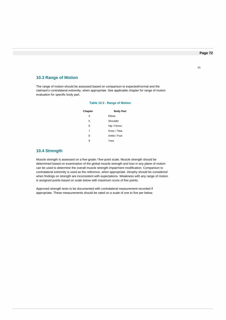
Page 72
65
10.3 Range of Motion
The range of motion should be assessed based on comparison to expected/normal and theclaimant’s contralateral extremity, when appropriate. See applicable chapter for range of motionevaluation for specific body part.
Table 10.3 - Range of Motion
Chapter Body Part
4 Elbow
5 Shoulder
6 Hip / Femur
7 Knee / Tibia
8 Ankle / Foot
9 Toes
10.4 Strength
Muscle strength is assessed on a five-grade / five-point scale. Muscle strength should bedetermined based on examination of the global muscle strength and loss in any plane of motioncan be used to determine the overall muscle strength impairment modification. Comparison tocontralateral extremity is used as the reference, when appropriate. Atrophy should be consideredwhen findings on strength are inconsistent with expectations. Weakness with any range of motionis assigned points based on scale below with maximum score of five points.
Approved strength tests to be documented with contralateral measurement recorded ifappropriate. These measurements should be rated on a scale of one to five per below.
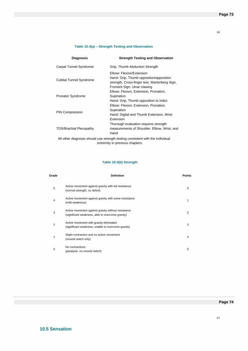
Page 73
66
Table 10.4(a) – Strength Testing and Observation
Diagnosis Strength Testing and Observation
Carpal Tunnel Syndrome Grip, Thumb Abduction Strength
Cubital Tunnel Syndrome
Elbow: Flexion/ExtensionHand: Grip, Thumb opposition/appositionstrength, Cross-finger test, Wartenberg Sign,Froment Sign, Ulnar clawing
Pronator SyndromeElbow: Flexion, Extension, Pronation,SupinationHand: Grip, Thumb opposition to index
PIN Compression
Elbow: Flexion, Extension, Pronation,SupinationHand: Digital and Thumb Extension, WristExtension
TOS/Brachial PlexopathyThorough evaluation requires strengthmeasurements of Shoulder, Elbow, Wrist, andHand
All other diagnosis should use strength testing consistent with the individualextremity in previous chapters.
Table 10.4(b) Strength
Grade Definition Points
5Active movement against gravity with full resistance(normal strength, no deficit)
0
4Active movement against gravity with some resistance(mild weakness)
1
3Active movement against gravity without resistance(significant weakness, able to overcome gravity)
2
2Active movement with gravity eliminated(significant weakness, unable to overcome gravity)
3
1Slight contraction and no active movement(muscle twitch only)
4
0No contractions(paralysis, no muscle twitch)
5
Page 74
67
10.5 Sensation
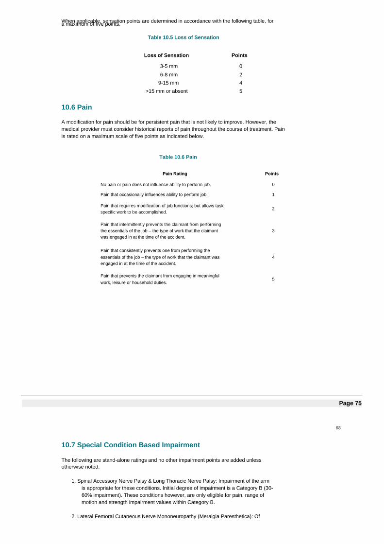
When applicable, sensation points are determined in accordance with the following table, fora maximum of five points.
Table 10.5 Loss of Sensation
Loss of Sensation Points
3-5 mm 0
6-8 mm 2
9-15 mm 4
>15 mm or absent 5
10.6 Pain
A modification for pain should be for persistent pain that is not likely to improve. However, themedical provider must consider historical reports of pain throughout the course of treatment. Painis rated on a maximum scale of five points as indicated below.
Table 10.6 Pain
Pain Rating Points
No pain or pain does not influence ability to perform job. 0
Pain that occasionally influences ability to perform job. 1
Pain that requires modification of job functions; but allows taskspecific work to be accomplished.
2
Pain that intermittently prevents the claimant from performingthe essentials of the job – the type of work that the claimantwas engaged in at the time of the accident.
3
Pain that consistently prevents one from performing theessentials of the job – the type of work that the claimant wasengaged in at the time of the accident.
4
Pain that prevents the claimant from engaging in meaningfulwork, leisure or household duties.
5
Page 75
68
10.7 Special Condition Based Impairment
The following are stand-alone ratings and no other impairment points are added unlessotherwise noted.
1. Spinal Accessory Nerve Palsy & Long Thoracic Nerve Palsy: Impairment of the armis appropriate for these conditions. Initial degree of impairment is a Category B (30-60% impairment). These conditions however, are only eligible for pain, range ofmotion and strength impairment values within Category B.
2. Lateral Femoral Cutaneous Nerve Mononeuropathy (Meralgia Paresthetica): Of
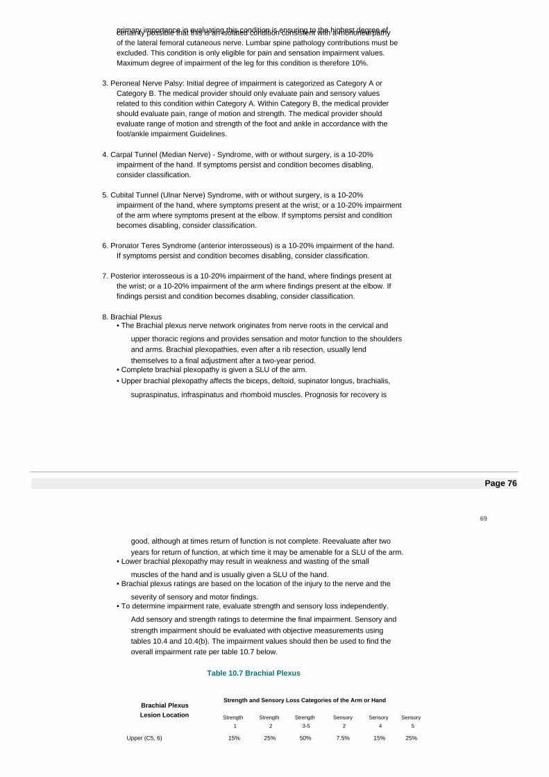
primary importance in evaluating this condition is ensuring to the highest degree ofcertainty possible that this is an isolated condition consistent with a mononeurpathyof the lateral femoral cutaneous nerve. Lumbar spine pathology contributions must beexcluded. This condition is only eligible for pain and sensation impairment values.Maximum degree of impairment of the leg for this condition is therefore 10%.
3. Peroneal Nerve Palsy: Initial degree of impairment is categorized as Category A orCategory B. The medical provider should only evaluate pain and sensory valuesrelated to this condition within Category A. Within Category B, the medical providershould evaluate pain, range of motion and strength. The medical provider shouldevaluate range of motion and strength of the foot and ankle in accordance with thefoot/ankle impairment Guidelines.
4. Carpal Tunnel (Median Nerve) - Syndrome, with or without surgery, is a 10-20%impairment of the hand. If symptoms persist and condition becomes disabling,consider classification.
5. Cubital Tunnel (Ulnar Nerve) Syndrome, with or without surgery, is a 10-20%impairment of the hand, where symptoms present at the wrist; or a 10-20% impairmentof the arm where symptoms present at the elbow. If symptoms persist and conditionbecomes disabling, consider classification.
6. Pronator Teres Syndrome (anterior interosseous) is a 10-20% impairment of the hand.If symptoms persist and condition becomes disabling, consider classification.
7. Posterior interosseous is a 10-20% impairment of the hand, where findings present atthe wrist; or a 10-20% impairment of the arm where findings present at the elbow. Iffindings persist and condition becomes disabling, consider classification.
8. Brachial Plexus• The Brachial plexus nerve network originates from nerve roots in the cervical and
upper thoracic regions and provides sensation and motor function to the shouldersand arms. Brachial plexopathies, even after a rib resection, usually lendthemselves to a final adjustment after a two-year period.
• Complete brachial plexopathy is given a SLU of the arm.• Upper brachial plexopathy affects the biceps, deltoid, supinator longus, brachialis,
supraspinatus, infraspinatus and rhomboid muscles. Prognosis for recovery is
Page 76
69
good, although at times return of function is not complete. Reevaluate after twoyears for return of function, at which time it may be amenable for a SLU of the arm.
• Lower brachial plexopathy may result in weakness and wasting of the small
muscles of the hand and is usually given a SLU of the hand.• Brachial plexus ratings are based on the location of the injury to the nerve and the
severity of sensory and motor findings.• To determine impairment rate, evaluate strength and sensory loss independently.
Add sensory and strength ratings to determine the final impairment. Sensory andstrength impairment should be evaluated with objective measurements usingtables 10.4 and 10.4(b). The impairment values should then be used to find theoverall impairment rate per table 10.7 below.
Table 10.7 Brachial Plexus
Brachial PlexusLesion Location
Strength and Sensory Loss Categories of the Arm or Hand
Strength
1
Strength
2
Strength
3-5
Sensory
2
Sensory
4
Sensory
5
Upper (C5, 6) 15% 25% 50% 7.5% 15% 25%
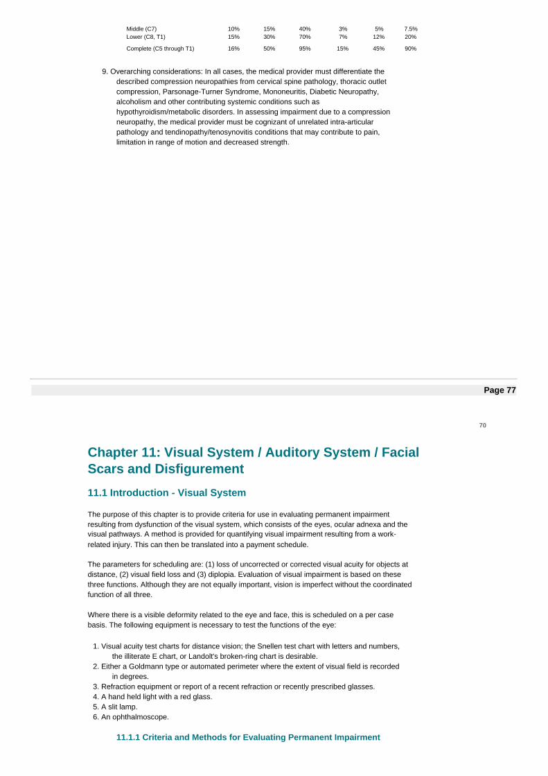
Middle (C7) 10% 15% 40% 3% 5% 7.5%Lower (C8, T1) 15% 30% 70% 7% 12% 20%
Complete (C5 through T1) 16% 50% 95% 15% 45% 90%
9. Overarching considerations: In all cases, the medical provider must differentiate thedescribed compression neuropathies from cervical spine pathology, thoracic outletcompression, Parsonage-Turner Syndrome, Mononeuritis, Diabetic Neuropathy,alcoholism and other contributing systemic conditions such ashypothyroidism/metabolic disorders. In assessing impairment due to a compressionneuropathy, the medical provider must be cognizant of unrelated intra-articularpathology and tendinopathy/tenosynovitis conditions that may contribute to pain,limitation in range of motion and decreased strength.
Page 77
70
Chapter 11: Visual System / Auditory System / FacialScars and Disfigurement
11.1 Introduction - Visual System
The purpose of this chapter is to provide criteria for use in evaluating permanent impairmentresulting from dysfunction of the visual system, which consists of the eyes, ocular adnexa and thevisual pathways. A method is provided for quantifying visual impairment resulting from a work-related injury. This can then be translated into a payment schedule.
The parameters for scheduling are: (1) loss of uncorrected or corrected visual acuity for objects atdistance, (2) visual field loss and (3) diplopia. Evaluation of visual impairment is based on thesethree functions. Although they are not equally important, vision is imperfect without the coordinatedfunction of all three.
Where there is a visible deformity related to the eye and face, this is scheduled on a per casebasis. The following equipment is necessary to test the functions of the eye:
1. Visual acuity test charts for distance vision; the Snellen test chart with letters and numbers,the illiterate E chart, or Landolt's broken-ring chart is desirable.
2. Either a Goldmann type or automated perimeter where the extent of visual field is recordedin degrees.
3. Refraction equipment or report of a recent refraction or recently prescribed glasses.4. A hand held light with a red glass.5. A slit lamp.6. An ophthalmoscope.
11.1.1 Criteria and Methods for Evaluating Permanent Impairment
Central Visual Acuity
The chart or reflecting surface should not be dirty or discolored. The far test distancesimulates infinity at 6 m (20 ft.) or at no less than 4 m (13 ft. 1 in.).
The central vision should be measured and recorded for distance with and without wearingconventional spectacles. The use of contact lens may further improve vision reduced byirregular astigmatism due to corneal injury or disease. In the absence of contraindications,if the patient is well adapted to contact lenses and wishes to wear them, correction bycontact lenses is acceptable.
Visual acuity for distance should be recorded in the Snellen notation, using a fraction–where the numerator is the test distance in feet or meters – and the denominator is thedistance at which the smallest letter discriminated by the patient would subtend fiveminutes of arc, that is, the distance at which an eye with 20/20 vision would see that letter.The fraction notation is one of convenience that does not imply percentage of visual acuity.
The procedure for determining the loss of central vision in one eye is as follows:
Page 78
71
(1) Measure and record best central visual acuity for distance with and withoutconventional corrective spectacles or contact lens.
(2) Schedule according to the Table 11.1.1(a) for uncorrected or corrected visual loss(in the injured eye) whichever is greater.
Table 11.1.1(a) - Visual Loss
Visual Fields
The extent of the visual field is determined by using a perimetric method with a whitetarget. If the Goldmann 30 cm. radius bowl perimeter is used, the III/4 e target in the kineticmode should be employed.
Determining Loss of Visual Field
The following steps are taken to determine the loss of visual field:
(1) Plot the extent of the visual field on each of the eight principal meridians of avisual field chart using Figure 11.1.1(b).
(2) Determine the percentage loss to schedule according to Table 11.1.1(c)
Visual Acuity Schedule %20/20 0
20/20-1 5
20/20-2 7.5
20/20-3 10
20/20-4 1520/25 20
20/25-1 22.5
20/25-2 25
20/30 33.3
20/30-1 3520/30-2 37.5
20/30+1 30
Visual Acuity Schedule %20/30 + 2 or 3 27.5
20/40 50
20/40+2 45
20/40+3 40
20/40-1 or 2 51.5
20/40-3 55
20/50 60
20/60 65
20/70 70
20/70-1 or 20/70-2 75
Over 75% 100%
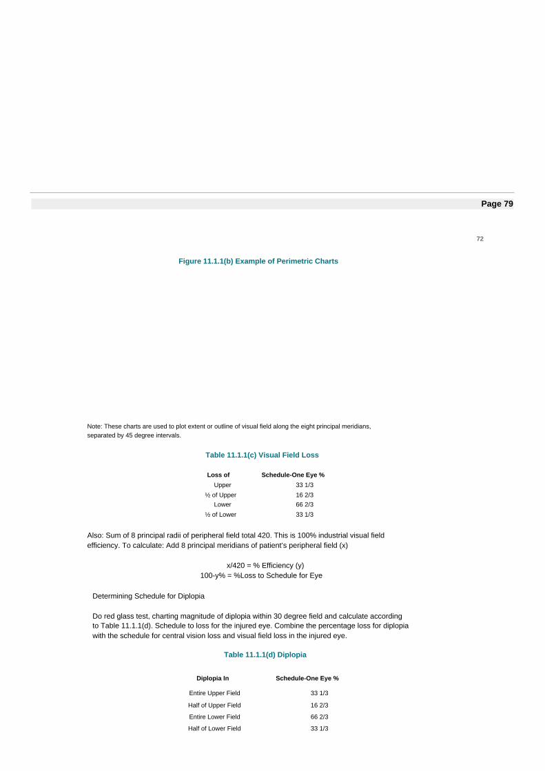
Page 79
72
Figure 11.1.1(b) Example of Perimetric Charts
Note: These charts are used to plot extent or outline of visual field along the eight principal meridians,separated by 45 degree intervals.
Table 11.1.1(c) Visual Field Loss
Loss of Schedule-One Eye %Upper 33 1/3
½ of Upper 16 2/3Lower 66 2/3
½ of Lower 33 1/3
Also: Sum of 8 principal radii of peripheral field total 420. This is 100% industrial visual fieldefficiency. To calculate: Add 8 principal meridians of patient’s peripheral field (x)
x/420 = % Efficiency (y)100-y% = %Loss to Schedule for Eye
Determining Schedule for Diplopia
Do red glass test, charting magnitude of diplopia within 30 degree field and calculate accordingto Table 11.1.1(d). Schedule to loss for the injured eye. Combine the percentage loss for diplopiawith the schedule for central vision loss and visual field loss in the injured eye.
Table 11.1.1(d) Diplopia
Diplopia In Schedule-One Eye %
Entire Upper Field 33 1/3
Half of Upper Field 16 2/3
Entire Lower Field 66 2/3
Half of Lower Field 33 1/3
Consider for 30 degree field
Page 80
73
11.2 Introduction - Auditory System
The waiting period for a worker to file a claim for a job-related hearing loss is three months fromthe date the worker leaves employment or is removed from exposure to harmful noise in theworkplace (can be by way of effective protective devices). The last day of the three-month periodof removal is considered the worker's date of disablement.
11.2.1 Occupational Loss of Hearing
Under these standards which in effect measure the ability to hear normal speech,audiometric tone tests at varying intensity of sound are conducted at frequency levels of500, 1000, 2000, 3000 Hertz (Hz).
Results at the four frequency levels are averaged and if the threshold necessary for theindividual to hear sound is 25 decibels (dB) or less, no hearing impairment is consideredto be present.
For every decibel that the hearing level of an ear exceeds 25 dB, hearing loss is calculatedat 1 1/2 percent, up to 100 percent at 92 dB. Thus, if the claimant's hearing level is 41 dB,he/she would have a hearing loss of 24 percent in that ear.
The percentage of hearing loss in the claimant's better ear is multiplied by 5, and theresulting figure is added to the percentage of hearing loss in the claimant's poorer ear.The total is divided by 6 and this represents the claimant's overall percentage of hearingloss for which benefits are awarded.
11.2.2 Traumatic Loss of Hearing
Traumatic Hearing Loss - May occur as a result of a blow to the head, a strong blast of airinto the ear, etc.
A different method is used to determine the degree of hearing loss as a result of traumathan as a result of occupational disease.
The scale used to measure percentage is based upon 250 Cycles Per Second (CPS) to4000 CPS.The schedule for complete loss of hearing for both ears is 150 weeks, and the schedulefor each ear is 60 weeks. The method used to compute the loss is to take the percentageof loss in each ear, total it, and then divide it by 2.
For example: 25% in right ear40% in left ear65% total loss
Divide 65% by 2, which equals 32.5 %.
Page 81
74
11.3 Facial Scars and Disfigurement
1. Permanent scars and disfigurement of the face and neck are usually evaluated one-year post-injury and/or one year after the last surgical procedure was performed.
2. Scars and disfigurement involving the neck are limited to the region above the clavicle.
3. The scar and disfigurement should be described accurately, using such parameters as length,width, color, contour, and exact location.
4. Specific disfigurements of the eye, ear, nose and mouth are also to be noted.
a. Common disfigurements of the eye include corneal scarring; defects of the iris and in someinstances total loss of the eye with use of a prosthesis.
b. Common disfigurements of the nose include nasal septal deviation, enlargement andtissue loss.
c. Common disfigurements of the lips include loss of soft tissue, enlargement, and alterationof normal contour of the lips.
d. Common disfigurements of the ear include loss of tissue and alteration of normal contourof the ear.
e. If teeth are damaged, the dentist’s report should be consulted.
![PRODUCT MONOGRAPH - Biogen · renal impairment [Creatinine Clearance (CrCl) ≤80 mL/min] (see CONTRAINDICATIONS). Determining renal function before treatment, and regular monitoring](https://img.pdfslide.net/doc/110x75/5ec2f27c11b3031eb36856ee/product-monograph-biogen-renal-impairment-creatinine-clearance-crcl-a80-mlmin.jpg)


![Psychological Disability Evaluation [Read-Only] · • Factors in Determining Causation ... • The difference between impairment and disability ... Distinguishing normal distress](https://img.pdfslide.net/doc/110x75/5b16b0257f8b9a5e6d8d3c37/psychological-disability-evaluation-read-only-factors-in-determining-causation.jpg)