Embed Size (px)
Citation preview
Inshore spawning of cobia (Rachycentron canadum) in South Carolina
Lefebvre and Denson, 2012
SEDAR28‐RD10
5 January 2012
SEDAR28-RD10
1
INSHORE SPAWNING OF COBIA (RACHYCENTRON CANADUM) IN SOUTH CAROLINA
LYNDSEY S. LEFEBVRE
* Grice Marine Laboratory
College of Charleston
205 Fort Johnson Road
Charleston, South Carolina 29412
**Fisheries Ecology Division
Southwest Fisheries Science Center
National Marine Fisheries Service
110 Shaffer Road
Santa Cruz, California 95060
Phone: (831) 345-0827
Fax: (831) 420-3977
MICHAEL R. DENSON
Marine Resources Research Institute
South Carolina Department of Natural Resources
217 Fort Johnson Road
Charleston, South Carolina 29412
Key words: cobia, spawning, embryo development, ovarian development,
ichythyoplankton
* Work conducted while affiliated with
** Current contact information
SEDAR28-RD10
2
ABSTRACT
Inshore spawning of the recreationally important cobia, Rachycentron canadum, was
documented in Port Royal Sound (PRS) and St. Helena Sound (SHS), South Carolina
between April and June of 2007 and 2008. Histological analysis of ovaries confirmed the
presence of gravid females inshore, and gonadosomatic index (GSI) values from females
collected inshore (mean=7.8) were higher than those caught offshore (mean=5.6), both of
which indicate spawning was occurring locally. Additionally, an ichthyoplankton survey
found cobia eggs and larvae as far as 10 and 15 km upriver from the mouths of SHS and
PRS, respectively. An egg development study of hatchery-reared cobia eggs provided
descriptions of embryological development of cobia. The comparison of visual and
quantitative characteristics allowed for positive identification of eggs collected in
plankton samples. We show that the inshore migration of cobia during April through
June, presence of gravid females, significantly higher GSI values, and collection of eggs
inside the estuaries, all confirm that PRS and SHS provide spawning habitat for cobia.
Due to the potential for heavy exploitation by recreational anglers as cobia move inshore
to spawn in South Carolina, current management strategies may need review.
SEDAR28-RD10
3
INTRODUCTION
Cobia, Rachycentron canadum, is a migratory, stenohaline benthopelagic species
distributed worldwide in tropic, subtropic, and warm temperate waters, except the eastern
Pacific (Briggs, 1960). In the United States, cobia are found throughout the Gulf of
Mexico and along the Atlantic coast from Florida to Massachusetts (Shaffer and
Nakamura, 1989). Cobia are moderately long-lived, with a maximum reported age of 15
years (Shaffer and Nakamura, 1989), and have fast growth rates, with both sexes reaching
sexual maturity by age two (males 60 cm fork length [FL]; females 80 cm FL; (Smith,
1996; Burns et al., 1998). Currently, no substantial commercial fishery exists for cobia in
the United States, with most commercial landings resulting from incidental catch in other
fisheries (Shaffer and Nakamura, 1989). Cobia are sought recreationally throughout their
range, and the majority of the annual catch in the U.S. comes from the recreational
fishery (National Marine Fisheries Services Statistics Division, personal communication).
The current fishery management plan, which imposes a bag limit of two fish per person
per day and a minimum FL of 84 cm (33 inches), was established by the Gulf of Mexico
(GMFMC) and South Atlantic Fishery Management Councils (SAFMC) in 19831 with
the aim of conserving a population that was considered overexploited at that time. The
restrictions were enacted under the assumptions that cobia are widely dispersed, are
primarily commercial bycatch, constitute a recreational fishery, and comprise a single
population in the U.S. These restrictions were meant to reduce catches and allow females
the opportunity to reproduce before entering the fishery. Regional fishing effort and
1 SAFMC and GMFMC. 1983. Fishery management plan final, environmental impact statement, regulatory impact review, final regulations for coastal migratory pelagic resources (mackerels). SAFMC, 4055 Faber Place, Suite 201, North Charleston, SC 29405. 321 pp.
SEDAR28-RD10
4
catch data indicate fishing pressure for cobia has increased over the past decade (Steele2).
Cobia continues to gain socioeconomic importance as a game fish throughout much of its
range, supporting an expanding charterboat industry; however, the current health of the
stock is unknown along the southeastern U.S.
In the spring and early summer months, cobia in the western North Atlantic are
thought to migrate with warming waters from Florida to as far as Massachusetts (Shaffer
and Nakamura, 1989). During this presumed northward migration, cobia enter high
salinity bays and estuaries, including Port Royal Sound (PRS) and St. Helena Sound
(SHS) in South Carolina, Pamlico Sound in North Carolina (Smith, 1996), and the
Chesapeake Bay (Shaffer and Nakamura, 1989), where they are more readily available to
recreational anglers. Reasons for the inshore movement are not fully understood, but it is
hypothesized they may be following prey species or aggregating to spawn.
On the east coast of the U.S., the cobia spawning season extends from April
though September (Lotz et al., 1996; Smith, 1996; Burns et al., 1998; Brown-Peterson et
al., 2001) with cobia being batch spawners, capable of spawning multiple times during a
season (Biesiot et al., 1994; Lotz et al., 1996). Regional peaks in spawning, as
designated by peaks in the gonadosomatic index (GSI), correlate with the migration of
cobia from Florida to Massachusetts. Spawning peaks along the southeastern U.S.
Atlantic coast in May (Shaffer and Nakamura, 1989; Burns et al., 1998), in North
Carolina in June (Smith, 1996), and in the Chesapeake Bay region in June and July
(Joseph et al., 1964). In South Carolina the peak in spawning activity in May
corresponds to the highest fishing effort in the region, as evidenced by the increased
2 Steele, G. 2009. Personal communication. South Carolina Department of Natural Resources, Charleston, SC 29412.
SEDAR28-RD10
5
landings during this month (Steele1). The South Carolina recreational cobia fishery, in
which the fish are easily accessible to anglers April through June when located inside
PRS and SHS, may be similar to the fishery in other states where cobia enter inland
waters. Because the inshore migrations correspond with the spawning season of cobia, it
is probable the sounds serve as spawning habitat.
Beyond the general knowledge of spawning season, a paucity of information
exists on spawning habitat and time of day of wild cobia spawning because much of the
previous research has focused on age and growth (Smith, 1996; Franks et al., 1999),
feeding habits (Smith, 1996; Arendt et al., 2001), and general reproductive biology
(Biesiot et al., 1994; Lotz et al., 1996; Smith, 1996; Brown-Peterson et al., 2001; van der
Velde et al., 2010). Collection of eggs in the Gulf Stream off North Carolina by Hassler
and Rainville (1975) suggested spawning took place offshore. Offshore spawning was
also proposed by Burns et al. (1998), due to a scarcity of fish with histological signs of
final oocyte maturation (FOM) collected in nearshore waters of the Gulf of Mexico and
the southeast U.S. Atlantic coast. Inshore spawning of cobia has been proposed in the
lower regions of Chesapeake Bay, based on the collection of eggs immediately south of
the bay (Joseph et al., 1964) and the ovarian condition of females collected in the bay
(Richards, 1967). In North Carolina Smith (1996) suggested cobia spawned adjacent to
inlets, by reason of neuston net collection of eggs within inlets and a lack of females
caught inshore undergoing FOM.
The main goal of the current study was to determine if cobia spawning occurs
within two high salinity South Carolina estuaries, PRS and SHS. Histological analysis of
ovarian tissue was used to evaluate the reproductive status of female cobia collected by
SEDAR28-RD10

6
the recreational fishery both inshore and offshore of these estuaries. A development
study using hatchery-reared cobia eggs provided embryological development
characteristics and insight on time of day of spawning of wild cobia. An ichthyoplankton
survey targeting cobia eggs and larvae in PRS and SHS was conducted during the
spawning season to provide further evidence of spawning locations. Inshore spawning of
cobia in South Carolina may suggest that spawning aggregations occur in other inland
and nearshore waters that cobia frequent during the spawning season and that cobia may
be subject to substantial fishing pressure before having the opportunity to reproduce.
MATERIALS AND METHODS
Reproductive Biology
Fresh and frozen cobia specimens were collected via hook and line from PRS and
SHS, South Carolina (Fig. 1) in April-June 2007 and 2008, from fishing tournaments,
cooperating anglers, recreational fishing guides, and SCDNR employees. For all whole
fish collected, total length (TL, mm), FL (mm), and fish weight (FW, kg) were measured.
Sex was determined macroscopically and gonads were excised, stored on ice, and
transported to the SCDNR Marine Resources Research Institute (MRRI). Date, time, and
location of capture were noted when available, with fish collected within PRS and SHS
designated as “inshore” and fish collected outside the two estuaries (defined as outside
the barrier islands) designated as “offshore”. For carcasses collected through SCDNR’s
freezer program, in which recreational anglers donate the fish carcass after filleting, the
aforementioned information was recorded when provided by anglers.
At the MRRI, gonads were weighed to the nearest gram and the GSI was
calculated as
SEDAR28-RD10
7
GSI GWSW 100
where GW = gonad weight (g) and SW = somatic weight (g). To determine if GSI values
for male and female cobia were significantly different, a Wilcoxon rank test was
performed. Cobia are batch spawners with indeterminate fecundity and group-
synchronous oocyte development (Lotz et al., 1996; Brown-Peterson et al., 2001; van der
Velde et al., 2010). Ovarian growth in other species with the same reproductive strategy
is isometric with body growth (Taylor et al., 1998; Somarakis et al., 2004). Additionally,
previous work from cobia ovaries with FOM has shown that there is no relationship
between relative fecundity and either body weight or fork length (Brown-Peterson et al.,
2001; van der Velde et al., 2010). Therefore, female GSI was used as a proxy for ovarian
maturation for statistical purposes. To evaluate if ovarian maturation differed between
cobia collected inshore and offshore, a Wilcoxon rank test was performed using GSI as a
proxy for maturation. Non-parametric Wilcoxon tests were performed due to non-
normality in the data resulting from the capture of two gravid females. A t-test was used
to determine if differences in GSI between inshore and offshore females persisted with
the exclusion of the GSI values from the two gravid females.
Histological analyses were limited to samples collected from female cobia, as
males are capable of spawning year-round (Brown-Peterson et al., 2001). Homogenous
ovarian development has been documented previously for cobia (Lotz et al., 1996);
therefore, a single portion of tissue (approximately 50-100 mg) from the middle of one
ovarian lobe was fixed in 10% neutral buffered formalin before being rinsed in
freshwater and stored in 50% isopropyl alcohol at least 24 hours prior to processing.
Tissue samples were dehydrated; infiltrated and blocked in paraffin; sectioned to 6 µm
SEDAR28-RD10
8
using a rotary microtome; mounted on glass slides; and stained using hematoxylin and
eosin-y according to standard histological techniques (Humason, 1972). Slides were
examined under a compound microscope at 100x magnification and staged according to
ovarian development. Ovarian stages (Table 1) are based on descriptions of teleost
oocyte development in Wallace and Selman (1981) and with modifications from
Roumillat and Brouwer (2004). When present, postovulatory follicles (POFs) were
categorized as either less than or equal to, or greater than 12 hours old, based on rates of
POFs atresia found in Cynoscion nebulosus (spotted sea trout; Roumillat and Brouwer,
2004). All samples were staged by a second, independent reader. Discrepancies were
resolved by simultaneous viewing of the slides by both readers, with all discrepancies
resolved. Percent composition (PC) of females in each of the ovarian maturation stages
was calculated separately for females collected inshore and offshore as
PC nsT 100,
where ns = the number of female samples in stage s, and T = the total number of female
samples.
Egg Development
To obtain a time-series reference collection of cobia eggs to assist in positively
identifying and aging candidate eggs from plankton collections, development studies
were conducted in 2007 and 2008 using fertilized eggs obtained from wild-caught adult
cobia spawned in the laboratory. Cobia yolk-sac larvae from the 2007 study served as
reference for the identification of young larvae in plankton collections. The experimental
temperature treatments covered the measured range of surface temperatures encountered
in PRS and SHS during May and June of both years.
SEDAR28-RD10

9
In 2007 at the Hollings Marine Laboratory (HML), four fiberglass hatching cones
(170 L) were equipped with aerators and heaters, and filled with seawater (34.5 parts per
thousand [ppt]) from Charleston Harbor that had been filtered (5 µm) and UV sanitized
after settling for three days. The water was heated to 22.5, 25.0, 26.5, and 29.0 ºC,
respectively, and maintained for 48 hours prior to the start of the study. Four 50 mL
aliquots of eggs (~2 x 10⁴ eggs) were used in the trial. Prior to the addition of eggs to the
water baths at approximately 11 hours post-spawn, digital micrographs of eggs were
recorded using a Nikon SMZ1500 stereo microscope (Nikon Instruments Inc., Melville,
NY) mounted with a Micropublisher 3.3 camera (Q Imaging, Surrey, BC, Canada).
Thereafter, approximately 10 eggs and/or larvae were collected from each tank and
micrographs were recorded every four hours until 61 hours post-spawn. After images
were recorded, eggs and/or larvae were preserved in 10% neutral buffered formalin.
In order to capture earlier stages of egg development and to expand experimental
temperatures, a second development study was conducted in 2008. Three water baths
were heated to and maintained at 24.0, 26.0, and 28.0 ºC in a temperature-controlled
laboratory 30 hours prior to the beginning of the study. At two hours post-spawn, a 25
mL aliquot of cobia eggs (~1 x 10⁴ eggs) was divided between petri dishes in each
temperature bath. Every hour, until 13 hours post-spawn, and every other hour thereafter,
to 25 hours post-spawn, a sample of approximately 10 eggs was removed from each
water bath, digital micrographs were recorded, and eggs were preserved in 10% neutral
buffered formalin.
Micrographs of live eggs, egg diameters and oil droplet diameters were measured
to the nearest µm using ImageJ image analysis software (ImageJ, vers. 1.38, Bethesda,
SEDAR28-RD10
10
MD). To determine if damage and diameter changes occurred with preservation, changes
in appearance were noted and measurements of egg and oil droplet diameters were
measured to the nearest µm using ImageJ from micrographs of preserved eggs taken
approximately one year from the date of collection in 2007 and two months from the date
of collection in 2008. Diameters were only measured in undamaged, preserved eggs.
Percent shrinkage was calculated as change in egg and oil droplet diameter between the
live and corresponding preserved eggs multiplied by 100. To determine if there was a
significant decrease in diameter due to preservation, two-sample t-tests were performed
independently for 2007 egg and oil droplet diameters and 2008 egg diameters. Due to
non-normality of the data, a Wilcoxon rank test was performed using 2008 oil droplet
diameters to determine if there was significant shrinkage after preservation.
Based on the micrographs of live eggs from the development studies, coarse
stages of embryological development were described. Because only the 2008 study
captured the earliest stages of development and larvae only hatched in the 2007 study, the
two temperature treatments that were closest in temperature (26.5 ºC and 26 ºC in 2007
and 2008, respectively) were used for the description of development to capture the
earliest stages of development through hatching.
Ichthyoplankton Survey
Field Collection
Five stations in each estuary (Fig. 1), encompassing known cobia fishing grounds,
were sampled weekly from May 6, 2008 through June 15, 2008 in PRS and May 8
through June 8, 2008 in SHS. Collections were made from 0745 to 1945 hours to
accomplish plankton captures during similar stages of the incoming tide. Stations in PRS
SEDAR28-RD10
11
were located between 12.1 and 20.6 km from the mouth of the estuary, as measured from
a navigation buoy (GC “25) located in the channel between St. Helena Island and Hilton
Head Island. Stations in SHS were located between 5.1 and 14.3 km from the mouth of
the estuary, as referenced from navigation buoy GC “9. Stations were positioned upriver
of particular bathymetric features where anglers typically target cobia. These are areas
where sand bars and banks result in water depth rising from 10 to 12 m mean low water
(mlw) to 2 to 3 m (mlw). Depths of stations ranged from 5.5 to 9.6 m at the time of net
deployment. Anchored plankton nets (Fig. 2) were deployed at slack water prior to the
daylight flood tide. Floats attached to the frame maintained the position of the net at
approximately 1 m below the surface. Flow meters were mounted in the center of the
nets at the most seaward and most upriver stations. At the time of net deployment,
ancillary water data were collected (temperature, salinity, dissolved oxygen) at the
surface and bottom of the water column using a handheld YSI model 556 (YSI Inc.,
Yellow Springs, OH). Plankton nets were deployed for the length of time required to set
all nets and return to retrieve the first (70-150 minutes). The average current speed
during the time of collection was calculated using the flow meter reading. Volume of
water filtered was calculated directly from flow meter readings in nets. For nets without
flow meters, current speeds and volumes filtered were estimated by taking the average of
the values from the most seaward and most upriver stations.
Egg and Larval Identification
Plankton samples were rinsed and sorted under a dissecting microscope. When
settled plankton volumes were greater than 1 L, samples were split using a Burrell
plankton splitter (Burrell et al., 1974) until a settled plankton volume of 0.5 L or less was
SEDAR28-RD10

12
attained. All non-clupeiform larvae were removed, identified to lowest possible
taxonomic level, and preserved in 70% isopropyl alcohol. Cobia larvae were identified
from descriptions of Ditty and Shaw (1992). All eggs measuring between 1.0 and 1.4
mm on an ocular micrometer and with one or more of the morphological characteristics
corresponding to cobia eggs (single, large [300 to 600 µm], pigmented oil droplet;
heavily pigmented embryo; narrow perivitelline space; (Ditty, 2006) were removed and
preserved in 10% neutral buffered formalin.
Digital micrographs of preliminarily identified cobia eggs were recorded. Egg
and oil droplet diameters of preserved eggs were measured to the nearest µm using
ImageJ. To aid in identification, eggs from the two development studies were viewed as
references. Eggs were positively identified as cobia if their diameter fell within the range
noted in the literature and if the morphological characteristics, with the exception of
number and diameter of oil droplets, matched the hatchery-reared eggs and published
description of cobia eggs (Ditty, 2006). The number and diameter of oil droplets were
excluded as positive identifying characters because of damage occurring due to
preservation. For further analyses eggs were labeled as early- (from fertilization to
blastopore closure; embryo not visible in preserved eggs; Ahlstrom and Moser, 1980) or
late- (from blastopore closure to hatching; embryo evident in preserved eggs; mid and
late stages in Ahlstrom and Moser 1980) stage.
Maternal condition in other species has been shown to have a significant effect on
oil droplet size of eggs (Gagliano and McCormick, 2007) and larvae (Berkeley et al.,
2004; Sogard et al., 2008), but no significant effect on egg size. As secondary evidence
for correct identification of eggs, an analysis of covariance (ANCOVA) was conducted to
SEDAR28-RD10

13
test for differences in the relationship of egg and oil droplet diameters between known
cobia eggs (early- and late-stage eggs from the 2007 and 2008 development studies) and
eggs identified from plankton samples. If the relationship was not significantly different
between hatchery-reared and wild eggs, the wild eggs were confirmed as cobia. Only
measurements from eggs with single, intact oil droplets were used in the ANCOVA.
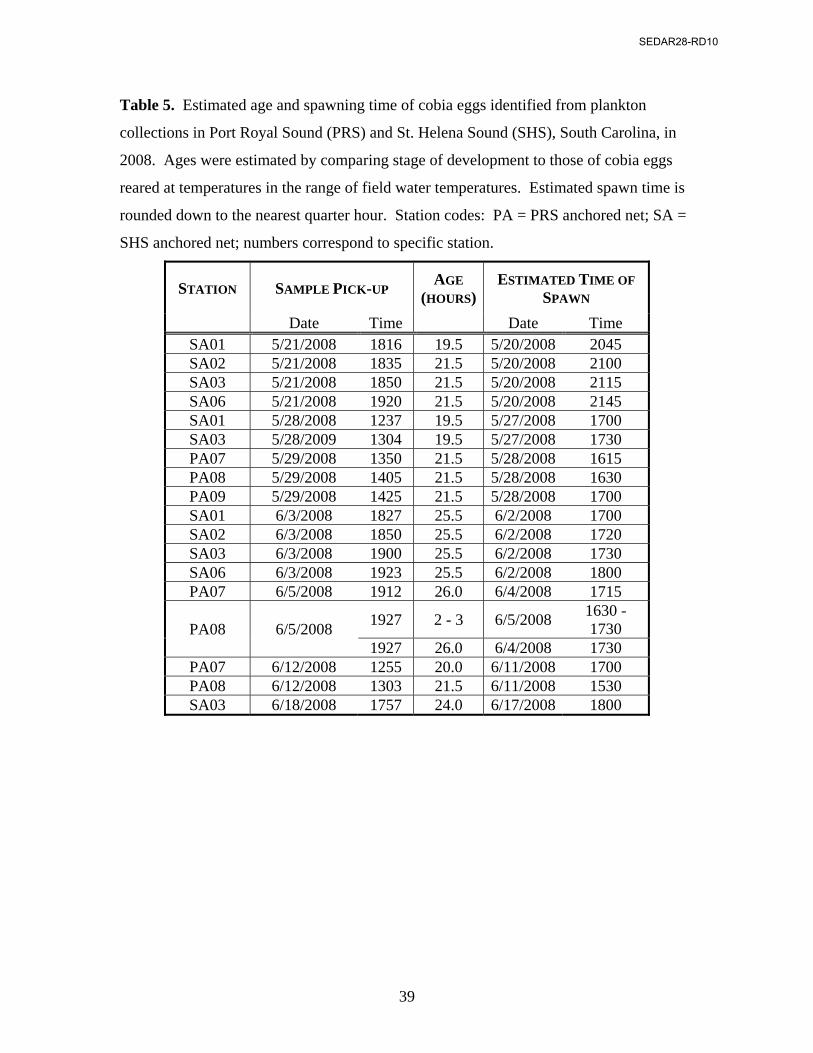
Ages of cobia eggs collected from the plankton samples were estimated via side-
by-side comparison to eggs reared at the closest matching temperature from either the
2007 or 2008 development study. For a given sample, time of spawning was
approximated using the estimated age of eggs by back-calculating from the time of
sample collection. Numbers of cobia eggs and larvae found in samples that had been
split were estimated based on the fraction of the sample sorted. Concentrations of cobia
eggs and larvae collected in the ichthyoplankton survey were obtained using calculated
and estimated sample volume filtered for the corresponding sample.
All statistical analyses were conducted using R statistical software (R
Development Core Team 2009). The significance level of = 0.05 was used for all tests.
RESULTS
Cobia (278 females, 283 males) were collected between April 2007 and August
2008. Two-hundred sixty-one fish came from fishing tournaments, 37 from SCDNR
efforts, and 263 from donations to the SCDNR freezer program. Specimens ranged in
size from 850 to 1425 mm FL (mean=1042 mm) for females and 386 to 1215 mm FL
(mean=930 mm) for males. Weights ranged from 6.7 to 38.3 kg (mean=15.0 kg) for
females and 0.5 to 23.0 kg (mean=9.9 kg) for males. Eighty percent of the fish 1000 mm
FL or larger and 79% of the fish with a total weight equal to or greater than 10 kgs were
female. Capture locations were available for 183 fish (44 offshore, 101 PRS, 39 SHS).
SEDAR28-RD10

14
Reproductive Biology
GSIs were calculated for 278 cobia (164 females; 114 males) and were combined
for April, May and June of both years. GSI for all cobia ranged between 0.7 and 22.5
(mean=6.1), and females had a significantly higher mean (7.3) than males (4.4; p<0.05).
Females collected inshore (mean=7.8; n=64) had a statistically significant higher average
GSI than females collected offshore (mean=5.6; n=34; p=0.002), suggesting the ovaries
were in a more developed state in inshore fish. The comparatively higher mean GSI from
females collected inshore was not biased by the collection of gravid females inshore or
spent females offshore as the mean inshore GSI (7.4) remained statistically higher than
the mean offshore GSI (5.6; p=0.005) with removal of the gravid and spent females.
Histological analysis of 213 ovaries showed that female cobia were capable of
spawning during May and June, when they are also most heavily targeted by anglers in
South Carolina (Steele1). All ovarian stages were represented in the female cobia
samples examined except for the immature and recovering stages (Fig. 3). Immature
specimens were absent because current fishing regulations impose a minimum FL (84
cm) larger than the length at female first maturity (80 cm). The lack of females in the
recovering state is a result of sample collection occurring only within the known
spawning season of cobia. Further histological analysis of female ovarian samples was
limited to the 98 specimens for which location of capture was known (64 inshore and 34
offshore). The majority (72%) of female cobia collected from both inshore and offshore
waters had ovaries in the late stage of development (Table 2). Two females with ovaries
SEDAR28-RD10

15
in the early developing stage were collected offshore in early April 2007 and inshore in
early May 2007. Both females (89 cm FL and 93 cm FL, respectively) were larger than
the size at maturity, and had no evidence of prior spawns (POFs), and were, therefore,
likely maturing for the first time of the spawning season. A single female in the spent
stage was collected in offshore waters in early June 2008. Females with histological
signs of a previous spawn (visible POFs) were found in cobia collected inshore as well as
offshore. All POFs were estimated to be greater than 12 hours old (Hunter and
Macewicz, 1985; Roumillat and Brouwer, 2004), with the exception of one specimen
collected in the morning hours in May 2007, in which the POFs were estimated to be 12
hours old.
Two gravid females were collected from inshore waters in 2007. The first (121.6
cm FL and 27 kg TW) was collected in mid-morning on May 12, 2007 in the Broad River
(PRS). The second (93.4 cm FL and 11 kg TW) was caught by SCDNR employees in
SHS on June 8 at 1030 hours. Both of these samples showed ovaries in the late stage of
final oocyte maturation (FOM), in which yolk coalescence and hydration occurs
immediately prior to ovulation. The time of collection of both the females, the state of
hydration of the oocytes, and the rapid nature of FOM found in other multiple spawning
species with distributional overlaps with cobia (Brown-Peterson et al., 1988; Fitzhugh et
al., 1993; Roumillat and Brouwer, 2004) indicates they would have spawned the
afternoon of capture. Damage occurred to some tissue samples that had either been hard
frozen or that had decayed prior to collection, which resulted in all size groups of
vitellogenic oocytes superficially resembling oocytes undergoing the beginning stages of
FOM (early lipid coalescence and migration of the nucleus). In order to avoid confusion
SEDAR28-RD10
16
between FOM and damage caused to the tissue due to decay or freezing, as well as to
maintain consistency across all samples, FOM stages earlier than hydration were not
addressed.
Egg Development
Micrographs of cobia eggs from the 2007 and 2008 development studies were
taken at 13 and 19 time periods, respectively. In 2007, the eggs hatched between 39 and
43 hours post-spawn at 22.5 ºC; 29 and 33 hours at 25.0 ºC; and 25 and 29 hours at 26.5.
Eggs incubated at 29 ºC were observed hatching at 26 hours post-spawn as micrographs
were being recorded. At the end of the 2007 study, all larvae still had yolk-sacs and were
3.8 to 4.6 mm TL. The 2008 development study ended before any eggs hatched. Mean
egg diameters of live cobia eggs were 1241 and 1337 µm in 2007 and 2008, respectively.
Mean oil droplet diameters were 359 µm in 2007 and 403 µm in 2008.
Micrographs of preserved eggs from development studies from each year revealed
damage to the oil droplet in many specimens due to preservation and/or handling (Fig. 4).
Deformation of the oil droplet (irregularly shaped oil droplet) occurred most often in
early-stage eggs. Damage in late-stage eggs included split oil droplets or droplets in
which the pigmented portion had detached from the lipid. Measurements of preserved
eggs were limited to specimens that had a single, intact oil droplet. The mean egg
diameter of preserved eggs from both years was 1280 µm, and the mean oil droplet
diameter was 380 µm. The averages of egg and oil droplet diameters for both preserved
and live cobia eggs fell within the published range (Table 3; Ditty, 2006). There was
significant shrinkage in cobia egg diameters with preservation, with a rate of 1.7% in
SEDAR28-RD10
17
2007 (p<<0.05) and 1.0% in 2008 (p<<0.05). No shrinkage occurred in the oil droplet
diameters during either year (2007 p=0.13; 2008 p=0.92).
Five stages of embryological development were described for cobia (Fig. 5) and
are detailed below. Duration of each stage is approximated in hours. There is overlap in
the durations of stages III and IV due to minor differences between eggs in 2007 and
2008 development studies.
Stage I (0-7 h): Newly fertilized eggs have a distinct, translucent oil droplet. Early cell
divisions are evident at the animal pole (opposite of the oil droplet) and eventually form
the blastodisc. Continuing divisions progress until individual blastomeres are no longer
distinguishable. Stage ends when germ ring is visible and encloses approximately 1/3 of
the yolk mass. Stage II (7 -13 h): The germ ring becomes distinct and epiboly proceeds
until the germ ring is in center of the egg. Stage ends when the blastopore is closed and
the optic cups distinguish head and tail regions of the embryo. Stage III (13-19 h): The
embryo is greater than ¼ internal circumference of the egg. Somites are distinct. Stellate
melanophores are scattered around the outside of the yolk. Through the stage,
pigmentation on the embryo (from head to anterior of caudal region) increases. Stage
ends with first appearance of melanophores on oil globule and when embryo is
approximately ½ internal circumference of egg. Stage IV (14-21 h): The embryo
continues to become more heavily pigmented. “Free” melanophores continue to
congregate on oil globule until there are few to no free melanophores around outside of
yolk. The embryo begins to move, as evidenced by flexion of the body. Stage ends when
tail is visibly detached from yolk, and embryo is approximately ¾ internal circumference
of egg. Stage V (21-29 h): The embryo continues to become more heavily pigmented.
SEDAR28-RD10

18
More movement of the embryo is evident. The embryo is greater than 100% internal
circumference of the egg before hatching, with the tail extending beyond the head.
Ichthyoplankton Survey
A total of 52 anchored plankton samples (26 PRS; 26 SHS) were collected
between May 6 and June 18, 2008. All samples from PRS were sorted completely.
Seventeen of the 26 samples from SHS were split to 1/2 (n=1), 1/4 (n=14), or 1/8 (n=2)
the original settled plankton volume. Measured current speeds ranged from 0.08 to 0.93
m/s (0.29 to 3.35 km/hr), with most speeds falling between 0.14 and 0.71 m/s (0.50 to
2.56 km/hr). Volumes filtered through the plankton nets ranged from 120 to 1156 m3.
Surface water temperatures in both estuaries ranged from 20.1 to 30.0 ºC. Salinities
ranged from 31.6 to 34.3 ppt in PRS and 28.4 to 32.7 ppt in SHS, which are within
tolerable ranges for larval, juvenile, and adult cobia (Hassler and Rainville, 1975; Shaffer
and Nakamura, 1989; Denson et al., 2003). The water columns in both PRS and SHS
appeared to be well mixed, with surface and bottom temperatures and salinities varying
by no more than 0.8 ºC and 0.8 ppt, respectively.
A total of 923 eggs were identified as cobia based on size and morphological
characteristics (562 early-stage and 364 late-stage; Tables 3 and 4; Fig. 6). Late-stage
eggs were found in samples collected at all stations in PRS (n=59). The 27 early-stage
eggs from PRS were collected at 1930 hours on June 5, 2008 at a single station (PA08,
Fig. 1; Table 4) approximately 15 km upriver in the Broad river. The majority of the
eggs were collected in SHS, with 535 early-stage eggs and 305 late-stage eggs collected
among all stations. Four hundred ninety-six early-stage eggs found in SHS came from a
single sample collected at 1900 hours on June 3, 2008 at a station (SA03) approximately
SEDAR28-RD10
19
9.7 km inshore (Fig. 1; Table 4). Late-stage eggs were collected between 1230 and 1945
hours in both estuaries on 8 sampling days. Egg concentrations ranged from 0.14 to
62.51 per 100 m3 (Table 4).
Oil droplets in many of the preserved eggs collected in the plankton survey
resembled damaged oil droplets of preserved cobia eggs from the development study. All
egg diameters were measured from micrographs, but oil droplet diameter was only
measured when a single oil droplet was present (n=73). Egg diameters ranged from 1116
to 1393 µm, and oil droplet diameters ranged from 275 to 420 µm. The mean egg and oil
droplet diameters were similar to those for hatchery-reared cobia eggs and to those
reported elsewhere (Table 3; Ditty and Shaw, 1992; Ditty, 2006).
Early- (n=48) and late-stage (n=25) egg measurements were combined for the
ANCOVA. Due to the significant shrinking of egg diameter with preservation, only
measurements from preserved hatchery-reared eggs were used for the ANCOVA. Mean
oil droplet diameters for field-collected eggs (357 µm) was lower than that for hatchery-
reared cobia eggs (380 µm); however, the ANCOVA showed the slopes of the regression
lines were not statistically different (p=0.35; overall R2=0.61; Fig. 7). The lack of a
significant difference in the relationship of egg to oil droplet diameter between hatchery-
reared cobia eggs and eggs collected in the plankton samples supports the visual
identification of those from plankton collections.
Ages of field-collected eggs were estimated for samples that contained more than
one undamaged egg (Table 5). Early-stage eggs tended to turn opaque in preservation,
and, as a result, it was difficult to discern their stage of cell division or cleavage:
therefore, ages were estimated for early eggs from a single sample collected June 5 in
SEDAR28-RD10
20
PRS. When eggs resembled an intermediate between two time periods, ages were
estimated to be midway. Late-stage eggs were estimated to be between 18 and 26 hours
old. Time of spawning was estimated to range from the late afternoon (1530 hours) to late
evening (2145 hours) hours (Table 5), with most of the spawning between 1530 and 1800
hours. The three exceptions were from a single sample day in SHS, when spawning was
estimated to have occurred near midnight. While the estimates of the ages were
determined subjectively by comparing wild-caught plankton to hatchery reared eggs,
eggs collected from multiple stations on the same day were aged independently and
estimated all to be nearly the same age.
A total of 42 cobia larvae (18 PRS; 24 SHS; Fig. 8) were collected on eight
sampling days. Larval concentrations ranged between 0.169 and 1.989 larva per 100 m3
(Table 5). Five yolk-sac cobia larvae were collected, two on May 14 (stations PA05 and
PA08), one on May 29 (station PA07), and two on May 21 (station SA01).
DISCUSSION
The results of this study indicate cobia spawn within PRS and SHS, South
Carolina, as evidenced by the high mean GSI value of females caught, the collection of
gravid females, the presence of recently fertilized eggs, and the presence of larvae in
these two estuaries. Using estimated ages of field-collected eggs, in combination with
time and location of capture data from adult cobia collected at fishing tournaments, this
study provides evidence of spawning of wild cobia in inshore waters in the late afternoon
and early evening hours.
The mean GSI of female cobia collected in this study peaked at a higher value
(7.3) than previously reported for cobia in the Gulf of Mexico by Biesiot et al. (1994; 5.5)
SEDAR28-RD10
21
and Lotz et al. (1996; 5), and North Carolina by Smith (1996; 5.7). The higher mean GSI
value reported in this study is not biased by the collection of two gravid females, as
Smith (1996) and Biesiot et al. (1994) also collected females with ovaries undergoing
FOM. Even when these females were removed from analyses, the GSI values still
yielded an average value well above that reported elsewhere. It is unlikely the high mean
GSI was due to regional differences between collected specimens. Brown-Peterson et al.
(2001) found that female cobia collected from the southeast Atlantic U.S. had higher
mean GSI values (~5.5) than those from either the eastern (~5) or north-central Gulf of
Mexico (~4.5). However, their reported peak mean GSI value for fish from the southeast
Atlantic coast was comparable to the peak GSI values reported in fish from the Gulf of
Mexico by Biesiot et al. (1994; 5.5) and Lotz et al. (1996; 5). In addition to a higher GSI
value, the size range of oocytes (500-850 µm) in late developing ovaries, which
dominated collections in this study, was higher than the group with the most developed
oocytes reported elsewhere (500-650 µm; Lotz et al., 1996). Fully developed and
fertilized cobia eggs in this study and Ditty and Shaw (1992) ranged in size from 1.15
mm to 1.42, so the larger oocyte sizes found in developing ovaries suggest the oocytes
were more highly developed in the current study. Brown-Peterson et al. (2001) and van
der Velde et al. (2010) reported that monthly GSI values corresponded with histological
evidence of when spawning was occurring, supporting GSI as a good proxy of the
developmental state of ovaries. In the current study, the larger size range of oocytes
together with the higher GSI values indicate female cobia collected in South Carolina
were closer to the time of spawning than previous studies.
SEDAR28-RD10
22
Differences in the stage of cobia ovarian development between this and other
studies may stem from female capture location. In previous work, females were collected
from coastal waters in the Gulf of Mexico (Lotz et al., 1996; Brown-Peterson et al.,
2001), as well as off the southeast Atlantic coast of the U.S. (Smith, 1996; Brown-
Peterson et al., 2001). Of the samples we examined, the mean GSI of females collected
offshore (5.6) was closer to the means reported elsewhere (Biesiot et al., 1994; Lotz et al.,
1996; Smith, 1996; Brown-Peterson et al., 2001). The dissimilarity of GSI values
between female cobia collected inshore and offshore is likely because those caught
inshore were part of a spawning aggregation whereas those caught offshore were caught
before or after spawning or intercepted while migrating to spawning grounds. This
hypothesis is supported by recent genetic evidence showing the presence of distinct
population segments in South Carolina (Darden3). In a study of the genetic structure of
cobia caught offshore from Florida to Virginia and from inshore locations in South
Carolina and Virginia, Darden et al. found that fish collected offshore comprised a single
population. Furthermore, the fish collected in inshore waters were genetically distinct
from the offshore group and from other estuaries. If there is a distinct inshore South
Carolina population segment, it could be maintained through spawning of cobia in their
natal estuaries.
Histological analysis of the ovaries demonstrated that the fish were in the middle
of their reproductive season, with the majority in the late developing stage. The
distribution of females between the reproductive states may not represent the actual
distribution in the population because the primary means of specimen collection was
3 Darden, T. 2009. Personal communication. South Carolina Department of Natural Resources, Charleston, SC 29412.
SEDAR28-RD10
23
fishery-dependent. In particular, the low percentage of females with early developing
and spent ovaries is likely result of fishery practices. Cobia are generally thought to enter
inshore waters when water temperatures reach 20 ºC (Richards, 1967; Smith, 1996), and
water temperatures in PRS and SHS reached 20 ºC before the end of April in 2008.
Recreational anglers, in contrast, targeted cobia most heavily in May. Similarly, the
fishing season for cobia may end before the fish leave inshore waters: the final plankton
collections in PRS and SHS in mid-June contained cobia eggs and larvae, respectively,
indicating spawning was still occurring in the area despite a decrease in availability of
adult carcasses from recreational anglers. Female cobia with histological signs of recent
spawns (POFs) were collected inshore, but may be underrepresented, again, due to the
primary means of collection being heavily dependent on recreational fishermen.
Evidence from both the histological analysis and plankton collections of eggs in this
study suggest that spawning of wild cobia in South Carolina inshore waters occurred
from mid afternoon to late evening, with most spawning activity occurring between 1530
and 1800 hours. Unfortunately, fish collected through tournament donations, where
weigh-ins occurred between 1500 and 1800 hours, were the primary means of attaining
samples. Few fish were collected during this critical period of time, when females would
have shown indications of a recent spawn, and, as expected, most POFs were found to be
> 12 hours old.
The best evidence produced from histological analysis that cobia were spawning
inshore in South Carolina was the collection of gravid females. The duration of FOM is
unknown for cobia, though this hormonally controlled “point-of-no-return” on the path to
spawning has been found to commence as late as 13 or as early as six hours prior to
SEDAR28-RD10

24
spawning in spotted sea trout (Brown-Peterson et al., 1988; Roumillat and Brouwer,
2004), which spawn in estuaries during the same time of year and within the same range
as cobia. Because of potential damage to tissues caused by freezing or decay prior to
sample collection, the earliest stages of FOM were not examined in this study and we,
therefore, do not have an estimate as to the time of onset of FOM in cobia. However,
both gravid females we collected had oocytes that were in the late stages of FOM (near
the mid-point of hydration), suggesting they both would have spawned in the mid
afternoon, only a few hours after their time of capture. The nearness to spawning of these
two females provides strong evidence of spawning activity occurring in the proximity of
their capture.
Few studies have collected females undergoing FOM, although there are
exceptions (Biesiot et al., 1994; Smith, 1996; van der Velde et al., 2010). Gravid females
may, again, comprise a larger percentage of the South Carolina inshore cobia population
than reported here, but capture of them may be precluded due to pre-spawning behavior.
As oocytes hydrate during FOM, their volume can nearly quadruple (Wallace and
Selman, 1981). In the PRS gravid female, the ovaries comprised 1/6 of the total body
weight, nearly filling the entire body cavity of the fish. It is probable that feeding activity
reduces or ceases altogether by the time ovaries are this far developed, which would
result few of these females being available to the hook-and-line fishery.
Supporting evidence of inshore spawning was also found by the collection of eggs
identified as cobia in both PRS and SHS. Identification of fish eggs by morphological
characteristics can be problematic for several reasons. First, the egg stage is the most
difficult life stage to identify due to transitory features and a small number of permanent
SEDAR28-RD10

25
attributes as compared to larvae (Fuiman, 2002). Secondly, preservation can alter
morphological features, such as the oil droplet, which may detach or rupture (Gates et al.,
1987; Klinger and Van Den Avyle, 1993). These two challenges were manageable due in
large part to the availability of preserved hatchery-reared cobia eggs, which provided
references for the identification of eggs collected in plankton samples. While
preservation damage did occur in hatchery eggs and appeared to have occurred in the
plankton samples, there were a number of eggs in both groups that remained intact. The
identification of late-stage eggs from the plankton samples, due to the distinct features
and size of cobia eggs, was done confidently, and side-by-side examination of
undamaged cobia and plankton eggs demonstrated the two were visually
indistinguishable.
The positive morphological identification of cobia eggs in plankton collections is
supported with the quantitative comparison of egg characteristics with known cobia eggs.
The eggs identified from the plankton samples had egg and oil droplet diameters within
the range reported for cobia (Ditty and Shaw, 1992); however, the oil droplet diameters
were smaller compared to the hatchery-reared eggs. The hatchery-reared eggs came from
the spawns of only two females that had been hormonally induced to spawn, and
maternal condition is known to affect egg quality and size in fishes (Berkeley et al., 2004;
Gagliano and McCormick, 2007; Sogard et al., 2008). Gagliano and McCormick (2007)
compared egg and oil droplet sizes from wild Pomacentrus ambionensis that were kept
on experimental reefs that were either supplemented with additional food or not, and
found that while egg size was not affected, females at supplemented reefs had eggs with
significantly larger oil droplets. Additionally, oil droplet size of larvae is significantly
SEDAR28-RD10

26
correlated with maternal age and length in several Sebastes species (Berkeley et al., 2004;
Sogard et al., 2008). It is likely that eggs collected in the plankton samples came from
several females which encountered a variety of environmental conditions and whose
physiological conditions were variable, as compared to the eggs from females reared in a
controlled environment with a regular, strictly controlled diet.
The collection of eggs and larvae within PRS and SHS alone, while highly
suggestive of spawning, does not alone positively confer spawning habitat. Origin of
large larvae (those without a yolk-sac) cannot be determined due to the ability of these
larvae to migrate (Clark and Levy, 1988) through selective tidal stream transport
(Boehlert and Mundy, 1988). However, the presence of cobia larvae in PRS and SHS
suggests that both inshore environments provide favorable habitat for larval survival and
development. The best evidence of inshore spawning of cobia comes from eggs collected
inshore. Cobia eggs in PRS occurred 12 to 20.5 km landward of the mouth of the
estuary. Based on the measured current speeds, a floating object would travel 7-15 km
over the course of a single flood tide, making it improbable that all eggs were spawned
outside of the system. The most conclusive evidence for spawning within PRS came
from eggs that were estimated to be only two to three hours old collected in the Broad
River, 15.0 km from the mouth. With an average current speed of 0.7 m/s measured
during the time of collection, transport could not have carried these eggs to this location
from outside of the estuary. In SHS, the early evening (1900 hours) collection of 496
early-stage eggs in a single sample 9.7 km (station SA03) from the mouth of the sound
lends compelling evidence that spawning had occurred recently and in the immediate
vicinity in that estuary as well.
SEDAR28-RD10
27
CONCLUSION
Based on the evidence provided here, cobia spawn in PRS and SHS during the
months of May and June. The collection of cobia eggs in early stages of development,
the high average inshore GSI value and the presence of gravid females in PRS and SHS
makes this the first study to positively document spawning of cobia in inshore waters.
The examination of embryological development and subsequent aging of wild-caught
eggs concurs with estimates based on histology of spawning in the afternoon and evening
hours (primarily between 1530 and 1800 hours). While other studies suggest daytime
and evening spawning (Shaffer and Nakamura, 1989; Ditty and Shaw, 1992; Weirich et
al., 2006), this is the first study with a specific aim of documenting spawning and to
positively document it through multiple methods.
Recreational fishing is generally considered to be less harmful to fish populations
than commercial fishing; however, intense recreational fishing can produce changes in
fish populations and communities in ways similar to commercial fishing (Coleman et al.,
2004; Cooke and Cowx, 2006). Additionally, hyperstability, whereby catches remain
constant as population declines are occurring, may mask population declines at spawning
aggregations (Sadovy and Domeier, 2005), and fishing of spawning aggregations has
been shown to be unsustainable in other aggregating species such as orange roughy
(Koslow et al., 2000; Clark et al., 2000) and Nassau grouper (Sala et al., 2001). Cobia
found on the Atlantic coast of the U.S. was considered to be overfished by the SAFMC
and GMFMC in 1983, although no recent fishery assessments have been conducted in the
region. Since that time, cobia has gained popularity among recreational anglers, as
SEDAR28-RD10
28
evidenced by the continual increase in effort by South Carolina charter boats since1997
(G. Steele, SCDNR, personal communication).
Further research needs to be conducted to determine the contribution of inshore
spawning cobia to the overall U.S. Atlantic population, as other studies have
hypothesized spawning may occur offshore as well (Hassler and Rainville, 1975; Smith,
1996; Burns et al., 1998). However, the documented spawning in PRS and SHS,
suggests inshore waters elsewhere in their region may also provide critical habitat for
cobia. In combination with the documented spawning, the discovery of a unique
population segment within South Carolina inshore waters (Darden 20094) provides a
compelling reason for management agencies to reconsider current management strategies.
Treating all cobia in U.S. waters as a single population may no longer be appropriate, and
the possible existence of regional, self-sustaining population segments should be taken
into account.
ACKNOWLEDGEMENTS
A sincere thanks to Bill Roumillat (SCDNR) for his guidance with the development of
histology criteria and staging of ovarian samples. A special thanks to the late John Olney
(Virginia Institute of Marine Science [VIMS], Department of Fisheries Science) for
helping with the design of the ichthyoplankton study, providing sampling equipment, and
assisting with the sorting and identification of eggs and larvae; and to Pat Crewe (VIMS)
for substantial assistance in the sorting of plankton samples. We would like to thank the
members of the Estuarine Finfish Research group at SCDNR who helped with data
collection, especially Justin Yost, Matt Perkinson, and Brock Renkas; Allison Williams
4 Darden, T. Personal communication. South Carolina Department of Natural Resources
SEDAR28-RD10
29
(SCDNR) and Lauri DiJoy (SCDNR) for conducting the second reads on all histology
slides; Colden Battey (NOAA) and Karl Brenkert (SCDNR) for assistance with setup for
the development study at HML; Nora Sturgeon for plankton sorting assistance; and David
Knott and the Southeastern Regional Taxonomic Center at SCDNR for providing sorting
space. Tanya Darden, John Leffler, and Tracey Smart (SCDNR) provided helpful
suggestions on the manuscript. This project was funded by the SCDNR. This is
Contribution No. XXX of the Grice Marine Laboratory, College of Charleston,
Charleston, South Carolina and Contribution XXX of the South Carolina Department of
Natural Resources.
LITERATURE CITED
Ahlstrom, E. H. and G. H. Moser. 1980. Characters useful in identification of pelagic
marine fish eggs. Cal. Coop. Ocean Fish.. 21:121-131.
Arendt, M. D., J. E. Olney, and J. A. Lucy. 2001. Stomach content analysis of cobia,
Rachycentron canadum, from lower Chesapeake Bay. Fish. Bull. 99:665-670.
Berkeley, S. A., C. Chapman, and S. M. Sogard. 2004. Maternal age as a determinant of
larval growth and survival in a marine fish, Sebastes melanops. Ecology.
85:1258-1264.
Biesiot, P. M., R. E. Caylor, and J. S. Franks. 1994. Biochemical and histological
changes during ovarian development of cobia, Rachycentron canadum, from the
northern Gulf of Mexico. Fish. Bull. 92:686-696.
SEDAR28-RD10
30
Boehlert, G. W. and B. C. Mundy. 1988. Roles of behavioral and physical factors in
larval and juvenile fish recruitment to estuarine nursery areasp. Am. Fish. Soc.
Symp. 3:51-67.
Briggs, J. C. 1960. Fishes of worldwide (circumtropical) distribution. Copeia.
1960:171-180.
Brown-Peterson, N., P. Thomas, and C. R. Arnold. 1988. Reproductive biology of the
spotted seatrout, Cynoscion nebulosus, in South Texas. Fish. Bull. 86:373-388.
Brown-Peterson, N. J., R. M. Overstreet, J. M. Lotz, J. S. Franks, and K. M. Burns.
2001. Reproductive biology of cobia, Rachycentron canadum, from coastal
waters of the southern United States. Fish Bull. 99:15-28.
Burns, K. M., C. Neidig, J. Lotz, and R. Overstreet. 1998. Cobia (Rachycentron
canadum) stock assessment study in the Gulf of Mexico and in the South Atlantic.
Mote Marine Lab. Tech. Rep.. No. 571:pp. 108.
Burrell, V. G., W. A. Van Engel, and S. G. Hummel. 1974. A new device for
subsampling plankton samples. J. Conseil Int. Exp. Mer. 35:364-366.
Clark, C. W. and D. A. Levy. 1988. Diel vertical migrations by juvenile sockeye salmon
and the antipredation window. Am Nat. 121:271-290.
Clark, M. R., O. F. Anderson, R. I. C. Chris Francis, and D. M. Tracey. 2000. The
effects of commercial exploitation on orange roughy (Hoplostethus atlanticus)
from the continental slope of the Chatham Rise, New Zealand, from 1979 to 1997.
Fish. Res. 45:217-238.
SEDAR28-RD10

31
Coleman, F. C., W. F. Figueira, J. S. Ueland, and L. B. Crowder. 2004. The impact of
United States recreational fisheries on marine fish populations. Science.
305:1958-1960.
Cooke, S. J. and I. G. Cowx. 2006. Contrasting recreational and commercial fishing:
Searching for common issues to promote unified conservation of fisheries
resources and aquatic environments. Biol. Conserv. 128:93-108.
Denson, M. R., K. R. Stuart, T. I. J. Smith, C. R. Weirich, and A. Segars. 2003. Effects
of salinity on growth, survival, and selected hematological parameters of juvenile
cobia Rachycentron canadum. J. World Aquacult. Soc. 34:496-504.
Ditty, J. G. 2006. Rachycentridae: Cobias, p. 1429-1431. In: Early Stages of Atlantic
Fishes: An Identification Guide for the Western Central North Atlantic. Vol. 2.
W. J. Richards (ed.). Taylor and Francis, Boca Raton.
Ditty, J. G. and R. F. Shaw. 1992. Larval development, distribution, and ecology of
cobia Rachycentron canadum (Family: Rachycentridae) in the northern Gulf of
Mexico. Fish. Bull. 90:668-677.
Fitzhugh, G. R., B. A. Thompson, and T. G. Snider Iii. 1993. Ovarian development,
fecundity, and spawning frequency of black drum Pogonias cromis in Louisiana.
Fish Bull. 91:244-253.
Franks, J. S., J. R. Warren, and B. V. Buchanan. 1999. Age and growth of cobia,
Rachycentron canadum, from the northeaster Gulf of Mexico. Fish. Bull.
97:459-471.
SEDAR28-RD10

32
Fuiman, L. A. 2002. Special Considerations of Fish Eggs and Larvae. In: Fishery
Science: The Unique Contributions of Early Life Stages. L. A. Fuiman and R. G.
Wenner (eds.), pp. 1-32. Blackwell Science, Ltd., Oxford.
Gagliano, M. and M. I. Mccormick. 2007. Maternal condition influences phenotypic
selection on offspring. J. Anim. Ecol. 76:174-182.
Gates, D. W., J. S. Bulak, and J. S. Crane. 1987. Preservation of striped bass eggs
collected from a low-hardness, freshwater system in South Carolina. Prog. Fish-
Cult. 49:230-232.
Hassler, W. W. and R. P. Rainville. 1975. Techniques for hatching and rearing cobia,
Rachycentron canadum, through larval and juvenile stages. University of North
Carolina Sea Grant Program, p. 26.
Humason, G. L. 1972. Animal tissue techniques. W. H. Freeman and Co., San Francisco
London.
Hunter, J. R. and B. J. Macewicz. 1985. Measurement of spawning frequency in
multiple spawning fishes. In: An egg production method for estimating spawning
biomass of pelagic fish: application to the northern anchovy, Engraulis mordax.
R. L. Lasker (ed.), pp. 79-94. NOAA Technical Report.
Joseph, E. B., J. J. Norcross, and W. H. Massmann. 1964. Spawning of the cobia,
Rachycentron canadum, in the Chesapeake Bay Area, with observations of
juvenile specimens. Chesapeake Sci. 5:67-71.
Klinger, R. C. and M. J. Van Den Avyle. 1993. Preservation of striped bass eggs:
effects of formalin concentration, buffering, stain, and initial stages of
development. Copeia. 1993:1114-1119.
SEDAR28-RD10

33
Koslow, J.A., G.W. Boehlert, J.D.M. Gordon, R.L. Haedrich, P. Lorance, and N. Parin.
2000. Continental slope and deep-sea fisheries: implications for a fragile
ecosystem. ICES J. Mar. Sci. 57:548-557.
Lotz, J. M., R. M. Overstreet, and J. S. Franks. 1996. Gonadal maturation in the cobia,
Rachycentron canadum, from the Northcentral Gulf of Mexico. Gulf Res. Rep.
9:147-159.
Richards, C. E. 1967. Age, growth and fecundity of the cobia, Rachycentron canadum,
from Chesapeake Bay and adjacent mid-Atlantic waters. Trans. Am. Fish. Soc.
96:343-350.
Roumillat, W. A. and M. C. Brouwer. 2004. Reproductive dynamics of female spotted
seatrout (Cynoscion nebulosus) in South Carolina. Fish. Bull. 102:473-487.
Sadovy, Y. and M. Domeier. 2005. Are aggreation-fisheries sustainable? Reef fish
fisheries as a case study. Coral Reefs. 24:254-262.
Sala, E., E. Ballesteros, and R. M. Starr. 2001. Rapid decline of Nassau grouper
spawning aggregations in Belize: fishery management and conservation needs.
Fisheries. 26:23-30.
Shaffer, R. V. and E. L. Nakamura. 1989. Synopsis of biological data on the cobia,
Rachycentron candum (Pisces: Rachycentridae). In: NOAA Tech. Rep. NMFS82
FAO Fish. Synop. 153.
Smith, J. W. 1996. Life history of cobia, Rachycentron canadum (Osteichthyes:
Rachycentridae), in North Carolina waters. Brimleyana. 23:1-23.
SEDAR28-RD10

34
Sogard, S. M., S. A. Berkeley, and R. Fisher. 2008. Maternal effects in rockfishes
Sebastes spp.: a comparison among species. Mar. Ecol. Progr. Ser. 360: 227–
236.
Somarakis, S., K. Ganias, G. Tserpes, and C. Koutsikopoulos. 2004. Ovarian allometry
and the use of the gonosomatic index: a case study in the Mediterranean sardine,
Sardina pilchardus. Mar. Biol. 16:181-189.
Taylor, R. G., H. J. Grier, and J. A. Whittington. 1998. Spawning rhythms of common
snook in Florida. J. Fish Biol. 53:502-520.
Van Der Velde, T. D., S. P. Griffiths, and G. C. Fry. 2010. Reproductive biology of the
commercially and recreationally important cobia Rachycentron canadum in
northeastern Australia. Fisheries Sci. 76:33-43.
Wallace, R. A. and K. Selman. 1981. Cellular and dynamic aspects of oocyte growth in
teleosts. Am. Zool. 21:325-343.
Weirich, C. R., A. D. Stokes, T. I. J. Smith, W. E. Jenkins, and M. R. Denson. 2006.
Outdoor tank and pond spawning of cobia, Rachycentron canadum, in coastal
South Carolina. J. Appl. Aquaculture. 18:1-16.
SEDAR28-RD10

35
Table 1. Stages of ovarian development, based on descriptions of teleost oocyte
development by Wallace and Selman (1981) and modified from Roumillat and Brouwer
(2004). FOM = final oocyte maturation; POFs = postovulatory follicle.
Stage Description
Immature Only oogonia and primary oocytes present. Fish has not yet reached sexual maturity and is incapable of spawning.
Early Developing
Primary growth oocytes dominate. A few early vitellogenic oocytes may be present and are <500 µm in diameter. Cortical alveoli visible. Based on diameter, there is a dominant batch of small vitellogenic oocytes and a few larger vitellogenic oocytes. Fish has not yet spawned this season.
Late Developing
Primary growth and advanced vitellogenic oocytes present, with the diameter of the largest batch between 500-850 µm. Cortical alveoli visible. Based on diameter, there are at least 2 distinct batches of vitellogenic oocytes. Some atresia may be present. Fish is capable of spawning and may have previously spawned.
Gravid
One batch of oocytes undergoing FOM (through hydration), as evidenced by lipid coalescence and diameter (>850 µm). More advanced stages of FOM will also show migration of nucleus to animal pole. Next largest batch of oocytes is 300-500 µm in diameter. Spawn imminent.
Postovulatory 1-Recent Spawn
Recent POFs are abundant, and distinguished by size (>250 µm across longest axis) and morphology. Recent POFs are amorphous that clearly show multiple infoldings of thecal and granulose cells. Largest batch of oocytes is 300-550 µm in diameter. Fish has spawned within hours 0-24 hours.
Postovulatory 2-Prior Spawn
Degradation of POFs indicate spawn was >24 hours prior. Older POFs are triangular in shape, condensed, smaller, and less numerous compared to recent counterparts. Largest batch of oocytes is 550-700 µm in diameter.
Spent Majority of largest batch of vitellogenic oocytes undergoing atresia. Oogonia and primary growth oocytes may be present. Indicative of cessation of spawning for the season.
Recovering Oogonia and primary growth oocytes dominate. Other oocytes are in late stages of atresia.
SEDAR28-RD10

36
Table 2. State of ovary development of female cobia caught in South Carolina in 2007
and 2008. n = number of fish; PC = percent composition.
STAGE INSHORE OFFSHORE UNKNOWN n PC n PC n PC
Immature 0 0 0 0 0 0 Early developing 1 2 1 3 1 1 Late Developing 51 80 20 59 97 84 Gravid 2 3 0 0 3 3 Postovulatory 1-Recent spawn
3 5 1 3 4 4
Postovulatory 2-Prior spawn
7 11 11 32 9 8
Spent 0 0 1 3 1 1 Recovering 0 0 0 0 0 0
SEDAR28-RD10

37
Table 3. Descriptions of cobia eggs from hatchery-reared eggs, eggs collected in
plankton samples in 2008 and from Ditty (2006). Asterisks indicate only preserved eggs
with a single oil droplet were measured.
PARAMETER HATCHERY PLANKTON
SAMPLES LITERATURE
Live Preserved* Preserved*
Diameter 1.21 - 1.38 mm
(Mean: 1.30 mm) 1.18 - 1.37 mm
(Mean: 1.28 mm) 1.15 - 1.39 mm
(Mean: 1.29 mm) 1.15 - 1.42 mm
(Mean: 1.24 mm) Number of
Oil Droplets 1
1 to several, often irregular in shape
1 to several, often irregular in shape
1
Oil Droplet Diameter
0.34 - 0.42 mm (Mean: 0.38 mm)
0.32 - 0.42 mm (Mean: 0.38 mm)
0.28 - 0.42 mm (Mean: 0.36 mm)
0.34 - 0.65 mm (Mean: 0.45 mm)
Oil Droplet Pigment
present present; in some eggs, pigment
separated from oil
present; in some eggs, pigment
separated from oil present
Perivitelline Space
narrow narrow narrow narrow
Embryonic Pigment
heavy, except on caudal peduncle
heavy, except on caudal peduncle
heavy, except on caudal peduncle
heavy, except on caudal peduncle
SEDAR28-RD10

38
Table 4. Cobia eggs and larvae identified from plankton collections in Port Royal Sound
(PRS) and St. Helena Sound (SHS), South Carolina in May and June 2008. Station
codes: PA = PRS anchored net; PT = PRS towed net; SA = SHS anchored net; ST = SHS
towed net; numbers correspond to specific station. Early eggs are from fertilization to
blastopore closure; late eggs are from blastopore closure to hatching. Concentrations
(number per 100 m3) were calculated for all anchored stations. Station numbers with an
asterisk were subsampled: numbers of eggs and larvae are estimated based on the
fraction sorted. n = number of eggs or larvae; C = concentration; na = not applicable due
to no volume filtered estimates.
Date Station Early eggs Late eggs Larvae n C n C n C
6-May PA-5 3 0.61
8-May SA-3 1 0.14
14-May PA-8 3 1.66
PA-9 1 0.55
21-May
SA-1* 6 0.93 88 13.58 4 0.62
SA-2 12 4.6
SA-3 2 0.28 9 1.27
SA-6* 4 0.56 12 1.69
28-May SA-1 2 0.38 16 3.08 1 0.19
SA-3 13 2.64 5 1.01
29-May
PA-7 10 2.95 1 0.3
PA-8 15 3.22
PA-9 3 0.64
PA-10 1 0.17 1 0.17
3-Jun
SA-1* 24 2.08 12 1.04 4 0.35
SA-2* 56 7.06
SA-3* 496 62.51 64 8.07 4 0.5
SA-6* 16 2.02
SA-7* 4 0.93 4 0.93
5-Jun PA-7 4 0.57
PA-8 27 3.18 3 0.35 2 0.24
12-Jun
PA-7 15 4.26
PA-8 5 1.42 2 0.57
PA-9 7 1.99
PA10 1 0.19 18-Jun SA-3 3 0.54 2 0.36
SEDAR28-RD10
39
Table 5. Estimated age and spawning time of cobia eggs identified from plankton
collections in Port Royal Sound (PRS) and St. Helena Sound (SHS), South Carolina, in
2008. Ages were estimated by comparing stage of development to those of cobia eggs
reared at temperatures in the range of field water temperatures. Estimated spawn time is
rounded down to the nearest quarter hour. Station codes: PA = PRS anchored net; SA =
SHS anchored net; numbers correspond to specific station.
STATION SAMPLE PICK-UP AGE
(HOURS)ESTIMATED TIME OF
SPAWN
Date Time Date Time SA01 5/21/2008 1816 19.5 5/20/2008 2045 SA02 5/21/2008 1835 21.5 5/20/2008 2100 SA03 5/21/2008 1850 21.5 5/20/2008 2115 SA06 5/21/2008 1920 21.5 5/20/2008 2145 SA01 5/28/2008 1237 19.5 5/27/2008 1700 SA03 5/28/2009 1304 19.5 5/27/2008 1730 PA07 5/29/2008 1350 21.5 5/28/2008 1615 PA08 5/29/2008 1405 21.5 5/28/2008 1630 PA09 5/29/2008 1425 21.5 5/28/2008 1700 SA01 6/3/2008 1827 25.5 6/2/2008 1700 SA02 6/3/2008 1850 25.5 6/2/2008 1720 SA03 6/3/2008 1900 25.5 6/2/2008 1730 SA06 6/3/2008 1923 25.5 6/2/2008 1800 PA07 6/5/2008 1912 26.0 6/4/2008 1715
PA08 6/5/2008 1927 2 - 3 6/5/2008
1630 - 1730
1927 26.0 6/4/2008 1730 PA07 6/12/2008 1255 20.0 6/11/2008 1700 PA08 6/12/2008 1303 21.5 6/11/2008 1530 SA03 6/18/2008 1757 24.0 6/17/2008 1800
SEDAR28-RD10