Embed Size (px)
Citation preview
(12) INTERNATIONAL APPLICATION PUBLISHED UNDER THE PATENT COOPERATION TREATY (PCT)
(19) World Intellectual PropertyOrganization
International Bureau (10) International Publication Number
(43) International Publication Date WO 2018/179003 A204 October 2018 (04.10.2018) W !P O PCT
(51) International Patent Classification: (84) Designated States (unless otherwise indicated, for everyNot classified kind of regional protection available): ARIPO (BW, GH,
GM, KE, LR, LS, MW, MZ, NA, RW, SD, SL, ST, SZ, TZ,(21) International Application Number:
UG, ZM, ZW), Eurasian (AM, AZ, BY, KG, KZ, RU, TJ,PCT/IN20 18/050 158
TM), European (AL, AT, BE, BG, CH, CY, CZ, DE, DK,(22) International Filing Date: EE, ES, FI, FR, GB, GR, HR, HU, IE, IS, IT, LT, LU, LV,
2 1 March 2018 (21 .03.2018) MC, MK, MT, NL, NO, PL, PT, RO, RS, SE, SI, SK, SM,TR), OAPI (BF, BJ, CF, CG, CI, CM, GA, GN, GQ, GW,
(25) Filing Language: English KM, ML, MR, NE, SN, TD, TG).(26) Publication Langi English
Published:(30) Priority Data: — without international search report and to be republished
20171 101 1707 31 March 2017 (3 1.03.2017) IN upon receipt of that report (Rule 48.2(g))(71) Applicant: INDIAN COUNCIL OF MEDICAL
RESEARCH [IN/IN]; V-Ramalingaswami Bhawan,P.O.Box 491 1, Ansari Nagar, New Delhi 110029 (IN).
(72) Inventors: KOLEY, Hemanta; National Institute ofCholera and Enteric Diseases, Division of Bacteriology,National Institute of Cholera and Enteric Diseases.P-33,C.I.T. Road, Scheme XM, Beliaghata, Kolkata 700010 (IN).HOWLADER RANJAN, Debaki; National Institute ofCholera and Enteric Diseases, Division of Bacteriology,National Institute of Cholera and Enteric Diseases.P-33,C.I.T. Road, Scheme XM, Beliaghata, Kolkata 700010 (IN).DUTTA, Shanta; National Institute of Cholera and En
teric Diseases, Division of Bacteriology, National Instituteof Cholera and Enteric Diseases.P-33, C.I.T. Road, SchemeXM, Beliaghata, Kolkata 700010 (IN).
(81) Designated States (unless otherwise indicated, for everykind of national protection available): AE, AG, AL, AM,AO, AT, AU, AZ, BA, BB, BG, BH, BN, BR, BW, BY, BZ,CA, CH, CL, CN, CO, CR, CU, CZ, DE, DJ, DK, DM, DO,DZ, EC, EE, EG, ES, FI, GB, GD, GE, GH, GM, GT, HN,HR, HU, ID, IL, IN, IR, IS, JO, JP, KE, KG, KH, KN, KP,KR, KW, KZ, LA, LC, LK, LR, LS, LU, LY, MA, MD, ME,MG, MK, MN, MW, MX, MY, MZ, NA, NG, NI, NO, NZ,OM, PA, PE, PG, PH, PL, PT, QA, RO, RS, RU, RW, SA,SC, SD, SE, SG, SK, SL, SM, ST, SV, SY, TH, TJ, TM, TN,TR, TT, TZ, UA, UG, US, UZ, VC, VN, ZA, ZM, ZW.
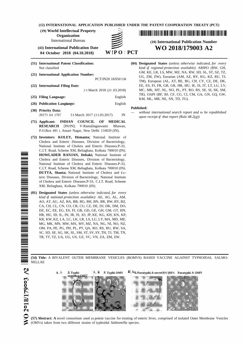
(54) Title: A BIVALENT OUTER MEMBRANE VESICLES (BOMVS) BASED VACCINE AGAINST TYPHOIDAL SALMO-NELLAE
< A . i. , Typhi- A. ii _ Typhi OMV . , Paratyphi A-secreOi¾'(ftI¾ Paratyphi A OMV
ool
00o
(57) Abstract: A novel consortium used as potent vaccine for treating of enteric fever, comprised of isolated Outer Membrane Vesicles(OMVs) taken from two different strains of typhoidal Salmonella species.

FIELD OF INVENTION:
The present invention relates to a novel consortium for a potent vaccine
for enteric fever, comprised of specific strains Salmonella Typhi and
Salmonella paratyphi A in equal proportion.
The Present invention also relates to a methodology for preparing the
said novel consortium based on outer membrane vesicles (OMV) .
BACKGROUND AND PRIOR ART OF THE INVENTION:
Enteric fever, a serious invasive bacterial infection, caused by Salmonella
enterica serovers Typhi and Paratyphi A (hereafter, S . Typhi and S.
Paratyphi A, respectively) is a major global burden in developed and
developing countries like India. Although S. Typhi is more prevalent,
affects 21.7 million cases and 200,000 deaths per year worldwide, S.
Paratyphi (A, B and C ) can cause significant enteric fever especially in
Asia as well as in travelers returning from these endemic areas (1, 2, 3).
Currently, there are only two licensed vaccines are available against S.
Typhi; a live attenuated galE mutant and a Vi-polysaccharide vaccine (4).
Although somewhat effective, they have their limitations such as they do
not provide long-term protection in children and they do not provide
significant long-term immunity in adults too.
Presently, the focus of vaccine research is on acellular vaccine because of
the certain drawbacks of conventional immunogens. In this changing
state of vaccine research, Outer Membrane Vesicles (OMVs) has got
significant importance. Neisseria meningitis OMV based licensed vaccine
is now presently available in market (5). As OMVs has got both LPS and
proteins, they do not need any artificial adjuvants (6) .
Though OMVs are used in drug development against various
microorganisms, such methods are often expensive and complicated as
most of the techniques employ infusion of different proteins from outside.
As the enteric fever is most predominant in developing countries, the
cost-effectivity should be the prime concern in field of vaccine
development.
Further, the conventional technology often devoid of providing
substantial protection against enteric fever caused by S. Typhi and also
results some hazardous side-effects in human.
Hence, there is always a need to provide an innovative formulation by
using a process based on Outer Membrane Vesicles (OMV) which are
overcoming the drawbacks of the conventional practice.
The present invention meets the above-mentioned long-felt need.
OBJECTIVES OF THE INVENTION:
The principal object of the present invention is to provide a simple yet
effective consortium comprised of isolated Outer Membrane Vesicles
(OMV) of Salmonella Typhi and Salmonella Paratyphi A in equal
proportion.
Another objective of the present invention is to provide a consortium
which is effective in treating enteric fever.
Yet another objective of the present invention is to provide a simple
consortium wherein any mutation or deletion of gene has not been
adopted to provide the end product.
Further objective of the present invention is to provide a simple
consortium wherein the mutation or deletion of gene is not incorporated
to reduce the expression of immune-dominant non-protective antigens.
Another objective of the present invention is to provide a simple
consortium wherein no antibiotics or any excipients have been used.
Yet another objective of the present invention is to provide a simple
consortium wherein no protective proteins is incorporated from outside.
Further objective of the present invention is to provide a simple
consortium wherein only isolated OMVs of Salmonella strain is used,
hence, it is cost-effective and environment friendly.
BRIEF DESCRIPTION OF THE ACCOMPANYING DRAWINGS:
It is to be noted, however, that the appended drawings illustrate only
typical embodiments of the present subject matter and are therefore not
to be considered for limiting of its scope, for the invention may admit to
other equally effective embodiments. The detailed description is
described with reference to the accompanying figures.
Some embodiments of system or methods in accordance with
embodiments of the present subject matter are now described, by way of
example, and with reference to the accompanying figures, in which:
Fig 1 and l a illustrate an electron micrograph of OMVs attached to
bacteria and isolated OMVs and characterization of isolated OMVs.
Fig 2a illustrates mice immunization by the consortium.
Fig 2b illustrates immunization and challenge regimen in mice.
Fig 3 illustrates a representative immunoblot analysis against OMVs,
from two typhoidal strains.
Fig 4 illustrates Dot blot analysis against extracted LPS from two
typhoidal strains.
Fig 5 illustrates a comparison of serum immunoglobulin titers in
immunized sera separately measured against each component OMVs of
bivalent OMV and heat-killed (HK) formulations.
Fig 5a illustrates serum immunoglobulin titers in immunized sera
separately measured against each component OMVs of bivalent OMV
formulation.
Fig 6 illustrates BOMVs induces the production of Thl and Thl7
polarizing cytokines in Ag-presenting BMDCs and splenic cells after
treatment.
Fig 7 illustrates Serum from bivalent immunized mice inhibits S. Typhi
and S. Paratyphi A motility.
Fig 8 illustrates immunization with the bivalent OMVs provides
protection in adult mice model.
Fig 9 illustrates colonization of Salmonella Typhi and Paratyphi A clinical
isolate in Salmonella Typhi monovalent OMV immunized mice.
Fig 10 illustrates colonization of Salmonella Typhi and Paratyphi A
clinical isolate in Salmonella Paratyphi A monovalent OMV immunized
mice.
Fig 11 illustrates the assessment of the effect of OMVs isolated from 24-
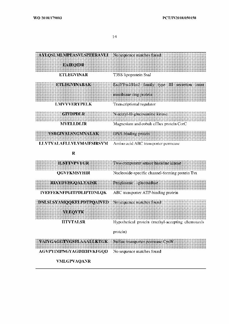
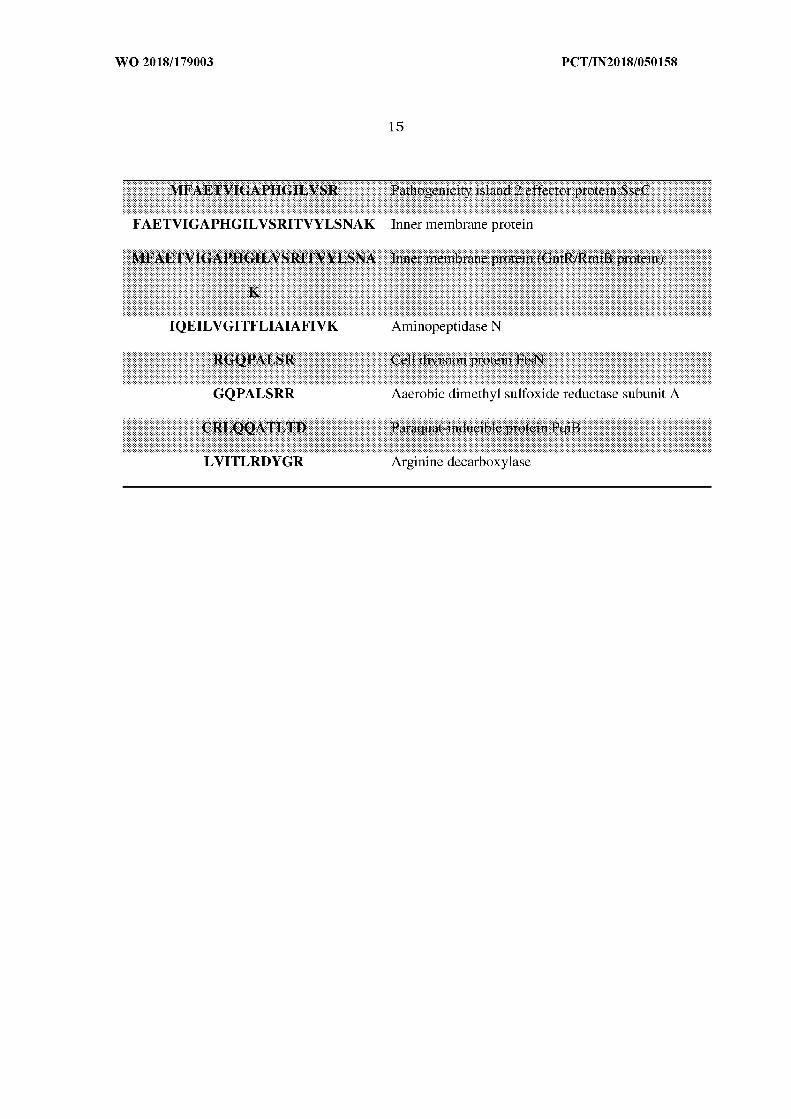
hours culture.
Fig 12a illustrates classification of Salmonella Typhi OMV-associated
proteins based on their location of appearance in the bacteria.
Fig 12b illustrates classification of Salmonella Parathyphi A OMV-
associated proteins based on their position in the bacteria.
Fig 12c illustrates OMV associated proteins.
DETAILED DESCRIPTION OF THE INVENTION:
The present subject matter relates to a novel formulation comprised of
isolated outer membrane vesicles from two thyphoidal Salmonella strains
such as Salmonella Typhi C-6953 and Salmonella Paratyphi A C-6915.
In the said formulations, two Salmonella strains are mixed in 1:1 ratio
i.e. 50% of Salmonella Typhi C-6953 and 50% of Salmonella paratyphi A-
6915.
The immunogenicity and protective efficacy have been studied on adult
mice after oral immunization with the said formulation.
The evaluation of the generation of humoral as well as cell mediated
immune response after oral immunization by measuring different
immunologic markers as well as anti-Vi polysaccharide specific serum
immunoglobulin and Thl/Thl7 specific cytokine response from splenic
and DCs (Dendritic Cells) were performed.
This bivalent OMVs based vaccine could be an ideal human vaccine
candidate against enteric fever.
The strains which are used in the said formulation are clinical isolates
and thus do not have any modification or induced mutation in them.
Mutating a specific gene for over-expression of protective antigen may
reduce immune-dominant non-protective antigen by mutating or deleting
it. Moreover, LPS mutants might have some adverse effects on the end
product; i.e., secreted OMVs. Mutating a gene also changes the bacterial
genetic make-up and might eventually produce a specific type of protein
which is not needed. Many other useful proteins could be lost in the
process.
But, the said formulation does not change the genetic make-up of the
microorganism and hence no such unwanted protein is produced.
No stress has been induced on the cultured bacteria in the form of
antibiotics or using minimal medium, which eventually increases the
cost-effectiveness of the final product.
Further, the said formulation does not incorporate antibiotics in it. Thus,
reducing the chance of spreading anti-microbial resistance.
Unlike conventional practice, the present invention does not add any
protein from outside or any other excipients such as lactose, sucrose,
gelatin, sorbitol, human serum albumin and hence it is free from post-
isolation purification steps.
From the analysis of the consortium, it has found that the consortium
also comprises substantially high number of outer, inner, periplasmic
and cytoplasmic protein, which have not infused from outside but were
found to be present naturally.
It also possesses high number of cytosolic proteins (even proteins like
DNA polymerase III, helicase, primase).
Any mutant stains has not been used in the novel formulation, the
instant invention only uses their native form to deliver their natural
contents in the host's body.
Further, effective short duration of immunization schedule can be
achieved by the novel formulation. The protection can stay for 3 to 6
months without any further booster doses than the regimen stated.
The process for preparing the novel consortium has used log-phase
culture of bacteria to isolate OMVs thus increasing the amount of TTSS
proteins which are more potent in nature as an immunogen.
Also, as per the present invention the OMVs contain Vi-polysaccharide of
Salmonella Typhi. The content of Vi-polysaccharide in the bivalent
formulation has been measured. The presence of Vi-polysaccharide in
vaccine constituents makes the vaccine more effective against Salmonella
Typhi infection because, Salmonella Typhi is covered with Vi-
polysaccharide, presence of anti-Vi antibiotics in the serum would
certainly elevated the level of protection. Presence of Paratyphi A OMVs
enlarges its protective nature further against Paratyphi A.
The detailed result has given below:

Bacterial strains and culture conditions
OMV antigens were prepared from S. Typhi C-6953 and S. Paratyphi A C-
6915, and S. Typhi C-6.946 and S. ParatyphiA BCR 148 for challenge
study were collected from National Institute of Cholera and Enteric
Diseases (NICED) culture bank. All strains were kept in 20% glycerol in
brain heart infusion broth (Difco, USA) at 80°C. Prior to experimentation,
each strain was grown in Tryptic Soy Broth (TSB; Difco, USA) at 37°C
under shaking conditions (100 rpm) or on plates in Tryptic Soy Agar
(TSA; Difco, USA).
Preparation of OMVs
OMVs were prepared from two Salmonella entenca strains with slight
modifications where cells were grown at 37°C under shaking condition
followed by centrifugation at 8000 rpm for 40 minutes at 4°C. Following
filtration by 0.22 µ m bacterial filters (Millipore, USA), OMVs were
subsequently purified by ultracentrifugation (4 h , 140,000 x g, 4°C)
using a Sorvall T-865 rotor, and re-suspended in Phosphate-Buffered
Saline (PBS, pH 7.4). The protein concentration was determined by the
modified Lowry protein assay kit (Pierce, USA). LPS O-Ag concentration
was determined by a method used by Dubois et al.
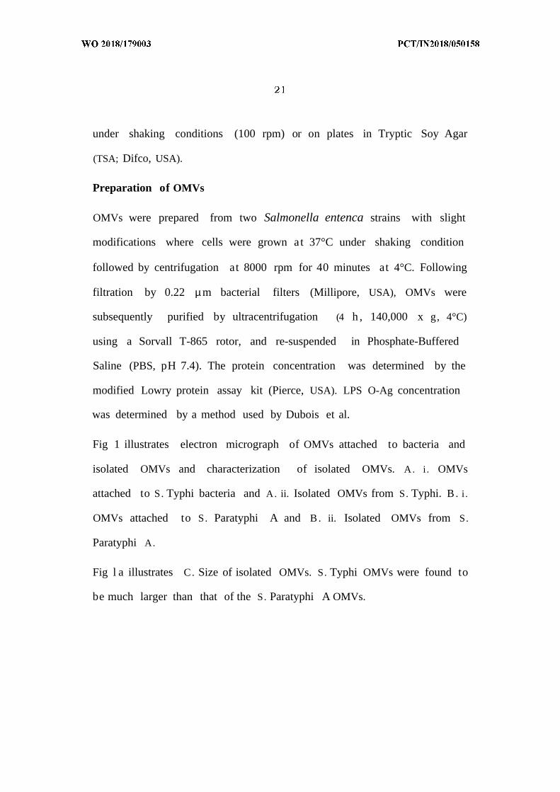
Fig 1 illustrates electron micrograph of OMVs attached to bacteria and
isolated OMVs and characterization of isolated OMVs. A. i . OMVs
attached to S. Typhi bacteria and A. ii. Isolated OMVs from S. Typhi. B. i .
OMVs attached to S. Paratyphi A and B. ii. Isolated OMVs from S.
Paratyphi A.
Fig l a illustrates C. Size of isolated OMVs. S. Typhi OMVs were found to
be much larger than that of the S. Paratyphi A OMVs.
Negative staining of OMVs and OMV-secreting bacteria
A 5µ1aliquot of secreted OMVs were placed on a carbon coated grid and
left for 1 minute for proper absorption. The grid was then washed with
two drops of Tris-HCl buffer. After blotting excess fluid, the sample was
stained with 2% aqueous solution of uranyl acetate. In case of negative
staining of bacteria-secreting OMVs, the same procedure was followed
with log-phase live bacterial cells. Both the negatively stained OMVs and
bacteria-secreting OMVs were observed under Tecnai 12 (as given in Fig.
1)·
Figure 2a illustrates BALB/c mice were immunized by oral gavage on day
0 and then two subsequent booster doses follow as stated. Mice were
challenged on day 35 via an intra-peritoneal challenge model.
From Figure 2b, the immunization and challenge regimen in mice can be
understood clearly. Mice were immunized on days 0, 14 and 28 and
challenged on 35th days after 1st immunization. Blood were collected
from on indicated days.
The experiments which have performed are given below:
Animals
Seven weeks old, BALB/c mice of either sex were taken from the animal
resource division of NICED, Kolkata. Male and female mice were caged
separately groups of 10 and maintained at a temperature of 25°C with
humidity at 75%. Mice were fed sterile food and water. All the animal
experiments were conducted following the standard operating procedure
as outlined by Committee for the Purpose of Control and Supervision of
Experiments on Animal (CPCSEA), Ministry of environment and forest,
Government of India. The animal experimental protocol was approved by
the Institutional Animal Ethical Committee of NICED with the project
approval no. PRO/ 108 May, 20 14-July, 2017.
Oral immunization
7 weeks old female BALB/c mice were kept empty stomach 24 hours
before the immunization date, water adlibitum. Mice were immunized
orally on days 0th, 14 th and 28th (Fig. 2 ) with 25 µg of purified S. Typhi
and S. Paratyphi A OMVs (1: 1) in 200 µ L of PBS following the protocol as
explained previously.
Collection of serum and stool
Blood was collected from the lateral tail vein at different time intervals on
the 0th, 14th, 21st, 28th, 35th, 78th, 90thday of first oral immunization.
The collected blood was taken in BD Microtainer (BD, NJ, USA) followed
by centrifugation (1000 rpm, 10 min and 4°C). Stools from immunized
and non-immunized mice were collected in an aseptic Eppendorf by
pressing the abdominal region. Stools were then homogenized by a
plastic homogenizer and centrifuged at 10000 x g for 10 min to remove
the debris. The supernatant was collected and stored.
The results of representative immunoblot analysis against OMVs, from
two typhoidal strains are given in figure 3 .A. SDS-PAGE profile of OMVs
extracted from two strains of typhoidal salmonellae. Lane M: Low
molecular weight marker (Bangalore GeNei), Lane 1: S. Typhi, Lane 2 : S.
Paratyphi A.B. Immunoblot against each component of the OMVs of the
bivalent formulation probed with 28th days anti- bivalent OMVs serum
from mice. Lane M: Pre-stained molecular weight marker (Bangalore
Genei), Lane 1: S. Typhi, Lane 2 : S. Paratyphi A OMV.
SDS-PAGE and immunoblot
The protein content of the OMVs recovered from Salmonella strains were
determined as described earlier in this paper. 80 µg of proteins were
boiled in 5x SDS-PAGE buffer and loaded onto a 12% SDS-PAGE gel. The
gel was then stained by either Coomassie or silver stain. For immunoblot
assay, gel was transferred onto nitrocellulose membrane (Bio-Rad, USA)
by using the ATTO AE-6687 (Japan) blot apparatus. The polyclonal
antibody rose in mice and HRP-conjugated rabbit anti-mouse secondary
IgG were used to detect the proteins which were immunogenic.
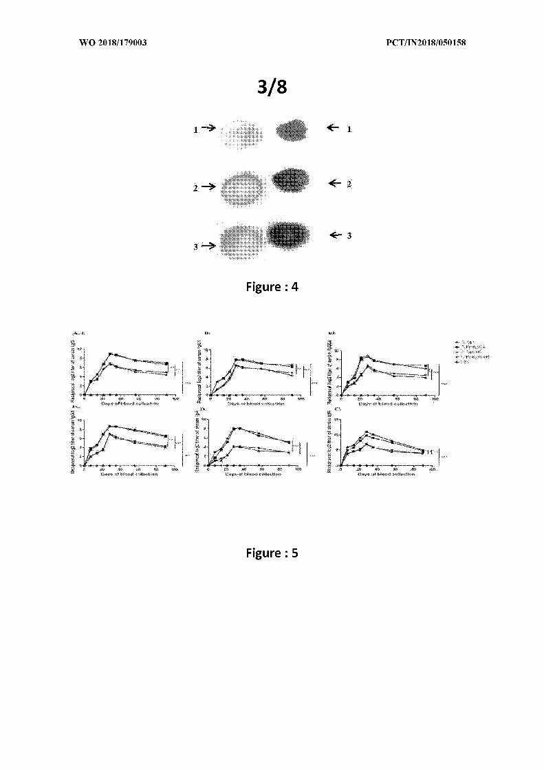
Fig. 4 illustrates dot blot analysis against extracted LPS from two
typhoidal strains. Lane 1 : S. Typhi LPS, Lane 2 : S. Paratyphi A LPS.
Here, 1, 2, and 3 denotes three different concentrations of LPSs against
which the dot blot analysis was performed.
Dot blot assay.
Dot blot analysis was done as described previously. Briefly, LPS of the
two strains were taken and blotted onto a nitrocellulose membrane. The
membrane was then washed with Tris-Buffered Saline (TBS) contains
0.1% Tween-20. The membrane was then incubated with primary and
secondary antibody successively, where OMV-immunized mice serum
was serving the purpose of a primary antibody and the blot was then
finally developed by chemiluminescence.
ELISA
Different immunoglobulins; e.g. IgG and its sub-types (IgGl, IgG2a,
IgG3), and IgA, slgA and IgM were measured by ELISA as stated by Keren
(23). Briefly, disposable polystyrene micro-titer wells (Nunc, Denmark)
were separately coated with OMVs 5 g/well) from either strains of the
immunogens (Table 1) and incubated for 18 h at 4 C. Wells were washed
and blocked with Bovine Serum Albumin (BSA; Sigma Chemical, USA).
After washing the wells with PBS-T (PBS with 0.5% Tween-20, Sigma
Chemicals, USA) and incubated with serially diluted serum samples,
100µ L HRP conjugated goat anti-mouse immunoglobulin was added and
incubated. After washing with PBS, the substrate o-phenyl-Di-amine
(OPD) was added to each well followed by stopping the reaction after 10
min by adding 100 µ L of 2 N sulphuric acid. OD492 was taken. The
experiments were repeated three times for each immunoglobulin, with
the immunized and non-immunized serum, collected from individual
mice, before, during and after immunization. The same procedure was
carried out when ELISA were done against Vi-polysaccharide of S. Typhi.
A serum immunoglobulin titer in immunized sera were separately
measured against each component OMVs of bivalent OMV and heat-
killed (HK) formulations. A. Serum IgG (i), IgGl (ii), IgG2a (iii), IgG3 (iv);
B. Serum IgA; C. Serum IgM response against each of the two OMVs and
heat-killed immunogens at pre-immunization, immunization and
postimmunization. The horizontal axis indicates the days of blood
collection. Data represented here are the mean values +/- Standard
Deviation (SD) of three independent experiments. The differences in post-
immunization day wise response of each of the studied antibodies
against each of the two OMVs were highly significant (P value< 0.005)
(shown in Figure 5).
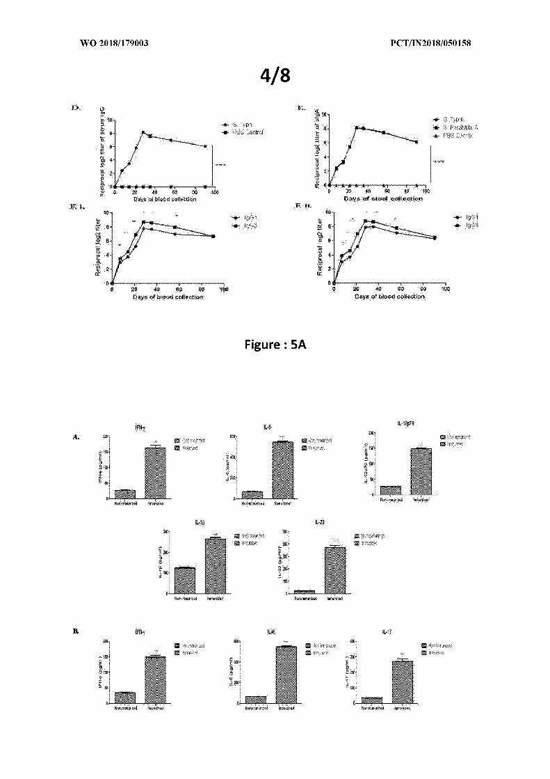
Fig 5a illustrates anti-Vi serum IgG; E. Secretory IgA; F. i . ii. Serum IgGl
and IgG3 response against each of the two OMVs, Salmonella Typhi and
Paratyphi A, respectively at pre-immunization, immunization and post-
immunization periods. The high serum IgG3 titer against serum IgGl
titer indicates higher Thl cell-mediated immune response in adult mice
sera after three doses of immunization. The horizontal axis indicates the
days of blood collection. Data represented here are the mean values +/ -
Standard Deviation (SD) of three independent experiments. The
differences in post-immunization day wise response of each of the
studied antibodies against each of the two OMVs were highly significant
(P value< 0.005).
Ex vivo studies on isolated dendritic cells
Dendritic cells from bone marrow of non-immunized BALB/c mice were
cultured for 7 days in complete RPMI containing 10% FBS in the
presence of 20 ng/ml GM-CSF (Tonbo). Cells were then treated with 100
ng/ml bivalent OMV and incubated in 37° C for 24 hours in presence of
5% C02. Different cytokines, namely IFN-γ , IL-4, IL-12p70, IL- 1β and
IL-23 were then measured (refer Fig. 6 A) by cytokine ELISA kit.
Splenocyte re-stimulation assay.
After 2 weeks from the end of last immunization, splenic cells from
immunized BALB/c mice were cultured for 2 hours in complete RPMI
containing 10% FBS. Cells were then treated with 100 ng/ml bivalent
OMV and incubated in 37 C for 24 hours in presence of 5% C02.
Different cytokines, namely IFN-γ , IL-6 and IL-17 were then measured
(Fig. 6 B) by cytokine ELISA kit (Invitrogen, USA) (8).
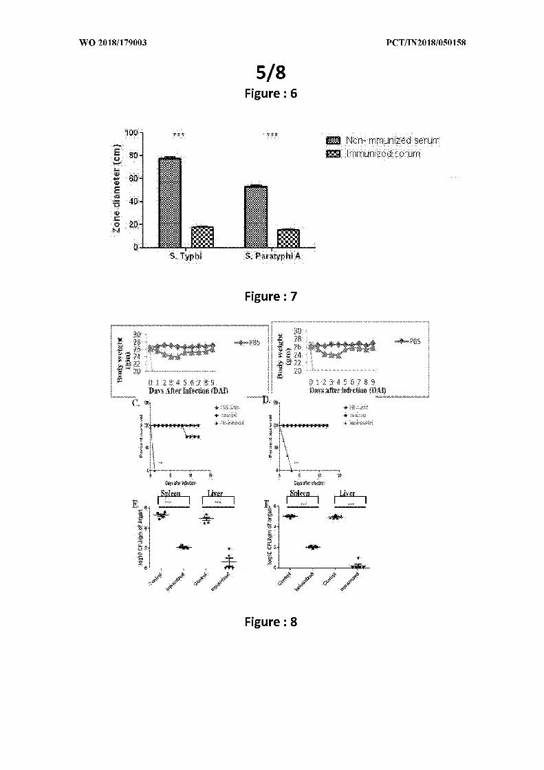
Figure 7 illustrates serum from bivalent immunized mice inhibits S.
Typhi and S . Paratyphi A motility.
Motility assay
Motility assay was done as previously described, with modifications.
Briefly, the immunized and non-immunized serum samples were mixed
with PBS at a concentration of 1:400 and poured on soft agar (0.3%)
plates. The plates were kept for an hour to get the serum mixed PBS
soak in the plate. After the plates became dry, log-phase bacteria (OD600
= 0.8) were pricked in the middle of the plate. The plates were then
incubated at 37°C for 24 hours. After 24 hours, the results were seen as
in Fig. 7 .
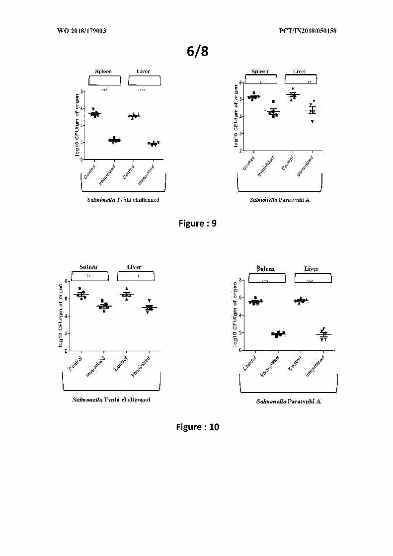
Figure 8 illustrates an Immunization with the bivalent OMVs provides
protection in adult mice model. Mice were immunized with 1:1 mixture of
typhoidal OMVs using 25 µg total OMVs per dose and a three-dose
immunization. Mice were then challenged with 1 x 10 6 CFU/ml of each
challenge strains intra-peritoneally and observed for the period of
survival for 12 days. A., B. Body weight was measured for each mouse
until 9th day post-infection and C , D. percent survival was calculated.
E., F. Mice were further challenged with 2 x 10 8 CFU/ml heterologous
strains of challenge bacteria and the systemic infection of the bacteria in
these mice were determined by serial dilution of the spleen and liver.
Bivalent S. Typhi and S. Paratyphi A OMV protect adult mice from S.
Typhi and S. Paratyphi A challenge
After four successive oral immunizations with bivalent OMVs
formulation, protective efficacy was observed in an adult mice intra
peritoneal model (Fig. 8). At 9 DPI (Days Post Infection), it was observed
that 1 x 10 s 1 x 10⁶ CFU/gm of spleen in non-immunized mice, whereas,
2 x 10² CFU/gm of spleen was the highest colonization found in
immunized mice's spleens.
Tissue homogenates from liver reveals only 10-100 organisms/ g in
case of immunized mice, whereas, in non-immunized mice, 5-log fold
higher colonization ability was observed.
Both immunized and non-immunized mice were challenged with 2 x 10⁶
CFU/ml intra-peritoneally and kept them for 12 days for survival assay.
In case of non-immunized mice, all the mice died within 1 4 days. But,
80% and 100% immunized mice were still alive. This result suggests
that, our bivalent formulation is inhibiting the systemic infection of
typhoidal salmonellae in mice and it indeed protecting the mice from
lethal infection.
In the majority of cases, the data presented are not normally distributed
due to biological variation. Therefore, non-parametric tests were used for

all data analysis. Comparison between two categorical variables was
made using the two-tailed student's test.
Comparison between multiple categorical variables was made using the
one-tailed student's test. Each experiment was repeated at least three
times. A P value of <0.05 or <0.01 were considered significant GraphPad
Prism 5 for Windows OS was used for all statistical analyses.
The effectiveness of both Salmonella Typhi and Paratyphi A OMVs have
been studied through various experiments.
When the mice were immunized with Salmonella Typhi OMVs, they were
protected from Salmonella Typhi infection (evaluated from the bacterial
count from spleen and liver 3 days' post infection). But when the same
Salmonella Typhi OMVs-immunized mice were challenged with
Salmonella Paratyphi A, very less amount of protection was found. The
same trend was seen when monovalent Salmonella Paratyphi A OMVs is
used to immunize mice. They were protected from Salmonella Paratyphi
A infection, but not protected from Salmonella Typhi infection. The
results are following:
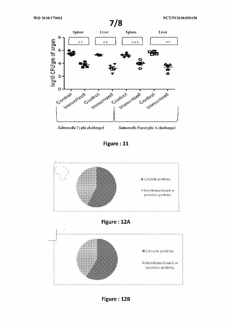
Figure 9 illustrates the first panel shows colonization of Salmonella Typhi
clinical isolate in Salmonella Typhi OMV-immunized mice. On the other
hand, the second panel shows colonization of Salmonella Paratyphi A
clinical isolate in Salmonella Typhi OMV-immunized mice. At least 2 fold
more colonization was seen when Salmonella Typhi OMV-immunized
mice were challenged with Salmonella Paratyphi A rather than
Salmonella Typhi clinical isolate. Homologous protection was seen, but
no heterologous or cross-protection was observed.
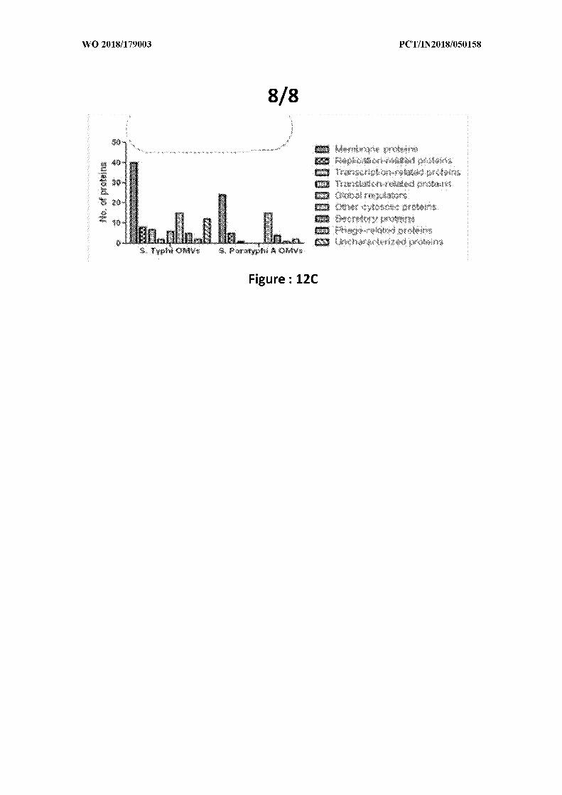
Figure 10 illustrates the first panel shows colonization of Salmonella
Typhi clinical isolate in Salmonella Paratyphi A OMV-immunized mice.
On the other hand, the second panel shows colonization of Salmonella
Paratyphi A clinical isolate in Salmonella Paratyphi A OMV-immunized
mice. At least 2 fold more colonization was seen when Salmonella
Paratyphi A OMV-immunized mice were challenged with Salmonella
Typhi rather than Salmonella Paratyphi A clinical isolate. Homologous
protection was seen, but no heterologous or cross -protection was
observed in this case also.
The effect of OMVs isolated from 24 hours' culture was assessed.
Although this result shows that the immunized mice are significantly
protected from the challenge, but the level of protection is much higher
in case of OMVs isolated from 5 hours' culture (refer to Patent
application fig. 8. E, F ) (as shown in figure 11).
The OMVs from log-phase culture bacteria caused much less colonization
in spleen and liver and substantially much more effective them any other
formulations. It is rich in many proteins which were reported to be
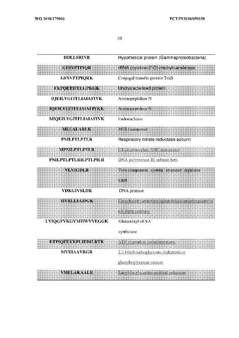
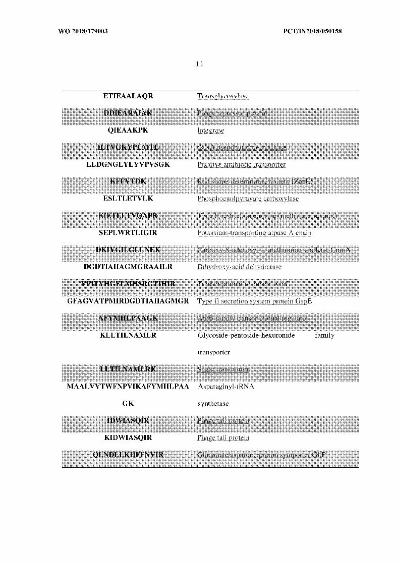
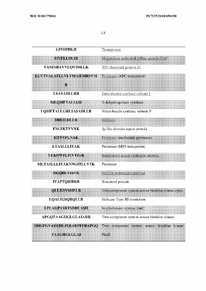
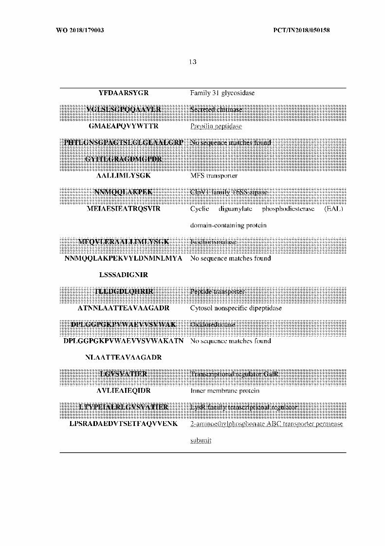
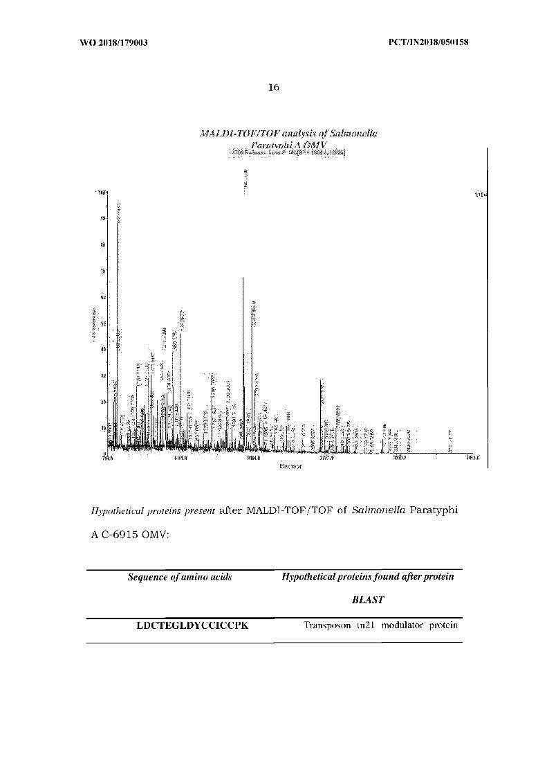
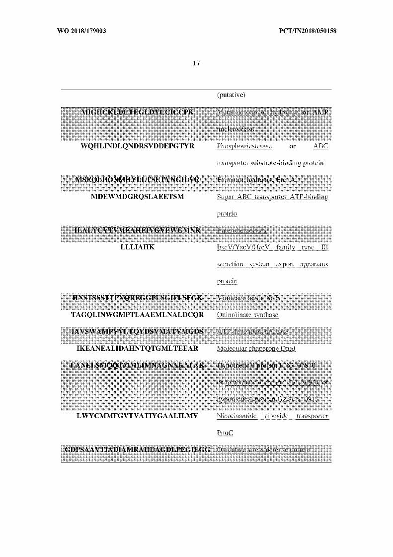
secreting via OMVs until recently (Fig. 12).
The OMVs of two strains of bacteria as claimed contains proteins of
different sizes as well as LPS in them. Western blot analysis indicates the
presence of strong immune response against the immunogens present in
BOMVs. Dot blot analysis serves the purpose of proving this immunogen
to be effective and immunogenic against LPS of these two strains. Three
doses of oral immunization of these BOMVs formulation in mice induces
a significant rise in the level of immunoglobulins specific for isolated
OMVs. Presence of slgA against OMVs formulation and IgG specific for
Vi-polysaccharide of S. Typhi shows the potency of our bivalent
formulation against S. Typhi infection.
As Vi-polysaccharide is the outer covering of S. Typhi, immunoglobulins
present against this component indicates the presence of Vi-
polysaccharide in the BOMVs. Because of their intra-cellular nature,
both S. Typhi and S. Paratyphi A can only be eradicated from the host in
the presence of significant Thl cell-mediated immune response along
with humoral immune response. A Thl biased immune response was
seen in the ELISA data.
Also, a significant up-regulation in the level of IFN-γ , IL-6, IL-12p70, IL-
1β and IL-23 from the isolated BMDCs and IFN-γ , IL-6 and IL-17 from
splenocytes shows that the induced response was a result of mainly a
Thl and Thl7 cell mediated immune response. Moreover, as verified by
sera and splenocytes adoptive transfer experiments, the protective effect
of BOMVs vaccination was dependent on both humoral and cellular
immunity. So, both humoral as well as cellular arms of the host's
immune system are being activated upon the exposure of BOMVs in
mice. Our BOMVs immunized mice sera can also inhibit the motility of
the wild type strains of typhoidal salmonellae. Inhibition of motility
means the bacteria will no longer be able to find their receptors for
binding on the human epithelium thus, rendering their inability to cause
infection. MTT assay was done to check the reactogenicity of BOMVs. It
was found BOMVs were less reactogenic than the conventional heat-
killed and whole cell lysate immunogens.
Inhibition to cause infection in mice was further confirmed by anti-
colonization and survival assays. In our anti-colonization assay,
immunized mice were challenged with circulating strains of typhoidal
salmonellae via the intra-peritoneal route. Significant increase in the
level of survival in the BOMVs immunized group was seen. Also, the
presence of typhoidal salmonellae in spleen and liver were found to be
significantly less in immunized mice. Taken together, these findings
suggested us that BOMVs could be used as a novel non-living human
vaccine candidate against S.Typhi and S.Paratyphi A infections in future.
Statistical analysis
In the majority of cases, the data presented are not normally distributed
due to biological variation. Therefore, non-parametric tests were used for
all data analysis. Comparison between two categorical variables was
made using the two-tailed student's t test. Comparison between multiple
categorical variables was made using the one-tailed student's t test. Each
experiment was repeated at least three times. A P value of <0.05 or <0.01
were considered significant. GraphPad Prism 5 for Windows OS was used
for all statistical analyses.
The non-limiting advantages are given below:
• The consortium consists of OMVs (BOMVs) together as an
immunogen and contain proteins of different sizes and LPS among
other constituents.
• The said consortium induces significant immune response after
three doses of oral immunization in mice. Western blot analysis
assures their immunogenicity. Dot blot analysis shows the ability
of induction of immunogenicity against LPS of circulating strains.
• Significant rise in the serum IgG, IgM, IgA, slgA was seen after
three doses of oral immunization. High level of serum IgG3 instead
of serum IgGl indicates a Thl-baised immune response. A high
titer against the Vi-polysaccharide of S. Typhi was seen. This
indicates the induction of a humoral immune response in the mice.
• Treating the isolated BMDCs with the said consortium results in
the elevation of Thl -biased cytokines. Rise in Thl and Thl7-baised
cytokines were seen in isolated splenocytes from immunized mice.
This indicates the induction of a strong Thl-cell mediated
immunity in mice.
• The said consortium immunized mice sera can significantly inhibit
the motility of wild type circulating strains of typhoidal
salmonellae. Inhibition of motility renders the bacteria ineffective
in the induction of its virulence.
• The said formulation is much less reactogenic than the
conventional heat-killed and whole cell lysate immunogens.
• The formulation immunized mice were protected against wild type
circulating strains of typhoidal salmonellae. The level of
colonization in spleen and liver were also found to be significantly
less than that of the non-immunized mice.
WE CLAIM:
1. A novel consortium used as potent vaccine for treating of enteric fever,
comprised of isolated Outer Membrane Vesicles (OMVs) taken from two
different strains of typhoidal Salmonella species.
2. The consortium as claimed in claim 1, wherein the Salmonella strains
are Salmonella Typhi C-6953 and Salmonella paratyphi A C-6915.
3. The consortium as claimed in claim 1, wherein said Salmonella strains
are mixed in 1:1 ratio i.e, 50% of Salmonella Typhi C-6953 and 50%
Salmonella Paratyphi A C-6915.
4. The consortium claimed in claim 1, further comprises high number of
outer, inner, periplasmic and cytoplasmic proteins among others.
5. The consortium as claimed in claim 1, further comprises high number
of cytosolic proteins such as DNA, polymerase, helicase and primase
which were never been addressed to be secreting through OMVs.
6. A method for preparing outer membrane vesicles (OMV) comprises the
steps of:
i ) Growing of cells at 37°C, under shaking condition followed by
centrifugation at a suitable condition;
ii) Subjected to filtration by 0.22 µ bacterial filtration followed by
purification though ultracentrifugation at a suitable condition by using a
rotor;
iii) Re-suspension in phosphate-buffered saline at pH 7.4.
7. The method for preparing outer membrane vesicles as claimed in claim
6, wherein the centrifugation occurs at 8000rpm for 40 min at 4°C.
8. The method for preparing Outer Membrane Vesicles (OMVs) as claimed
in claim 6, wherein the ultra centrifugation occurs for 4 hours at
140,000 x g at 4°C.


![Intellectual Property Rights [IPR] · 2017-03-08 · intellectual property either by development and commercialization after securing available protection, by publication, or both](https://img.pdfslide.net/doc/110x75/5f5fe0d3302c7e6d3d0fb0d1/intellectual-property-rights-ipr-2017-03-08-intellectual-property-either-by.jpg)