Embed Size (px)
Citation preview
World Journal ofMeta-Analysis
World J Meta-Anal 2019 May 31; 7(5): 184-258
ISSN 2308-3840 (online)
Published by Baishideng Publishing Group Inc
W J M A World Journal ofMeta-Analysis
Contents Irregular Volume 7 Number 5 May 31, 2019
REVIEW184 Current state and future direction of screening tool for colorectal cancer
Hong JT, Kim ER
MINIREVIEWS209 Hepatitis B reactivation in patients with hepatitis B core antibody positive and surface antigen negative on
immunosuppressantsWu CCH, Kumar R
218 Present state of endoscopic ultrasonography-guided fine needle aspiration for the diagnosis of autoimmune
pancreatitis type 1Sugimoto M, Takagi T, Suzuki R, Konno N, Asama H, Sato Y, Irie H, Watanabe K, Nakamura J, Kikuchi H, Takasumi M,
Hashimoto M, Hikichi T, Ohira H
SYSTEMATIC REVIEW224 Hepatic gastrointestinal stromal tumor: Systematic review of an exceptional location
Manuel-Vázquez A, Latorre-Fragua R, de la Plaza-Llamas R, Ramia JM
234 Scoring criteria for determining the safety of liver resection for malignant liver tumorsHarada K, Nagayama M, Ohashi Y, Chiba A, Numasawa K, Meguro M, Kimura Y, Yamaguchi H, Kobayashi M, Miyanishi K,
Kato J, Mizuguchi T
META-ANALYSIS249 Pancreatic stents to prevent post-endoscopic retrograde cholangiopancreatography pancreatitis: A meta-
analysisSugimoto M, Takagi T, Suzuki R, Konno N, Asama H, Sato Y, Irie H, Watanabe K, Nakamura J, Kikuchi H, Takasumi M,
Hashimoto M, Hikichi T, Ohira H
WJMA https://www.wjgnet.com May 31, 2019 Volume 7 Issue 5I

ContentsWorld Journal of Meta-Analysis
Volume 7 Number 5 May 31, 2019
ABOUT COVER Editorial Board Member of World Journal of Meta-Analysis, Singh Rajender,PhD, Senior Scientist, Endocrinology Division, Central Drug ResearchInsititue, Lucknow 226001, India
AIMS AND SCOPE The World Journal of Meta-Analysis (WJMA) is an online, open-access, single-blind peer-reviewed journal that is published by Baishideng PublishingGroup. Accepting both solicited and unsolicited manuscripts, the WJMAmainly publishes high quality meta-analysis and systematic review articlesto lead the development of various fields of clinical medicine. The type ofarticles published in the WJMA include the editorial, minireview, review,meta-analysis, meta-regression, systematic review, and evidence-basedpractice. The primary aim of the WJMA is to provide scholars and readers fromvarious fields of clinical medicine with a platform to publish high qualitymeta-analysis and systematic review articles and communicate theirresearch findings online. The WJMA mainly publishes research results and findings obtainedthrough meta-analysis and systematic review in a wide range of areas,including medicine, pharmacy, preventive medicine, stomatology, nursing,medical imaging, and laboratory medicine.
INDEXING/ABSTRACTING The WJMA is now abstracted and indexed in Emerging Sources Citation Index (Web
of Science), China National Knowledge Infrastructure (CNKI), China Science and
Technology Journal Database (CSTJ), and Superstar Journals Database
RESPONSIBLE EDITORSFOR THIS ISSUE
Responsible Electronic Editor: Yun-Xiaojian Wu Proofing Editorial Office Director: Jin-Lei Wang
NAME OF JOURNALWorld Journal of Meta-Analysis
ISSNISSN 2308-3840 (online)
LAUNCH DATEMay 26, 2013
FREQUENCYIrregular
EDITORS-IN-CHIEFGiuseppe Biondi-Zoccai
EDITORIAL BOARD MEMBERShttps://www.wjgnet.com/2308-3840/editorialboard.htm
EDITORIAL OFFICEJin-Lei Wang, Director
PUBLICATION DATEMay 31, 2019
COPYRIGHT© 2019 Baishideng Publishing Group Inc
INSTRUCTIONS TO AUTHORShttps://www.wjgnet.com/bpg/gerinfo/204
GUIDELINES FOR ETHICS DOCUMENTShttps://www.wjgnet.com/bpg/GerInfo/287
GUIDELINES FOR NON-NATIVE SPEAKERS OF ENGLISHhttps://www.wjgnet.com/bpg/gerinfo/240
PUBLICATION MISCONDUCThttps://www.wjgnet.com/bpg/gerinfo/208
ARTICLE PROCESSING CHARGEhttps://www.wjgnet.com/bpg/gerinfo/242
STEPS FOR SUBMITTING MANUSCRIPTShttps://www.wjgnet.com/bpg/GerInfo/239
ONLINE SUBMISSIONhttps://www.f6publishing.com
© 2019 Baishideng Publishing Group Inc. All rights reserved. 7041 Koll Center Parkway, Suite 160, Pleasanton, CA 94566, USA
E-mail: [email protected] https://www.wjgnet.com
WJMA https://www.wjgnet.com May 31, 2019 Volume 7 Issue 5II
W J M A World Journal ofMeta-Analysis
Submit a Manuscript: https://www.f6publishing.com World J Meta-Anal 2019 May 31; 7(5): 234-248
DOI: 10.13105/wjma.v7.i5.234 ISSN 2308-3840 (online)
SYSTEMATIC REVIEW
Scoring criteria for determining the safety of liver resection formalignant liver tumors
Kohei Harada, Minoru Nagayama, Yoshiya Ohashi, Ayaka Chiba, Kanako Numasawa, Makoto Meguro,Yasutoshi Kimura, Hiroshi Yamaguchi, Masahiro Kobayashi, Koji Miyanishi, Junji Kato, Toru Mizuguchi
ORCID number: Kohei Harada(0000-0002-3245-6980); MinoruNagayama (0000-0003-3309-4309);Yoshiya Ohashi(0000-0002-0287-6722); Ayaka Chiba(0000-0002-4602-3852); KanakoNumasawa (0000-0001-9375-1310);Makoto Meguro(0000-0002-9170-6919); YasutoshiKimura (0000-0002-7790-9250);Horoshi Yamaguchi(0000-0002-8121-1277); MasahiroKobayashi (0000-0002-4714-2183);Koji Miyanishi(0000-0002-6466-3458); Junji Kato(0000-0002-7367-8344); ToruMizuguchi (0000-0002-8225-7461).
Author contributions: Harada Kand Miyanishi K conceptualizedand designed the systematicreview; Ohashi Y, Chiba A, andNumasawa K collected data fromthe radiological database;Nagayama M and Meguro Msearched for and screened thearticles; Kimura Y, Yamaguchi H,and Kobayashi M assessed thearticles for eligibility; Harada Kand Yamaguchi H carried out thestatistical analyses; Kato Jsupervised and audited thepreparation of the manuscript;Harada K and Mizuguchi T draftedthe initial manuscript; andMizuguchi T finalized themanuscript. All of the authorsreviewed and approved the finalmanuscript as submitted.
Supported by a Grant-in-Aid forScientific Research from theMinistry of Education, Culture,Sports, Science, and Technology,Japan, No. 23591993 to TM, andNo. 24791437 to MM, No.
Kohei Harada, Minoru Nagayama, Makoto Meguro, Yasutoshi Kimura, Hiroshi Yamaguchi, ToruMizuguchi, Departments of Surgery, Surgical Science, and Oncology, Sapporo MedicalUniversity, Sapporo, Hokkaido 060-8556, Japan
Kohei Harada, Yoshiya Ohashi, Ayaka Chiba, Kanako Numasawa, Division of Radiology,Sapporo Medical University Hospital, Sapporo, Hokkaido 060-8556, Japan
Kohei Harada, Toru Mizuguchi, Sapporo Medical University Postgraduate School of HealthScience and Medicine, Sapporo Medical University, Sapporo, Hokkaido 060-8556, Japan
Masahiro Kobayashi, Research and Education Center for Clinical Pharmacy, KitasatoUniversity School of Pharmacy, Tokyo 108-8641, Japan
Koji Miyanishi, Junji Kato, Department of Internal Medicine IV, Sapporo Medical University,Sapporo, Hokkaido 060-8556, Japan
Toru Mizuguchi, Department of Nursing and Surgical Science, Sapporo Medical University,Sapporo 0608543, Japan
Corresponding author: Toru Mizuguchi, MD, PhD, Professor, Surgeon, Department of Nursingand Surgical Science, Sapporo Medical University, S-1, W-17, Chuo-ku, Sapporo, Hokkaido060-8556, Japan. [email protected]: +81-11-612111Fax: +81-11-6125525
AbstractBACKGROUNDLiver resection has become safer as it has become less invasive. However, theminimum residual liver volume (RLV) required to maintain homeostasis isunclear. Furthermore, the formulae used to calculate standard liver volume (SLV)are complex.
AIMTo review previously reported SLV formulae and the methods used to evaluatethe minimum RLV, and explore the association between liver volume andmortality.
METHODSA systematic review of Medline, PubMed, and grey literature was performed.References in the retrieved articles were cross-checked manually to obtain furtherstudies. The last search was conducted on January 20, 2019. We developed an
WJMA https://www.wjgnet.com May 31, 2019 Volume 7 Issue 5234
17K10672; to T Mizuguchi. Part ofthis study was also supported byAstellas Pharma, Inc., No.RS2018A000763, Tokyo, Japan;Daiichi Sankyo Company, No.1800461, Tokyo, Japan; Shionogi &Co., No. RS2018A000439931,Osaka, Japan; Merk Serono, No.MSJS20180613001, Tokyo, Japan;Sapporo Doto Hospital, No.30037656, Sapporo, Japan; NoguchiHospital, No. 30047663, Otaru,Japan; Doki-kai TomakomaiHospital, No. 30047674,Tomakomai, Japan; TsuchidaHospital, No. 30057704, Sapporo,Japan; Ikuta Hospital, No.30057704, Shiraoi, Japan was givento TM.
Conflict-of-interest statement: Theauthors declare that they have noconflicts of interest.
Open-Access: This article is anopen-access article which wasselected by an in-house editor andfully peer-reviewed by externalreviewers. It is distributed inaccordance with the CreativeCommons Attribution NonCommercial (CC BY-NC 4.0)license, which permits others todistribute, remix, adapt, buildupon this work non-commercially,and license their derivative workson different terms, provided theoriginal work is properly cited andthe use is non-commercial. See:http://creativecommons.org/licenses/by-nc/4.0/
Manuscript source: Invitedmanuscript
Received: March 26, 2019Peer-review started: March 26, 2019First decision: April 30, 2019Revised: May 20, 2019Accepted: May 22, 2019Article in press: May 22, 2019Published online: May 31, 2019
P-Reviewer: Shen ZY, Gumbs A,Lee HCS-Editor: Dou YL-Editor: AE-Editor: Wu YXJ
SLV formula using data for 86 consecutive patients who underwent hepatectomyat our institution between July 2009 and August 2011.
RESULTSLinear regression analysis revealed the following formula: SLV (mL) = 822.7 ×body surface area (BSA) − 183.2 (R2 = 0.419 and R = 0.644, P < 0.001). We retrieved25 studies relating to SLV formulae and 12 studies about the RLV required forsafe liver resection. Although the previously reported formulae included variouscoefficient and constant values, a simplified version of the SLV, the common SLV(cSLV), can be calculated as follows: cSLV (mL) = 710 or 770 × BSA. Theminimum RLV for normal and damaged livers ranged from 20%-40% and 30%-50%, respectively. The Sapporo score indicated that the minimum RLV rangesfrom 35%-95% depending on liver function.
CONCLUSIONWe reviewed SLV formulae and the minimum RLV required for safe liverresection. The Sapporo score is the only liver function-based method fordetermining the minimum RLV.
Key words: Standard liver volume; Residual liver volume; Hepatectomy; Mortality; Liverfailure; Liver function
©The Author(s) 2019. Published by Baishideng Publishing Group Inc. All rights reserved.
Core tip: We systematically reviewed standard liver volume (SLV) formulae, methodsfor assessing the minimum residual liver volume (RLV) required for safe liver resection,and the association between liver volume and mortality. Although the reported SLVformulae contained different coefficient/constant values, a simplified version of the SLV,the common SLV (cSLV), can be calculated as follows: cSLV (mL) = 710 or 770 × bodysurface area. The Sapporo score is the only liver function-based method for determiningthe minimum RLV.
Citation: Harada K, Nagayama M, Ohashi Y, Chiba A, Numasawa K, Meguro M, Kimura Y,Yamaguchi H, Kobayashi M, Miyanishi K, Kato J, Mizuguchi T. Scoring criteria fordetermining the safety of liver resection for malignant liver tumors. World J Meta-Anal 2019;7(5): 234-248URL: https://www.wjgnet.com/2308-3840/full/v7/i5/234.htmDOI: https://dx.doi.org/10.13105/wjma.v7.i5.234
INTRODUCTIONLiver resection is a potentially curative treatment for malignant liver tumors, such ashepatocellular carcinoma (HCC) and metastatic liver cancer, in cases in which nometastasis is present in other organs[1,2]. Although the mortality rate associated withliver resection has decreased, surgical complications still occur[3-7]. To ensure that liverresection is performed safely, it is important to preoperatively evaluate patients’ liverfunction so that it is possible to estimate the maximum liver volume that can be safelyremoved[8,9]. The Child-Pugh classification and the liver damage classificationestablished by the Liver Cancer Study Group of Japan are used to evaluate liverfunction[10-13]. The indications for liver resection are grade A or B liver function,according to either classification system. However, liver function varies greatlybetween grade A and B in patients with HCC. Therefore, in HCC patients it is difficultto accurately predict the maximum safe extent of liver resection.
Recent advances in radiological assessments of the liver have made it possible toprecisely calculate liver volume prior to liver resection[8,9,14-16]. Multi-detector-rowcomputed tomography (MDCT) can be used to evaluate not only liver volume, butalso patients’ individual anatomies prior to liver resection[17-19]. The aim of thissystematic review was to summarize the methods used to assess liver volume in orderto aid the establishment of a standard formula for calculating standard liver volume(SLV). In addition, we attempted to summarize the relationship between liver volumeand liver failure in order to facilitate safe liver resection.
WJMA https://www.wjgnet.com May 31, 2019 Volume 7 Issue 5
Harada K et al. Scoring criteria for liver resection
235
MATERIALS AND METHODSThis study was approved by the internal review board of Sapporo Medical University(approval ID: 302-195 and approval date: February 14, 2019). The Preferred ReportingItems for Systematic Reviews and Meta-Analyses (PRISMA) statement guidelines forconducting and reporting meta-analyses were followed[15]. To conduct this study, thestudy protocol was published on PROSPERO, which is the international prospectiveregister of systematic reviews (reference: No. CRD42019123642).
Estimation of SLVBetween July 2009 and August 2011, 86 consecutive patients who underwent liverresection for malignant tumors were enrolled in this study. Clinical laboratory tests,including of the serum levels of aspartate aminotransferase, alanine aminotransferase,albumin, hyaluronic acid, hepatocyte growth factor, and antithrombin III (ATIII); theprothrombin time (PT); the indocyanine green retention rate at 15 min (ICGR15); andthe platelet count were evaluated prior to liver resection. The uptake ratio of the heartat 15 min to that seen at 3 min (HH15) and the uptake ratio of the liver to the liverplus heart at 15 min (LHL15) were obtained from time activity curves of 99 m Tc-galactosyl human serum albumin scintigraphy.
Liver volume was evaluated using 64-row MDCT (LightSpeed VCT VISION; GEHealthcare, Milwaukee, WI, United States). The images were obtained in four phases,the early arterial phase, portal vein phase, hepatic vein phase, and late phase. A ZIOSTATION 2 (Ziosoft Inc., Tokyo, Japan) was used to calculate liver volume. Theimages of the hepatic vein phase were used for volumetry, and image analysis wasrestricted to the first and second branches of the portal and hepatic veins, as describedpreviously[20].
Definition of liver dysfunctionLiver failure was defined as a serum bilirubin concentration of > 10 mg/dL for > 2 d.Liver dysfunction was defined as a total bilirubin level of ≥ 3 mg/dL and a PT valueof < 50% within 7 d after liver resection[21].
Database searchesA systematic review of Medline, PubMed, and grey literature was performed.References from the retrieved articles were also cross-checked manually to obtainfurther studies. When more than one study from the same institution was found, onlythe publication with the most complete data was included. The last search wasconducted on January 20, 2019. The search strategy for the PubMed database was asfollows: {[“liver” (MeSH Terms) OR “liver” (All Fields)] AND volume (All Fields)}AND calculation (All Fields). The searches of other databases were conducted usingthe same medical subject headings (MeSH) and keywords in various combinations.
Statistical analysesPatient demographics and perioperative laboratory tests were extracted from thedatabase, and differences between the groups were compared using the chi-squaretest followed by a post-hoc 2 × 2 Fisher’s exact test. The unpaired t-test was used forcomparisons between the no liver dysfunction group (n = 78) and the liverdysfunction group (n = 8). The relationships among the various clinical parameterswere evaluated using Spearman’s rank correlation coefficient. The intraclasscorrelation coefficient (ICC) was used to assess inter-rater reliability. All calculationswere performed using the SPSS 20.0 software program (SPSS Inc., Chicago, IL, UnitedStates). All results are expressed as the mean together with minimum and maximumlevels. P-values of < 0.05 were considered to be statistically significant.
RESULTS
SLV at our institutionWe investigated the cases of patients who underwent hepatectomy for variousmalignancies, including HCC, at our institution between July 2009 and August 2011.Table 1 shows the clinical demographics of the patients in three groups; i.e., allpatients (n = 86), the no liver dysfunction group (n = 78), and the liver dysfunctiongroup (n = 8).
The ICGR15, serum ATIII level, operation time, the background of the malignancy,and the reduction in liver volume differed significantly between the no liverdysfunction group and liver dysfunction group (Table 1). The results of the linearregression analysis of the relationship between resectable liver volume and bodysurface area (BSA) are shown in Figure 1. The latter analysis resulted in the deve-
WJMA https://www.wjgnet.com May 31, 2019 Volume 7 Issue 5
Harada K et al. Scoring criteria for liver resection
236
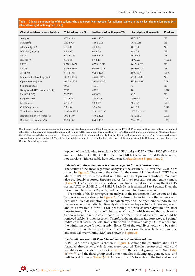
Table 1 Clinical demographics of the patients who underwent liver resection for malignant tumors in the no liver dysfunction group (n =78) and liver dysfunction group (n = 8)
Clinical variables / characteristics Total values (n = 86) No liver dysfunction (n = 78) Liver dysfunction (n = 8) P-values
Age (yr) 67.0 ± 10.3 66.8 ± 10.5 68.7 ± 9.3 NS
BSA (cm2) 1.61 ± 0.18 1.60 ± 0.18 1.65 ± 0.18 NS
Albumin (g/dL) 4.0 ± 0.4 4.0 ± 0.4 3.8 ± 0.4 NS
Bilirubin (mg/dL) 0.7 ± 0.3 0.6 ± 0.3 0.8 ± 0.4 NS
PT (%) 93.4 ± 11.9 93.9 ± 12.1 88.4 ± 9.7 NS
ICGR15 (%) 9.0 ± 4.6 8.4 ± 4.3 14.9 ± 2.5 < 0.001
HH15 0.578 ± 0.079 0.575 ± 0.078 0.617 ± 0.010 NS
LHL15 0.940 ± 0.027 0.940 ± 0.028 0.935 ± 0.024 NS
ATIII (%) 94.9 ± 17.2 96.0 ± 17.3 83.9 ± 13.4 0.036
Intraoperative bleeding (mL) 481.2 ± 468.5 455.8 ± 472.4 675.0 ± 430.0 NS
Operative time (min) 404.5 ± 135.2 390.8 ± 129.3 525.9 ± 140.9 0.014
Sex (male:female) 49:37 44:34 5:3 NS
Background (HCC: meta or CCC) 57:29 49:29 8:0 0.047
Hr (0/S:1:2/3) 53:17:16 49:16:13 4:1:3 NS
Sapporo score 13.2 ± 2.4 13.4 ± 2.5 11.0 ± 3.2 0.037
MELD score 7.6 ± 1.6 7.6 ± 1.7 7.8 ± 0.7 0.103
Child-Pugh score 3.2 ± 0.4 3.2 ± 0.4 3.4 ± 0.5 0.119
Total liver volume (cc) 1137.8 ± 222.9 1136.2 ± 228.5 1153.5 ± 221.4 NS
Reduction in liver volume (%) 19.0 ± 13.0 17.6 ± 12.1 32.8 ± 15.0 0.006
Residual liver volume (%) 85.1 ± 14.4 86.0 ± 13.7 75.4 ± 17.9 0.039
Continuous variables are expressed as the mean and standard deviation. BSA: Body surface area; PT-INR: Prothrombin time-international normalizedratio; ICG15: Indocyanine green retention rate at 15 min; ATIII: Serum anti-thrombin III level; HCC: Hepatocellular carcinoma; meta: Metastatic tumor;CCC: Cholangiocellular carcinoma; Hr: Hepatic resection; HH15: Uptake ratio of the heart at 15 min to that seen at 3 min on 99m Tc-galactosyl humanserum albumin scintigraphy (GSA); LHL15: The uptake ratio of the liver to the liver plus heart at 15 min on GSA; MELD: Model For End-Stage LiverDisease; NS: Not significant.
lopment of the following formula for SLV: SLV (mL) = 822.7 × BSA - 183.2 (R2 = 0.419and R = 0.644, P < 0.001). On the other hand, MELD score and Child-Pugh score didnot correlate with resectable liver volume at all (Supplement Figure 1 and 2).
Estimation of the minimum liver volume required for safe hepatectomyThe results of the linear regression analysis of the serum ATIII level and ICGR15 areshown in Figure 2. The sum of the values for the serum ATIII level and ICGR15 wasalmost 100%, which is consistent with the findings of previous studies[20]. We havealso previously reported Sapporo scores for liver resection for malignant tumors(Table 2). The Sapporo score consists of four clinical variables, including the ICGR15,serum ATIII level, HH15, and LHL15. Each factor is awarded 1 to 4 points. Thus, themaximum total score is 16 points, and the minimum total score is 4 points.
The results of the linear regression analysis of the reduction in liver volume and theSapporo score are shown in Figure 3. The closed circles indicate the patients whoexhibited liver dysfunction after hepatectomy, and the open circles indicate thepatients who did not display liver dysfunction after hepatectomy. Linear regressionanalysis revealed a formula for predicting the risk of liver dysfunction afterhepatectomy. The linear coefficient was almost 5, which meant that each extraSapporo score point indicated that a further 5% of the total liver volume could beremoved safely via liver resection. Therefore, the maximum Sapporo score (16 points)indicates that 65% of the total liver volume can be removed safely. On the other hand,the minimum score (4 points) only allows 5% of the total liver volume to be safelyremoved. The relationships between the Sapporo score, the resectable liver volume,and residual liver volume (RLV) are shown in Figure 3B.
Systematic review of SLV and the minimum residual liver volumeA PRISMA flow diagram is shown in Figure 4. Among the 25 studies about SLVformulae, three types of calculations were reported. The first group used height andweight as independent factors (Table 3)[22-28]; the second group used BSA (Table4)[24,25,29-36]; and the third group used other variables including age, gender, race, andradiological findings (Table 5)[37-43]. Although the SLV formulae in the first and second
WJMA https://www.wjgnet.com May 31, 2019 Volume 7 Issue 5
Harada K et al. Scoring criteria for liver resection
237
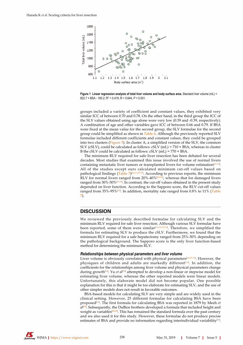
Figure 1
Figure 1 Linear regression analysis of total liver volume and body surface area. Standard liver volume (mL) =822.7 × BSA - 183.2; R2 = 0.419, R = 0.644; P < 0.001.
groups included a variety of coefficient and constant values, they exhibited verysimilar ICC of between 0.70 and 0.78. On the other hand, in the third group the ICC ofthe SLV values obtained using age alone were very low (0.39 and -0.39, respectively).A combination of age and other variables gave ICC of between 0.66 and 0.79. If BSAwere fixed at the mean value for the second group, the SLV formulae for the secondgroup could be simplified as shown in Table 6. Although the previously reported SLVformulae included different coefficients and constant values, they could be groupedinto two clusters (Figure 5). In cluster A, a simplified version of the SLV, the commonSLV (cSLV), could be calculated as follows: cSLV (mL) = 710 × BSA, whereas in clusterB the cSLV could be calculated as follows: cSLV (mL) = 770 × BSA.
The minimum RLV required for safe liver resection has been debated for severaldecades. Most studies that examined this issue involved the use of normal liverscontaining metastatic liver tumors or transplanted livers for volume estimation[44-48].All of the studies except ours calculated minimum cut-off values based onpathological findings (Table 7)[16,17,49-57]. According to previous reports, the minimumRLV for normal livers ranged from 20%-40%[54-56], whereas that for damaged liversranged from 30%-50%[16,52]. In contrast, the cut-off values obtained in the present studydepended on liver function. According to the Sapporo score, the RLV cut-off valuesranged from 35%-95%[20]. In addition, mortality rate ranged from 0.8% to 11% (Table7).
DISCUSSIONWe reviewed the previously described formulae for calculating SLV and theminimum RLV required for safe liver resection. Although various SLV formulae havebeen reported, some of them were similar[24,30,32-35]. Therefore, we simplified theformula for estimating SLV to produce the cSLV. Furthermore, we found that theminimum RLV required for a safe hepatectomy ranged from 25%-50% depending onthe pathological background. The Sapporo score is the only liver function-basedmethod for determining the minimum RLV.
Relationships between physical parameters and liver volumeLiver volume is obviously correlated with physical parameters[24,32-35]. However, thephysiques of children and adults are markedly different[24]. In addition, thecoefficients for the relationships among liver volume and physical parameters changeduring growth[24]. Yu et al[24] attempted to develop a non-linear or stepwise model forestimating liver volume, whereas the other reported models were linear models.Unfortunately, this elaborate model did not become popular. One possibleexplanation for this is that it might be too elaborate for estimating SLV, and the use ofother simpler models does not result in favorable outcomes.
BSA-based models for calculating SLV are very simple and are widely used in theclinical setting. However, 25 different formulae for calculating BSA have beenproposed[58]. The first formula for calculating BSA was reported in 1879 by Meeh etal[59]. Subsequently, the DuBios brothers developed a formula that included height andweight as variables[59,60]. This has remained the standard formula over the past centuryand we also used it for this study. However, these formulae do not produce preciseestimates of BSA and provide no information regarding interindividual variability[58].
WJMA https://www.wjgnet.com May 31, 2019 Volume 7 Issue 5
Harada K et al. Scoring criteria for liver resection
238

Table 2 Sapporo scores for liver resection of malignant tumors
FactorsScores
4 3 2 1
ICGR15 (%) ≤ 10 10-19 20-29 ≥ 30
ATIII (%) ≥ 90 80-89 70-79 ≤ 69
HH15 ≤ 0.55 0.56-0.59 0.60-0.64 ≥ 0.65
LHL15 ≥ 0.95 0.90-0.94 0.85-0.89 ≤ 0.84
The total number of points was calculated as the sum of the scores for the ICGR15, serum ATIII level, HH15,and LHL15. Hr 2 or Hr 3: 16 points, Hr: 1 ≥ 12 points, Hr S: ≥ 8 points, Hr 0: ≤ 7 points. ICG15: Indocyaninegreen retention rate at 15 min; ATIII: Serum anti-thrombin III level; HH15: Uptake ratio of the heart at 15 minto that seen at 3 min on 99m Tc-galactosyl human serum albumin scintigraphy (GSA); LHL15: The uptakeratio of the liver to the liver plus heart at 15 min on GSA.
Therefore, SLV varies markedly depending on which BSA formula is used.Since the variation in SLV is not as large as that in BSA, similar coefficient and
constant values were used to calculate SLV in previous studies. We identified twoclusters of SLV formulae, as shown in Figure 5, and created a simplified cSLV formulafor each cluster. The cluster analysis actually identified three clusters, but two of theclusters were very similar and not significantly different (data not shown). Therefore,we combined them together as cluster B. The differences among the clusters related toage or BSA. The age and BSA of cluster A tended to be younger and smaller,respectively, than those of cluster B. Therefore, differences in the patients’background data might have affected the coefficients used and the resultant ICC.Cluster B displayed ICC greater values than cluster A, although the exact reason forthis was unclear. One possible explanation is that cSLV stabilized in elderly patients,and so the error range became smaller than that found in younger patients.
Aging and SLVSLV is affected by aging; i.e., it was reported to be 4% of body weight at birth, but only2%-2.7% of body weight in adults[30,61]. Therefore, age is an important factor whencomparing the formulae used to calculate SLV. A study by Urata et al[30] involvedyoung patients, whereas other studies involved adults[24,30,32-35]. Our study populationwas older than those employed in previous studies. However, the formulae producedin each study were very similar. Although the SLV is affected by aging, it mightremain relatively constant in all patients.
Takahashi et al[37] and Kanamori et al[38] proposed that SLV can be assessed using agealone. However, their approach would not have been appropriate for our patientpopulation, in which most patients were elderly. On the other hand, a combination ofage and other variables provided ICC of between 0.66 and 0.79. Thus, it is likely thatSLV is partially affected by aging. Although elaborate formulae were created in thethird group, this did not result in better ICC compared with those seen in the othergroups. Therefore, simple SLV formulas could be applied to patients who are > 10years old.
Minimum RLV required for maintaining homeostasis after surgeryThe issue of the minimum RLV does not only involve the reduction in liver volume,but also several other factors. For example, bile duct reconstruction could be one ofthe predictors of short-term clinical outcomes[62,63]. The frequency of bile leakage ishigher in cases involving biliary reconstruction after hepatectomy than in cases inwhich biliary reconstruction is not performed[64]. In addition, biliary reconstructioncan cause intra-abdominal leakage followed by intra-abdominal infection[64,65].Therefore, the minimum RLV might differ between cases that do and do not involvebiliary reconstruction. Second, the background of the liver also plays an importantrole in determining clinical outcomes. The general question is how we could evaluateliver damage before surgery. Several liver function evaluation methods have beenproposed, including methods based on serum protein levels, serum enzyme levels,the ICGR15, and radiological assessments[66-68]. However, none of them represent liverfunction perfectly. For example, ICGR15 has been used for several decades; however,it does not reflect liver function in patients that exhibit ICG intolerance or possess anarteriovenous shunt. Radiological evaluations are also affected by the systemiccirculation, e.g., by dehydration and heart failure. Serum protein and enzyme levelsare too stable to allow them to be used to evaluate liver function in the initial stages ofliver damage, and they might not be valuable until the terminal stages of disease
WJMA https://www.wjgnet.com May 31, 2019 Volume 7 Issue 5
Harada K et al. Scoring criteria for liver resection
239
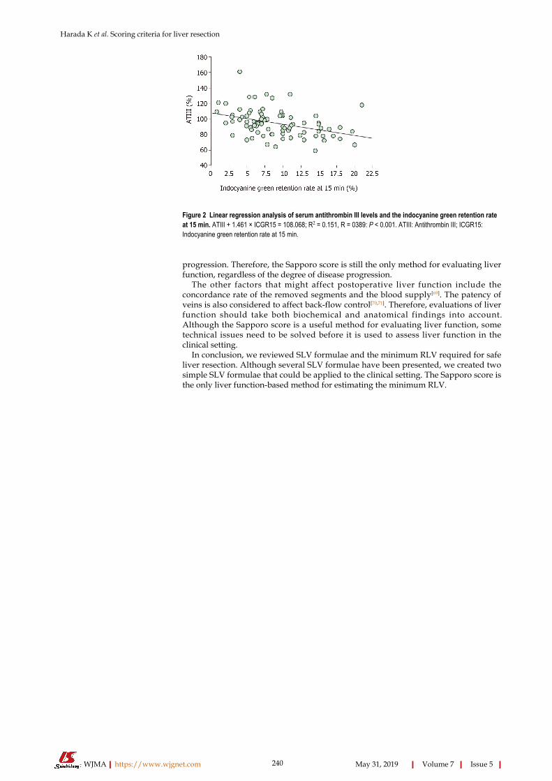
Figure 2
Figure 2 Linear regression analysis of serum antithrombin III levels and the indocyanine green retention rateat 15 min. ATIII + 1.461 × ICGR15 = 108.068; R2 = 0.151, R = 0389: P < 0.001. ATIII: Antithrombin III; ICGR15:Indocyanine green retention rate at 15 min.
progression. Therefore, the Sapporo score is still the only method for evaluating liverfunction, regardless of the degree of disease progression.
The other factors that might affect postoperative liver function include theconcordance rate of the removed segments and the blood supply[69]. The patency ofveins is also considered to affect back-flow control[70,71]. Therefore, evaluations of liverfunction should take both biochemical and anatomical findings into account.Although the Sapporo score is a useful method for evaluating liver function, sometechnical issues need to be solved before it is used to assess liver function in theclinical setting.
In conclusion, we reviewed SLV formulae and the minimum RLV required for safeliver resection. Although several SLV formulae have been presented, we created twosimple SLV formulae that could be applied to the clinical setting. The Sapporo score isthe only liver function-based method for estimating the minimum RLV.
WJMA https://www.wjgnet.com May 31, 2019 Volume 7 Issue 5
Harada K et al. Scoring criteria for liver resection
240
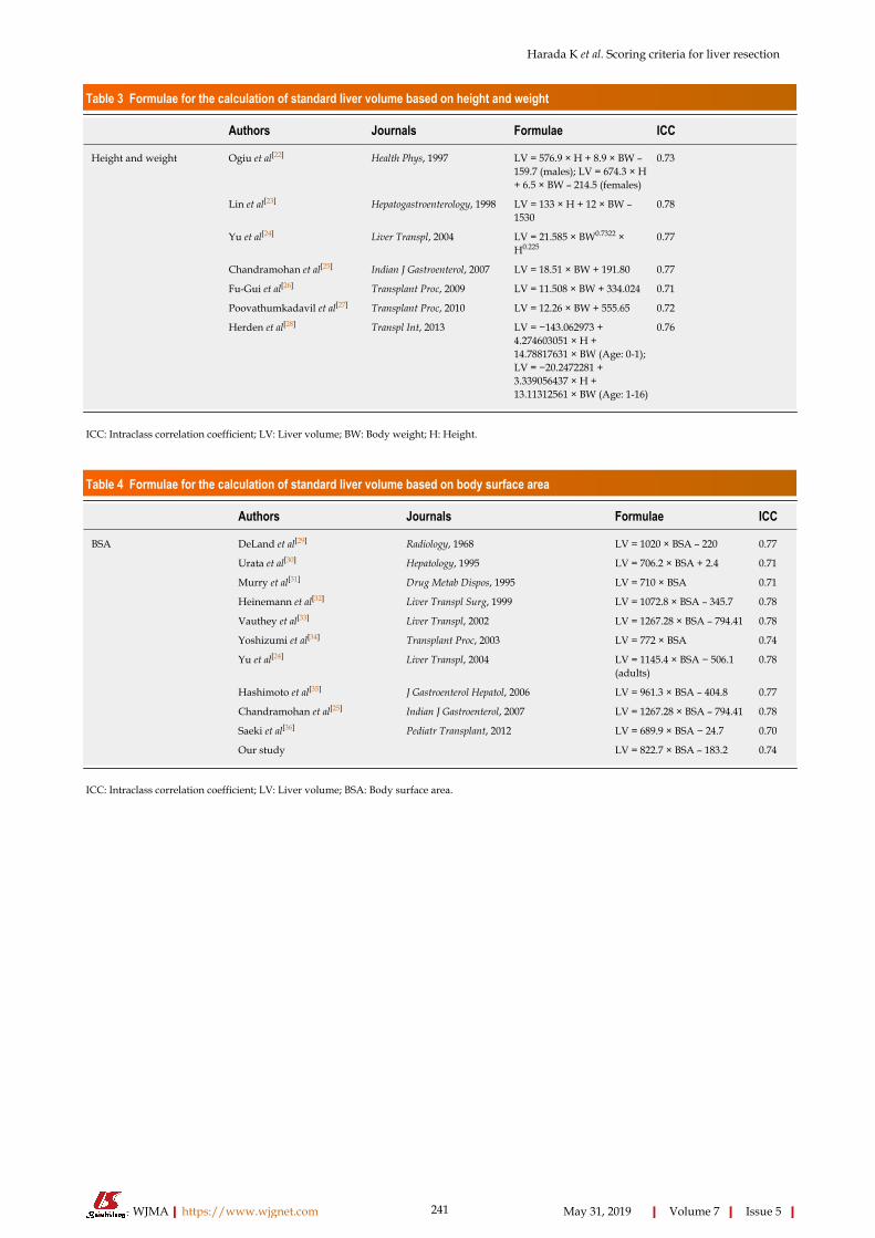
Table 3 Formulae for the calculation of standard liver volume based on height and weight
Authors Journals Formulae ICC
Height and weight Ogiu et al[22] Health Phys, 1997 LV = 576.9 × H + 8.9 × BW –159.7 (males); LV = 674.3 × H+ 6.5 × BW – 214.5 (females)
0.73
Lin et al[23] Hepatogastroenterology, 1998 LV = 133 × H + 12 × BW –1530
0.78
Yu et al[24] Liver Transpl, 2004 LV = 21.585 × BW0.7322 ×H0.225
0.77
Chandramohan et al[25] Indian J Gastroenterol, 2007 LV = 18.51 × BW + 191.80 0.77
Fu-Gui et al[26] Transplant Proc, 2009 LV = 11.508 × BW + 334.024 0.71
Poovathumkadavil et al[27] Transplant Proc, 2010 LV = 12.26 × BW + 555.65 0.72
Herden et al[28] Transpl Int, 2013 LV = −143.062973 +4.274603051 × H +14.78817631 × BW (Age: 0-1);LV = −20.2472281 +3.339056437 × H +13.11312561 × BW (Age: 1-16)
0.76
ICC: Intraclass correlation coefficient; LV: Liver volume; BW: Body weight; H: Height.
Table 4 Formulae for the calculation of standard liver volume based on body surface area
Authors Journals Formulae ICC
BSA DeLand et al[29] Radiology, 1968 LV = 1020 × BSA – 220 0.77
Urata et al[30] Hepatology, 1995 LV = 706.2 × BSA + 2.4 0.71
Murry et al[31] Drug Metab Dispos, 1995 LV = 710 × BSA 0.71
Heinemann et al[32] Liver Transpl Surg, 1999 LV = 1072.8 × BSA – 345.7 0.78
Vauthey et al[33] Liver Transpl, 2002 LV = 1267.28 × BSA – 794.41 0.78
Yoshizumi et al[34] Transplant Proc, 2003 LV = 772 × BSA 0.74
Yu et al[24] Liver Transpl, 2004 LV = 1145.4 × BSA − 506.1(adults)
0.78
Hashimoto et al[35] J Gastroenterol Hepatol, 2006 LV = 961.3 × BSA – 404.8 0.77
Chandramohan et al[25] Indian J Gastroenterol, 2007 LV = 1267.28 × BSA – 794.41 0.78
Saeki et al[36] Pediatr Transplant, 2012 LV = 689.9 × BSA − 24.7 0.70
Our study LV = 822.7 × BSA – 183.2 0.74
ICC: Intraclass correlation coefficient; LV: Liver volume; BSA: Body surface area.
WJMA https://www.wjgnet.com May 31, 2019 Volume 7 Issue 5
Harada K et al. Scoring criteria for liver resection
241

Table 5 Formulae for the calculation of standard liver volume based on age, gender, or radiological findings
Authors Journals Formulae ICC
Others Takahashi et al[37] Clin Pharmacol Ther, 2000 LV = 15 × (4.6 × Age + 19.8),Age: 1-18; LV = 15 × (0.31 ×Age + 97.8), Age: 30-40; LV =15 × (–0.91 × Age + 149), Age:≥ 41
0.39
Kanamori et al[38] Int J Clin Pharmacol Ther, 2002 LV = 67.3 × Age + 229.8 −0.39
Choukèr et al[39] Liver Transpl, 2004 LV=452 + 16.34 × BW + 11.85× Age – 166 × Gender (Age:16–50, M: 0, F: 1); LV=1390 +15.94 × BW – 12.86 × Age(Age: 51–70)
0.79
Chan et al[40] World J Gastroenterol, 2006 LV = 218 + BW × 12.3 +Gender × 51 (F: 0, M: 1)
0.74
Yuan et al[41] Transplant Proc, 2008 LV = 949.7 × BSA − 48.3 ×Age − 247.4 (Age: 1: < 40, 2:41–60, 3: > 60)
0.77
Kokudo et al[42] J Hepatol, 2015 LV = 203.3 − 3.61 × Age +58.7 × Thoracic width − 463.7× Race (Asian: 1, Caucasian:0)
0.66
Ma et al[43] Liver Transpl, 2017 LV = (2 × Depth) + (10 × BW)+ 190
0.75
ICC: Intraclass correlation coefficient; LV: Liver volume; BSA: Body surface area; BW: Body weight; F: Female; M: Male.
Table 6 Characteristics of the patients and simple standard liver volume formulae based on a mean body surface area of 1.61
Authors Mean age ± SD(range) BSA
Simple formulae(tentative mean BSA= 1.61)
ICC Clusters
DeLand et al[29] ND ND LV = 883 × BSA 0.75 B
Urata et al[30] 11.1 ± 8.8 1.078 ± 0.528(0.248–1.935)
LV = 707 × BSA 0.71 A
Murry et al[31] 9.7 (3.3–18.8) Median: 1.37 (0.57–2.0) LV = 710 × BSA 0.71 A
Heinemann et al[32] 50.6±18.9 ND LV = 858 × BSA 0.75 B
Vauthey et al[33] Mean: 54, Median: 56(14-90)
Median: 1.82 (1.32–2.90) LV = 770 × BSA 0.74 B
Yoshizumi et al[34] 38.6 ± 20.6 (0–87) formales; 47.0 ± 19.7 (0–85)for females
1.86 ± 0.36 (0.24-2.88);1.68 ± 0.28 (0.28-2.38)
LV = 772 × BSA 0.74 B
Yu et al[24] 42.4 ± 16.5 1.65 ± 0.26 LV = 831 × BSA 0.74 B
Hashimoto et al[35] (17-66) 1.67 ± 0.18 (1.25-2.56) LV = 710 × BSA 0.71 A
Chandramohan et al[25] 46.5 (10-70) Median: 1.60 (0.88-2.25) LV = 774 × BSA 0.73 B
Saeki et al[36] 5.8 (0 d-15) ND LV = 675 × BSA 0.70 A
Our study 67.0 ± 10.3 1.61 ± 0.18 LV = 709 × BSA 0.71 A
ICC: Intraclass correlation coefficient; BSA: Body surface area; ND: Not describe; LV: Liver volume.
WJMA https://www.wjgnet.com May 31, 2019 Volume 7 Issue 5
Harada K et al. Scoring criteria for liver resection
242
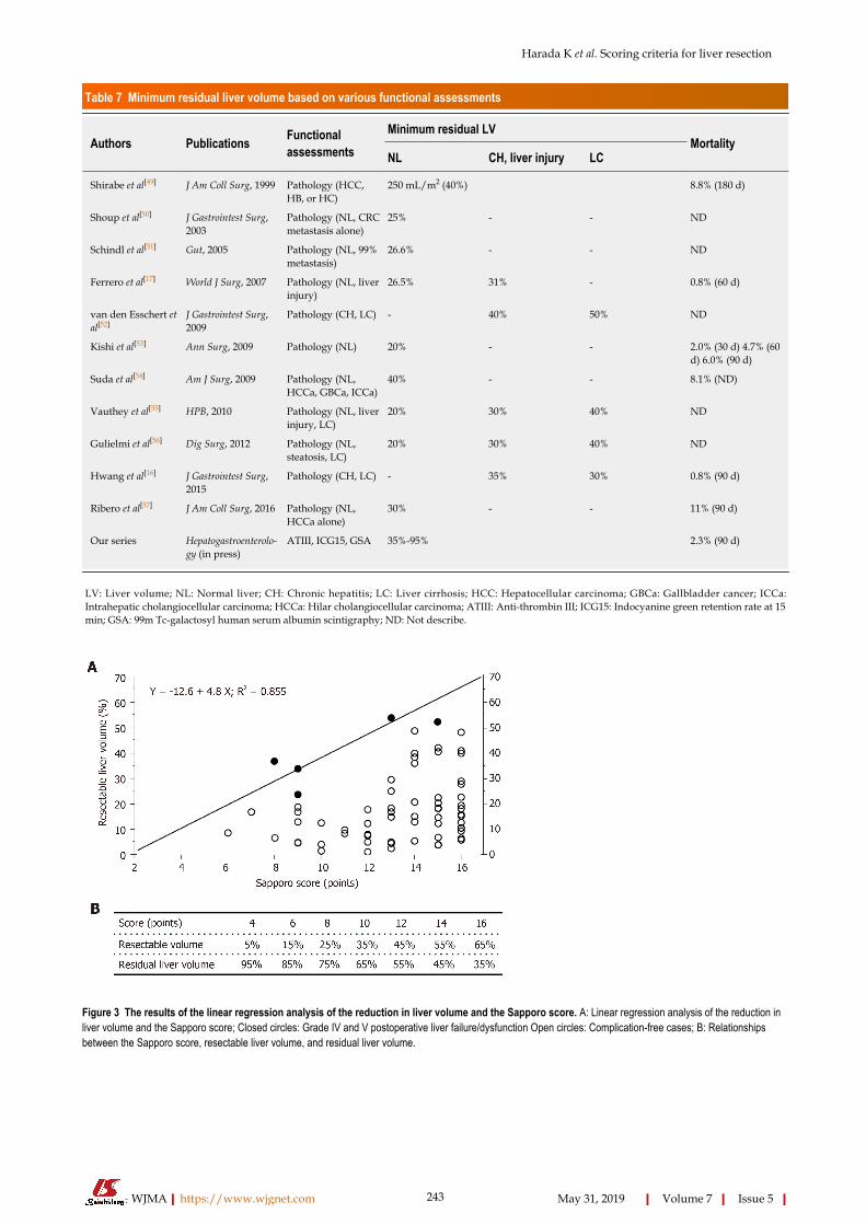
Table 7 Minimum residual liver volume based on various functional assessments
Authors Publications Functionalassessments
Minimum residual LVMortality
NL CH, liver injury LC
Shirabe et al[49] J Am Coll Surg, 1999 Pathology (HCC,HB, or HC)
250 mL/m2 (40%) 8.8% (180 d)
Shoup et al[50] J Gastrointest Surg,2003
Pathology (NL, CRCmetastasis alone)
25% - - ND
Schindl et al[51] Gut, 2005 Pathology (NL, 99%metastasis)
26.6% - - ND
Ferrero et al[17] World J Surg, 2007 Pathology (NL, liverinjury)
26.5% 31% - 0.8% (60 d)
van den Esschert etal[52]
J Gastrointest Surg,2009
Pathology (CH, LC) - 40% 50% ND
Kishi et al[53] Ann Surg, 2009 Pathology (NL) 20% - - 2.0% (30 d) 4.7% (60d) 6.0% (90 d)
Suda et al[54] Am J Surg, 2009 Pathology (NL,HCCa, GBCa, ICCa)
40% - - 8.1% (ND)
Vauthey et al[55] HPB, 2010 Pathology (NL, liverinjury, LC)
20% 30% 40% ND
Gulielmi et al[56] Dig Surg, 2012 Pathology (NL,steatosis, LC)
20% 30% 40% ND
Hwang et al[16] J Gastrointest Surg,2015
Pathology (CH, LC) - 35% 30% 0.8% (90 d)
Ribero et al[57] J Am Coll Surg, 2016 Pathology (NL,HCCa alone)
30% - - 11% (90 d)
Our series Hepatogastroenterolo-gy (in press)
ATIII, ICG15, GSA 35%-95% 2.3% (90 d)
LV: Liver volume; NL: Normal liver; CH: Chronic hepatitis; LC: Liver cirrhosis; HCC: Hepatocellular carcinoma; GBCa: Gallbladder cancer; ICCa:Intrahepatic cholangiocellular carcinoma; HCCa: Hilar cholangiocellular carcinoma; ATIII: Anti-thrombin III; ICG15: Indocyanine green retention rate at 15min; GSA: 99m Tc-galactosyl human serum albumin scintigraphy; ND: Not describe.
Figure 3
Figure 3 The results of the linear regression analysis of the reduction in liver volume and the Sapporo score. A: Linear regression analysis of the reduction inliver volume and the Sapporo score; Closed circles: Grade IV and V postoperative liver failure/dysfunction Open circles: Complication-free cases; B: Relationshipsbetween the Sapporo score, resectable liver volume, and residual liver volume.
WJMA https://www.wjgnet.com May 31, 2019 Volume 7 Issue 5
Harada K et al. Scoring criteria for liver resection
243

Figure 4
Figure 4 A PRISMA flow diagram.
Figure 5
Figure 5 Three-dimensional scatterplot of simple standard liver volume formulae and their intraclass correlation coefficients. ICC: Intraclass correlationcoefficients.
ARTICLE HIGHLIGHTSResearch backgroundVarious minimum residual liver volume (RLV) has been presented. In addition, many formulasof standard liver volume (SLV) were also established.
Research motivationWhen we planned hepatectomy for malignant tumors, we had not proven which methods werethe best reliable assessment to estimate minimum RLV and SLV.
Research objectivesAim of this study was to review previous SLV formulae and the methods used to evaluate theminimum RLV, and explore the association between liver volume and mortality.
Research methodsA systematic review was performed (No. CRD42019123642). We developed an SLV formulausing data for 86 consecutive patients who underwent hepatectomy at our institution between
WJMA https://www.wjgnet.com May 31, 2019 Volume 7 Issue 5
Harada K et al. Scoring criteria for liver resection
244
July 2009 and August 2011.
Research resultsOur formula: SLV (mL) = 822.7 × BSA − 183.2 (R2 = 0.419 and R = 0.644, P < 0.001). We retrieved25 studies relating to SLV formulae and 12 studies about the RLV required for safe liverresection. The minimum RLV for normal and damaged livers ranged from 20%-40% and 30%-50%, respectively. The Sapporo score indicated that the minimum RLV ranges from 35%-95%depending on liver function.
Research conclusionsWe reviewed SLV formulae and the minimum RLV required for safe liver resection. Althoughseveral SLV formulae have been presented, we created two simple SLV formulae that could beapplied to the clinical setting. The Sapporo score is the only liver function-based method forestimating the minimum RLV.
Research perspectivesThe Sapporo score should be validated by large study with prospective registration. Thecommon SLV, which is cSLV (mL) = 710 or 770 × body surface area, needs to verify in thespecific population.
ACKNOWLEDGEMENTSWe thank Sandy Tan and Thomas Hui for their help in preparing this manuscript andtheir valuable discussions.
REFERENCES1 Llovet JM, Brú C, Bruix J. Prognosis of hepatocellular carcinoma: the BCLC staging classification. Semin
Liver Dis 1999; 19: 329-338 [PMID: 10518312 DOI: 10.1055/s-2007-1007122]2 Forner A, Llovet JM, Bruix J. Hepatocellular carcinoma. Lancet 2012; 379: 1245-1255 [PMID: 22353262
DOI: 10.1016/S0140-6736(11)61347-0]3 Belghiti J, Hiramatsu K, Benoist S, Massault P, Sauvanet A, Farges O. Seven hundred forty-seven
hepatectomies in the 1990s: an update to evaluate the actual risk of liver resection. J Am Coll Surg 2000;191: 38-46 [PMID: 10898182 DOI: 10.1016/S1072-7515(00)00261-1]
4 Imamura H, Seyama Y, Kokudo N, Maema A, Sugawara Y, Sano K, Takayama T, Makuuchi M. Onethousand fifty-six hepatectomies without mortality in 8 years. Arch Surg 2003; 138: 1198-206; discussion1206 [PMID: 14609867 DOI: 10.1001/archsurg.138.11.1198]
5 Poon RT, Fan ST. Hepatectomy for hepatocellular carcinoma: patient selection and postoperativeoutcome. Liver Transpl 2004; 10: S39-S45 [PMID: 14762838 DOI: 10.1002/lt.20040]
6 Wu CC, Cheng SB, Ho WM, Chen JT, Liu TJ, P'eng FK. Liver resection for hepatocellular carcinoma inpatients with cirrhosis. Br J Surg 2005; 92: 348-355 [PMID: 15672423 DOI: 10.1002/bjs.4838]
7 Eguchi S, Kanematsu T, Arii S, Omata M, Kudo M, Sakamoto M, Takayasu K, Makuuchi M, MatsuyamaY, Monden M; Liver Cancer Study Group of Japan. Recurrence-free survival more than 10 years after liverresection for hepatocellular carcinoma. Br J Surg 2011; 98: 552-557 [PMID: 21267990 DOI:10.1002/bjs.7393]
8 Rau HG, Schauer R, Helmberger T, Holzknecht N, von Rückmann B, Meyer L, Buttler E, Kessler M,Zahlmann G, Schuhmann D, Schildberg FW. Impact of virtual reality imaging on hepatic liver tumorresection: calculation of risk. Langenbecks Arch Surg 2000; 385: 162-170 [PMID: 10857486]
9 Wigmore SJ, Redhead DN, Yan XJ, Casey J, Madhavan K, Dejong CH, Currie EJ, Garden OJ. Virtualhepatic resection using three-dimensional reconstruction of helical computed tomographyangioportograms. Ann Surg 2001; 233: 221-226 [PMID: 11176128 DOI:10.1097/00000658-200102000-00011]
10 Child CG, Turcotte JG. Surgery and portal hypertension. Major Probl Clin Surg 1964; 1: 1-85 [PMID:4950264]
11 Pugh RN, Murray-Lyon IM, Dawson JL, Pietroni MC, Williams R. Transection of the oesophagus forbleeding oesophageal varices. Br J Surg 1973; 60: 646-649 [PMID: 4541913 DOI:10.1002/bjs.1800600817]
12 Kudo M, Kubo S, Takayasu K, Sakamoto M, Tanaka M, Ikai I, Furuse J, Nakamura K, Makuuchi M;Liver Cancer Study Group of Japan (Committee for Response Evaluation Criteria in Cancer of the Liver,Liver Cancer Study Group of Japan). Response Evaluation Criteria in Cancer of the Liver (RECICL)proposed by the Liver Cancer Study Group of Japan (2009 Revised Version). Hepatol Res 2010; 40: 686-692 [PMID: 20633194 DOI: 10.1111/j.1872-034X.2010.00674.x]
13 Mizuguchi T, Kawamoto M, Meguro M, Nakamura Y, Harada K, Kukita K, Hirata K. Prognostic impactof preoperative the branched-chain amino acid to the tyrosine ratio in hepatocellular carcinoma patientsafter initial hepatectomy. J Gastrointest Surg 2011; 15: 1433-1439 [PMID: 21607795 DOI:10.1007/s11605-011-1566-y]
14 Mosteller RD. Simplified calculation of body-surface area. N Engl J Med 1987; 317: 1098 [PMID:3657876 DOI: 10.1056/NEJM198710223171717]
15 Hutton B, Salanti G, Caldwell DM, Chaimani A, Schmid CH, Cameron C, Ioannidis JP, Straus S,Thorlund K, Jansen JP, Mulrow C, Catalá-López F, Gøtzsche PC, Dickersin K, Boutron I, Altman DG,Moher D. The PRISMA extension statement for reporting of systematic reviews incorporating networkmeta-analyses of health care interventions: checklist and explanations. Ann Intern Med 2015; 162: 777-784[PMID: 26030634 DOI: 10.7326/M14-2385]
16 Hwang S, Ha TY, Song GW, Jung DH, Ahn CS, Moon DB, Kim KH, Lee YJ, Lee SG. Quantified RiskAssessment for Major Hepatectomy via the Indocyanine Green Clearance Rate and Liver Volumetry
WJMA https://www.wjgnet.com May 31, 2019 Volume 7 Issue 5
Harada K et al. Scoring criteria for liver resection
245
Combined with Standard Liver Volume. J Gastrointest Surg 2015; 19: 1305-1314 [PMID: 25947549 DOI:10.1007/s11605-015-2846-8]
17 Ferrero A, Viganò L, Polastri R, Muratore A, Eminefendic H, Regge D, Capussotti L. Postoperative liverdysfunction and future remnant liver: where is the limit? Results of a prospective study. World J Surg2007; 31: 1643-1651 [PMID: 17551779 DOI: 10.1007/s00268-007-9123-2]
18 Yamanaka J, Okada T, Saito S, Kondo Y, Yoshida Y, Suzumura K, Hirano T, Iimuro Y, Fujimoto J.Minimally invasive laparoscopic liver resection: 3D MDCT simulation for preoperative planning. JHepatobiliary Pancreat Surg 2009; 16: 808-815 [PMID: 19466379 DOI: 10.1007/s00534-009-0112-8]
19 Hori M, Suzuki K, Epstein ML, Baron RL. Computed tomography liver volumetry using 3-dimensionalimage data in living donor liver transplantation: effects of the slice thickness on the volume calculation.Liver Transpl 2011; 17: 1427-1436 [PMID: 21850689 DOI: 10.1002/lt.22419]
20 Harada K, Mizuguchi T, Kawamoto M, Meguro M, Ota S, Sasaki S, Miyanishi K, Hatakenaka M,Shinomura Y, Kato J, Hirata K. Prediction of postoperative liver failure and evaluation of modified criteriafor liver resection with computed volume analysis. Hepatogastroenterology 2014; In press
21 Balzan S, Belghiti J, Farges O, Ogata S, Sauvanet A, Delefosse D, Durand F. The "50-50 criteria" onpostoperative day 5: an accurate predictor of liver failure and death after hepatectomy. Ann Surg 2005;242: 824-828, discussion 828-discussion 829 [PMID: 16327492 DOI:10.1097/01.sla.0000189131.90876.9e]
22 Ogiu N, Nakamura Y, Ijiri I, Hiraiwa K, Ogiu T. A statistical analysis of the internal organ weights ofnormal Japanese people. Health Phys 1997; 72: 368-383 [PMID: 9030838 DOI:10.1097/00004032-199703000-00004]
23 Lin XZ, Sun YN, Liu YH, Sheu BS, Cheng BN, Chen CY, Tsai HM, Shen CL. Liver volume in patientswith or without chronic liver diseases. Hepatogastroenterology 1998; 45: 1069-1074 [PMID: 9756008]
24 Yu HC, You H, Lee H, Jin ZW, Moon JI, Cho BH. Estimation of standard liver volume for livertransplantation in the Korean population. Liver Transpl 2004; 10: 779-783 [PMID: 15162473 DOI:10.1002/lt.20188]
25 Chandramohan A, Eapen A, Govil S, Govil S, Jeyaseelan V. Determining standard liver volume:assessment of existing formulae in Indian population. Indian J Gastroenterol 2007; 26: 22-25 [PMID:17401231]
26 Fu-Gui L, Lu-Nan Y, Bo L, Yong Z, Tian-Fu W, Ming-Qing X, Wen-Tao W, Zhe-Yu C. Estimation ofstandard liver volume in Chinese adult living donors. Transplant Proc 2009; 41: 4052-4056 [PMID:20005340 DOI: 10.1016/j.transproceed.2009.08.079]
27 Poovathumkadavil A, Leung KF, Al Ghamdi HM, Othman Iel H, Meshikhes AW. Standard formula forliver volume in Middle Eastern Arabic adults. Transplant Proc 2010; 42: 3600-3605 [PMID: 21094823DOI: 10.1016/j.transproceed.2010.07.098]
28 Herden U, Wischhusen F, Heinemann A, Ganschow R, Grabhorn E, Vettorazzi E, Nashan B, Fischer L. Aformula to calculate the standard liver volume in children and its application in pediatric livertransplantation. Transpl Int 2013; 26: 1217-1224 [PMID: 24118382 DOI: 10.1111/tri.12198]
29 DeLand FH, North WA. Relationship between liver size and body size. Radiology 1968; 91: 1195-1198[PMID: 5699624 DOI: 10.1148/91.6.1195]
30 Urata K, Kawasaki S, Matsunami H, Hashikura Y, Ikegami T, Ishizone S, Momose Y, Komiyama A,Makuuchi M. Calculation of child and adult standard liver volume for liver transplantation. Hepatology1995; 21: 1317-1321 [PMID: 7737637 DOI: 10.1002/hep.1840210515]
31 Murry DJ, Crom WR, Reddick WE, Bhargava R, Evans WE. Liver volume as a determinant of drugclearance in children and adolescents. Drug Metab Dispos 1995; 23: 1110-1116 [PMID: 8654200]
32 Heinemann A, Wischhusen F, Püschel K, Rogiers X. Standard liver volume in the Caucasian population.Liver Transpl Surg 1999; 5: 366-368 [PMID: 10477836 DOI: 10.1002/lt.500050516]
33 Vauthey JN, Abdalla EK, Doherty DA, Gertsch P, Fenstermacher MJ, Loyer EM, Lerut J, Materne R,Wang X, Encarnacion A, Herron D, Mathey C, Ferrari G, Charnsangavej C, Do KA, Denys A. Bodysurface area and body weight predict total liver volume in Western adults. Liver Transpl 2002; 8: 233-240[PMID: 11910568 DOI: 10.1053/jlts.2002.31654]
34 Yoshizumi T, Gondolesi GE, Bodian CA, Jeon H, Schwartz ME, Fishbein TM, Miller CM, Emre S. Asimple new formula to assess liver weight. Transplant Proc 2003; 35: 1415-1420 [PMID: 12826175 DOI:10.1016/S0041-1345(03)00482-2]
35 Hashimoto T, Sugawara Y, Tamura S, Hasegawa K, Kishi Y, Kokudo N, Makuuchi M. Estimation ofstandard liver volume in Japanese living liver donors. J Gastroenterol Hepatol 2006; 21: 1710-1713[PMID: 16984594 DOI: 10.1111/j.1440-1746.2006.04433.x]
36 Saeki I, Tokunaga S, Matsuura T, Hayashida M, Yanagi Y, Taguchi T. A formula for determining thestandard liver volume in children: a special reference for neonates and infants. Pediatr Transplant 2012;16: 244-249 [PMID: 22151603 DOI: 10.1111/j.1399-3046.2011.01624.x]
37 Takahashi H, Ishikawa S, Nomoto S, Nishigaki Y, Ando F, Kashima T, Kimura S, Kanamori M, EchizenH. Developmental changes in pharmacokinetics and pharmacodynamics of warfarin enantiomers inJapanese children. Clin Pharmacol Ther 2000; 68: 541-555 [PMID: 11103757 DOI:10.1067/mcp.2000.110977]
38 Kanamori M, Takahashi H, Echizen H. Developmental changes in the liver weight- and body weight-normalized clearance of theophylline, phenytoin and cyclosporine in children. Int J Clin Pharmacol Ther2002; 40: 485-492 [PMID: 12698985 DOI: 10.5414/CPP40485]
39 Choukèr A, Martignoni A, Dugas M, Eisenmenger W, Schauer R, Kaufmann I, Schelling G, Löhe F,Jauch KW, Peter K, Thiel M. Estimation of liver size for liver transplantation: the impact of age andgender. Liver Transpl 2004; 10: 678-685 [PMID: 15108261 DOI: 10.1002/lt.20113]
40 Chan SC, Liu CL, Lo CM, Lam BK, Lee EW, Wong Y, Fan ST. Estimating liver weight of adults by bodyweight and gender. World J Gastroenterol 2006; 12: 2217-2222 [PMID: 16610024 DOI:10.3748/wjg.v12.i14.2217]
41 Yuan D, Lu T, Wei YG, Li B, Yan LN, Zeng Y, Wen TF, Zhao JC. Estimation of standard liver volumefor liver transplantation in the Chinese population. Transplant Proc 2008; 40: 3536-3540 [PMID:19100432 DOI: 10.1016/j.transproceed.2008.07.135]
42 Kokudo T, Hasegawa K, Uldry E, Matsuyama Y, Kaneko J, Akamatsu N, Aoki T, Sakamoto Y,Demartines N, Sugawara Y, Kokudo N, Halkic N. A new formula for calculating standard liver volume forliving donor liver transplantation without using body weight. J Hepatol 2015; 63: 848-854 [PMID:26057995 DOI: 10.1016/j.jhep.2015.05.026]
43 Ma KW, Chok KSH, Chan ACY, Tam HSC, Dai WC, Cheung TT, Fung JYY, Lo CM. A new formula for
WJMA https://www.wjgnet.com May 31, 2019 Volume 7 Issue 5
Harada K et al. Scoring criteria for liver resection
246
estimation of standard liver volume using computed tomography-measured body thickness. Liver Transpl2017; 23: 1113-1122 [PMID: 28650089 DOI: 10.1002/lt.24807]
44 Vibert E, Pittau G, Gelli M, Cunha AS, Jamot L, Faivre J, Castro Benitez C, Castaing D, Adam R. Actualincidence and long-term consequences of posthepatectomy liver failure after hepatectomy for colorectalliver metastases. Surgery 2014; 155: 94-105 [PMID: 24694360 DOI: 10.1016/j.surg.2013.05.039]
45 Calatayud D, Sánchez Cabús S, Sampson J, Resendiz A, Molina V, Fondevila C, Fuster J, García-Valdecasas JC. Hepatic resection: a safe and effective surgery. Cir Esp 2017; 95: 437-446 [PMID:28818290 DOI: 10.1016/j.ciresp.2017.06.006]
46 Olthof PB, Tomassini F, Huespe PE, Truant S, Pruvot FR, Troisi RI, Castro C, Schadde E, Axelsson R,Sparrelid E, Bennink RJ, Adam R, van Gulik TM, de Santibanes E. Hepatobiliary scintigraphy to evaluateliver function in associating liver partition and portal vein ligation for staged hepatectomy: Liver volumeoverestimates liver function. Surgery 2017; 162: 775-783 [PMID: 28732555 DOI:10.1016/j.surg.2017.05.022]
47 Pietrasz D, Fuks D, Subar D, Donatelli G, Ferretti C, Lamer C, Portigliotti L, Ward M, Cowan J, Nomi T,Beaussier M, Gayet B. Laparoscopic extended liver resection: are postoperative outcomes different? SurgEndosc 2018; 32: 4833-4840 [PMID: 29770886 DOI: 10.1007/s00464-018-6234-9]
48 Watanabe Y, Kuboki S, Shimizu H, Ohtsuka M, Yoshitomi H, Furukawa K, Miyazaki M. A NewProposal of Criteria for the Future Remnant Liver Volume in Older Patients Undergoing MajorHepatectomy for Biliary Tract Cancer. Ann Surg 2018; 267: 338-345 [PMID: 27849659 DOI:10.1097/SLA.0000000000002080]
49 Shirabe K, Shimada M, Gion T, Hasegawa H, Takenaka K, Utsunomiya T, Sugimachi K. Postoperativeliver failure after major hepatic resection for hepatocellular carcinoma in the modern era with specialreference to remnant liver volume. J Am Coll Surg 1999; 188: 304-309 [PMID: 10065820 DOI:10.1016/S1072-7515(98)00301-9]
50 Shoup M, Gonen M, D'Angelica M, Jarnagin WR, DeMatteo RP, Schwartz LH, Tuorto S, Blumgart LH,Fong Y. Volumetric analysis predicts hepatic dysfunction in patients undergoing major liver resection. JGastrointest Surg 2003; 7: 325-330 [PMID: 12654556 DOI: 10.1016/S1091-255X(02)00370-0]
51 Schindl MJ, Redhead DN, Fearon KC, Garden OJ, Wigmore SJ; Edinburgh Liver Surgery andTransplantation Experimental Research Group (eLISTER). The value of residual liver volume as apredictor of hepatic dysfunction and infection after major liver resection. Gut 2005; 54: 289-296 [PMID:15647196 DOI: 10.1136/gut.2004.046524]
52 van den Esschert JW, de Graaf W, van Lienden KP, Busch OR, Heger M, van Delden OM, Gouma DJ,Bennink RJ, Laméris JS, van Gulik TM. Volumetric and functional recovery of the remnant liver aftermajor liver resection with prior portal vein embolization : recovery after PVE and liver resection. JGastrointest Surg 2009; 13: 1464-1469 [PMID: 19475462 DOI: 10.1007/s11605-009-0929-0]
53 Kishi Y, Abdalla EK, Chun YS, Zorzi D, Madoff DC, Wallace MJ, Curley SA, Vauthey JN. Threehundred and one consecutive extended right hepatectomies: evaluation of outcome based on systematicliver volumetry. Ann Surg 2009; 250: 540-548 [PMID: 19730239 DOI: 10.1097/SLA.0b013e3181b674df]
54 Suda K, Ohtsuka M, Ambiru S, Kimura F, Shimizu H, Yoshidome H, Miyazaki M. Risk factors of liverdysfunction after extended hepatic resection in biliary tract malignancies. Am J Surg 2009; 197: 752-758[PMID: 18778802 DOI: 10.1016/j.amjsurg.2008.05.007]
55 Vauthey JN, Dixon E, Abdalla EK, Helton WS, Pawlik TM, Taouli B, Brouquet A, Adams RB; AmericanHepato-Pancreato-Biliary Association; Society of Surgical Oncology; Society for Surgery of theAlimentary Tract. Pretreatment assessment of hepatocellular carcinoma: expert consensus statement. HPB(Oxford) 2010; 12: 289-299 [PMID: 20590901 DOI: 10.1111/j.1477-2574.2010.00181.x]
56 Guglielmi A, Ruzzenente A, Conci S, Valdegamberi A, Iacono C. How much remnant is enough in liverresection? Dig Surg 2012; 29: 6-17 [PMID: 22441614 DOI: 10.1159/000335713]
57 Ribero D, Zimmitti G, Aloia TA, Shindoh J, Fabio F, Amisano M, Passot G, Ferrero A, Vauthey JN.Preoperative Cholangitis and Future Liver Remnant Volume Determine the Risk of Liver Failure inPatients Undergoing Resection for Hilar Cholangiocarcinoma. J Am Coll Surg 2016; 223: 87-97 [PMID:27049784 DOI: 10.1016/j.jamcollsurg.2016.01.060]
58 Redlarski G, Palkowski A, Krawczuk M. Body surface area formulae: an alarming ambiguity. Sci Rep2016; 6: 27966 [PMID: 27323883 DOI: 10.1038/srep27966]
59 Meeh K. Oberflächenmessungen des menschlichen Körpers. Ztschr f Biol 1879; 1: 458-42560 Du Bois D, Du Bois E. A formula to estimate the approximate surface area if height and weight are
known. Arch Intern Med 1916; 17: 863–871 [DOI: 10.1001/archinte.1916.00080130010002]61 Balistreri W. Nelson textbook of pediatrics. Philadelphia: Saunders 1992; 1001-100462 Kadaba RS, Bowers KA, Khorsandi S, Hutchins RR, Abraham AT, Sarker SJ, Bhattacharya S, Kocher
HM. Complications of biliary-enteric anastomoses. Ann R Coll Surg Engl 2017; 99: 210-215 [PMID:27659373 DOI: 10.1308/rcsann.2016.0293]
63 Otto W, Sierdziński J, Smaga J, Dudek K, Zieniewicz K. Long-term effects and quality of life followingdefinitive bile duct reconstruction. Medicine (Baltimore) 2018; 97: e12684 [PMID: 30313064 DOI:10.1097/MD.0000000000012684]
64 Martin AN, Narayanan S, Turrentine FE, Bauer TW, Adams RB, Stukenborg GJ, Zaydfudim VM.Clinical Factors and Postoperative Impact of Bile Leak After Liver Resection. J Gastrointest Surg 2018;22: 661-667 [PMID: 29247421 DOI: 10.1007/s11605-017-3650-4]
65 Jin S, Fu Q, Wuyun G, Wuyun T. Management of post-hepatectomy complications. World JGastroenterol 2013; 19: 7983-7991 [PMID: 24307791 DOI: 10.3748/wjg.v19.i44.7983]
66 Mizuguchi T, Kawamoto M, Meguro M, Son S, Nakamura Y, Harada K, Shibata T, Ota S, Hirata K.Serum antithrombin III level is well correlated with multiple indicators for assessment of liver function anddiagnostic accuracy for predicting postoperative liver failure in hepatocellular carcinoma patients.Hepatogastroenterology 2012; 59: 551-557 [PMID: 22353523]
67 Harada K, Mizuguchi T, Katagiri Y, Kawamoto M, Nakamura Y, Meguro M, Ota S, Sasaki S, MiyanishiK, Sonoda T, Mori M, Shinomura Y, Kato J, Hirata K. Area between the hepatic and heart curves of(99m)Tc-galactosyl-human serum albumin scintigraphy represents liver function and disease progressionfor preoperative evaluation in hepatocellular carcinoma patients. J Hepatobiliary Pancreat Sci 2012; 19:667-673 [PMID: 22179579 DOI: 10.1007/s00534-011-0486-2]
68 Ishii M, Mizuguchi T, Harada K, Ota S, Meguro M, Ueki T, Nishidate T, Okita K, Hirata K.Comprehensive review of post-liver resection surgical complications and a new universal classificationand grading system. World J Hepatol 2014; 6: 745-751 [PMID: 25349645 DOI: 10.4254/wjh.v6.i10.745]
69 Sanjeevi S, Sparrelid E, Gilg S, Jonas E, Isaksson B. Arterial ischemia in the deportalized liver following
WJMA https://www.wjgnet.com May 31, 2019 Volume 7 Issue 5
Harada K et al. Scoring criteria for liver resection
247
associating liver partition and portal vein ligation for staged hepatectomy. World J Hepatol 2015; 7: 2492-2496 [PMID: 26483871 DOI: 10.4254/wjh.v7.i23.2492]
70 Balzan SM, Gava VG, Magalhaes MA, Dotto ML. Outflow modulation to target liver regeneration:something old, something new. Eur J Surg Oncol 2014; 40: 140-143 [PMID: 24075823 DOI:10.1016/j.ejso.2013.08.012]
71 Radtke A, Sgourakis G, Molmenti EP, Beckebaum S, Cicinnati VR, Schmidt H, Peitgen HO, BroelschCE, Malagó M, Schroeder T. Risk of venous congestion in live donors of extended right liver graft. WorldJ Gastroenterol 2015; 21: 6008-6017 [PMID: 26019467 DOI: 10.3748/wjg.v21.i19.6008]
WJMA https://www.wjgnet.com May 31, 2019 Volume 7 Issue 5
Harada K et al. Scoring criteria for liver resection
248
Published By Baishideng Publishing Group Inc
7041 Koll Center Parkway, Suite 160, Pleasanton, CA 94566, USA
Telephone: +1-925-2238242
Fax: +1-925-2238243
E-mail: [email protected]
Help Desk: https://www.f6publishing.com/helpdesk
https://www.wjgnet.com
© 2019 Baishideng Publishing Group Inc. All rights reserved.