Embed Size (px)
Citation preview

World Journal ofRadiology
World J Radiol 2020 January 28; 12(1): 1-9
ISSN 1949-8470 (online)
Published by Baishideng Publishing Group Inc
W J R World Journal ofRadiology
Contents Monthly Volume 12 Number 1 January 28, 2020
ORIGINAL ARTICLE
Observational Study
1 Segmentation of carotid arterial walls using neural networksSamber DD, Ramachandran S, Sahota A, Naidu S, Pruzan A, Fayad ZA, Mani V
WJR https://www.wjgnet.com January 28, 2020 Volume 12 Issue 1I
ContentsWorld Journal of Radiology
Volume 12 Number 1 January 28, 2020
ABOUT COVER Editorial Board Member of World Journal of Radiology, Jyoti Kumar, DNB,MD, Professor, Department of Radiodiagnosis, Maulana Azad MedicalCollege, New Delhi 110002, India
AIMS AND SCOPE The primary aim of World Journal of Radiology (WJR, World J Radiol) is toprovide scholars and readers from various fields of radiology with aplatform to publish high-quality basic and clinical research articles andcommunicate their research findings online. WJR mainly publishes articles reporting research results and findingsobtained in the field of radiology and covering a wide range of topicsincluding state of the art information on cardiopulmonary imaging,gastrointestinal imaging, genitourinary imaging, musculoskeletal imaging,neuroradiology/head and neck imaging, nuclear medicine and molecularimaging, pediatric imaging, vascular and interventional radiology, andwomen's imaging.
INDEXING/ABSTRACTING The WJR is now abstracted and indexed in Emerging Sources Citation Index (Web of
Science), PubMed, PubMed Central, China National Knowledge Infrastructure
(CNKI), China Science and Technology Journal Database (CSTJ), and Superstar
Journals Database.
RESPONSIBLE EDITORS FORTHIS ISSUE
Responsible Electronic Editor: Xiang Li
Proofing Production Department Director: Yun-Xiaojian Wu
NAME OF JOURNALWorld Journal of Radiology
ISSNISSN 1949-8470 (online)
LAUNCH DATEJanuary 31, 2009
FREQUENCYMonthly
EDITORS-IN-CHIEFVenkatesh Mani
EDITORIAL BOARD MEMBERShttps://www.wjgnet.com/1949-8470/editorialboard.htm
EDITORIAL OFFICERuo-Yu Ma, Director
PUBLICATION DATEJanuary 28, 2020
COPYRIGHT© 2020 Baishideng Publishing Group Inc
INSTRUCTIONS TO AUTHORShttps://www.wjgnet.com/bpg/gerinfo/204
GUIDELINES FOR ETHICS DOCUMENTShttps://www.wjgnet.com/bpg/GerInfo/287
GUIDELINES FOR NON-NATIVE SPEAKERS OF ENGLISHhttps://www.wjgnet.com/bpg/gerinfo/240
PUBLICATION MISCONDUCThttps://www.wjgnet.com/bpg/gerinfo/208
ARTICLE PROCESSING CHARGEhttps://www.wjgnet.com/bpg/gerinfo/242
STEPS FOR SUBMITTING MANUSCRIPTShttps://www.wjgnet.com/bpg/GerInfo/239
ONLINE SUBMISSIONhttps://www.f6publishing.com
© 2020 Baishideng Publishing Group Inc. All rights reserved. 7041 Koll Center Parkway, Suite 160, Pleasanton, CA 94566, USA
E-mail: [email protected] https://www.wjgnet.com
WJR https://www.wjgnet.com January 28, 2020 Volume 12 Issue 1II
W J R World Journal ofRadiology
Submit a Manuscript: https://www.f6publishing.com World J Radiol 2020 January 28; 12(1): 1-9
DOI: 10.4329/wjr.v12.i1.1 ISSN 1949-8470 (online)
ORIGINAL ARTICLE
Observational Study
Segmentation of carotid arterial walls using neural networks
Daniel D Samber, Sarayu Ramachandran, Anoop Sahota, Sonum Naidu, Alison Pruzan, Zahi A Fayad,Venkatesh Mani
ORCID number: Daniel D Samber(0000-0002-9241-2330); SarayuRamachandran(0000-0002-9917-5876); AnoopSahota (0000-0002-9278-5030);Sonum Naidu(0000-0003-4175-3933); AlisonPruzan (0000-0002-3054-6341); ZahiA Fayad (0000-0002-3439-7347);Venkatesh Mani(0000-0002-0432-2918).
Author contributions: Samber DDprogrammed the analysis softwareand wrote the draft of themanuscript; Ramachandran Sassembled and pre-processedimaging data; Naidu S, Sahota A,and Pruzan A performed theimage analysis; Fayad ZA andMani V oversaw the analysis; allauthors critically reviewed themanuscript.
Supported by American HeartAssociation Grant in Aid FoundersAffiliate No. 17GRNT33420119(Mani V), NIH NHLBI2R01HL070121 (Fayad ZA) andNIH NHLBI 1R01HL135878 (FayadZA).
Institutional review boardstatement: The study wasapproved by the InstitutionalReview Board of the Icahn Schoolof Medicine at Mount Sinai.
Informed consent statement:Waiver of institutional reviewboard (IRB) approval was obtainedfrom the IRB as only deidentifieddata was used in this study. Theimages analyzed for this studywere anonymized and devoid ofany Protected Health Information.
Conflict-of-interest statement: No
Daniel D Samber, Sarayu Ramachandran, Anoop Sahota, Sonum Naidu, Alison Pruzan, Zahi AFayad, Venkatesh Mani, Translational and Molecular Imaging Institute (TMII), Icahn School ofMedicine at Mount Sinai, New York, NY 10029, United States
Corresponding author: Daniel D Samber, BSc, Research Scientist, Translational MolecularImaging Institute (TMII), Icahn School of Medicine at Mount Sinai, 1470 Madison Avenue,New York, NY 10029, United States. [email protected]
AbstractBACKGROUNDAutomated, accurate, objective, and quantitative medical image segmentation hasremained a challenging goal in computer science since its inception. This studyapplies the technique of convolutional neural networks (CNNs) to the task ofsegmenting carotid arteries to aid in the assessment of pathology.
AIMTo investigate CNN’s utility as an ancillary tool for researchers who requireaccurate segmentation of carotid vessels.
METHODSAn expert reader delineated vessel wall boundaries on 4422 axial T2-weightedmagnetic resonance images of bilateral carotid arteries from 189 subjects withclinically evident atherosclerotic disease. A portion of this dataset was used totrain two CNNs (one to segment the vessel lumen and the other to segment thevessel wall) with the remaining portion used to test the algorithm’s efficacy bycomparing CNN segmented images with those of an expert reader.
RESULTSOverall quantitative assessment between automated and manual segmentationswas determined by computing the DICE coefficient for each pair of segmentedimages in the test dataset for each CNN applied. The average DICE coefficient forthe test dataset (CNN segmentations compared to expert’s segmentations) was0.96 for the lumen and 0.87 for the vessel wall. Pearson correlation values and theintra-class correlation coefficient (ICC) were computed for the lumen (Pearson =0.98, ICC = 0.98) and vessel wall (Pearson = 0.88, ICC = 0.86) segmentations.Bland-Altman plots of area measurements for the CNN and expert readersindicate good agreement with a mean bias of 1%-8%.
CONCLUSIONAlthough the technique produces reasonable results that are on par with experthuman assessments, our application requires human supervision and monitoring
WJR https://www.wjgnet.com January 28, 2020 Volume 12 Issue 11
conflicts to disclose.
Data sharing statement: Oncepublished and after appropriatesafeguard to ensure that the data isdevoid of any identifiers, the dataused for the analysis for this studywill be shared on the Mount Sinaidata sharing portal according toInstitutional guidelines.
STROBE statement: The authorshave read the STROBE Statement-checklist of items, and themanuscript was prepared andrevised according to the STROBEStatement-checklist of items.
Open-Access: This article is anopen-access article that wasselected by an in-house editor andfully peer-reviewed by externalreviewers. It is distributed inaccordance with the CreativeCommons Attribution NonCommercial (CC BY-NC 4.0)license, which permits others todistribute, remix, adapt, buildupon this work non-commercially,and license their derivative workson different terms, provided theoriginal work is properly cited andthe use is non-commercial. See:http://creativecommons.org/licenses/by-nc/4.0/
Manuscript source: Unsolicitedmanuscript
Received: July 19, 2019Peer-review started: July 21, 2019First decision: September 21, 2019Revised: October 11, 2019Accepted: November 20, 2019Article in press: November 20, 2019Published online: January 28, 2020
P-Reviewer: Qi XS, Tajiri KS-Editor: Yan JPL-Editor: AE-Editor: Li X
to ensure consistent results. We intend to deploy this algorithm as part of asoftware platform to lessen researchers’ workload to more quickly obtain reliableresults.
Key words: Carotid arteries; Segmentation; Convolutional neural network; Magneticresonance imaging; Vessel wall
©The Author(s) 2020. Published by Baishideng Publishing Group Inc. All rights reserved.
Core tip: Accurate segmentation of carotid arteries is useful in assessing the degree ofheart disease in general and vascular diseases (such as atherosclerosis) in particular.Until recently, obtaining accurate segmentation could only be accomplished through thework of an experienced researcher requiring a large investment of time and effort. Overthe last several years, the method of convolutional neural networks has demonstrated itsefficacy in a number of fields. In this study, we apply this method to magnetic resonanceimages acquired from subjects with clinically evident atherosclerotic disease andcompare the resulting segmentations with those determined by experienced researchers.
Citation: Samber DD, Ramachandran S, Sahota A, Naidu S, Pruzan A, Fayad ZA, Mani V.Segmentation of carotid arterial walls using neural networks. World J Radiol 2020; 12(1): 1-9URL: https://www.wjgnet.com/1949-8470/full/v12/i1/1.htmDOI: https://dx.doi.org/10.4329/wjr.v12.i1.1
INTRODUCTIONCarotid artery disease and stroke have been cited as the leading cause of death in theUnited States and worldwide[1]. Notably, atherosclerotic disease of the carotid vesselsaccounts for up to 20% of transient ischemic attacks or ischemic strokes[2]. Ischemicstroke arising from the development of vascular disease and associated plaqueformation can cause vessel stenosis resulting in compromised hemodynamics[3]. Thedegree of the vessel stenosis has been shown to be related the risk of recurrentstroke[4].
Magnetic resonance imaging (MRI), can provide both structural and functionalinformation, including lumen stenosis [5], vessel wall measurements, plaquecomposition, blood flow velocity, and flow rate [6 ]. Additionally, MRI hasdemonstrated the ability to characterize morphological aspects of the carotids such aslumen and wall area[7].
Despite MRI’s capabilities, automated, accurate, objective, and repeatablequantitative analysis of MRI data has proven a challenging goal in computer sciencefor years. In the field of vessel wall imaging, virtually every image-processingalgorithm has been applied to the task of accurately and objectively delineating vesselwall boundaries in order to segment the vessels. These techniques include deformablesplines[8], active contours[9], active shapes[10], level sets[11], cluster analysis[12], andcountless others.
Although many of these techniques worked well in a restricted regime, morecomplex analysis frequently required combining the approaches to achieve reasonableresults. For example, gradient-based ellipse fitting has been combined with fuzzyclustering to delineate carotid artery walls in a semi-automated approach[13]. Similarly,segmentation of arterial vessel walls, both on MR and computed tomography datausing a combination of techniques has been proposed. In another study, deformablemodels were used to segment the aortic lumen while a K nearest neighbor (KNN)classifier was employed to identify the associated thrombus in an aneurysm[14].
Recent advances in convolutional neural networks (CNN)[15,16] have dramaticallyimproved efforts in both the classification of images as well as the segmentation ofobjects within images themselves. In the field of medical image analysis, theapplication of CNNs to the task of image segmentation has recently becomeubiquitous[17]. Whether it is applied to facilitate the identification of brain regions[18] orto segment skeletal structures[19], the CNN approach has proven to be both general-purpose and remarkably effective. Recently CNNs have been employed in thesegmentation of blood vessels in the retina [20 ], heart [21 ], as well as in thecharacterization of plaque in the carotid vessels[22,23].
WJR https://www.wjgnet.com January 28, 2020 Volume 12 Issue 1
Samber DD et al. Segmentation of carotid arterial walls using CNNs
2
This study applies the technique of CNNs to the task of segmenting carotid arteriesin order to aid in the assessment of pathology. Accumulated plaque in carotid arterieshas been established as a reliable marker of underlying cardiovascular disease. Intimamedia thickness (IMT) measured by ultrasound has been shown to be associated withfuture cardiovascular events[24]. Previous studies have also shown a strong correlativerelationship between wall thickness and wall area measures using dark blood MRIand ultrasound based IMT[25]. Studies in the MESA cohort have also establishedrelationships between wall morphology measurements by MRI and futurecardiovascular events[26].
We anticipate incorporating CNN technology into a software platform designed tofacilitate the analysis of carotid images thereby streamlining what was once anonerous time-consuming task into something more manageable.
MATERIALS AND METHODSImages analyzed in this study were culled from a prior study[27] in which 189 subjects(ranging in age from 18 to 74 years) with clinically evident atherosclerotic diseaseunderwent MRI of the carotid arteries. The subjects from that study presented witheither type 2 diabetes mellitus or impaired glucose tolerance were randomized toreceive placebo (n = 94) or canakinumab 150 mg monthly (n = 95) for 12 mo. Imagingwas performed at multiple sites using a 3.0-T whole-body MRI scanner, includingTrio, TIM Trio, or Verio (Siemens Medical SolutionsUSA, Inc., Malvern, PA, UnitedStates) or Achieva (Philips, Amsterdam, the Netherlands) platforms. The localInstitutional Review Board approved both studies with all subjects providinginformed consent.
A range of 6 to 13 high-resolution axial ECG-gated T2-weighted MR images ofbilateral carotid arteries were acquired using a 4-channel carotid array (MachnetB.V.,Roden, Netherlands) on Siemens scanners and an equivalent multi-channel (4 to 8)phased array carotid coil (Shanghai Medical, Shanghai, China) was used on thePhilips scanners. In plane pixel resolution for the acquired images ranged from 0.52mm to 0.7 mm with a slice thickness of 3 mm. These images were subsequentlyanalyzed by an expert reader who manually segmented individual images of thecarotid wall using VesselMass (LKEB, the Division of Image Processing, Departmentof Radiology, the Leiden University Medical Center, The Netherlands) resulting in adataset of 4422 segmented images. Based on this manual segmentation, metricsdescribing the carotid vessel were generated and recorded thereby establishing astandard against which the efficacy of automated analysis could be judged.
In preparation for automated segmentation, the original dataset was divided into 3groups: A “training dataset” (3581 images), a “validation dataset” (398 images), and a“test dataset” (443 images). Each dataset was composed of anatomical images andtheir associated manually segmented images. While the training dataset was used todynamically adjust the CNN’s parameters during training, the validation dataset wasused only to evaluate model performance during training and avoid over-fitting.These datasets were used to train two separate CNNs (one for carotid lumensegmentation and the other for carotid wall segmentation).
To prepare the CNNs for training, each manually segmented image was identifiedand the information describing that image’s segmented vessel was converted fromVesselMass contour information to a binary mask image using a custom Matlab(Mathworks, Natick, MA, United States) script. Additionally, each image was croppedto a fixed size (64 × 64 pixel) to center on the carotid vessel with the luminanceadjusted to maximize brightness and contrast. The order of images was randomlyshuffled before they were submitted to the CNN for training.
For this study, we used a segmentation model developed for spinal cord graymatter segmentation by Perone[28]. The CNN trained to segment the carotid vessel wallwas compiled to use an Adam optimizer, a learning rate of 0.001, with the DICEmetric[29] selected as the loss function to be minimized. The network was trained witha batch size of 32 for 1000 epochs. The data was hosted on an AMD Ryzen7 3.6GHzcomputer although the actual neural network training ran on the computer’s NvidiaGeForce 1080 Ti GPU card. Training software was implemented using Tensorflowversion 1.10 and Keras version 2.2.2. The CNN trained to segment the carotid vessel’slumen was configured similarly but required far less training and converged in 100epochs.
Once the CNNs had been trained, the images from the test dataset were processedto produce segmentations as binary images. The resulting binary images were thencompared with expert segmentations to determine the overall efficacy of theapproach. In order to compute traditional vessel metrics on the binary images, some
WJR https://www.wjgnet.com January 28, 2020 Volume 12 Issue 1
Samber DD et al. Segmentation of carotid arterial walls using CNNs
3
minor post-processing (such as removing isolated pixels identified as noise) wasnecessary. Vessel wall metrics of the CNN’s test dataset were compared with thoseindependently produced by expert reader to determine acceptability.
RESULTSThe DICE loss metric for the CNN used to segment the carotid’s wall decreasedsteadily during training with a final score of 0.85 for the training dataset and 0.87 forthe validation dataset. The loss metric for the CNN used to segment the carotid’slumen decreased very quickly with a final DICE score of 0.96 for both the training andthe validation dataset. Following the training phase, the network used the computedweights to process 398 images of the test dataset.
Initial qualitative evaluation of the images produced by the CNN when applied tothe test dataset was very good although technical issues stemming from some imageswith extremely narrow carotid walls affected the analysis. Visual inspectionsuggested that the majority of segmentations achieved by automated means closelyapproximated those that were segmented manually, with many appearing almostindistinguishable.
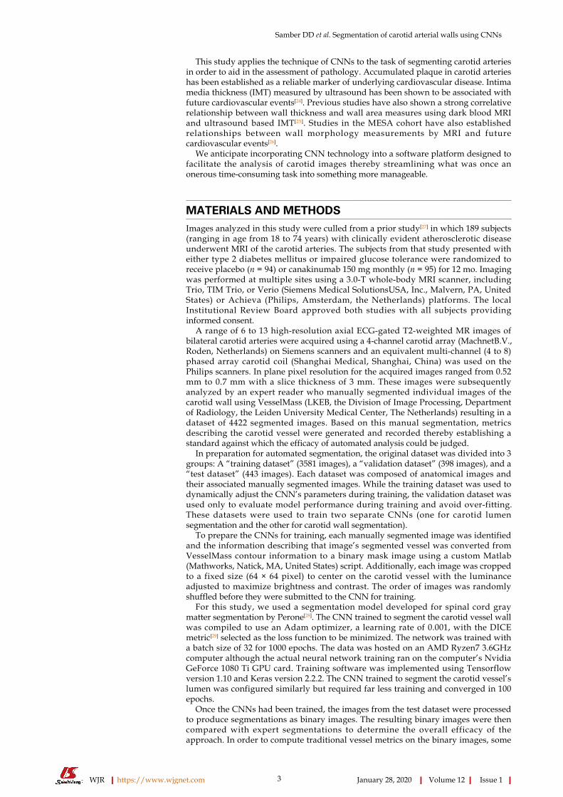
Overall quantitative assessment between automated and manual segmentationswas determined by computing the DICE coefficient for each pair of segmented imagesin the test dataset (Figure 1). Applying the CNN trained to segment the carotid vesselwall to the test dataset resulted in segmented images with an average DICE coefficientof 0.87. Applying the CNN trained to segment the carotid lumen to the test datasetresulted in segmented images with an average DICE coefficient of 0.96.
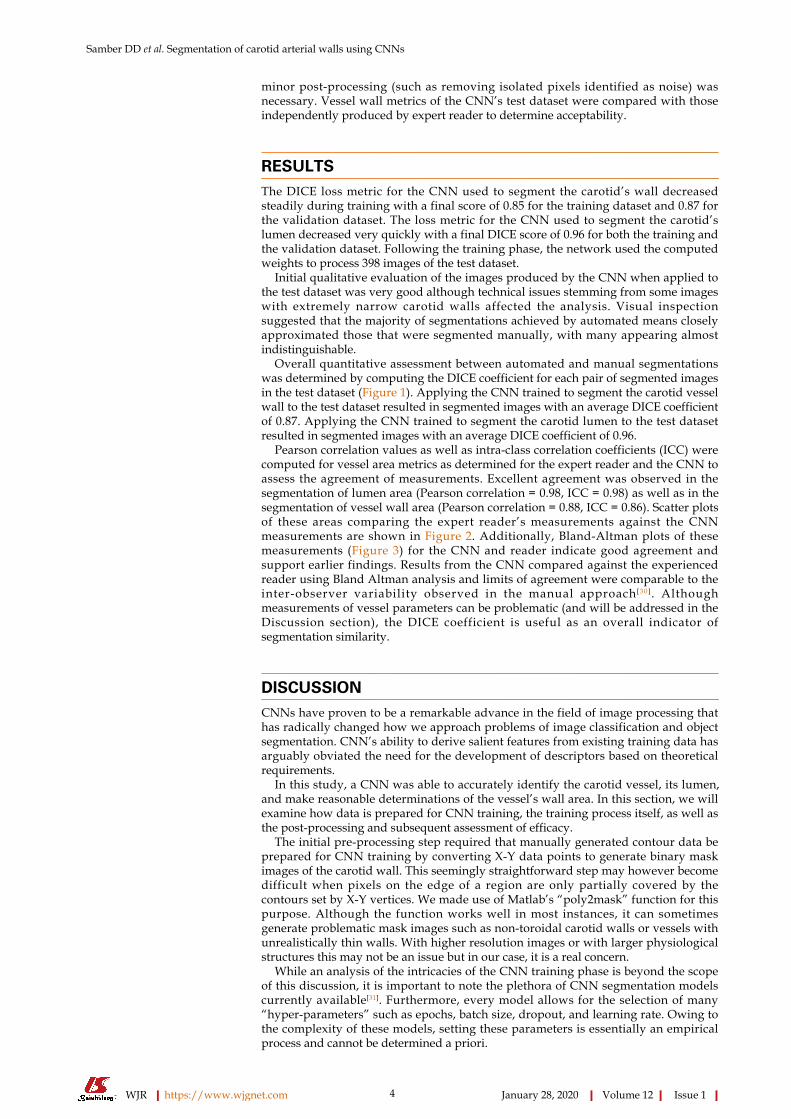
Pearson correlation values as well as intra-class correlation coefficients (ICC) werecomputed for vessel area metrics as determined for the expert reader and the CNN toassess the agreement of measurements. Excellent agreement was observed in thesegmentation of lumen area (Pearson correlation = 0.98, ICC = 0.98) as well as in thesegmentation of vessel wall area (Pearson correlation = 0.88, ICC = 0.86). Scatter plotsof these areas comparing the expert reader’s measurements against the CNNmeasurements are shown in Figure 2. Additionally, Bland-Altman plots of thesemeasurements (Figure 3) for the CNN and reader indicate good agreement andsupport earlier findings. Results from the CNN compared against the experiencedreader using Bland Altman analysis and limits of agreement were comparable to theinter-observer variability observed in the manual approach [ 3 0 ]. Althoughmeasurements of vessel parameters can be problematic (and will be addressed in theDiscussion section), the DICE coefficient is useful as an overall indicator ofsegmentation similarity.
DISCUSSIONCNNs have proven to be a remarkable advance in the field of image processing thathas radically changed how we approach problems of image classification and objectsegmentation. CNN’s ability to derive salient features from existing training data hasarguably obviated the need for the development of descriptors based on theoreticalrequirements.
In this study, a CNN was able to accurately identify the carotid vessel, its lumen,and make reasonable determinations of the vessel’s wall area. In this section, we willexamine how data is prepared for CNN training, the training process itself, as well asthe post-processing and subsequent assessment of efficacy.
The initial pre-processing step required that manually generated contour data beprepared for CNN training by converting X-Y data points to generate binary maskimages of the carotid wall. This seemingly straightforward step may however becomedifficult when pixels on the edge of a region are only partially covered by thecontours set by X-Y vertices. We made use of Matlab’s “poly2mask” function for thispurpose. Although the function works well in most instances, it can sometimesgenerate problematic mask images such as non-toroidal carotid walls or vessels withunrealistically thin walls. With higher resolution images or with larger physiologicalstructures this may not be an issue but in our case, it is a real concern.
While an analysis of the intricacies of the CNN training phase is beyond the scopeof this discussion, it is important to note the plethora of CNN segmentation modelscurrently available[31]. Furthermore, every model allows for the selection of many“hyper-parameters” such as epochs, batch size, dropout, and learning rate. Owing tothe complexity of these models, setting these parameters is essentially an empiricalprocess and cannot be determined a priori.
WJR https://www.wjgnet.com January 28, 2020 Volume 12 Issue 1
Samber DD et al. Segmentation of carotid arterial walls using CNNs
4

Figure 1
Figure 1 Representative axial T2-weighted magnetic resonance image of bilateral carotid arteries with cropped vessel area highlighted (A). Carotid vesselwall segmentations using convolutional neural networks (B) and expert reader (C). DICE = 0.91.
Originally, we trained a single model to segment the vessel wall and attempted tolabel the resulting images using conventional image processing techniques. Thisostensibly simple task of identifying lumen, vessel wall, and background regions waspotentially complicated as the segmented vessel wall border could narrow to a singlepixel and make region identification based on pixel connectivity challenging. Itquickly became apparent that training a separate CNN to segment the vessel lumenwould result in a less ambiguous result. In addition, the typically high contrastbetween the lumen and surrounding tissue allowed the CNN to converge extremelyquickly with very high accuracy.
In the interest of simplicity, we directed each CNN to segment the region of interest(either vessel wall or lumen). In that same spirit, we minimized experimentation toallow for reasonable processing time and adapt to memory constraints imposed byour hardware. Following the training phase, test data that had been set aside wasprocessed by the CNN to generate segmented images. Post processing of the resultantimages was limited to removing pixel “noise” that established isolated regions toosmall to be of significance for our purpose.
Our initial cursory side-by-side examination of manual and post-processed CNNimage segmentations was encouraging. Our subjective impression was subsequentlyborne out by comparing traditional area measures of the vessel wall, lumen, and totalvessel area for both the CNN and manual methods. As these measures may notconvey morphological differences between the two segmentations, we also calculatedthe DICE coefficient for corresponding images from these datasets. It should be notedthat although the DICE metric objectively describes the degree of overlap of twosegmentations, a relatively low score could sometimes be misleading. Alternatively,calculation of traditional vessel metrics was complicated by the fact that the neuralnetwork approach is a pixel-based technique that may result in obvious erroneousresults despite their high DICE score. For example, the neural network may producean image with pixels sparsely distributed making identification of vessel structuresproblematic.
Ultimately, establishing the utility of CNNs must be assessed not in absolute terms,but in comparing its performance to that of manual analysis carried out by experts. Inessence, a CNN must be shown to be accurate to within limits established by expertreaders and be judged by that same criteria.
In some respects, the effectiveness of this technology depends on its intended use.For example, we initially applied the technique to images with a much larger field ofview and a trained network was able to both identify and segment the vessel wall butwith occasional failures. Most failures occurred when the CNN incorrectly identifiedthe vessel of interest but largely succeeded in delineating vessel wall boundaries forthat incorrect vessel. For our purposes, it was decided to focus less on vesselidentification and concentrate on aspects of the vessel itself. As a practical matter, thisshifts the burden of identifying the vessel to a user but potentially just for the initialimage in a series. Consequently, we intend to implement this approach into aworkflow whereby a researcher first identifies a vessel of interest in its initialpresentation in an axial view. The researcher will then establish a bounding box of thevessel in that image and specify the number of axial images to process. Alternatively,these parameters could be established as a function of the subject’s physicaldimensions and adjusted for established physiological landmarks. Once theseparameters have been set, the CNN will proceed to process each image in turn withthe segmentation of one image's vessel providing the bounding box for itsneighboring slice in the series.
In conclusion, in this study, we have demonstrated the effectiveness of CNNtechnology in its application to the task of delineating carotid vessel walls thereby
WJR https://www.wjgnet.com January 28, 2020 Volume 12 Issue 1
Samber DD et al. Segmentation of carotid arterial walls using CNNs
5
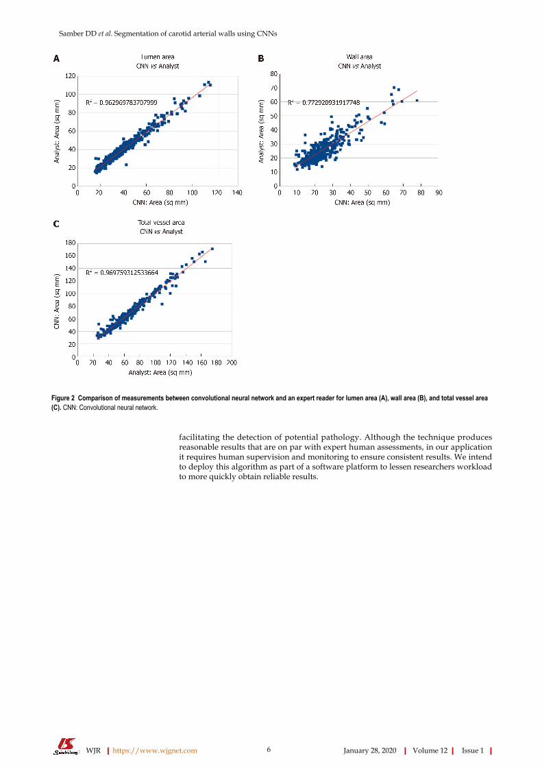
Figure 2
Figure 2 Comparison of measurements between convolutional neural network and an expert reader for lumen area (A), wall area (B), and total vessel area(C). CNN: Convolutional neural network.
facilitating the detection of potential pathology. Although the technique producesreasonable results that are on par with expert human assessments, in our applicationit requires human supervision and monitoring to ensure consistent results. We intendto deploy this algorithm as part of a software platform to lessen researchers workloadto more quickly obtain reliable results.
WJR https://www.wjgnet.com January 28, 2020 Volume 12 Issue 1
Samber DD et al. Segmentation of carotid arterial walls using CNNs
6
Figure 3
Figure 3 Bland-Altman plots demonstrating agreement of convolutional neural networks and expert reader in assessing lumen area (A), wall area (B), andtotal vessel area (C). CNN: Convolutional neural network.
ARTICLE HIGHLIGHTSResearch backgroundSegmentation of arterial vessels is an important step is the assessment of vascular disease. Formany years, the accepted method of producing segmentations was through manual approachperformed by expert researchers. We apply the technique of convolutional neural networks(CNNs) to the task of segmentation of carotid arteries and compare the results to the manualmethod.
Research motivationThe accepted standard of manual segmentation by expert researchers is an onerous and time-consuming task that is inherently subjective. Consequently, constructing an algorithm from suchan opaque process is problematic. Creation and adoption of a reliable segmentation algorithmcould lead to significant savings through automation.
Research objectivesThe objective in this study was to examine the feasibility of applying CNNs to the task ofsegmenting carotid arteries of subjects with vascular disease.
Research methodsSubsets of magnetic resonance images of the carotid arteries of 189 subjects with atheroscleroticdisease were used to train and subsequently validate the CNN. Image segmentations used totrain the CNN were produced by an expert reader who manually segmented individual imagesof the carotid wall using conventional means resulting in a dataset of 4422 segmented images. Inpreparation for automated segmentation, the original dataset was divided into 3 groups: A“training dataset” (3581 images), a “validation dataset” (398 images), and a “test dataset” (443images). These datasets were used to train two separate segmentation CNNs (one for carotidlumen and the other for carotid wall). After training, images from the test dataset were processedto produce segmentations as binary images.
Research results
WJR https://www.wjgnet.com January 28, 2020 Volume 12 Issue 1
Samber DD et al. Segmentation of carotid arterial walls using CNNs
7
Overall quantitative assessment between manual and automated segmentations was determinedby computing the DICE coefficient for each pair of segmented images in the test dataset. Theaverage DICE coefficient between automated and manual segmentations was 0.87 for the carotidvessel wall and 0.96 for the carotid lumen. Intra-class correlation coefficients (ICC) as well asPearson correlation values were computed for vessel area metrics as determined for the expertreader and the CNN to assess the agreement of measurements. Excellent agreement wasobserved in the segmentation of lumen area (Pearson correlation = 0.98, ICC = 0.98) as well as inthe segmentation of vessel wall area (Pearson correlation = 0.88, ICC = 0.86). Additionally,Bland-Altman plots of these measurements for the CNN and reader indicate good agreement.
Research conclusionsIn this study, we have demonstrated the effectiveness of CNN technology in its application tothe task of delineating carotid vessel walls thereby facilitating the detection of potentialpathology.
Research perspectivesAlthough the technique produces reasonable results that are on par with expert humanassessments, in our application it requires human supervision and monitoring to ensureconsistent results. We intend to deploy this algorithm as part of a software platform to lessenresearchers workload to more quickly obtain reliable results.
REFERENCES1 Writing Group Members, Mozaffarian D, Benjamin EJ, Go AS, Arnett DK, Blaha MJ, Cushman M, Das
SR, de Ferranti S, Després JP, Fullerton HJ, Howard VJ, Huffman MD, Isasi CR, Jiménez MC, Judd SE,Kissela BM, Lichtman JH, Lisabeth LD, Liu S, Mackey RH, Magid DJ, McGuire DK, Mohler ER 3rd,Moy CS, Muntner P, Mussolino ME, Nasir K, Neumar RW, Nichol G, Palaniappan L, Pandey DK, ReevesMJ, Rodriguez CJ, Rosamond W, Sorlie PD, Stein J, Towfighi A, Turan TN, Virani SS, Woo D, Yeh RW,Turner MB; American Heart Association Statistics Committee; Stroke Statistics Subcommittee. ExecutiveSummary: Heart Disease and Stroke Statistics--2016 Update: A Report From the American HeartAssociation. Circulation 2016; 133: 447-454 [PMID: 26811276 DOI: 10.1161/CIR.0000000000000366]
2 Fairhead JF, Rothwell PM. The need for urgency in identification and treatment of symptomatic carotidstenosis is already established. Cerebrovasc Dis 2005; 19: 355-358 [PMID: 15838162 DOI:10.1159/000085201]
3 Libby P, Ridker PM, Hansson GK. Progress and challenges in translating the biology of atherosclerosis.Nature 2011; 473: 317-325 [PMID: 21593864 DOI: 10.1038/nature10146]
4 North American Symptomatic Carotid Endarterectomy Trial Collaborators, Barnett HJM, TaylorDW, Haynes RB, Sackett DL, Peerless SJ, Ferguson GG, Fox AJ, Rankin RN, Hachinski VC, WiebersDO, Eliasziw M. Beneficial effect of carotid endarterectomy in symptomatic patients with high-gradecarotid stenosis. N Engl J Med 1991; 325: 445-453 [PMID: 1852179 DOI:10.1056/NEJM199108153250701]
5 Nederkoorn PJ, van der Graaf Y, Hunink MG. Duplex ultrasound and magnetic resonance angiographycompared with digital subtraction angiography in carotid artery stenosis: a systematic review. Stroke 2003;34: 1324-1332 [PMID: 12690221 DOI: 10.1161/01.STR.0000068367.08991.A2]
6 Markl M, Schnell S, Wu C, Bollache E, Jarvis K, Barker AJ, Robinson JD, Rigsby CK. Advanced flowMRI: emerging techniques and applications. Clin Radiol 2016; 71: 779-795 [PMID: 26944696 DOI:10.1016/j.crad.2016.01.011]
7 Yuan C, Beach KW, Smith LH, Hatsukami TS. Measurement of atherosclerotic carotid plaque size in vivousing high resolution magnetic resonance imaging. Circulation 1998; 98: 2666-2671 [PMID: 9851951DOI: 10.1161/01.cir.98.24.2666]
8 Klein AK, Lee F, Amini AA. Quantitative coronary angiography with deformable spline models. IEEETrans Med Imaging 1997; 16: 468-482 [PMID: 9368103 DOI: 10.1109/42.640737]
9 Yan P, Kassim AA. Segmentation of volumetric MRA images by using capillary active contour. MedImage Anal 2006; 10: 317-329 [PMID: 16464631 DOI: 10.1016/j.media.2005.12.002]
10 de Bruijne M, van Ginneken B, Viergever MA, Niessen WJ. Interactive segmentation of abdominal aorticaneurysms in CTA images. Med Image Anal 2004; 8: 127-138 [PMID: 15063862 DOI:10.1016/j.media.2004.01.001]
11 Manniesing R, Velthuis BK, van Leeuwen MS, van der Schaaf IC, van Laar PJ, Niessen WJ. Level setbased cerebral vasculature segmentation and diameter quantification in CT angiography. Med Image Anal2006; 10: 200-214 [PMID: 16263325 DOI: 10.1016/j.media.2005.09.001]
12 Itskovich VV, Samber DD, Mani V, Aguinaldo JG, Fallon JT, Tang CY, Fuster V, Fayad ZA.Quantification of human atherosclerotic plaques using spatially enhanced cluster analysis of multicontrast-weighted magnetic resonance images. Magn Reson Med 2004; 52: 515-523 [PMID: 15334569 DOI:10.1002/mrm.20154]
13 Adame IM, van der Geest RJ, Wasserman BA, Mohamed MA, Reiber JH, Lelieveldt BP. Automaticsegmentation and plaque characterization in atherosclerotic carotid artery MR images. MAGMA 2004; 16:227-234 [PMID: 15029508 DOI: 10.1007/s10334-003-0030-8]
14 Olabarriaga SD, Rouet JM, Fradkin M, Breeuwer M, Niessen WJ. Segmentation of thrombus inabdominal aortic aneurysms from CTA with nonparametric statistical grey level appearance modeling.IEEE Trans Med Imaging 2005; 24: 477-485 [PMID: 15822806 DOI: 10.1109/tmi.2004.843260]
15 Fukushima K. Neocognitron: a self organizing neural network model for a mechanism of patternrecognition unaffected by shift in position. Biol Cybern 1980; 36: 193-202 [PMID: 7370364 DOI:10.1007/bf00344251]
16 Lecun Y, Bengio Y, Arbib MA. Convolutional Networks for Images, Speech, and Time Series. In: ArbibMA, editor. The handbook of brain theory and neural networks. 1st ed. Arbib MA. Cambridge, MA: MITPress 1998; 255-258
17 Litjens G, Kooi T, Bejnordi BE, Setio AAA, Ciompi F, Ghafoorian M, van der Laak JAWM, van
WJR https://www.wjgnet.com January 28, 2020 Volume 12 Issue 1
Samber DD et al. Segmentation of carotid arterial walls using CNNs
8
Ginneken B, Sánchez CI. A survey on deep learning in medical image analysis. Med Image Anal 2017; 42:60-88 [PMID: 28778026 DOI: 10.1016/j.media.2017.07.005]
18 de Brebisson A, Montana G. Deep neural networks for anatomical brain segmentation. 2015 Preprint.Available from: https://arxiv.org/abs/1502.02445 [DOI: 10.1109/CVPRW.2015.7301312]
19 Cernazanu-Glavan C, Holban S. Segmentation of bone structure in x-ray images using convolutionalneural network. Adv Electr Comput En 2013; 13: 87-94 [DOI: 10.4316/aece.2013.01015]
20 Liskowski P, Krawiec K. Segmenting Retinal Blood Vessels With Deep Neural Networks. IEEE TransMed Imaging 2016; 35: 2369-2380 [PMID: 27046869 DOI: 10.1109/TMI.2016.2546227]
21 López-Linares K, Aranjuelo N, Kabongo L, Maclair G, Lete N, Ceresa M, García-Familiar A, Macía I,González Ballester MA. Fully automatic detection and segmentation of abdominal aortic thrombus in post-operative CTA images using Deep Convolutional Neural Networks. Med Image Anal 2018; 46: 202-214[PMID: 29609054 DOI: 10.1016/j.media.2018.03.010]
22 Lekadir K, Galimzianova A, Betriu A, Del Mar Vila M, Igual L, Rubin DL, Fernandez E, Radeva P,Napel S. A Convolutional Neural Network for Automatic Characterization of Plaque Composition inCarotid Ultrasound. IEEE J Biomed Health Inform 2017; 21: 48-55 [PMID: 27893402 DOI:10.1109/JBHI.2016.2631401]
23 Sudha S, Jayanthi KB, Ramalingan C, Madian N, Sunder T. Convolutional Neural Network forSegmentation and Measurement of Intima Media Thickness. J Med Syst 2018; 42: 154 [PMID: 29987622DOI: 10.1007/s10916-018-1001-y]
24 Lorenz MW, Gao L, Ziegelbauer K, Norata GD, Empana JP, Schmidtmann I, Lin HJ, McLachlan S,Bokemark L, Ronkainen K, Amato M, Schminke U, Srinivasan SR, Lind L, Okazaki S, Stehouwer CDA,Willeit P, Polak JF, Steinmetz H, Sander D, Poppert H, Desvarieux M, Ikram MA, Johnsen SH, Staub D,Sirtori CR, Iglseder B, Beloqui O, Engström G, Friera A, Rozza F, Xie W, Parraga G, Grigore L, PlichartM, Blankenberg S, Su TC, Schmidt C, Tuomainen TP, Veglia F, Völzke H, Nijpels G, Willeit J, Sacco RL,Franco OH, Uthoff H, Hedblad B, Suarez C, Izzo R, Zhao D, Wannarong T, Catapano A, Ducimetiere P,Espinola-Klein C, Chien KL, Price JF, Bergström G, Kauhanen J, Tremoli E, Dörr M, Berenson G,Kitagawa K, Dekker JM, Kiechl S, Sitzer M, Bickel H, Rundek T, Hofman A, Mathiesen EB, CastelnuovoS, Landecho MF, Rosvall M, Gabriel R, de Luca N, Liu J, Baldassarre D, Kavousi M, de Groot E, BotsML, Yanez DN, Thompson SG; PROG-IMT study group. Predictive value for cardiovascular events ofcommon carotid intima media thickness and its rate of change in individuals at high cardiovascular risk -Results from the PROG-IMT collaboration. PLoS One 2018; 13: e0191172 [PMID: 29649236 DOI:10.1371/journal.pone.0191172]
25 Mani V, Aguiar SH, Itskovich VV, Weinshelbaum KB, Postley JE, Wasenda EJ, Aguinaldo JG, SamberDD, Fayad ZA. Carotid black blood MRI burden of atherosclerotic disease assessment correlates withultrasound intima-media thickness. J Cardiovasc Magn Reson 2006; 8: 529-534 [PMID: 16755842 DOI:10.1080/10976640600675245]
26 Zhang Y, Guallar E, Malhotra S, Astor BC, Polak JF, Qiao Y, Gomes AS, Herrington DM, Sharrett AR,Bluemke DA, Wasserman BA. Carotid Artery Wall Thickness and Incident Cardiovascular Events: AComparison between US and MRI in the Multi-Ethnic Study of Atherosclerosis (MESA). Radiology 2018;289: 649-657 [PMID: 30299234 DOI: 10.1148/radiol.2018173069]
27 Choudhury RP, Birks JS, Mani V, Biasiolli L, Robson MD, L'Allier PL, Gingras MA, Alie N,McLaughlin MA, Basson CT, Schecter AD, Svensson EC, Zhang Y, Yates D, Tardif JC, Fayad ZA.Arterial Effects of Canakinumab in Patients With Atherosclerosis and Type 2 Diabetes or GlucoseIntolerance. J Am Coll Cardiol 2016; 68: 1769-1780 [PMID: 27737744 DOI: 10.1016/j.jacc.2016.07.768]
28 Perone CS, Calabrese E, Cohen-Adad J. Spinal cord gray matter segmentation using deep dilatedconvolutions. Sci Rep 2018; 8: 5966 [PMID: 29654236 DOI: 10.1038/s41598-018-24304-3]
29 Dice LR. Measures of the amount of ecologic association between species. Ecology 1945; 26: 297-302[DOI: 10.2307/1932409]
30 El Aidi H, Mani V, Weinshelbaum KB, Aguiar SH, Taniguchi H, Postley JE, Samber DD, Cohen EI,Stern J, van der Geest RJ, Reiber JH, Woodward M, Fuster V, Gidding SS, Fayad ZA. Cross-sectional,prospective study of MRI reproducibility in the assessment of plaque burden of the carotid arteries andaorta. Nat Clin Pract Cardiovasc Med 2009; 6: 219-228 [PMID: 19174763 DOI: 10.1038/ncpcardio1444]
31 Garcia-Garcia A, Orts-Escolano S, Oprea S, Villena-Martinez V, Garcia-Rodriguez J. A Review on DeepLearning Techniques Applied to Semantic Segmentation. 2017 Preprint. Available from:https://arxiv.org/abs/1704.06857
WJR https://www.wjgnet.com January 28, 2020 Volume 12 Issue 1
Samber DD et al. Segmentation of carotid arterial walls using CNNs
9

Published By Baishideng Publishing Group Inc
7041 Koll Center Parkway, Suite 160, Pleasanton, CA 94566, USA
Telephone: +1-925-2238242
E-mail: [email protected]
Help Desk: https://www.f6publishing.com/helpdesk
https://www.wjgnet.com
© 2020 Baishideng Publishing Group Inc. All rights reserved.


![European Journal of Radiology · Journal of Radiology 87 (2017) 76–82 Contents lists available at ScienceDirect European Journal of Radiology j ... [MRI]) [2,4] have reported that](https://img.pdfslide.net/doc/110x75/5cb0677988c993767e8c67df/european-journal-of-radiology-journal-of-radiology-87-2017-7682-contents.jpg)