Embed Size (px)
Citation preview
H. Alan Arbuckle, MD, FAAP, FAAD, AAWCPA, IIWCC-Can Wound Care Consultant, Advanced Wound Care Department
Section Head, Pediatric Dermatology Department of Dermatology Kaiser Permanente Colorado
Conflicts of Interest
Consultant, Shire Regenerative Medicine
Objectives Discuss venous stasis, venous stasis dermatitis and
venous insufficiency
When to use compression and how much
Develop a basic and rational understanding of patients with chronic wounds

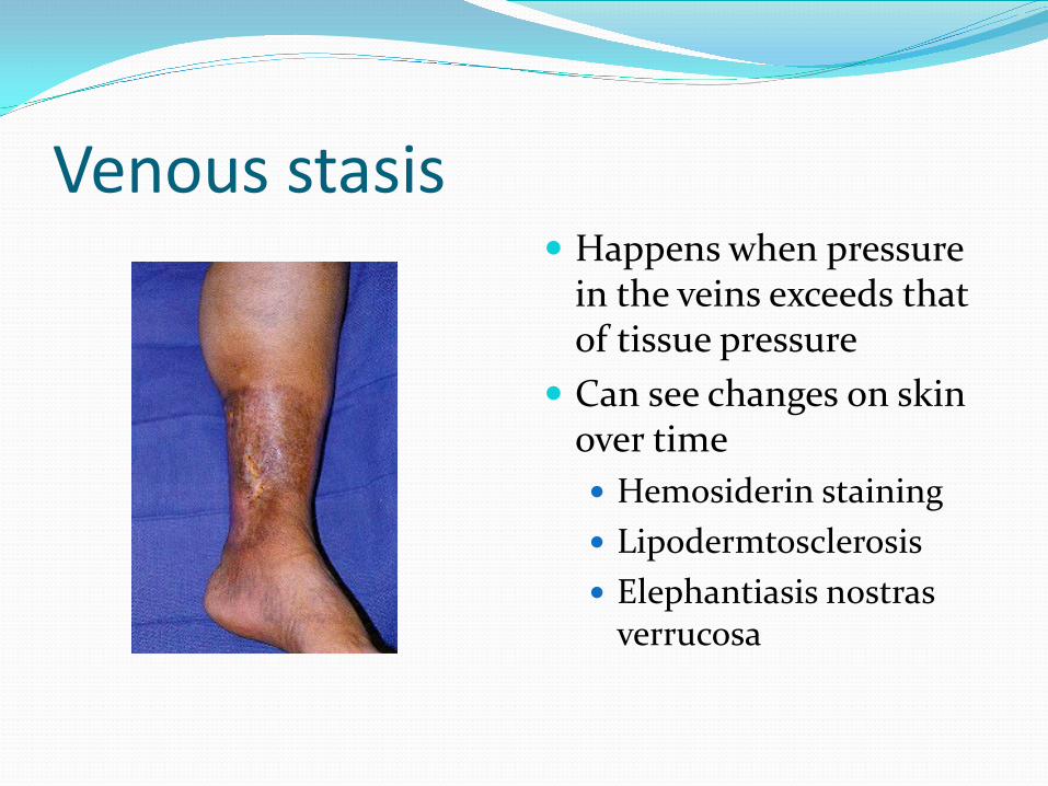
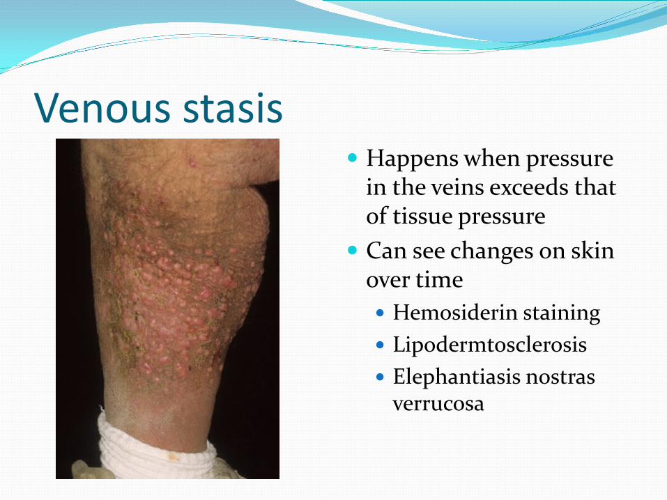
Venous stasis Happens when pressure
in the veins exceeds that of tissue pressure
Can see changes on skin over time Hemosiderin staining Lipodermtosclerosis Elephantiasis nostras
verrucosa
Venous stasis Happens when pressure
in the veins exceeds that of tissue pressure
Can see changes on skin over time Hemosiderin staining Lipodermtosclerosis Elephantiasis nostras
verrucosa
Venous stasis Happens when pressure
in the veins exceeds that of tissue pressure
Can see changes on skin over time Hemosiderin staining Lipodermtosclerosis Elephantiasis nostras
verrucosa
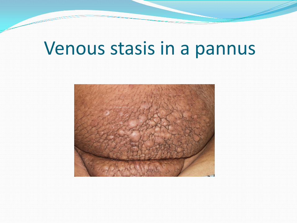
Venous stasis in a pannus
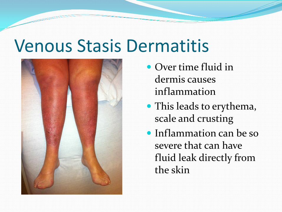
Venous Stasis Dermatitis Over time fluid in
dermis causes inflammation
This leads to erythema, scale and crusting
Inflammation can be so severe that can have fluid leak directly from the skin
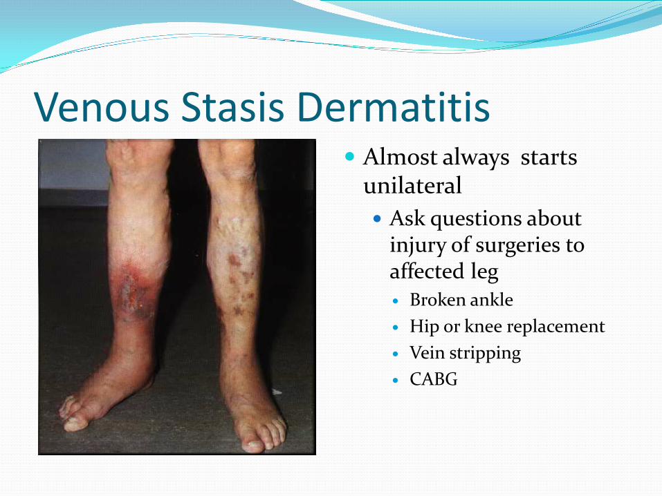
Venous Stasis Dermatitis Almost always starts
unilateral Ask questions about
injury of surgeries to affected leg Broken ankle Hip or knee replacement Vein stripping CABG
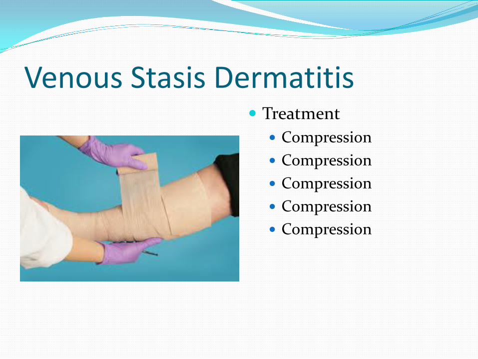
Venous Stasis Dermatitis Treatment
Compression Compression Compression Compression Compression

Venous Stasis Dermatitis Treatment
Some compression is better than no compression
ABI should be >0.6 Dorsalis pedis pulse 40
mmHg Start with light
compression and then move up gradually
Strong topical steroids Clobetasol Fluocinonide
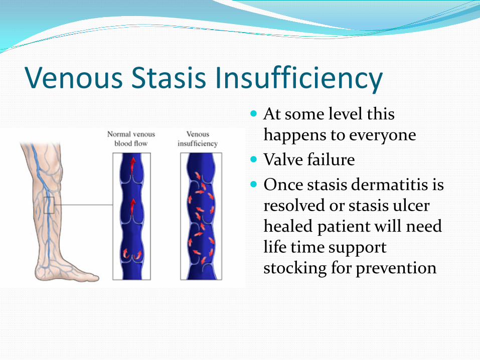
Venous Stasis Insufficiency At some level this
happens to everyone Valve failure Once stasis dermatitis is
resolved or stasis ulcer healed patient will need life time support stocking for prevention

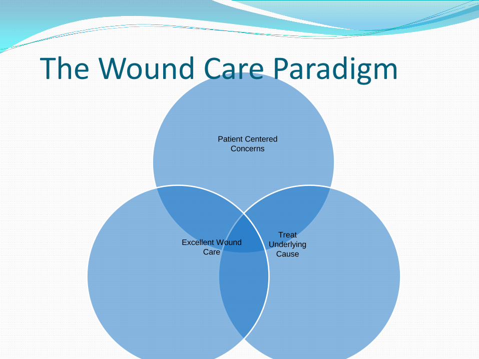
The Wound Care Paradigm
Patient Centered Concerns
Treat Underlying
Cause Excellent Wound
Care
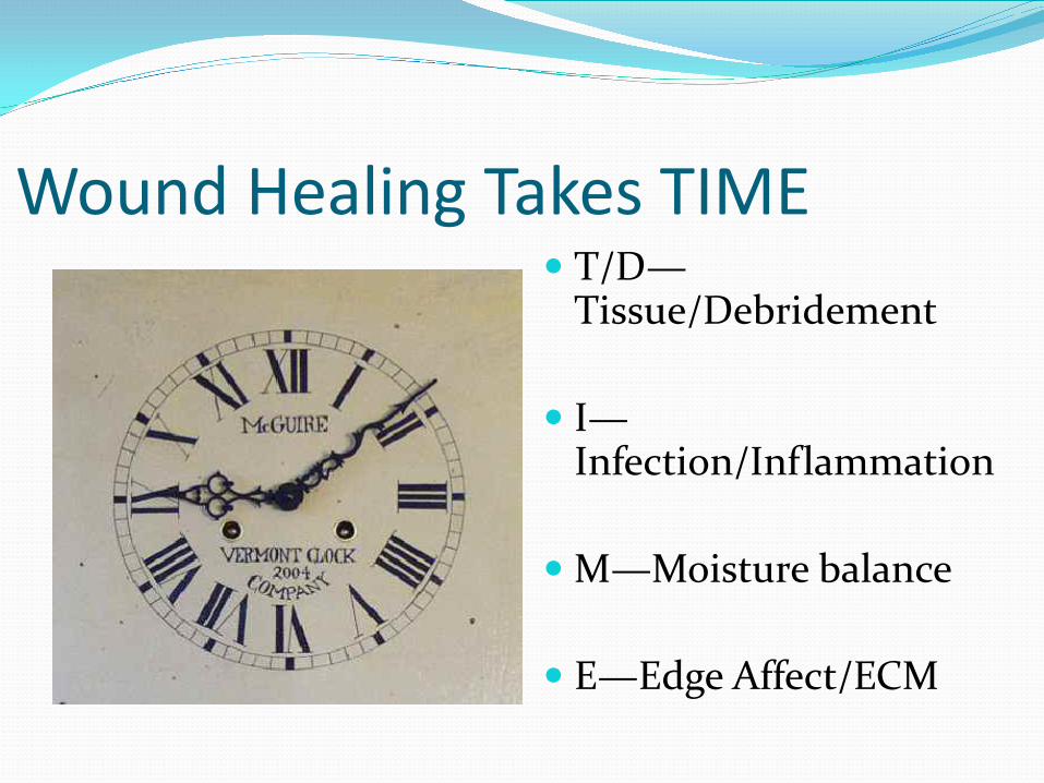
Wound Healing Takes TIME T/D—
Tissue/Debridement
I—Infection/Inflammation
M—Moisture balance
E—Edge Affect/ECM
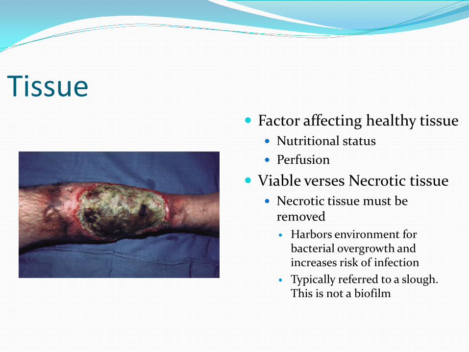
Tissue Factor affecting healthy tissue
Nutritional status Perfusion
Viable verses Necrotic tissue Necrotic tissue must be
removed Harbors environment for
bacterial overgrowth and increases risk of infection
Typically referred to a slough. This is not a biofilm

Infection All chronic wounds are
colonized Wounds go through a
step wise process of: Contamination Colonization Critical colonization Infection
www.scharfphoto.com/fine_art_prints/archives
Infection Wound infection is
defined as the presence of replicating microorganisms within a wound with subsequent host injury†
Infection= dose x
virulence/host resistance†
†Dow G. Infection in chronic wounds. In: Krasner DL, Rodeheaver GT, Sibbald RG (eds). Chronic Wound Care: A Clinical Source Book for Healthcare Professionals, Third Edition. Wayne, PA:HMP Communications, 2001:343-356
www.johnclearygallery.com/pics/smith/twomen.jpg
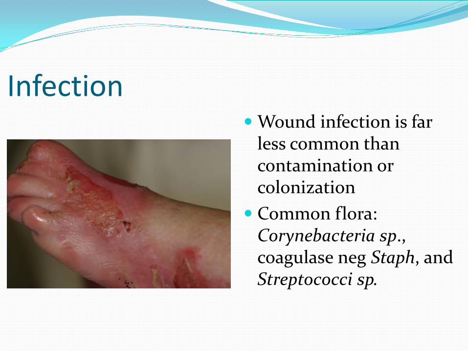
Infection Wound infection is far
less common than contamination or colonization
Common flora: Corynebacteria sp., coagulase neg Staph, and Streptococci sp.
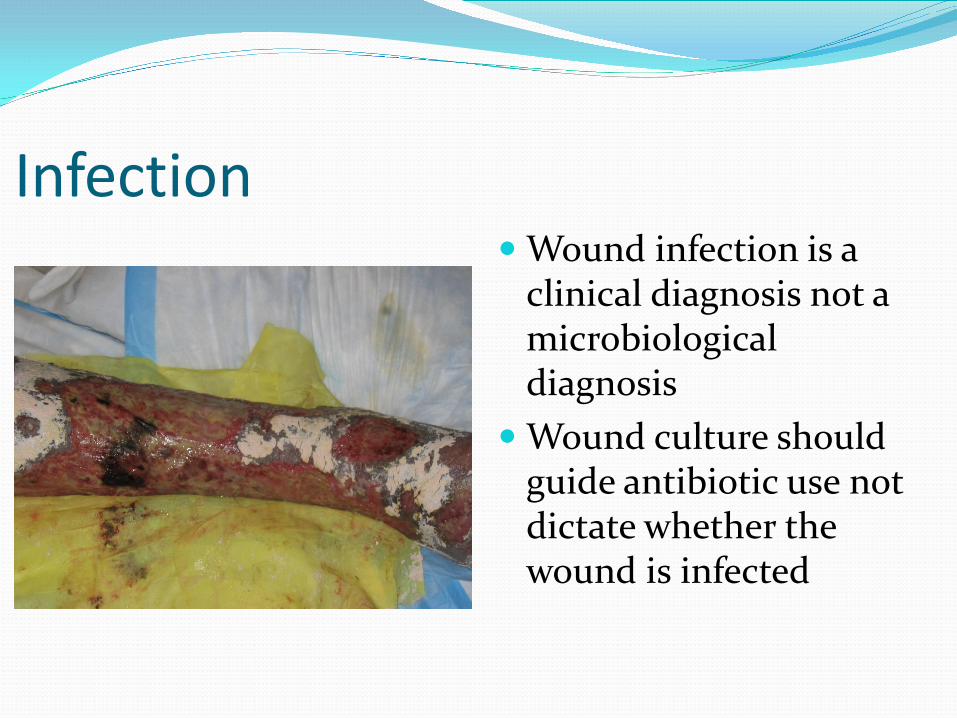
Infection Wound infection is a
clinical diagnosis not a microbiological diagnosis
Wound culture should guide antibiotic use not dictate whether the wound is infected
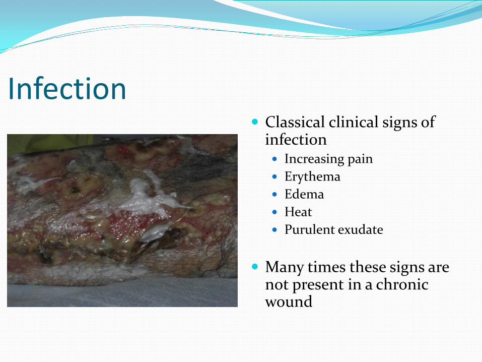
Infection Classical clinical signs of
infection Increasing pain Erythema Edema Heat Purulent exudate
Many times these signs are
not present in a chronic wound
Infection It is important to differentiate between a superficial
infection and deep infection In superficial infections topical antibiotics can typically
be utilized In deeper infections systemic therapy is usually
necessary There is no data that adding a topical antimicrobial
agent while a patient is on systemic therapy results in increased wound healing
Superficial Infection (Critical Colonization) NERDS
N:Nonhealing wound E: Exudate R: Red granulation tissue D:Debris—Tissue—Nonviable S: Smell
Deep Infection True Infection STONEES
S: Size of wound is increasing T: Temperature—surrounding tissue O:Osteomyelitis N:New wound breakdown E: Exudate/Edema S: Smell
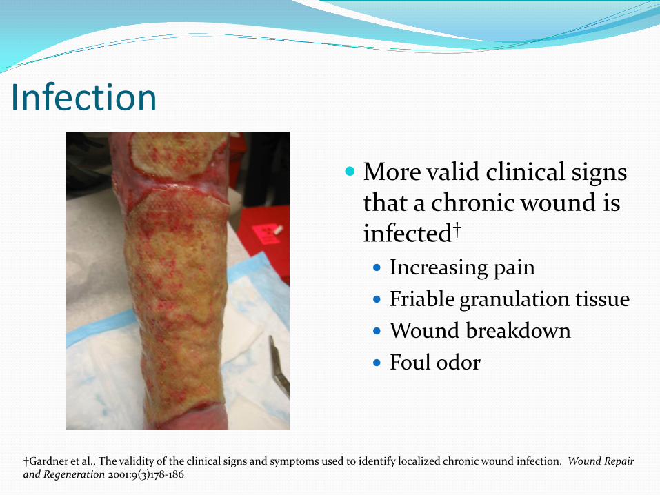
Infection
More valid clinical signs that a chronic wound is infected†
Increasing pain Friable granulation tissue Wound breakdown Foul odor
†Gardner et al., The validity of the clinical signs and symptoms used to identify localized chronic wound infection. Wound Repair and Regeneration 2001:9(3)178-186
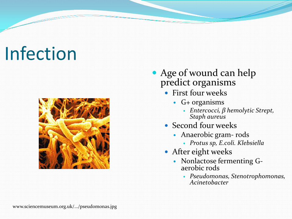
Infection Age of wound can help
predict organisms First four weeks
G+ organisms Entercocci, β hemolytic Strept,
Staph aureus Second four weeks
Anaerobic gram- rods Protus sp, E.coli. Klebsiella
After eight weeks Nonlactose fermenting G-
aerobic rods Pseudomonas, Stenotrophomonas,
Acinetobacter
www.sciencemuseum.org.uk/.../pseudomonas.jpg
Treatment of Critical Colonization NERDS It is reasonable to try a topical therapeutic approach
using antiseptics or topical antibiotics Povidone-iodine/Cadexomer iodine Hydrogen Peroxide Acetic acid Chlorhexidine Topical antibiotic
Silver, Bacitracin, Neosporin, white petrolatum
Treatment of Deep Infection STONEES
“Early” chronic wound Cephalexin or
Clindamycin “Late” chronic wound
Amoxicillin-clavulanate Cephalexin +
metronidazole Ciprofloxacin +
clindamycin Doxycycline +
trimethoprim
Moisture Balance “To date, research and clinical experience have identified that in a moist environment exudate controls infection, provides the cells involved in wound repair with nutrients, and provides the best environment for healing.”
Jones V, Harding K, Moist wound healing. In: Kransner DL, Rodeheaver GT, Sibbald RG (eds). Chronic Wound Care: A Clinical Source Book for Healthcare Professionals, Third Edition. Wayne, PA: HMP Communications, 2001:245-252
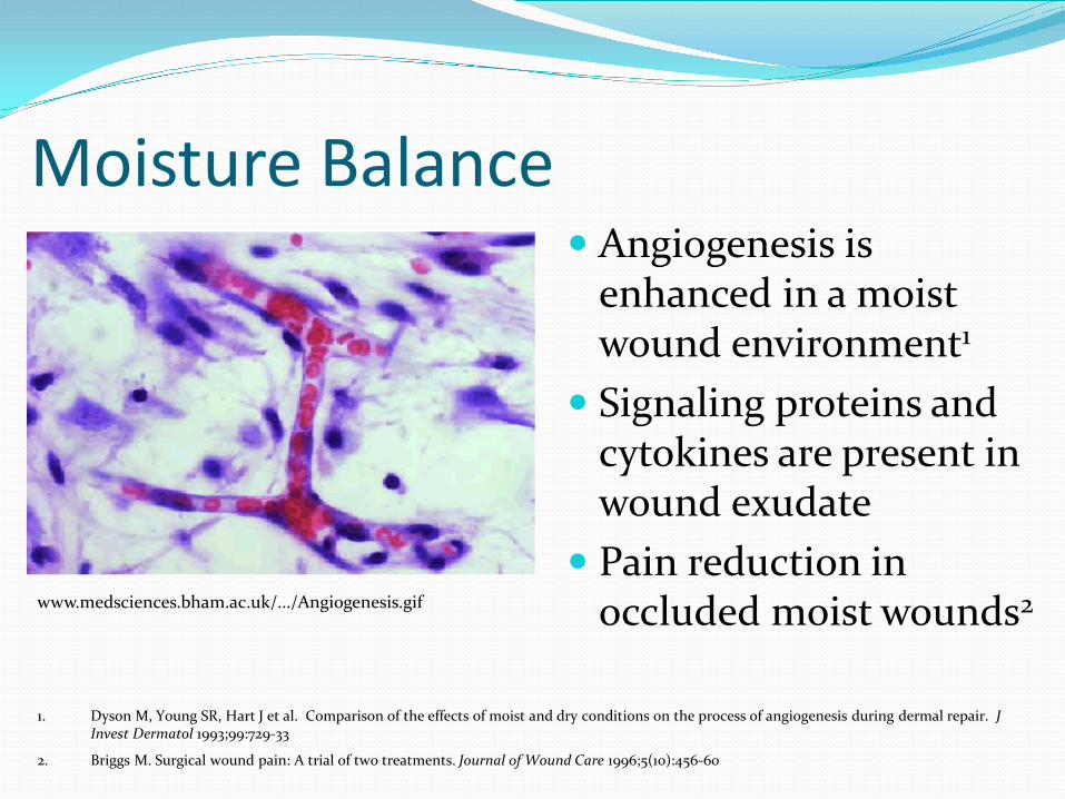
Moisture Balance Angiogenesis is
enhanced in a moist wound environment1
Signaling proteins and cytokines are present in wound exudate
Pain reduction in occluded moist wounds2
1. Dyson M, Young SR, Hart J et al. Comparison of the effects of moist and dry conditions on the process of angiogenesis during dermal repair. J Invest Dermatol 1993;99:729-33
2. Briggs M. Surgical wound pain: A trial of two treatments. Journal of Wound Care 1996;5(10):456-60
www.medsciences.bham.ac.uk/.../Angiogenesis.gif
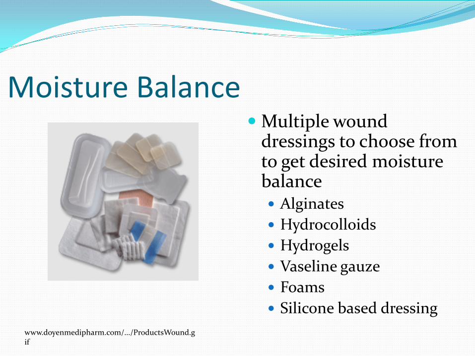
Moisture Balance Multiple wound
dressings to choose from to get desired moisture balance Alginates Hydrocolloids Hydrogels Vaseline gauze Foams Silicone based dressing
www.doyenmedipharm.com/.../ProductsWound.gif
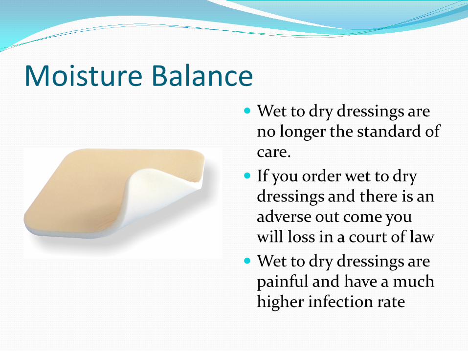
Moisture Balance Wet to dry dressings are
no longer the standard of care.
If you order wet to dry dressings and there is an adverse out come you will loss in a court of law
Wet to dry dressings are painful and have a much higher infection rate

Moisture Balance When all else fails and
you don’t know what to do: Vaseline and some type
of dressing
Refer to wound center

Edge Affect Escar delays keratinocyte
migration Hyperkeratosis (callus)
on the wound edge will also delay keratinocyte migration Callus tells you that there
has been long standing pressure and that pressure redistribution is necessary
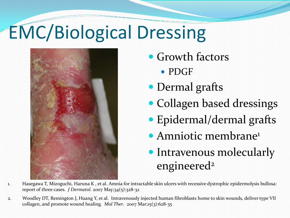
EMC/Biological Dressing Growth factors
PDGF
Dermal grafts Collagen based dressings Epidermal/dermal grafts Amniotic membrane1
Intravenous molecularly engineered2
1. Hasegawa T, Mizoguchi, Haruna K , et al. Amnia for intractable skin ulcers with recessive dystrophic epidermolysis bullosa: report of three cases. J Dermatol. 2007 May;34(5):328-32
2. Woodley DT, Remington J, Huang Y, et al. Intravenously injected human fibroblasts home to skin wounds, deliver type VII collagen, and promote wound healing. Mol Ther. 2007 Mar;15(3):628-35
Wound Care
“Maintenance wound” verses a healing wound
Thank You