Embed Size (px)
Citation preview
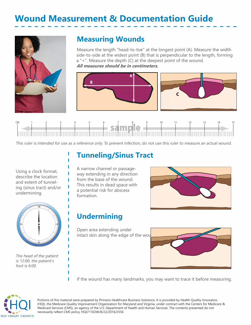
Wound Measurement & Documentation Guide
Portions of this material were prepared by Primaris Healthcare Business Solutions. It is provided by Health Quality Innovators (HQI), the Medicare Quality Improvement Organization for Maryland and Virginia, under contract with the Centers for Medicare & Medicaid Services (CMS), an agency of the U.S. Department of Health and Human Services. The contents presented do not necessarily reflect CMS policy. HQI/11SOW/8/22/2016/2556
Measuring WoundsMeasure the length "head-to-toe" at the longest point (A). Measure the width side-to-side at the widest point (B) that is perpendicular to the length, forming a "+". Measure the depth (C) at the deepest point of the wound. All measures should be in centimeters.
A
B
C
sampleCM 1 2 3 4 8765 131211109 1514
This ruler is intended for use as a reference only. To prevent infection, do not use this ruler to measure an actual wound.
Using a clock format, describe the location and extent of tunnel-ing (sinus tract) and/or undermining.
6
12
39
The head of the patient is 12:00, the patient's foot is 6:00.
If the wound has many landmarks, you may want to trace it before measuring.
Tunneling/Sinus TractA narrow channel or passage-way extending in any direction from the base of the wound. This results in dead space with a potential risk for abscess formation.
UnderminingOpen area extending under intact skin along the edge of the wound.
Wound Measurement & Documentation Guide
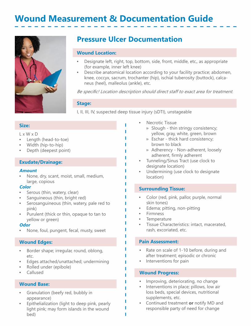
Wound Location: • Designate left, right, top, bottom, side, front, middle, etc., as appropriate
(for example, inner left knee)• Describe anatomical location according to your facility practice; abdomen,
knee, coccyx, sacrum, trochanter (hip), ischial tuberosity (buttock), calca-neus (heel), malleolus (ankle), etc.
Be specific! Location description should direct staff to exact area for treatment.
Stage: I, II, III, IV, suspected deep tissue injury (sDTI), unstageable
Pressure Ulcer Documentation
Size: L x W x D• Length (head-to-toe)• Width (hip-to-hip)• Depth (deepest point)
Exudate/Drainage: Amount• None, dry, scant, moist, small, medium,
large, copiousColor• Serous (thin, watery, clear)• Sanguineous (thin, bright red)• Serosanguineous (thin, watery, pale red to
pink)• Purulent (thick or thin, opaque to tan to
yellow or green)Odor• None, foul, pungent, fecal, musty, sweet
Wound Edges: • Border shape; irregular, round, oblong,
etc.• Edges attached/unattached; undermining• Rolled under (epibole)• Callused
Wound Base: • Granulation (beefy red, bubbly in
appearance)• Epithelialization (light to deep pink, pearly
light pink; may form islands in the woundbed)
• Necrotic Tissue» Slough - thin stringy consistency;
yellow, gray, white, green, brown» Eschar - thick hard consistency;
brown to black» Adherency - Non-adherent, loosely
adherent, firmly adherent• Tunneling/Sinus Tract (use clock to
designate location)• Undermining (use clock to designate
location)
Surrounding Tissue: • Color (red, pink, pallor, purple, normal
skin tones)• Edema; pitting, non-pitting• Firmness• Temperature• Tissue Characteristics: intact, macerated,
rash, excoriated, etc.
Pain Assessment: • Rate on scale of 1-10 before, during and
after treatment; episodic or chronic• Interventions for pain
Wound Progress: • Improving, deteriorating, no change• Interventions in place; pillows, low air
loss beds, special devices, nutritionalsupplements, etc.
• Continued treatment or notify MD andresponsible party of need for change